The Real-World Clinical Utility of NGS in Oncology: From Genomic Profiling to Precision Treatment and Improved Patient Outcomes
This article provides a comprehensive analysis of the real-world clinical utility of Next-Generation Sequencing (NGS) in oncology, tailored for researchers, scientists, and drug development professionals.
The Real-World Clinical Utility of NGS in Oncology: From Genomic Profiling to Precision Treatment and Improved Patient Outcomes
Abstract
This article provides a comprehensive analysis of the real-world clinical utility of Next-Generation Sequencing (NGS) in oncology, tailored for researchers, scientists, and drug development professionals. It explores the foundational technology shift from single-gene tests to massively parallel sequencing, detailing its core applications in tumor genomic profiling, hereditary cancer syndrome detection, and therapy selection. The content examines current methodological approaches, including whole-genome, whole-exome, and targeted panels, while addressing significant implementation challenges such as data interpretation, cost, reimbursement, and integration into clinical workflows. Through a validation lens, the article compares NGS with traditional sequencing methods and assesses its impact on patient survival, quality of life, and clinical trial stratification. The synthesis provides a forward-looking perspective on emerging trends and the evolving role of NGS in personalized cancer care.
The NGS Revolution: Understanding the Core Technology and Its Transformative Role in Cancer Genomics
The evolution of DNA sequencing technologies from first-generation Sanger methods to massively parallel Next-Generation Sequencing (NGS) represents one of the most transformative advancements in modern molecular biology. This paradigm shift has fundamentally altered the landscape of genomic research and clinical diagnostics, particularly in oncology, where comprehensive genomic profiling has become indispensable for precision medicine. The dramatic improvements in throughput and cost-efficiency afforded by NGS have enabled researchers and clinicians to pursue questions previously deemed technologically or economically unfeasible. This review quantitatively compares the technical capabilities, operational parameters, and economic considerations of both sequencing approaches, with specific emphasis on their application in cancer research and clinical oncology. We further examine experimental protocols from key studies demonstrating the real-world utility of NGS in characterizing tumor genomes and informing therapeutic decisions.
The fundamental principles of DNA sequencing remained largely unchanged for three decades following Frederick Sanger's development of the chain-termination method in 1977 [1]. Sanger sequencing, also known as first-generation sequencing, revolutionized molecular biology by enabling researchers to read DNA sequences with high accuracy [1]. For years, it served as the gold standard for clinical diagnostic applications, including sequencing individual genes implicated in hereditary disorders and validating critical research findings [2]. However, its reliance on capillary electrophoresis and its inherently linear, low-throughput workflow made large-scale projects like whole-genome sequencing prohibitively costly and time-consuming [3].
The advent of massively parallel sequencing, commonly termed Next-Generation Sequencing (NGS), in the mid-2000s marked a revolutionary departure from this model [3]. Unlike Sanger sequencing, which processes a single DNA fragment per reaction, NGS technologies simultaneously sequence millions to billions of DNA fragments [4] [5]. This core difference in parallelism has driven a seismic shift in genomics, drastically reducing the cost and time required to sequence entire genomes while exponentially increasing data output [6]. In oncology, this technological leap has been particularly impactful, moving the field from a focus on single-gene alterations to a comprehensive understanding of the complex genomic landscape of cancer, thereby laying the foundation for truly personalized cancer therapy [6] [7].
Fundamental Technological Differences
The operational divergence between Sanger and NGS methodologies stems from their distinct underlying biochemical approaches and detection systems.
Sanger Sequencing: Chain Termination Chemistry
The Sanger method is based on the selective incorporation of chain-terminating dideoxynucleotides (ddNTPs) during in vitro DNA synthesis [8] [1]. In modern implementations, fluorescently labeled ddNTPs are used in a single reaction. As DNA polymerase synthesizes a new strand complementary to the template, random incorporation of a ddNTP terminates the growing chain. The resulting mixture of fluorescently labeled DNA fragments of varying lengths is then separated by capillary electrophoresis, which resolves the fragments by size [8]. A laser detects the fluorescent tag of the terminal ddNTP on each fragment, and the sequence is determined based on the order of the colored peaks as the fragments pass the detector [2]. This process generates long, contiguous reads (500-1000 base pairs) with exceptionally high per-base accuracy, exceeding 99.999% (Phred score Q50) in the central portion of the read [8].
Massively Parallel Sequencing: Diverse Parallelized Chemistries
NGS encompasses several different technologies, all sharing the common principle of massive parallelism [1] [5]. The process typically begins with shearing genomic DNA into a library of small fragments. Adapters are ligated to these fragments, enabling them to be immobilized on a solid surface or microbeads [6] [1]. The critical differentiator is that millions to billions of these fragments are sequenced simultaneously in a single run [4]. The most common chemistry, Sequencing by Synthesis (SBS), used by Illumina platforms, employs fluorescently labeled, reversible terminator nucleotides [1]. These nucleotides are added iteratively in cycles: incorporation, imaging to identify the base, and then cleavage of the terminator and fluorophore to enable the next cycle [4] [1]. Other NGS chemistries include pyrosequencing (Roche/454), sequencing by ligation (SOLiD), and ion semiconductor sequencing (Ion Torrent) [1]. Unlike Sanger, NGS generates millions of short reads (50-300 base pairs) that must be computationally aligned to a reference genome [8].
Comparative Technical Specifications
The table below summarizes the core technical differences between Sanger sequencing and NGS.
Table 1: Key technical characteristics of Sanger and Massively Parallel Sequencing
| Feature | Sanger Sequencing | Massively Parallel Sequencing (NGS) |
|---|---|---|
| Fundamental Method | Chain termination with ddNTPs [8] | Massively parallel sequencing (e.g., SBS, ligation, ion detection) [8] |
| Read Length | Long (500–1000 bp) [8] | Short (50-300 bp for short-read platforms) [8] |
| Throughput per Run | Low (single fragment) [4] | Extremely High (millions to billions of fragments) [4] [5] |
| Detection System | Capillary electrophoresis & fluorescent detection [8] | High-resolution optical imaging or ion sensor detection [8] |
| Data Output | Single sequence per reaction [8] | Gigabases to Terabases of data [8] |
| Typical Applications | Single gene sequencing, variant validation [4] [2] | Whole genomes, exomes, transcriptomes, targeted panels [4] [8] |
Quantitative Comparison: Throughput and Cost
The paradigm shift becomes most evident when examining the quantitative metrics of throughput, scalability, and cost-efficiency.
Throughput and Scalability
The throughput of Sanger sequencing is fundamentally limited by its one-fragment-per-capillary design. Scaling up requires a linear increase in reactions, reagents, and time. In contrast, NGS achieves monumental throughput by processing all fragments in a single, highly parallelized reaction [4]. This difference is not merely incremental but logarithmic.
For example, while Sanger sequencing is practical for interrogating up to approximately 20 genomic targets, it becomes prohibitively costly and time-consuming beyond this point [4]. A single Sanger run can process up to 96 samples, but each sample typically targets only one specific region. Conversely, a single NGS run on a platform like the Illumina MiSeq can generate up to 15 Gb of data, which is sufficient to sequence multiple samples at high depth for hundreds to thousands of genes simultaneously [4] [3]. This allows researchers to screen entire pathways or gene networks in a single experiment, a capability that is simply not feasible with Sanger sequencing.
Cost Efficiency and Economic Considerations
The economic advantage of NGS is a direct result of its massive parallelism. The key differentiator is the cost per base pair, which is dramatically lower for NGS than for Sanger sequencing [8]. While the upfront capital investment for an NGS instrument is substantial, the economy of scale makes it the undisputed choice for large-scale projects [8] [3].
Table 2: Economic and operational comparison of sequencing technologies
| Parameter | Sanger Sequencing | Massively Parallel Sequencing (NGS) |
|---|---|---|
| Cost per Base | High [8] | Very Low [8] |
| Cost per Sample | Low for small-scale projects [4] [2] | Low for large-scale projects [4] [2] |
| Instrument Cost | Lower (capillary electrophoresis systems) [8] | Higher (e.g., \$90K-\$1M+ for Illumina platforms) [3] |
| Multiplexing Capability | Limited | High (hundreds of samples can be barcoded and pooled) [8] |
| Labor Intensity | High for large projects (many reactions) | Lower per unit of data (automated library prep) |
| Ideal Use Case | Cost-effective for 1-20 targets [4] | Cost-effective for large genes, panels, or whole genomes [4] |
The Human Genome Project, which relied on Sanger sequencing, cost an estimated \$2.7 billion. Today, due to NGS, a human genome can be sequenced for a few hundred dollars, a reduction in cost of several million-fold [3]. This staggering drop has democratized genomic research and made clinical whole-genome sequencing an attainable reality.
Experimental Protocols in Oncology Research
The application of NGS in oncology follows well-established workflows, from sample preparation to data interpretation. The following protocols highlight its implementation in large-scale clinical studies.
Protocol 1: Targeted NGS for Population-Based Cancer Genomics
The Cancer 2015 Study was a prospective, multisite cohort study designed to assess the feasibility of using targeted NGS for routine cancer care [9]. Its methodology serves as a model for large-scale somatic variant screening.
- Sample Type: Formalin-Fixed Paraffin-Embedded (FFPE) tumor blocks or sections from 1,094 newly diagnosed cancer patients.
- DNA Extraction: DNA was extracted from up to ten 5-μm thick FFPE sections using the DNeasy Blood and Tissue Kit (Qiagen). Quantification was performed using the Qubit dsDNA HS Assay.
- Library Preparation & Sequencing: A minimum of 50 ng of DNA was used as input for the Illumina TruSeq Amplicon Cancer Panel (TSACP), which targets 212 amplicons across 48 cancer-related genes. Sequencing was performed on the Illumina MiSeq system with 150-bp paired-end kits.
- Bioinformatic Analysis: Demultiplexing and FASTQ file generation used CASAVA v1.8.2. Reads were aligned to hg19 amplicon reference sequences. Variant calling was performed using VarScan2. To ensure high confidence, strict filters were applied: minimum coverage of 100 reads, at least 50 variant reads, and a minimum variant allele frequency of 8%.
- Key Findings: The study successfully sequenced 854 patients (78% success rate). Clinically relevant mutations were identified in 63% of patients, with 26% harboring mutations with direct therapeutic implications. Orthogonal validation using the MassARRAY system showed 100% concordance for a subset of samples, confirming the high reliability of the NGS workflow [9].
Protocol 2: Whole-Genome Sequencing in a Clinical Setting
The 100,000 Genomes Project and subsequent studies have established protocols for using WGS as a comprehensive diagnostic tool in oncology [7] [10]. The following workflow is based on these initiatives.
- Sample Type: Fresh frozen tumor tissue or FFPE blocks, paired with a matched normal sample (typically peripheral blood).
- DNA Extraction & Library Preparation: DNA is extracted using kits such as the AllPrep DNA/RNA Mini Kit (Qiagen). For WGS, library preparation uses PCR-free or low-PCR methods (e.g., Illumina TruSeq DNA PCR-Free) to minimize bias. Libraries are prepared for both tumor and normal samples.
- Sequencing: Sequencing is performed on high-throughput platforms like the Illumina NovaSeq 6000 to achieve a mean coverage of 30-40x for tumor and 20-30x for the matched normal sample [10].
- Bioinformatic Analysis: This is a critical and complex step for WGS. The pipeline includes:
- Alignment: Mapping of reads to a reference genome (e.g., GRCh38) using aligners like BWA-MEM.
- Variant Calling: Somatic single nucleotide variants (SNVs) and small indels are called using tools like Mutect2 and Strelka2 [10]. Copy number alterations (CNAs) are identified using tools like Sequenza, and structural variants (SVs) are detected using tools like Delly.
- Annotation & Interpretation: Variants are annotated for predicted functional impact using tools like Ensembl VEP. They are then filtered against population databases and curated for clinical actionability using resources like OncoKB and COSMIC.
- Key Findings: A 2024 study implementing this WGS protocol for 95 solid cancer patients found that 72% (68/95) of patients received clinically relevant insights from WGS. This included therapeutic actionability for 69% of patients and clarity on clinical questions for 81% of applicable cases, demonstrating the profound utility of a comprehensive NGS approach [10].
The Scientist's Toolkit: Essential Research Reagents and Materials
Successful implementation of NGS in a research or clinical setting relies on a suite of specialized reagents and tools. The following table details key solutions used in standard NGS workflows.
Table 3: Essential research reagents and materials for NGS workflows
| Reagent/Material | Function | Example Products/Kits |
|---|---|---|
| Nucleic Acid Extraction Kits | Isolate high-quality DNA (and RNA) from diverse sample types (tissue, blood, FFPE). | DNeasy Blood & Tissue Kit (Qiagen) [9], AllPrep DNA/RNA kits [10] |
| Library Preparation Kits | Fragment DNA and ligate platform-specific adapters; often include barcodes for sample multiplexing. | Illumina TruSeq DNA PCR-Free, TruSeq Amplicon Cancer Panel [9] [10] |
| Target Enrichment Panels | Capture genomic regions of interest (e.g., cancer gene panels) from a complex whole-genome library. | Illumina TruSeq Amplicon Cancer Panel [9] |
| Sequence Reaction Kits | Contain the enzymes, buffers, and labeled nucleotides required for the sequencing chemistry itself. | Illumina MiSeq, HiSeq, or NovaSeq reagent kits [9] [3] |
| Bioinformatics Software | Analyze raw sequencing data; includes tools for alignment, variant calling, annotation, and visualization. | VarScan2 [9], Mutect2, Strelka2, Delly, Sequenza [10], BWA, GATK |
Clinical Utility in Oncology: A New Paradigm for Patient Care
The transition to NGS has had its most profound impact in the field of oncology, enabling a shift from histology-based to genomics-driven cancer care.
Comprehensive Genomic Profiling and Actionable Mutations
Targeted sequencing panels and WGS can identify a wide spectrum of genomic alterations—including SNVs, indels, CNAs, and SVs—in a single assay [6] [10]. This comprehensive nature is critical because it reveals not only common driver mutations but also rare and novel variants that would be missed by single-gene Sanger tests. The 100,000 Genomes Project demonstrated this power: in an evaluation of 933 cancer participants with reported variants, 59.2% (552 patients) had clinical recommendations made based on their genomic findings, including potential therapy changes or eligibility for clinical trials [7].
Detection of Low-Frequency Variants and Tumor Heterogeneity
NGS's ability to achieve high depth of coverage (often hundreds to thousands of reads per base) provides the sensitivity needed to detect variants present at low allele frequencies in heterogeneous tumor samples [4] [8]. Sanger sequencing has a limited sensitivity, with a typical limit of detection around 15-20% variant allele frequency [4]. In contrast, targeted NGS can reliably detect variants present at frequencies as low as 1-5% [4] [9]. This is essential for identifying emerging resistance mutations in cell-free DNA (liquid biopsies) or for characterizing sub-clonal populations within a tumor that may influence treatment outcomes.
Integration with Immunotherapy and Prognostic Stratification
NGS enables the calculation of tumor mutational burden (TMB), a key biomarker for predicting response to immune checkpoint inhibitors [7] [10]. While TMB can be estimated from large targeted panels, WGS provides the most accurate and comprehensive measurement of TMB because it surveys the entire genome without the bias inherent in targeted capture [10]. Furthermore, NGS can identify specific mutational signatures that reveal the underlying causes of carcinogenesis (e.g., UV exposure, BRCA deficiency) and have prognostic value [7].
The paradigm shift from Sanger to massively parallel sequencing is unequivocal. The dramatic orders-of-magnitude improvement in throughput and cost-efficiency have fundamentally changed the scale and scope of scientific inquiry and clinical practice in oncology. While Sanger sequencing retains its vital role as a gold standard for confirming specific variants and for sequencing single genes, NGS has become the indispensable tool for comprehensive genomic analysis. Its ability to simultaneously interrogate thousands of genes at unprecedented depth allows for the discovery of novel biomarkers, the sensitive detection of tumor heterogeneity, and the identification of actionable mutations that directly inform personalized therapeutic strategies. As NGS technologies continue to evolve, becoming faster, more accurate, and more affordable, their integration into routine clinical workflows will undoubtedly deepen, solidifying their role as the cornerstone of precision oncology.
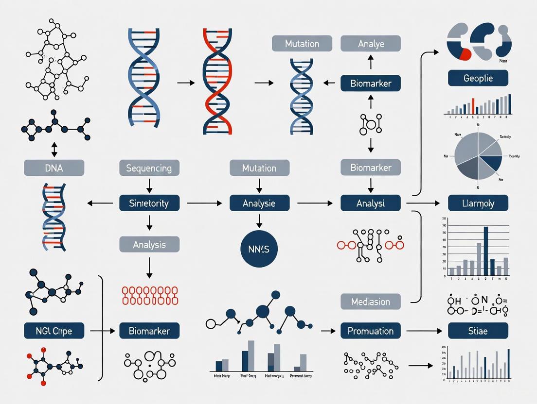
Next-generation sequencing (NGS) has revolutionized oncology research by enabling comprehensive molecular profiling of tumors, facilitating the discovery of disease mechanisms, and advancing personalized cancer treatment strategies. In clinical oncology, tumor molecular profiling is essential for therapeutic management of advanced cancers, with NGS enabling the identification of actionable mutations that guide targeted therapy selection [11] [12]. The technology provides unprecedented insights into genetic alterations, gene expression patterns, and epigenetic modifications that drive oncogenesis. The core NGS workflow encompasses multiple critical phases, from sample preparation through data analysis, each requiring meticulous optimization to generate clinically relevant data. This guide examines each component of the NGS workflow, compares established and emerging methodologies, and evaluates their performance in the context of oncology research applications, with particular emphasis on real-world clinical utility in cancer studies.
Sample Preparation: Foundation of Quality Results
Nucleic Acid Extraction and Quality Control
The initial step in any NGS workflow involves extracting high-quality nucleic acids (DNA or RNA) from various biological samples, which may include tumor tissues, blood, cultured cells, or liquid biopsy samples [13] [14]. In oncology research, sample quality directly impacts sequencing success, particularly with challenging specimens like formalin-fixed paraffin-embedded (FFPE) tissues which may contain degraded DNA [15]. The extraction process must yield sufficient quantity and purity of genetic material while preserving its molecular integrity. For liquid biopsy applications, circulating tumor DNA (ctDNA) is isolated from plasma, requiring specialized handling to capture often low-abundance tumor-derived fragments [12]. Best practices include using fresh starting material when possible, proper storage at specified temperatures, and working in dedicated pre-amplification areas to minimize contamination risk [13].
Library Construction Methodologies
Library preparation converts extracted nucleic acids into a format compatible with sequencing instruments by creating a library of uniformly sized, adapter-ligated DNA fragments [15]. This process involves several key steps:
- Fragmentation: DNA or cDNA is fragmented to desired sizes using physical methods (sonication, acoustic shearing), enzymatic approaches (non-specific endonucleases), or tagmentation (simultaneous fragmentation and tagging using transposase) [15] [16]. Physical methods generally produce more random fragmentation, while tagmentation offers workflow advantages.
- End Repair and A-Tailing: The fragmented DNA ends are repaired to create blunt ends, phosphorylated at the 5' ends, and a single A nucleotide is added to the 3' ends to facilitate adapter ligation [15] [16].
- Adapter Ligation: Platform-specific adapters containing sequencing primer binding sites are ligated to the fragments. These adapters often include barcode sequences that enable sample multiplexing [15] [13].
- Library Amplification: PCR amplification may be used to enrich for adapter-ligated fragments and generate sufficient material for sequencing, though this step can introduce biases if not carefully optimized [13] [16].
Table 1: Comparison of DNA Fragmentation Methods in Library Preparation
| Method | Principle | Advantages | Limitations | Best Applications |
|---|---|---|---|---|
| Acoustic Shearing | Physical shearing via sound waves | Minimal sequence bias, controllable size distribution | Specialized equipment required, additional cleanup steps | Whole genome sequencing, applications requiring uniform coverage |
| Enzymatic Fragmentation | DNA digestion via enzyme cocktails | Simple, fast, no specialized equipment | Potential sequence bias, higher indel artifacts | Routine sequencing, high-throughput applications |
| Tagmentation | Simultaneous fragmentation and adapter insertion via transposase | Rapid, reduced handling steps, integrated adapter ligation | Higher sequence bias, optimization challenges | High-throughput sequencing, limited input samples |
RNA library preparation presents additional challenges, including RNA susceptibility to degradation and the need for reverse transcription to generate cDNA before library construction [15]. The process typically includes steps for ribosomal RNA depletion or mRNA enrichment to focus sequencing on biologically relevant transcripts. For fusion gene detection in cancer research, RNA sequencing provides critical information that may complement DNA-based analyses [11] [13].
Sequencing Platforms and Performance Comparison
Platform Selection Considerations
NGS platforms vary significantly in their specifications, capabilities, and optimal applications. Selection depends on multiple factors including required throughput, read length, run time, and cost considerations [14] [17]. Different sequencing methods serve distinct research purposes in oncology:
- Whole Genome Sequencing (WGS) provides a comprehensive view of the entire genome, including coding and non-coding regions, enabling detection of novel variants across the genome [13] [14].
- Whole Exome Sequencing (WES) focuses on protein-coding regions, offering a cost-effective approach for identifying variants most likely to impact protein function [13] [14].
- Targeted Sequencing allows deep sequencing of specific genes or regions of interest, making it ideal for profiling known cancer-related genes with high sensitivity [13] [12].
- RNA Sequencing reveals gene expression patterns, alternative splicing, fusion genes, and mutations in the transcriptome [13] [14].
Table 2: Comparison of NGS Platforms and Their Specifications
| Platform | Output Range | Maximum Read Length | Run Time | Best Applications in Oncology |
|---|---|---|---|---|
| NovaSeq 6000 | 167-6000 Gb | 2 × 150 bp | 19-40 hours | Large cohort studies, whole genomes, multi-omics profiling |
| HiSeq X Series | 900-1800 Gb | 2 × 150 bp | <3 days | Population-scale genomics, cancer atlas projects |
| NextSeq 500 | 20-120 Gb | 2 × 150 bp | 11-29 hours | Medium-scale studies, tumor-normal pairs, transcriptomes |
| MiSeq Series | 0.3-15 Gb | 2 × 300 bp | 4-55 hours | Targeted panels, validation sequencing, small studies |
| MiniSeq | 1.8-7.5 Gb | 2 × 150 bp | 4-24 hours | Small targeted panels, rapid turnaround applications |
Impact of Sequencing Parameters on Data Quality
Sequencing depth (coverage) and read length significantly impact data quality and interpretive power. Higher coverage increases confidence in variant calling, particularly for heterogeneous tumor samples or low-frequency variants, while longer reads improve alignment in complex genomic regions and structural variant detection [14]. In clinical oncology applications, the optimal balance of these parameters depends on the specific clinical question, with targeted therapies often requiring deep sequencing to detect low-frequency resistance mutations, while novel biomarker discovery may benefit from broader genomic coverage [11] [12].
Data Analysis: Bioinformatics Pipelines
Genome Assembly Approaches
Bioinformatics analysis transforms raw sequencing data into biologically meaningful information. The primary approaches for genome assembly include:
- Reference-based assembly aligns sequencing reads to an existing reference sequence, offering computational efficiency but potential reference bias [18].
- De novo assembly constructs sequences without a reference, avoiding bias but struggling with gaps in coverage [18].
- Hybrid approaches combine strengths of both methods, leveraging reference guidance while accommodating sequence divergence [18].
In oncology, reference-based assembly typically using the human reference genome is most common, though hybrid approaches show promise for detecting novel cancer-specific alterations not present in reference databases.
Performance Comparison of Bioinformatics Pipelines
A comparative evaluation of open-source bioinformatics pipelines revealed significant differences in performance characteristics. When assembling viral genomes from NGS data, shiver and SmaltAlign pipelines showed robust performance with divergent samples, while viral-ngs and V-Pipe demonstrated advantages in runtime efficiency [18]. These findings highlight the importance of selecting analysis tools appropriate for specific research contexts and sample types.
Table 3: Comparison of Bioinformatics Pipelines for NGS Data Analysis
| Pipeline | Assembly Approach | Strengths | Limitations | Runtime Performance |
|---|---|---|---|---|
| shiver | Reference-based with iterative refinement | Handles divergent sequences well, high accuracy | Longer runtime, computationally intensive | Slower (benefits from Dockerized version) |
| SmaltAlign | Reference-based | Robust with non-matching subtypes, user-friendly | Limited functionality beyond core assembly | Fast (order of magnitude faster than shiver) |
| viral-ngs | Hybrid | Broad functionality, efficient resource use | Performance drops with divergent references | Fast (similar to SmaltAlign) |
| V-Pipe | Modular workflow | Extensive functionality, standardized variant calling | Complex setup, computationally demanding | Slower (similar to shiver) |
Cloud-based bioinformatics solutions offer promising approaches for the computationally intensive tasks required by NGS data analysis, providing scalability and accessibility for research teams without extensive local computational resources [19]. These platforms vary in their user interfaces, setup requirements, and analytical capabilities, requiring careful evaluation to match research needs.
Clinical Utility in Oncology Research
Real-World Evidence from Oncology Studies
The clinical utility of NGS in oncology is demonstrated through its ability to identify actionable genomic alterations that inform therapeutic decisions. In adolescent and young adult patients with advanced sarcoma, tumor NGS identified actionable mutations in 24.4% of cases, though fewer than 5% derived clinical benefit from NGS-directed therapy, highlighting both the promise and limitations of current approaches [11]. A systematic review and meta-analysis of NGS applications in childhood and AYA solid tumors found a pooled proportion of actionable alterations of 57.9%, with 22.8% of cases resulting in changes in clinical decision-making [20]. These findings underscore the potential of genomic profiling to guide personalized treatment strategies in challenging malignancies.
In non-small cell lung cancer, liquid biopsy NGS approaches using circulating tumor DNA (ctDNA) have demonstrated clinical utility, particularly when tissue samples are unavailable. Studies show 71.2% concordance between standard tissue-based genotyping and ctDNA-NGS, with ctDNA analysis offering a minimally invasive alternative for tumor profiling [12]. However, biological factors such as low ctDNA shed and assay-specific limitations can impact sensitivity, emphasizing the need for complementary approaches in clinical practice.
Methodological Standards for Clinical Research
Significant variability in NGS methodologies across studies influences the interpretation and comparability of results. Differences in sequencing techniques (targeted panels, whole exome, whole genome), tumor sampling strategies (primary vs. relapsed disease), and definitions of "actionable alterations" create challenges for pooled analyses and clinical implementation [20]. Standardization of sequencing methodologies, sample collection practices, and reporting standards using established frameworks such as the ESMO Scale for Clinical Actionability of Molecular Targets (ESCAT) can enhance methodological consistency and translational impact [20].
The Scientist's Toolkit: Essential Research Reagents and Materials
Table 4: Essential Research Reagents and Materials for NGS Workflows
| Reagent/Material | Function | Application Notes |
|---|---|---|
| Nucleic Acid Extraction Kits | Isolate DNA/RNA from various sample types | Selection depends on sample source (tissue, blood, FFPE); specialized kits needed for ctDNA |
| Fragmentation Enzymes/Reagents | Fragment DNA to optimal size distributions | Enzymatic methods offer convenience; physical methods may reduce bias |
| End-Repair Enzymes | Create blunt-ended, phosphorylated DNA fragments | Typically uses T4 DNA polymerase and T4 PNK |
| Adapter Ligases and Oligos | Attach platform-specific adapters to fragments | May include unique molecular identifiers (UMIs) for error correction |
| High-Fidelity Polymerases | Amplify library fragments with minimal errors | Critical for maintaining sequence accuracy during amplification |
| Size Selection Beads/Kits | Select fragments within specific size ranges | Magnetic bead-based methods enable automation and high-throughput |
| Quality Control Instruments | Assess library quality and quantity | Fluorometric methods preferred for accuracy over spectrophotometry |
| Target Enrichment Probes | Capture specific genomic regions of interest | Critical for targeted sequencing; design impacts coverage uniformity |
| Unique Molecular Identifiers (UMIs) | Tag individual molecules for error correction | Essential for liquid biopsy applications and low-frequency variant detection |
Experimental Workflow Visualization
NGS Library Preparation Workflow
NGS Data Analysis Pipeline
The core NGS workflow represents an integrated system where each component—from sample preparation through data analysis—contributes significantly to the quality and interpretability of final results. As NGS technologies continue to evolve, balancing increasing throughput with maintaining data quality remains paramount. In oncology research, standardization of methodologies and analytical approaches will enhance the comparability of findings across studies and maximize the clinical utility of genomic profiling. The ongoing refinement of NGS technologies and analytical methods promises to further advance our understanding of cancer biology and expand opportunities for personalized cancer therapy.
Next-Generation Sequencing (NGS) has revolutionized oncology research, but its true value is ultimately defined by its impact on patient care in clinical settings. This guide objectively compares the performance of NGS-based comprehensive genomic profiling (CGP) against traditional testing methods and evaluates the evidence for its real-world clinical utility, providing researchers and drug developers with the data and frameworks needed for its effective implementation.
◍ NGS versus Traditional Single-Gene Testing
The transition from single-gene testing to NGS represents a paradigm shift in molecular oncology. The table below provides a performance and outcome comparison between these testing approaches.
| Feature | Next-Generation Sequencing (NGS) | Traditional Single-Gene Assays |
|---|---|---|
| Testing Approach | Multigene panel in a single assay [6] [21] | Focus on a small set of genes, typically one per test [6] |
| Throughput & Speed | Massive parallel sequencing; rapid turnaround for multiple genes [6] [21] | Sequential testing; time-consuming for broad profiling [6] |
| Data Output | Large amount of data (SNVs, Indels, CNVs, fusions, TMB, MSI) [22] [6] | Limited data output; focused on specific known mutations [6] |
| Diagnostic Yield | Higher; identifies co-mutations and rare variants missed by limited panels [22] [23] | Lower; may miss clinically relevant alterations in non-targeted genes [6] |
| Tissue Utilization | More economical; reduces risk of exhausting precious biopsy samples [21] | Poor; multiple tests can consume limited tissue [6] |
| Clinical Utility (Matched Therapy Rate) | Enables matching to targeted therapy or clinical trials [22] [23] | Limited to therapies for the specific genes tested [6] |
| Cost-Effectiveness | Higher for large-scale profiling; cost-saving versus sequential single tests [24] | Lower for testing a single gene, but costly for broad profiling [6] |
◍ Quantifying Clinical Impact: Real-World Evidence of NGS Utility
Clinical utility is demonstrated when diagnostic testing leads to improved patient outcomes. The following table summarizes key real-world evidence on the survival benefits of NGS-informed therapy.
| Study / Review Description | Patient Population | Key Findings on NGS-Informed Therapy |
|---|---|---|
| SNUBH Real-World Study (2025) [22] | 990 advanced solid tumor patients | • 37.5% of patients with measurable lesions achieved a partial response.• 34.4% achieved stable disease.• Median treatment duration was 6.4 months. |
| US Literature Review (2023) [23] | 31 studies across multiple tumor types | • 11 publications reported significantly longer Progression-Free Survival (PFS) with matched therapy.• 16 publications reported significantly longer Overall Survival (OS).• Hazard Ratios (HRs) for PFS favored matched therapy (range: 0.24-0.67). |
| Below-LoD Biomarker Study (2025) [25] | 129 advanced NSCLC patients | • Patients with biomarkers detected below the assay's Limit of Detection (LoD) showed 67% (tissue) and 72% (liquid) real-world response rates when given matched therapy, far exceeding the 30% historical benchmark for chemotherapy. |
◍ Experimental Protocols for Validating Clinical NGS
For a novel NGS assay to be deployed in a clinical setting, it must undergo rigorous validation to ensure analytical and clinical performance. The following protocol outlines the key stages of this process.
▸ Protocol 1: Analytical Validation of a Targeted NGS Panel
This protocol is adapted from a 2022 study that validated an NGS panel for pediatric acute leukemia, detailing the steps to establish analytical sensitivity, specificity, and reproducibility [26].
- Step 1: Sample Selection and Preparation
- Commercial Controls: Use commercially available reference DNA and RNA controls with known mutations and fusions at specified variant allele frequencies (VAFs). These are essential for establishing sensitivity and limit of detection (LOD) [26].
- Patient Samples: Select patient samples with well-characterized genetic alterations via orthogonal methods (e.g., Sanger sequencing, qRT-PCR). Include samples with a range of VAFs and alteration types (SNVs, Indels, fusions) [26].
- Step 2: Nucleic Acid Extraction and QC
- Extract DNA and RNA using standardized kits (e.g., QIAamp for DNA, TriPure for RNA).
- Assess purity using spectrophotometry (OD260/280 ratio >1.8). Determine concentration by fluorometric quantification (e.g., Qubit) and assess integrity using automated electrophoresis systems (e.g., Bioanalyzer, TapeStation) [26].
- Step 3: Library Preparation and Sequencing
- Use the targeted panel kit (e.g., AmpliSeq Childhood Cancer Panel) according to manufacturer's instructions.
- Input 100 ng of DNA and RNA per sample.
- Perform library amplification to generate target amplicons. Sequence on a platform such as Illumina NextSeq with a minimum mean read depth of 1000x [26].
- Step 4: Data Analysis and Variant Calling
- Step 5: Determination of Key Validation Metrics
- Sensitivity: Calculate as (True Positives / (True Positives + False Negatives)) × 100. The validated panel achieved >98% for DNA and >94% for RNA [26].
- Specificity: Calculate as (True Negatives / (True Negatives + False Positives)) × 100. The goal is 100% [26].
- Reproducibility: Perform replicate runs of the same sample across different days/operators. Calculate concordance; the benchmark is 100% for DNA and >89% for RNA [26].
- Limit of Detection (LOD): Serially dilute positive controls to determine the lowest VAF at which a variant is detected with ≥95% confidence [25] [26].
▸ Protocol 2: Assessing Clinical Utility in a Real-World Cohort
This protocol describes a methodology for evaluating whether NGS testing leads to improved patient management and outcomes, as seen in large-scale real-world studies [22] [23].
- Step 1: Cohort Definition and NGS Testing
- Step 2: Variant Classification and Actionability Assessment
- Classify all genetic alterations using a standardized tier system (e.g., Association for Molecular Pathology guidelines):
- Crucial Step: Define "NGS-based therapy" as treatment selected based only on novel information from the NGS test, excluding therapies identifiable via prior conventional testing [22].
- Step 3: Outcome Measurement and Statistical Analysis
- For patients receiving NGS-based therapy, measure objective outcomes:
- Objective Response Rate (ORR): Proportion of patients with a partial or complete response per RECIST criteria [22].
- Treatment Duration: Median duration of therapy [22].
- Overall Survival (OS): Median time from treatment start until death from any cause [22] [23].
- Progression-Free Survival (PFS): Median time from treatment start until disease progression or death [23].
- Use statistical software (e.g., SPSS, R) for survival analysis, generating Kaplan-Meier curves and calculating hazard ratios to compare outcomes where possible [22].
- For patients receiving NGS-based therapy, measure objective outcomes:
◍ The NGS Clinical Pathway: From Sample to Report
The following diagram visualizes the end-to-end process of using NGS in clinical oncology, from sample acquisition to therapeutic decision-making, highlighting key steps that impact patient care.
◍ The Scientist's Toolkit: Essential Reagents for NGS Workflows
Successful implementation of clinical NGS requires a suite of specialized reagents and tools. This table details key components of a typical targeted NGS workflow.
| Item | Function |
|---|---|
| FFPE Tumor Specimens | The primary source material for DNA extraction; requires pathologist review for tumor content [22]. |
| Nucleic Acid Extraction Kits (e.g., QIAamp DNA FFPE Tissue kit) | Isolate and purify high-quality genomic DNA from formalin-fixed, paraffin-embedded tissue samples [22]. |
| Targeted NGS Panel (e.g., SNUBH Pan-Cancer v2, AmpliSeq Childhood Cancer Panel) | A predefined set of probes designed to capture and sequence hundreds of cancer-related genes simultaneously [22] [26]. |
| Library Preparation Kit (e.g., Agilent SureSelectXT) | Prepares the fragmented DNA for sequencing by adding platform-specific adapters and indices [22]. |
| NGS Platform (e.g., Illumina NextSeq) | The instrument that performs massively parallel sequencing, generating millions of short DNA reads [22] [27]. |
| Bioinformatics Pipelines (e.g., MuTect2, CNVkit, LUMPY) | Software tools for aligning sequences to a reference genome and identifying variants (SNVs, CNVs, fusions) [22]. |
| Variant Classification Guidelines (e.g., AMP/ASCO/CAP) | A standardized framework for interpreting and reporting sequence variants based on clinical significance [22]. |
| Positive Control DNA/RNA (e.g., SeraSeq) | Reference materials with known mutations used for assay validation, quality control, and monitoring performance [26]. |
The collective evidence demonstrates that NGS has decisively moved beyond a research tool to become a cornerstone of modern oncology care. Its comprehensive nature offers a clear advantage over sequential single-gene testing, enabling the identification of actionable biomarkers that directly inform treatment. Real-world data consistently shows that this leads to improved patient response rates and survival outcomes across multiple cancer types. For researchers and drug developers, this underscores the importance of designing therapies against a expanding list of molecular targets and of integrating robust NGS-based biomarkers into clinical trials to accurately identify the patients most likely to benefit.
Next-generation sequencing (NGS) has emerged as a transformative technology in oncology, enabling comprehensive genomic profiling that informs diagnosis, risk assessment, and treatment monitoring [6]. This high-throughput methodology allows for massive parallel sequencing of millions of DNA fragments simultaneously, providing unprecedented insights into the molecular architecture of cancers [6]. The real-world clinical utility of NGS extends across three fundamental applications: characterizing tumor genomes for actionable mutations, identifying hereditary cancer susceptibility, and monitoring minimal residual disease (MRD) with exceptional sensitivity. As precision oncology advances, NGS technologies have become indispensable tools for researchers and drug development professionals seeking to understand cancer biology and develop targeted therapeutic strategies. This guide objectively compares NGS performance against alternative methodologies across these key applications, supported by experimental data and implementation protocols.
Technical Foundations of Next-Generation Sequencing
Core Sequencing Methodology and Comparison with Traditional Approaches
The NGS workflow encompasses multiple critical steps: nucleic acid extraction, library preparation, massive parallel sequencing, and bioinformatic analysis [6]. During library construction, DNA is fragmented into segments of approximately 300 base pairs, followed by adapter ligation that facilitates amplification and sequencing [6]. Cluster generation then occurs on flow cells through bridge amplification, creating millions of identical DNA templates. Finally, sequencing-by-synthesis incorporates fluorescently-labeled nucleotides, with optical detection of incorporated bases determining the sequence [6]. This fundamental process enables the high-throughput capabilities that distinguish NGS from traditional sequencing methods.
Table 1: Comparison of Next-Generation Sequencing and Sanger Sequencing
| Feature | Next-Generation Sequencing | Sanger Sequencing |
|---|---|---|
| Cost-effectiveness | Higher for large-scale projects | Lower for small-scale projects |
| Speed | Rapid sequencing | Time-consuming |
| Application | Whole-genome sequencing, targeted sequencing | Ideal for sequencing single genes |
| Throughput | Multiple sequences simultaneously | Single sequence at a time |
| Data output | Large amount of data | Limited data output |
| Clinical utility | Detects mutations, structural variants | Identifies specific mutations |
Source: Adapted from [6]
Essential Research Reagent Solutions for NGS Applications
Successful implementation of NGS in oncology research requires specific reagent systems and analytical tools:
- Nucleic Acid Extraction Kits: Automated systems like Maxwell DNA Purification provide high-quality DNA from various sample types, crucial for reliable sequencing results [28].
- Targeted Sequencing Panels: Commercial panels (FoundationOne, Tempus, LymphoTrack) enable focused analysis of cancer-related genes with optimized coverage and sensitivity [11] [28] [29].
- Library Preparation Systems: Kit-based solutions (Twist Library Preparation) facilitate fragment end-repair, adapter ligation, and library amplification with minimal bias [12].
- Unique Molecular Identifiers (UMIs): xGEN UMI adapters enable error correction and accurate quantification by tagging individual DNA molecules before amplification [12].
- Hybrid Capture Reagents: Custom probe sets (Twist Biosciences) enrich target genomic regions through solution-based hybridization for comprehensive variant detection [12].
- Bioinformatic Platforms: Specialized software (LymphoTrack Analysis, GATK Mutect2) processes raw sequencing data, detects variants, and interprets clinical significance [28] [12].
Application 1: Comprehensive Tumor Genomic Profiling
Experimental Protocols for Tumor Molecular Characterization
Tumor profiling via NGS follows standardized protocols to ensure reproducible results. The BALLETT study, a nationwide comprehensive genomic profiling initiative, demonstrated a 93% success rate across 756 advanced cancer patients using a 523-gene panel [30]. Their methodology involved: (1) DNA extraction from formalin-fixed paraffin-embedded (FFPE) tumor tissue with quality control measures; (2) library preparation using standardized kits across nine participating laboratories; (3) sequencing on Illumina platforms with a minimum coverage of 500x; and (4) bioinformatic analysis for single nucleotide variants, insertions/deletions, copy number alterations, and gene fusions [30]. This protocol identified actionable genomic markers in 81% of patients, substantially higher than the 21% actionability rate with smaller, nationally reimbursed panels [30].
In sarcoma research, a multicenter analysis of 81 patients utilized four commercial NGS kits (FoundationOne, Tempus, OncoDEEP, MI Profile) to investigate mutation profiles [29]. The protocol specified: (1) tumor content >20% in samples; (2) sequencing of 223 genomic alterations with average of 2.74 alterations per patient; (3) assessment of tumor mutational burden and microsatellite instability; and (4) annotation of variants according to OncoKB criteria for therapeutic actionability [29]. This approach detected genomic alterations in 90.1% of patients, with TP53 (38%), RB1 (22%), and CDKN2A (14%) emerging as most frequently mutated genes [29].
Performance Comparison: NGS versus Traditional Molecular Assays
NGS-based tumor profiling demonstrates significant advantages over single-gene testing approaches in comprehensiveness and efficiency. The BALLETT study directly compared actionability rates between CGP and standard-of-care panels, revealing that conventional testing would have identified actionable markers in only 21% of patients compared to 81% with CGP [30]. In adolescent and young adult sarcoma patients, tumor NGS successfully identified actionable mutations in 24.4% of cases (28/115 patients), enabling molecular confirmation of diagnosis in 3.5% [11]. This demonstrates the diagnostic utility of NGS beyond therapeutic guidance.
Table 2: Tumor Profiling Performance of NGS Versus Traditional Methods
| Parameter | Comprehensive Genomic Profiling | Single-Gene/Small Panel Testing |
|---|---|---|
| Actionable mutation detection rate | 81% (BALLETT study, n=756) [30] | 21% (BALLETT study, n=756) [30] |
| Genes analyzed simultaneously | 523 genes (BALLETT study) [30] | Typically 1-50 genes |
| Therapeutic impact in sarcoma | 14.8% received NGS-directed therapy [11] | Limited by narrow scope |
| Turnaround time | Median 29 days (BALLETT study) [30] | Variable based on number of tests required |
| Additional biomarkers | TMB, MSI, HRD simultaneously assessed [30] | Requires separate testing |
Application 2: Hereditary Cancer Risk Assessment
Methodologies for Germline Variant Detection from Tumor Sequencing
Tumor-based NGS profiling can identify potential pathogenic germline variants (PPGVs) through specialized bioinformatic filtering approaches. Foundation Medicine developed a validated workflow incorporating: (1) filtering for short variants in 24 cancer susceptibility genes with high germline conversion rates; (2) application of variant allele frequency thresholds (>10% for tissue, >30% for liquid biopsies); and (3) pathogenicity assessment using ClinVar classifications [31]. This method identified PPGVs in 9.7% of 125,128 advanced cancer patients, with BRCA2 (16.9%), MUTYH (15.0%), and ATM (13.4%) representing the most frequently implicated genes [31].
Research by Tung et al. applied similar filtering criteria to tumor-only sequencing data from 125,000 patients, demonstrating that 9.7% harbored likely pathogenic germline variants [32]. Their protocol included: (1) variant calling from tumor sequencing; (2) filtering based on population frequency databases; (3) application of variant allele frequency thresholds suggestive of germline origin; and (4) confirmation using ClinVar pathogenicity classifications [32]. This approach successfully detected PPGVs across a broad cancer spectrum, including instances where traditional clinical criteria would not have prompted germline testing.
Comparative Performance: Tumor-Based NGS Versus Traditional Germline Testing
Tumor-based NGS approaches complement traditional germline testing by identifying hereditary risk in patients who might not otherwise meet testing criteria. Research demonstrates that PPGVs are identified in similar proportions of cancers with NCCN recommendations for germline testing (11%) as in cancer types without such recommendations (9%) [31]. This suggests tumor NGS can detect hereditary risk outside established clinical parameters. Among 81 sarcoma patients undergoing NGS, two confirmed germline mutations (BLM, TP53, ATM) were identified, followed by genetic counseling and family risk assessment [29].
Table 3: Germline Variant Detection Performance
| Metric | Tumor-Based NGS with PPGV Filtering | Traditional Clinical Criteria-Based Testing |
|---|---|---|
| Detection rate pan-cancer | 9.7% (n=125,128) [31] | 3-17% (varies by cancer type) [32] |
| Detection in "off-tumor" cancers | 9% of cases without NCCN testing guidelines [31] | Typically missed without family history |
| Most commonly detected genes | BRCA2 (16.9%), MUTYH (15.0%), ATM (13.4%) [31] | BRCA1/2 predominant in breast/ovarian cancer |
| Multiple PPGV cases | 10% of PPGV+ cases [31] | Rare without specific syndromes |
| Confirmation rate | High germline conversion rate for selected CSGs [31] | Gold standard |
Application 3: Monitoring Disease Progression and MRD
Experimental Protocols for Minimal Residual Disease Detection
In multiple myeloma, NGS-based MRD detection utilizes the LymphoTrack IGH panel with the following protocol: (1) DNA extraction from bone marrow aspirates with quality control; (2) PCR amplification of IGH rearrangements using BIOMED-2 primers; (3) library preparation with spike-in control cells for absolute quantification; (4) sequencing on MiSeq platforms targeting 1 million reads per sample; and (5) data analysis using LymphoTrack software to identify clonotypic sequences [28]. This approach achieved a sensitivity of 10^-5, detecting one malignant cell per 100,000 normal cells [28].
A comparative study in acute myeloid leukemia implemented both NGS and multiparameter flow cytometry (MFC) on 107 patients with 717 MFC and 247 NGS studies [33]. The NGS methodology included: (1) targeted sequencing of recurrently mutated genes in AML; (2) unique molecular identifier incorporation to reduce errors; (3) deep sequencing (>1000x coverage) for sensitive variant detection; and (4) bioinformatic analysis comparing mutation profiles at diagnosis and follow-up [33]. This protocol revealed that 44 instances were MFC-negative/NGS-positive, with 64% of these occurring within 6 months post-treatment [33].
Comparative Performance: NGS Versus Flow Cytometry and PCR
NGS demonstrates complementary value to established MRD monitoring techniques, offering different advantages and limitations. In multiple myeloma, NGS and next-generation flow (NGF) showed high correlation (R²=0.905), with 3-year progression-free survival significantly longer for MRD-negative patients by either method (NGS: 88.7% vs. 56.6%; NGF: 91.4% vs. 50%) [28]. In AML monitoring, discordant results between MFC and NGS occurred in 47 of 247 paired samples, with most (44/47) being MFC-negative/NGS-positive [33]. This suggests NGS can detect residual disease not identified by immunophenotypic methods.
Table 4: MRD Monitoring Method Comparison in Hematologic Malignancies
| Parameter | Next-Generation Sequencing | Multiparameter Flow Cytometry | Quantitative PCR |
|---|---|---|---|
| Sensitivity | 10^-5 to 10^-6 [28] | 10^-4 to 10^-5 [33] | 10^-4 to 10^-6 [28] |
| Applicability | ~90% for B-cell malignancies [28] | >95% [33] | 40-75% in myeloma [28] |
| Turnaround time | 7-14 days | 1-2 days | 5-10 days |
| Standardization | Requires validated bioinformatic pipelines [28] | Operator-dependent [33] | Requires patient-specific primers |
| Additional information | Provides clonal evolution data [33] | Limited to immunophenotype | Limited to known targets |
Integrated Analysis of NGS Clinical Utility
The cumulative evidence from recent studies demonstrates that NGS delivers substantial clinical value across the cancer care continuum. The BALLETT study reported that 23% of patients ultimately received matched therapies based on CGP results, with 69% receiving specific treatment recommendations from a molecular tumor board [30]. In sarcoma patients, however, the direct clinical benefit was more modest, with only 4.4% deriving clinical benefit from NGS-directed therapy despite 24.4% having actionable mutations [11]. This highlights tumor-type variability in NGS utility that researchers should consider.
The integration of liquid biopsy NGS approaches expands applications to instances where tissue is limited. In NSCLC, ctDNA-NGS demonstrated 71.2% concordance with standard-of-care tissue genotyping, though it missed actionable drivers in 3.4% of cases [12]. Implementation modeling predicted that offering ctDNA-NGS only to patients not testable by standard methods would increase diagnostic yield by 6.7% [12], highlighting its complementary role rather than replacement for tissue profiling.
Next-generation sequencing technologies provide researchers and drug development professionals with powerful tools for comprehensive genomic characterization across oncology applications. Performance data demonstrates that NGS outperforms traditional single-gene assays in detecting actionable alterations (81% vs. 21% actionability), identifies hereditary cancer risk in nearly 10% of patients beyond standard clinical criteria, and enables highly sensitive disease monitoring at 10^-5 to 10^-6 sensitivity. Each application requires specialized experimental protocols and bioinformatic approaches to maximize clinical utility. While implementation challenges remain regarding standardization, turnaround time, and clinical interpretation, the strategic integration of NGS technologies continues to advance precision oncology by uncovering molecular insights that inform targeted therapeutic strategies and improve patient outcomes.
NGS in Action: Methodological Approaches and Real-World Clinical Applications in Oncology
Next-generation sequencing (NGS) has revolutionized molecular diagnostics and precision oncology, enabling comprehensive genomic profiling that guides therapeutic decisions. The three primary NGS approaches—whole-genome sequencing (WGS), whole-exome sequencing (WES), and targeted gene panels—differ fundamentally in scope, resolution, and clinical application [34]. In oncology research, selecting the appropriate method requires careful consideration of each technology's capabilities to detect clinically actionable biomarkers, including single nucleotide variants (SNVs), insertions/deletions (indels), copy number variations (CNVs), structural variants (SVs), and complex biomarkers like tumor mutational burden (TMB) and microsatellite instability (MSI) [35] [36]. This guide provides an objective comparison of these methodologies, supported by recent experimental data and analytical protocols, to inform their strategic application in cancer research and drug development.
The fundamental distinction between NGS approaches lies in the genomic regions they target, directly influencing the variants detectable in oncology applications.
Whole-genome sequencing (WGS) interrogates the entire 3 billion base pair human genome, comprising both coding (exonic) and non-coding regions [37] [34]. This comprehensive coverage enables detection of virtually all variant types, including those in regulatory regions that may influence gene expression in cancer pathogenesis [38].
Whole-exome sequencing (WES) targets approximately 1-2% of the genome encompassing all protein-coding exons (∼30 million base pairs) [39] [34]. This focus on functionally consequential regions provides a balance between comprehensiveness and data management for identifying driver mutations in oncology.
Targeted gene panels sequence a predefined set of genes known to be associated with specific cancer types or therapeutic pathways [34] [40]. These panels range from dozens to hundreds of genes, with extreme depth of coverage facilitating sensitive detection of low-frequency variants in heterogeneous tumor samples [34].
The diagram below illustrates the fundamental relationship between the sequencing approaches in terms of genomic coverage and typical sequencing depth:
Table 1: Technical comparison of NGS approaches
| Parameter | Whole Genome Sequencing | Whole Exome Sequencing | Targeted Gene Panels |
|---|---|---|---|
| Sequencing Region | Entire genome (∼3 GB) [34] | Protein-coding exons only (∼30 MB) [34] | Selected genes/regions (varies) [34] |
| Typical Depth | >30X [34] | 50-150X [34] | >500X [34] |
| Data Volume | >90 GB [34] | 5-10 GB [34] | Varies by panel size |
| Detectable Variants | SNPs, InDels, CNV, Fusion, SV [34] | SNPs, InDels, CNV, Fusion [34] | SNPs, InDels, CNV, Fusion [34] |
| Non-Coding Variants | Yes [38] | Limited | No |
| Turnaround Time | ∼4 days [38] | Varies | Typically fastest [40] |
Experimental Data: Diagnostic Yield and Clinical Actionability
Recent large-scale studies provide robust quantitative comparisons of the clinical performance of different NGS approaches in oncology and genetic disease diagnostics.
Diagnostic Yield Across Clinical Indications
A comprehensive Brazilian study analyzing 3,025 patients provides direct comparative data on diagnostic yield across sequencing approaches [39].
Table 2: Diagnostic yield by NGS approach and clinical indication
| Clinical Indication | WES Detection Rate | Multi-Gene Panel Detection Rate | Notable Findings |
|---|---|---|---|
| Overall | 32.7% [39] | Not directly reported | WES had highest detection rate but also highest inconclusive rate [39] |
| Skeletal Disorders | 55% [39] | Not specified | Highest diagnostic yield among all indications with WES [39] |
| Hearing Disorders | 50% [39] | Not specified | Second highest yield with WES [39] |
| Syndromic Disorders | 76.2% of tests were WES [39] | 13.9% of tests [39] | Most frequent indication for WES [39] |
| Neurodevelopmental | 74% of tests were WES [39] | 22% of tests [39] | Second most frequent indication for WES [39] |
Clinical Actionability in Oncology
The clinical utility of NGS approaches in oncology is demonstrated by their ability to identify actionable biomarkers that inform treatment decisions.
Table 3: Biomarker detection and clinical actionability in oncology
| Biomarker Category | WES/WGS/TS Performance | Panel Performance | Clinical Impact |
|---|---|---|---|
| Therapy Recommendations | 3.5 per patient (median) [35] | 2.5 per patient (median) [35] | WES/WGS/TS provided more treatment options [35] |
| Tumor Mutational Burden | Detected [35] | Detected by some comprehensive panels [36] | Eligibility for immunotherapy [36] |
| Microsatellite Instability | Detected [35] | Detected by some comprehensive panels [36] | Eligibility for immunotherapy [36] |
| Structural Variants/Gene Fusions | Comprehensive detection [35] | Limited to targeted genes [35] | Critical for fusion-driven cancers [35] |
| Homologous Recombination Deficiency | Detectable [35] | Limited detection [36] | Predicts response to PARP inhibitors [36] |
A 2025 study comparing WES/WGS/transcriptome sequencing (TS) to panel sequencing (TruSight Oncology 500/TruSight Tumor 170) in 20 rare or advanced tumor patients found that approximately half of the therapy recommendations were identical between approaches [35]. However, approximately one-third of therapy recommendations in WES/WGS/TS relied on biomarkers not covered by the panel, highlighting the additional clinical value of comprehensive approaches [35].
In a real-world study of tumor NGS in adolescent and young adult sarcoma patients, actionable mutations were identified in 24.4% of cases (28/115), though only 4.4% ultimately derived clinical benefit from NGS-directed therapy [11]. This underscores that detection of actionable alterations does not invariably translate to clinical benefit, emphasizing the need for robust biomarker-therapy associations.
Methodological Protocols and Workflows
Understanding the experimental protocols for each NGS approach is essential for selecting appropriate methodologies for specific research questions.
Laboratory Workflows
The core laboratory workflow for NGS includes sample preparation, library construction, sequencing, and data analysis, with key differences in target enrichment.
Key Methodological Considerations
WGS Protocols: Current short-read WGS protocols typically achieve >95% of the human genome at 10X coverage with median coverage of 30X, sufficient for germline analysis [38]. Tumor analysis requires higher coverage (~90X) to identify minority clones [38]. Laboratory procedures can be performed in conventional molecular biology laboratories, with protocols taking approximately four working days that are less labor-intensive than panel or exome sequencing due to the absence of capture and amplification steps [38].
WES Target Enrichment: Critical probe performance metrics include:
- On-target rate: Percentage of sequencing data aligning to the target region [34]
- Coverage uniformity: Evenness of coverage across target regions [34]
- Sensitivity: Ability to detect target regions effectively [34]
- Duplication rate: Percentage of duplicate reads, affecting usable data [34]
Panel Design Strategies: Gene panels employ either hybridization capture or multiplex amplicon sequencing [34]. The number of genes ranges from dozens focused on specific cancer types (e.g., NSCLC panels) to comprehensive pan-cancer panels covering 500+ genes [36].
Analytical Validation and Quality Metrics
Robust quality control is essential for clinical-grade NGS across all approaches:
- Sequencing depth: Critical for detection sensitivity, particularly for subclonal variants in tumor samples [37]
- Coverage uniformity: Impacts ability to call variants consistently across all target regions [34]
- Mapping rates: Measures proportion of bases aligning to reference genome, with higher rates indicating better data quality [37]
- Contamination monitoring: Particularly crucial for WGS, which is unlikely to be repeated [38]
For tumor sequencing, matched normal samples are essential for distinguishing somatic from germline variants, with specialized bioinformatics pipelines required for accurate variant calling in complex cancer genomes [35] [38].
Essential Research Reagents and Platforms
Successful implementation of NGS in oncology research requires specific reagent systems and analytical tools.
Table 4: Essential research reagents and platforms for NGS in oncology
| Reagent Category | Specific Examples | Research Function |
|---|---|---|
| Commercial Panels | TruSight Oncology 500, FoundationOne CDx [11] [35] | Standardized detection of actionable cancer mutations |
| Hybridization Capture Kits | Various exome capture panels [34] | Target enrichment for WES and large panels |
| Library Prep Kits | Platform-specific kits (Illumina, etc.) [34] | Fragment processing and adapter ligation |
| Sequencing Platforms | Illumina, PacBio, Oxford Nanopore [37] | DNA sequencing with various read lengths and outputs |
| Bioinformatics Tools | GATK, DRAGEN, Sentieon [38] | Variant calling, annotation, and filtering |
| Reference Databases | gnomAD, COSMIC, ClinVar [34] | Variant annotation and pathogenicity assessment |
The comparative analysis of WGS, WES, and targeted panels reveals a nuanced landscape where each approach offers distinct advantages for specific research contexts in oncology. WGS provides the most comprehensive variant detection, including non-coding regions and complex structural variants, making it invaluable for discovery research and cases where previous testing has been uninformative [41] [38]. WES balances comprehensiveness with practical data management, offering substantial diagnostic yield across diverse cancer types while focusing on protein-coding regions that harbor most known pathogenic variants [39] [42]. Targeted panels deliver cost-effective, rapid analysis of known actionable genes with exceptional depth of coverage, ideal for routine clinical profiling and detecting low-frequency variants [36] [40].
The choice between these approaches depends on multiple factors, including research objectives, sample quality, bioinformatics capabilities, and budgetary constraints. For discovery-oriented research and complex cases with unknown etiology, WGS and WES offer clear advantages in identifying novel alterations and complex biomarkers [35]. For focused investigation of established cancer genes and clinical applications requiring rapid turnaround, targeted panels provide efficient and sensitive mutation detection [36]. As sequencing costs continue to decline and analytical methods improve, the integration of multiple approaches—particularly WGS/WES with transcriptome sequencing—will likely expand, offering increasingly comprehensive molecular profiling to advance precision oncology and therapeutic development.
Identifying Actionable Mutations and Guiding Targeted Therapy Selection
Next-generation sequencing (NGS) has revolutionized oncology by enabling comprehensive molecular profiling of tumors, moving cancer care beyond histology-based classification to a genetically-guided paradigm. The identification of actionable mutations—genomic alterations susceptible to targeted therapeutic agents—represents a cornerstone of precision medicine. The clinical utility of NGS extends across multiple domains: facilitating accurate diagnosis, informing prognosis, predicting treatment response, and identifying resistance mechanisms. In the current oncology landscape, NGS testing provides the critical molecular data necessary to match patients with targeted therapies, often within clinical trial frameworks, based on their tumor's unique genetic signature. This guide objectively compares the performance characteristics of various NGS methodologies and platforms, providing researchers and drug development professionals with experimental data to inform their genomic strategy.
Actionable Mutations: Prevalence and Clinical Significance
Actionable genomic alterations (AGAs) represent the fundamental targets of precision oncology. These mutations occur primarily in driver genes that promote cancer progression and can be therapeutically targeted with specific agents. In non-small cell lung cancer (NSCLC), a model disease for targeted therapy, AGAs are predominantly found in lung adenocarcinomas, with varying prevalence across different populations [43].
Table 1: Prevalence of Actionable Genomic Alterations in NSCLC [43]
| Gene | Alteration Type | Prevalence (%) | Notes |
|---|---|---|---|
| EGFR | Common mutations (del19, L858R) | 15% | 50-60% in Asian populations |
| EGFR | Uncommon mutations (G719X, L861Q, S768I) | 10% | |
| EGFR | Exon 20 insertions | 2% | |
| ALK | Fusions | 5% | |
| ROS1 | Fusions | 1-2% | |
| BRAF | V600E mutations | 2% | |
| MET | Exon 14-skipping mutations | 3% | |
| RET | Fusions | 1-2% | |
| KRAS | G12C mutations | 12% | |
| ERBB2 (HER2) | Mutations | 2-5% | |
| NTRK | Fusions | 0.23-3% |
The clinical significance of identifying these alterations is profound. For EGFR-mutant NSCLC, the development of tyrosine kinase inhibitors (TKIs) has dramatically improved outcomes. The FLAURA trial established osimertinib, a third-generation TKI, as standard of care with median progression-free survival (PFS) of 18.9 months versus 10.2 months for first-generation TKIs (HR 0.46) and overall survival of 38.6 months versus 31.8 months (HR 0.80) [43]. Recent combination approaches such as osimertinib with chemotherapy (FLAURA2) or amivantamab with lazertinib (MARIPOSA) have further improved outcomes, demonstrating the continuous evolution of targeted therapy strategies [43].
Comparative Methodologies for Mutation Detection
Tissue-Based NGS Approaches
Tissue-based genomic profiling remains the gold standard for detecting actionable mutations. Various technological platforms offer different advantages in terms of throughput, sensitivity, and comprehensiveness.
Table 2: Comparison of NGS Methodologies for Actionable Mutation Detection
| Methodology | Genes Covered | Sensitivity | Turnaround Time | Key Advantages | Limitations |
|---|---|---|---|---|---|
| Targeted DNA Panel (TTSH-oncopanel) [44] | 61 cancer-associated genes | 98.23% for unique variants | 4 days | High throughput, cost-effective for focused analysis | Limited to predefined genomic regions |
| Hybrid Capture-Based NGS [12] | 45 genes (hotspot regions) | 70-80% for ctDNA vs tissue | 3-5 days | Can be applied to ctDNA; UMI error correction | Lower sensitivity for fusion detection in ctDNA |
| Targeted RNA-Seq [45] | Varies by panel (e.g., 593 genes in Afirma XA) | Detects expressed variants only | Varies | Confirms functional expression of mutations; detects fusions | Dependent on gene expression levels |
The TTSH-oncopanel represents an optimized targeted approach, demonstrating high performance metrics including 99.99% repeatability and 99.98% reproducibility across validation studies. This panel requires ≥50ng DNA input and achieves a limit of detection at 2.9% variant allele frequency (VAF) for both SNVs and INDELs [44]. The assay's relatively short turnaround time (4 days) addresses a critical clinical need for timely results to guide therapeutic decisions.
Liquid Biopsy Approaches
Liquid biopsy via circulating tumor DNA (ctDNA) analysis offers a minimally invasive alternative to tissue biopsy, particularly valuable when tissue is insufficient or serial monitoring is required. The LICA study evaluating ctDNA-NGS in advanced NSCLC demonstrated 71.2% concordance with standard tissue-based genotyping [12]. Discordant results occurred in 25.4% of cases, though without direct therapeutic impact in most instances. In 3.4% of patients, ctDNA-NGS missed an actionable driver that would impact therapy selection [12].
Modeling the diagnostic yield of a "ctDNA-first" strategy predicted a 7.0% decrease in diagnostic yield for actionable drivers if all patients underwent ctDNA-NGS instead of tissue testing. However, offering ctDNA-NGS only to patients not tested by standard of care would increase diagnostic yield by 6.7% [12], highlighting the complementary role of liquid biopsy in comprehensive molecular profiling.
Experimental Protocols and Workflows
Targeted DNA Sequencing Protocol
The TTSH-oncopanel protocol employs a hybridization-capture based DNA target enrichment method [44]:
- DNA Extraction: Isolate DNA from tumor tissue (≥50ng input required)
- Library Preparation: Use automated MGI SP-100RS system with Sophia Genetics library kits
- Target Capture: Custom probe set covering 61 cancer-associated genes
- Sequencing: Perform on MGI DNBSEQ-G50RS sequencer with cPAS technology
- Data Analysis: Utilize Sophia DDM software with machine learning for variant calling
- Variant Interpretation: Classify somatic variations using four-tiered system (benign, likely benign, variant of uncertain significance, likely pathogenic, pathogenic)
Quality control metrics include: percentage of target regions with coverage ≥100× unique molecules (>98%), coverage 10% quantile (251×-329×), and median read coverage of 1671× [44].
Liquid Biopsy NGS Protocol
The ctDNA-NGS protocol from the LICA study [12]:
- Sample Collection: Peripheral blood in cell-stabilizing Roche Cell-Free DNA collection tubes
- Processing: Centrifuge at 1,600g for 10min, then supernatant at 16,000g for 10min
- DNA Isolation: Use QIAamp Circulating Nucleic Acid kit (elution volume: 50μL)
- Library Preparation: Twist Library Preparation Kit with xGEN dual index UMIs
- Sequencing: NovaSeq6000 system (2×150bp paired-end reads)
- Bioinformatics: Mapping to Hg19, deduplication with Fgbio, variant detection with GATK Mutect2
This protocol achieves median deduplicated read depth of 4,029× (IQR 2,907-5,016×) and incorporates unique molecular identifiers for error correction [12].
Integrated DNA-RNA Sequencing Approach
Targeted RNA-seq complements DNA-based mutation detection by confirming functional expression of variants [45]:
- Parallel Sequencing: Perform targeted DNA-seq and RNA-seq on same samples
- Panel Selection: Utilize panels with overlapping gene content (e.g., Agilent Clear-seq or Roche Comprehensive Cancer panels)
- Variant Calling: Apply multiple callers (VarDict, Mutect2, LoFreq) with consensus approach
- Expression Confirmation: Verify DNA variants are transcribed (VAF ≥2%, DP ≥20, ADP ≥2)
- False Positive Control: Implement stringent measures using high-confidence negative position lists
This integrated approach enables researchers to distinguish between potentially consequential expressed mutations and silent DNA alterations that may not drive functional protein changes [45].
Diagram 1: Integrated DNA and RNA Sequencing Workflow for Actionable Mutation Detection. This workflow demonstrates the parallel processing of DNA and RNA from tumor samples to generate a comprehensive actionable mutation profile for targeted therapy selection [44] [45].
Resistance Mechanisms and Therapeutic Implications
Despite initial responses to targeted therapies, resistance remains a significant challenge in oncology. Understanding resistance mechanisms is essential for developing next-generation treatment strategies.
Primary Resistance Mechanisms
- On-target resistance: Reactivation of the targeted oncogene through secondary mutations or amplification. The EGFR T790M "gatekeeper" mutation emerges in approximately half of NSCLC patients treated with first-generation EGFR TKIs [46].
- Bypass pathway activation: Engagement of alternative signaling pathways that circumvent the targeted node. In BRAF-mutant melanoma, resistance to BRAF inhibitors frequently occurs through mutation or amplification of RAS, or loss of NF1 [46].
- Histologic transformation: Lineage switch to a different histologic type, such as transformation to small-cell lung cancer in EGFR-mutant NSCLC progressing on osimertinib [43].
- EcDNA-mediated amplification: Extrachromosomal DNA (ecDNA) formation enables high-level amplification of resistance genes. Inhibition of the non-homologous end-joining (NHEJ) DNA repair pathway can prevent ecDNA formation and delay resistance [47].
Diagram 2: Therapeutic Resistance Mechanisms and Overcoming Strategies. This diagram illustrates the primary pathways through which cancers develop resistance to targeted therapies and corresponding approaches to overcome resistance [43] [46] [47].
The Scientist's Toolkit: Essential Research Reagents
Table 3: Key Research Reagent Solutions for NGS-Based Mutation Detection
| Reagent/Kit | Manufacturer | Function | Key Performance Characteristics |
|---|---|---|---|
| QIAamp Circulating Nucleic Acid Kit | Qiagen | ctDNA isolation from plasma | Enables extraction of low-abundance ctDNA; elution volume 50μL [12] |
| Twist Library Preparation Kit | Twist Biosciences | Library preparation for NGS | Compatible with hybrid-capture; incorporates UMIs for error correction [12] |
| Roche Cell-Free DNA Collection Tubes | Roche | Blood sample collection | Cell-stabilizing; enables sample stability during transport [12] |
| Sophia Genetics DDM Software | Sophia Genetics | Variant calling and interpretation | Machine learning algorithms; integrates with OncoPortal for clinical insights [44] |
| xGEN Dual Index UMI Adapters | Integrated DNA Technologies | Unique molecular identifiers | Error correction; molecular consensus sequencing [12] |
| Custom Hybridization Capture Probes | Twist Biosciences | Target enrichment | 117kb coverage; 45-61 cancer genes; design flexibility [44] [12] |
The field of actionable mutation detection continues to evolve with several promising developments. Integrated DNA-RNA sequencing approaches provide greater confidence in variant prioritization by confirming functional expression [45]. Machine learning algorithms are increasingly being deployed to predict test outcomes and guide testing decisions, with models demonstrating performance comparable to expert clinicians (AUROC 0.77-0.78) [48]. The ongoing challenge of therapeutic resistance is being addressed through novel strategies targeting resistance mechanisms themselves, such as DNA-PK inhibition to prevent ecDNA-mediated resistance [47]. As NGS technologies become more accessible and cost-effective, their implementation in routine clinical practice will expand, further enabling personalized cancer therapy based on individual tumor molecular profiles. For research and drug development professionals, understanding the comparative performance characteristics of different NGS methodologies is essential for optimizing biomarker discovery and therapeutic development strategies.
Detecting Hereditary Cancer Syndromes with Multi-Gene Panels
Next-generation sequencing (NGS) has revolutionized cancer genetics by enabling simultaneous analysis of multiple susceptibility genes. While traditional clinical testing focused predominantly on high-penetrance genes like BRCA1 and BRCA2, this single-gene approach failed to identify a considerable number of individuals with hereditary cancer syndromes [49]. Multi-gene panel testing addresses this "missing heritability" by comprehensively evaluating numerous cancer predisposition genes concurrently, significantly improving detection rates and providing more complete genetic risk assessment [49] [50].
The transition to multi-gene panels represents a paradigm shift in oncology research and clinical practice, offering researchers and clinicians a powerful tool for understanding the complex genetic architecture of cancer predisposition. This approach has demonstrated particular utility in cancers with heterogeneous genetic backgrounds, including breast, ovarian, pancreatic, and colorectal cancers, where pathogenic variants can occur across multiple genes involved in critical cellular pathways such as DNA damage repair [49]. The real-world clinical utility of this comprehensive testing approach continues to be validated across diverse patient populations and cancer types.
Performance Comparison: Multi-Gene Panels Versus Traditional Testing
Enhanced Detection Rates Across Multiple Cancer Types
Multi-gene panels significantly increase the identification of pathogenic variants compared to traditional BRCA1/2-only testing approaches. A 2023 study of 546 patients with breast, ovarian, or pancreatic cancer demonstrated that multi-gene panel testing increased the mutation detection rate by 15% in pancreatic cancer, 8% in breast cancer, and 5% in ovarian cancer cases [49]. Without this comprehensive approach, a considerable percentage of mutations would have been missed, potentially affecting clinical management for these patients and their families.
Table 1: Detection Rates of Pathogenic Variants with Multi-Gene Panels vs. BRCA1/2 Testing Only
| Cancer Type | Number of Patients | BRCA1/2 PV/LPV Detection Rate | Additional Genes PV/LPV Detection Rate | Overall Detection Increase |
|---|---|---|---|---|
| Breast Cancer | 423 | 7% (32/423) | 8% (33/423) | 8% |
| Ovarian Cancer | 59 | 15% (9/59) | 5% (3/59) | 5% |
| Pancreatic Cancer | 64 | 5% (3/64) | 16% (10/64) | 15% |
| Overall | 546 | 8% (44/546) | 8% (46/546) | 8% |
PV/LPV: Pathogenic Variant/Likely Pathogenic Variant [49]
The distribution of pathogenic variants across genes beyond BRCA1/2 highlights the importance of comprehensive testing. In the aforementioned study, the additional pathogenic variants were found primarily in MUTYH (37%) and CHEK2 (24%), among other susceptibility genes [49]. This distribution pattern varies across different patient populations and ethnic groups, emphasizing the need for broad panel testing.
Identifying At-Risk Individuals Beyond Current Guidelines
Current genetic testing guidelines based primarily on personal and family history inevitably miss a substantial proportion of at-risk individuals. A large-scale study of 23,179 individuals referred for hereditary cancer risk assessment found that 38.7% would not have met National Comprehensive Cancer Network (NCCN) criteria for genetic testing, yet multi-gene panel testing identified pathogenic variants in this group [50]. Furthermore, the research revealed that 21.7% of individuals with pathogenic variants in genes with well-established genetic testing recommendations did not meet corresponding NCCN criteria [50].
Table 2: Diagnostic Yield of Multi-Gene Panel Testing Across Clinical Presentations
| Clinical Presentation | Number of Patients | Damaging Mutation Detection Rate | Actionable Findings | VUS Rate (Initial Report) | VUS Rate After Reclassification |
|---|---|---|---|---|---|
| All Patients | 1,462 | 12% | - | 10.5% | 7.5% |
| Breast Cancer Patients | - | - | 13% | - | - |
| Colorectal Cancer Patients | - | - | 13% | - | - |
| Cancer-Free Subjects | - | - | 4% | - | - |
VUS: Variant of Uncertain Significance [51]
The study also highlighted ethnic variations in pathogenic variant distribution. Among individuals of Ashkenazi Jewish descent, three-quarters of pathogenic variants were outside of the three common BRCA1 and BRCA2 founder alleles, supporting the use of comprehensive panel testing even in populations with known founder mutations [50].
Experimental Approaches and Methodological Frameworks
Laboratory Protocols for Multi-Gene Panel Testing
The technical workflow for multi-gene panel testing involves several critical steps that ensure accurate and reliable results:
DNA Extraction and Library Preparation: Genomic DNA is typically collected from patient samples using buccal swabs or peripheral blood. Extraction is performed using specialized kits such as the MagPurix instrument and Forensic DNA Extraction Kit [49]. For formalin-fixed, paraffin-embedded (FFPE) tissue samples, DNA extraction requires specific protocols to address DNA fragmentation and cross-linking [52].
Library Preparation and Sequencing: DNA libraries are prepared through fragmentation and adapter ligation, followed by clonal amplification. Studies have successfully utilized the Ion Torrent S5 system (Thermo Fisher Scientific) with automatic library preparation using Ion Chef [49]. The prepared samples are loaded onto sequencing chips (e.g., Ion 530 chip) and sequenced using instruments such as the Ion S5 Plus [49].
Data Analysis and Variant Calling: Sequencing data analysis employs specialized plugins including "SampleId" and "Coverageanalysis" on platforms such as the Torrent Suite. The base coverage uniformity should exceed 98% in all batches, with minimum coverage of 20x at all target regions to ensure variant detection accuracy [49].
Variant Confirmation: Traditional Sanger sequencing using systems like SeqStudio Genetic Analyzer System with BigDye Terminator 3.1 Cycle Sequencing Kit is often employed to confirm all pathogenic/likely pathogenic variants identified through NGS [49] [52].
Analytical Validation Frameworks
Establishing robust analytical validation protocols is essential for clinical implementation of multi-gene panels. The Medical Genome Initiative has developed consensus recommendations for clinical whole-genome sequencing validation, many of which apply to multi-gene panels [53]. These recommendations cover:
Test Development and Optimization: Clearly define variant types to be reported (SNVs, indels, CNVs) and regions of the genome interrogated. Clinical WGS tests should aim to analyze and report on all possible detectable variant types, with SNVs, indels, and CNVs as a minimally appropriate set [53].
Test Validation Practices: Establish performance metrics for sensitivity, specificity, and reproducibility. The validation should demonstrate that multi-gene panel performance meets or exceeds that of any tests it is replacing [53].
Ongoing Quality Management: Implement metrics to monitor test performance in clinical use, including periodic reanalysis to leverage updated annotation and analysis techniques [53].
NGS Multi-Gene Panel Testing Workflow
Variant Interpretation and Classification
Accurate variant interpretation represents a critical challenge in multi-gene panel testing. An expert-driven approach that exploits all available information significantly improves classification accuracy [51]. The International Agency for Research on Cancer (IARC) guidelines provide a framework for variant classification:
Evidence-Based Classification: Variants are classified into five categories: benign (C1), likely benign (C2), variant of uncertain significance (VUS, C3), likely pathogenic (C4), and pathogenic (C5) [49]. This classification incorporates evolutionary conservation, known and predicted variant consequences, and personal and family cancer history [51].
Multi-Expert Review: Implementing independent variant review by multiple experts followed by consensus conference reduces misinterpretation. This approach has been shown to maximize actionable results while minimizing unhelpful VUS reports [51].
Utilization of Multiple Databases: Clinical significance is evaluated using resources including ClinVar, Varsome, Franklin Genoox, and disease-specific databases such as LOVD-InSIGHT for certain susceptibility genes [49].
Key Biological Pathways in Hereditary Cancer Syndromes
DNA Damage Repair Pathways
Most genes included in hereditary cancer panels participate in critical biological pathways, with DNA damage response representing the most prominent. Genes such as BRCA1, BRCA2, ATM, CHEK2, PALB2, BARD1, BRIP1, RAD51C, and RAD51D function collaboratively in DNA repair mechanisms, particularly homologous recombination [49]. Understanding these pathway relationships is essential for comprehending the functional consequences of pathogenic variants and their association with specific cancer types.
The fundamental comprehension of disease drivers in these cascades facilitates accurate evaluation of genetic cancer risk and informs targeted therapeutic approaches [49]. For instance, PARP inhibitors exploit synthetic lethality in tumors with homologous recombination deficiency, making detection of pathogenic variants in these genes therapeutically significant beyond risk assessment.
DNA Damage Response Pathway in Hereditary Cancer
The Researcher's Toolkit: Essential Materials and Reagents
Table 3: Key Research Reagent Solutions for Multi-Gene Panel Testing
| Reagent/Instrument | Manufacturer | Function in Experimental Protocol |
|---|---|---|
| MagPurix Instrument + Forensic DNA Extraction Kit | Zinexts Life Science Corp. | Genomic DNA collection and extraction from patient samples |
| Ion Torrent S5 System | Thermo Fisher Scientific | Next-generation sequencing platform |
| Ion Chef System | Thermo Fisher Scientific | Automated library preparation including fragmentation and adapter ligation |
| Ion 530 Chip | Thermo Fisher Scientific | Sequencing chip for high-throughput analysis |
| BigDye Terminator 3.1 Cycle Sequencing Kit | Thermo Fisher Scientific | Sanger sequencing confirmation of identified variants |
| SeqStudio Genetic Analyzer | Thermo Fisher Scientific | Capillary electrophoresis system for Sanger sequencing |
| SureSelect Library of cRNA Capture Probes | Agilent Technologies | Target enrichment for specific gene panels |
| Illumina Sequencing Adapters | Illumina | Library preparation for sequencing platforms |
Multi-gene panel testing represents a significant advancement in detecting hereditary cancer syndromes, offering substantially improved detection rates compared to traditional single-gene testing approaches. The experimental data demonstrates that this comprehensive testing method identifies 8-15% additional pathogenic variants across various cancer types, enabling more accurate risk assessment and personalized management strategies [49].
For oncology researchers and drug development professionals, these findings highlight the importance of comprehensive genetic profiling in understanding cancer biology and developing targeted therapies. The identification of pathogenic variants beyond BRCA1/2 expands potential applications for PARP inhibitors and other targeted therapies to additional patient populations with homologous recombination deficiencies [49]. Furthermore, the robust methodological frameworks and validation protocols established for multi-gene panels provide a foundation for implementing these technologies in both research and clinical settings.
As NGS technologies continue to evolve, multi-gene panels will likely incorporate additional gene targets and variant types, further enhancing their clinical utility. The ongoing challenge of variant interpretation underscores the need for continued research into functional characterization of genetic variants and development of more sophisticated bioinformatics approaches. For the research community, multi-gene panels offer a powerful tool for advancing our understanding of cancer genetics and developing more effective prevention and treatment strategies.
The Growing Role of Liquid Biopsies and ctDNA for Monitoring Treatment Response and Resistance
The therapeutic landscape of oncology has been transformed by precision medicine, creating an urgent need for biomarker technologies that can dynamically track tumor evolution. While traditional tissue biopsy and imaging techniques like RECIST criteria remain the gold standard, they offer a limited, static view and are often unable to detect microscopic residual disease or emerging resistance mechanisms in real-time [54]. Liquid biopsy, particularly the analysis of circulating tumor DNA (ctDNA), has emerged as a revolutionary approach that addresses these critical limitations. As tumor cells undergo apoptosis and necrosis, they release fragmented DNA into the bloodstream, carrying tumor-specific genetic and epigenetic alterations that provide a comprehensive window into tumor heterogeneity and dynamics [55] [56]. For researchers and drug development professionals, ctDNA analysis represents not merely an alternative to tissue biopsy, but a paradigm-shifting tool for monitoring treatment response, detecting minimal residual disease (MRD), and identifying resistance mechanisms with unprecedented temporal resolution and minimal invasiveness [57] [58].
The clinical adoption of ctDNA has been accelerated by technological advances in next-generation sequencing (NGS) that enable detection of extremely low variant allele frequencies (down to 0.001% in some assays), allowing researchers to identify molecular responses long before they become clinically apparent [57]. This review examines the growing role of ctDNA in monitoring treatment response and resistance across solid tumors, with a focus on the experimental methodologies, clinical validations, and real-world utility that position this technology at the forefront of precision oncology research.
Analytical Methodologies: From Basic Detection to Ultra-Sensitive Platforms
The effective monitoring of treatment response via ctDNA requires sophisticated analytical platforms capable of detecting extremely low-abundance mutations amidst a background of predominantly wild-type cell-free DNA. The methodological landscape encompasses both targeted and comprehensive approaches, each with distinct strengths for specific research applications.
Core Detection Technologies
Table 1: Comparison of Major ctDNA Analysis Technologies
| Technology | Detection Principle | Sensitivity | Multiplexing Capacity | Primary Applications | Key Limitations |
|---|---|---|---|---|---|
| Digital PCR (dPCR/ddPCR) | Partitioning and endpoint PCR | ~0.01%-0.001% | Low (1-10 mutations) | Tracking known mutations, therapy response monitoring | Limited to predefined targets, low multiplexing [54] |
| BEAMing | Beads, emulsion, amplification, and magnetics | ~0.01% | Moderate | Mutation quantification, therapy response | Complex workflow, limited targets [54] |
| Targeted NGS Panels | Hybrid capture or amplicon-based NGS | ~0.1%-0.01% | High (dozens to hundreds of genes) | Comprehensive profiling, resistance mutation detection | Background sequencing errors, requires error correction [54] [12] |
| Whole Exome/Genome Sequencing | Comprehensive sequencing of exonic or entire genomic regions | ~1%-0.1% | Very high (entire exome/genome) | Discovery, heterogeneity studies, novel alteration identification | High cost, extensive data analysis, lower sensitivity [6] |
| PhasED-Seq/SV-based Approaches | Detection of phased mutations or structural variants | ~0.001% (parts-per-million) | Moderate to High | MRD, early-stage disease, ultra-sensitive monitoring | Requires tumor tissue for SV identification [57] |
Advanced Ultrasensitive Platforms
Recent technological innovations have dramatically improved the sensitivity of ctDNA detection, enabling researchers to monitor treatment response with unprecedented precision. Structural variant (SV)-based ctDNA assays represent a particularly promising approach, identifying tumor-specific chromosomal rearrangements that are essentially absent from normal cell-free DNA. These assays can achieve parts-per-million sensitivity and have demonstrated capability to detect ctDNA in 96% of early-stage breast cancer patients at baseline, with 10% of positive cases having variant allele frequencies below 0.01% [57].
Electrochemical biosensors utilizing nanomaterials constitute another emerging technology with significant potential for monitoring applications. These platforms employ magnetic nanoparticles coated with gold and conjugated with complementary DNA probes that capture and enrich target ctDNA fragments, achieving attomolar detection limits within 20 minutes. Similarly, magnetic nano-electrode systems combine nucleic acid amplification with superparamagnetic Fe₃O₄–Au core–shell particles, enabling detection at three attomolar concentrations within 7 minutes of PCR amplification [57].
Fragmentomic approaches leverage the distinct size characteristics of ctDNA (typically 90-150 base pairs) compared to non-tumor cfDNA. Through specialized library preparation methods that enrich for shorter fragments, researchers can significantly increase the fractional abundance of ctDNA in sequencing libraries, thereby enhancing the detection of low-frequency variants without increasing sequencing depth [57].
Critical Methodological Considerations for Treatment Monitoring
For researchers designing ctDNA monitoring studies, several pre-analytical and analytical factors require careful consideration:
Tumor-informed vs. tumor-agnostic approaches: Tumor-informed assays (e.g., using whole-exome sequencing of tumor tissue to identify patient-specific mutations) typically offer higher sensitivity for MRD detection, while tumor-agnostic panels (focusing on recurrent mutations in cancer genes) provide greater convenience and faster turnaround times [54].
Unique Molecular Identifiers (UMIs): These molecular barcodes tagged onto DNA fragments before PCR amplification are essential for distinguishing true low-frequency mutations from sequencing artifacts. Advanced error-correction methods like Duplex Sequencing, SaferSeqS, and CODEC (Concatenating Original Duplex for Error Correction) significantly improve detection accuracy [54].
Multi-analyte approaches: Combining ctDNA analysis with other liquid biopsy components such as circulating tumor cells (CTCs), extracellular vesicles (EVs), and methylome analysis provides complementary information for comprehensive response monitoring [54] [56].
Experimental Workflows: From Sample Collection to Data Interpretation
The reliability of ctDNA analysis for monitoring treatment response depends on rigorous standardized protocols throughout the experimental workflow. The following diagram illustrates the complete process from sample collection to clinical interpretation:
Diagram 1: Comprehensive ctDNA Analysis Workflow. This workflow illustrates the key steps in processing liquid biopsy samples for treatment monitoring applications, highlighting critical quality control checkpoints.
Pre-analytical Considerations
The pre-analytical phase is particularly critical for ctDNA monitoring studies, as variations in sample handling can significantly impact results. Blood collection should be performed using cell-stabilizing tubes (e.g., Roche Cell-Free DNA collection tubes) that prevent leukocyte lysis and dilution of ctDNA with genomic DNA. Processing within five days of collection is recommended, with sequential centrifugation steps (10 minutes at 1,600g followed by 10 minutes at 16,000g) to generate cell-free plasma [12]. ctDNA isolation using silica membrane-based methods (e.g., QIAamp Circulating Nucleic Acid kit) typically yields sufficient quantity and quality for downstream analysis. Quality assessment using fluorometric methods (e.g., Qubit High Sensitivity dsDNA kit) is essential before library preparation [12].
Library Preparation and Sequencing
Library preparation methodologies vary significantly depending on the analytical platform. Hybrid capture-based approaches (used with Illumina platforms) employ custom probe sets covering relevant genomic regions (e.g., 117 kb covering 45 genes in the LICA study [12]), while amplicon-based methods (e.g., Oncomine Precision Assay on Thermo Fisher platforms) use targeted PCR amplification [59]. Incorporation of unique molecular identifiers (UMIs) before amplification is critical for error correction, enabling distinction between true low-frequency variants and PCR/sequencing artifacts [54] [12].
Sequencing depth requirements depend on the intended application, with MRD detection typically requiring higher coverage (>10,000x) compared to mutation profiling in advanced disease (1,000-3,000x). The LICA study achieved a median deduplicated read depth of 4,029x for ctDNA analysis in NSCLC [12].
Bioinformatic Analysis and Interpretation
Bioinformatic processing begins with read alignment to a reference genome (e.g., Hg19), followed by deduplication using UMI information. Variant calling algorithms (e.g., GATK Mutect2) identify somatic mutations with additional filtering against background noise using control cfDNA samples from healthy individuals [12]. For treatment monitoring applications, variant annotation and interpretation according to established guidelines (ACMG/AMP) is essential, with classification of variants based on clinical actionability [12] [59]. The entire workflow, from sample collection to final interpretation, typically requires 7-10 days, enabling relatively rapid therapeutic decision-making [12].
Clinical Applications: Monitoring Treatment Response Across Solid Tumors
The utility of ctDNA for monitoring treatment response extends across multiple cancer types and therapeutic modalities. The dynamic changes in ctDNA levels and mutation profiles provide researchers and clinicians with critical insights into treatment efficacy and resistance development.
Monitoring Minimal Residual Disease (MRD)
The detection of MRD following curative-intent therapy represents one of the most promising applications of ctDNA monitoring. In colorectal cancer, numerous prospective studies have demonstrated that ctDNA positivity after surgery predicts recurrence with significantly higher accuracy than conventional clinicopathologic criteria [58]. The presence of ctDNA after definitive treatment is associated with a high risk of recurrence (>80% in some studies), while its absence correlates with excellent long-term outcomes [58]. Similar findings have been reported in breast cancer, where structural variant-informed ctDNA assays can detect residual disease months to years before clinical recurrence, creating opportunities for early intervention [57].
Table 2: ctDNA for MRD Detection Across Cancer Types
| Cancer Type | Timing of Assessment | Predictive Value for Recurrence | Lead Time Before Clinical Recurrence | Key Studies/Evidence |
|---|---|---|---|---|
| Colorectal Cancer | Post-surgery (4-8 weeks) and post-adjuvant therapy | >80% for ctDNA+ patients | 8.7 months median lead time | DYNAMIC, CIRCULATE, GALAXY [58] |
| Breast Cancer | Post-surgery and during/after adjuvant therapy | ~90% for ctDNA+ patients | Up to 2 years | PhasED-Seq, SV-based assays [57] |
| Lung Cancer | Post-resection and post-adjuvant therapy | ~85% for ctDNA+ patients | 5.2 months median lead time | LICA, BESPOKE [12] |
| Lymphoma | During and after immunochemotherapy | Superior to PET/CT imaging | Variable | Fragment size analysis [57] |
Assessing Targeted Therapy Response and Resistance
For patients receiving targeted therapies, ctDNA monitoring provides real-time assessment of treatment response and early detection of resistance mechanisms. In EGFR-mutant NSCLC, declining ctDNA levels after initiation of EGFR tyrosine kinase inhibitor (TKI) therapy often precede radiographic response by several weeks [57]. Conversely, the emergence of resistance mutations (e.g., T790M, C797S) can be detected in plasma weeks to months before clinical progression, enabling timely therapeutic modifications [57]. Similar patterns have been observed in other malignancies, including BRAF-mutant melanoma, HER2-positive breast cancer, and ALK-rearranged lung cancer [54].
The concordance between tissue and liquid biopsy genotyping has been extensively validated. A recent study of advanced NSCLC demonstrated 71.2% concordance between standard-of-care tissue genotyping and ctDNA NGS, with ctDNA successfully identifying actionable drivers in cases where tissue was unavailable [12]. In real-world clinical settings, ctDNA-based genomic profiling has revealed mutation frequencies consistent with tissue-based cohorts (e.g., TP53 mutations in 43% of lung cancer patients, EGFR mutations in 44%), further supporting its reliability for response monitoring [59].
Evaluating Immunotherapy and Chemotherapy Response
ctDNA monitoring has emerged as a valuable tool for assessing response to immunotherapies, where traditional response criteria (RECIST) may be limited by pseudo-progression. Rapid clearance of ctDNA following immunotherapy initiation has been correlated with improved survival outcomes across multiple cancer types [54]. In patients with aggressive B-cell lymphoma, ctDNA-based MRD assessment has proven more sensitive and informative than standard PET or CT imaging for guiding immunochemotherapy [57].
During chemotherapy, ctDNA dynamics provide early indication of treatment efficacy. In colorectal cancer, longitudinal ctDNA monitoring during adjuvant chemotherapy has been shown to be significantly faster and more reliable than carcinoembryonic antigen (CEA) measurement and imaging assessment [57]. A decline in ctDNA levels predicts radiographic response more accurately than follow-up imaging in NSCLC patients treated with cytotoxic agents [57].
The Scientist's Toolkit: Essential Reagents and Materials
Successful implementation of ctDNA monitoring requires specialized reagents and materials optimized for low-abundance analyte detection. The following table details key research solutions for ctDNA analysis:
Table 3: Essential Research Reagents for ctDNA Analysis
| Product Category | Specific Examples | Primary Function | Key Considerations |
|---|---|---|---|
| Blood Collection Tubes | Roche Cell-Free DNA collection tubes, Streck cfDNA BCT | Cellular fraction stabilization | Prevent leukocyte lysis, maintain ctDNA integrity during transport [12] |
| Nucleic Acid Isolation Kits | QIAamp Circulating Nucleic Acid Kit, Maxwell RSC ccfDNA Plasma Kit | ctDNA extraction from plasma | High recovery of short fragments, removal of PCR inhibitors [12] |
| Library Preparation Kits | Twist Library Preparation Kit, Illumina DNA Prep | NGS library construction from ctDNA | Efficient conversion of low-input material, minimal bias [12] |
| Target Enrichment Panels | Oncomine Precision Assay, Custom Solid Tumor Panel (SOPHiA) | Mutation capture and amplification | Comprehensive coverage of relevant genes, flexibility for customization [59] |
| Unique Molecular Identifiers | xGEN dual index UMIs, Integrated DNA Technologies | Error correction and quantification | Molecular barcoding for distinguishing true mutations from artifacts [54] [12] |
| Sequencing Platforms | Illumina NovaSeq6000, Thermo Fisher platforms | High-throughput sequencing | Sufficient depth for low VAF detection, low error rates [12] [59] |
| Bioinformatic Tools | GATK Mutect2, Fgbio, Alamut Visual Plus | Variant calling and annotation | Sensitive detection with low false-positive rates, clinical interpretation [12] |
Clinical Decision Pathways: Integrating ctDNA into Therapeutic Strategy
The integration of ctDNA monitoring into clinical decision-making requires carefully defined pathways that translate molecular findings into therapeutic actions. The following diagram illustrates a representative decision pathway for treatment management based on ctDNA dynamics:
Diagram 2: ctDNA-Guided Treatment Decision Pathway. This clinical algorithm illustrates how ctDNA dynamics can inform therapeutic decisions throughout the cancer treatment continuum, from initial response assessment to long-term surveillance.
Challenges and Future Directions
Despite significant advances, several challenges remain in the widespread implementation of ctDNA for treatment monitoring. Pre-analytical variability, lack of standardized protocols, and cost considerations present barriers to routine clinical adoption [57]. The lower sensitivity of ctDNA assays in low-shedding tumors (e.g., lung cancers confined to the pulmonary parenchyma) and central nervous system malignancies requires further technological refinement [58]. Additionally, distinguishing clonal hematopoiesis of indeterminate potential (CHIP) from true tumor-derived mutations remains a diagnostic challenge that can be addressed through simultaneous sequencing of white blood cell DNA [12].
Future directions in ctDNA monitoring include the development of multi-modal liquid biopsy approaches that integrate fragmentomics, methylation patterns, and end motif analysis to improve sensitivity and tissue-of-origin determination [55] [57]. The application of artificial intelligence for error suppression and variant calling, along with point-of-care microfluidic devices for rapid ctDNA detection, represent promising technological innovations [57]. Furthermore, large-scale prospective clinical trials validating ctDNA-guided intervention strategies (e.g., treatment escalation for MRD-positive patients and de-escalation for MRD-negative patients) are needed to firmly establish the utility of ctDNA monitoring in clinical practice [58].
For researchers and drug development professionals, ctDNA monitoring offers an unprecedented opportunity to understand therapeutic resistance mechanisms and develop more effective treatment strategies. As technology continues to evolve toward increasingly sensitive detection capabilities, ctDNA analysis is poised to become an indispensable tool in precision oncology, transforming how treatment response is assessed across the spectrum of malignant disease.
Enabling Comprehensive Genomic Profiling (CGP) for Immunotherapy Biomarker Discovery
Comprehensive Genomic Profiling (CGP) represents a transformative approach in oncology research, enabling simultaneous analysis of hundreds of cancer-related genes to identify biomarkers predictive of immunotherapy response. Unlike traditional single-biomarker tests, CGP utilizes next-generation sequencing (NGS) technologies to provide a complete molecular portrait of tumors from both tissue and liquid biopsies [60]. This paradigm has revolutionized biomarker discovery by moving beyond conventional biomarkers like PD-L1 expression to incorporate complex genomic signatures including tumor mutational burden (TMB), microsatellite instability (MSI), and homologous recombination repair (HRR) deficiencies [61]. For researchers and drug development professionals, understanding the technical capabilities and comparative performance of available CGP platforms is essential for advancing personalized immunotherapy and identifying novel predictive biomarkers.
The clinical imperative for comprehensive biomarker assessment is underscored by the observation that despite demonstrated successes with immune checkpoint inhibitors, responses only typically occur in a minority of patients across most cancer histologies [62]. This response limitation, coupled with the potential for immune-related adverse events and high treatment costs, creates a compelling need for better predictive biomarkers. CGP addresses this need by enabling a multidimensional assessment of the tumor-immune interface, capturing the genomic complexity and heterogeneity that underlie differential responses to immunotherapy [61].
Comparative Performance of Leading CGP Platforms
Analytical Concordance Across Testing Modalities
Recent comparative studies have evaluated the technical performance of major CGP platforms, providing crucial data for researchers selecting appropriate methodologies. A 2025 study directly compared the Ion Torrent Genexus Sequencer with the established FoundationOne CDx system across both tissue and liquid biopsy samples [60]. The research analyzed variant concordance using the Genexus-OCA v3 (for tissue) and Genexus OPA (for blood) compared to FoundationOne CDx and FoundationOne Liquid CDx.
Table 1: Analytical Concordance Between Genexus and FoundationOne Platforms
| Performance Metric | Tissue Analysis (OCA vs F1) | Liquid Biopsy Analysis (OPA vs F1L) |
|---|---|---|
| Common Genes Analyzed | 130 genes | 41 genes |
| Sensitivity | 55% | 55% |
| Specificity | 99% | 99% |
| Concordant Variants | 9 SNVs, 1 CNA, 1 fusion | Not specified |
| Genexus-Only Variants | 1 SNV (MAP2K1 F53V), 2 CNAs (AKT3, MYC), 1 fusion (ESR-CCDC170) | Not specified |
| FoundationOne-Only Variants | 2 SNVs (TP53 Q331*, KRAS G12V) | Not specified |
This comparative analysis revealed that while the two platforms showed substantial agreement, they were not perfectly equivalent, with each system detecting unique variants that the other missed [60]. The researchers concluded that different assay methodologies and analytical approaches likely influenced these divergent results, highlighting the importance of understanding platform-specific strengths and limitations when designing biomarker discovery studies.
Clinical Utility in Real-World Settings
Beyond analytical performance, the real-world clinical utility of NGS-guided therapy selection has been demonstrated in prospective studies. A 2025 investigation examining advanced non-small cell lung cancer (NSCLC) patients found that NGS testing significantly improved overall survival compared to non-NGS approaches (P = .0038) [63]. The study further revealed striking differences in outcomes based on whether patients received targeted therapies matched to their genomic alterations.
Table 2: Clinical Outcomes in Advanced NSCLC Based on NGS Testing
| Patient Cohort | Progression-Free Survival | Overall Survival |
|---|---|---|
| NGS vs Non-NGS Groups | Not specified | Significant difference (P = .0038) |
| NGS Targetable vs Non-Targetable | Significant difference (P = .0016) | Significant difference (P < .0001) |
| NGS-Matched vs Non-Matched | Significant difference (P < .00011) | Significant difference (P < .0001) |
These findings demonstrate that NGS-based treatment selection translates to meaningful clinical benefits, with patients receiving genomically matched therapies experiencing significantly longer survival [63]. For drug development professionals, these real-world outcomes validate the importance of comprehensive biomarker discovery in optimizing immunotherapy development and deployment.
Experimental Methodologies for CGP Biomarker Discovery
Standardized CGP Workflow Protocols
Robust biomarker discovery requires standardized experimental methodologies. The typical CGP workflow incorporates multiple steps from sample preparation through clinical interpretation:
Sample Preparation and Quality Control
- Tumor Content Assessment: H&E-stained slides are examined by a pathologist to identify areas with viable tumor cells. Specimens with ≥25% tumor nuclei are typically required for reliable analysis [61].
- DNA/RNA Extraction: Nucleic acids are extracted from formalin-fixed, paraffin-embedded (FFPE) tissue samples or circulating tumor DNA (ctDNA) from liquid biopsies.
- Library Preparation: Fragmented DNA undergoes adapter ligation and amplification to create sequencing libraries. For integrated DNA/RNA panels like the TruSight Oncology 500, this process includes both nucleic acid types in a unified workflow [61].
Sequencing and Data Analysis
- Sequencing: Libraries are sequenced using massively parallel sequencing technology, generating millions of short reads (typically 50-600 base pairs) [64].
- Variant Calling: Bioinformatics pipelines identify single nucleotide variants (SNVs), insertions/deletions (indels), copy number alterations (CNAs), and structural variants (SVs).
- Immunotherapy Biomarker Assessment: Specialized algorithms calculate TMB, MSI status, and genomic loss of heterozygosity (gLOH) from the sequencing data.
Diagram 1: CGP Workflow for Biomarker Discovery
Emerging Methodological Innovations
Advanced methodologies are expanding the capabilities of CGP for immunotherapy biomarker discovery:
Integrated Genopathomic Approaches Recent innovations combine digital histopathology with genomic profiling through confounder-aware representation learning. One such system, the genopathomic biomarker for immunotherapy response (PITER), uses diagnosis-acquired H&E-stained pathological slides leveraged with histopathological and genetic characteristics to identify immunotherapy candidates [65]. This approach was validated across multiple datasets containing 1,944 slides from 1,239 patients and successfully identified lung adenocarcinoma patients with favorable progression-free and overall survival following immunotherapy (p < 0.05) [65].
Liquid Biopsy Applications Liquid biopsy-based CGP analyzes circulating tumor DNA (ctDNA) from blood samples, enabling non-invasive assessment of immunotherapy biomarkers. This approach facilitates dynamic monitoring of treatment response and detection of resistance mechanisms [60]. Comparative studies have demonstrated that liquid biopsy CGP panels can identify key variants with high specificity (99%), though with lower sensitivity (55%) compared to tissue-based approaches [60].
Key Biomarkers Detectable by CGP Platforms
Established and Emerging Immunotherapy Biomarkers
CGP enables simultaneous assessment of multiple biomarker classes relevant to immunotherapy response prediction:
Table 3: Key Immunotherapy Biomarkers Detectable via CGP
| Biomarker Class | Specific Markers | Therapeutic Implications | Detection Rate |
|---|---|---|---|
| Tumor Mutational Burden | TMB-H (≥10 mut/Mb) | Predicts response to immune checkpoint inhibitors | 16% of solid tumors [61] |
| Microsatellite Instability | MSI-H | Eligibility for pembrolizumab regardless of cancer type | Part of 16% with IO biomarkers [61] |
| Homologous Recombination Repair | sBRCA1/2, other HRR genes | Sensitivity to PARP inhibitors | 13.5% of cohort [61] |
| Viral Presence | HPV, EBV, others | Potential targets for therapeutic vaccines | Not specified |
| Gene Fusions | NTRK, RET, ALK, ROS1 | Targeted therapy options | 55+ RNA alterations detected [61] |
| Somatic Mutations | EGFR, KRAS, BRAF, PIK3CA | Targeted therapy options | Mean 1.7 mutations/sample [61] |
Biomarker Interrelationships and Composite Signatures
The true power of CGP lies in its ability to detect multiple biomarker classes simultaneously, enabling the development of composite signatures that more accurately predict immunotherapy response. Research indicates that integrating multiple parameters - including protein expression, genomics, and transcriptomics - may be necessary for accurate prediction of clinical benefit from immunotherapies [62]. CGP facilitates this integrative approach by providing a unified assessment of diverse biomarker types from a single assay.
Diagram 2: Biomarker Network Influencing Immunotherapy Response
Research Reagent Solutions for CGP Implementation
Successful implementation of CGP for biomarker discovery requires specialized reagents and platforms. The following essential materials represent key solutions currently employed in the field:
Table 4: Essential Research Reagents for CGP Biomarker Discovery
| Reagent Category | Specific Examples | Research Function | Key Characteristics |
|---|---|---|---|
| Integrated CGP Panels | TruSight Oncology 500 | Comprehensive analysis of 523 cancer-related genes from DNA and RNA | Detects SNVs, indels, fusions, TMB, MSI in single workflow [61] |
| Liquid Biopsy Assays | Genexus OPA, FoundationOne Liquid CDx | ctDNA-based biomarker detection from blood samples | Enables non-invasive monitoring of immunotherapy response [60] |
| Automated Sequencing Systems | Ion Torrent Genexus Sequencer | Automated NGS from sample to result | Rapid turnaround time; minimal hands-on time [60] |
| Bioinformatics Tools | TSO 500 Local App, FoundationOne Analysis Pipeline | Variant calling, TMB/MSI calculation, interpretation | Standardized biomarker assessment from sequencing data [61] |
| Tumor Enrichment Kits | Immunodepletion reagents, ctDNA extraction kits | Enrichment of tumor-derived material from complex samples | Improves detection sensitivity; enables low-input samples [61] |
Clinical Validation and Utility Assessment
Evidence for Clinical Impact
The clinical utility of CGP extends beyond biomarker discovery to direct impacts on patient management and outcomes. A comprehensive study of 1,000 Indian cancer patients demonstrated that CGP revealed a greater number of druggable genes (47%) compared to small panels (14%) [61]. This comprehensive assessment directly influenced treatment decisions, with therapeutic changes implemented in 43% of patients based on CGP findings [61].
Interim analysis with a median follow-up of 18 months after therapy changes based on CGP results showed that 71% of patients were still alive, establishing the importance of comprehensive genomic assessment in personalized, genomics-driven cancer treatment [61]. These findings validate the real-world impact of CGP in oncology research and clinical practice.
Validation Frameworks for Novel Biomarkers
As novel immunotherapy biomarkers emerge through CGP, rigorous validation frameworks are essential. The Society for Immunotherapy of Cancer (SITC) has established guidelines for advancing biomarker assays through validation to clinical application [62]. This process involves:
- Analytical Validation: Assessment of basic assay performance characteristics including sensitivity, specificity, and reproducibility.
- Clinical Validation: Characterization of assay performance regarding its intended clinical use.
- Regulatory Qualification: Establishment of biomarkers for specific contexts of use through regulatory review.
These validation frameworks ensure that biomarkers discovered through CGP platforms meet the rigorous standards required for clinical application and drug development decision-making.
Comprehensive Genomic Profiling represents a cornerstone technology in the advancing field of immuno-oncology, enabling multidimensional assessment of biomarkers predictive of immunotherapy response. While current platforms demonstrate strong performance characteristics, the ongoing evolution of CGP technologies - including the integration of artificial intelligence, transcriptomic analysis, and epigenomic profiling - promises to further enhance biomarker discovery capabilities.
For researchers and drug development professionals, understanding the comparative strengths of available CGP platforms and implementing rigorous experimental methodologies is essential for advancing the field. As evidence continues to accumulate demonstrating the real-world clinical utility of CGP-guided therapy selection, these comprehensive approaches are poised to become increasingly central to personalized cancer immunotherapy and the development of novel treatment strategies.
The integration of CGP into standard research practice enables a more complete understanding of the complex interactions between tumor genomics and immune response, ultimately accelerating the development of more effective immunotherapies and companion biomarkers to guide their application.
Navigating Implementation Hurdles: Data, Cost, and Reimbursement Challenges in NGS Testing
Overcoming Bioinformatics Bottlenecks and Data Interpretation Complexities
Next-generation sequencing (NGS) has fundamentally transformed oncology research and clinical practice by enabling comprehensive genomic profiling of tumors. However, the implementation of NGS in real-world clinical settings presents significant challenges in bioinformatics processing and data interpretation. These bottlenecks can delay treatment decisions and obscure clinically actionable findings. This guide examines the current methodologies, compares alternative approaches, and provides structured frameworks to optimize NGS data analysis in oncology research, ultimately enhancing the clinical utility of genomic findings for drug development and personalized treatment strategies.
Experimental Protocols & Methodologies
Targeted Sequencing for Measurable Residual Disease (MRD) Monitoring
Objective: To evaluate the prognostic value of NGS-based MRD assessment in acute myeloid leukemia (AML) and establish variant allele frequency (VAF) thresholds predictive of clinical outcomes [66].
Methodology Details:
- Panel Design: Targeted sequencing of 47 AML-associated genes (including ASXL1, BCOR, CEBPA, DNMT3A, ETV6, FLT3, IDH1/2, NPM1, RUNX1, TP53) [66]
- Sample Processing: DNA extraction from bone marrow specimens, followed by library preparation using multiplex PCR amplification [66]
- Sequencing Parameters: Illumina NovaSeq platform with minimum coverage of 1000× to detect low-frequency variants [66]
- Variant Calling: Bioinformatic pipeline with VAF threshold ≥0.1% for MRD detection, excluding germline mutations and clonal hematopoiesis of indeterminate potential (CHIP) variants [66]
- Statistical Analysis: Kaplan-Meier survival analysis correlated with VAF thresholds at consolidation therapy (C1D1) and during monitoring [66]
Comprehensive Pan-Cancer Profiling
Objective: To implement NGS tumor profiling in routine clinical practice and evaluate the frequency of actionable mutations across advanced solid tumors [22].
Methodology Details:
- Panel Design: SNUBH Pan-Cancer v2.0 targeting 544 genes with simultaneous assessment of tumor mutational burden (TMB) and microsatellite instability (MSI) status [22]
- Sample Requirements: Formalin-fixed paraffin-embedded (FFPE) tumor specimens with minimum 20ng DNA, A260/A280 ratio 1.7-2.2 [22]
- Library Preparation: Hybrid capture-based target enrichment using Agilent SureSelectXT kit [22]
- Sequencing Platform: Illumina NextSeq 550Dx with average mean depth of 677.8× and >80% of bases covered at 100× [22]
- Variant Classification: Association for Molecular Pathology (AMP) tier system for clinical significance [22]
Comparative Performance Data
Detection Sensitivity Across NGS Approaches
Table 1: Analytical Performance of NGS Methodologies in Clinical Oncology
| Sequencing Approach | Detection Sensitivity | Turnaround Time | Actionable Variant Rate | Key Limitations |
|---|---|---|---|---|
| Targeted Panels (47-544 genes) | VAF ≥0.1%-2.0% [66] [22] | 7-14 days [22] | 24.4%-26.0% (Tier I) [22] | Limited to predefined genes; may miss novel biomarkers |
| Whole Exome Sequencing (WES) | VAF ~5% [67] | 4-6 weeks [67] | 10-15% (estimated) | Higher incidental findings; complex interpretation |
| Whole Genome Sequencing (WGS) | VAF ~5% [67] | 6-8 weeks [67] | 10-15% (estimated) | Highest cost; extensive data storage requirements |
Clinical Utility Metrics in Real-World Practice
Table 2: Clinical Outcomes Based on NGS Implementation
| Cancer Type | Patients with Actionable Mutations | Received NGS-Directed Therapy | Clinical Benefit Rate | Key Predictive Biomarkers |
|---|---|---|---|---|
| AML (MRD Monitoring) | 69.57% (C1D1 detection rate) [66] | Not specified | VAF ≤0.004 (C1D1) associated with better prognosis [66] | ETV6 mutations predictive of relapse (p<0.05) [66] |
| Advanced Solid Tumors | 26.0% (Tier I variants) [22] | 13.7% of Tier I cases [22] | 37.5% partial response; 34.4% stable disease [22] | KRAS (10.7%), EGFR (2.7%), BRAF (1.7%) [22] |
| AYA Sarcoma | 24.4% (actionable mutations) [11] | 14.8% [11] | 4.4% derived clinical benefit [11] | Limited by absence of RNA sequencing for fusion detection [11] |
Bioinformatics Workflow Visualization
The Scientist's Toolkit: Essential Research Reagents & Materials
Table 3: Key Research Reagents for NGS Implementation in Oncology
| Reagent/Material | Function | Application Example |
|---|---|---|
| QIAamp DNA FFPE Tissue Kit (Qiagen) | DNA extraction from archived tumor samples | Isolation of high-quality DNA from FFPE specimens for pan-cancer panels [22] |
| Agencourt AMPure XP Beads (Beckman) | PCR product purification and size selection | Library cleanup in AML MRD monitoring panel [66] |
| SureSelectXT Target Enrichment (Agilent) | Hybrid capture-based library preparation | Enrichment of 544-gene targets in pan-cancer profiling [22] |
| Qubit dsDNA HS Assay Kit (Thermo Fisher) | Accurate DNA quantification | Quality control of input DNA and final libraries [66] [22] |
| MultipSeq Custom Panel (Rightongene) | Targeted amplification of disease-specific genes | Simultaneous analysis of 47 AML-associated genes [66] |
Data Interpretation Frameworks
Variant Classification System
The Association for Molecular Pathology (AMP) guidelines provide a standardized framework for variant interpretation in oncology [22]:
- Tier I: Variants of strong clinical significance (FDA-approved or professional guidelines)
- Tier II: Variants of potential clinical significance (investigational therapies)
- Tier III: Variants of unknown clinical significance
- Tier IV: Benign or likely benign variants
MRD Assessment Criteria
In AML monitoring, specific VAF thresholds provide prognostic stratification [66]:
- Favorable Prognosis: VAF ≤0.004 at consolidation therapy (C1D1)
- Monitoring Threshold: VAF ≤0.020 during 2-year MRD monitoring
- High-Risk Indicator: Mean VAF 0.160±0.155 in relapsed patients
Integrated Approaches for Enhanced Clinical Utility
The combination of multiple MRD detection methods significantly improves prognostic stratification. In AML, integrating multiparameter flow cytometry (MFC) with NGS-based MRD assessment demonstrated that patients negative by both methods had substantially longer survival compared to those positive by either method alone [66]. This multimodal approach leverages the sensitivity of NGS for specific mutations while maintaining the broad detection capability of MFC for phenotypic abnormalities.
Future directions to address current bottlenecks include the integration of RNA sequencing for improved fusion detection [11], artificial intelligence tools for variant interpretation [68], and liquid biopsy approaches for serial monitoring. The continuous evolution of bioinformatics pipelines and validation frameworks remains essential for maximizing the clinical utility of NGS in oncology drug development and personalized treatment strategies.
Addressing Cost Barriers and Inconsistent Payer Coverage for NGS Tests
Next-generation sequencing (NGS) has fundamentally transformed oncology research and clinical practice by enabling comprehensive molecular profiling that guides targeted therapy selection. The real-world clinical utility of NGS is well-established across multiple cancer types, from metastatic breast cancer to advanced non-small cell lung cancer (NSCLC) and rare sarcomas [69] [11] [70]. Clinical guidelines from leading organizations including the National Comprehensive Cancer Network (NCCN), American Society of Clinical Oncology (ASCO), and European Society for Medical Oncology (ESMO) now recommend NGS-based molecular profiling for patients with advanced cancers to identify actionable genomic alterations [69]. Despite these clear clinical benefits, significant economic barriers and inconsistent payer coverage prevent optimal implementation of NGS testing, potentially compromising patient outcomes and hindering research progress. This guide examines the current landscape of NGS testing barriers, compares potential solutions, and provides methodological frameworks for assessing testing utility in research settings.
NGS Clinical Utility Across Cancer Types: A Comparative Analysis
The clinical value of NGS testing varies substantially across different malignancies and patient populations. Understanding these variations is crucial for researchers designing studies and advocating for appropriate test coverage.
Table 1: Clinical Utility Metrics of NGS Testing Across Cancer Types
| Cancer Type | Actionable Alteration Rate | Clinical Decision Impact | Therapeutic Benefit | Evidence Source |
|---|---|---|---|---|
| Childhood/AYA Solid Tumors | 57.9% (95% CI: 49.0-66.5%) | 22.8% (95% CI: 16.4-29.9%) | Variable across tumor types | [20] |
| AYA Sarcoma | 24.4% | 14.8% received NGS-directed therapy | 4.4% derived clinical benefit | [11] |
| Advanced NSCLC (Tissue) | High for EGFR, ALK, BRAF, KRAS | Guides first-line therapy decisions | Improved progression-free survival with matched therapy | [70] [12] |
| HR+/HER2− Metastatic Breast Cancer | Dependent on ESCAT tier I/II alterations | Enables CDK4/6, PI3K, PARP inhibitor selection | Prolonged PFS with targeted therapy | [69] |
The pooled proportion of actionable alterations in childhood and adolescent/young adult (AYA) solid tumors reaches 57.9%, though this translates to actual treatment changes in only approximately 22.8% of cases [20]. This discrepancy between actionable findings and clinical implementation highlights the complex translational pathway from genomic identification to therapeutic application. In AYA sarcoma patients, while 24.4% had actionable mutations identified by NGS, only 14.8% actually received NGS-directed therapy, with a mere 4.4% deriving clinical benefit [11]. This demonstrates that identification of actionable alterations does not automatically guarantee successful therapeutic intervention, emphasizing the need for better access to targeted therapies and clinical trials.
Economic and Coverage Barriers: Multi-Stakeholder Perspectives
Payer Coverage Restrictions and Knowledge Gaps
A comprehensive multi-stakeholder survey revealed that inconsistent payer coverage represents the most significant barrier to optimal NGS implementation [69]. The survey, which included medical oncologists, nurses, physician assistants, lab directors, pathologists, payers, and patients, identified critical gaps in payer awareness of current testing recommendations. Surprisingly, 33% of payers were not aware of the current somatic biomarker testing recommendations from NCCN guidelines [69]. Payers identified the lack of clear clinical guidelines (74% ranked this as a top-3 barrier), lack of internal consensus on which NGS tests to cover (45%), and absence of internal expertise on NGS (39%) as primary hurdles for broader NGS access [69].
Disparities in Test Access and Utilization
Predictive modeling using machine learning methods has identified specific patient factors associated with lower NGS testing rates in advanced NSCLC [71]. Factors associated with never receiving NGS testing included older age, lower performance status, Black race, higher number of single-gene tests, public insurance, and treatment in geographies with Molecular Diagnostics Services Program adoption [71]. These findings highlight concerning healthcare disparities in precision medicine access that warrant further investigation and intervention.
Table 2: NGS Testing Barriers and Contributing Factors
| Barrier Category | Specific Challenges | Affected Populations | Potential Solutions |
|---|---|---|---|
| Payer Coverage | Inconsistent coverage policies across payers; High out-of-pocket costs; Challenging prior authorization processes | Patients with restrictive insurance plans; Medicare/Medicaid beneficiaries | Payer education on clinical guidelines; Demonstration of cost-effectiveness |
| Knowledge Gaps | 33% of payers unaware of NCCN biomarker testing recommendations; Lack of internal NGS expertise | Patients treated in regions with lower biomarker testing adoption | Development of clear clinical guidelines; Continuing medical education initiatives |
| Demographic Disparities | Older age; Black race; Public insurance; Lower performance status | Elderly patients; Racial minorities; Socioeconomically disadvantaged groups | Equitable test access initiatives; Patient navigation programs |
| Test Methodology Uncertainty | Payer skepticism about liquid biopsy efficacy; Limited data on cost savings | Patients requiring liquid biopsy due to contraindicated tissue biopsy | Additional validation studies; Cost-effectiveness analyses |
Methodological Frameworks for NGS Utility Assessment
Multi-Stakeholder Survey Methodology
The survey examining NGS barriers employed rigorous methodology to ensure representative sampling and minimize bias [69]. Four individual quantitative online surveys were conducted with United States-based medical oncologists (n=109), nurses and physician assistants (n=50), lab directors and pathologists (n=40), payers (n=31), and patients diagnosed with metastatic breast cancer (n=137) [69]. To minimize selection bias, invitations were distributed across diverse clinical settings, clinical laboratories, payer types, and US geographies with stratification quotas in place. The survey employed multiple recruitment channels including two major market research vendors supplemented with an internally developed database of healthcare professionals. The anonymous survey design helped reduce social desirability bias, encouraging more honest responses about perceived barriers and testing behaviors.
Diagnostic Accuracy Assessment Protocols
For researchers evaluating NGS test performance, systematic reviews and meta-analyses provide robust methodological frameworks. The meta-analysis of NGS diagnostic accuracy in advanced NSCLC followed rigorous PRISMA guidelines and included 56 studies involving 7,143 patients [70]. The protocol was registered in PROSPERO (CRD42023450465) before conduct, enhancing methodological transparency [70]. Key methodological considerations included:
- Inclusion Criteria: Confirmed NSCLC diagnosis based on cytological/histopathological evaluation; Paired tissue and/or blood samples from same patient; Detection of actionable mutations using standard diagnostic techniques; Specification of test performed for each sample type [70]
- Quality Assessment: The Quality Assessment of Diagnostic Accuracy Studies 2 (QUADAS-2) tool was applied to evaluate risk of bias and applicability concerns [70]
- Statistical Analysis: Pooled sensitivities and specificities were calculated by mutation and evaluation technique using Stata 17 with the "metadta" command [70]
Machine Learning Approaches for Predicting Test Utility
Machine learning models are emerging as valuable tools for predicting NGS test results and optimizing test utilization [72]. One prospective study integrated a custom machine learning model into a live clinical environment to predict outcomes of Heme-STAMP testing (an NGS panel for hematopoietic and lymphoid malignancies) [72]. The model was trained on 3,472 orders placed between May 2018 and September 2021 and demonstrated comparable performance to expert hematologists (AUC score: 0.77 [0.66, 0.87] for model vs. 0.78 [0.68, 0.86] for ordering clinicians) [72]. Importantly, the model achieved this performance using only structured EHR data, while clinicians had access to additional unstructured data, clinical notes, and direct patient interactions [72].
Research Reagent Solutions for NGS Implementation
Table 3: Essential Research Reagents and Platforms for NGS Studies
| Reagent/Platform Category | Specific Examples | Research Function | Considerations for Implementation |
|---|---|---|---|
| NGS Sequencing Platforms | Illumina NovaSeq6000; Twist Library Preparation Kit | High-throughput sequencing; Library preparation | Ensure compatibility with targeted gene panels; Consider throughput requirements |
| ctDNA Isolation Kits | QIAamp Circulating Nucleic Acid Kit | Isolation of cell-free DNA from plasma samples | Maximize yield from limited plasma volumes; Maintain DNA integrity |
| Hybrid Capture Panels | Custom probe sets (e.g., Twist Biosciences); FoundationOne CDx | Target enrichment for specific genomic regions | Customize based on cancer type; Balance comprehensiveness with cost |
| UMI Adapters | xGEN dual index UMIs (Integrated DNA Technologies) | Error correction; Deduplication; Template molecule quantification | Essential for low-frequency variant detection in ctDNA |
| Bioinformatic Tools | GATK Mutect2; Conpair; Contamination Source Predictor (ConSPr) | Variant calling; Contamination detection; Quality control | Implement robust QC pipelines; Address cross-sample contamination |
The real-world clinical utility of NGS testing in oncology is well-established, yet significant economic and coverage barriers limit its potential impact. Research demonstrates that payer education on clinical guidelines and test utility is crucial for enhanced NGS adoption [69]. The development of machine learning tools for predicting test outcomes shows promise for optimizing test utilization and reducing unnecessary spending [72]. Future research should focus on standardizing testing protocols, demonstrating cost-effectiveness, and addressing disparities in test access to ensure that all patients who can benefit from precision oncology approaches have equitable access to these transformative technologies.
Strategies for Optimizing Reimbursement and Managing Prior Authorization Processes
The integration of next-generation sequencing (NGS) into standard oncology practice represents a cornerstone of precision medicine. However, its widespread adoption is heavily influenced by the complex landscape of reimbursement and prior authorization. These processes determine the financial viability of utilizing advanced genomic technologies in both clinical and research settings. Framing this discussion within the context of real-world clinical utility is paramount, as evidence of improved patient outcomes directly informs coverage policies and payment models. A 2025 study on the real-world utility of NGS in adolescent and young adult (AYA) patients with sarcoma underscored this point, finding that while NGS was critical for diagnosis and clinical trial access, fewer than 5% of patients derived direct clinical benefit from NGS-directed therapy [11]. This juxtaposition of molecular promise against practical clinical benefit highlights the critical need for strategic management of the reimbursement pathway to ensure patient access and sustainable implementation.
The Reimbursement Landscape for NGS Testing
Core Components of Reimbursement
Reimbursement for molecular diagnostics in the United States is a three-part system. The absence of any single component can result in non-payment, making a thorough understanding of each element essential [73].
- Coverage: This refers to the insurer's policy defining the range and extent of services they will pay for. It establishes the clinical circumstances under which a test is considered appropriate.
- Coding: Coding provides the standardized nomenclature used to identify specific services and procedures for claims submission. Accurate coding is the language that links a provided service to a payment.
- Payment: This is the final step, encompassing the process and the actual monetary amount paid to the provider for a covered and correctly coded service [73].
Key Payer Systems and Their Evidence Requirements
Coverage decisions are made by different payer types, each with its own processes and evidence standards.
Table 1: Key Payer Systems for NGS Testing in the United States
| Payer System | Description | Evidence & Policy Focus |
|---|---|---|
| Medicare (National Coverage Determination) | A national policy providing consistent testing access for all Medicare patients. | Highly influential on private payers; establishes a "reasonable and necessary" standard [73]. |
| Medicare (Local Coverage Determination) | Jurisdiction-specific policies issued by Medicare Administrative Contractors (MACs). | The MolDx program requires detailed evidence of analytical validity, clinical validity, and clinical utility for coverage [73]. |
| Commercial/Private Payers | Individual policies created by each private insurance company. | Standard is "medical necessity"; coverage can be highly variable and may lag behind Medicare decisions [73]. |
| Medicaid | Coverage is determined on a state-by-state basis. | Often lags behind Medicare and private payer coverage policies [73]. |
Internationally, similar health technology assessment (HTA) processes are used. For example, France's National Authority for Health (HAS) recently issued a positive opinion for regular reimbursement of NGS panels in multiple myeloma and non-small cell lung cancer (NSCLC) after a period of temporary funding for innovation [74].
Comparative Analysis of Reimbursement Strategy Effectiveness
A comparison of different reimbursement strategies reveals distinct advantages, challenges, and real-world outcomes. The evidence base, particularly clinical utility, is a common determinant of success across all approaches.
Table 2: Comparison of Reimbursement Strategies for NGS in Oncology
| Strategy | Real-World Evidence & Outcomes | Key Challenges |
|---|---|---|
| Evidence-Driven Payer Policy (e.g., MolDx) | Requires comprehensive dossiers on clinical utility. The 2025 AYA sarcoma study exemplifies the data—such as a 24.4% actionable mutation rate—used to inform such policies [11] [73]. | High evidence barrier; process can be slow and varies by MAC jurisdiction [73]. |
| National Coverage Policy | Creates uniform access. Influenced by large-scale real-world data (RWD) studies, such as those using automated NLP to analyze outcomes in thousands of patients [75]. | The "reasonable and necessary" standard may still require extensive proof of patient benefit [73]. |
| Prior Authorization Management | Proactively managing requirements, like the new rules for CPT codes 0211U (pan-tumor NGS) effective October 2025, prevents claim denials [76]. | Administrative burden; requires dedicated staff and systems to track evolving payer-specific rules [76] [73]. |
| International HTA Pathway (e.g., France) | The French HAS transitioned NGS for NSCLC and myeloma from temporary to regular reimbursement based on sufficient "Actual Benefit," providing a model for progressive coverage [74]. | Not directly applicable to U.S. systems, but demonstrates the global trend of requiring proven value for permanent funding. |
Experimental Protocols for Demonstrating Clinical Utility
To meet the evidence requirements of payers, robust experimental and data-generation protocols are essential. The following methodologies are critical for building a compelling case for reimbursement.
Real-World Data Integration and Natural Language Processing
Protocol Objective: To integrate disparate clinical and genomic data sources to generate robust, real-world evidence of clinical utility for outcomes prediction.
Detailed Methodology:
- Cohort Formation: Assemble a large, clinicogenomic dataset from electronic health records (EHRs), tumour registries, and sequencing data. The MSK-CHORD study, for example, integrated data from 24,950 patients with various cancers [75].
- Automated Feature Annotation: Apply Natural Language Processing (NLP) transformer models to automatically annotate unstructured free-text notes, such as radiology and pathology reports. These models are trained to extract features like cancer progression, tumour sites, and receptor status.
- Model Training and Validation: Combine NLP-derived features with structured genomic and clinical data. Train machine learning models to predict key outcomes like overall survival. Validate model performance using cross-validation and external, multi-institution datasets [75].
Supporting Experimental Data: In the MSK-CHORD study, this protocol involved annotating 705,241 radiology reports. The resulting NLP models achieved an area under the curve (AUC) of >0.9, with precision and recall exceeding 0.78 for most extracted features. Models that included NLP-derived features outperformed those based on genomic data or cancer stage alone in predicting overall survival [75].
NLP-Driven RWD Integration Workflow
Assessing Real-World Clinical Utility in a Defined Cohort
Protocol Objective: To quantitatively evaluate the frequency of actionable findings and subsequent patient outcomes from NGS testing in a specific patient population.
Detailed Methodology:
- Patient Selection: Conduct a retrospective cohort study of consecutive patients, such as AYA patients with advanced sarcoma, who underwent tumour NGS over a defined period [11].
- Sequencing and Analysis: Perform sequencing via whole-genome, whole-exome, or targeted panels (e.g., 324-523 genes). Analyze biomarkers including somatic mutations, gene fusions, tumour mutational burden (TMB), and microsatellite instability.
- Outcome Measures: Track key endpoints including the frequency of actionable mutations, changes in treatment based on NGS results, and patient outcomes (stable disease, partial response, complete response, or progression) on NGS-directed therapies [11].
Supporting Experimental Data: In the 2025 AYA sarcoma study, this protocol was successfully applied to 108 patients. It found that 24.4% had an actionable mutation, and 14.8% received NGS-directed therapy, mostly through clinical trials. However, only 4.4% of the total cohort derived clinical benefit, underscoring the nuanced reality of clinical utility [11].
The Scientist's Toolkit: Essential Research Reagent Solutions
Table 3: Key Reagents and Platforms for NGS-Based Clinical Utility Research
| Item | Function in Reimbursement-Focused Research |
|---|---|
| NGS Platforms (Illumina, Thermo Fisher) | Generate the primary genomic data (mutations, TMB, MSI) that forms the basis for assessing actionability and clinical utility [11] [77]. |
| NLP Transformer Models | Automate the extraction of critical clinical features (e.g., progression, metastasis sites) from unstructured EHR text, enabling large-scale outcomes research [75]. |
| Targeted NGS Panels (e.g., FoundationOne CDx) | Provide standardized, clinically validated panels for consistent biomarker assessment across a patient cohort, which is vital for generating comparable real-world evidence [11]. |
| Liquid Biopsy (ctDNA) Assays | Allow for molecular profiling in cases where tissue is unavailable or insufficient, expanding the potential patient population for study and clinical use, as recognized in recent French HTA decisions [74]. |
| Bioinformatics Pipelines (e.g., DRAGEN) | Perform secondary analysis of NGS data, which is critical for ensuring the accuracy and reproducibility of genomic findings that link to patient outcomes [77]. |
Navigating the reimbursement landscape for NGS in oncology requires a multifaceted strategy that is deeply informed by real-world clinical utility. The process is iterative: robust experimental protocols generate high-quality evidence of clinical utility, which in turn supports the development of favorable coverage policies and enables effective management of prior authorization. As the market for NGS continues to grow, projected to reach US$16.57 billion by 2033, the importance of these strategies will only intensify [77]. The fundamental lesson for researchers and drug developers is that demonstrating diagnostic value is no longer sufficient. Success in securing reimbursement hinges on proving, through rigorous real-world data, that NGS testing translates into tangible, beneficial outcomes for patients within the healthcare system.
Ensuring Sample Sufficiency and Navigating Tissue versus Liquid Biopsy Decisions
In the era of precision medicine, next-generation sequencing (NGS) has become indispensable for molecular profiling in oncology, enabling tailored therapeutic decisions. However, a significant challenge persists: ensuring sufficient sample quality and quantity for comprehensive analysis while navigating the choice between traditional tissue biopsy and minimally invasive liquid biopsy. For researchers and drug development professionals, this decision directly impacts diagnostic yield, data reliability, and ultimately, the success of clinical trials and therapeutic development. Tissue biopsies, while historically the gold standard, are invasive, susceptible to tumor heterogeneity, and sometimes difficult to obtain in sufficient quantities. Liquid biopsies, which analyze circulating tumor DNA (ctDNA) and other biomarkers from blood, offer a less invasive alternative for serial monitoring but present their own challenges regarding sensitivity and standardization. This guide provides an objective comparison of these approaches within the context of NGS-based oncology research, supported by experimental data and methodological protocols.
Technical Performance and Diagnostic Yield: A Data-Driven Comparison
Concordance and Sensitivity in Detection
Direct comparative studies reveal critical differences in the ability of tissue and liquid NGS biopsies to detect clinically relevant genomic alterations. The following table summarizes key performance metrics from recent clinical studies.
Table 1: Performance Comparison of Tissue NGS versus Liquid Biopsy NGS
| Study and Context | Sensitivity of Liquid Biopsy | Concordance with Tissue NGS | Key Findings |
|---|---|---|---|
| LICA Study (NSCLC) [12] | Not explicitly stated | 71.2% (for small variants) | In 3.4% of cases, ctDNA-NGS missed an actionable driver with direct therapeutic impact. |
| LIQUIK Study (mNSCLC) [78] | 48.2% (ctDNA only) vs Tissue NGS (59.6%) | 72.1% (LHM ctDNA vs Tissue) | The combination of ctDNA and ctRNA increased diagnostic yield for actionable biomarkers by 15.6% versus ctDNA alone. |
| Retrospective Analysis (Lung Adenocarcinoma) [79] | 52.6% (Plasma-NGS) | Not explicitly stated | Tissue-NGS identified 74 clinically relevant mutations (94.8% sensitivity) vs 41 by plasma-NGS (p<0.001). |
The data consistently demonstrates that tissue NGS currently maintains a higher sensitivity and diagnostic yield compared to liquid biopsy alone [79]. The "actionable missed" rate of 3.4% in the LICA study is particularly significant for clinical decision-making and trial enrollment [12]. However, incorporating multiple analytes, such as circulating tumor RNA (ctRNA), can meaningfully close this gap by improving the detection of gene fusions, which are often challenging to identify with ctDNA-only assays [78].
Practical and Operational Considerations
Beyond pure performance metrics, practical workflow considerations significantly influence the choice of biopsy method in a research setting.
Table 2: Operational and Practical Comparison
| Parameter | Tissue Biopsy | Liquid Biopsy |
|---|---|---|
| Invasiveness | Invasive surgical procedure, patient risk [80] | Minimally invasive (blood draw) [80] [81] |
| Tumor Representation | Captures spatial heterogeneity but may miss it due to sampling a single site [81] | Potentially captures a more global representation of tumor heterogeneity, including subclones from multiple sites [80] [81] |
| Serial Monitoring | Difficult and impractical for repeated sampling [80] | Ideal for longitudinal studies and real-time monitoring of tumor evolution and treatment resistance [80] [56] |
| Turnaround Time (TAT) | Often longer due to complex sample processing [78] | Shorter; mean TAT of 9.7 days vs 21.7 days for tissue in one study [78] |
| Sample Sufficiency | Insufficient material in ~30% of advanced NSCLC cases [12] | Generally sufficient sample volume, but ctDNA concentration can be very low in early-stage disease [81] [12] |
The operational advantages of liquid biopsy are clear: it enables longitudinal, dynamic insights into tumor evolution with a faster turnaround time, which is invaluable for monitoring therapy response and resistance mechanisms in clinical trials [80] [78]. Conversely, tissue biopsy's limitation regarding sample sufficiency is a major practical hurdle, leaving a substantial fraction of patients without a molecular profile for research or clinical purposes [12].
Methodologies for Critical Experiments and Workflows
Protocol for a Concordance Study: Tissue NGS vs. ctDNA-NGS
The following methodology, based on the LICA study, provides a framework for objectively comparing the two approaches [12].
- Study Design: A prospective, multicenter, observational study with retrospective ctDNA-NGS analysis. Patients are enrolled at diagnosis of advanced cancer (e.g., stage IV NSCLC).
- Sample Collection:
- Tissue: Tumor tissue or cytological material is obtained via standard clinical procedures as part of the standard of care (SoC).
- Blood: Peripheral blood (e.g., 2x10mL tubes) is collected in cell-stabilizing cell-free DNA collection tubes. Samples are processed centrally within a strict timeline (e.g., 96 hours). Plasma is separated via double centrifugation (e.g., 1,600g for 10 min, then 16,000g for 10 min) and stored at -80°C.
- NGS Analysis:
- Tissue DNA: SoC analysis is performed per local institutional protocols.
- ctDNA Isolation: Using a commercial kit (e.g., QIAamp Circulating Nucleic Acid Kit).
- Library Preparation & Sequencing: Employ a hybrid-capture-based approach with a custom probe set covering relevant genes. Incorporate Unique Molecular Identifiers (UMIs) to correct for PCR errors and artifacts. Sequence on a platform such as Illumina NovaSeq6000.
- Data Analysis:
- Bioinformatics: Align reads to a reference genome (e.g., Hg19). Perform variant calling with a validated pipeline (e.g., GATK Mutect2). Apply filters (e.g., minimum read depth, remove germline polymorphisms) and use UMIs for deduplication.
- Variant Interpretation: Classify variants using a five-tier system (e.g., based on ACMG/AMP guidelines). Only likely pathogenic and pathogenic variants are considered clinically relevant.
- Concordance Calculation: Calculate the concordance rate between SoC tissue results and ctDNA-NGS for all small variants. The True-Positive Rate (TPR) for ctDNA-NGS is calculated as the number of small variants detected by ctDNA-NGS divided by the number of true positive small variants detected by SoC.
Protocol for a Combined ctDNA/ctRNA Liquid Biopsy Assay
This protocol, derived from the LIQUIK study, details how to enhance liquid biopsy yield by combining analytes [78].
- Sample Collection: Collect peripheral blood in appropriate tubes for plasma separation.
- Plasma Processing: Double-centrifuge blood to obtain cell-free plasma.
- Nucleic Acid Co-Isolation: Extract total nucleic acids (including both ctDNA and ctRNA) from plasma using a commercial silica membrane-based kit.
- Library Preparation:
- ctDNA Library: A portion of the nucleic acid is used for a hybrid-capture-based ctDNA NGS library, similar to the protocol above.
- ctRNA Library: Another portion is used for ctRNA analysis. This involves steps such as ribosomal RNA depletion, reverse transcription to cDNA, and hybridization-based capture targeting relevant RNA fusion transcripts.
- Sequencing and Analysis: Pooled libraries are sequenced on a high-throughput NGS platform. Bioinformatic analysis is performed for somatic variant calling (from ctDNA) and fusion/gene expression analysis (from ctRNA).
- Result Integration: Clinically actionable findings from both the DNA and RNA streams are combined into a final report. The diagnostic yield is compared against ctDNA-only assays and tissue NGS.
Visualizing the Decision Pathway and Workflow
The following diagram illustrates the logical decision-making process for selecting between tissue and liquid biopsy in an oncology research context, integrating key considerations from the comparative data.
Diagram 1: Biopsy Selection Workflow. This decision pathway integrates key findings from clinical studies. Tissue NGS is prioritized when available for maximum sensitivity. Liquid biopsy is a viable alternative in advanced disease, particularly when combining ctDNA and ctRNA. Negative liquid biopsy results in this context should be confirmed with tissue if possible.
The experimental workflow for processing and analyzing liquid biopsy samples is complex, requiring careful attention at each step to ensure data quality and reliability.
Diagram 2: Liquid Biopsy NGS Workflow. The process for liquid biopsy analysis is divided into three critical phases, each requiring standardization to ensure reliable results, as emphasized by the International Society of Liquid Biopsy (ISLB) [82]. The pre-analytical phase (red) involves sample collection and processing, which can significantly impact ctDNA yield. The analytical phase (yellow/green) covers the wet-lab procedures and sequencing. The post-analytical phase (blue) involves data analysis and interpretation, where factors like clonal hematopoiesis must be considered to avoid false positives [81] [12].
The Scientist's Toolkit: Essential Reagents and Materials
Successful implementation of NGS-based biopsy analysis requires a suite of specialized reagents and tools. The following table details key solutions for a typical liquid biopsy workflow.
Table 3: Research Reagent Solutions for Liquid Biopsy NGS
| Research Reagent | Function | Example Product/Category |
|---|---|---|
| Cell-Free DNA Blood Collection Tubes | Stabilizes nucleated blood cells to prevent genomic DNA release, preserving the native cfDNA profile during transport. | Roche Cell-Free DNA Collection Tubes [12] |
| Nucleic Acid Isolation Kits | Efficiently extracts short-fragment, low-concentration ctDNA from large-volume plasma samples. | QIAamp Circulating Nucleic Acid Kit (Qiagen) [12] |
| Hybrid-Capture Probe Panels | Biotinylated oligonucleotide probes designed to enrich specific genomic regions of interest (e.g., cancer genes) prior to sequencing. | Custom Panels (e.g., Twist Biosciences) [12] |
| Unique Molecular Identifiers (UMIs) | Short random nucleotide sequences added to each DNA molecule during library prep, enabling bioinformatic correction of PCR errors and artifacts for ultra-sensitive variant detection. | xGEN Dual Index UMI Adapters (Integrated DNA Technologies) [12] |
| Library Preparation Master Mixes | Enzymatic mixes for end-repair, adapter ligation, and PCR amplification to convert isolated ctDNA into sequencer-compatible libraries. | Twist Library Preparation Kit [12] |
The choice between tissue and liquid biopsy in oncology research is not a simple binary. The data indicates that tissue NGS remains the more sensitive single modality, crucial for initial diagnosis and when maximal genomic yield is paramount [79]. However, liquid biopsy is an indispensable complementary tool, offering unique advantages for longitudinal monitoring, assessing tumor heterogeneity, and profiling patients where tissue is unavailable [80] [78].
For researchers and drug developers, the optimal strategy involves a synergistic approach. Initial tumor characterization should leverage tissue NGS where feasible. Subsequent monitoring of tumor evolution, therapy response, and emergent resistance mechanisms can be powerfully achieved through serial liquid biopsies. Furthermore, the integration of multiple analytes—specifically the combination of ctDNA and ctRNA—is a key advancement that significantly closes the sensitivity gap with tissue and enhances the detection of critical fusions [78]. As standardization efforts led by organizations like the ISLB progress [82], liquid biopsy will undoubtedly become an even more robust and reliable pillar of oncology research, accelerating the development of next-generation cancer therapies.
The Critical Need for HCP and Payer Education on Clinical Guidelines (e.g., NCCN, ASCO)
The rapid integration of next-generation sequencing (NGS) into standard oncology practice represents one of the most significant advancements in cancer care over the past decade. This technology provides a foundation for targeted therapy and precision oncology by identifying actionable alterations and enabling the development of treatments that substantially improve clinical outcomes [83]. As of December 2024, over 200 targeted therapies have received approval, demonstrating the growing interest in delivering the right treatment to the right patient at the right time [83]. Major oncology guideline bodies, including the National Comprehensive Cancer Network (NCCN) and the American Society of Clinical Oncology (ASCO), have responded to this rapid evolution by consistently updating their recommendations to incorporate NGS-based approaches for multiple cancer types [83] [84].
However, the translation of these evidence-based guidelines into routine clinical practice faces substantial challenges, creating a critical need for comprehensive education of both healthcare providers (HCPs) and payers. The disconnect between guideline recommendations and real-world implementation can lead to disparities in patient access to optimal care, particularly in the context of molecularly-driven treatment selection. This article examines the performance characteristics of NGS technologies compared to conventional testing methods, provides detailed experimental data supporting its clinical utility, and outlines the essential educational imperatives for ensuring uniform adoption of guideline-recommended practices across the oncology care continuum.
NGS Performance Versus Conventional Testing Modalities
Diagnostic Accuracy Metrics Across Platforms
The transition from single-gene testing to comprehensive genomic profiling represents a paradigm shift in oncology diagnostics. The diagnostic accuracy of NGS has been rigorously evaluated against conventional techniques across multiple cancer types and sample types. A recent systematic review and meta-analysis of 56 studies involving 7,143 patients with advanced non-small cell lung cancer (NSCLC) provides compelling evidence for the robustness of NGS technologies [85].
Table 1: Diagnostic Performance of NGS Versus Conventional Methods in Advanced NSCLC [85]
| Molecular Alteration | Sample Type | Sensitivity (%) | Specificity (%) | Conventional Comparator |
|---|---|---|---|---|
| EGFR mutations | Tissue | 93 | 97 | PCR, Sanger sequencing |
| ALK rearrangements | Tissue | 99 | 98 | FISH, IHC |
| BRAF V600E | Liquid Biopsy | 80 | 99 | PCR |
| KRAS G12C | Liquid Biopsy | 80 | 99 | PCR |
| HER2 mutations | Liquid Biopsy | 80 | 99 | PCR |
| ALK rearrangements | Liquid Biopsy | Limited sensitivity | 99 | FISH |
The meta-analysis found no significant differences in valid result percentages between standard tests and NGS in tissue (85.57% vs. 85.78%; p = 0.99) and liquid biopsy (81.50% vs. 91.72%; p = 0.277) [85]. However, liquid biopsy demonstrated a significantly shorter turnaround time (8.18 vs. 19.75 days; p < 0.001), highlighting one of the practical advantages of NGS-based approaches in clinical settings where rapid treatment decisions are critical [85].
Technical Specifications of Commercial NGS Platforms
Multiple NGS platforms have received regulatory approval and are increasingly utilized in clinical practice. Each platform offers distinct technical characteristics, gene coverage, and performance specifications that influence their appropriate application in different clinical scenarios.
Table 2: Commercial NGS Platforms for Solid Tumor Profiling [83]
| NGS Test | Technology Used | Sample Type | Key Genes Detected | Turnaround Time |
|---|---|---|---|---|
| FoundationOne CDx | Hybrid Capture NGS | Tumor Tissue | EGFR, ALK, BRAF, BRCA1/2, MET, NTRK, ROS1, PIK3CA, RET, ERBB2 (HER2), KRAS | 14 days |
| FoundationOne Liquid CDx | cfDNA Sequencing | Blood (Liquid Biopsy) | EGFR, ALK, BRAF, BRCA1/2, MET, NTRK, ROS1, PIK3CA, RET, ERBB2 (HER2), KRAS | 10-14 days |
| Guardant360 CDx | cfDNA Sequencing | Blood (Liquid Biopsy) | EGFR, ALK, BRAF, BRCA1/2, MET, RET, ERBB2 (HER2), PIK3CA, KRAS | 7 days |
| MSK-IMPACT | Hybrid Capture NGS | Tumor Tissue | EGFR, ALK, BRAF, BRCA1/2, MET, NTRK, ROS1, PIK3CA, RET, ERBB2 (HER2), KRAS | 21 days |
| Oncomine Dx Target Test | AmpliSeq NGS | Tumor Tissue | EGFR, ALK, ROS1, BRAF, RET, MET, KRAS | 7-10 days |
| Tempus xT | Hybrid Capture NGS | Tumor Tissue | EGFR, ALK, BRAF, BRCA1/2, MET, NTRK, ROS1, PIK3CA, RET, ERBB2 (HER2), KRAS | 10-14 days |
The comprehensive nature of these NGS panels enables simultaneous assessment of multiple actionable mutations, significantly expanding the therapeutic options available to patients with advanced cancers [83]. Current NCCN, ESMO, and ASCO guidelines recommend NGS in patients with advanced lung, breast, colorectal, prostate, and ovarian cancer, with indications continually expanding alongside the development of novel targeted therapies [83].
Experimental Evidence and Clinical Validation
Methodological Framework for NGS Validation Studies
The validation of NGS technologies for clinical application requires rigorous experimental methodologies and analytical frameworks. The systematic review and meta-analysis conducted by Téllez Castillo et al. followed a structured approach that exemplifies the standards required for diagnostic test accuracy studies [85].
Experimental Protocol:
- Study Registration: The review was registered with PROSPERO (CRD42023450465) prior to commencement [85].
- Literature Search: Comprehensive search strategies encompassing multiple electronic databases (PubMed, Embase, Cochrane Library) were employed to identify relevant diagnostic accuracy studies [85].
- Inclusion Criteria: Studies with sufficient comparative data between NGS and conventional methods for detecting actionable mutations in advanced NSCLC were included. Both tissue and liquid biopsy samples were considered [85].
- Statistical Analysis: Researchers used a t-test to analyze turnaround time differences and hypothesis testing for valid result proportions (p < 0.05). The meta-analysis, performed in Stata 17, pooled sensitivities and specificities by mutation and evaluation technique. The QUADAS-2 tool was utilized to assess study quality [85].
This methodological rigor ensures that performance claims regarding NGS technologies are evidence-based and statistically validated, providing the necessary foundation for clinical guideline recommendations and reimbursement policies.
Case Study: Diagnostic Reclassification Through NGS
The impact of NGS testing extends beyond mere mutation detection to enabling complete diagnostic reclassification in clinically challenging cases. A recent case report illustrates this transformative potential [86].
A 33-year-old female presented with diffuse osteolytic bone lesions that on biopsy showed an undifferentiated malignancy. Initially diagnosed with poorly differentiated sarcoma at an outside institution, she began treatment with carboplatin and paclitaxel with minimal clinical improvement [86]. Subsequent NGS testing revealed a TRAF1::ALK translocation, which prompted a revised diagnosis of stage IV ALK-positive anaplastic large cell lymphoma (ALCL) - a potentially curable cancer with distinct therapeutic approaches [86].
Experimental Protocol for NGS Testing:
- DNA Sequencing: Comprehensive analysis covering all coding exons of 177 genes [86].
- RNA Sequencing: Targeted RNA sequencing of 1408 genes for expression quantification and fusion detection [86].
- Technology: Hybrid capture-based NGS technology was employed [86].
- Correlative Testing: Results were confirmed through additional immunohistochemistry (IHC) showing CD30 and CD4 positivity, validating the ALK-positive ALCL diagnosis [86].
Following diagnostic reclassification, the patient received appropriate lymphoma-directed therapy including brentuximab vedotin, cyclophosphamide, doxorubicin, etoposide, and prednisone (BV-CHEP) followed by autologous stem cell transplantation, achieving continued complete remission at 28 months [86]. This case underscores how NGS can serve as a critical diagnostic tool when histopathological findings are ambiguous, directly guiding therapeutic decision-making and dramatically altering patient outcomes.
Guideline Evolution and Educational Imperatives
Dynamic Landscape of Oncology Guidelines
The rapid pace of discovery in precision oncology has necessitated frequent updates to clinical practice guidelines. The National Comprehensive Cancer Network (NCCN) and American Society of Clinical Oncology (ASCO) have emerged as the two primary organizations publishing clinical practice guidelines in medical oncology, though with slightly different approaches [84]. While ASCO prioritizes rigorous guideline development methodology, NCCN emphasizes speed in updating recommendations to reflect emerging evidence [84].
Recent guideline updates highlight the expanding role of NGS in clinical practice. In January 2025 alone, NCCN published updated guidelines for numerous malignancies including Non-Small Cell Lung Cancer, Colon Cancer, Rectal Cancer, Breast Cancer, and multiple other cancer types [84]. These updates frequently incorporate new evidence regarding molecular testing and targeted therapy approaches. Simultaneously, ASCO has enhanced accessibility to their guidelines through AI-powered tools like the ASCO Guidelines Assistant, developed in partnership with Google Cloud, which allows members to search clinical practice guidelines through an intuitive chat interface [87].
Educational Gaps and Implementation Barriers
Despite clear guideline recommendations, significant gaps persist in the uniform implementation of NGS testing across clinical practice. Several barriers contribute to these disparities:
- Knowledge Gaps Among HCPs: Variable understanding of the technical aspects, interpretation, and clinical implications of NGS results among practicing oncologists.
- Payment Policy Misalignment: Inconsistent coverage policies across payers that may not reflect current guideline recommendations for comprehensive genomic profiling.
- Result Interpretation Challenges: Difficulty integrating complex genomic data into routine treatment decision-making, particularly for community oncologists.
- Logistical Implementation Barriers: Operational challenges related to tissue acquisition, test turnaround times, and integration of results into clinical workflow.
These implementation barriers highlight the critical need for targeted education programs that address both the clinical utility of NGS technologies and the practical aspects of their integration into routine oncology practice.
The Scientist's Toolkit: Essential Research Reagents and Platforms
The advancement of NGS technologies and their application in clinical oncology relies on a sophisticated ecosystem of research reagents, analytical tools, and platforms. The following table details key components essential for conducting rigorous NGS-based research and clinical studies.
Table 3: Essential Research Reagents and Platforms for NGS Oncology Applications [83] [86]
| Research Tool Category | Specific Examples | Primary Function | Application in NGS Workflow |
|---|---|---|---|
| Commercial NGS Panels | FoundationOne CDx, MSK-IMPACT, Tempus xT | Comprehensive genomic profiling using hybrid capture technology | Simultaneous interrogation of hundreds of cancer-related genes for mutations, fusions, and copy number variations |
| Liquid Biopsy Platforms | FoundationOne Liquid CDx, Guardant360 CDx | Circulating tumor DNA analysis from blood samples | Non-invasive tumor genotyping, therapy response monitoring, and resistance mutation detection |
| Targeted RNA Sequencing | Fusion-specific panels (e.g., TRAF1::ALK detection) | Detection of gene rearrangements and fusion transcripts | Identification of oncogenic fusions that may be missed by DNA-only sequencing approaches |
| Hybrid Capture Reagents | Biotinylated probe sets, library preparation kits | Target enrichment for specific genomic regions | Selection and amplification of genes of interest from complex genomic DNA samples |
| Bioinformatic Tools | Variant callers, fusion detection algorithms, expression quantifiers | Computational analysis of NGS data | Identification and interpretation of pathogenic variants from raw sequencing data |
These research tools form the foundation of modern cancer genomics and enable the translation of complex molecular data into clinically actionable insights. Their continued refinement and validation are essential for advancing precision oncology approaches across diverse cancer types.
The evidence supporting the clinical utility of NGS technologies in oncology continues to accumulate, with demonstrated benefits in diagnostic accuracy, therapeutic decision-making, and patient outcomes across multiple cancer types. The rigorous validation of these technologies through systematic reviews, meta-analyses, and clinical case series provides a solid evidence base for their incorporation into major oncology guidelines [85] [83] [86]. However, the full potential of precision oncology will only be realized through concerted efforts to educate both healthcare providers and payers on the appropriate application of these complex technologies in routine clinical practice.
The dynamic nature of cancer genomics necessitates ongoing education regarding guideline updates, technology capabilities, and interpretation frameworks. As new biomarkers and targeted therapies continue to emerge at an accelerated pace, the critical need for HCP and payer education on clinical guidelines becomes increasingly paramount. By bridging the current knowledge and implementation gaps, the oncology community can ensure that patients uniformly benefit from the transformative potential of precision oncology approaches, regardless of practice setting or geographic location.
Proving Value: Comparative Effectiveness and Validation of NGS in Clinical Practice
The advent of precision oncology has fundamentally shifted the paradigm of cancer diagnosis and treatment, making the accurate identification of tumor-specific genetic alterations a critical component of clinical management. For years, traditional single-gene assays were the standard for detecting these mutations. However, the rapidly expanding knowledge of cancer genomics and the growing number of clinically actionable biomarkers have exposed the limitations of this piecemeal approach. Next-generation sequencing (NGS) has emerged as a comprehensive alternative, enabling the simultaneous analysis of hundreds of cancer-related genes from a single sample. Within the context of a broader thesis on the real-world clinical utility of NGS in oncology research, this guide provides an objective comparative analysis of the diagnostic yield of NGS versus traditional single-gene assays. We will synthesize current experimental data, detail key methodologies, and visualize workflows to provide researchers, scientists, and drug development professionals with a clear understanding of the performance and applications of each technology.
Defining the Technologies
Traditional Single-Gene Assays
Traditional single-gene assays, most commonly Sanger sequencing, are based on the chain-termination method developed by Frederick Sanger [88]. This method sequences a single DNA fragment at a time, making it ideal for confirming or ruling out a specific diagnosis when a particular mutation is strongly suspected [39] [4]. It is considered the "gold standard" for sequence validation due to its high accuracy for small genomic regions, with reads of up to 500-700 base pairs [88]. However, its low sensitivity (limit of detection ~15-20%) and low throughput make it poorly suited for analyzing complex tumor samples or screening multiple genomic regions simultaneously [4] [88].
Next-Generation Sequencing
Next-generation sequencing represents a revolutionary leap in genomic technology. Unlike Sanger sequencing, NGS is massively parallel, processing millions of DNA fragments simultaneously per run [6] [4]. This high-throughput capability allows for the sequencing of hundreds to thousands of genes at one time. In clinical oncology, the most frequently used NGS approaches are targeted gene panels and whole-exome sequencing (ES) [39] [67]. Targeted panels focus on a predefined set of genes with known relevance to cancer, offering deep coverage and streamlined interpretation. In contrast, ES evaluates all protein-coding regions of the genome (~2%), providing a broader, hypothesis-free approach that is particularly valuable for complex or undiagnosed conditions [39] [67].
Table 1: Fundamental Characteristics of Sequencing Technologies
| Feature | Sanger Sequencing | Targeted NGS |
|---|---|---|
| Sequencing Principle | Sequential sequencing of a single DNA fragment | Massively parallel sequencing of millions of fragments |
| Throughput | Low | Very High |
| Limit of Detection | ~15-20% [4] | As low as 1-5% [4] [21] |
| Discovery Power | Low; ideal for known targets | High; can identify novel and rare variants [4] |
| Cost-Effectiveness | For 1-20 targets [4] | For large numbers of targets or samples [4] |
Comparative Diagnostic Yield
Diagnostic yield—the proportion of tests that successfully identify a pathogenic variant—is a primary metric for evaluating the effectiveness of genetic tests. A large-scale retrospective study analyzing 3,025 patients found that exome sequencing (ES) had the highest detection rate at 32.7%, outperforming multi-gene panels [39]. The diagnostic yield further varied significantly by clinical indication, with ES achieving a remarkable 55% yield for skeletal disorders and 50% for hearing disorders [39].
The superiority of genome-wide sequencing is corroborated by a meta-analysis of pediatric rare diseases, which found that the pooled diagnostic yield for genome/exome sequencing (GWS) was 34.2%, compared to 18.1% for non-GWS methods (including single-gene tests and small panels). This translates to 2.4-times the odds of reaching a diagnosis with GWS [89].
In oncology, the value of multi-gene panels over single-gene tests is demonstrated by their ability to uncover a broader landscape of actionable alterations. A 2025 prospective study in advanced HR+/HER2- breast cancer compared a single-gene PIK3CA assay with the 77-gene AVENIO ctDNA Expanded assay [90]. While there was a high concordance (92.6%) for PIK3CA mutations, the broader NGS panel identified additional actionable alterations in key genes such as ESR1 (17.5% of cases) and other PI3K pathway alterations (40.6% of cases) [90]. This finding underscores a critical advantage of NGS: the ability to comprehensively profile a tumor's genomic landscape in a single assay, which directly informs therapeutic decisions and can reveal resistance mechanisms.
Table 2: Comparative Diagnostic Yield Across Studies
| Study / Context | NGS Diagnostic Yield | Traditional Method Yield | Key Findings |
|---|---|---|---|
| General Genetic Disorders [39] | ES: 32.7% (n=1,447) | Multi-gene panels: Lower than ES | ES had the highest detection rate but also the highest inconclusive rate. |
| Pediatric Rare Diseases (Meta-analysis) [89] | GWS: 34.2% (95% CI: 27.6-41.5) | Non-GWS: 18.1% (95% CI: 13.1-24.6) | GWS provided 2.4-times the odds of diagnosis (P < 0.05). |
| Skeletal Disorders (via ES) [39] | 55% | Information not specified | ES showed particularly high yield for this indication. |
| HR+/HER2- Breast Cancer [90] | Panel identified ESR1 mutations in 17.5% and PI3K alt. in 40.6% | Single-gene detected PIK3CA only | NGS revealed a wider spectrum of clinically actionable mutations beyond PIK3CA. |
Experimental Protocols and Data
Methodology: Targeted NGS Gene Panel Validation
The analytical validation of a targeted NGS panel for somatic variants is a rigorous process, as outlined by the Association of Molecular Pathology (AMP) guidelines [91]. The key steps include:
- Panel Design: The laboratory must define the intended use of the panel. The gene set can range from a core group of genes with strong clinical validity to larger sets that include investigational genes. The panel must be designed to detect specific variant types: single-nucleotide variants (SNVs), small insertions and deletions (indels), copy number alterations (CNAs), and structural variants (SVs) like gene fusions [91].
- Sample Preparation: For solid tumors, pathologic review of the sample is mandatory to ensure sufficient tumor content and to mark areas for macrodissection. The estimation of tumor cell fraction is critical for interpreting variant allele frequencies and CNAs [91].
- Library Preparation: Two major approaches are used:
- Hybrid capture-based: Uses long, biotinylated oligonucleotide probes to hybridize and capture regions of interest. This method is more tolerant of mismatches and avoids allele dropout [91].
- Amplification-based: Uses PCR primers to amplify targeted regions. This approach is highly efficient but can be susceptible to allele dropout due to polymorphisms in primer binding sites [91].
- Sequencing and Data Analysis: The sequenced data is processed through a bioinformatics pipeline for alignment, variant calling, and annotation. The pipeline must be rigorously validated for each type of variant the test is intended to report [91].
- Validation Metrics: Performance characteristics are established by evaluating:
- Positive Percentage Agreement (PPA): The assay's sensitivity in detecting known positive variants.
- Positive Predictive Value (PPV): The accuracy of a positive result.
- Limit of Detection (LOD): The lowest variant allele fraction reliably detected by the assay [91].
Key Experimental Data: Concordance and Added Value
The 2025 breast cancer study by Fassl et al. provides a direct, real-world comparison of a single-gene assay (SiMSen-Seq, SSS) and a multi-gene NGS panel (AVENIO) [90]. The experimental protocol involved:
- Samples: 161 prospectively collected plasma samples from 146 patients with advanced HR+/HER2- breast cancer.
- Methods: Each sample was analyzed in parallel using the SSS assay (for 11 PIK3CA hotspots) and the AVENIO ctDNA Expanded assay (77 genes). Tumor fraction was estimated using mFAST-SeqS.
- Results: The two assays showed 92.6% overall agreement for PIK3CA mutation detection. The positive percent agreement (PPA) was 88.7% and the negative percent agreement (NPA) was 94.3%. Most discordant results occurred in samples with low tumor fraction, where the NGS panel's broader scope helped validate true negative results by confirming the presence of other somatic mutations, thereby proving the sample was informative [90].
Diagram 1: Simplified NGS Clinical Workflow. The core differentiator is the massively parallel sequencing step, which allows for the simultaneous analysis of millions of DNA fragments.
Clinical Utility in Oncology Research
The transition from single-gene assays to NGS in oncology is driven by the tangible clinical and research utilities that NGS provides.
- Comprehensive Genomic Profiling: NGS consolidates the detection of SNVs, indels, CNAs, and SVs into a single assay. This eliminates the need for multiple sequential tests, which conserves precious biopsy material, reduces turnaround time, and provides a more complete molecular picture of the tumor [6] [21] [67].
- Guiding Immunotherapy: NGS is instrumental in identifying biomarkers that predict response to immunotherapy, such as high tumor mutational burden (TMB), which is difficult to assess with single-gene tests [6] [21].
- Monitoring Tumor Evolution: The sensitivity of NGS enables the detection of minimal residual disease (MRD) and early signs of relapse through liquid biopsy, allowing for dynamic monitoring of treatment response and emerging resistance mechanisms [6] [90].
Diagram 2: Clinical Decision Pathway. The choice between Sanger sequencing and NGS depends on the clinical context, with NGS being superior for complex or heterogeneous conditions.
The Scientist's Toolkit: Research Reagent Solutions
Successful implementation of NGS in research requires a suite of reliable reagents and tools. The following table details essential components for a targeted NGS workflow.
Table 3: Essential Research Reagent Solutions for Targeted NGS
| Item | Function | Considerations |
|---|---|---|
| Hybrid Capture Probes | Solution-based, biotinylated oligonucleotides designed to hybridize and enrich for genomic regions of interest. | Probe length and design impact tolerance for mismatches and prevent allele dropout [91]. |
| Library Preparation Kit | Converts extracted DNA into a library of fragments of specific size with adapter sequences ligated for sequencing. | Choose between hybrid-capture or amplicon-based kits based on required uniformity, off-target rates, and input DNA needs [91]. |
| Bench-top Sequencer | In-lab instrument (e.g., Illumina MiSeq/Iseq, Ion Torrent PGM) for generating sequencing data. | Selection depends on required throughput, read length, and cost per sample [4]. |
| Bioinformatics Software | Pipeline for sequence alignment, variant calling, annotation, and filtering. | Must be validated for each variant type (SNV, indel, CNA, fusion). Relies on public databases (e.g., ClinGen, COSMIC) for clinical interpretation [91] [67]. |
| Reference Cell Lines | Genomically characterized controls (e.g., from Coriell Institute) used during assay validation. | Essential for determining PPA, PPV, and LOD for different variant types [91]. |
Challenges and Future Directions
Despite its advantages, the implementation of NGS presents challenges. The broader scope of ES and GS leads to an increased rate of inconclusive results, primarily due to the identification of variants of uncertain significance (VUS) [39]. Furthermore, the high cost of implementation, the need for robust bioinformatics infrastructure and expertise, and the complexities of data interpretation and storage remain significant hurdles [6] [67]. Ethical considerations, including patient consent for incidental findings and data privacy, must also be addressed [6].
Future developments are poised to further enhance the capabilities of NGS. Long-read sequencing technologies (e.g., PacBio, Oxford Nanopore) are being integrated to better resolve complex genomic regions and detect epigenetic modifications [67]. The use of liquid biopsies for circulating tumor DNA (ctDNA) analysis is becoming more widespread for monitoring treatment response and tumor evolution with minimal invasiveness [6] [21] [90]. Finally, ongoing efforts to standardize bioinformatics pipelines and reporting will be crucial for ensuring consistency and reliability across different laboratories and research studies [91].
The comparative analysis of diagnostic yield unequivocally demonstrates the superior performance of NGS over traditional single-gene assays in the context of oncology research. While single-gene tests retain their utility for targeted analysis of a limited number of genes, NGS provides a transformative, high-throughput approach that captures the complex and heterogeneous nature of cancer genomes. The significantly higher diagnostic yield, the ability to identify a wide spectrum of actionable alterations in a single test, and the power to guide immunotherapy and monitor tumor dynamics make NGS an indispensable tool for advancing personalized cancer care. As the technology continues to evolve and challenges related to interpretation and cost are mitigated, NGS is firmly established as the cornerstone of modern genomic-driven oncology research.
Next-generation sequencing (NGS) has revolutionized oncology by enabling comprehensive genomic profiling of tumors, facilitating personalized treatment strategies through identification of actionable mutations [6]. While the technical capabilities of NGS are well-established, demonstrating its impact on real-world patient outcomes remains critical for validating its clinical utility, guiding resource allocation, and informing clinical practice guidelines, especially in diverse healthcare settings and across various cancer types [92] [22]. This guide objectively compares the performance of NGS-based approaches against conventional diagnostic methods, synthesizing current evidence on survival endpoints, quality of life, and practical implementation challenges.
Real-World Survival Outcomes with NGS-Guided Therapy
Evidence from multiple real-world studies across different geographical regions and cancer types consistently demonstrates a positive association between NGS-guided treatment and improved survival parameters.
Table 1: Real-World Survival Outcomes Associated with NGS-Guided Therapy
| Cancer Type/ Population | Study Design | NGS-Guided Therapy Impact | Comparison Group | Statistical Significance | Citation |
|---|---|---|---|---|---|
| Mixed Cancers (MENA Region) | Retrospective cohort (N=180) | Median OS: 59 months; PFS: 5.32 months | Median OS: 23 months; PFS: 3.28 months | PFS: p=0.023; OS: p=0.096 | [92] |
| Advanced Solid Tumors (South Korea) | Real-world cohort (N=990) | 37.5% partial response; 34.4% stable disease | Not directly reported | Treatment duration: 6.4 months | [22] |
| Advanced HER2-mutant NSCLC (Europe) | Retrospective cohort (N=55) | Median OS from advanced diagnosis: 14.2 months | Historical context of poor prognosis | Highlights unmet need | [93] |
| AYA Sarcoma (Australia) | Retrospective cohort (N=108) | <5% derived clinical benefit | Most experienced progression | Limited utility in this population | [11] |
The data indicates that the benefit of NGS is not uniform across all cancer types. For instance, a study of Adolescent and Young Adult (AYA) patients with sarcoma found that less than 5% derived clinical benefit from NGS-directed therapy, with 75% of those treated still experiencing disease progression [11]. This underscores the importance of tumor context in evaluating NGS utility.
Diagnostic Performance and Yield of NGS
The foundation for improved outcomes lies in the superior diagnostic capability of NGS to identify actionable mutations compared to conventional methods.
Table 2: Diagnostic Performance of NGS vs. Conventional Methods in Advanced NSCLC
| Metric | Tissue NGS | Liquid Biopsy NGS | Conventional Methods | Citation |
|---|---|---|---|---|
| Overall Valid Result Rate | 85.78% | 91.72% | 85.57% (tissue) | [70] |
| Turnaround Time (Days) | ~19.75 days | 8.18 days | Not specified | [70] |
| EGFR Sensitivity/Specificity | 93% / 97% | 80% / 99% | Varies by method | [70] |
| ALK Rearrangement Sensitivity/Specificity | 99% / 98% | Limited sensitivity | FISH/IHC | [70] |
| Proportion with Actionable Alterations (Pediatric/AYA) | 57.9% (pooled) | Not extensively studied | Lower (inferred) | [20] |
A meta-analysis of 56 studies confirmed the high accuracy of tissue NGS for detecting key mutations like EGFR and ALK in NSCLC [70]. Liquid biopsy NGS offers a significantly faster turnaround time, a critical factor in advanced cancer management, though with lower sensitivity for certain fusion genes [12] [70].
Experimental Protocols and Methodologies in Real-World Studies
The real-world data cited in this guide are derived from rigorous observational studies. The following represents a synthesis of their core methodologies.
Common Workflow for Real-World NGS Utility Studies
Diagram 1: Core study workflow for real-world NGS utility studies.
Detailed Methodological Components
Patient Cohort and Study Design: Most are retrospective cohort studies analyzing data from patients with advanced cancers who underwent NGS testing. For example, the South Korean study included 990 patients with advanced solid tumors, 82.5% of whom had Stage IV disease [22]. Similarly, the MENA region study was a retrospective chart review of 180 patients with advanced, metastatic, or recurrent cancer [92].
Sample Processing and NGS Analysis:
- Sample Type: Studies used Formalin-Fixed Paraffin-Embedded (FFPE) tumor tissue as the primary source [92] [22]. Liquid biopsy (ctDNA) from blood plasma was also used, especially when tissue was unavailable [12].
- DNA Extraction: DNA was extracted using commercial kits (e.g., QIAamp DNA FFPE Tissue kit) and quantified using fluorometric methods like the Qubit dsDNA HS Assay [22] [12].
- Library Preparation: Two main methods were employed:
- Hybridization Capture: Library preparation used kits like KAPA HyperPlus with probe sets (e.g., Agilent SureSelectXT, custom panels like SNUBH Pan-Cancer v2.0 covering 544 genes) for target enrichment [22] [94].
- Amplicon-Based: Alternatively, PCR-based panels like Illumina's AmpliSeq Cancer Hotspot Panel were used to target specific genomic regions [94].
- Sequencing: Libraries were sequenced on platforms such as Illumina's NextSeq 550Dx or MiSeq [22] [94].
Bioinformatic Analysis:
- Alignment & Variant Calling: Sequenced reads were aligned to a reference genome (e.g., hg19). Somatic variants were identified using tools like GATK Mutect2 [22] [12].
- Variant Annotation & Filtering: Detected variants were annotated and filtered based on population frequency (e.g., gnomAD), variant allele frequency (VAF), and read quality. Unique Molecular Indices were used in some liquid biopsy assays to correct for sequencing errors [12].
- Actionability Assessment: Variants were classified using established guidelines like the Association for Molecular Pathology (AMP) tiers. Tier I variants (strong clinical significance) were the primary focus for informing treatment [22].
Outcomes Measurement:
- Overall Survival was measured from diagnosis of advanced disease or start of treatment until death from any cause.
- Progression-Free Survival was measured from treatment initiation until disease progression or death.
- Survival analyses were performed using Kaplan-Meier curves and compared with the log-rank test [92] [22]. Statistical significance was typically set at p < 0.05.
The Scientist's Toolkit: Essential Research Reagents and Platforms
Table 3: Key Research Reagents and Platforms for NGS Implementation
| Category | Specific Examples | Primary Function | Research Context |
|---|---|---|---|
| Nucleic Acid Extraction | QIAamp DNA FFPE Tissue Kit, QIAamp Circulating Nucleic Acid Kit | Isolation of high-quality DNA from tumor tissue or plasma | Foundational step for all downstream NGS processes [22] [12] |
| Target Enrichment | Illumina AmpliSeq Panels, Agilent SureSelectXT, Custom Panels (e.g., SNUBH v2.0, TumorSecTM) | Enrichment of genomic regions of interest prior to sequencing | Determines the genes and variants accessible for detection [22] [94] |
| Library Prep & Sequencing | KAPA HyperPlus Kit, Illumina NextSeq/MiSeq | Preparation of sequencing libraries and massive parallel sequencing | Core sequencing infrastructure [22] [94] |
| Bioinformatics Tools | GATK Mutect2, CNVkit, LUMPY, SnpEff, Custom Pipelines (e.g., TumorSecTM) | Raw data processing, variant calling, annotation, and interpretation | Translates raw sequence data into clinically interpretable results [22] [12] [94] |
| Variant Databases | ClinVar, OncoKB, Franklin by Genoox | Classifying variant pathogenicity and clinical actionability | Essential for determining clinical relevance of detected mutations [12] [94] |
Real-world evidence confirms that NGS-guided therapy can significantly improve patient outcomes, including progression-free survival and overall survival, across various advanced solid tumors [92] [22]. The diagnostic superiority of NGS over conventional methods, its ability to comprehensively profile tumors, and the emergence of liquid biopsy as a less invasive alternative with a faster turnaround time underpin this clinical utility [6] [12] [70]. However, the extent of benefit is context-dependent, varying by cancer type, disease stage, and the availability of matched targeted therapies. Successful implementation, particularly in resource-limited settings, may be facilitated by cost-effective, targeted NGS panels and validated, automated bioinformatics pipelines tailored to specific patient populations [94]. Future efforts should focus on standardizing testing protocols, overcoming barriers to the implementation of NGS-based treatment recommendations, and generating more real-world evidence on quality-of-life metrics to fully establish the value of precision oncology.
The Utility of NGS in Clinical Trial Stratification and Enrichment for Drug Development
Next-generation sequencing (NGS) has fundamentally transformed oncology research and drug development by enabling deep molecular characterization of tumors. This technology provides the critical genomic evidence required to stratify patient populations and enrich clinical trials, ensuring that therapeutic interventions are tested in the patients most likely to benefit from them. The integration of NGS into clinical development pipelines allows researchers to identify actionable genetic alterations, monitor treatment response, and ultimately accelerate the path to precision medicine. This guide objectively compares the performance of NGS methodologies and platforms, supported by experimental data from real-world clinical studies, to inform strategic implementation in oncology drug development.
NGS Workflow and Technology Platforms
Core NGS Workflow in Clinical Oncology
The clinical application of NGS in oncology follows a standardized workflow to ensure reliable and reproducible results. This process begins with sample preparation and culminates in data analysis and clinical interpretation [6].
Sample Preparation and Library Construction: The initial step involves extracting DNA from tumor samples, typically formalin-fixed paraffin-embedded (FFPE) tissue specimens. The quality and quantity of nucleic acids are rigorously assessed to meet sequencing requirements. For library preparation, genomic DNA is fragmented into small segments (approximately 300 bp), and adapters are attached to these fragments. These adapters are essential for attaching DNA fragments to the sequencing platform and for subsequent amplification. Library construction may involve enrichment of coding sequences through PCR with specific primers or exon-specific hybridization probes [22].
Sequencing Reaction and Data Analysis: The library fragments are immobilized on a solid surface (flow cell) and amplified to form clusters of identical sequences. During sequencing, nucleotides labeled with fluorescent dyes are incorporated into growing DNA strands, with the instrument detecting fluorescence to determine sequences. The final stage involves analyzing the massive datasets generated using specialized software packages that assemble sequences, identify variants by comparison to reference genomes, and generate interpretable files detailing mutation information and variant locations [6].
Comparison of NGS Technology Platforms
Various NGS platforms employ different sequencing chemistries and detection methods, each with distinct performance characteristics affecting their utility in clinical trial contexts.
NGS Technology Platforms and Their Core Sequencing Principles
The table below summarizes the key characteristics of major NGS platforms used in clinical oncology research:
Table 1: Performance Comparison of NGS Platforms
| Platform | Technology | Read Length | Key Strengths | Key Limitations |
|---|---|---|---|---|
| Illumina | Sequencing by synthesis | 36-300 bp | High accuracy (∼99.5%), high throughput | Substitution errors, GC bias [95] [96] |
| Ion Torrent | Semiconductor sequencing | 200-400 bp | Fast run times, simple workflow | Homopolymer sequencing errors [95] |
| PacBio SMRT | Single-molecule real-time | 10,000-25,000 bp | Very long reads, low GC bias | Higher cost, lower throughput [95] |
| Oxford Nanopore | Nanopore electrical detection | 10,000-30,000 bp | Ultra-long reads, real-time analysis | Higher error rate (up to 15%) [95] |
NGS in Clinical Trial Stratification: Real-World Evidence
Impact on Patient Outcomes in Advanced NSCLC
A 2025 prospective observational study conducted with 322 participants with advanced non-small cell lung cancer (NSCLC) demonstrates the significant clinical utility of NGS in stratifying patients for targeted therapies [63]. The study design categorized patients into NGS and non-NGS groups, with the NGS group further divided into those with targetable mutations and those without.
Methodology: The study included newly diagnosed metastatic NSCLC patients aged 18-70 years planned for first-line treatment. All patients underwent NGS testing with a multi-gene panel covering 9 molecular biomarkers (EGFR, ALK, ROS1, KRAS, HER2, BRAF, MET, RET, and NTRK) as per National Comprehensive Cancer Network guidelines. Patients with identified oncogenic driver mutations received targeted therapy, while those without detectable targetable mutations received standard chemotherapy or chemo-immunotherapy. Therapeutic efficacy was evaluated by measuring progression-free survival (PFS) and overall survival (OS) [63].
Results: The study revealed a significant difference in overall survival between NGS and non-NGS groups (P = .0038). More notably, there was a substantial difference between NGS targetable and non-targetable groups in terms of both PFS (P = .0016) and OS (P < .0001). Patients who received treatments matched to their NGS results experienced significantly longer survival compared to those with non-matched treatments (P < .0001 for both PFS and OS) [63].
Implementation in Clinical Practice
A large-scale real-world study from South Korea further validates the utility of NGS in clinical practice [22]. This study analyzed results from NGS cancer panel tests ordered for 990 patients with advanced solid tumors (median age: 62, Stage IV: 82.5%).
Methodology: The study utilized the SNUBH Pan-Cancer v2.0 Panel, a targeted sequencing platform covering 544 genes. Testing was performed on FFPE tumor specimens, with DNA extraction using a QIAamp DNA FFPE Tissue kit. Libraries were prepared using hybrid capture method with an Agilent SureSelectXT Target Enrichment Kit, and sequencing was performed on Illumina NextSeq 550Dx with a mean depth of 677.8×. Genetic alterations were classified into tiers according to Association for Molecular Pathology guidelines [22].
Results: The study found that 257 (26.0%) patients harbored tier I variants (strong clinical significance), and 859 (86.8%) patients carried tier II variants (potential clinical significance). Among patients with tier I variants, 13.7% received NGS-based therapy, with the highest rates in thyroid cancer (28.6%), skin cancer (25.0%), gynecologic cancer (10.8%), and lung cancer (10.7%). Of 32 patients with measurable lesions who received NGS-based therapy, 12 (37.5%) achieved partial response, and 11 (34.4%) achieved stable disease, demonstrating the significant clinical benefit of NGS-guided therapy [22].
Quantitative Comparison of NGS Platform Performance
Detection Sensitivity for Minor Variants
The ability to detect minor variants is crucial for identifying resistant subclones and understanding tumor heterogeneity. A comprehensive performance evaluation compared different NGS platforms for their sensitivity in detecting minor populations [97].
Table 2: Sensitivity Analysis of NGS Platforms for Minor Variant Detection
| Platform | Error Rate | Sensitivity Threshold (2000 reads) | Sensitivity Threshold (5000 reads) | Linearity (R²) |
|---|---|---|---|---|
| Illumina MiSeq | 0.078% | 0.35% | 0.26% | 0.926 |
| 454 GS-Junior | 0.018% | 0.25% | - | 0.993 |
The study used artificial mixtures of pure X4 and R5 virus clones to determine sensitivity for detecting minor variants. The MiSeq system demonstrated superior sensitivity (0.5-1%) compared to the 454 GS-Junior system (1-5%), attributed to higher read counts achievable with MiSeq. However, the MiSeq system tended to overestimate variant frequency by approximately 20% for low values, indicating that technical optimization is needed for precise quantification of minor populations [97].
Platform-Specific Error Profiles
Understanding platform-specific error profiles is essential for accurate variant interpretation in clinical trials. Each NGS technology demonstrates characteristic error patterns that must be considered during data analysis [95] [97].
Illumina Platforms: Exhibit predominantly substitution errors, with error rates potentially spiking to 1% in cases of sample overloading or signal overlapping. These platforms may also under-represent GC-rich regions [95].
Ion Torrent and 454 Pyrosequencing: Show limitations in homopolymer regions, where accurate determination of homopolymer length can be challenging, leading to insertion or deletion errors [95].
Third-Generation Platforms: PacBio SMRT and Oxford Nanopore technologies have higher overall error rates (5-20%) compared to second-generation platforms, with Oxford Nanopore particularly prone to indel errors [95] [96].
Experimental Protocols for NGS-Based Biomarker Discovery
Protocol for NGS-Based Patient Stratification in Oncology Trials
The following detailed methodology is adapted from real-world NGS implementation studies in oncology [63] [22]:
Sample Collection and Processing:
- Tissue Collection: Obtain FFPE tumor specimens with appropriate tumor cellularity.
- Macrodissection: Identify representative tumor areas with sufficient tumor cellularity for manual microdissection.
- DNA Extraction: Use QIAamp DNA FFPE Tissue kit for genomic DNA extraction. Quantity DNA concentration using Qubit dsDNA HS Assay kit on Qubit 3.0 Fluorometer.
- Quality Control: Assess DNA purity using NanoDrop Spectrophotometer, requiring A260/A280 ratio between 1.7 and 2.2. Minimum input: 20 ng DNA.
Library Preparation and Sequencing:
- Library Generation: Use hybrid capture method with Agilent SureSelectXT Target Enrichment Kit following Illumina's standard protocol.
- Library QC: Assess average library size and quantity using Agilent 2100 Bioanalyzer system with Agilent High Sensitivity DNA Kit. Criteria: library size 250-400 bp, concentration ≥2 nM.
- Sequencing: Perform sequencing on Illumina NextSeq 550Dx or similar platform. Target: minimum 80% of bases at 100× coverage, with average mean depth of 600-800×.
Data Analysis:
- Alignment: Map reads to human reference genome (hg19).
- Variant Calling: Use Mutect2 for detecting SNVs and small indels. Apply variant allele frequency (VAF) threshold ≥2%.
- Copy Number Analysis: Use CNVkit with average CN ≥5 considered as amplification.
- Fusion Detection: Apply LUMPY with read counts ≥3 interpreted as positive for structural variations.
- MSI and TMB Assessment: Determine microsatellite instability using mSINGs and calculate tumor mutational burden as number of eligible variants within panel size.
Protocol for Cross-Contamination Detection
Cross-sample contamination can significantly impact NGS results, particularly in somatic variant detection. A comprehensive performance evaluation recommends specific computational methods for contamination detection [98]:
- Quality Control Implementation: Integrate contamination identification tools as a crucial QC step in routine NGS bioinformatic pipelines.
- Tool Selection: Based on comprehensive evaluation, Conpair achieved the best performance for identifying contamination and predicting contamination levels in solid tumor NGS analysis.
- Source Identification: Implement Contamination Source Predictor (ConSPr), a Python script developed based on Conpair, to identify the source of contamination.
- Preventive Measures: Establish laboratory procedures to minimize cross-contamination during sample processing, especially when handling low-input samples.
Research Reagent Solutions for NGS Implementation
The successful implementation of NGS in clinical trials requires carefully selected reagents and materials optimized for genomic analysis.
Table 3: Essential Research Reagents for NGS-Based Clinical Trials
| Reagent/Material | Function | Example Products |
|---|---|---|
| FFPE DNA Extraction Kit | DNA extraction from archival tissue samples | QIAamp DNA FFPE Tissue Kit |
| DNA Quantitation Assay | Accurate quantification of DNA concentration | Qubit dsDNA HS Assay Kit |
| Library Prep Kit | Fragment end-repair, adapter ligation, and library amplification | Agilent SureSelectXT Target Enrichment Kit |
| Target Enrichment Probes | Hybrid capture-based enrichment of target genes | Custom or pan-cancer targeted panels |
| Sequencing Chemistry | Nucleotide incorporation and detection | Illumina sequencing reagents |
| Bioinformatics Tools | Variant calling, annotation, and interpretation | Mutect2, CNVkit, LUMPY, Conpair |
NGS in Biomarker-Driven Trial Designs
NGS enables sophisticated biomarker-driven trial designs that optimize patient selection and accelerate oncology drug development [99]. The integration of NGS data allows researchers to implement several strategic approaches:
NGS-Enabled Clinical Trial Designs and Their Key Characteristics
Enrichment Designs: These designs enroll only biomarker-positive patients, providing efficient signal detection for therapies with strong mechanistic rationale linked to specific biomarkers. For example, enrichment based on EGFR mutations in NSCLC enables efficient evaluation of EGFR-targeted therapies [99].
Stratified Randomization: This approach enrolls all patients but randomizes within biomarker subgroups, removing confounding when biomarkers have prognostic significance. PD-L1 expression in NSCLC represents one such biomarker used for stratification [99].
All-Comers Designs with Exploratory Biomarkers: These trials enroll both biomarker-positive and negative patients without stratification, enabling hypothesis generation for future studies. This design is particularly valuable in early-phase trials where biomarker effects are not yet fully understood [99].
Tumor-Agnostic Basket Trials: These innovative designs enroll patients with specific biomarker-positive tumors across different cancer types into separate study arms. This approach is ideal for therapies targeting biomarkers with strong predictive value across multiple tumor types, such as BRAF V600 mutations [99].
Next-generation sequencing has emerged as an indispensable tool for clinical trial stratification and enrichment in oncology drug development. Real-world evidence demonstrates that NGS-guided patient selection significantly improves survival outcomes in advanced cancers. The comparative performance data presented in this guide provides researchers with critical insights for selecting appropriate NGS platforms and methodologies based on specific trial requirements. As NGS technologies continue to evolve, with emerging approaches like single-cell sequencing and liquid biopsies promising to further enhance precision, their integration into biomarker-driven trial designs will remain fundamental to advancing personalized cancer care and bringing more effective treatments to patients faster.
Validation of NGS for Detecting Novel and Rare Cancer Mutations
The integration of Next-Generation Sequencing (NGS) into clinical oncology represents a paradigm shift in cancer management, enabling comprehensive genomic profiling that guides precision therapy. However, the detection of novel and rare cancer mutations presents significant technical challenges, primarily due to the inherent error rates of conventional NGS platforms and the low variant allele frequencies (VAFs) characteristic of subclonal populations [100]. Standard NGS methods typically achieve error rates around 1%, fundamentally limiting reliable detection of variants present in fewer than 1% of DNA molecules in a sample [100]. This limitation is particularly problematic in clinical scenarios such as minimal residual disease monitoring, early cancer detection, and heterogeneous tumor profiling.
Robust validation frameworks are therefore essential to establish the accuracy, sensitivity, specificity, and reproducibility of NGS assays for identifying these clinically significant but technically challenging mutations. The Next-Generation Sequencing Quality Initiative (NGS QI), established by the Centers for Disease Control and Prevention and the Association of Public Health Laboratories, addresses these challenges by providing standardized tools and resources for implementing quality management systems in clinical laboratories performing NGS [101]. This guide systematically compares validation approaches and performance data across multiple NGS methodologies, providing researchers and clinicians with objective criteria for assay selection and implementation.
Performance Comparison of NGS Assays and Technologies
The analytical performance of NGS assays varies significantly based on technology platform, gene content, and application requirements. The following tables summarize key performance metrics across multiple recently validated NGS approaches.
Table 1: Comparative Analytical Performance of Validated NGS Panels
| Assay / Study | Gene Target Number | Sensitivity (%) | Specificity (%) | Minimum VAF | Key Technological Features |
|---|---|---|---|---|---|
| TTSH Oncopanel [44] | 61 genes | 98.23% (unique variants) | 99.99% | 2.9% | Hybridization-capture, MGI DNBSEQ-G50RS platform |
| Hedera HP2 (Liquid Biopsy) [102] | 32 genes | 96.92% (SNVs/Indels), 100% (fusions) | 99.67% | 0.5% | Hybrid-capture, ctDNA application |
| K-MASTER Panel [103] | 409 genes | Variable by gene (87.4% KRAS, 86.2% EGFR) | Variable by gene (79.3% KRAS, 97.5% EGFR) | 1% (actionable variants) | Hybrid-capture, whole exomes of 409 genes |
| SNUBH Pan-Cancer v2.0 [22] | 544 genes | Not specified | Not specified | 2% | Hybrid-capture, includes TMB and MSI status |
Table 2: Concordance Rates with Orthogonal Methods in the K-MASTER Study [103]
| Cancer Type | Gene/Marker | Sensitivity (%) | Specificity (%) | Notes |
|---|---|---|---|---|
| Colorectal Cancer (n=225) | KRAS | 87.4 | 79.3 | Compared with PCR |
| NRAS | 88.9 | 98.9 | Compared with PCR | |
| BRAF | 77.8 | 100.0 | Compared with PCR | |
| NSCLC (n=109) | EGFR | 86.2 | 97.5 | Compared with pyrosequencing or real-time PCR |
| ALK fusion | 100.0 | 100.0 | Compared with IHC/FISH | |
| ROS1 fusion | 33.3 (1/3) | 100.0 | Compared with real-time PCR | |
| Breast Cancer (n=260) | ERBB2 amplification | 53.7 | 99.4 | Compared with IHC/ISH |
| Gastric Cancer (n=64) | ERBB2 amplification | 62.5 | 98.2 | Compared with IHC/ISH |
The data reveals several important trends: targeted panels with fewer genes (e.g., TTSH Oncopanel with 61 genes) generally demonstrate higher overall sensitivity and specificity for variant detection compared to comprehensive panels covering hundreds of genes. However, larger panels provide more extensive genomic coverage, enabling detection of novel mutations beyond established hotspots. The K-MASTER study highlights that concordance with orthogonal methods varies substantially by specific genetic alteration, with fusion detection showing particularly variable performance depending on the methodology [103].
Methodological Approaches to NGS Validation
Experimental Design for Assay Validation
Comprehensive validation of NGS assays requires a multi-faceted approach incorporating reference standards, clinical samples, and replicate analyses. The TTSH Oncopanel validation exemplifies this strategy, utilizing 43 unique samples including clinical tissues, external quality assessment samples, and reference controls to establish performance metrics [44]. Key validation components include:
- Input DNA Titration: Determining the minimum DNA input requirement through serial dilution (e.g., 10-100 ng), with the TTSH panel establishing ≥50 ng as optimal [44]
- Limit of Detection (LOD) Studies: Establishing the minimum detectable VAF using diluted reference standards, with the TTSH panel achieving 2.9% VAF for both SNVs and INDELs [44]
- Precision Assessment: Evaluating both intra-run (repeatability) and inter-run (reproducibility) precision through replicate sequencing of the same samples
- Long-term Reproducibility: Repeated testing of positive controls over extended periods to assess assay stability
For liquid biopsy applications, the Hedera HP2 validation demonstrated exceptional sensitivity (96.92%) and specificity (99.67%) for SNVs/Indels at 0.5% VAF using reference standards, with 100% sensitivity for fusion detection [102]. This highlights the advanced capabilities of modern ctDNA assays for detecting rare variants in blood samples.
Error Correction and Sensitivity Enhancement Methods
Conventional NGS technologies face fundamental limitations in detecting rare variants due to errors introduced during library preparation, amplification, and sequencing. Three primary strategies have emerged to enhance accuracy:
- Computational Artifact Reduction: Bioinformatic tools such as MuTect2 and VarScan2 apply filters to exclude low-confidence variants based on quality scores, read orientation bias, and positional artifacts [100]
- Biochemical Reduction of Artefacts: Optimized library preparation methods minimize DNA damage through pH buffering, cation chelation, and enzymatic fragmentation instead of ultrasonic shearing [100]
- Single-Molecule Consensus Sequencing: The most effective approach, utilizing unique molecular identifiers (UMIs) to tag original DNA molecules, enabling bioinformatic consensus generation from multiple reads of the same template to distinguish true mutations from PCR and sequencing errors [100]
Table 3: Comparison of Error Correction Strategies for Rare Variant Detection
| Strategy | Mechanism | Advantages | Limitations |
|---|---|---|---|
| Computational Filtering | Quality score thresholding, positional filters, statistical models | No additional wet-lab steps; compatible with standard workflows | Limited ability to detect variants <1% VAF; risks excluding true positives |
| Biochemical Optimization | High-fidelity enzymes, damage-reduction buffers, optimized fragmentation | Reduces errors at source; improves overall data quality | Cannot eliminate all errors; platform-dependent efficacy |
| Molecular Barcoding (UMIs) | Unique tagging of original DNA molecules followed by consensus generation | Can reduce error rates by >100-fold; enables detection to 0.1% VAF | Increased cost and complexity; higher DNA input requirements |
The implementation of single-molecule consensus sequencing with UMIs has demonstrated particularly significant improvements, potentially reducing error rates by more than 100-fold compared to conventional NGS [100]. This approach is especially valuable for liquid biopsy applications where variant allele frequencies are typically very low.
Experimental Workflow and Signaling Pathways
Comprehensive NGS Validation Workflow
The following diagram illustrates the key stages in a rigorous NGS validation workflow, from initial sample preparation through final clinical reporting:
NGS Assay Validation Decision Pathway
The validation pathway for NGS assays requires systematic evaluation of multiple performance characteristics, as illustrated below:
Essential Research Reagents and Platforms
Successful implementation of validated NGS assays requires specific reagents, controls, and instrumentation. The following table details essential components for establishing robust NGS workflows in clinical and research settings.
Table 4: Essential Research Reagent Solutions for NGS Validation
| Category | Specific Product/Platform | Function/Application | Validation Role |
|---|---|---|---|
| DNA Extraction Kits | QIAamp DNA FFPE Tissue Kit (Qiagen) [22] | DNA extraction from formalin-fixed paraffin-embedded (FFPE) tissues | Ensures high-quality input material from challenging clinical samples |
| Library Preparation | Agilent SureSelectXT Target Enrichment [22], Sophia Genetics Library Kit [44] | Target enrichment via hybridization capture | Enables focused sequencing of cancer-relevant genomic regions |
| Reference Standards | Horizon HD701 Reference Standard [44] | Contains multiple known mutations at defined allelic frequencies | Establishes sensitivity, specificity, and limit of detection |
| Quantification Methods | Qubit dsDNA HS Assay, Bioanalyzer HS DNA Kit [22] | Accurate quantification and quality assessment of DNA libraries | Ensures optimal loading and sequencing performance |
| Sequencing Platforms | MGI DNBSEQ-G50RS [44], Illumina NextSeq 550Dx [22] | High-throughput sequencing with combinatorial Probe-Anchor Synthesis (cPAS) or sequencing-by-synthesis | Generates high-quality sequencing data with minimal errors |
| Bioinformatics Tools | Sophia DDM with machine learning [44], MuTect2, VarScan2 [100] | Variant calling, annotation, and clinical interpretation | Distinguishes true mutations from sequencing artifacts |
Real-World Clinical Utility and Actionable Findings
The ultimate validation of NGS technologies rests on their ability to generate clinically actionable information that improves patient outcomes. Real-world studies demonstrate significant clinical utility across diverse cancer types:
Actionable Mutation Rates: Comprehensive genomic profiling (CGP) identifies actionable biomarkers in 62.3% of tumor samples in Asian cohorts, with 8.4% harboring established tumor-agnostic biomarkers (TMB-high, MSI-high, NTRK/RET fusions, BRAF V600E) [36]. In sarcoma, actionable mutations are detected in 24.4% of adolescent and young adult (AYA) patients [11].
Therapy Guidance and Outcomes: Implementation of the SNUBH Pan-Cancer v2.0 panel enabled NGS-directed therapy in 13.7% of patients with Tier I alterations, with 37.5% of treated patients achieving partial response and 34.4% stable disease [22]. Similar real-world data shows that 17.3% of patients with actionable mutations received treatment modifications based on NGS findings [104].
Tumor-Agnostic Biomarkers: CGP identifies tumor-agnostic biomarkers across 89.7% of cancer types, with homologous recombination deficiency (HRD) observed in 34.9% of samples and ERBB2 amplification in 3.6% of cases [36]. This highlights the potential for histology-independent treatment approaches.
Despite these advances, challenges remain in translating molecular findings into clinical benefit. In AYA sarcoma patients with actionable mutations, only 4.4% derived clinical benefit from NGS-directed therapy, underscoring the complex relationship between target identification and therapeutic efficacy [11]. This emphasizes the need for continued refinement of both detection technologies and targeted therapeutic approaches.
Validation of NGS for detecting novel and rare cancer mutations requires rigorous, multi-faceted approaches that address pre-analytical, analytical, and post-analytical variables. Targeted panels with optimized error-correction strategies can achieve sensitivities below 1% VAF, enabling detection of clinically relevant subclonal mutations. The establishment of standardized validation frameworks through initiatives like the NGS Quality Initiative provides essential guidance for laboratories implementing these complex assays.
As NGS technologies continue to evolve, with emerging platforms offering improved accuracy and emerging methodologies like single-molecule consensus sequencing, the detection limits for rare variants will further decrease. This progression will enhance our ability to identify novel cancer mutations, monitor treatment resistance, and detect minimal residual disease, ultimately advancing the goals of precision oncology through more sensitive and comprehensive genomic profiling.
Economic Impact and Cost-Effectiveness of NGS-Based Treatment Pathways
The integration of next-generation sequencing (NGS) into oncology represents a paradigm shift from traditional histology-based classification to molecularly-driven cancer care. While the clinical benefits of comprehensive genomic profiling are increasingly recognized, its economic impact remains a critical consideration for healthcare systems, researchers, and drug developers. The cost-effectiveness of NGS-based pathways compared to conventional single-gene testing approaches is not merely a question of test price, but a complex calculation involving diagnostic yield, therapeutic optimization, and long-term patient outcomes. This analysis examines the economic evidence for NGS in oncology, detailing the specific conditions under which it provides value, the methodologies for assessing its economic impact, and the remaining barriers to its implementation within the broader thesis of real-world clinical utility in cancer research.
Economic Evidence: NGS Versus Single-Gene Testing
Recent systematic analyses demonstrate that the cost-effectiveness of NGS is highly dependent on clinical context, testing methodology, and healthcare setting. The evidence reveals a consistent pattern: targeted NGS panels (testing 2-52 genes) become cost-effective compared to single-gene tests when four or more genes require analysis [105]. The economic advantage expands when evaluations consider holistic testing costs, including turnaround time, healthcare personnel requirements, and hospital visits [105].
Table 1: Cost-Effectiveness Evidence Across Cancer Types
| Cancer Type | Testing Scenario | Key Economic Findings | Sources |
|---|---|---|---|
| Advanced NSCLC | NGS (panel) vs. Single-Gene Testing | ICER: €25,895 per QALY gained (below cost-effectiveness thresholds); 1,188 additional QALYs; 1,873 more alterations detected | [106] |
| Multiple Oncology Indications | Targeted Panel (2-52 genes) | Cost-effective when 4+ genes require assessment | [105] |
| Multiple Oncology Indications | Large Panels (hundreds of genes) | Generally not cost-effective | [105] |
| Breast & Blood Cancers | Genomic-guided therapy | Highly likely to be cost-effective | [107] |
| Myeloid Malignancies | 37-gene panel | ICER: AU$153,854 per QALY gained (only 1% chance of being cost-effective) | [108] |
The economic value of NGS extends beyond immediate diagnostic outcomes. In advanced NSCLC, the use of NGS in Spanish reference centers would enable 82 more patients to be enrolled in clinical trials annually due to improved detection of actionable mutations [106]. This demonstrates how NGS contributes to research advancement while simultaneously benefiting individual patients.
Holistic Cost Considerations
Traditional cost analyses focusing solely on direct testing expenses often overlook the full economic value of NGS. When holistic testing costs are incorporated—including turnaround time, healthcare staff requirements, number of hospital visits, and associated hospital costs—targeted panel testing consistently demonstrates cost savings versus single-gene testing [105]. The streamlined NGS workflow reduces the administrative and operational burdens on healthcare systems, creating efficiencies that partially offset higher upfront testing costs.
The comprehensive genomic profiling (CGP) approach enabled by NGS offers additional advantages over traditional methods by requiring smaller tissue samples and reducing the time needed to test for multiple biomarkers [109]. This is particularly valuable in advanced cancers where treatment decisions are time-sensitive and tissue samples are often limited.
Methodological Approaches for Economic Evaluation
Health Economic Modeling Techniques
Economic evaluations of NGS technologies employ sophisticated modeling approaches to capture both short-term diagnostic phases and long-term treatment outcomes:
Joint Decision Tree-Partitioned Survival Models: These models combine a decision tree representing the diagnostic phase with partitioned survival models assessing long-term costs and health consequences [106]. The diagnostic phase captures testing rates, turnaround times, rebiopsy probabilities, and initial treatment assignment, while survival models project long-term outcomes using metrics like progression-free survival and overall survival.
Cost-Utility Analysis: The most common approach measures outcomes in quality-adjusted life-years (QALYs) and calculates incremental cost-effectiveness ratios (ICERs) comparing NGS to standard testing strategies [108] [106]. This methodology allows for standardized comparisons across different healthcare interventions and settings.
Lifetime Horizon Analysis: Economic evaluations typically adopt a lifetime perspective (e.g., 3% discount rate for future costs and outcomes) to capture the full impact of testing decisions on long-term treatment pathways and survival [106].
Table 2: Key Parameters in NGS Economic Evaluations
| Parameter Category | Specific Metrics | Data Sources |
|---|---|---|
| Diagnostic Phase | Testing rates, turnaround time, tissue exhaustion/rebiopsy rates, staff time/costs | Expert consensus panels, time-motion studies, institutional cost accounting |
| Clinical Outcomes | Alteration detection rates, treatment allocation, clinical trial enrollment | Prospective observational studies, real-world evidence, clinical trials |
| Long-term Outcomes | Progression-free survival, overall survival, quality-of-life metrics | Published literature, clinical trials, real-world evidence |
| Cost Components | Test costs, personnel costs, treatment costs, adverse event management | Institutional databases, national fee schedules, literature estimates |
Real-World Clinical Outcome Studies
Beyond modeling studies, prospective observational trials provide critical data on how NGS testing impacts actual patient outcomes. A 2024 study of 322 advanced NSCLC patients demonstrated significantly improved overall survival in the NGS group compared to non-NGS testing (p = 0.0038) [63]. More importantly, patients who received treatments matched to their NGS results experienced significantly longer progression-free survival (p = 0.0016) and overall survival (p < 0.0001) compared to those with non-matched treatments [63]. These real-world outcomes directly inform economic models by providing evidence of the clinical utility that underpins cost-effectiveness calculations.
The methodology for such real-world studies typically involves:
- Prospective observational design tracking newly diagnosed patients
- Stratification into NGS and non-NGS cohorts
- Multi-gene panel testing aligned with clinical guidelines (e.g., EGFR, ALK, ROS1, KRAS, HER2, BRAF, MET, RET, NTRK)
- Survival analysis using Kaplan-Meier curves and Cox proportional hazards models
- Treatment matching assessment based on molecular results
Visualization of NGS Testing Pathways and Economic Impact
NGS Testing Pathway and Economic Evaluation Framework
Comprehensive Genomic Profiling Value Assessment
Research Reagent Solutions for NGS Implementation
Essential Materials and Technologies
Table 3: Key Research Reagents and Platforms for NGS in Oncology
| Category | Specific Examples | Function in NGS Workflow |
|---|---|---|
| Nucleic Acid Sources | Genomic DNA (gDNA), RNA, cell-free DNA (cfDNA), circulating tumor DNA (ctDNA) | Input material for sequencing; cfDNA/ctDNA enable liquid biopsy applications |
| Library Preparation | Fragmentation enzymes, adapters, magnetic beads, quantitative PCR reagents | Fragment DNA, attach sequencing adapters, amplify and quantify libraries |
| Sequencing Platforms | Illumina (reversible dye terminators), Ion Torrent (hydrogen ion detection) | Massive parallel sequencing using different detection chemistries |
| Target Enrichment | Whole-genome, whole-exome, or targeted panels (e.g., 50-gene cancer panels) | Focus sequencing on regions of interest; balance cost and coverage |
| Bioinformatics Tools | Variant callers, sequence alignment software, annotation databases | Analyze raw sequence data, identify mutations, interpret clinical significance |
Barriers and Implementation Challenges
Despite compelling evidence for cost-effectiveness under specific conditions, significant barriers impede widespread NGS adoption. A systematic review identified complex reimbursement processes and uncertainties around clinical utility as key obstacles [24]. Additional challenges include lack of staff training, inadequate infrastructure, and perceptions about limited therapeutic benefit [110] [24].
Specialty-specific perspectives also influence NGS implementation. Oncologists prioritize expanding testing coverage, while pathologists focus on educational content, and surgeons emphasize improved communication among multidisciplinary teams [110]. These differing priorities highlight the need for tailored implementation strategies across stakeholder groups.
Insurance policies often lag behind clinical guidelines, with coverage restrictions that limit appropriate use of NGS [24]. While Medicare's National Coverage Determination (2018) improved access for advanced cancer patients, commercial insurers frequently maintain more restrictive policies [24].
The economic impact of NGS-based treatment pathways in oncology is demonstrated across multiple cancer types, particularly in advanced NSCLC where comprehensive evidence confirms cost-effectiveness compared to single-gene testing approaches. The value proposition of NGS extends beyond simple cost-per-test calculations to encompass broader benefits including improved diagnostic yield, faster time to appropriate treatment, enhanced clinical trial enrollment, and ultimately, better patient outcomes. The methodological frameworks for evaluating NGS economics continue to evolve, incorporating both conventional cost-effectiveness metrics and novel value assessments that capture the full clinical and research utility of comprehensive genomic profiling.
For researchers and drug development professionals, these findings underscore the importance of considering economic evidence alongside clinical data when evaluating precision oncology approaches. Future efforts should focus on addressing implementation barriers, standardizing economic evaluation methodologies, and expanding the evidence base to include underrepresented cancer types and healthcare settings. As NGS technologies continue to advance and become more accessible, their integration into routine cancer care promises to deliver both clinical and economic value across the healthcare system.
Conclusion
The integration of NGS into clinical oncology represents a cornerstone of precision medicine, fundamentally improving how cancer is diagnosed, treated, and monitored. Evidence confirms its utility in guiding targeted therapy, identifying hereditary risk, and improving patient outcomes. However, widespread adoption requires overcoming significant hurdles in data interpretation, cost, and reimbursement. Future directions will be shaped by emerging innovations such as single-cell sequencing, liquid biopsies, and the integration of AI and multi-omics data, which promise to further refine the precision of cancer diagnostics and therapeutics. For researchers and drug developers, these advancements underscore the necessity of embedding NGS into the core of clinical trial design and drug development pipelines to unlock the next generation of personalized cancer treatments.
References
- 1. Review of massively parallel DNA sequencing technologies [https://pmc.ncbi.nlm.nih.gov/articles/PMC3238019/]
- 2. How do I choose the right sequencing technology? [https://www.thermofisher.com/us/en/home/life-science/sequencing/sequencing-learning-center/sequencing-basics/choose-right-sequencing-technology.html]
- 3. How Much Does a Next-Generation Sequencer Cost? [https://www.excedr.com/blog/how-much-does-a-next-generation-sequencer-cost]
- 4. NGS vs Sanger Sequencing [https://www.illumina.com/science/technology/next-generation-sequencing/beginners/advantages/ngs-vs-sanger.html]
- 5. Massively parallel sequencing — Knowledge Hub [https://www.genomicseducation.hee.nhs.uk/genotes/knowledge-hub/massively-parallel-sequencing/]
- 6. Next-generation sequencing in cancer diagnosis and ... [https://pmc.ncbi.nlm.nih.gov/articles/PMC12009796/]
- 7. The potential clinical utility of Whole Genome Sequencing ... [https://www.nature.com/articles/s41416-024-02890-6]
- 8. Next-Generation Sequencers vs. Sanger ... [https://www.labx.com/resources/next-generation-sequencers-vs-sanger-sequencers-key-differences/5597]
- 9. Assessing the clinical value of targeted massively parallel ... [https://pmc.ncbi.nlm.nih.gov/articles/PMC4402458/]
- 10. Clinical application of whole-genome sequencing of solid ... [https://www.nature.com/articles/s12276-024-01288-x]
- 11. Real-World Clinical Utility of Tumor Next-Generation ... [https://pubmed.ncbi.nlm.nih.gov/40601894/]
- 12. Clinical utility of liquid biopsy next-generation sequencing ... [https://www.nature.com/articles/s41598-025-13667-z]
- 13. Sample Preparation for NGS – A Comprehensive Guide [https://frontlinegenomics.com/sample-preparation-for-ngs-a-comprehensive-guide/]
- 14. NGS Sequencing Methods: A Comprehensive Beginner's Guide [https://mgi-tech.eu/solutions/ngs-beginers-guide]
- 15. NGS library preparation [https://www.qiagen.com/us/knowledge-and-support/knowledge-hub/bench-guide/ngs/dna-sequencing/library-preparation]
- 16. Library construction for next-generation sequencing [https://pmc.ncbi.nlm.nih.gov/articles/PMC4351865/]
- 17. Sequencing Platform Comparison Tool [https://emea.illumina.com/systems/sequencing-platforms/comparison-tool_msm_moved_msm_moved.html]
- 18. Comparative Evaluation of Open-Source Bioinformatics ... [https://pmc.ncbi.nlm.nih.gov/articles/PMC11680378/]
- 19. Comprehensive comparison of cloud-based NGS data ... [https://www.sciencedirect.com/science/article/pii/S2352914819303673]
- 20. The Clinical Utility of Next-Generation Sequencing in ... [https://www.mdpi.com/2072-6694/17/8/1292]
- 21. Benefits of NGS in Oncology [https://www.illumina.com/areas-of-interest/cancer/ngs-in-oncology.html]
- 22. Clinical implementation of next-generation sequencing testing and genomically-matched therapy: a real-world data in a tertiary hospital | Scientific Reports [https://www.nature.com/articles/s41598-024-84909-9]
- 23. Comprehensive Review on the Clinical Impact of Next ... [https://pmc.ncbi.nlm.nih.gov/articles/PMC10309568/]
- 24. Barriers and facilitators to next-generation sequencing use in ... [https://pmc.ncbi.nlm.nih.gov/articles/PMC11572137/]
- 25. Clinical utility of companion diagnostic biomarker results ... [https://pmc.ncbi.nlm.nih.gov/articles/PMC12205977/]
- 26. Technical Validation and Clinical Utility of an NGS ... [https://www.frontiersin.org/journals/molecular-biosciences/articles/10.3389/fmolb.2022.854098/full]
- 27. Next Generation Sequencing: From Research Area to Clinical ... [https://pmc.ncbi.nlm.nih.gov/articles/PMC6247137/]
- 28. Comparison of next-generation sequencing (NGS) and ... [https://pmc.ncbi.nlm.nih.gov/articles/PMC7603393/]
- 29. Next-generation sequencing-based genomic profiling of ... [https://www.frontiersin.org/journals/oncology/articles/10.3389/fonc.2025.1627452/full]
- 30. A nationwide comprehensive genomic profiling and ... [https://www.nature.com/articles/s41698-025-00858-0]
- 31. Potential pathogenic germline variant reporting from tumor ... [https://www.nature.com/articles/s41698-023-00429-1]
- 32. Detecting likely germline variants during tumor-based ... [https://pmc.ncbi.nlm.nih.gov/articles/PMC12321393/]
- 33. Comparison of flow cytometry and next-generation ... [https://pubmed.ncbi.nlm.nih.gov/34585519/]
- 34. WGS vs. WES vs. Targeted Sequencing Panels [https://www.cd-genomics.com/resource-wgs-vs-wes-vs-targeted-panels.html]
- 35. Translational and clinical comparison of whole genome ... [https://www.nature.com/articles/s41698-024-00788-3]
- 36. High clinical actionability of a pan-cancer tissue-based ... [https://www.nature.com/articles/s41698-025-01126-x]
- 37. Advances in Whole Genome Sequencing: Methods, Tools ... [https://pmc.ncbi.nlm.nih.gov/articles/PMC11719799/]
- 38. Whole genome sequencing in clinical practice [https://bmcmedgenomics.biomedcentral.com/articles/10.1186/s12920-024-01795-w]
- 39. Assessing the impact of exome sequencing on diagnostic ... [https://pmc.ncbi.nlm.nih.gov/articles/PMC12356641/]
- 40. Gene Panel vs Clinical Whole Exome Sequencing [https://3billion.io/blog/gene-panel-clinical-whole-exome-sequencing]
- 41. The Benefits and Limitations of Genome Sequencing [https://www.mybiosource.com/learn/the-benefits-and-limitations-of-genome-sequencing/]
- 42. A first large study of whole-exome sequencing (WES) in ... [https://www.sciencedirect.com/science/article/pii/S2950008725000468]
- 43. Lung Cancer: Targeted Therapy in 2025 - PMC [https://pmc.ncbi.nlm.nih.gov/articles/PMC11941068/]
- 44. A targeted next-generation sequencing panel for ... [https://www.nature.com/articles/s41598-025-08039-6]
- 45. Augmenting precision medicine via targeted RNA-Seq ... [https://www.nature.com/articles/s41698-025-00993-8]
- 46. Principles of resistance to targeted cancer therapy [https://pmc.ncbi.nlm.nih.gov/articles/PMC6401263/]
- 47. Preventing Resistance to Cancer Targeted Therapies - NCI [https://www.cancer.gov/news-events/cancer-currents-blog/2023/preventing-resistance-cancer-targeted-therapies]
- 48. Real time machine learning prediction of next generation ... [https://pmc.ncbi.nlm.nih.gov/articles/PMC12365101/]
- 49. Clinical usefulness of NGS multi-gene panel testing in ... [https://pmc.ncbi.nlm.nih.gov/articles/PMC10104445/]
- 50. Multi-Gene Panel Testing of 23179 Individuals for ... [https://www.sciencedirect.com/science/article/pii/S1525157818303349]
- 51. Improving performance of multigene panels for genomic ... [https://www.nature.com/articles/gim2015212]
- 52. Validation of multi-gene panel next-generation sequencing ... [https://www.sciencedirect.com/science/article/pii/S1028455922003448]
- 53. Best practices for the analytical validation of clinical whole ... [https://www.nature.com/articles/s41525-020-00154-9]
- 54. Circulating tumor DNA to monitor treatment response in ... [https://www.nature.com/articles/s41698-025-00876-y]
- 55. Circulating Tumor DNA as a Cancer Biomarker [https://pmc.ncbi.nlm.nih.gov/articles/PMC9350013/]
- 56. Liquid biopsy in cancer: current status, challenges and ... [https://www.nature.com/articles/s41392-024-02021-w]
- 57. Monitoring and Assessment of Circulating Tumor DNA in ... [https://pmc.ncbi.nlm.nih.gov/articles/PMC12550271/]
- 58. The Evolving Paradigm of Circulating Tumor DNA (ctDNA) [https://www.mdpi.com/resolver?pii=cancers14133078]
- 59. Implementation of Circulating Tumor DNA (ctDNA) Testing ... [https://www.sciencedirect.com/science/article/pii/S2950195425000359]
- 60. Comparison of Comprehensive Genomic Profiling Testing ... [https://pubmed.ncbi.nlm.nih.gov/40750431/]
- 61. Comprehensive genomic profiling reveals a unique ... [https://www.nature.com/articles/s41598-025-94762-z]
- 62. Validation of biomarkers to predict response ... - PubMed Central [https://pmc.ncbi.nlm.nih.gov/articles/PMC5109744/]
- 63. Utility and real-world clinical outcomes of next-generation ... [https://pmc.ncbi.nlm.nih.gov/articles/PMC12311926/]
- 64. Next-generation sequencing: 2025 Revolution [https://lifebit.ai/blog/next-generation-sequencing/]
- 65. Article Genopathomic profiling identifies signatures for ... [https://www.sciencedirect.com/science/article/pii/S2589004222016546]
- 66. The clinical utility and prognostic value of next-generation ... [https://pmc.ncbi.nlm.nih.gov/articles/PMC12202931/]
- 67. NGS Approaches in Clinical Diagnostics: From Workflow to ... [https://www.mdpi.com/1422-0067/26/19/9597]
- 68. Top Oncology Innovations That Shaped the First Half of 2025 [https://binaytara.org/cancernews/article/top-oncology-innovations-that-shaped-the-first-half-of-2025]
- 69. Perceived Barriers to NGS-Based Molecular Profiling ... [https://pmc.ncbi.nlm.nih.gov/articles/PMC12564138/]
- 70. Diagnostic accuracy of next-generation sequencing (NGS) ... [https://link.springer.com/article/10.1007/s12094-025-04040-7]
- 71. Predictive Modeling Using Machine Learning Methods [https://pubmed.ncbi.nlm.nih.gov/40497643/]
- 72. Real time machine learning prediction of next generation ... [https://www.nature.com/articles/s41746-025-01816-7]
- 73. Navigating the Reimbursement Landscape for Clinical ... [https://www.pieriandx.com/webinars/navigating-the-reimbursement-landscape]
- 74. Positive HAS opinion for regular reimbursement of NGS ... [https://mtrconsult.com/news/positive-has-opinion-regular-reimbursement-ngs-panels-two-indications-france-0]
- 75. Automated real-world data integration improves cancer ... [https://www.nature.com/articles/s41586-024-08167-5]
- 76. New Prior Authorization Requirement for Certain Genetic ... [https://www.ambetterhealth.com/en/tx/provider-resources/provider-news/effective-october-2025-new-prior-authorization-requirement-certain-genetic-molecular-testing/]
- 77. United States Next Generation Sequencing (NGS) Market ... [https://crisprmedicinenews.com/press-release-service/card/united-states-next-generation-sequencing-ngs-market-research-2025-2033-personalized-medicine-res/]
- 78. Prospective Multicenter Study Evaluating a Combined ... [https://pubmed.ncbi.nlm.nih.gov/40570258/]
- 79. Comparison of solid tissue sequencing and liquid biopsy ... [https://www.sciencedirect.com/science/article/pii/S0893395222003775]
- 80. Liquid biopsy – a narrative review with an update on current ... [https://pmc.ncbi.nlm.nih.gov/articles/PMC12333414/]
- 81. Liquid biopsy: advantages, limitations & outlook [https://www.ogt.com/resources/ngs-resources-and-support/ngs-resources/liquid-biopsy-advantages-limitations-and-outlook/]
- 82. International society of liquid biopsy (ISLB) perspective on ... [https://www.sciencedirect.com/science/article/pii/S2950195425000177]
- 83. Next-Generation Sequencing in Oncology—A Guiding ... [https://pmc.ncbi.nlm.nih.gov/articles/PMC11988731/]
- 84. NCCN and ASCO New Guidelines ... [https://www.guidelinecentral.com/insights/oncology-guidelines-january-2025-rundown]
- 85. Diagnostic accuracy of next-generation sequencing (NGS ... [https://pubmed.ncbi.nlm.nih.gov/40944810/]
- 86. Next-generation sequencing guides diagnosis and ... [https://www.frontiersin.org/journals/oncology/articles/10.3389/fonc.2025.1502782/full]
- 87. Guidelines [https://www.asco.org/guidelines]
- 88. Sanger Sequencing vs. Next-Generation Sequencing (NGS) [https://www.genscript.com/gene-news/sanger-sequencing-vs-next-generation-sequencing.html]
- 89. A meta-analysis of diagnostic yield and clinical utility ... [https://www.sciencedirect.com/science/article/pii/S1098360025000450]
- 90. Clinical impact of single-gene vs. panel sequencing in ... [https://www.nature.com/articles/s41523-025-00805-z]
- 91. Guidelines for Validation of Next-Generation Sequencing– ... [https://pmc.ncbi.nlm.nih.gov/articles/PMC6941185/]
- 92. Real-World Data: Implementation and Outcomes of Next- ... [https://pmc.ncbi.nlm.nih.gov/articles/PMC12110402/]
- 93. Real-World Characteristics, Treatment Patterns, and ... [https://www.sciencedirect.com/science/article/pii/S0149291825003455]
- 94. A Feasibility Study of NGS-Based Variant Analysis of FFPE ... [https://www.mdpi.com/1467-3045/47/8/599]
- 95. Next-Generation Sequencing Technology: Current Trends ... [https://pmc.ncbi.nlm.nih.gov/articles/PMC10376292/]
- 96. A practical comparison of the next-generation sequencing ... [https://www.life-science-alliance.org/content/6/4/e202201744]
- 97. Performance comparison of next-generation sequencing ... [https://www.nature.com/articles/srep42215]
- 98. A comprehensive performance evaluation, comparison ... [https://www.sciencedirect.com/science/article/pii/S1532046424000431]
- 99. Biomarker-Driven Clinical Trials in Oncology [https://www.precisionformedicine.com/blog/biomarker-driven-clinical-trials-in-oncology-enrichment-stratification-all-comers-basket-when-to-use-what-and-why]
- 100. Enhancing the accuracy of next-generation sequencing for ... [https://pmc.ncbi.nlm.nih.gov/articles/PMC6485430/]
- 101. The Next-Generation Sequencing Quality Initiative and ... [https://wwwnc.cdc.gov/eid/article/31/13/24-1175_article]
- 102. Analytical Validation of a Pan-Cancer NGS Assay for In- ... [https://www.sciencedirect.com/science/article/pii/S152515782500220X]
- 103. Comparison of the Data of a Next-Generation Sequencing ... [https://pmc.ncbi.nlm.nih.gov/articles/PMC8756135/]
- 104. Advances in personalized medicine: translating genomic ... [https://pmc.ncbi.nlm.nih.gov/articles/PMC12106117/]
- 105. Assessing the Cost-Effectiveness of Next-Generation ... [https://www.sciencedirect.com/science/article/pii/S109830152402357X]
- 106. Cost-Effectiveness of Next-Generation Sequencing Versus ... [https://pmc.ncbi.nlm.nih.gov/articles/PMC10309530/]
- 107. The Cost Effectiveness of Genomic Medicine in Cancer ... [https://link.springer.com/article/10.1007/s40258-025-00949-w]
- 108. The Health Economics of Genomic Technologies [https://pmc.ncbi.nlm.nih.gov/articles/PMC12053066/]
- 109. Next-generation sequencing impact on cancer care [https://www.frontiersin.org/journals/genetics/articles/10.3389/fgene.2024.1420190/full]
- 110. Breaking the Barriers to NGS Testing in Oncology Care [https://oncpracticemanagement.com/lung-cancer-monthly-minutes/breaking-the-barriers-to-ngs-testing-in-oncology-care-physician-reported-strategies]