Structure-Based Drug Design for Cancer Targets: Principles, AI-Driven Methods, and Clinical Applications
This article provides a comprehensive overview of Structure-Based Drug Design (SBDD) and its pivotal role in modern oncology drug discovery.
Structure-Based Drug Design for Cancer Targets: Principles, AI-Driven Methods, and Clinical Applications
Abstract
This article provides a comprehensive overview of Structure-Based Drug Design (SBDD) and its pivotal role in modern oncology drug discovery. Tailored for researchers and drug development professionals, it covers the foundational principles of SBDD, from target identification to lead optimization. The scope extends to detailed methodological applications, including virtual screening and molecular dynamics, the integration of artificial intelligence to overcome traditional challenges, and rigorous validation techniques through case studies. By synthesizing current methodologies with emerging trends, this article serves as a guide for developing more effective and targeted cancer therapeutics.
The Foundation of SBDD: From Target Identification to Druggability Assessment
Structure-Based Drug Design (SBDD) represents a fundamental shift in modern oncology drug discovery, moving from traditional empirical screening to a rational, target-driven approach. SBDD is defined as the design and optimization of a drug's chemical structure based on the three-dimensional structure of its biological target [1]. In the context of cancer, which remains a global health threat characterized by complex tumor mechanisms and limitations of single-target therapies, SBDD provides a powerful framework for developing more precise and effective treatments [2]. The completion of the Human Genome Project and advances in structural biology have provided hundreds of potential cancer targets and their three-dimensional structures, creating unprecedented opportunities for SBDD to address previously "undruggable" oncogenic proteins [3]. This guide examines the core principles, techniques, and applications of SBDD specifically within oncology research, providing scientists and drug development professionals with a comprehensive technical framework for targeted cancer therapeutic development.
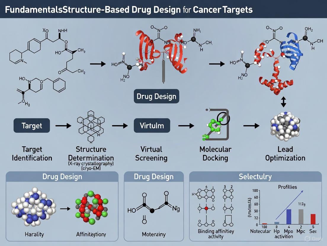
Fundamental Principles and Workflow of SBDD
Core Concepts and Definitions
At its essence, SBDD leverages the atomic-level understanding of a protein target's structure to guide the identification and optimization of small molecules that can modulate its function. The approach is considered "reverse pharmacology" because it begins with target identification rather than compound screening [3]. The binding site or pocket—a small cavity on the target protein where ligands bind—serves as the molecular blueprint for design [3] [1]. SBDD encompasses several specific applications, including structure-based virtual screening (SBVS) of compound libraries and de novo drug design, which involves piecing together molecular subunits to create novel compounds predicted to fit into selected binding sites [1].
The Iterative SBDD Cycle
The SBDD process is fundamentally iterative, proceeding through multiple cycles that progressively optimize a drug candidate [3]. The standard workflow encompasses several key phases, visualized in the following diagram:
SBDD Workflow
This iterative cycle begins with target identification and validation, where potential therapeutic proteins implicated in cancer pathways are selected [3]. The subsequent structure determination phase utilizes techniques such as X-ray crystallography, NMR spectroscopy, or cryo-electron microscopy to resolve the three-dimensional structure of the target protein [3] [4]. With the structure in hand, researchers identify binding pockets—a step increasingly aided by computational methods like Q-SiteFinder, which calculates van der Waals interaction energies to locate favorable binding regions [3].
The core design phase employs computational docking to screen large databases of small molecules or design novel compounds that complement the binding site's steric and electrostatic properties [3] [1]. Top-ranked compounds from virtual screening are then synthesized and progress to experimental testing in biochemical and cellular assays to evaluate affinity, potency, and specificity [3]. A crucial feedback loop involves determining the co-crystal structure of promising ligands bound to their target, providing detailed insights into molecular recognition and binding interactions that inform the next round of optimization [3]. This iterative process continues until a candidate with sufficient efficacy and specificity progresses to clinical trials.
Key Experimental Methodologies in SBDD
Structural Determination Techniques
High-resolution structural information forms the foundation of SBDD. Several complementary techniques enable researchers to determine the three-dimensional structures of cancer targets and their complexes with ligands:
X-ray Crystallography has been the workhorse of structural biology, responsible for over 85% of structures in the Protein Data Bank [5]. The traditional approach involves growing protein crystals, introducing ligands through co-crystallization or soaking, and collecting diffraction data at cryogenic temperatures [5]. Recent advances in room-temperature serial crystallography have enabled the study of protein dynamics and the identification of conformational changes in inhibitors that were not detectable at cryogenic temperatures [5]. This approach has proven particularly valuable for studying allosteric binding sites and explaining differences in inhibitor potency [5].
Cryo-Electron Microscopy (Cryo-EM) has emerged as a transformative technique, especially for large protein complexes and membrane proteins that are difficult to crystallize [5] [4]. While historically achieving lower resolution than crystallography, Cryo-EM has seen remarkable advances, with approximately 55% of Cryo-EM maps deposited in the PDB in 2021 achieving resolutions better than 3.5Å [5].
Nuclear Magnetic Resonance (NMR) Spectroscopy provides valuable information about protein dynamics and structure in solution, making it particularly useful for studying flexible regions of proteins that may be important for function and drug binding [4].
Integrated Experimental Workflow
A comprehensive SBDD campaign typically integrates multiple structural techniques to overcome the limitations of any single method. The following diagram illustrates how these methodologies combine in a modern SBDD pipeline:
Structural Techniques Pipeline
Experimental Protocols for Key Techniques
Room-Temperature Serial Crystallography Protocol
Application: Ideal for studying conformational dynamics, allosteric binding sites, and intermediate states in cancer targets that may be masked by cryo-cooling [5].
Detailed Methodology:
- Microcrystal Growth: Generate microcrystals (10μm or smaller) via batch crystallization with crystal seeding to boost density and quality [5].
- Sample Delivery:
- Fixed Target Approach: Pipette or directly grow microcrystals onto silicon, polymer, or polyimide chips.
- Moving Target Approach: Use viscous jets or tape-drive methods to continuously supply crystals.
- Data Collection: Raster scan a micro-focused X-ray beam across the sample support, collecting hundreds to thousands of diffraction patterns from multiple randomly oriented crystals [5].
- Data Processing: Scale, filter, and merge partial diffraction patterns from multiple microcrystals to generate a complete dataset.
Advantages in Oncology: This protocol has been successfully applied to explain potency differences in glutaminase C inhibitors (targeted in cancer metabolism) and to identify allosteric sites in KRAS, a previously "undruggable" oncogene [5].
Mix-and-Inject Serial Crystallography (MISC) Protocol
Application: Time-resolved studies of ligand binding on millisecond to second timescales [5].
Detailed Methodology:
- Microfluidic Mixing: Combine protein microcrystals with ligand solutions using flow-focused diffusive mixers.
- Reaction Initiation: Allow binding reactions to proceed for precise time intervals before exposure to X-rays.
- Serial Data Collection: Capture structural snapshots at multiple time points to reconstruct binding pathways.
This approach enables researchers to visualize the dynamic process of drug binding to cancer targets, providing insights that can guide optimization of binding kinetics.
Computational Approaches in SBDD
Molecular Docking and Dynamics
Computational methods form the backbone of modern SBDD, enabling high-throughput screening and optimization that would be infeasible through experimental approaches alone. Molecular docking calculates the conformation and orientation (the "docking pose") of compounds at targeted binding sites using scoring functions to predict interaction stability [1]. Molecular dynamics (MD) simulations extend beyond static docking by modeling the behavior of complex molecular systems based on fundamental chemical properties, providing a dynamic view of protein-ligand interactions [1]. Although MD offers greater precision, it comes with high computational costs and sensitivity to force field parameters [2].
Artificial Intelligence and Machine Learning
Recent advances in AI have revolutionized SBDD by enabling the analysis and systemization of large datasets through statistical machine learning methods [3]. Equivariant diffusion models represent a cutting-edge approach for generative SBDD. These models, such as DiffSBDD, formulate drug design as a three-dimensional conditional generation problem and can generate novel ligands conditioned on protein pockets while respecting rotational and translation symmetries [6]. The diffusion process involves training a neural network to predict noiseless features of molecules, then using these predictions to parameterize denoising transition probabilities that gradually move samples from a normal distribution onto the data manifold [6].
Virtual Screening and de novo Design
Structure-based virtual screening (SBVS) computationally screens large compound libraries against a target structure, prioritizing molecules with favorable binding predictions for experimental testing [3]. This approach dramatically reduces the time and cost associated with experimental high-throughput screening. In contrast, de novo drug design pieces together molecular subunits to create completely novel compounds predicted to fit into selected binding sites [1]. AI-based generative models have significantly advanced this field by creating chemically viable molecules that satisfy multiple constraints simultaneously [6].
The Scientist's Toolkit: Essential Research Reagents and Materials
Successful SBDD campaigns require carefully selected reagents and computational resources. The following table details key solutions used in modern SBDD pipelines:
Table 1: Essential Research Reagents and Computational Tools for SBDD
| Category | Specific Examples | Function in SBDD |
|---|---|---|
| Protein Production Systems | E. coli, insect cells, mammalian cells, yeast, cell-free systems [7] | Heterologous expression of target proteins for structural studies |
| Structural Biology Platforms | X-ray crystallography, Cryo-EM, NMR spectroscopy [5] [4] | Determination of 3D protein structures and protein-ligand complexes |
| Compound Libraries | DNA-encoded libraries, fragment libraries, virtual compound databases [7] [1] | Source of chemical starting points for screening and optimization |
| Computational Docking Software | Molecular docking packages, virtual screening platforms [3] | Prediction of ligand binding poses and affinity scoring |
| Molecular Dynamics Packages | GROMACS, AMBER, CHARMM [8] | Simulation of protein-ligand interactions and conformational dynamics |
| AI/ML Platforms | DiffSBDD, Pocket2Mol, ResGen [6] | Generative design of novel ligands and property optimization |
| Binding Assay Technologies | CETSA, activity-based protein profiling, biochemical assays [7] | Experimental validation of target engagement and binding affinity |
| Data Integration Platforms | Proasis, Protein Data Bank, binding affinity databases [8] | Management and integration of structural and chemical data |
Success Stories and Clinical Applications in Oncology
SBDD has contributed to several notable successes in oncology drug development. The following table summarizes key examples:
Table 2: Success Stories of SBDD in Oncology Drug Development
| Drug/Target | Target Disease | SBDD Approach | Key Outcome |
|---|---|---|---|
| KRASG12C Inhibitors (FMC-376) | Lung cancer, Pancreatic cancer | Dual inhibitor targeting both active and inactive KRAS states [7] | Overcomes resistance to first-generation inhibitors |
| pan-RAS Inhibitors (ADT-1004) | Pancreatic cancer | Broad-spectrum RAS inhibition with low resistance potential [7] | Superior activity in mouse models compared to mutant-specific inhibitors |
| WRN Helicase Inhibitors (VVD-214/RO7589831) | MSI-High Cancers | Covalent allosteric inhibition targeting DNA repair dependency [7] | First-in-class approach for cancers with microsatellite instability |
| STAT3 Inhibitors (STX-0119) | Lymphoma | Structure-based virtual screening [3] | Targeted inhibition of signal transduction and transcription activation |
| Pim-1 Kinase Inhibitors | Cancer | Hierarchical multistage virtual screening [3] | Selective kinase inhibition for oncology applications |
| KRAS Degraders | KRAS-driven cancers | Targeted protein degradation to eliminate mutant KRAS [7] | Novel approach addressing resistance to conventional inhibitors |
These case studies demonstrate how SBDD enables targeting of challenging oncoproteins and provides strategies to overcome drug resistance. For instance, the development of KRASG12C inhibitors exemplifies how SBDD can transform previously "undruggable" targets into tractable ones by identifying novel binding pockets [5]. The recent emphasis on degraders and allosteric inhibitors further expands the toolbox against cancer targets that defy conventional occupancy-based inhibition [7].
Emerging Trends and Future Perspectives
The future of SBDD in oncology is being shaped by several converging technological trends. Multimodal data integration combines structural information with genomics, proteomics, and metabolomics to create comprehensive target profiles [2]. AI-driven high-throughput screening leverages machine learning to predict binding affinities and optimize multi-target drug design [2]. The emergence of federated data ecosystems enables organizations to share structural information while protecting proprietary interests, accelerating discovery across the research community [8].
Treating data as a product represents a paradigm shift in SBDD, where well-curated bioinformatics and cheminformatics datasets become valuable assets rather than mere research byproducts [8]. High-value structural data products are characterized by rigorous validation, standardized formats, comprehensive metadata, and intuitive interfaces that democratize access across multidisciplinary teams [8].
As these trends converge, SBDD is poised to enable truly personalized cancer medicine, where treatments are tailored to an individual's unique genetic makeup and protein structures [4] [2]. The ongoing development of more sophisticated AI tools, combined with exponential growth in structural data, promises to further accelerate the design of precision oncology therapeutics in the coming years.
Structure-Based Drug Design (SBDD) is a rational approach to drug discovery and development that uses the three-dimensional (3D) structure of a biological target—typically a protein—to design and optimize drug candidates [9]. This methodology has become fundamental in modern pharmaceutical research, particularly for developing cancer therapeutics, where understanding precise molecular interactions is crucial for developing targeted treatments with improved efficacy and reduced side effects [2]. The core principle of SBDD involves utilizing detailed structural information about the target protein to guide the design of small molecules that can modulate its function, significantly accelerating the drug discovery timeline compared to traditional methods [10].
The SBDD approach is especially valuable in oncology, where researchers can leverage the structural differences between cancerous and normal cells to design selective inhibitors. Modern SBDD integrates computational methods with experimental structural biology, creating an iterative process where each cycle of design and testing provides more refined structural data to inform subsequent optimization [11]. This review will examine the key stages of the SBDD workflow, from initial target identification to candidate drug selection, with specific emphasis on applications in cancer drug development.
Key Stages of the SBDD Workflow
Target Identification and Validation
The initial stage in the SBDD workflow involves identifying and validating a biological target with a confirmed role in cancer pathology. Targets are typically molecules involved in disease processes, such as enzymes in biochemical pathways, receptors, or proteins within cellular signaling cascades [12]. For cancer therapeutics, potential targets may include overexpressed growth factor receptors, mutated signaling proteins, or enzymes essential for tumor survival and proliferation.
Target validation requires thorough investigation of the molecular biology and biochemistry of the disease to establish that modulating the target will produce a therapeutic effect [12]. In this phase, structural bioinformatics plays a crucial role in assessing target "druggability" by identifying functional regions such as active sites, co-factor binding areas, allosteric sites, or surfaces involved in protein-protein interactions [12]. For cancer targets, this may involve analyzing the structural consequences of mutations observed in tumors and determining whether these alterations create unique binding sites that can be selectively targeted.
Structure Determination and Preparation
Once a target is validated, obtaining its high-resolution 3D structure is essential. The three-dimensional structure of a target protein can typically be found in the RCSB Protein Data Bank [13]. Experimental methods for structure determination include:
- X-ray crystallography: The most common method providing high-resolution structures [9] [14]
- Cryo-electron microscopy (cryo-EM): Particularly valuable for large protein complexes [9]
- NMR spectroscopy: Useful for studying protein dynamics and transient states [14]
When experimental structures are unavailable, researchers can construct homology models based on related protein structures or apply AI-based methods for structure prediction [9]. Protein preparation involves several critical steps: adding hydrogen atoms, assigning partial charges, optimizing hydrogen bonds, treating metal cofactors, and addressing missing residues or loops [10]. Proper assignment of protonation states for amino acid residues is crucial for accurate simulation of binding interactions.
Binding Site Identification and Analysis
Identifying the precise binding site where small molecules will interact with the target protein is a critical step that significantly influences SBDD outcomes [13]. The binding site (or pocket) is the location on the protein where the drug binds, and its definition requires careful consideration of the desired mechanism of action (MOA) [13]. For example, in kinase targets, researchers may target the ATP-binding site for competitive inhibitors or identify allosteric sites for developing non-competitive inhibitors.
Proteins are dynamic structures that undergo conformational changes when binding drugs or cofactors [13]. Understanding this structural flexibility is essential for effective SBDD. For instance, nuclear receptors exhibit different conformational states when binding agonists versus antagonists, which must be considered when selecting protein structures for docking studies [13]. Binding site analysis also involves examining potential interactions with cofactors (e.g., SAM in methyltransferases) or metal ions (e.g., Zn²⁺ in metalloenzymes) that may need to be included as part of the binding site definition [13].
Virtual Screening and Hit Identification
Virtual screening (VS) uses computational methods to identify potential hit compounds from large chemical libraries that are likely to bind to the target protein [10]. This approach serves as an efficient, cost-effective alternative to experimental high-throughput screening (HTS) [10]. The virtual screening process involves several key components:
- Library preparation: Compound libraries are pre-processed to generate 3D structures, assign proper stereochemistry, and determine likely tautomeric and protonation states [10]
- Molecular docking: Specialized software positions each compound within the binding site and scores its complementarity [11]
- Post-processing: Top-ranking compounds are evaluated for binding poses, undesirable chemical features, and drug-like properties [10]
Table 1: Common Molecular Docking Software Tools
| Software | Key Features | Availability |
|---|---|---|
| DOCK 6 | Uses incremental construction for ligands; includes solvent effects | Free for academic use [11] |
| AutoDock | Uses interaction grids and simulated annealing | Free [11] |
| Glide | Performs complete conformational, orientational, and positional search | Commercial [11] |
| GOLD | Uses genetic algorithms; allows partial protein flexibility | Commercial [11] |
Hit-to-Lead Optimization
Once hit compounds are identified, the hit-to-lead optimization phase begins, focusing on improving various properties of the initial hits [9]. This iterative process involves structural biologists and medicinal chemists working closely to enhance:
- Binding affinity: Improving the strength of interaction with the target protein
- Selectivity: Reducing off-target effects by minimizing interactions with related proteins [12]
- ADME properties: Optimizing absorption, distribution, metabolism, and excretion profiles [12]
- Solubility: Enhancing compound solubility for better bioavailability [12]
During this phase, researchers typically use co-crystallization of compounds with the target protein to obtain detailed structural information about binding interactions [12]. This structural data guides rational chemical modifications to improve compound properties. Computational methods, including molecular dynamics (MD) simulations, provide dynamic views of ligand-receptor complexes, capturing conformational changes and binding flexibility that influence drug behavior [9]. Advanced MD techniques such as steered MD and umbrella sampling can study the kinetics and thermodynamics of ligand binding and unbinding processes [9].
Lead Optimization to Candidate Drug
The final stage of the SBDD workflow focuses on transforming lead compounds into a candidate drug (CD) ready for clinical trials [12]. This involves iterative cycles of computational modeling, chemical modification, biological testing, and structure-based design to identify an optimized lead molecule that meets specific criteria:
- Potency: Typically low nM to μM activity against the target [12]
- Selectivity: Minimal off-target effects due to binding to other proteins [12]
- ADMET profile: Optimal pharmacokinetics and low toxicity in preclinical studies [12]
- Efficacy: Demonstrated activity in disease models (usually animals) [12]
- Synthetic feasibility: Cost-effective synthesis demonstrated in the laboratory [12]
At this stage, researchers also address potential issues such as toxicity (including cytotoxicity and genotoxicity) and conduct thorough assessment of off-target effects by evaluating interactions with other proteins [12]. The candidate drug should represent a balance of optimal molecular properties within a patentable chemical scaffold [12].
Experimental Protocols and Methodologies
Molecular Docking Protocol
Molecular docking is a fundamental technique in SBDD that predicts how small molecules bind to a protein target [11]. A standard docking protocol includes these critical steps:
Ligand Preparation
- Convert 2D chemical representations to 3D structures using programs like CONCORD or CORINA [11]
- Assign proper protonation states for the pH conditions of the target environment [11]
- Generate possible tautomers and stereoisomers as separate structures [11]
- Energy minimization to ensure proper molecular geometry [11]
Receptor Preparation
- Add hydrogen atoms to the protein structure [11]
- Assign partial charges to individual residues [11]
- Define the docking site, typically using a 3.5-6 Å radius around a known ligand or binding site [11]
- Decide on treatment of water molecules, metals, and cofactors in the binding site [11]
- For flexible docking, define which residues can move and their degrees of freedom [11]
Docking Execution
Post-Docking Analysis
Molecular Dynamics Simulation Protocol
Molecular dynamics (MD) simulations provide a dynamic view of ligand-receptor complexes, capturing conformational changes and binding flexibility [9]. A typical MD protocol includes:
System Setup
- Solvate the protein-ligand complex in a water box with appropriate dimensions
- Add counterions to neutralize system charge
- Apply force field parameters (e.g., CHARMM, AMBER) for the protein and ligand
Energy Minimization
- Use steepest descent or conjugate gradient algorithms to relieve steric clashes
- Gradually reduce position restraints on protein and ligand atoms
System Equilibration
- Perform gradual heating from 0K to target temperature (typically 310K)
- Equilibrate density with position restraints on heavy atoms
- Conduct unrestrained equilibration until system properties stabilize
Production Simulation
- Run extended simulations (typically 100ns-1μs) for analysis
- Maintain constant temperature and pressure using appropriate thermostats and barostats
- Save trajectory frames at regular intervals (e.g., every 100ps)
Trajectory Analysis
- Calculate root mean square deviation (RMSD) to assess system stability
- Analyze protein-ligand interactions over time (hydrogen bonds, hydrophobic contacts)
- Identify transient binding pockets and conformational changes
- Use MM/PBSA or related methods to estimate binding free energies
Advanced SBDD Techniques for Cancer Targets
Recent advances in SBDD have introduced sophisticated approaches specifically valuable for cancer drug discovery:
Ensemble Docking: This technique addresses receptor flexibility by docking compounds against multiple protein conformations rather than a single static structure [10]. For cancer targets that exhibit significant conformational heterogeneity, ensemble docking improves virtual screening accuracy by accounting for different binding site shapes [10].
AI-Driven Methods: Modern SBDD incorporates artificial intelligence to enhance various stages of the workflow. For example, TransDiffSBDD is a novel framework that integrates autoregressive transformers and diffusion models to generate hybrid-modal sequences for protein-ligand complexes, effectively handling both discrete molecular graph information and continuous 3D structural data [15].
Free Energy Pertigation (FEP): FEP calculations provide a rigorous measure of the changes in free energy between unbound and bound complexes in solvent, offering more accurate binding affinity predictions than standard docking scores [11]. This approach is particularly valuable during lead optimization to prioritize compound synthesis.
Table 2: Key Research Reagent Solutions for SBDD
| Category | Specific Resources | Function in SBDD |
|---|---|---|
| Structural Databases | RCSB PDB, PDBe Chemical Components Library [12] | Source of 3D protein structures and ligand information for target analysis and binding site characterization |
| Compound Libraries | ZINC database [11], commercial screening libraries | Collections of purchasable compounds for virtual screening and hit identification |
| Bioactivity Databases | ChEMBL, PubChem, DrugBank, BindingDB [16] | Target-annotated ligand information for validation and similarity searching |
| Protein Preparation Tools | PROPKA [10], H++ [10], PDB2PQR [10] | Software for assigning protonation states, adding hydrogens, and optimizing protein structures |
| Docking Software | DOCK, AutoDock, Glide, GOLD [11] | Programs for predicting binding modes and scoring protein-ligand interactions |
| MD Software | GROMACS, AMBER, NAMD | Packages for running molecular dynamics simulations to study binding stability and conformational changes |
| Visualization Tools | PyMOL, Chimera, Maestro | Software for visual analysis of protein-ligand complexes and interaction mapping |
| Analysis Tools | WaterMap [10], 3D RISM [10] | Specialized software for analyzing water networks and solvation effects in binding sites |
Workflow Visualization
SBDD Workflow Overview - This diagram illustrates the key stages and iterative nature of the structure-based drug design process, from target identification through candidate drug selection.
The SBDD workflow represents a powerful, rational approach to drug discovery that has become increasingly sophisticated with advances in structural biology, computational methods, and artificial intelligence. For cancer drug development, this methodology offers the potential to design highly specific therapeutics that target molecular vulnerabilities in tumor cells while minimizing effects on healthy tissues. The iterative nature of SBDD—cycling between design, synthesis, testing, and structural analysis—creates a feedback loop that systematically improves compound properties.
Future directions in SBDD point toward increased integration of multi-modal data, enhanced AI-driven high-throughput screening, and the development of standardized platforms for data integration and analysis [2]. As these technologies mature, SBDD will continue to transform cancer drug discovery, enabling more precise and personalized therapeutic approaches that significantly improve treatment efficacy and patient quality of life [2].
The foundation of modern, targeted cancer therapy rests on the precise identification and validation of key proteins and pathways that drive oncogenesis. Within the framework of structure-based drug design (SBDD), this initial target discovery and validation phase is critical, as it determines the feasibility and direction of subsequent drug development efforts [17]. This guide synthesizes contemporary methodologies, integrating multi-omics data and computational approaches to deconvolute the complex molecular mechanisms of cancer and establish robust, druggable targets.
Core Concepts in Cancer Target Identification
Defining Cancer Hallmarks through Molecular Pathways
Cancer phenotypes are sustained by alterations in core biological pathways. Identifying these pathways provides a systems-level understanding of the disease and reveals potential nodes for therapeutic intervention. These pathways often involve dysregulated cell cycle progression, resistance to cell death, sustained proliferative signaling, and activation of invasion and metastasis.
Systematic analyses across multiple cancer types have identified both common and unique pathway dependencies. For instance, the olfactory transduction pathway was identified as a significant pathway in numerous cancers, including acute myeloid leukemia (AML), breast cancer, colorectal cancer, and non-small cell lung carcinoma (NSCLC), suggesting a previously underappreciated role in oncogenesis [18]. Other key pathways frequently altered include signaling by GPCR, messenger RNA processing, and axon guidance [18].
The Role of Key Proteins as Molecular Targets
Within dysregulated pathways, specific proteins often serve as critical drivers and are therefore prime candidates for therapeutic targeting. These proteins can be transcription factors, kinases, receptors, or structural proteins.
A prominent example is the βIII-tubulin isotype, a component of microtubules. Its significant overexpression in various cancers is closely associated with resistance to anticancer agents like Taxol, making it an attractive target for novel therapies [19]. Another example is Discoidin Domain Receptor 1 (DDR1), identified as a molecular target specific for pancreatic cancer, enabling the development of selective inhibitors [18].
Methodological Approaches for Target Identification
The identification of cancer targets leverages a suite of high-throughput technologies and computational analyses. The integrative workflow, outlined in the diagram below, combines multi-omics data to pinpoint and prioritize potential targets.
Multi-Omics Data Integration
Integrating data from various molecular levels provides a comprehensive view of cancer biology. Key data types include:
- Transcriptomics: RNA sequencing (RNA-Seq) measures RNA transcript abundance, identifying genes that are differentially expressed in specific cancer types. Large-scale resources like the Cancer Cell Line Encyclopedia (CCLE) provide RNA-Seq data for over 1,000 cancer cell lines [18].
- Proteomics: Tandem mass tag (TMT)-based quantitative proteomics provides large-scale protein quantification, directly reflecting functional cellular components. A key study profiled 375 cell lines across diverse cancer types, creating a rich resource for protein expression exploration [18].
The power of multi-omics is demonstrated by studies that collectively analyze transcriptomics and proteomics data from 16 common types of human cancer. This integration allows for the identification of "significant transcripts" and "significant proteins" characteristic of each cancer type, which are then used for pathway enrichment analysis [18]. The consistency between these data layers is often high; for example, in liver cancer, 234 protein-coding biotypes were found in both the significant transcript set and the significant protein set [18].
Computational and AI-Driven Approaches
Computational methods have become indispensable for processing complex biological data and predicting interactions.
- Structure-Based Virtual Screening (SBVS): This computational technique screens large libraries of compounds against a 3D protein structure to identify potential binders. For example, screening 89,399 natural compounds from the ZINC database against the βIII-tubulin isotype identified 1,000 initial hits based on binding energy [19].
- Machine Learning (ML) for Hit Refinement: Supervised ML models can distinguish between active and inactive molecules based on chemical descriptor properties. This approach was used to narrow 1,000 virtual screening hits against βIII-tubulin down to 20 high-confidence active natural compounds [19].
- Artificial Intelligence in Target Prediction: AI-driven models, particularly machine learning algorithms, enhance the target identification process for natural products by processing complex proteomic data and predicting potential NP-protein interactions, thereby accelerating discovery [20].
- Chemical Proteomics: This powerful experimental approach uses chemical probes derived from bioactive molecules, such as natural products, to pull down and identify their direct protein targets from complex biological mixtures. When integrated with AI, it provides a robust method for deconvoluting the mechanisms of complex natural products [20].
Table 1: Summary of Significant Omics Findings Across 16 Cancer Types [18]
| Cancer Type | Significant Transcripts | Significant Proteins | Characteristic Pathways (Examples) |
|---|---|---|---|
| Acute Myeloid Leukemia (AML) | ~11,000 | 2,443 | Various (112 overlapping pathways) |
| Breast Cancer | ~9,256 (median) | ~1,344 (median) | Olfactory Transduction, Signaling by GPCR |
| Colorectal Cancer | ~9,256 (median) | ~1,344 (median) | Olfactory Transduction, Signaling by GPCR |
| Glioma | ~9,256 (median) | ~1,344 (median) | Olfactory Transduction, Messenger RNA Processing |
| Liver Cancer | 5,756 | 825 | Olfactory Transduction |
| Melanoma | 11,143 | ~1,344 (median) | Olfactory Transduction, Signaling by GPCR |
| Non-Small Cell Lung Carcinoma (NSCLC) | ~9,256 (median) | ~1,344 (median) | Olfactory Transduction, Signaling by GPCR |
| Ovarian Cancer | ~9,256 (median) | ~1,344 (median) | Olfactory Transduction |
| Stomach Cancer | ~9,256 (median) | 409 | Axon Guidance |
| Urinary Tract Cancer | ~9,256 (median) | ~1,344 (median) | Alpha-6 Beta-1/Alpha-6 Beta-4 Integrin Signaling |
Experimental Protocols for Target Validation
After initial identification, putative targets must be rigorously validated. The following section details key experimental methodologies.
Protocol: In Silico Target Validation via Molecular Docking and Dynamics
This protocol is used for the initial computational validation of a small molecule's interaction with a protein target [19] [17].
Protein Structure Preparation:
- If an experimental crystal structure is unavailable, construct a homology model using software like Modeller. The template structure should have high sequence identity (e.g., the bovine αIBβIIB tubulin structure (PDB: 1JFF) shares 100% identity with human β-tubulin and can be used for modeling the human βIII isotype).
- Select the final model based on assessment scores like DOPE (Discrete Optimized Protein Energy) and validate stereo-chemical quality using a Ramachandran plot (e.g., via PROCHECK).
Ligand Library Preparation:
- Retrieve compound structures from databases like ZINC in SDF format.
- Convert files to PDBQT format using Open-Babel software and add polar hydrogens and Gasteiger charges.
Molecular Docking:
- Perform high-throughput virtual screening against the target's binding site (e.g., the 'Taxol site' on βIII-tubulin) using AutoDock Vina or InstaDock.
- Screen compounds based on binding energy (kcal/mol) and select top hits (e.g., top 1,000) for further analysis.
Machine Learning Classification:
- Generate molecular descriptors for the top hits and a training dataset of known active/inactive compounds using PaDEL-Descriptor.
- Train a supervised ML classifier (e.g., with 5-fold cross-validation) to distinguish active from inactive molecules. Filter the virtual screening hits using this model to identify high-confidence active compounds (e.g., 20 compounds).
ADME-T and Toxicity (ADME-T) Prediction:
- Analyze the top ML-ranked compounds for drug-like properties, including Absorption, Distribution, Metabolism, Excretion, and Toxicity.
Molecular Dynamics (MD) Simulations:
- Simulate the dynamics of the top ligand-protein complexes (e.g., for 100-200 ns) in a solvated system.
- Analyze trajectories using metrics like Root Mean Square Deviation (RMSD), Root Mean Square Fluctuation (RMSF), Radius of Gyration (Rg), and Solvent Accessible Surface Area (SASA) to evaluate complex stability and binding mode.
Protocol: Chemical Proteomics for Natural Product Target Identification
This protocol identifies the protein targets of natural products (NPs) using pull-down assays [20].
Probe Design and Synthesis:
- Design a chemical probe by incorporating a photoaffinity label (e.g., a diazirine) and a bio-orthogonal handle (e.g., an alkyne) into the native NP structure without destroying its bioactivity. The alkyne allows for subsequent "click chemistry" conjugation.
Cell Lysate Preparation and Pull-Down:
- Treat live cells or prepare lysates from relevant cancer cell lines.
- Incubate the lysate with the NP probe. A control probe (a structurally similar but inactive molecule) should be used in parallel.
- Activate the photoaffinity label with UV light to cross-link the probe to its interacting proteins.
Enrichment of Probe-Protein Complexes:
- Use click chemistry to conjugate the alkyne on the probe to an azide-functionalized solid support (e.g., agarose beads).
- Incubate the mixture to allow conjugation, then wash the beads thoroughly to remove non-specifically bound proteins.
Protein Identification and Quantification:
- Elute the bound proteins from the beads.
- Digest the proteins with trypsin and analyze the resulting peptides by liquid chromatography-tandem mass spectrometry (LC-MS/MS).
- Use label-free or isobaric tagging (e.g., TMT) methods to quantify proteins enriched in the NP probe sample compared to the control probe sample.
Protocol: Functional Validation via Gene Silencing
This protocol tests the functional necessity of a putative target in cancer cell survival and drug response [19].
- Cell Line Selection: Choose cancer cell lines that express the target protein (e.g., βIII-tubulin) and relevant control lines.
- siRNA Transfection: Design and transfert small interfering RNAs (siRNAs) specifically targeting the mRNA of the gene of interest. A non-targeting (scrambled) siRNA should be used as a negative control.
- Efficiency Knockdown Validation: After 48-72 hours, validate knockdown efficiency at the mRNA level (using qRT-PCR) and/or protein level (using western blotting).
- Phenotypic Assays:
- Viability/Drug Sensitivity: Treat siRNA-transfected cells with a range of concentrations of a relevant chemotherapeutic agent (e.g., Paclitaxel). Measure cell viability after 72-96 hours using assays like MTT or CellTiter-Glo.
- Proliferation and Clonogenic Assays: Monitor long-term proliferation and colony-forming ability post-knockdown.
The workflow below illustrates the logical progression from initial computational screening to experimental validation, highlighting the iterative nature of modern cancer target identification.
The Scientist's Toolkit: Research Reagent Solutions
A successful target identification and validation pipeline relies on a suite of essential reagents, databases, and software tools.
Table 2: Essential Research Reagents and Resources for Cancer Target Identification
| Category / Item | Specific Example(s) | Function and Application |
|---|---|---|
| Biological Models | ||
| Cancer Cell Line Encyclopedia (CCLE) | >1,000 cell lines, 40+ cancer types [18] | Provides standardized, well-characterized in vitro models for transcriptomic, proteomic, and functional studies. |
| Omics Databases & Software | ||
| Transcriptomics Data | RNA-Seq data from CCLE [18] | Identifies differentially expressed genes and transcripts specific to cancer types. |
| Proteomics Data | TMT-based quantitative data (e.g., 375 cell lines) [18] | Quantifies protein expression levels to identify overexpressed or dysregulated proteins. |
| Pathway Analysis Tools | Enrichment analysis software (e.g., GSEA) | Identifies biological pathways significantly altered in a specific cancer type from omics data. |
| Computational & SBDD Tools | ||
| Homology Modeling | Modeller [19] | Generates 3D protein structures when experimental structures are unavailable. |
| Virtual Screening | AutoDock Vina, InstaDock [19] | Rapidly docks thousands to millions of compounds into a target binding site to predict binding affinity. |
| Molecular Descriptor Calculator | PaDEL-Descriptor [19] | Calculates chemical properties and fingerprints from molecular structures for machine learning. |
| Molecular Dynamics Software | GROMACS, AMBER, NAMD | Simulates the physical movement of atoms and molecules over time to assess complex stability. |
| Experimental Validation Reagents | ||
| Chemical Proteomics Probes | Photoaffinity-labeled NPs with alkyne handles [20] | Used to covalently capture and identify direct protein targets of natural products in complex lysates. |
| Gene Silencing Tools | siRNA oligos [19] | Knocks down expression of a target gene to study its functional role in cancer phenotypes and drug response. |
Case Studies in Cancer Target Discovery
Case Study 1: Targeting βIII-Tubulin in Resistant Cancers
The βIII-tubulin isotype exemplifies a resistance-associated target identified and validated through integrated methods. Target Identification: Overexpression of βIII-tubulin was correlated with resistance to taxanes in clinical samples of ovarian, breast, and NSCLC cancers [19]. Validation: siRNA-mediated knockdown of βIII-tubulin in resistant NSCLC cell lines (NCI-H460, Calu-6) restored sensitivity to Paclitaxel, Vincristine, and Vinorelbine, functionally validating its role in resistance [19]. Drug Discovery: A structure-based drug design campaign screened 89,399 natural compounds against the 'Taxol site' of a homology model of αβIII-tubulin. Machine learning refined 1,000 initial hits to 20 active compounds. Four (ZINC12889138, ZINC08952577, ZINC08952607, ZINC03847075) showed exceptional binding affinity, ADME-T properties, and stabilized the αβIII-tubulin heterodimer in MD simulations, identifying them as promising leads for targeting βIII-tubulin-overexpressing carcinomas [19].
Case Study 2: Multi-Omics Driven Pathway and Drug Repurposing
A large-scale integrative analysis demonstrated a systematic approach to identifying cancer-type-specific pathways and corresponding drugs. Methodology: Researchers analyzed transcriptomics and proteomics data from 16 common cancer types, identifying significant transcripts and proteins for each [18]. Pathway Identification: Overlapping pathways from both omics layers were considered characteristic. The number of these pathways ranged from 4 (stomach cancer) to 112 (AML) [18]. Drug Discovery: Potential anti-cancer drugs were retrieved based on their ability to target these identified pathways. The number of therapeutic drugs ranged from one (ovarian cancer) to 97 (AML and NSCLC). The method was validated by the fact that some of these drugs are already FDA-approved for their corresponding cancer type, while others represent new repurposing opportunities [18].
In the field of structure-based drug design, particularly for cancer targets, determining the three-dimensional atomic structure of biological macromolecules is a fundamental step. It provides the crucial blueprint for understanding disease mechanisms and designing novel therapeutics. Among the techniques used to obtain these structures, X-ray crystallography, cryo-electron microscopy (cryo-EM), and computational homology modeling form a powerful triad. This guide details the principles, advanced methodologies, and integrated applications of these techniques, with a specific focus on their use in cancer drug discovery. Recent breakthroughs, including the integration of artificial intelligence (AI) with cryo-EM and advanced homology modeling, are revolutionizing the speed and accuracy of structural biology, enabling the study of challenging cancer-related targets like membrane proteins and large macromolecular complexes [21].
Core Techniques and Methodologies
X-ray Crystallography
X-ray crystallography has long been a cornerstone of structural biology, enabling the determination of high-resolution structures of proteins, nucleic acids, and their complexes by analyzing the diffraction patterns of X-rays passing through crystallized samples [21] [22].
- Principles and Workflow: The technique relies on directing a monochromatic X-ray beam at a purified protein crystal. The atoms within the crystal lattice cause the X-rays to diffract, producing a characteristic pattern of spots on a detector. The angles and intensities of these diffracted beams are used to calculate an electron density map, into which an atomic model of the protein is built [23] [22]. The key steps are summarized in the workflow below.
Advanced Applications and Protocol: The field has been transformed by serial crystallography (SX), conducted at synchrotrons and X-ray free-electron lasers (XFELs). This approach uses microcrystals and allows for time-resolved studies of reaction mechanisms, known as "molecular movies" [24]. A critical application in cancer research is determining the structures of drug-target complexes, such as the SARS-CoV-2 main protease with the inhibitor nirmatrelvir, a strategy directly applicable to oncology drug development [21].
- Detailed Protocol for Sample Delivery in Serial Crystallography [24]:
- Crystal Preparation: Generate a slurry of microcrystals (typically 1-10 µm in size) in their mother liquor.
- Delivery System Selection: Choose an appropriate low-consumption method:
- Liquid Injection: A slurry is jetted as a continuous stream or in droplets across the X-ray beam. Advanced systems can achieve flow rates as low as 0.1 µL/min to conserve precious sample.
- Fixed-Target: Crystals are loaded onto a microfluidic chip with thousands of microscopic wells. The chip is rastered through the beam, exposing one crystal at a time. This method minimizes sample waste.
- Data Collection: The X-ray pulse (femtoseconds at XFELs, milliseconds at synchrotrons) hits a crystal, producing a single diffraction pattern before the crystal is destroyed. Tens of thousands of such patterns are collected from fresh crystals.
- Detailed Protocol for Sample Delivery in Serial Crystallography [24]:
Quantitative Data:
Table 1: Sample Consumption in Modern Serial Crystallography [24]
| Sample Delivery Method | Typical Sample Consumption for a Full Dataset | Key Advantages | Key Challenges |
|---|---|---|---|
| Liquid Injection | ~1-100 mg | Compatible with time-resolved studies (mix-and-inject) | Sample waste between X-ray pulses |
| Fixed-Target | < 1 mg (micrograms in ideal cases) | Minimal sample waste; high data collection efficiency | Potential crystal harvesting issues; chip background scattering |
Cryo-Electron Microscopy (Cryo-EM)
Cryo-EM has undergone a "resolution revolution," making it a dominant technique for determining high-resolution structures of large complexes and flexible proteins that are difficult to crystallize, such as many cancer drug targets [21].
- Principles and Workflow: In single-particle cryo-EM, a purified protein solution is applied to a grid and rapidly frozen in liquid ethane, embedding the particles in a thin layer of vitreous ice. This preserves their native state. An electron beam is used to capture thousands of 2D micrographs of the randomly oriented particles. Computational algorithms then classify, average, and reconstruct these 2D images into a high-resolution 3D density map [21] [25].
Advanced Applications and Protocol: A major challenge has been sample preparation, where proteins can be denatured at the air-water interface. A recent breakthrough is high-speed droplet vitrification, which avoids this damage [25]. Furthermore, for thick samples like intact bacterial cells, a new technique called tilt-corrected bright-field STEM (tcBF-STEM) offers a 3–5x improvement in dose efficiency compared to conventional methods, enabling structural studies in a more native cellular context [26].
- Detailed Protocol for High-Speed Droplet Vitrification [25]:
- Setup: A custom-built droplet sprayer delivers microscopic droplets of protein solution at high speed (approaching 100 m/s) onto a cryogenically cooled grid coated with liquid ethane.
- Spraying and Impact: The droplets flatten and freeze in under 10 microseconds upon impact with the ethane-coated grid.
- Outcome: This ultra-fast process locks proteins in place before they can diffuse to the air-water interface, preventing structural damage and yielding more uniform particle distributions for imaging.
- Detailed Protocol for High-Speed Droplet Vitrification [25]:
Homology Modeling
When experimental structure determination is not feasible, homology modeling provides a powerful computational alternative for predicting a protein's 3D structure based on its amino acid sequence.
- Principles and Workflow: Also known as comparative modeling, this method relies on the observation that protein structure is more conserved than sequence. If the sequence of a target protein shares significant similarity with a protein of known structure (the template), a model of the target can be built by aligning the sequences and copying the coordinates of conserved regions from the template [19].
Advanced Applications and Protocol: The field has been revolutionized by AI-driven tools like AlphaFold2, which accurately predict protein monomer structures [21]. A key challenge remains the prediction of protein-protein complexes, which are critical for understanding signaling pathways in cancer. The newly developed DeepSCFold pipeline addresses this by using deep learning to predict structure complementarity and interaction probability directly from sequence, significantly improving complex structure prediction over tools like AlphaFold-Multimer and AlphaFold3 [27].
- Detailed Protocol for Modeling a Protein Complex with DeepSCFold [27]:
- Input: Provide the amino acid sequences of the individual protein chains believed to form a complex.
- Feature Prediction: The pipeline uses deep learning models to predict:
- pSS-score: The structural similarity between the input sequence and its homologs.
- pIA-score: The interaction probability between pairs of sequence homologs from different subunits.
- Paired MSA Construction: These scores are used to systematically rank and concatenate monomeric multiple sequence alignments (MSAs) into high-quality paired MSAs, which capture inter-chain interaction signals.
- Structure Prediction: The paired MSAs are fed into a structure prediction engine (e.g., AlphaFold-Multimer) to generate the final quaternary structure model of the complex.
- Detailed Protocol for Modeling a Protein Complex with DeepSCFold [27]:
Integrated Applications in Cancer Drug Discovery
The synergy of these techniques is powerfully illustrated in the search for inhibitors of the human βIII-tubulin isotype, a protein overexpressed in various cancers and linked to resistance to anticancer agents like Taxol [19].
- Step 1: Target Selection and Structure Preparation: The βIII-tubulin isotype was established as a critical cancer drug target. Since its experimental structure was unavailable, a homology model was built using the crystal structure of a closely related bovine tubulin isotype (PDB: 1JFF) as a template [19].
- Step 2: Structure-Based Virtual Screening (SBVS): The homology model of the αβIII-tubulin heterodimer, specifically the 'Taxol site', was used to computationally screen 89,399 natural compounds from the ZINC database. The top 1,000 hits were selected based on binding energy calculated by molecular docking [19].
- Step 3: Machine Learning and Experimental Validation: A machine learning classifier was trained on known Taxol-site binders to refine the 1,000 hits down to 20 high-probability active compounds. Four leads (e.g., ZINC12889138) showed exceptional binding affinity and ADME-T properties. Their stability and interaction with the target were confirmed through molecular dynamics simulations [19]. This integrated computational workflow, which can be initiated with a homology model and validated by experimental data, efficiently identifies promising drug candidates.
Table 2: The Scientist's Toolkit for Structure-Based Drug Design
| Research Reagent / Material | Function in Experimental Workflow |
|---|---|
| Purified Protein Sample | The fundamental starting material for both crystallization (X-ray) and vitrification (Cryo-EM). |
| Crystallization Solutions | Specialized buffers to slowly precipitate protein molecules into an ordered crystal lattice [23]. |
| Cryo-EM Grids | Tiny metal meshes used to support the thin layer of vitrified ice containing the protein sample [25]. |
| Liquid Ethane | A cryogen used for rapid vitrification of water to preserve protein structure in a native, hydrated state [25]. |
| Template Structure (PDB) | A previously solved protein structure from the Protein Data Bank, used as a reference for homology modeling [19]. |
| Compound Library (e.g., ZINC) | A database of small molecules for virtual screening to identify potential drug leads that bind to the target structure [19]. |
X-ray crystallography, cryo-EM, and homology modeling are complementary and indispensable tools for obtaining 3D protein structures in cancer research. The ongoing integration of these techniques with artificial intelligence and machine learning is creating a powerful new paradigm. As highlighted in recent evaluations like CASP16, AI-driven prediction tools are achieving remarkable accuracy, pushing the field toward a discovery-driven science where structural insights can be rapidly translated into therapeutic hypotheses [21] [28]. For cancer drug development professionals, mastering the principles, protocols, and synergistic application of this toolkit is fundamental to accelerating the design of next-generation, targeted therapies.
The systematic assessment of target druggability is a foundational step in modern oncology drug discovery, serving as a critical gatekeeper to ensure efficient resource allocation and increase the probability of clinical success. Druggability analysis fundamentally involves the computational and experimental evaluation of a protein's ability to bind small molecules with high affinity and specificity, particularly focusing on the structural characteristics of binding pockets and interaction sites. Within cancer biology, where targets often involve mutated signaling proteins, transcription factors, and regulatory elements, druggability assessment provides the strategic framework for distinguishing viable drug targets from those that may consume significant R&D investment without yielding therapeutic candidates.
The emergence of challenging target classes, including protein-protein interactions and intrinsically disordered proteins, has necessitated advanced methods for identifying and characterizing cryptic and allosteric binding sites. Contemporary approaches have evolved beyond simple structural analysis to integrate dynamic pocket prediction, chemo-proteomic mapping, and machine learning algorithms that collectively provide a multidimensional view of target tractability. This guide examines the core principles, methodologies, and experimental frameworks for comprehensive druggability assessment, with specific emphasis on applications in oncology drug discovery where overcoming resistance and targeting previously "undruggable" oncoproteins remains a priority.
Fundamental Concepts and Definitions
Key Terminology
- Druggability: The propensity of a target to be modulated by a small-molecule drug with adequate potency, selectivity, and pharmacokinetic properties to achieve therapeutic efficacy. Druggability assessment specifically evaluates the structural and chemical features of a protein that enable high-affinity binding to drug-like molecules.
- Binding Pocket: A region on a protein surface characterized by concavity, distinct physicochemical properties, and the ability to accommodate ligand binding. Conventional binding pockets typically exhibit defined boundaries, sufficient volume (>150 ų), and hydrophobic character mixed with polar functionality for specific molecular recognition.
- Interaction Sites: Specific residues within binding pockets that form direct non-covalent interactions with ligands, including hydrogen bonds, ionic interactions, π-π stacking, and van der Waals contacts. The spatial arrangement and complementarity of these sites determine binding affinity and specificity.
- Cryptic Pockets: Binding sites that are not apparent in static crystal structures but become accessible through protein dynamics, conformational changes, or upon ligand binding. These pockets represent significant opportunities for targeting proteins lacking obvious binding cavities.
- Allosteric Sites: Binding pockets topographically distinct from a protein's active site that modulate function through induced conformational changes. Allosteric modulation offers advantages for targeting essential proteins where orthosteric inhibition proves problematic due to conservation or structural constraints.
Structural Determinants of Druggability
The druggability of a binding pocket is determined by a combination of structural, physicochemical, and dynamic properties that collectively influence ligand binding. Key determinants include:
- Pocket Volume and Depth: Sufficient volume (>150 ų) to accommodate drug-like molecules and adequate depth to enable high-affinity interactions beyond surface contacts.
- Surface Complexity: Presence of invaginations, ridges, and sub-pockets that increase interaction surface area and provide opportunities for specific molecular recognition.
- Hydrophobic Character: Proportion of hydrophobic residues that drive binding through the hydrophobic effect, typically constituting 40-70% of pocket surface area in druggable sites.
- Polar Functionality: Strategic placement of hydrogen bond donors/acceptors that enable specific directional interactions with ligands, typically at pocket edges or defining specificity sub-pockets.
- Structural Plasticity: The ability of a pocket to adapt to different ligand shapes through sidechain rearrangements or backbone movements without compromising protein stability.
- Solvent Accessibility: The balance between buried and solvent-exposed regions, with optimal pockets having limited water access to maximize hydrophobic interactions while maintaining solubility requirements.
Table 1: Structural Properties of Different Binding Pocket Classes
| Pocket Class | Typical Volume (ų) | Key Features | Druggability Potential | Example Cancer Targets |
|---|---|---|---|---|
| Conventional Active Site | 300-1000 | Well-defined, deep, mixed hydrophobicity | High | Kinase ATP sites, Protease active sites |
| Protein-Protein Interface | 200-600 | Extended, relatively flat, mixed functionality | Moderate to Low | BCL-2 family, RAS-effector interfaces |
| Allosteric Site | 150-500 | Often cryptic, lower conservation | Variable | SHP2, KRAS allosteric sites |
| Shallow Surface Groove | 100-300 | Minimal depth, highly solvent exposed | Low | Transcription factor interfaces |
Methodologies for Binding Pocket Analysis
Structure-Based Computational Approaches
Computational methods for binding pocket analysis leverage three-dimensional structural information to identify, characterize, and prioritize potential drug binding sites.
Homology Modeling for Pocket Prediction When experimental structures are unavailable, homology modeling generates reliable protein models based on closely related templates. For example, in studying the human βIII tubulin isotype, researchers employed Modeller 10.2 using the bovine αIBβIIB tubulin isotype (PDB ID: 1JFF) as a template, which shares 100% sequence identity with human β-tubulin. The resulting model was evaluated using DOPE (Discrete Optimized Protein Energy) scores and stereo-chemical quality assessment via Ramachandran plots to ensure reliability before pocket analysis [19].
Molecular Docking and Virtual Screening Structure-based virtual screening (SBVS) systematically evaluates compound libraries against target binding pockets. A standard protocol involves:
- Preparation of target protein structure (adding hydrogens, assigning charges)
- Definition of binding site grid coordinates based on known ligand positions
- Conversion of compound libraries into appropriate formats (e.g., PDBQT using Open-Babel)
- High-throughput docking using programs like AutoDock Vina
- Hit identification based on binding energy thresholds [19]
In practice, screening 89,399 natural compounds from the ZINC database against the 'Taxol site' of αβIII-tubulin identified 1,000 initial hits based on binding energy, which were subsequently refined using machine learning approaches [19].
Binding Pocket Detection Algorithms Multiple algorithms exist for systematic binding pocket identification:
- FPOCKET: Utilizes Voronoi tessellation and alpha spheres to detect cavities based on geometry and physicochemical parameters
- SiteMap: Employs grid-based searching to identify regions with favorable binding properties including enclosure, hydrophobicity, and hydrogen bonding capacity
- CASTp: Computes surface topology using the alpha shape theory to measure pocket areas and volumes
- MetaPocket: Combines multiple prediction methods to improve consensus accuracy
Quantitative Structure-Activity Relationship (QSAR) Analysis
QSAR modeling establishes quantitative correlations between molecular descriptors of ligands and their biological activity, providing insights into pocket-specific pharmacophore requirements. A recent study on acylshikonin derivatives demonstrated the application of QSAR for anticancer activity prediction, where molecular descriptors were calculated and reduced via principal component analysis followed by QSAR modeling using partial least squares, principal component regression, and multiple linear regression [29].
The principal component regression (PCR) model demonstrated superior predictive performance (R² = 0.912, RMSE = 0.119), highlighting the significance of electronic and hydrophobic descriptors as determinants of cytotoxic activity [29]. This approach reveals critical structure-activity relationships that inform the design of optimized compounds with enhanced binding affinity and specificity.
Table 2: Key Molecular Descriptors in Druggability Assessment
| Descriptor Category | Specific Descriptors | Structural Interpretation | Impact on Binding |
|---|---|---|---|
| Electronic | Partial charges, HOMO/LUMO energies, Polarizability | Electron distribution and orbital energies | Hydrogen bonding, cation-π interactions |
| Hydrophobic | LogP, Molar refractivity, Surface area | Lipophilicity and dispersion potential | Hydrophobic effect, desolvation penalty |
| Steric | Molecular volume, Rotatable bonds, Shape indices | Molecular size and flexibility | Entropic contributions, conformational adaptation |
| Topological | Connectivity indices, Molecular graphs | Bond connectivity and branching patterns | Spatial complementarity to pocket shape |
Machine Learning and Artificial Intelligence Approaches
Machine learning has transformed druggability assessment by enabling pattern recognition in complex structural and chemical data that eludes traditional methods. Supervised ML approaches differentiate between active and inactive molecules based on chemical descriptor properties, allowing identification of potential drug compounds even with limited experimental data [19].
In practice, researchers have employed training datasets consisting of known active compounds (Taxol-site targeting drugs) and inactive compounds (non-Taxol targeting drugs) to build classifiers. Molecular descriptors and fingerprints are generated using tools like PaDEL-Descriptor, which calculates 797 descriptors and 10 types of fingerprints primarily using the Chemistry Development Kit [19]. Performance evaluation through 5-fold cross-validation incorporating metrics such as precision, recall, F-score, accuracy, Matthews Correlation Coefficient (MCC), and Area Under Curve (AUC) ensures model robustness [19].
Recent advances include deep graph networks for molecular generation, as demonstrated in a 2025 study that generated 26,000+ virtual analogs, resulting in sub-nanomolar inhibitors with over 4,500-fold potency improvement over initial hits [30]. AI-based molecular generation techniques are now being applied to natural product scaffolds like β-elemene to explore structure-activity relationships and design novel derivatives with optimized binding properties [17].
Experimental Validation Protocols
Biochemical and Biophysical Assays
Experimental validation of computational druggability predictions requires a hierarchy of assays progressing from simple binding measurements to functional cellular responses.
Surface Plasmon Resonance (SPR) SPR provides label-free quantification of binding kinetics and affinity through real-time monitoring of molecular interactions.
- Protocol: Immobilize purified target protein on sensor chip; flow compounds at varying concentrations; measure association/dissociation rates; calculate KD from kinetic constants
- Data Interpretation: High-affinity interactions (KD < 100 nM) with slow off-rates suggest strong binding; stoichiometry analysis confirms binding at intended site
- Throughput: Medium (50-100 compounds/day)
Isothermal Titration Calorimetry (ITC) ITC directly measures binding thermodynamics by quantifying heat changes during complex formation.
- Protocol: Fill sample cell with protein solution; titrate with ligand from syringe; integrate heat pulses to determine binding enthalpy; calculate KD, ΔH, ΔS, and stoichiometry
- Data Interpretation: Favorable enthalpy-entropy balance indicates quality binding; heat capacity changes reflect hydrophobic interactions
- Sample Requirements: High protein concentration (10-100 μM) and solubility
Cellular Thermal Shift Assay (CETSA) CETSA validates target engagement in physiologically relevant cellular environments by measuring ligand-induced thermal stabilization.
- Protocol: Treat intact cells with compound; heat to different temperatures; separate soluble protein; quantify remaining target by immunoblotting or MS; generate melting curves
- Data Interpretation: Right-shift in melting temperature indicates stabilization due to binding; dose-dependent stabilization confirms specificity
- Advantages: Works in cellular context, compatible with native proteins and complexes
Recent work has applied CETSA in combination with high-resolution mass spectrometry to quantify drug-target engagement of DPP9 in rat tissue, confirming dose- and temperature-dependent stabilization ex vivo and in vivo [30]. These approaches bridge the critical gap between biochemical potency and cellular efficacy.
Structural Biology Methods
High-resolution structural characterization provides atomic-level insights into binding modes and pocket architecture.
X-ray Crystallography
- Protocol: Purify and crystallize target protein; soak with compounds or co-crystallize; collect diffraction data; solve structure by molecular replacement; refine model
- Information Gained: Precise ligand positioning, interaction geometry, conformational changes, water networks
- Challenge: Requires high-quality crystals and diffraction resolution (<2.5 Å for drug design)
Cryo-Electron Microscopy (Cryo-EM)
- Protocol: Flash-freeze protein-ligand complexes on grids; collect micrographs; reconstruct 3D density maps; build atomic models
- Applications: Large complexes, membrane proteins, flexible systems refractory to crystallization
- Current Limitations: Resolution limitations for small molecule visualization (<3 Å ideal)
Functional Cellular Assays
Cellular assays contextualize binding events within pharmacological responses and pathway modulation.
Pathway Reporter Assays
- Design: Engineer cells with luciferase or fluorescent reporters downstream of target pathway; treat with compounds; measure signal modulation
- Interpretation: EC50 values reflect functional potency; maximal efficacy indicates mechanism of action
- Validation: Confirm target specificity with genetic knockdown/knockout controls
Phenotypic Screening
- Approach: Monitor complex phenotypic endpoints (viability, morphology, migration) without presupposed molecular target
- Advantage: Identifies compounds with desired functional outcomes regardless of binding site characteristics
- Integration: Follow-up with target deconvolution for novel pocket identification
Diagram 1: Experimental validation workflow for assessing target druggability.
Case Studies in Oncology Targets
Targeting βIII-Tubulin in Drug-Resistant Cancers
Microtubules composed of α-/β-tubulin heterodimers are established anticancer targets, but resistance frequently emerges through overexpression of specific β-tubulin isotypes, particularly βIII-tubulin. This isotype is significantly overexpressed in various cancers and associated with resistance to anticancer agents, making it an attractive target for novel therapies [19].
A comprehensive study employed structure-based drug design to identify natural compounds targeting the 'Taxol site' of the αβIII-tubulin isotype. The approach integrated:
- Homology modeling of human αβIII tubulin isotype
- Virtual screening of 89,399 natural compounds
- Machine learning classification to identify active compounds
- ADME-T and PASS biological property evaluations
- Molecular docking and molecular dynamics simulations [19]
This systematic workflow identified four natural compounds (ZINC12889138, ZINC08952577, ZINC08952607, and ZINC03847075) with exceptional binding properties and anti-tubulin activity. Molecular dynamics simulations using RMSD, RMSF, Rg, and SASA analysis revealed that these compounds significantly influenced the structural stability of the αβIII-tubulin heterodimer compared to the apo form [19]. The success of this approach demonstrates how comprehensive druggability assessment can identify novel therapeutic options for resistant cancers.
Targeting Lipid Pockets in Undruggable Proteins
Many membrane-associated proteins have been considered "undruggable" due to their dynamic, hydrophobic pockets that resist conventional screening approaches. Lipid modifications such as palmitoylation control how these proteins anchor to membranes and relay growth signals, yet their transient nature has complicated drug discovery efforts [31].
Tasca Therapeutics has pioneered a platform that maps and modulates auto-palmitoylation – a self-driven lipid modification that shapes protein localization and activity. Using mass-spectrometry-based proteomics, the company precisely maps lipid-binding pockets and exact auto-palmitoylation sites, enabling structure-based design of small molecules that occupy or modify these cavities [31]. This approach combines chemical biology, computational modeling, and AI-facilitated structural prediction to convert previously undruggable cancer drivers into viable therapeutic targets.
The lead molecule emerging from this platform, CP-383, is a small-molecule inhibitor designed to modulate a palmitoylation-dependent oncogenic pathway and is currently in Phase I/II clinical trials for advanced solid tumors [31]. This case demonstrates how innovative druggability assessment of challenging target classes can open new therapeutic avenues.
Natural Product Derivative Optimization
Natural products represent valuable scaffolds for anticancer drug discovery due to their diverse biological activities and structural complexity. However, systematic identification of structural modifications that optimize pharmacological profiles requires sophisticated druggability assessment.
A study on acylshikonin derivatives implemented an integrated in silico framework to evaluate 24 compounds, combining QSAR modeling, molecular docking against cancer-associated target 4ZAU, and ADMET/drug-likeness assessments [29]. Docking simulations identified compound D1 as the most promising derivative, forming multiple stabilizing hydrogen bonds and hydrophobic interactions with key residues [29]. The integrated computational framework demonstrated how systematic analysis of structure-activity relationships can prioritize lead candidates with optimized binding characteristics.
Similarly, research on β-elemene, a bioactive compound derived from traditional Chinese medicine, has employed structure-based drug design approaches to hypothesize methyltransferase-like 3 (METTL3) as a potential target, establishing a scientific foundation for integrating advanced drug design strategies with natural product scaffolds [17].
The Scientist's Toolkit: Essential Research Reagents and Materials
Table 3: Essential Research Reagents for Druggability Assessment
| Reagent/Material | Application | Key Features | Example Vendors/Platforms |
|---|---|---|---|
| Modeller | Homology Modeling | 3D structure prediction from sequence | UCSF Modeller |
| AutoDock Vina | Molecular Docking | Automated molecular docking | Scripps Research |
| PaDEL-Descriptor | Molecular Descriptors | 797 molecular descriptors calculation | CDKN PaDEL |
| FPOCKET/SiteMap | Binding Pocket Detection | Cavity detection and characterization | BioLuminate, Schrödinger |
| CETSA Reagents | Cellular Target Engagement | In-cell thermal shift assays | Pelago Biosciences |
| SPR Sensor Chips | Biophysical Binding | Label-free interaction analysis | Cytiva, Bruker |
| Crystallization Screens | Structural Studies | Crystal formation optimization | Hampton Research, Molecular Dimensions |
| Pathway Reporter Cells | Functional Validation | Pathway activation measurement | Promega, Thermo Fisher |
The systematic assessment of target druggability through binding pocket analysis has evolved from a supplementary analysis to a central discipline in oncology drug discovery. The integration of computational predictions with experimental validation creates a powerful framework for prioritizing targets and designing effective therapeutic agents. As structural biology methods advance, providing deeper insights into dynamic protein states and transient pockets, and machine learning algorithms become increasingly sophisticated at predicting interaction patterns, the scope of druggable targets will continue to expand.
The most significant advances are emerging at the intersection of computational prediction and experimental validation, where methods like CETSA provide direct evidence of target engagement in physiologically relevant environments [30]. Furthermore, the mapping of previously challenging target classes such as lipid-binding pockets demonstrates how innovative approaches can transform undruggable targets into tractable opportunities [31]. As these technologies mature and integrate into standardized workflows, the pharmaceutical industry will be better positioned to address the complex challenges of cancer therapeutics, particularly in overcoming drug resistance and targeting personalized oncology targets.
Core Methods and AI Integration: Virtual Screening, Docking, and Dynamics
Structure-Based Virtual Screening (SBVS) is a powerful computational methodology within the broader field of Structure-Based Drug Design (SBDD). It serves as an efficient, alternative approach to experimental high-throughput screening (HTS) by leveraging the three-dimensional structural information of biological targets to identify potential drug candidates from vast libraries of compounds [10]. SBVS has proven to be more efficient than traditional drug discovery approaches because it aims to understand the molecular basis of disease at an atomic level and utilizes this knowledge to rationally design or identify therapeutic compounds [10]. The method attempts to predict the best interaction mode between two molecules to form a stable complex and uses scoring functions to estimate the force of non-covalent interactions between a ligand and its molecular target [32]. Within the specific context of cancer research, SBVS has become indispensable for identifying novel compounds that target oncogenic proteins, with recent studies successfully applying these methods to identify natural inhibitors against specific cancer-associated isotypes such as the human αβIII tubulin isotype, which is significantly overexpressed in various cancers and associated with resistance to anticancer agents [19].
Fundamental Principles of SBVS
Molecular Recognition and Docking
At the core of SBVS lies the principle of molecular recognition, which governs how small molecules (ligands) interact with biological targets (receptors). This recognition is driven by complementary molecular features between the ligand and receptor binding site, often described by the lock-and-key model (rigid complementarity) or the more dynamic induced fit theory (conformational adjustments upon binding) [33]. The process is driven by fundamental thermodynamic factors where enthalpy and entropy changes determine the strength and specificity of ligand-receptor interactions [33]. The docking process itself aims to predict the ligand-protein complex structure by exploring the conformational space of ligands within the binding site of the protein, followed by scoring to approximate the free energy of binding for each docking pose [10].
Key Interactions in Ligand Binding
The binding affinity between a ligand and its protein target is determined by a combination of non-covalent interactions:
- Hydrogen bonding: Directional, electrostatic attractions between hydrogen bond donors and acceptors
- Van der Waals forces: Weak, short-range interactions arising from electron distribution fluctuations
- Hydrophobic effects: Driving force that clusters non-polar regions together in aqueous environments
- Electrostatic interactions: Attractive or repulsive forces between charged groups on ligand and protein
- Pi-stacking: Interactions involving aromatic ring systems in ligands and amino acid side chains [33]
The SBVS Workflow: A Step-by-Step Technical Guide
The SBVS process follows a systematic workflow that transforms raw structural data into prioritized experimental candidates. This workflow can be divided into three major phases: preparation, docking and scoring, and post-processing.
Phase 1: System Preparation
Protein Preparation
The success of an SBVS campaign largely depends on reasonable starting structures for both the protein and the ligand [10]. A typical PDB structure file requires significant preprocessing before it can be used for virtual screening. The preparation steps include:
- Determination of protonation states of amino acid residues using software such as PROPKA [10] or H++ [10]
- Assignment of hydrogen atoms and optimization of protein hydrogen bonds according to an optimal hydrogen bond network using tools like PDB2PQR [10]
- Assignment of partial charges, capping of terminal residues, and treatment of metal ions and cofactors
- Handling missing components such as loops and side chains, often through homology modeling or molecular dynamics
- Structural minimization to relieve steric clashes and optimize the structure
- Critical decision-making regarding water molecules in the binding site, which may be addressed using methods like 3D RISM, SZMAP, JAWS, or WaterMap [10]
For cancer drug discovery, this phase may involve constructing three-dimensional atomic coordinates through homology modeling when experimental structures are unavailable, as demonstrated in recent research targeting the human αβIII tubulin isotype [19].
Compound Library Preparation
Simultaneously, the compound library undergoes rigorous preprocessing:
- Format standardization and removal of duplicates
- Assignment of proper stereochemistry, tautomeric, and protonation states at physiological pH
- Generation of multiple conformers to account for ligand flexibility
- Filtering based on drug-likeness using rules such as Lipinski's Rule of Five
- Assessment of chemical diversity to ensure broad coverage of chemical space
Table 1: Common Types of Compound Libraries for SBVS in Cancer Research
| Library Type | Number of Compounds | Characteristics | Common Sources |
|---|---|---|---|
| Commercial Screening Libraries | 1-5 million | Drug-like molecules, lead-like compounds | ZINC, eMolecules |
| Natural Product Libraries | 50,000-500,000 | Structurally diverse, biologically pre-validated | ZINC Natural Products [19] |
| Fragment Libraries | 1,000-20,000 | Low molecular weight, high ligand efficiency | Various fragment databases |
| Targeted Libraries | 1,000-100,000 | Focused on specific protein families | Kinase-focused, GPCR-focused |
Phase 2: Docking and Scoring
Molecular Docking Algorithms
Docking algorithms explore the conformational and orientational space of a ligand within a defined binding site. Major algorithmic approaches include:
- Genetic algorithms (e.g., AutoDock, GOLD) that evolve populations of ligand poses through selection, crossover, and mutation operations [33]
- Incremental construction approaches (e.g., FlexX) that build ligands within the binding site piece by piece [33]
- Shape-matching algorithms (e.g., DOCK) that use geometric complementarity to fit ligands into binding sites [33]
- Hierarchical filtering (e.g., Glide) that employs a series of filters of increasing complexity to search for possible ligand positions [33]
In recent applications for cancer targets, studies have utilized AutoDock Vina for virtual screening against the 'Taxol site' of the αβIII-tubulin isotype, screening 89,399 natural compounds from the ZINC database [19].
Scoring Functions
Scoring functions are mathematical approximations used to predict the binding affinity of a ligand to its target. They represent the primary determinant of success or failure in SBVS [32]. The main categories include:
- Force field-based functions that calculate the sum of bonded and non-bonded energy terms using molecular mechanics force fields [33]
- Empirical scoring functions that use a weighted sum of uncorrelated terms derived from fitting to experimental binding affinity data [33]
- Knowledge-based functions that derive potentials of mean force from statistical analysis of known protein-ligand structures [33]
- Machine learning-based scoring that incorporates complex non-linear relationships through algorithms like random forest or neural networks [33]
Table 2: Comparison of Scoring Function Types in SBVS
| Scoring Function Type | Theoretical Basis | Advantages | Limitations |
|---|---|---|---|
| Force Field-Based | Molecular mechanics principles | Physical meaningfulness, transferability | Sensitive to protonation states, neglects entropy |
| Empirical | Linear regression of interaction terms | Fast computation, optimized for binding | Parameter correlation, limited transferability |
| Knowledge-Based | Statistical analysis of structural databases | Implicit solvation effects, fast | Dependence on database size and quality |
| Machine Learning-Based | Pattern recognition in training data | Ability to capture complex relationships | Black box nature, requires large training sets |
Phase 3: Post-Processing and Hit Selection
After docking and scoring, post-processing techniques are applied to prioritize the most promising candidates:
- Visual inspection of top-ranking poses to assess binding mode validity
- Consensus scoring that combines multiple scoring functions to improve accuracy and reduce false positives [32]
- Interaction pattern analysis to ensure key interactions with the target are formed
- Assessment of undesirable chemical moieties that may cause toxicity or metabolic instability
- Evaluation of physicochemical properties and lead-likeness according to established criteria
- Chemical diversity analysis to select structurally distinct chemotypes for experimental validation
Recent advances incorporate machine learning classifiers to further refine hits identified through virtual screening. In the study targeting αβIII-tubulin, researchers employed a supervised machine learning approach based on chemical descriptor properties to differentiate between active and inactive molecules, narrowing 1,000 initial virtual screening hits down to 20 active natural compounds [19].
Advanced SBVS Techniques for Complex Cancer Targets
Accounting for Protein Flexibility
Traditional rigid docking approaches often fail to account for the dynamic nature of proteins, which is particularly important for flexible cancer targets. Advanced methods to address this limitation include:
- Ensemble docking that uses multiple protein conformations derived from different crystal structures, molecular dynamics simulations, or normal mode analysis [10] [33]
- Induced fit docking that allows for local side chain or backbone movements during ligand binding [33]
- Molecular dynamics simulations that reveal protein conformational changes over time and can generate representative structural ensembles [33]
In recent cancer drug discovery efforts, ensemble docking has been employed to enhance inhibitor selectivity. For instance, in designing selective binders for the RXRα nuclear receptor, researchers constructed a set of target structures based on binding site shape characterization and clustering to enhance the hit rate of selective inhibitors [10].
Consensus and Machine Learning Approaches
To improve the accuracy of virtual screening, consensus approaches have gained popularity:
- Consensus docking that combines results from multiple docking programs to reduce method-specific biases [32]
- Consensus scoring that aggregates predictions from different scoring functions to improve hit identification [33]
- Machine learning classifiers that use chemical descriptor properties to differentiate between active and inactive molecules, as demonstrated in the recent αβIII-tubulin study where this approach narrowed 1,000 initial hits to 20 active natural compounds [19]
AI-Enhanced Virtual Screening
Artificial intelligence, particularly machine learning and deep learning, is transforming SBVS:
- Deep generative models such as variational autoencoders and generative adversarial networks can create novel chemical structures with desired pharmacological properties [34]
- Reinforcement learning optimizes molecular structures to balance potency, selectivity, solubility, and toxicity [34]
- Neural network-based scoring functions can capture complex, non-linear relationships that traditional functions might miss [33]
Companies such as Insilico Medicine and Exscientia have reported AI-designed molecules reaching clinical trials in record times, with applications expanding to oncology targets [34].
Table 3: Key Research Reagent Solutions for SBVS Implementation
| Resource Category | Specific Tools/Software | Function/Purpose | Availability |
|---|---|---|---|
| Protein Structure Preparation | PROPKA, H++ | Determination of amino acid protonation states | Free academic [10] |
| PDB2PQR | Assignment of hydrogen atoms and optimization of hydrogen bond network | Free academic [10] | |
| Protein Preparation Wizard (Maestro) | Comprehensive protein structure preparation | Commercial [10] | |
| Molecular Docking | AutoDock Vina | Molecular docking with advanced scoring function | Free academic [19] |
| GOLD | Genetic algorithm-based docking with flexible ligand handling | Commercial [33] | |
| Glide | Hierarchical docking with precision scoring | Commercial [33] | |
| DOCK | Geometric matching algorithm for ligand placement | Free academic [33] | |
| Compound Libraries | ZINC Database | Curated database of commercially available compounds | Free access [19] |
| PDBe Chemical Components Library | Database of small molecule components from PDB structures | Free access [12] | |
| Virtual Screening Pipelines | InstaDock | Automated docking and filtering pipeline | Free academic [19] |
| CLEVER | Library design and virtual screening platform | Free academic [10] | |
| Pipeline Pilot | Comprehensive informatics platform for screening workflows | Commercial [10] |
Case Study: SBVS Protocol for Selective Inhibitor Design
To illustrate the practical application of SBVS principles, we examine two promising protocols recently developed to increase inhibitor selectivity:
Discovery of PI3Kα H1047R Mutant Inhibitors
The first protocol focused on inhibiting the mutant H1047R PI3Kα kinase, a common oncogenic driver in cancer. The approach involved:
- Target selection: Focusing on the mutant form of PI3Kα that is frequently implicated in oncogenic signaling
- Structure preparation: Curating multiple structures of the mutant kinase domain
- Library screening: Using SBVS to identify initial hit compounds
- Experimental validation: Confirming micromolar inhibitors directly from the virtual screening process [10]
Selective Binders for RXRα Nuclear Receptor
The second protocol addressed the challenge of achieving selectivity for the RXRα nuclear receptor:
- Ensemble construction: Generating a set of target structures based on binding site shape characterization and clustering
- Shape-based clustering: Grouping similar binding site conformations to create representative structures
- Ensemble docking: Screening compounds against multiple receptor conformations
- Selectivity enhancement: Using the ensemble approach to enhance the hit rate of selective inhibitors for the desired protein target through the SBVS process [10]
This strategy demonstrates how advanced SBVS techniques can address the critical challenge of selectivity in cancer drug discovery, where off-target effects can lead to dose-limiting toxicities.
Structure-Based Virtual Screening represents a powerful methodology that continues to evolve with advances in structural biology, computational chemistry, and artificial intelligence. By leveraging the three-dimensional structural information of cancer targets, SBVS enables the rapid identification of novel therapeutic candidates with greater efficiency and lower cost than traditional screening approaches. The integration of machine learning, consensus methods, and sophisticated handling of protein flexibility has further enhanced the accuracy and applicability of SBVS in oncology drug discovery. As structural information continues to expand through experimental methods and homology modeling, and computational power increases, SBVS is poised to play an increasingly central role in the identification of next-generation cancer therapeutics.
Molecular docking stands as a pivotal component of computer-aided drug design (CADD), consistently contributing to advancements in pharmaceutical research, particularly in oncology [35]. At its core, molecular docking employs computational algorithms to identify the optimal fit between a small molecule (ligand) and a target protein's binding site, akin to solving intricate three-dimensional puzzles [35]. This process predicts the bound conformation (pose) and estimates the binding affinity of the ligand-receptor complex, which is crucial for understanding molecular recognition mechanisms at an atomic scale [35]. In the context of cancer therapeutics, where targeting specific oncogenic proteins is paramount, docking provides an automatic way to manipulate the recognition of a drug by its protein target through capturing physical principles, thereby accelerating structure-based drug design (SBDD) [35]. The rapid growth of protein structures in databases like the Protein Data Bank has transformed molecular docking into an invaluable tool for mechanistic biological research and pharmaceutical drug discovery [35].
Physical Basis and Molecular Recognition
Protein-ligand interactions are central to understanding biological function and form the physical foundation of molecular docking. In biological systems, these interactions are primarily governed by four types of non-covalent forces that collectively determine binding specificity and strength [35]:
- Hydrogen Bonds: Polar electrostatic interactions between a hydrogen atom bonded to an electronegative donor atom and another electronegative acceptor atom, with a strength of approximately 5 kcal/mol [35].
- Ionic Interactions: Electronic attractions between oppositely charged ionic pairs, characterized by high specificity [35].
- Van der Waals Interactions: Nonspecific forces arising from transient dipoles in electron clouds when atoms approach closely, with strengths around 1 kcal/mol [35].
- Hydrophobic Interactions: Entropy-driven associations where nonpolar molecules aggregate to exclude themselves from the aqueous solvent [35].
The cumulative effect of these multiple weak interactions produces highly stable and specific associations critical for complex formation [35]. The net driving force for binding is balanced between entropy (the tendency to achieve the highest degree of randomness) and enthalpy (the tendency to achieve the most stable bonding state), quantified by the Gibbs free energy equation: ΔGbind = ΔH - TΔS [35].
Three conceptual models explain the mechanisms of molecular recognition in ligand-protein binding [35]:
- Lock-and-Key Model: Theorizes complementary geometric matching between rigid binding interfaces, dominated by entropy [35].
- Induced-Fit Model: Proposes conformational changes in the protein during binding to best accommodate the ligand [35].
- Conformational Selection Model: Suggests ligands selectively bind to the most suitable conformational state among an ensemble of protein substates [35].
Methodological Workflow and Experimental Protocols
A comprehensive molecular docking protocol involves multiple stages, from target preparation to result validation. Below is a standardized workflow detailing key experimental methodologies.
Target and Ligand Preparation
Protein Target Preparation
- Obtain the three-dimensional structure of the target protein from experimental methods (X-ray crystallography, cryo-EM, NMR) or computational modeling [35].
- Remove water molecules and co-crystallized ligands, except those critical for catalytic activity or structural integrity.
- Add hydrogen atoms and assign appropriate protonation states for amino acid residues at physiological pH.
- Define the binding site coordinates based on known catalytic residues or from bound ligands in homologous structures.
Ligand Preparation
- Generate three-dimensional structures of small molecule ligands from chemical databases or through molecular modeling.
- Optimize ligand geometry using molecular mechanics force fields or quantum chemical calculations.
- Assign appropriate bond orders, formal charges, and tautomeric states.
- Generate multiple conformational states for flexible ligands.
Docking Execution and Pose Prediction
The core docking process involves searching the conformational space of the ligand within the defined binding site and scoring the resulting poses. Key methodological considerations include:
- Search Algorithm Selection: Choose appropriate algorithms (systematic, stochastic, or deterministic) based on ligand flexibility and computational resources [36].
- Protein Flexibility Handling: Implement methods to account for side-chain or backbone flexibility through ensemble docking or flexible residue approaches [36].
- Pose Generation: Generate multiple candidate binding poses by exploring rotational, translational, and conformational degrees of freedom [35].
- Scoring and Ranking: Evaluate generated poses using scoring functions to predict binding affinities and identify the most probable binding mode [35].
The following diagram illustrates the comprehensive molecular docking workflow:
Diagram 1: Comprehensive molecular docking workflow from target preparation to validation.
Advanced Docking Methodologies
Recent methodological advances have expanded docking capabilities for specialized applications [36]:
- Fragment-Based Docking: Docks small molecular fragments to identify key binding interactions, followed by fragment linking or growing [36].
- Covalent Docking: Predicts interactions between ligands and protein residues that form covalent bonds, particularly valuable for targeting drug-resistant mutations [36].
- Virtual Screening: Automatically screens large chemical libraries against target proteins to identify potential hit compounds [36] [35].
Applications in Cancer Drug Discovery
Molecular docking plays a transformative role in oncology drug development, enabling more efficient targeting of cancer-specific proteins and pathways.
Targeting Oncogenic Proteins
Docking techniques have been instrumental in developing inhibitors against challenging cancer targets. For instance, KRAS mutations at codon 12 are among the most frequent driver mutations in various cancers and have been historically difficult to target due to strong nucleotide binding and lack of druggable pockets [37]. Structure-guided drug design, leveraging molecular docking, has led to covalent inhibitors specifically targeting the KRAS G12C mutation, transforming KRAS from an "undruggable" target to a tractable one [37].
Natural Product Drug Development
Molecular docking facilitates the development of anticancer agents from natural products. β-elemene, a bioactive compound from traditional Chinese medicine, has been clinically used in cancer therapy, though its mechanisms remain incompletely understood [17]. Comprehensive docking studies have hypothesized that methyltransferase-like 3 (METTL3) may serve as a potential target of β-elemene, establishing a foundation for rational drug design strategies to enhance this natural product's therapeutic efficacy [17].
Selective Inhibitor Design
Docking enables the design of selective inhibitors for cancer-relevant targets. The CMD-GEN framework exemplifies this approach, utilizing coarse-grained pharmacophore points sampled from diffusion models to generate structure-specific molecules [38]. This method has demonstrated success in designing selective PARP1/2 inhibitors, showcasing molecular docking's potential for creating targeted cancer therapies with reduced off-target effects [38].
Computational Tools and Scoring Functions
The accuracy of molecular docking predictions depends heavily on the scoring functions and software tools employed. The table below summarizes key docking algorithms and their applications:
Table 1: Key Molecular Docking Software and Scoring Functions
| Software Tool | Scoring Function Type | Key Features | Applications in Cancer Research |
|---|---|---|---|
| AutoDock/Vina [36] | Empirical/Knowledge-based | Fast execution, user-friendly interface, open-source | Virtual screening of compound libraries against cancer targets |
| Glide [36] | Force field-based | High accuracy pose prediction, hierarchical screening | Lead optimization for kinase inhibitors in oncology |
| DiffDock [39] [40] | AI-driven diffusion model | Superior pose prediction for unknown targets | Binding site exploration for novel cancer targets |
| DockBind [39] | Physics-informed machine learning | Integrates multiple pose descriptors and ESM protein language model | Kinase-inhibitor binding affinity prediction |
Traditional scoring functions face limitations in accurately predicting binding affinities due to simplified energy calculations and challenges in modeling solvation effects and entropy [39]. Recent AI-driven approaches are addressing these limitations:
- Equivariant Graph Neural Networks (e.g., MACE) capture detailed atomic environments and interactions within binding pockets [39].
- Diffusion models enhance pose prediction accuracy through generative processes [40].
- Hybrid methods integrate physical constraints with deep learning to improve binding affinity estimation [40].
- Ensemble approaches combining predictions across multiple poses mitigate the impact of misranked conformations [39].
The following diagram illustrates the components of an advanced AI-enhanced scoring function:
Diagram 2: AI-enhanced scoring functions integrate multiple feature types for improved affinity prediction.
Successful implementation of molecular docking requires both computational and experimental resources. The following table details key research reagents and tools essential for molecular docking studies:
Table 2: Essential Research Reagents and Computational Tools for Molecular Docking
| Category | Specific Resources | Function and Application |
|---|---|---|
| Structural Databases | Protein Data Bank (PDB) [35] | Repository of experimentally determined 3D protein structures for target preparation and validation |
| Chemical Databases | ChEMBL [38], ZINC | Curated databases of bioactive molecules and commercially available compounds for virtual screening |
| Docking Software | AutoDock Vina [36], Glide [36] | Programs implementing docking algorithms for pose generation and scoring |
| Force Fields | CHARMM, AMBER, OPLS | Parameter sets describing atomic interactions and energies for molecular mechanics calculations |
| Analysis Tools | PyMOL, Chimera | Visualization and analysis of docking results and protein-ligand interactions |
| Validation Assays | Surface Plasmon Resonance (SPR), Isothermal Titration Calorimetry (ITC) | Experimental techniques for validating predicted binding affinities and kinetics |
Limitations and Future Perspectives
Despite its significant contributions to drug discovery, molecular docking has important limitations. Docking alone cannot ensure the safety and efficacy of a pharmacological agent for commercialization, as it primarily predicts binding affinity and interaction without fully accounting for pharmacokinetics, toxicity, off-target effects, or in vivo behavior [36]. Therefore, experimental validation through molecular dynamics simulation, ADMET profiling, in vitro and in vivo studies, and ultimately clinical trials remains essential [36].
Future advancements are focusing on several key areas:
- Integration of AI and Machine Learning: AI-driven methods are significantly enhancing key aspects of docking, including ligand binding site prediction, pose estimation, scoring function development, and virtual screening [40]. Geometric deep learning and sequence-based embeddings are refining the identification of potential druggable target sites [40].
- Addressing Protein Flexibility: Improved sampling techniques and sophisticated algorithms are increasing the efficiency and precision of simulations, enabling better investigation of conformational changes and protein flexibility during drug binding [36].
- Generalizability Across Targets: Overcoming challenges in generalization across diverse protein-ligand pairs remains a priority, with multi-task learning and transfer learning approaches showing promise [40].
- Hybrid Approaches: Combining sequence and structure-based embeddings with physical constraints is creating more robust predictive frameworks [40].
As these technologies continue to evolve, they are expected to further revolutionize molecular docking and affinity prediction, increasing both the accuracy and efficiency of structure-based drug discovery for cancer targets and beyond [40].
The Role of Molecular Dynamics (MD) Simulations in Evaluating Stability
Within the framework of structure-based drug design for cancer targets, the evaluation of stability is a critical determinant of therapeutic success. Molecular Dynamics (MD) simulations have emerged as an indispensable in silico technique that provides atomic-level insights into the dynamic behavior and stability of drug targets, their interactions with potential therapeutics, and the functional consequences of cancer-associated mutations [41] [42]. Unlike static experimental methods such as X-ray crystallography, MD simulations capture the temporal evolution of molecular systems, enabling researchers to quantify stability through rigorous thermodynamic and kinetic analyses [42]. This technical guide examines the fundamental principles, methodologies, and applications of MD simulations in evaluating stability within cancer drug discovery, providing researchers with a comprehensive framework for implementation.
Fundamental Principles of MD in Stability Analysis
MD simulations are computational approaches based on solving Newton's equations of motion for a system of interacting atoms, applying the principles of classical mechanics and statistical mechanics to model biomolecular behavior under conditions mimicking physiological environments [42]. The potential energy of the system, which determines the forces between atoms, is described by molecular mechanics force fields such as AMBER, CHARMM, and GROMOS [42]. These force fields parameterize key interactions including bonded terms (bonds, angles, dihedrals) and non-bonded terms (van der Waals forces, electrostatic interactions) as represented in this potential energy function from the GROMOS96 force field [42]:
[ V(r1,r2,...,rN) = \sum{bonds} \frac{1}{4}Kb(b^2 - b0^2)^2 + \sum{angles} \frac{1}{2}K{\theta}(cos\theta - cos\theta0)^2 + \sum{impropers} \frac{1}{2}K{\xi}(\xi - \xi0)^2 + \sum{dihedrals} K{\phi}[1 + cos\delta cos(m\phi)] + \sum{pairs} \left( \frac{C12{ij}}{r{ij}^{12}} - \frac{C6{ij}}{r{ij}^6} \right) + \sum{pairs} \frac{qi qj}{4\pi\varepsilon0\varepsilon1 r_{ij}} ]
The capability of MD simulations to model systems at varying pH, ionic concentrations, and even in the presence of lipid bilayers makes them particularly valuable for evaluating biological stability under diverse conditions [42]. For cancer drug discovery, this enables researchers to investigate how drug candidates interact with their targets in environments that closely resemble cellular conditions.
Key Stability Metrics and Analytical Parameters
MD simulations generate trajectories that contain rich information about system stability, which can be extracted through specific analytical approaches. The table below summarizes the key metrics used in stability assessment:
Table 1: Key Stability Metrics Derived from MD Simulations
| Metric | Description | Interpretation in Stability Assessment |
|---|---|---|
| Root Mean Square Deviation (RMSD) | Measures conformational drift of a structure relative to a reference | Low values indicate stable binding; high fluctuations suggest structural instability [43] [44] |
| Root Mean Square Fluctuation (RMSF) | Quantifies per-residue flexibility | Identifies regions of high flexibility or instability; pinpoints allosteric sites [43] |
| Radius of Gyration (Rg) | Measures structural compactness | Increasing values may indicate unfolding; stable values suggest maintained tertiary structure [44] |
| Solvent Accessible Surface Area (SASA) | Evaluates surface area exposed to solvent | Changes reflect alterations in folding state or protein-solvent interactions [19] |
| Hydrogen Bond Count | Tracks stability of specific molecular interactions | Consistent hydrogen bonding indicates stable binding interfaces [43] |
| Binding Free Energy (MM/PBSA, MM/GBSA) | Calculates thermodynamic affinity of binding | More negative values indicate stronger, more stable binding interactions [45] [46] |
These metrics provide complementary insights into different aspects of stability, from global structural integrity to specific molecular interactions critical for drug-target complex formation.
Methodological Workflow for Stability Assessment
A standardized workflow ensures comprehensive evaluation of stability through MD simulations. The following diagram illustrates the integrated process for stability assessment in cancer drug discovery:
System Preparation and Force Field Selection
The initial phase involves constructing the three-dimensional atomic system containing the target protein (e.g., a cancer-associated kinase) and the ligand (drug candidate). For cancer targets with limited structural data, homology modeling using tools like MODELLER can generate initial structures based on related proteins with known structures [47] [19]. Selection of appropriate force fields (AMBER, CHARMM, or GROMOS) is critical, as these mathematical models define the potential energy terms governing atomic interactions [42]. The system is then solvated in explicit water molecules and ionized to physiological concentration (typically 0.15M NaCl) to mimic the cellular environment [47].
Equilibration and Production Simulation
The system undergoes energy minimization to remove steric clashes, followed by a carefully designed equilibration protocol that gradually increases temperature and pressure to target values (typically 310K and 1 bar for biological systems) [47]. Production simulation then follows, with timescales dependent on the biological process of interest. While early simulations were limited to picosecond-nanosecond ranges, advances in computing now enable microsecond-to-millisecond simulations, allowing observation of complex events like protein folding and ligand binding [42].
Trajectory Analysis and Validation
The resulting trajectory is analyzed using the stability metrics in Table 1. For cancer drug discovery, particular emphasis is placed on binding free energy calculations using MM/PBSA (Molecular Mechanics Poisson-Boltzmann Surface Area) or MM/GBSA (Molecular Mechanics Generalized Born Surface Area) methods to quantify drug-target affinity [45] [46]. Crucially, findings should be validated through experimental techniques such as circular dichroism spectroscopy, differential scanning calorimetry, or functional assays, creating a feedback loop that refines computational models [47].
Essential Research Reagents and Computational Tools
Implementation of MD simulations for stability analysis requires specific computational "reagents" and tools. The table below catalogues essential resources for conducting robust MD studies in cancer drug discovery:
Table 2: Essential Research Reagent Solutions for MD Simulations
| Category | Specific Tools/Software | Function in Stability Analysis |
|---|---|---|
| Simulation Software | GROMACS, NAMD, AMBER, CHARMM | Core engines for running MD simulations with optimized algorithms [42] |
| Force Fields | AMBER, CHARMM, GROMOS | Parameter sets defining atomic interactions and potential energies [42] |
| System Preparation | MODELLER, PyMol, MolProbity | Structure modeling, refinement, and quality assessment [47] [19] |
| Visualization & Analysis | VMD, PyMOL, MDAnalysis | Trajectory visualization and calculation of stability metrics [43] [44] |
| Binding Affinity Calculation | MM/PBSA, MM/GBSA | Endpoint methods for estimating binding free energies [45] [46] |
| Enhanced Sampling | Metadynamics, Umbrella Sampling | Techniques for improving sampling of rare events and energy landscapes [47] |
Cancer-Specific Case Studies and Applications
Analyzing Oncogenic Mutations in Kinase Targets
MD simulations have proven invaluable for understanding how cancer-associated mutations alter protein stability and function. A seminal study on RET and MET kinases demonstrated that oncogenic mutations (M918T in RET and M1250T in MET) cause significant free energy destabilization of the inactive kinase state while stabilizing the active conformation [47]. This destabilization creates a detrimental imbalance that shifts the dynamic equilibrium toward the constitutively active form, driving uncontrolled cell proliferation. The computed protein stability differences between wild-type and mutant kinases showed remarkable consistency with experimental circular dichroism spectroscopy and differential scanning calorimetry data [47].
Evaluating Drug Resistance Mechanisms
In βIII-tubulin, an isotype overexpressed in various cancers and associated with resistance to taxane-based chemotherapy, MD simulations revealed how structural dynamics contribute to treatment failure [19]. Researchers employed integrated structure-based drug design and machine learning to identify natural compounds targeting the 'Taxol site' of αβIII-tubulin isotype. MD simulations of top candidates demonstrated significant influences on structural stability through comprehensive RMSD, RMSF, Rg, and SASA analyses [19]. The decreasing binding affinity order (ZINC12889138 > ZINC08952577 > ZINC08952607 > ZINC03847075) correlated with stability metrics, highlighting the relationship between binding stability and therapeutic potential.
Designing Small-Molecule Immunotherapy Agents
For immune checkpoint targets like PD-L1, MD simulations have guided the development of small-molecule inhibitors as alternatives to antibody-based therapies [44]. Virtual screening identified Lig_1 as a promising PD-L1 inhibitor with a docking score of -8.512 kcal/mol. A 100-ns MD simulation confirmed stable binding, with minimal structural fluctuations (via RMSD and Rg analyses) and maintained hydrophobic contacts and π-π stacking with Tyr56 [44]. This stability profile suggested the compound could effectively disrupt PD-1/PD-L1 interactions, representing a promising approach for cancer immunotherapy.
Advanced Protocols and Experimental Design
Quantitative Stability Assessment Protocol
For comprehensive stability assessment, researchers should implement this detailed protocol:
System Setup:
- Build initial structure using PDB files or homology modeling
- Parameterize ligands using GAFF or CGenFF
- Solvate in explicit water (TIP3P, SPC/E) with minimum 10Å padding
- Neutralize with ions (Na+/Cl-) to 0.15M concentration
Simulation Parameters:
- Use periodic boundary conditions
- Employ particle mesh Ewald for long-range electrostatics
- Apply constraints to bonds involving hydrogen (LINCS/SHAKE)
- Set temperature coupling (310K) and pressure coupling (1 bar) using Berendsen or Nosé-Hoover methods
Enhanced Sampling:
- Implement replica-exchange MD for improved conformational sampling
- Use accelerated MD to reduce time-scale limitations
- Apply umbrella sampling for free energy calculations along reaction coordinates
Integration with Machine Learning Approaches
Recent advances combine MD with machine learning to enhance stability predictions. In βIII-tubulin inhibitor discovery, researchers used ML classifiers to refine virtual screening hits, successfully identifying compounds with exceptional ADMET properties and anti-tubulin activity [19]. The integration of computational approaches creates a powerful pipeline for stability-focused drug design against cancer targets.
Molecular Dynamics simulations provide an unparalleled platform for evaluating stability in cancer drug discovery, offering atomic-resolution insights into dynamic processes that underlie drug-target interactions, mutation effects, and resistance mechanisms. By applying the methodologies, metrics, and protocols outlined in this technical guide, researchers can leverage MD simulations to advance structure-based drug design against challenging cancer targets, ultimately contributing to the development of more effective and stable therapeutic interventions.
Structure-Based Drug Design (SBDD) has been transformed by artificial intelligence (AI) and machine learning (ML), creating a paradigm shift in pharmaceutical innovation. Traditional drug discovery is characterized by high costs, lengthy timelines exceeding a decade, and high failure rates with approximately 90% of drugs failing during clinical development [48] [34]. AI technologies, particularly deep learning (DL) and generative models, are now accelerating various stages of drug development from target identification to lead optimization [49]. This revolution is especially impactful in oncology, where tumor heterogeneity and complex microenvironmental factors make effective targeting particularly challenging [34]. The integration of AI into SBDD addresses these challenges by enabling more efficient exploration of chemical space, more accurate prediction of protein-ligand interactions, and optimization of multiple drug properties simultaneously.
The foundational process of SBDD consists of four key phases: (1) receptor modeling, where a 3D model of the target protein is built or selected; (2) modeling of ligand-bound receptor complexes; (3) hit identification; and (4) hit-to-lead and lead optimization [50]. AI and ML enhance each of these phases, from predicting protein structures with AlphaFold2 to generating novel chemical entities with generative AI models [49] [50]. For cancer drug discovery, this AI-driven approach enables researchers to address unique challenges such as tumor heterogeneity, resistance mechanisms, and complex immune system interactions [34]. The following sections provide a comprehensive technical examination of how generative models and scoring functions are revolutionizing SBDD for cancer targets.
Generative AI Models in Molecular Design
Core Architectures and Mechanisms
Generative AI models have emerged as transformative tools for designing novel molecular structures with desired pharmacological properties. These models leverage different architectural approaches to explore chemical space efficiently, as summarized in Table 1.
Table 1: Key Generative AI Model Architectures in Drug Discovery
| Model Type | Key Mechanism | Strengths | Common Applications in SBDD |
|---|---|---|---|
| Variational Autoencoders (VAEs) | Encode inputs into latent space and decode to generate structures [51] | Smooth latent space enables interpolation and optimization [52] [51] | Generating novel molecular scaffolds with target properties |
| Generative Adversarial Networks (GANs) | Generator-discriminator competition improves output quality [51] | Capable of producing highly diverse chemical structures [52] | De novo design of inhibitors for specific binding pockets |
| Diffusion Models | Progressive denoising process generates structures through reverse diffusion [53] [51] | High-quality generation with strong performance on complex distributions [53] | Refining molecular structures to fit specific binding sites |
| Transformers | Self-attention mechanisms capture long-range dependencies [51] | Effective at learning subtle dependencies in sequential molecular representations [51] | Generating molecules represented as SMILES or SELFIES strings |
The IDOLpro platform exemplifies the advanced application of diffusion models combined with multi-objective optimization for structure-based drug design. This novel generative chemistry AI integrates diffusion with multi-objective optimization to generate novel ligands in silico, optimizing a plurality of target physicochemical properties simultaneously [53]. Differentiable scoring functions guide the latent variables of the diffusion model to explore uncharted chemical space, particularly for optimizing binding affinity and synthetic accessibility on cancer-related targets [53].
Optimization Strategies for Generative Models
Several advanced strategies have been developed to enhance the performance and applicability of generative AI models in molecular design:
Reinforcement Learning (RL): RL frameworks are increasingly combined with generative models to optimize molecular properties. The agent iteratively proposes molecular structures and receives rewards for generating drug-like, active, and synthetically accessible compounds [52]. Deep Q-learning and actor-critic methods have successfully designed compounds with optimized binding profiles and ADMET characteristics [52].
Multi-objective Optimization: This approach enables the simultaneous optimization of multiple drug properties, addressing the complex trade-offs between factors such as binding affinity, solubility, metabolic stability, and synthetic accessibility. Platforms like IDOLpro implement differentiable scoring functions that guide the generation process toward molecules satisfying all desired physicochemical properties [53].
Transfer Learning: Pre-trained models on large chemical databases can be fine-tuned for specific targets or therapeutic areas, significantly reducing the data requirements for specialized applications [51]. This is particularly valuable in oncology for targeting specific cancer pathways with limited known actives.
Table 2: Performance Comparison of AI Platforms in Drug Discovery
| Platform/Company | Core AI Technology | Reported Advantages | Clinical Stage Examples |
|---|---|---|---|
| IDOLpro | Diffusion models with multi-objective optimization [53] | Binding affinities 10-20% higher than state-of-the-art methods; >100× faster than exhaustive virtual screening [53] | Generated ligands with better binding affinities than experimentally observed ligands on test sets [53] |
| Exscientia | Generative AI with Centaur Chemist approach [54] | ~70% faster design cycles; 10× fewer synthesized compounds than industry norms [54] | DSP-1181 (Phase I for OCD); CDK7 inhibitor GTAEXS-617 (Phase I/II for solid tumors) [54] |
| Insilico Medicine | Generative models for de novo design [54] | Preclinical candidate developed in under 18 months vs. typical 3-6 years [54] [34] | ISM001-055 (Phase IIa for IPF); novel QPCTL inhibitors for oncology [54] [34] |
| Schrödinger | Physics-enabled ML design [54] | Combines physical principles with machine learning | TYK2 inhibitor zasocitinib (TAK-279) advanced to Phase III trials [54] |
Diagram 1: Generative AI Workflow for Molecular Design. This workflow illustrates the iterative process of generative molecular design, highlighting how latent space exploration is guided by multi-objective optimization and differentiable scoring functions.
Scoring Functions and Binding Affinity Prediction
The Scoring Challenge in SBDD
Accurately predicting protein-ligand binding affinity remains a fundamental challenge in structure-based drug discovery. Despite significant advances in protein structure prediction through AI systems like AlphaFold2, scoring methodologies have not kept pace [55]. The central challenge involves balancing the accuracy-speed tradeoff: physics-based methods like quantum mechanics offer high accuracy but are computationally expensive, while faster empirical scoring functions often sacrifice accuracy and miss crucial interactions [55].
Current ML approaches for scoring have faced generalization issues, often performing unpredictably when encountering chemical structures outside their training distribution [56]. This limitation restricts their real-world utility in drug discovery campaigns where novel chemotypes are frequently explored. Dr. Benjamin P. Brown from Vanderbilt University addresses this "generalizability gap" through a targeted approach that focuses learning specifically on the representation of protein-ligand interaction space rather than entire 3D structures [56]. This method captures distance-dependent physicochemical interactions between atom pairs, forcing the model to learn transferable principles of molecular binding rather than structural shortcuts present in training data [56].
Key Technical Challenges and Innovations
Several persistent technical challenges impact the accuracy and reliability of scoring functions in SBDD:
Protein Flexibility: Traditional scoring functions often treat proteins as relatively rigid structures, ignoring conformational flexibility that can significantly impact binding [55]. This limitation leads to missed interactions or false positives, particularly when protein movement plays a critical role in ligand binding.
Solvent Effects: Water molecules play essential roles in molecular recognition but are frequently oversimplified in scoring functions [55]. Explicit water molecules are computationally expensive to simulate, while implicit solvent models may miss critical water-mediated interactions, especially in binding pockets where water networks are essential.
Entropic Contributions: Most scoring functions focus predominantly on enthalpic contributions to binding while neglecting entropic effects such as conformational flexibility and water displacement [55]. Better modeling of entropy and its influence on binding could significantly enhance scoring function reliability.
Recent innovations address these challenges through specialized model architectures. Brown's generalizable deep learning framework employs a task-specific architecture intentionally restricted to learn only from representations of protein-ligand interaction space [56]. This approach captures distance-dependent physicochemical interactions between atom pairs, forcing the model to learn transferable binding principles rather than structural shortcuts [56]. The framework was rigorously evaluated using leave-out protein superfamilies to simulate real-world scenarios involving novel protein families, demonstrating significantly improved generalization compared to contemporary ML models [56].
Diagram 2: Generalizable Scoring Framework. This specialized architecture for binding affinity prediction focuses on interaction space representation rather than full 3D structures to improve generalization to novel protein families.
Experimental Protocols and Methodologies
Protocol for Generative Model-Based Hit Identification
This protocol outlines the methodology for employing generative AI models in hit identification for cancer targets, based on established approaches from platforms like IDOLpro and Exscientia [53] [54].
Step 1: Target Selection and Preparation
- Select cancer-related target with known or predicted 3D structure (experimental or AlphaFold2-predicted)
- Prepare protein structure by removing crystallographic artifacts, adding hydrogen atoms, and optimizing side-chain conformations
- Define binding site coordinates based on known ligand interactions or computational prediction
Step 2: Multi-Objective Property Definition
- Establish target product profile specifying desired properties:
- Binding affinity threshold (e.g., IC50 < 100 nM)
- Selectivity against related targets
- Drug-likeness parameters (Lipinski's Rule of Five, QED)
- Synthetic accessibility (SAscore)
- ADMET properties (predicted permeability, metabolic stability)
Step 3: Generative Model Configuration
- Select appropriate generative architecture (diffusion models, VAEs, GANs) based on available data
- Configure multi-objective optimization framework with differentiable scoring functions
- Set exploration parameters for chemical space sampling
Step 4: Iterative Generation and Optimization
- Execute generative process with iterative refinement based on scoring function feedback
- Apply reinforcement learning to optimize compounds toward desired properties
- Implement transfer learning from general chemical space to target-specific optimization
Step 5: Compound Selection and Validation
- Select top candidates based on multi-parameter optimization scores
- Synthesize selected compounds for experimental validation
- Validate through biochemical assays, structural biology, and cellular models
Protocol for Evaluation of Scoring Function Generalizability
This protocol, adapted from Brown's rigorous evaluation methodology, assesses scoring function performance on novel protein families [56].
Step 1: Dataset Curation and Partitioning
- Compile diverse set of protein-ligand complexes with known binding affinities
- Partition data using leave-out protein superfamilies approach:
- Identify distinct protein superfamilies in dataset
- Assign all complexes from specific superfamilies to test set
- Ensure no structural similarity between training and test superfamilies
Step 2: Model Architecture Implementation
- Implement task-specific model architecture focused on interaction space
- Represent protein-ligand interactions through distance-dependent physicochemical features
- Configure network to learn from interaction representation rather than full 3D structures
Step 3: Training Protocol
- Train model exclusively on training set complexes
- Implement early stopping based on validation loss
- Apply regularization techniques to prevent overfitting
Step 4: Performance Evaluation
- Evaluate model on held-out test set containing novel protein superfamilies
- Calculate key metrics: Pearson's R, RMSE, Kendall's τ between predicted and experimental affinities
- Compare performance against conventional scoring functions and other ML approaches
- Assess robustness across different target classes and chemotypes
Application to Cancer Drug Discovery
Targeting Cancer-Specific Pathways and Processes
AI-driven SBDD presents particular advantages for cancer drug discovery, where it enables targeting of complex pathways and resistance mechanisms. Key application areas include:
Immune Checkpoint Modulation: Small molecule inhibitors targeting PD-1/PD-L1 interaction have been designed using AI approaches, addressing the challenging large, flat binding interface through generative models [52]. Compounds like PIK-93, which enhances PD-L1 ubiquitination and degradation, demonstrate this approach [52].
Metabolic Pathways: AI-generated small molecules target metabolic enzymes like indoleamine 2,3-dioxygenase 1 (IDO1) and arginase that contribute to immunosuppression within the tumor microenvironment [52]. Inhibitors such as epacadostat have been developed to reverse immunosuppressive effects and reinvigorate T-cell responses [52].
Intracellular Signaling: AI models enable targeting of intracellular regulators such as transforming growth factor beta (TGF-β) signaling intermediates and the aryl hydrocarbon receptor, which controls PD-L1, PD-L2, and IDO1 expression [52].
Integration with Precision Oncology Approaches
AI-driven SBDD integrates with precision oncology through several advanced applications:
Patient Stratification: AI algorithms analyze multi-omics data (genomics, transcriptomics, proteomics) to identify patient subgroups most likely to respond to specific targeted therapies [52] [34].
Digital Twins: AI-powered digital twin simulations of patients allow virtual testing of drugs before actual clinical trials, enabling personalized therapeutic strategy optimization [52] [34].
Biomarker Discovery: Deep learning applied to pathology slides, circulating tumor DNA, and other biomedical data identifies complex biomarker signatures that predict response to targeted therapies [34].
The Scientist's Toolkit: Essential Research Reagents and Platforms
Table 3: Key Research Reagent Solutions for AI-Driven SBDD
| Tool/Category | Specific Examples | Function in AI-Driven SBDD |
|---|---|---|
| Generative AI Platforms | IDOLpro [53], Exscientia [54], Insilico Medicine [54] | De novo molecular design with multi-parameter optimization for cancer targets |
| Structure Prediction | AlphaFold2 [50], RoseTTAFold [50] | Accurate 3D protein structure prediction for targets lacking experimental structures |
| Specialized Scoring Functions | Generalizable DL frameworks [56], Physics-ML hybrids [54] | Predicting protein-ligand binding affinity with improved accuracy and generalizability |
| Cancer-Specific Data Resources | The Cancer Genome Atlas (TCGA) [34], Protein Data Bank (PDB) [50] | Training and validation data for target identification and model development |
| ADMET Prediction Tools | CODE-AE [52], Various QSAR platforms [48] | Predicting absorption, distribution, metabolism, excretion, and toxicity of AI-generated compounds |
AI and machine learning have fundamentally transformed structure-based drug design, creating powerful synergies between generative models and scoring functions. The integration of diffusion models with multi-objective optimization, as demonstrated by platforms like IDOLpro, enables simultaneous optimization of binding affinity, drug-likeness, and synthetic accessibility [53]. Meanwhile, advances in scoring function development address critical generalizability challenges through specialized architectures focused on protein-ligand interaction spaces [56].
For cancer drug discovery, these technologies offer unprecedented opportunities to target complex pathways, overcome resistance mechanisms, and develop personalized therapeutic approaches. The successful advancement of AI-designed molecules into clinical trials for cancer and other diseases demonstrates the tangible impact of these methodologies [54] [34]. Future directions will likely involve increased integration of physical principles with deep learning, improved handling of protein flexibility and solvent effects in scoring, and the development of more sophisticated multi-objective optimization frameworks that better capture the complexities of drug discovery.
As these technologies continue to mature, AI-driven SBDD will play an increasingly central role in oncology drug discovery, potentially reducing development timelines from years to months while increasing success rates in clinical translation. The convergence of generative AI, accurate scoring functions, and cancer biology expertise represents a powerful paradigm for addressing the persistent challenges of cancer drug development.
Cancer remains a leading cause of mortality worldwide, with oncogenic mutations driving uncontrolled cell proliferation and tumor progression [34]. The design of targeted inhibitors against these mutations represents a frontier in precision oncology. Traditional drug discovery approaches, constrained by high attrition rates and lengthy timelines, are increasingly being supplanted by artificial intelligence (AI)-driven methodologies that can rapidly identify and optimize therapeutic candidates [34] [57]. This case study examines the integration of AI into structure-based drug design (SBDD) for targeting cancer-related mutations, with a specific focus on KRAS mutations at codon 12—historically considered "undruggable" targets that exemplify both the challenge and promise of modern computational oncology [37].
The convergence of multi-omics data, advanced computing infrastructure, and sophisticated machine learning algorithms has created a paradigm shift in cancer drug discovery [58] [59]. AI platforms now leverage genomic, proteomic, and clinical data to generate predictive models that accelerate target identification, compound design, and optimization [34]. This technical guide explores the fundamental principles, experimental protocols, and practical implementations of AI-driven inhibitor design, providing researchers with a comprehensive framework for targeting oncogenic mutations in cancer.
AI Methodologies in Structure-Based Drug Design
Artificial intelligence encompasses a spectrum of computational approaches that are transforming structure-based drug design. Machine learning (ML) algorithms learn patterns from data to make predictions about compound activity, while deep learning (DL) utilizes neural networks to handle complex datasets such as histopathology images or multi-omics data [34]. Natural language processing (NLP) tools extract knowledge from unstructured biomedical literature and clinical notes, and reinforcement learning (RL) optimizes decision-making in de novo molecular design [34].
Recent advances have introduced specialized frameworks that address the unique challenges of inhibitor design. The Coarse-grained and Multi-dimensional Data-driven molecular generation (CMD-GEN) framework represents a significant innovation by bridging ligand-protein complexes with drug-like molecules through a hierarchical architecture [38]. This approach decomposes three-dimensional molecule generation within binding pockets into sequential sub-tasks: pharmacophore point sampling, chemical structure generation, and conformation alignment, effectively mitigating instability issues common in molecular conformation prediction [38].
AI-driven platforms have demonstrated remarkable efficiency improvements in early drug discovery. Companies such as Exscientia and Insilico Medicine have reported AI-designed molecules reaching clinical trials in record times, compressing discovery timelines that traditionally required 4-6 years into just 12-18 months [34] [54]. These platforms leverage generative models trained on vast chemical libraries and experimental data to propose novel molecular structures that satisfy precise target product profiles, including potency, selectivity, and ADMET (absorption, distribution, metabolism, excretion, and toxicity) properties [54].
Table 1: AI Methods in Cancer Drug Discovery
| AI Method | Primary Application | Key Advantages | Representative Platforms |
|---|---|---|---|
| Machine Learning (ML) | Target identification, QSAR modeling | Pattern recognition from complex datasets | Schrödinger, Atomwise |
| Deep Learning (DL) | Molecular generation, image analysis | Handles large, multimodal data | AlphaFold, CMD-GEN |
| Natural Language Processing (NLP) | Literature mining, EHR analysis | Knowledge extraction from unstructured data | IBM Watson, HopeLLM |
| Reinforcement Learning (RL) | De novo molecular design | Optimizes decision-making in compound design | Exscientia, Insilico Medicine |
| Generative Models | Compound design, lead optimization | Creates novel chemical structures | CMD-GEN, DiffSBDD |
Case Study: Targeting KRAS G12 Mutations
Biological Significance and Historical Challenges
KRAS mutations at codon 12 rank among the most frequent driver oncogenic alterations across various cancers, including pancreatic, colorectal, and non-small cell lung carcinomas [37]. These mutations are associated with aggressive disease phenotypes and poor clinical outcomes [37]. Historically, KRAS presented a formidable therapeutic challenge due to its strong binding affinity for GDP/GTP and the absence of readily druggable binding pockets on its smooth surface [37].
The turning point in KRAS targeting came with the discovery that the G12C mutation creates a cryptic pocket adjacent to the nucleotide-binding site, enabling covalent targeting of the mutant cysteine residue [37]. This breakthrough demonstrated that KRAS was not inherently undruggable but required innovative approaches to identify and exploit its structural vulnerabilities.
AI-Driven Design Strategies
AI platforms have employed multiple strategies to tackle KRAS inhibition. Structure-based drug design approaches leverage the atomic-resolution structures of KRAS mutants to identify potential binding sites and design complementary inhibitors [37]. Generative chemistry models create novel chemical entities with optimal properties for KRAS binding, while molecular dynamics simulations predict the stability and binding modes of candidate compounds [37] [2].
The CMD-GEN framework has shown particular promise in addressing the challenges of selective inhibitor design [38]. By utilizing coarse-grained pharmacophore points sampled from diffusion models and a hierarchical generation process, CMD-GEN bridges the gap between limited protein-ligand complex structures and the vast chemical space of drug-like molecules [38]. This approach enables the generation of molecules with specific binding patterns tailored to the unique structural features of KRAS mutants.
Experimental Validation and Clinical Translation
AI-designed KRAS inhibitors have progressed rapidly into clinical evaluation. The Nimbus-originated TYK2 inhibitor, zasocitinib (TAK-279), developed using Schrödinger's physics-enabled design strategy, has advanced to Phase III clinical trials, exemplifying the successful translation of computational design to late-stage clinical testing [54]. This achievement underscores the potential of AI-driven platforms to deliver clinically viable candidates for challenging targets.
Wet-lab validation remains essential for confirming computational predictions. For KRAS inhibitors, experimental protocols typically include:
- Biochemical assays to measure direct binding affinity and inhibition of GTPase activity
- Cellular assays assessing downstream signaling pathway modulation
- X-ray crystallography to verify binding modes and protein-inhibitor interactions
- In vivo efficacy studies in patient-derived xenograft models [37]
Experimental Protocols and Methodologies
AI-Driven Workflow for Inhibitor Design
The development of AI-driven inhibitors follows a structured workflow that integrates computational prediction with experimental validation. The following diagram illustrates this iterative process:
CMD-GEN Framework Implementation
The CMD-GEN framework implements a hierarchical approach to structure-based molecular generation [38]. The experimental protocol involves three distinct modules:
Coarse-grained 3D Pharmacophore Sampling: This module generates coarse-grained ligand pharmacophore points under protein pocket constraints using diffusion models. The training utilizes crossdocked dataset with protein pockets described using all atoms (except hydrogen) or alpha carbon atoms within residues [38].
Molecular Generation with Gating Condition Mechanism (GCPG): This module converts sampled pharmacophore point clouds into chemical structures using a transformer encoder-decoder architecture with gating mechanisms to control molecular properties including molecular weight, LogP, QED, and synthetic accessibility [38].
Conformation Prediction via Pharmacophore Alignment: This module aligns the pharmacophore point cloud with the generated chemical structure in three dimensions, ensuring physically meaningful molecular conformations [38].
Validation studies demonstrate that CMD-GEN outperforms other methods in benchmark tests and effectively controls drug-likeness while excelling at selective inhibitor design for challenging targets such as PARP1/2 [38].
KRAS Inhibitor Design Protocol
For KRAS-specific inhibitor design, the following specialized protocol has been employed successfully:
Target Analysis: Identify mutation-specific structural features using crystal structures of KRAS G12 mutants (e.g., G12C, G12D, G12V) [37].
Pocket Detection: Utilize computational methods to detect cryptic binding pockets and allosteric sites through molecular dynamics simulations [37].
Compound Generation: Employ generative models to design small molecules that covalently target cysteine residues (for G12C) or exploit other mutant-specific structural vulnerabilities [37].
Virtual Screening: Screen generated compounds against mutant and wild-type KRAS structures to prioritize selective candidates [37].
Free Energy Calculations: Perform MM/PBSA (Molecular Mechanics/Poisson-Boltzmann Surface Area) calculations to predict binding affinities [2].
Synthetic Feasibility Assessment: Evaluate synthetic accessibility of top candidates using retrosynthesis analysis [38].
Data Presentation and Analysis
Performance Metrics of AI Platforms
The quantitative assessment of AI drug discovery platforms reveals significant improvements in efficiency and success rates. The following table summarizes key performance metrics from leading AI-driven platforms:
Table 2: AI Drug Discovery Platform Performance Metrics
| Platform/Company | Discovery Timeline | Compounds Synthesized | Key Achievements | Clinical Stage |
|---|---|---|---|---|
| Exscientia | ~70% faster | 10× fewer compounds | First AI-designed molecule (DSP-1181) in human trials | Phase I/II for oncology candidates |
| Insilico Medicine | 18 months (vs. 3-6 years) | Not specified | AI-designed TNIK inhibitor for IPF; novel QPCTL inhibitors for cancer | Phase II for IPF candidate |
| Schrödinger | Not specified | Not specified | TYK2 inhibitor (zasocitinib) advancing to Phase III | Phase III |
| BenevolentAI | Not specified | Not specified | Novel glioblastoma targets identified via knowledge graphs | Preclinical |
| Recursion-Exscientia | Not specified | Not specified | Integrated phenomic screening with automated chemistry | Multiple Phase I/II |
Computational Resource Requirements
AI-driven drug discovery imposes significant computational demands, with resource requirements growing exponentially [59]. The following data illustrates the computational intensity of these approaches:
- AlphaFold 2/3: Required thousands of GPU-years for training and retraining, with each structure prediction requiring tens of GPU-minutes [59]
- AI-driven screening: Virtual screening of millions of compounds against cancer targets can be performed in weeks, dramatically cutting traditional discovery timelines [34]
- Infrastructure investment: Citigroup forecasts $2.8 trillion in AI-related infrastructure spending by 2029, with biotech representing a growing segment of this demand [59]
- Hardware utilization: Nvidia reported data-center AI sales of $41.1 billion in one quarter (2025), reflecting demand that includes biotech research laboratories [59]
The Scientist's Toolkit: Research Reagent Solutions
Successful implementation of AI-driven inhibitor design requires specialized computational and experimental resources. The following table details essential research reagents and their applications:
Table 3: Essential Research Reagents for AI-Driven Inhibitor Design
| Reagent/Resource | Function | Application in AI-Driven Design |
|---|---|---|
| Protein Data Bank (PDB) Structures | Provides 3D structural data of target proteins | Template for molecular docking and structure-based design |
| AlphaFold Database | Predicted protein structures for unavailable targets | Enables targeting of proteins without experimental structures |
| Molecular Docking Software (AutoDock, Glide) | Predicts ligand binding modes and affinity | Virtual screening of AI-generated compounds |
| MD Simulation Software (GROMACS, AMBER) | Models molecular movements over time | Validates binding stability and dynamics |
| Multi-omics Datasets (Genomics, Proteomics) | Comprehensive biological data | Trains AI models for target identification and biomarker discovery |
| CHEMBL Database | Curated bioactive molecules with drug-like properties | Training data for generative chemical models |
| CRISPR-Cas9 Screening Libraries | Functional genomics validation | Experimental confirmation of AI-predicted targets |
| Patient-Derived Xenograft (PDX) Models | In vivo efficacy testing | Validates AI-designed compounds in clinically relevant models |
Signaling Pathways and Molecular Interactions
Understanding the signaling networks involved in oncogenic mutations provides critical context for targeted inhibitor design. The following diagram illustrates the key pathways and intervention points for KRAS-directed therapies:
Challenges and Future Directions
Despite significant progress, AI-driven inhibitor design faces several formidable challenges. Data quality and availability remain fundamental constraints, as AI models are only as robust as the data on which they are trained [34]. Incomplete, biased, or noisy datasets can lead to flawed predictions and failed compounds. Model interpretability presents another significant hurdle, with many deep learning models operating as "black boxes" that limit mechanistic insight into their predictions [34]. This lack of transparency complicates both scientific understanding and regulatory approval.
Validation requirements constitute a critical bottleneck, as computational predictions demand extensive preclinical and clinical validation that remains resource-intensive [34]. Computational resource demands are growing exponentially, with AI compute demand rapidly outpacing available infrastructure [59]. This creates barriers to entry for smaller research organizations and academic institutions. Finally, integration into established workflows requires cultural shifts among researchers, clinicians, and regulators who may remain skeptical of AI-derived insights [34].
Future developments will likely focus on several key areas. Multi-modal AI approaches capable of integrating genomic, imaging, and clinical data promise more holistic insights into cancer biology and therapeutic response [34]. Federated learning techniques that train models across multiple institutions without sharing raw data can overcome privacy barriers while enhancing data diversity [34]. Quantum computing may eventually accelerate molecular simulations beyond current computational limits, enabling more accurate modeling of complex biological systems [59]. Additionally, the development of digital twins—virtual patient simulations—may allow for in silico testing of drugs before actual clinical trials, potentially reducing both costs and risks in drug development [34].
AI-driven design of inhibitors for cancer-related mutations represents a paradigm shift in oncology drug discovery. The integration of structure-based design with advanced machine learning algorithms has transformed previously "undruggable" targets like KRAS mutants into tractable therapeutic opportunities [37]. Frameworks such as CMD-GEN demonstrate how hierarchical approaches to molecular generation can produce selective inhibitors with optimized properties [38], while platforms from companies including Exscientia, Insilico Medicine, and Schrödinger have validated the accelerated timelines and improved efficiency offered by AI-driven methodologies [54].
As these technologies mature, their integration throughout the drug discovery pipeline will likely become standard practice rather than exception. The convergence of improved computational infrastructure, increasingly sophisticated algorithms, and growing biological datasets promises to further accelerate this transformation. For researchers and drug development professionals, understanding both the capabilities and limitations of these AI-driven approaches is essential for leveraging their full potential in the development of next-generation cancer therapeutics. The ultimate beneficiaries of these advances will be cancer patients worldwide, who may gain earlier access to safer, more effective, and highly personalized therapies targeting the specific molecular drivers of their disease.
Navigating Challenges: Protein Flexibility, Scoring, and Lead Optimization
Addressing Protein Flexibility and Binding Site Dynamics
Protein flexibility and binding site dynamics present a central challenge and opportunity in modern structure-based drug design (SBDD). Traditional structural biology techniques often provide static snapshots of protein targets, potentially overlooking the conformational ensembles that govern molecular recognition and function [5]. For cancer drug discovery, where targets frequently involve dynamic processes and allosteric regulation, accounting for these dynamics becomes particularly critical for designing effective therapeutics. This technical guide examines advanced experimental and computational methodologies that address protein flexibility, enabling more accurate drug design against complex cancer targets.
The Challenge of Protein Dynamics in Drug Design
Proteins exist as dynamic ensembles of conformations rather than static structures, and this flexibility directly influences ligand binding, allosteric regulation, and protein function. Traditional cryogenic X-ray crystallography, while responsible for over 85% of structures in the Protein Data Bank (PDB), traps proteins in a single, often low-energy conformation through freezing processes that can remove natural flexibility from the crystal lattice [5]. This limitation has profound implications for SBDD:
- Incomplete Binding Site Characterization: Cryo-cooling may obscure rare but functionally relevant conformational states, including transient allosteric pockets that could be targeted therapeutically.
- Potency Explanations Elusive: Structural studies of glutaminase C (GAC) inhibitors revealed that cryogenic structures could not distinguish binding modes between compounds with significantly different potencies, hindering rational optimization [5].
- Undruggable Targets: Proteins like KRAS were historically deemed "undruggable" because their functional dynamics and absence of apparent binding pockets prevented traditional inhibitor design [5].
The advent of techniques that capture protein dynamics has begun to transform this landscape, enabling drug design that accounts for the full conformational spectrum of therapeutic targets.
Experimental Techniques for Capturing Protein Dynamics
Room-Temperature Serial Crystallography
Serial crystallography at room temperature has emerged as a powerful method for probing protein structural dynamics. This approach avoids cryo-trapping and cryoprotectant effects, allowing proteins to maintain their natural flexibility within the crystal lattice [5].
Key Methodological Advancements:
- Microcrystal Utilization: Enables studies with crystals as small as 10 microns, overcoming the bottleneck of growing large single crystals
- Radiation Damage Mitigation: XFELs (X-ray Free Electron Lasers) deliver ultrashort pulses (10s of femtoseconds) that collect near damage-free diffraction patterns
- Sample Delivery Systems:
- Moving target approaches (viscous jets, tape-drive methods) continuously supply fresh crystals
- Fixed target approaches (silicon, polymer, polyimide chips) allow raster scanning of micro-focused X-ray beams
Applications in Cancer Drug Discovery: Room-temperature fixed target serial crystallography identified structural changes in GAC inhibitors that explained potency differences undetectable by cryo-cooled crystallography. These studies revealed disrupted hydrogen bonding and increased binding site flexibility that correlated with decreased inhibitor potency [5]. Additionally, room-temperature approaches have revealed previously hidden allosteric sites in GPCRs and other targets, enabling new therapeutic strategies [5].
Table 1: Comparison of Crystallographic Methods for Studying Protein Dynamics
| Method | Crystal Requirements | Temperature | Dynamic Information | Key Applications |
|---|---|---|---|---|
| Traditional Cryo-Crystallography | Large single crystals (≥100μm) | Cryogenic (≈100K) | Single conformation, limited dynamics | Standard SBDD, high-throughput structure determination |
| Serial Room-Temperature Crystallography | Microcrystals (≥10μm) | Room temperature | Conformational ensembles, intermediate states | Identifying hidden allosteric sites, explaining potency differences |
| Time-Resolved Serial Crystallography | Microcrystals (≥10μm) | Room temperature | Millisecond-second timescale dynamics | Ligand-binding studies, light-activated reactions |
Cryogenic Electron Microscopy (cryoEM) for Flexible Complexes
CryoEM has become increasingly valuable for studying proteins and protein complexes that are difficult to crystallize, including many cancer targets with inherent flexibility [5]. While still typically performed at cryogenic temperatures, cryoEM can capture multiple conformational states within a single sample, providing insights into functional dynamics, particularly for membrane proteins and large macromolecular complexes that are challenging for crystallographic methods [5].
Small Angle X-Ray Scattering (SAXS)
SAXS serves as a solution-based technique that can probe protein conformational changes and oligomerization states under native conditions [5]. As a potential high-throughput screening tool, SAXS can identify inhibitors that target protein complexes and oligomerization processes relevant to cancer biology, providing complementary information to high-resolution methods.
Computational Approaches for Modeling Flexibility
Computational methods have advanced significantly to address protein flexibility, providing powerful tools that complement experimental structural biology.
Geometric Deep Learning
Geometric deep learning applies neural-network-based machine learning to macromolecular structures, explicitly incorporating their three-dimensional geometric information [60]. These approaches have demonstrated particular utility for:
- Molecular Property Prediction: Estimating binding affinities and pharmacological properties while accounting for protein flexibility
- Ligand Binding Site and Pose Prediction: Identifying potential binding pockets including transient sites
- Structure-Based De Novo Molecular Design: Generating novel chemical entities optimized for dynamic target structures [60]
Molecular Dynamics (MD) Simulations
MD simulations track atomic movements over time, providing unprecedented atomic-level insights into protein flexibility and binding site dynamics [2]. Though computationally intensive, MD offers:
- Binding Free Energy Calculations: MM/PBSA (Molecular Mechanics/Poisson-Boltzmann Surface Area) methods quantify ligand-binding affinities
- Allosteric Pathway Identification: Revealing communication networks within protein structures
- Conformational Sampling: Capturing rare events and transient states difficult to observe experimentally
In recent studies, MD simulations have demonstrated that natural compounds targeting the αβIII-tubulin isotype significantly influence structural stability compared to the apo form, providing insights for combating taxane resistance in cancer [19].
Machine Learning-Enhanced Virtual Screening
Supervised machine learning approaches can differentiate between active and inactive molecules based on chemical descriptor properties, significantly accelerating the identification of potential drug compounds [19]. These methods have been successfully applied to screen natural compound databases for inhibitors targeting dynamic cancer targets like the αβIII-tubulin isotype [19].
Table 2: Computational Methods for Addressing Protein Flexibility
| Method | Timescale | Key Applications | Considerations |
|---|---|---|---|
| Geometric Deep Learning | N/A (structure-based) | Molecular property prediction, binding site identification, de novo design | Requires sufficient training data; captures geometric invariants |
| Molecular Dynamics (MD) | Femtoseconds to milliseconds | Binding free energy calculations, allosteric pathways, conformational sampling | Computationally expensive; force field sensitivity |
| Machine Learning Virtual Screening | N/A (classification-based) | High-throughput compound prioritization, active/inactive differentiation | Dependent on training data quality; may miss novel chemotypes |
| Homology Modeling | N/A | Structure prediction when experimental structures unavailable | Template-dependent accuracy; model quality assessment critical |
Integrated Methodologies and Workflows
Cutting-edge research increasingly combines multiple techniques to address protein flexibility comprehensively. A recent study targeting the αβIII-tubulin isotype exemplifies this integrated approach [19]:
Workflow for Identifying Tubulin-Targeting Natural Compounds
This workflow demonstrates how combining homology modeling, virtual screening, machine learning, ADME-T prediction, molecular docking, and MD simulations can identify natural compounds targeting dynamic cancer targets [19].
Research Reagent Solutions
Successful investigation of protein flexibility requires specialized reagents and computational resources:
Table 3: Essential Research Reagents and Tools for Studying Protein Dynamics
| Reagent/Tool | Function | Application Examples |
|---|---|---|
| Modeller | Homology modeling | Construction of 3D atomic coordinates when experimental structures unavailable [19] |
| AutoDock Vina | Molecular docking | Virtual screening of compound libraries against dynamic binding sites [19] |
| Gas Dynamic Virtual Nozzle (GDVN) | Sample delivery for serial crystallography | Produces thin liquid jets (<10μm) for XFEL experiments [5] |
| Fixed target chips (silicon, polymer) | Sample support for serial synchrotron crystallography | Raster scanning of microcrystals at room temperature [5] |
| PaDEL-Descriptor | Molecular descriptor calculation | Generates chemical descriptors for machine learning approaches [19] |
| Directory of Useful Decoys - Enhanced (DUD-E) | Decoy generation for virtual screening | Creates compounds with similar physicochemical properties but different topologies for control studies [19] |
Experimental Protocols
Room-Temperature Serial Crystallography Protocol
This protocol outlines the fixed-target approach for serial room-temperature crystallography studies [5]:
Crystal Preparation:
- Grow microcrystals via batch crystallization with seeding to boost crystal density and quality
- Harvest crystals directly into mother liquor without cryoprotectant
Sample Loading:
- Pipette microcrystal suspension (~10μL) onto fixed target chips (silicon, polymer, or polyimide)
- Alternatively, grow crystals directly on chip surfaces
Data Collection:
- Align chip in micro-focused X-ray beam at synchrotron source
- Raster scan across sample, collecting hundreds to thousands of diffraction images
- Optionally perform vector scanning on large single crystals at multiple points
Data Processing:
- Index and integrate partial diffraction patterns from multiple randomly oriented crystals
- Scale, filter, and merge data to create complete dataset
- Refine structural models with attention to conformational heterogeneity
Computational Screening Protocol for Dynamic Targets
This protocol describes an integrated computational approach for identifying inhibitors of dynamic cancer targets [19]:
Target Preparation:
- Perform homology modeling if experimental structure unavailable (using Modeller)
- Select model based on DOPE score and validate stereo-chemical quality with Ramachandran plots
Virtual Screening:
- Prepare compound library in PDBQT format (using Open-Babel)
- Perform high-throughput virtual screening against binding site (using AutoDock Vina)
- Select top hits based on binding energy (1,000 compounds in αβIII-tubulin study)
Machine Learning Filtering:
- Generate molecular descriptors for training and test sets (using PaDEL-Descriptor)
- Train classifiers on known active/inactive compounds
- Apply trained models to prioritize biologically active compounds from virtual screening hits
Binding Validation:
- Perform molecular docking with selected leads
- Conduct MD simulations (RMSD, RMSF, Rg, SASA analyses) to assess complex stability
- Calculate binding free energies to rank compound affinity
Addressing protein flexibility and binding site dynamics requires a multidisciplinary approach integrating advanced experimental techniques with sophisticated computational methods. Room-temperature serial crystallography captures conformational states invisible to cryogenic methods, while computational approaches like geometric deep learning and molecular dynamics simulations model the dynamic behavior of protein targets. For cancer drug discovery, where resistance mechanisms often involve dynamic processes, these methodologies provide powerful tools for designing therapeutics against challenging targets. The continued integration of these approaches promises to advance structure-based drug design from static snapshot-based methods toward dynamic ensemble-based strategies that account for the intrinsic flexibility of biological systems.
Overcoming Limitations in Docking and Scoring Functions
In the field of structure-based drug design, particularly for complex cancer targets such as membrane proteins and drug-resistant tubulin isotypes, molecular docking serves as a fundamental computational technique for predicting how small molecules interact with biological targets [61] [19]. The process typically involves two critical steps: sampling, which generates numerous candidate conformations or "poses" of the protein-ligand complex, and scoring, which evaluates and ranks these conformations based on their predicted binding affinity [62]. While sampling has benefited significantly from advances in computing hardware, scoring remains a substantial bottleneck in the accurate prediction of protein-ligand interactions [62].
The reliability of scoring functions directly impacts drug discovery outcomes, especially in oncology where targeting specific markers like prostate cancer membrane proteins or the βIII-tubulin isotype can determine therapeutic success [61] [19]. Without accurate and efficient scoring functions to differentiate between native and non-native binding complexes, the overall accuracy of docking tools cannot be guaranteed, potentially leading to false positives in virtual screening and inefficient allocation of experimental resources [62]. This technical review examines the fundamental limitations of current scoring methodologies and explores innovative computational strategies that are advancing the field toward more reliable binding affinity prediction.
Fundamental Limitations of Classical Scoring Functions
Classical scoring functions can be broadly categorized into four main types: physics-based, empirical-based, knowledge-based, and hybrid approaches [62]. Each category exhibits distinct limitations that impact their performance in real-world drug discovery applications, particularly when dealing with the complex binding interactions characteristic of cancer targets.
Table 1: Performance Characteristics of Classical Scoring Function Categories
| Category | Theoretical Basis | Key Limitations | Computational Cost |
|---|---|---|---|
| Physics-based | Classical force fields calculating van der Waals, electrostatic interactions, and solvation effects [62] | High computational cost; limited by force field accuracy and implicit solvation models [62] | Very High |
| Empirical-based | Weighted sum of energy terms parameterized against experimental binding affinity data [62] | Limited transferability; dependence on training dataset composition [62] | Moderate |
| Knowledge-based | Statistical potentials derived from pairwise atom/residue distances in known structures [62] | Reference state problem; limited by quantity and diversity of structural databases [62] | Low to Moderate |
| Hybrid Methods | Combination of elements from multiple scoring approaches [62] | Parameterization complexity; potential propagation of errors from constituent methods [62] | Variable |
The operational performance of scoring functions is governed by two theoretical aspects: location performance (the ability to identify the correct binding pose) and magnitude performance (the accurate prediction of binding affinity) [63]. While many functions perform adequately on location tasks, they show "widely varying performance" on magnitude estimation, which is crucial for correctly ranking true ligands in virtual screening [63]. This deficiency becomes particularly problematic when working with congeneric series of compounds during hit-to-lead optimization campaigns, where accurate relative affinity predictions are essential [64].
Comparative assessments reveal that scoring functions implemented in different docking software packages exhibit significant performance variations. A 2025 pairwise comparison of five scoring functions in Molecular Operating Environment (MOE) found that only two functions (Alpha HB and London dG) demonstrated high comparability, while others showed disparate behaviors across different evaluation metrics [65] [66]. This inconsistency raises troubling questions about whether these tools are fine-tuned and tested on specific "in-distributions" and whether they maintain performance with "out-of-distributions" datasets [62].
Emerging Machine Learning Approaches to Overcome Scoring Limitations
Machine learning (ML) and deep learning (DL) approaches represent a paradigm shift in scoring function development, moving beyond explicit empirical or mathematical functions to learned complex transfer functions that map interface features to binding affinity predictions [62]. These methods leverage increasingly large structural datasets to learn the intricate patterns underlying molecular recognition, offering potential solutions to long-standing limitations of classical scoring functions.
Graph Neural Network Architectures
Graph convolutional neural networks (GCNs) and related architectures have demonstrated remarkable success in developing target-specific scoring functions for challenging cancer targets such as cGAS and kRAS [67]. These approaches represent protein-ligand complexes as graph structures, with atoms as nodes and bonds/interactions as edges, allowing the model to learn directly from the topological features of the complex. Recent research shows that target-specific scoring functions developed using GCNs "significantly enhance the accuracy of virtual screening" compared to generic scoring functions [67]. These models exhibit remarkable robustness and accuracy in determining whether a molecule is active, indicating that GCNs can generalize to predict heterogeneous data based on learned complex patterns of molecular protein binding [67].
Advanced Featurization Strategies
Innovative featurization methods that more comprehensively represent protein-ligand interactions are emerging as a key strategy for improving scoring accuracy. The AEV-PLIG (Atomic Environment Vector-Protein Ligand Interaction Graph) model combines atomic environment vectors with protein-ligand interaction graphs using an expressive attentional GNN architecture [64]. This approach learns the relative importance of neighboring environments to capture complex and nuanced interactions between protein and ligand atoms, addressing limitations of simpler featurization schemes [64]. By typing atoms using extended connectivity interaction features (ECIF), which offer a richer set of 22 distinct protein atom types, AEV-PLIG provides a more detailed and informative representation of the chemical environment than traditional element-based typing [64].
Table 2: Comparison of Machine Learning Scoring Approaches
| Method | Architecture | Key Innovation | Reported Performance |
|---|---|---|---|
| Graph Convolutional Networks [67] | Graph convolutional neural networks | Direct learning from molecular graph representations | Significant superiority over generic scoring functions for cGAS and kRAS targets [67] |
| AEV-PLIG [64] | Attention-based graph neural network with atomic environment vectors | Combination of AEVs with protein-ligand interaction graphs | Competitive performance on CASF-2016; weighted mean PCC 0.59 on FEP benchmark [64] |
| 3D Convolutional Neural Networks [66] | 3D-CNN with spatial attention mechanisms | Volumetric representation of protein-ligand interactions | Strong performance on CASF-2013 benchmark [66] |
| Geometric Graph Learning [66] | Graph networks with extended atom-type features | Incorporation of geometric constraints in graph representation | Extensive validation of scoring power on CASF-2013 [66] |
Data Augmentation Strategies
To address the fundamental limitation of scarce training data, researchers are implementing data augmentation strategies that generate synthetic protein-ligand complexes to expand training datasets. By leveraging both experimentally determined structures and those generated through template-based ligand alignment and molecular docking, ML models can achieve significantly improved prediction correlation and ranking for congeneric series [64]. This approach has demonstrated particularly impressive results, with weighted mean Pearson correlation coefficient (PCC) and Kendall's τ increasing from 0.41 and 0.26 to 0.59 and 0.42 on FEP benchmarks, thereby narrowing the performance gap with more computationally expensive FEP calculations [64].
Experimental Protocols for Scoring Function Evaluation
Standardized Benchmarking Methodologies
Rigorous evaluation of scoring functions requires standardized benchmarking protocols using diverse, curated datasets. The Comparative Assessment of Scoring Functions (CASF) benchmark, particularly the CASF-2013 and CASF-2016 datasets, provides a widely adopted framework for this purpose [66] [64]. These benchmarks typically include hundreds of protein-ligand complexes with available binding affinity data, encompassing a wide range of protein families and ligand chemotypes to ensure comprehensive evaluation [66]. The standard evaluation metrics include:
- Pose Prediction Accuracy: Measured by the root mean square deviation (RMSD) between predicted poses and the co-crystallized ligand structure [65] [66]
- Screening Power: Ability to enrich true ligands over non-ligands in virtual screening [63]
- Ranking Power: Correlation between predicted and experimental binding affinities for known binders [63]
- Scoring Power: Absolute accuracy of binding affinity predictions [64]
A 2025 study implemented InterCriteria Analysis (ICrA) as a multi-criterion decision-making approach for pairwise comparison of scoring functions, evaluating five MOE scoring functions across multiple docking outputs including best docking score, lowest RMSD, and their combinations [66]. This methodology enables more nuanced understanding of scoring function performance across different evaluation dimensions.
Specialized Protocols for Cancer Target Applications
For specific cancer targets such as the βIII-tubulin isotype, specialized benchmarking protocols integrate multiple computational approaches. A comprehensive study targeting the 'Taxol site' of human αβIII tubulin implemented a multi-stage workflow including:
- Homology Modeling: Construction of 3D atomic coordinates using Modeller 10.2 based on template structures with high sequence identity [19]
- Virtual Screening: High-throughput docking of natural compound libraries using AutoDock Vina with binding energy thresholds for hit selection [19]
- Machine Learning Classification: Application of supervised ML algorithms to differentiate active and inactive molecules based on chemical descriptor properties [19]
- Binding Affinity Validation: Molecular dynamics simulations with RMSD, RMSF, Rg, and SASA analysis to evaluate complex stability [19]
This integrated approach successfully identified natural compounds with exceptional binding properties for the drug-resistant αβIII-tubulin isotype, demonstrating the power of combining classical and ML approaches for challenging cancer targets [19].
Diagram 1: Integrated Workflow for Cancer Target Drug Discovery. This protocol combines homology modeling, virtual screening, machine learning, and molecular dynamics validation for identifying compounds targeting specific cancer markers like the βIII-tubulin isotype [19].
Implementation Guide: Research Reagent Solutions
Table 3: Essential Computational Tools for Advanced Scoring Function Development
| Tool/Category | Specific Examples | Primary Function | Application Context |
|---|---|---|---|
| Docking Software | AutoDock Vina [19], MOE [66], HADDOCK [62] | Generation of protein-ligand complex conformations | Initial sampling and pose generation for virtual screening |
| Classical Scoring Functions | FireDock [62], PyDock [62], RosettaDock [62], ZRANK2 [62] | Binding affinity estimation using physical force fields or empirical potentials | Baseline comparison and hybrid scoring approaches |
| Machine Learning Frameworks | Graph Convolutional Networks [67], AEV-PLIG [64], 3D-CNN [66] | Learning complex protein-ligand interaction patterns from structural data | Development of target-specific and generalizable scoring functions |
| Benchmarking Datasets | CASF-2013/2016 [66] [64], PDBbind [66], OOD Test [64] | Standardized evaluation of scoring function performance | Method validation and comparative assessment |
| Molecular Dynamics | GROMACS, AMBER, CHARMM | All-atom simulation of protein-ligand complexes | Binding free energy validation and augmented data generation |
| Data Augmentation Tools | Template-based modeling [64], Molecular docking [64] | Generation of synthetic protein-ligand complexes | Expanding training datasets for improved ML model generalization |
The field of scoring function development is evolving toward hybrid approaches that leverage the complementary strengths of physical modeling principles and data-driven machine learning. Several promising directions are emerging to further bridge the gap between computational efficiency and predictive accuracy:
Integration of Quantum Mechanical Methods
Quantum mechanical (QM) approaches offer the potential for chemically accurate property predictions but face significant computational constraints when applied to large biological systems [68]. Emerging strategies focus on "preserving accuracy while optimizing the computational cost" through refined algorithms and computational approaches [68]. The development of QM-tailored physics-based force fields and the coupling of QM with machine learning, enhanced by supercomputing resources, represents a promising avenue for more accurate description of electronic effects in protein-ligand interactions [68].
Advanced Out-of-Distribution Benchmarking
The development of more realistic out-of-distribution test sets, such as the OOD Test introduced in recent research, addresses critical limitations of current benchmarks that may reward memorization rather than genuine learning of physical principles [64]. These benchmarks are specifically "designed to penalize ligand and/or protein memorization," providing more realistic assessment of model generalizability in real-world drug discovery scenarios [64].
Augmented Data Integration
The strategic use of augmented data represents one of the most promising approaches to address the fundamental data scarcity problem in structure-based binding affinity prediction. By leveraging both experimental structures and those generated through computational modeling, ML scoring functions can achieve significant improvements in prediction correlation and ranking [64]. This approach is particularly valuable for congeneric series typical of hit-to-lead optimization, where it demonstrably narrows "the performance gap with FEP calculations while being ~400,000 times faster" [64].
In conclusion, overcoming the limitations of classical scoring functions requires a multifaceted approach that integrates physical modeling principles with modern machine learning techniques. As these methods continue to mature and incorporate more diverse biological and chemical information, they hold the potential to dramatically accelerate structure-based drug design, particularly for challenging cancer targets where traditional approaches have shown limited success. The ongoing development of more robust benchmarking standards and augmented data generation strategies will be crucial for translating these advanced scoring functions from academic benchmarks to practical drug discovery applications.
Lead optimization is a critical phase in the structure-based drug design pipeline, serving as the bridge between identifying a initial hit compound and developing a viable clinical candidate. In the context of cancer therapeutics, this process involves the systematic refinement of chemical structures to achieve an optimal balance between potency, selectivity, and favorable ADMET (Absorption, Distribution, Metabolism, Excretion, and Toxicity) properties. The challenges in oncology are particularly pronounced due to tumor heterogeneity, resistance mechanisms, and the narrow therapeutic window often required for cytotoxic agents [34]. Modern lead optimization strategies have evolved beyond traditional iterative chemistry approaches, now incorporating sophisticated computational methods, artificial intelligence (AI), and multi-parameter optimization frameworks to accelerate the development of effective cancer treatments while minimizing off-target effects and toxicity [52] [69].
The fundamental goal of lead optimization is to transform a compound with demonstrated activity against a cancer target into a drug candidate with sufficient efficacy, safety, and pharmaceutical properties to succeed in clinical development. This requires careful consideration of structure-activity relationships (SAR), structure-property relationships (SPR), and the intricate balance between molecular characteristics that influence both pharmacodynamics and pharmacokinetics [69]. With the advent of AI-driven approaches and advanced structural biology techniques, the lead optimization process has become increasingly precise and efficient, enabling the rational design of compounds tailored to specific cancer targets and patient populations [34] [52].
Computational Framework for Lead Optimization
Structure-Based Drug Design Approaches
Structure-based drug design (SBDD) has revolutionized lead optimization by providing atomic-level insights into ligand-target interactions. The SBDD process is cyclical, beginning with a known target structure and proceeding through iterative design, synthesis, and testing phases to optimize compound properties [70]. Key to this approach is the use of molecular docking, which predicts how small-molecule ligands bind to their macromolecular targets and estimates binding affinity through scoring functions [70]. Docking programs employ various conformational search algorithms, including systematic methods (e.g., FRED, Surflex) and stochastic approaches (e.g., AutoDock, Gold), to explore possible binding modes and identify the most favorable conformations [70].
Recent advances in SBDD include the development of integrated frameworks that combine multiple computational techniques. The CMD-GEN (Coarse-grained and Multi-dimensional Data-driven molecular generation) framework represents a significant innovation, using a hierarchical architecture that decomposes three-dimensional molecule generation into pharmacophore point sampling, chemical structure generation, and conformation alignment [38]. This approach bridges ligand-protein complexes with drug-like molecules by utilizing coarse-grained pharmacophore points sampled from diffusion models, effectively addressing the challenge of limited pharmaceutical data that often plagues AI-driven drug design [38]. For cancer targets specifically, such frameworks enable the design of selective inhibitors that can distinguish between closely related protein isoforms or family members, a critical consideration for minimizing off-target effects in oncology therapeutics.
AI and Machine Learning Applications
Artificial intelligence has emerged as a transformative force in lead optimization, particularly through machine learning (ML) and deep learning (DL) approaches that can predict molecular properties, generate novel compounds, and optimize multiple parameters simultaneously [34] [52]. Supervised learning algorithms, including support vector machines (SVMs) and random forests, are widely used for quantitative structure-activity relationship (QSAR) modeling, toxicity prediction, and virtual screening [52]. These models learn from labeled datasets to map molecular descriptors to outputs such as binding affinity or ADMET properties [52].
Deep generative models have shown remarkable capabilities in de novo molecular design for cancer therapy. Variational autoencoders (VAEs) and generative adversarial networks (GANs) can create novel chemical structures with desired pharmacological properties, while reinforcement learning (RL) further optimizes these structures to balance potency, selectivity, and drug-likeness [34] [52]. For instance, AI-driven platforms have demonstrated the ability to design molecules that reach clinical trials in record times, such as Insilico Medicine's development of a preclinical candidate for idiopathic pulmonary fibrosis in under 18 months compared to the typical 3-6 years [34]. Similar approaches are being successfully applied to oncology lead optimization, generating small molecules and antibody designs with improved profiles [34].
Table 1: AI/ML Techniques in Lead Optimization
| Technique | Application in Lead Optimization | Key Advantages |
|---|---|---|
| Supervised Learning (SVMs, Random Forests) | QSAR modeling, toxicity prediction, virtual screening | High accuracy for property prediction with sufficient labeled data |
| Deep Learning (Neural Networks) | Compound classification, bioactivity prediction | Handles complex, non-linear relationships in high-dimensional data |
| Variational Autoencoders (VAEs) | De novo molecular generation with specific properties | Creates novel, synthetically accessible structures |
| Generative Adversarial Networks (GANs) | Generating diverse compounds with optimized binding profiles | Enhances chemical diversity and improves binding profiles |
| Reinforcement Learning (RL) | Multi-parameter optimization of lead compounds | Balances multiple properties simultaneously (potency, selectivity, ADMET) |
Key Parameters in Lead Optimization
Enhancing Potency and Efficacy
Potency optimization begins with analyzing and enhancing the binding interactions between a lead compound and its cancer target. Structure-based approaches utilize molecular docking and molecular dynamics (MD) simulations to understand binding conformations, key intermolecular interactions, and ligand-induced conformational changes in the target [70]. For example, in optimizing carbazole compounds as topoisomerase II inhibitors for breast and prostate cancer, researchers used molecular docking to analyze binding modes and identify critical interactions stabilizing the ligand-receptor complex [71]. This analysis guided strategic modifications to the carbazole scaffold at positions 1, 3, 4, and 9, resulting in derivatives with significantly improved potency (IC50 values of 5.35-8.47 μM compared to 10.20 μM for the initial hit) [71].
Lead optimization for potency must also consider the thermodynamic profile of binding and the structural flexibility of both ligand and target. Advanced simulation techniques like molecular dynamics provide insights into the stability of ligand-target complexes under physiological conditions. In a study identifying natural inhibitors of the human αβIII tubulin isotype, molecular dynamics simulations evaluated using RMSD, RMSF, Rg, and SASA analysis revealed that lead compounds significantly influenced the structural stability of the αβIII-tubulin heterodimer compared to the apo form [19]. Binding energy calculations further quantified the affinity of these interactions, establishing a clear hierarchy of potency among the candidate compounds [19].
Achieving Selectivity Against Cancer Targets
Selectivity is paramount in cancer drug design to minimize off-target effects and associated toxicities. Selective inhibitor design requires a deep understanding of structural differences between target proteins and related family members. The CMD-GEN framework addresses this challenge by incorporating matching analysis of pharmacophore point clouds, enabling the generation of compounds that selectively bind to specific targets while avoiding related off-targets [38]. This approach has proven effective in designing highly selective PARP1/2 inhibitors, where subtle differences in binding pockets can be exploited to achieve therapeutic specificity [38].
Structure-based strategies for enhancing selectivity often involve:
- Identifying unique subpockets or binding features in the target protein that are absent in related proteins
- Exploiting differential conformational flexibility between target and off-target proteins
- Designing compounds that form specific interactions with non-conserved residues in the binding site
- Utilizing water-mediated hydrogen bonding networks that differ between protein isoforms
For βIII-tubulin specific inhibitors, researchers employed homology modeling to construct the three-dimensional structure of this particular tubulin isotype, enabling virtual screening against a library of natural compounds specifically targeting the 'Taxol site' of βIII-tubulin while minimizing interactions with other tubulin isoforms [19]. This approach yielded several natural compounds with exceptional binding specificity for the target isotype, demonstrating the power of structure-based methods in achieving selectivity for challenging cancer targets [19].
Optimizing ADMET Properties
ADMET optimization is crucial for developing cancer therapeutics with acceptable safety profiles and suitable pharmacokinetics. In silico ADMET prediction tools have become indispensable in early lead optimization, allowing researchers to prioritize compounds with favorable properties before costly synthesis and testing [52]. Key ADMET parameters for cancer drugs include metabolic stability, plasma protein binding, membrane permeability, cytochrome P450 inhibition, and cardiotoxicity risk (hERG channel inhibition).
Machine learning models trained on large chemical datasets can accurately predict various ADMET endpoints, enabling multi-parameter optimization [52]. For instance, in the optimization of carbazole topoisomerase II inhibitors, researchers employed comprehensive ADMET profiling to select compounds with balanced properties, ensuring adequate solubility, metabolic stability, and low toxicity risks while maintaining anticancer activity [71]. Similarly, in the identification of natural inhibitors against αβIII tubulin, machine learning classifiers were used to narrow down virtual screening hits to compounds with favorable ADMET profiles, followed by experimental validation of the most promising candidates [19].
Table 2: Key ADMET Parameters in Cancer Lead Optimization
| ADMET Parameter | Optimization Goal | Experimental & Computational Methods |
|---|---|---|
| Absorption/Solubility | High oral bioavailability | cLogP, cLogS, HBD/HBA count, PSA, PAMPA |
| Distribution | Adequate tissue penetration, blood-brain barrier (if needed) | Plasma protein binding, volume of distribution |
| Metabolism | Stable against degradation | Cytochrome P450 inhibition/induction, metabolic soft spot prediction |
| Excretion | Balanced clearance | Renal/hepatic clearance prediction |
| Toxicity | Minimal off-target effects | hERG inhibition, genotoxicity, hepatotoxicity prediction |
The following diagram illustrates the interconnected relationship between potency, selectivity, and ADMET properties during lead optimization:
Diagram 1: Key Parameter Interrelationships in Lead Optimization. This diagram illustrates how potency, selectivity, and ADMET properties form the foundation of lead optimization, with each parameter encompassing multiple considerations that must be balanced to develop successful therapeutic candidates.
Integrated Methodologies and Workflows
Experimental Protocols for Lead Optimization
A robust lead optimization workflow integrates multiple computational and experimental techniques in an iterative cycle. The following protocol outlines a comprehensive approach for optimizing lead compounds against cancer targets:
Step 1: Structural Biology and Binding Site Analysis
- Obtain high-resolution structures of the target protein through X-ray crystallography, NMR, or cryo-EM [72]
- Perform binding site analysis to identify key interaction points, subpockets, and conserved vs. unique features
- Generate homology models if experimental structures are unavailable, as demonstrated for human βIII tubulin isotype [19]
Step 2: Virtual Screening and Hit Identification
- Prepare compound libraries (e.g., ZINC natural compounds, corporate collections) in suitable formats for docking [19]
- Perform structure-based virtual screening using docking programs like AutoDock Vina or FRED [19] [70]
- Apply machine learning classifiers to prioritize hits based on predicted activity and properties [19]
Step 3: Molecular Dynamics Simulations
- Set up simulation systems with the ligand-protein complex solvated in explicit water
- Run MD simulations (typically 50-100 ns) to assess complex stability and binding mode dynamics
- Analyze trajectories using RMSD, RMSF, Rg, and SASA to evaluate structural impacts [19]
Step 4: In Silico ADMET Profiling
- Calculate molecular descriptors related to absorption, distribution, and permeability
- Predict metabolic stability and potential toxicities using QSAR models
- Apply multi-parameter optimization to balance potency and ADMET properties [52]
Step 5: Synthesis and Biological Evaluation
- Design and synthesize focused libraries based on computational predictions
- Evaluate compounds in biochemical and cellular assays for potency and selectivity
- Assess ADMET properties experimentally using in vitro models [71]
Step 6: Iterative Optimization
- Analyze SAR from biological testing to refine computational models
- Design next-generation compounds addressing identified limitations
- Repeat cycle until candidate meets all optimization criteria
Case Study: Carbazole Topoisomerase II Inhibitors
A recent example of successful lead optimization involves the development of carbazole derivatives as topoisomerase II inhibitors for breast and prostate cancer [71]. The initial hit compound (4f) demonstrated promising activity but required optimization for improved potency and selectivity. Researchers employed a structure-based approach, beginning with molecular docking of 4f in the topoisomerase II binding site to identify key interactions. Based on this analysis, they systematically modified the carbazole scaffold at positions 1, 3, 4, and 9, synthesizing derivatives 5a-5j, 6a-6d, and 7a-7d [71].
The optimized leads 5a and 6a showed significantly improved anticancer activity (IC50 = 8.47 ± 0.29 μM and 5.35 ± 0.30 μM, respectively) compared to the original hit 4f (IC50 = 10.20 ± 0.44 μM and 8.564 ± 0.55 μM) in MCF-7 and PC-3 cells [71]. Mechanism of action studies confirmed that these compounds increased ROS generation, depolarized mitochondrial membrane potential, induced apoptosis via increased Bax/Bcl2 ratio, and arrested the cell cycle at G2/M phase. Molecular docking and MD simulation studies supported their binding mode in topoisomerase II, validating the structure-based design approach [71].
Table 3: Essential Research Reagents and Computational Tools for Lead Optimization
| Tool/Resource | Function in Lead Optimization | Specific Applications |
|---|---|---|
| Molecular Docking Software (AutoDock Vina, GLIDE, Gold) | Predict binding modes and affinity of lead compounds | Structure-based virtual screening, binding mode analysis [19] [70] |
| Molecular Dynamics Software (GROMACS, AMBER, NAMD) | Assess ligand-protein complex stability and dynamics | Binding free energy calculations, conformational sampling [19] |
| Homology Modeling Tools (Modeller, SWISS-MODEL) | Generate 3D structures when experimental structures unavailable | Target structure preparation for novel cancer targets [19] |
| AI/ML Platforms (GENTRL, CMD-GEN) | De novo molecular design and multi-parameter optimization | Generating novel scaffolds, optimizing selectivity and properties [52] [38] |
| Compound Databases (ZINC, NPASS, ChEMBL) | Source of chemical starting points and bioactivity data | Virtual screening libraries, SAR analysis [73] [19] |
| ADMET Prediction Tools (pkCSM, admetSAR) | Predict pharmacokinetic and toxicity properties | Early-stage prioritization of lead compounds [52] |
| X-ray Crystallography Systems | Determine high-resolution protein-ligand structures | Binding mode elucidation, structure-based design [72] |
Lead optimization in cancer drug discovery has evolved from a largely empirical process to a sophisticated, rational endeavor powered by structural insights and computational intelligence. The successful balancing of potency, selectivity, and ADMET properties requires integrated approaches that leverage the latest advances in structural biology, computational chemistry, and machine learning. Frameworks like CMD-GEN demonstrate how AI can address specialized design challenges such as selective inhibitor generation, while comprehensive workflows that combine virtual screening, molecular dynamics, and experimental validation continue to yield optimized candidates for challenging cancer targets [38].
Looking forward, the field is moving toward increasingly personalized approaches, with AI-driven platforms capable of designing compounds tailored to specific patient populations or resistance profiles [34]. The integration of multi-omics data into lead optimization workflows will enable more precise targeting of cancer vulnerabilities while minimizing off-target effects. Additionally, methods like federated learning promise to overcome data privacy barriers by training models across multiple institutions without sharing raw data, enhancing the diversity and representativeness of training datasets [34]. As these technologies mature, they will further accelerate the development of safer, more effective cancer therapeutics, ultimately improving outcomes for patients facing this complex disease.
Strategies for Tackling Drug Resistance in Cancer Targets
Drug resistance represents the principal obstacle to achieving durable responses and long-term survival in cancer patients, with an estimated 90% of chemotherapy failures and over 50% of failures in targeted or immunotherapy directly attributable to resistance mechanisms [74]. This challenge transcends treatment modalities, affecting chemotherapy, targeted therapy, and immunotherapy alike, and ultimately leads to disease progression, recurrence, and mortality [75] [74]. Within the framework of structure-based drug design (SBDD), overcoming resistance requires a multifaceted approach that integrates deep understanding of molecular mechanisms with advanced computational and experimental techniques.
The fundamental mechanisms driving resistance are diverse, encompassing genetic mutations, epigenetic adaptations, cellular plasticity, and microenvironmental influences [75]. Cancer cells employ sophisticated strategies to evade therapeutic pressure, including activating alternative survival pathways, enhancing drug efflux through transporter proteins, acquiring mutations that impair drug binding, and entering dormant states that confer temporary tolerance [76] [74]. Addressing these challenges requires targeting not only the cancer cells themselves but also their adaptive capabilities and supportive microenvironment.
Table 1: Major Categories of Cancer Drug Resistance
| Resistance Category | Key Characteristics | Clinical Manifestation |
|---|---|---|
| Intrinsic (Primary) Resistance | Pre-existing insensitivity before treatment initiation | Lack of initial tumor response to therapy |
| Acquired (Secondary) Resistance | Develops during or after treatment period | Initial response followed by disease progression |
| Multidrug Resistance | Cross-resistance to multiple structurally unrelated drugs | Failure of combination chemotherapy regimens |
Molecular Mechanisms of Drug Resistance
Genetic and Epigenetic Adaptations
At the genetic level, resistance emerges through somatic mutations that alter drug-target interactions, activate bypass signaling pathways, or enhance DNA repair capacity. For example, in non-small cell lung cancer (NSCLC) with EGFR mutations, the emergence of the T790M gatekeeper mutation following first-generation EGFR tyrosine kinase inhibitor (TKI) treatment represents a classic resistance mechanism that sterically hinders drug binding while maintaining kinase activity [74]. Similarly, the C797S mutation confers resistance to third-generation EGFR inhibitors like osimertinib by disrupting covalent binding [74].
Epigenetic regulation plays an equally critical role through chromatin remodeling and transcriptional reprogramming. The three-dimensional architecture of chromatin—how DNA is packaged with proteins within the nucleus—serves as a physical medium for cellular memory, determining which genes are expressed or suppressed in response to therapeutic stress [77]. When chromatin packing becomes disordered, cancer cells gain phenotypic plasticity, enhancing their ability to adapt and resist treatments [77]. This epigenetic flexibility allows cancer cells to dynamically switch between drug-sensitive and resistant states without permanent genetic alterations.
Efflux Transporters and Cellular Plasticity
The ATP-binding cassette (ABC) transporter family, including P-glycoprotein (P-gp), multidrug resistance proteins (MRPs), and breast cancer resistance protein (BCRP), actively efflux chemotherapeutic agents from cancer cells, significantly reducing intracellular drug concentrations [75]. These transporters recognize a broad spectrum of structurally unrelated compounds, leading to multidrug resistance (MDR) that undermines combination chemotherapy approaches [75].
Cancer cells also demonstrate remarkable phenotypic plasticity through transitions between functional states, including the acquisition of stem-like properties and entry into dormant or persister states [76] [74]. These slow-cycling populations evade therapies that target rapidly dividing cells and can subsequently regenerate tumor heterogeneity after treatment cessation. The emergence of these resistant subpopulations follows evolutionary dynamics that can be tracked through genetic barcoding approaches, revealing distinct trajectories including pre-existing resistance versus adaptively acquired resistance [76].
Strategic Approaches to Overcome Resistance
Targeting Chromatin Architecture and Cellular Plasticity
A novel strategy focuses on modulating chromatin architecture to restrict cancer cells' adaptive capacity rather than directly killing them. Northwestern University researchers demonstrated that targeting chromatin organization with Transcriptional Plasticity Regulators (TPRs) like celecoxib—an FDA-approved anti-inflammatory drug—can double the effectiveness of standard chemotherapy in ovarian cancer models [77]. This approach effectively removes the "superpower" of cancer cells to evolve resistance mechanisms, making them more vulnerable to conventional treatments [77].
The mathematical framework for understanding these phenotypic dynamics encompasses three progressively complex models: unidirectional transitions (Model A) with stable resistant subpopulations; bidirectional transitions (Model B) with reversible phenotype switching; and escape transitions (Model C) where drug pressure induces progression to fully resistant states [76]. These models help quantify resistance behaviors and inform therapeutic sequencing strategies.
Structure-Based Drug Design for Resistance-Prone Targets
Advanced structural techniques are revolutionizing SBDD for challenging cancer targets. Serial room-temperature crystallography enables visualization of previously hidden conformational dynamics in protein-inhibitor complexes, revealing allosteric binding sites and explaining potency variations that were mysterious from cryogenic structures alone [5]. For example, this approach identified a new conformation of glutaminase C inhibitors with disrupted hydrogen bonding that explained reduced potency, guiding rational design of more effective derivatives [5].
The successful targeting of KRAS(G12C) mutants, once considered "undruggable," exemplifies how structural insights can overcome resistance. Researchers identified a newly appreciated binding pocket between the switch II region and nucleotide binding site, enabling development of covalent inhibitors that have shown promising clinical results [5]. When resistance emerges to KRAS-G12C inhibitors like adagrasib, combination approaches targeting adaptive resistance mechanisms—such as SRC kinase inhibition with dasatinib—can restore therapeutic efficacy [78].
Computational and AI-Driven Approaches
Machine learning and computational methods are accelerating the identification of compounds that overcome specific resistance mechanisms. A comprehensive structure-based virtual screening of 89,399 natural compounds against the βIII-tubulin isotype—a key mediator of taxane resistance—employed machine learning classifiers to identify candidates with optimal binding affinities and drug-like properties [19]. This integrated computational pipeline combined molecular docking, ADME-T prediction, and molecular dynamics simulations to prioritize four natural compounds with exceptional potential to overcome tubulin-mediated resistance [19].
Artificial intelligence is further advancing the field through de novo molecular generation and lead optimization. For natural product-based drug discovery, such as derivatives of the anticancer compound β-elemene, AI models can generate novel chemical structures with improved properties while maintaining target engagement, efficiently exploring chemical space beyond human intuition [17].
Table 2: Emerging Computational Approaches in Anti-Resistance Drug Design
| Computational Method | Application in Resistance Management | Research Example |
|---|---|---|
| Structure-Based Virtual Screening | High-throughput identification of novel scaffolds | Screening 89,399 compounds for βIII-tubulin binding [19] |
| Machine Learning Classification | Predicting compound activity from chemical descriptors | Identifying active tubulin inhibitors from 1,000 initial hits [19] |
| Molecular Dynamics Simulations | Assessing compound effects on protein stability | RMSD, RMSF, Rg, and SASA analysis of αβIII-tubulin complexes [19] |
| AI-Based Molecular Generation | De novo design of derivatives with improved properties | Generating β-elemene variants with optimized target binding [17] |
Experimental Workflows and Technical Approaches
Integrated Computational-Experimental Pipeline for Resistance Targeting
The following workflow diagram illustrates a comprehensive approach for identifying and validating compounds that overcome specific drug resistance mechanisms:
Target Identification and Validation Strategies
Successful targeting of resistance mechanisms begins with comprehensive target identification. This involves genomic surveillance of resistant tumors through initiatives like the Hartwig Medical Foundation and TRACERx, which sequence cancer genomes pre- and post-treatment to identify mutational patterns associated with therapeutic failure [79]. Functional genomics approaches, including CRISPR-based saturation genome editing, enable high-throughput characterization of variant effects under drug selection pressure, mapping resistance mutations before they emerge clinically [79].
For structure-based approaches, homology modeling provides reliable protein structures when experimental coordinates are unavailable. The human βIII-tubulin isotype was effectively modeled using Modeller with the bovine αIBβIIB tubulin structure (PDB: 1JFF) as a template, achieving 100% sequence identity and enabling accurate prediction of Taxol-site binding [19]. Model quality is assessed using Discrete Optimized Protein Energy (DOPE) scores and Ramachandran plots to ensure stereochemical validity before proceeding with virtual screening [19].
Virtual Screening and Machine Learning Protocols
Structure-based virtual screening (SBVS) employs molecular docking to rapidly evaluate large compound libraries against resistance targets. Using tools like AutoDock Vina and InstaDock, researchers can screen 89,399 natural compounds from the ZINC database, ranking them by binding energy to identify top hits (e.g., selecting 1,000 from nearly 90,000 candidates) [19]. Compound libraries are prepared by converting SDF files to PDBQT format using Open-Babel software, ensuring proper assignment of torsion trees and atomic types for accurate docking [19].
Machine learning classifiers significantly enhance hit identification by distinguishing active from inactive compounds based on chemical descriptor properties. Training datasets include known Taxol-site targeting drugs as active compounds and non-Taxol targeting drugs as inactive compounds, with decoys generated by the Directory of Useful Decoys - Enhanced (DUD-E) server to account for physicochemical similarities without topological equivalence [19]. PaDEL-Descriptor software calculates 797 molecular descriptors and 10 fingerprint types from compound SMILES codes, enabling machine learning algorithms to identify patterns predictive of anti-resistance activity [19].
Molecular Dynamics and Binding Validation
Molecular dynamics (MD) simulations provide critical insights into compound effects on target protein stability and conformation. For the αβIII-tubulin heterodimer, simulations analyze root-mean-square deviation (RMSD), root-mean-square fluctuation (RMSF), radius of gyration (Rg), and solvent-accessible surface area (SASA) to assess structural stability compared to apo forms [19]. These analyses reveal whether identified compounds destabilize resistant targets or lock them in conformations susceptible to conventional therapies.
Binding energy calculations from MD trajectories, such as MM-GBSA or MM-PBSA methods, quantitatively rank compound affinity, revealing hierarchies of effectiveness (e.g., ZINC12889138 > ZINC08952577 > ZINC08952607 > ZINC03847075 for αβIII-tubulin) [19]. This computational validation prioritizes candidates for experimental testing, conserving resources by focusing only on the most promising anti-resistance compounds.
The Scientist's Toolkit: Essential Research Reagents
Table 3: Key Research Reagents for Anti-Resistance Drug Discovery
| Reagent/Resource | Function in Resistance Research | Application Example |
|---|---|---|
| Genetic Barcoding Libraries | Lineage tracing of resistance evolution | Tracking clonal dynamics in 5-FU resistant colorectal cancer cells [76] |
| Covalent SRC Inhibitors (DGY-06-116) | Overcoming adaptive resistance to KRAS-G12C inhibitors | Restoring adagrasib efficacy in NSCLC models [78] |
| Transcriptional Plasticity Regulators (Celecoxib) | Modulating chromatin architecture to prevent adaptation | Enhancing chemotherapy efficacy in ovarian cancer [77] |
| Machine Learning Classifiers | Predicting compound activity from chemical descriptors | Identifying active tubulin inhibitors from virtual screening hits [19] |
| Room-Temperature Crystallography Platforms | Capturing protein conformational dynamics | Identifying allosteric sites and hidden binding pockets [5] |
Overcoming drug resistance in cancer targets demands an integrated, multidisciplinary approach that combines deep biological understanding with cutting-edge technical capabilities. The most promising strategies target not only cancer cells but their evolutionary capacity, disrupting the physical and molecular mechanisms that enable adaptation and survival. As structural techniques advance to reveal previously hidden aspects of target proteins, and computational methods become increasingly sophisticated at predicting and preempting resistance mechanisms, the toolkit available to drug discovery scientists continues to expand. By embracing these innovative approaches within a framework of collaboration across disciplines—structural biology, computational chemistry, cancer evolution, and clinical oncology—we can systematically address the challenge of drug resistance and develop more durable therapeutic options for cancer patients.
The Critical Role of Water Molecules and Protonation States
In the field of structure-based drug design, particularly for challenging cancer targets, success has traditionally been attributed to optimizing direct interactions between drug candidates and their protein targets. However, recent computational and experimental advances have revealed that two invisible factors—structured water molecules and protein protonation states—play an equally critical role in determining binding affinity and drug efficacy. These elements form an intricate molecular framework that governs molecular recognition, with their omission from design strategies frequently leading to failed drug discovery programs.
The integration of water molecules and accurate protonation states into drug design represents a paradigm shift in medicinal chemistry, moving beyond static protein-ligand interactions to a dynamic understanding of the solvated binding interface. For cancer drug discovery, where targets often feature complex, water-filled binding pockets, mastering these elements can transform previously "undruggable" targets into tractable therapeutic opportunities. This whitepaper examines the fundamental principles, computational methodologies, and practical applications of water and protonation state management in modern drug design, providing researchers with the technical framework to leverage these critical factors in their work.
Fundamental Role of Water Molecules in Drug-Target Interactions
Water as a Structural and Energetic Determinant
Water molecules in protein binding sites form intricate hydrogen-bonded networks that significantly influence drug binding thermodynamics. Far from being passive spectators, these water molecules act as "invisible scaffolding" that maintains the structural integrity of the binding site [80]. Displacing a single strategically positioned water molecule can either enhance or weaken a drug's binding affinity by orders of magnitude, creating both challenges and opportunities for drug designers.
The thermodynamic properties of active-site water are highly position-dependent [81]. Displacing water from hydrophobic regions of a binding pocket typically provides an energetic driving force for ligand binding, while displacing tightly bound water molecules that form multiple hydrogen bonds with the protein often incurs a substantial energetic penalty. This understanding has led to the conceptual framework of "high-energy" and "low-energy" water molecules, where displacing the former can significantly enhance binding affinity.
Quantitative Impact on Drug Potency
Recent research on B-cell lymphoma 6 (BCL6), a protein implicated in several cancers, demonstrates the dramatic effects of water displacement on drug potency. In a systematic study, researchers designed compounds that sequentially displaced up to three water molecules from a hydrated subpocket, resulting in a 50-fold increase in potency across the compound series [80]. However, the relationship between water displacement and potency proved non-linear, emphasizing that simply displacing water molecules does not guarantee improved affinity.
Table 1: Impact of Sequential Water Displacement on BCL6 Inhibitor Potency
| Compound | Modification | Water Molecules Displaced | Potency Increase | Key Observations |
|---|---|---|---|---|
| Compound 1 | Baseline | 0 | Reference | Stable network of 5 water molecules |
| Compound 2 | Added ethylamine group | 1 | 2-fold | Destabilized remaining water network negated benefits |
| Compound 3 | Added pyrimidine ring | 2 | >10-fold | New hydrogen bonds stabilized remaining water network |
| Compound 4 | Added second methyl group | 3 | 2-fold | Conformational preorganization offset water network destabilization |
The BCL6 case study revealed that the cooperative nature of water networks means that gaining some interactions often comes at the cost of losing others [80]. Successful drug design requires quantifying this trade-off, as exemplified by Compound 3, which not only displaced a water molecule but also stabilized the remaining network through new hydrogen bonds, resulting in a substantial potency jump.
Significance of Protonation States in Molecular Simulations
Fundamental Challenge in Computational Drug Design
The protonation states of titratable amino acid residues represent a critical yet often overlooked variable in structure-based drug design. Conventional molecular dynamics (MD) simulations typically keep protonation states fixed, despite the fact that proton transfer reactions are central to protein function [82]. This simplification can lead to significant inaccuracies in simulating protein behavior and drug binding.
The challenge stems from the fact that most experimental techniques, including X-ray crystallography, cannot directly determine hydrogen atom positions, creating ambiguity in assigning protonation states, particularly for histidine residues which can adopt three different protonation configurations [83]. This uncertainty directly impacts the accuracy of binding mode and affinity predictions, potentially leading to false positives in virtual screening or missed bioactive compounds.
Consequences for Simulation Accuracy
Research on high-resolution cryo-EM structures of membrane proteins has demonstrated that simulations performed with standard protonation states (all amino acids in their charged states at pH 7) can cause the protein structure to diverge significantly from its starting conformation [82]. In contrast, simulations performed with carefully predetermined protonation states much more accurately reproduce the native structural conformation, protein hydration, and molecular interactions.
The protonation state of key residues can be inhibitor-dependent, as demonstrated in studies of HIV-1 protease complexes [83]. For cancer drug targets like KRAS with specific mutations (e.g., G12C, G12D), the local environment around the mutation site may alter the pKa values of nearby residues, necessitating careful protonation state assignment for meaningful simulations [84].
Computational Methodologies for Characterization
Advanced Simulation Techniques for Water Networks
State-of-the-art computational methods have emerged to characterize hydration structures with unprecedented accuracy. Grand Canonical Monte Carlo (GCMC) simulations have proven particularly effective for modeling water behavior in binding sites, successfully reproducing 94% of experimentally observed water sites in the BCL6 system, even when starting from different protein conformations [80].
Grid Inhomogeneous Solvation Theory (GIST) offers a complementary approach that discretizes water properties onto a fine three-dimensional grid, providing a more complete picture of complex water distributions than simplified hydration site models [81]. In studies of coagulation Factor Xa (FXa), GIST-based analysis revealed that the displacement of energetically unfavorable water serves as the dominant factor in scoring functions, with water entropy playing a secondary role.
Table 2: Comparison of Computational Methods for Analyzing Hydration Effects
| Method | Approach | Key Applications | Advantages | Limitations |
|---|---|---|---|---|
| GCMC | Models water occupancy fluctuations in equilibrium with external water reservoir | Mapping water networks in binding sites | High accuracy (94% agreement with crystal structures); Manages water cooperativity | Computationally intensive; Limited software availability |
| GIST | Discretizes water thermodynamics onto 3D grid | Analyzing solvation thermodynamics for ligand scoring | Captures complex-shaped hydration regions; Avoids simplifying assumptions | Requires substantial sampling |
| Alchemical Free Energy Calculations | Computes free energy differences through non-physical pathways | Predicting binding affinity changes from modifications | High accuracy for congeneric series; Direct thermodynamic interpretation | Computationally expensive (several days) |
| 3D-RISM | Integral equation theory of molecular liquids | Rapid mapping of solvent distributions | Fast calculation; No explicit sampling required | Less accurate for cooperative water networks |
Protonation State Prediction Methods
Accurate protonation state prediction begins with calculating theoretical pKa values of ionizable residues at physiological pH, accounting for the local microenvironment [83]. For critical applications, researchers can generate an ensemble of possible protonation states and use scoring functions to identify the most likely state based on comparison with experimental data or analysis of hydrogen bonding networks and steric clashes.
A combined approach of fast protonation state prediction followed by MD simulations has shown promise for improving not only protonation state assignments but also atomic modeling of experimental density data [82]. For systems where proton transfer plays a functional role, such as membrane proteins involved in proton transport, these careful protonation assignments are essential for meaningful simulations.
Figure 1: Integrated Computational Workflow for Incorporating Water Networks and Protonation States in Drug Design
Water Model Selection
The choice of water model (e.g., TIP3P vs. OPC) significantly impacts simulation outcomes, particularly for properties related to protein tunnels and transport pathways [85]. Studies on haloalkane dehalogenase LinB revealed that while overall tunnel topology remains similar across water models, geometrical characteristics of auxiliary tunnels and the stability of open tunnels show sensitivity to the water model used.
For projects focused on transport kinetics, the OPC model appears preferable, while TIP3P provides valid data on overall tunnel networks when computational resources are limited or compatibility issues exist [85]. This consideration is particularly relevant for cancer drug targets with buried active sites accessible only through tunnels, such as cytochrome P450 enzymes.
Experimental Protocols and Technical Approaches
Protocol: GCMC and Alchemical Free Energy Calculations
The following protocol, adapted from the BCL6 study [80], provides a methodology for quantifying water displacement effects in binding sites:
System Preparation:
- Obtain high-resolution crystal structure of the target protein with hydrated binding site
- Prepare protein structure using standard molecular modeling software, assigning appropriate protonation states based on pKa calculations
- Parameterize ligand molecules using appropriate force fields
- Solvate the system in explicit water molecules with neutralizing ions
GCMC Simulations:
- Define the binding site region for water sampling
- Perform GCMC simulations using available software implementations (e.g., in-house codes or commercial packages)
- Run simulations at physiological temperature (310 K) with chemical potential corresponding to pure water
- Use simulation lengths sufficient for convergence (typically overnight runs)
- Analyze resulting water occupations to identify high-occupancy sites and hydrogen-bonded networks
Alchemical Free Energy Calculations:
- Design thermodynamic cycle connecting compounds with different water displacement patterns
- Use free energy perturbation (FEP) or thermodynamic integration (TI) methods
- Run simulations with multiple independent replicates to assess convergence
- Employ advanced sampling techniques if necessary to improve phase space exploration
- Calculate binding free energy differences with careful error analysis
Data Analysis:
- Correlate water displacement with experimental potency measurements
- Deconstruct free energy contributions into water displacement and direct interaction components
- Identify opportunities for further optimization based on the trade-offs observed
Protocol: Protonation State Determination for MD Simulations
This protocol, based on methodologies for membrane protein simulations [82], ensures appropriate protonation states for stable and accurate MD simulations:
Initial Assessment:
- Identify all titratable residues in the protein (Asp, Glu, His, Lys, Arg, Tyr, Cys)
- Calculate theoretical pKa values using established software (e.g., PROPKA, H++)
- Consider the biological context, including membrane environment for membrane proteins
- Account for the presence of bound ligands that might alter local pKa values
Protonation State Assignment:
- Generate multiple protonation state combinations for residues with ambiguous pKa values
- For histidine residues, consider all three possible protonation states (HID, HIE, HIP)
- Use structural analysis to eliminate sterically impossible protonation states
- For crystal structures with bound ligands, analyze hydrogen-bonding patterns to constrain possibilities
Equilibration and Validation:
- Perform energy minimization with position restraints on heavy atoms
- Conduct gradual equilibration with decreasing restraints
- Monitor structural stability during initial MD simulations
- Compare simulated structures with experimental data (e.g., cryo-EM density maps)
- Iterate protonation assignments if significant deviations from experimental structures occur
Advanced Considerations:
- For proteins with proton transport function, consider using methods that allow protonation state changes during simulation
- For very long simulations, assess the potential for protonation events that might require adjustment of protonation states
Application to Cancer Drug Targets: KRAS Case Study
KRAS as a Paradigm for Water-Aware Drug Design
The KRAS oncoprotein represents a compelling case study in targeting previously "undruggable" cancer targets through careful consideration of water molecules and protein dynamics. Historically considered undruggable due to its strong nucleotide binding and lack of obvious binding pockets, KRAS has been successfully targeted through strategies that exploit dynamic pockets and water-mediated interactions [84].
KRAS functions as a molecular switch, toggling between GTP-bound (ON) and GDP-bound (OFF) states. The switch I and switch II regions undergo significant conformational changes during state transitions, altering the hydration patterns in key regions [84]. Successful inhibitors like sotorasib (AMG-510) and adagrasib (MRTX849) target the switch II pocket in the GDP-bound state, exploiting a cryptic pocket that becomes accessible in the G12C mutant.
Role of Water in KRAS Targeting Strategies
The design of KRAS G12C inhibitors exemplifies sophisticated water management in drug design. The covalent warhead that targets cysteine 12 displaces bound water molecules while forming a critical covalent bond. Extension into the switch II pocket involves displacing additional water molecules and forming new hydrogen bonds that stabilize the inactive conformation of KRAS.
For non-covalent KRAS inhibitors targeting other mutations (e.g., G12D), water displacement strategies become even more critical. The shallow, polar surface of KRAS contains extensive hydration networks that must be appropriately targeted or exploited. The MRTX1133 non-covalent inhibitor for KRAS G12D demonstrates how extending into hydrated regions with appropriate functional groups can achieve potent inhibition through optimized water displacement.
Figure 2: KRAS Drug Targeting Strategy Exploiting Hydrated Pockets
Table 3: Essential Research Reagents and Computational Tools
| Category | Item/Solution | Function/Application | Key Features |
|---|---|---|---|
| Computational Software | GCMC Software (e.g., in-house codes) | Modeling water occupancy in binding sites | Grand canonical ensemble sampling; Chemical potential control |
| Alchemical Free Energy Packages (e.g., FEP+) | Predicting binding affinity changes | Thermodynamic cycle calculations; High accuracy for congeneric series | |
| Molecular Dynamics Packages (e.g., AMBER, GROMACS) | Simulating protein-ligand dynamics with explicit solvent | Explicit water models; Long timescale simulations | |
| pKa Prediction Tools (e.g., PROPKA) | Determining residue protonation states | Structure-based pKa calculation; Microenvironment effects | |
| Water Models | TIP3P | Standard 3-point water model | Computational efficiency; Compatibility with most force fields |
| OPC | Optimized 4-point water model | Improved accuracy for diffusion and dielectric properties | |
| Experimental Techniques | X-ray Crystallography | Identifying structural water molecules | High-resolution hydration site mapping |
| Cryo-EM | Membrane protein structure determination | High-resolution structures without crystals; Hydration analysis | |
| Neutron Diffraction | Hydrogen atom positioning | Direct proton position determination | |
| ITC/SPR | Binding affinity measurement | Experimental validation of computational predictions |
The integration of water molecules and protonation states into structure-based drug design represents a critical advancement in cancer drug discovery. As demonstrated through techniques like GCMC simulations and advanced free energy calculations, quantitatively understanding the role of structured water networks enables more rational optimization of drug candidates, particularly for challenging targets like BCL6 and KRAS.
Similarly, careful attention to protonation states, especially for titratable residues in active sites and binding pockets, ensures more accurate simulations and predictions of binding behavior. The combined approach of managing both water networks and protonation states provides drug discovery researchers with a powerful framework for tackling targets once considered undruggable.
For the field of cancer drug discovery, where targets often feature complex, hydrated binding sites and sensitive protonation equilibria, these considerations may prove decisive in developing the next generation of targeted therapies. As computational methods continue to advance and integrate more sophisticated treatments of solvent and protonation effects, structure-based drug design will become increasingly predictive and effective in delivering novel cancer therapeutics.
Validation, Case Studies, and Comparative Analysis of SBDD Success
In the structured pipeline of modern, structure-based drug design (SBDD), the transition from a digital prediction to a physically validated result is the most critical step in de-risking a potential therapeutic candidate. Computational predictions, derived from methods like virtual screening and molecular docking, provide an efficient starting point for identifying hits. However, these in silico results are merely hypotheses until they are confirmed through experimental evidence in the laboratory. The process of validation bridges the gap between theoretical models and biological reality, ensuring that predicted interactions and activities hold true in a physiological context. This guide details the fundamental principles and practical methodologies for robustly validating computational predictions, with a specific focus on cancer drug discovery. The overarching goal is to provide researchers with a clear framework for confirming that their in silico findings against cancer targets, such as tubulin isotypes or mutant kinases, translate into tangible in vitro activity.
The necessity for rigorous validation is underscored by the high attrition rates in oncology drug development. While artificial intelligence and sophisticated machine learning tools have dramatically accelerated the initial phases of drug discovery, their predictions require extensive preclinical and clinical validation, which remains a resource-intensive process [34]. This guide, framed within the broader fundamentals of SBDD for cancer targets, will explore a real-world case study, provide detailed protocols for key experiments, and visualize the integrated workflow, offering a comprehensive resource for scientists and drug development professionals.
A Case Study: Identifying Natural Inhibitors of αβIII Tubulin Isotype
A recent study exemplifies a comprehensive validation workflow, moving from computational screening to in vitro confirmation for a relevant cancer target [19]. The study aimed to identify natural compounds that inhibit the human αβIII tubulin isotype, a protein significantly overexpressed in various cancers and closely associated with resistance to anticancer agents like Taxol.
The research employed a multi-stage approach [19]:
- Structure-Based Virtual Screening (SBVS): The researchers screened 89,399 natural compounds from the ZINC database against the 'Taxol site' of a homology-modeled αβIII tubulin structure. The top 1,000 hits were selected based on binding energy.
- Machine Learning (ML) Refinement: A supervised ML classifier was used to distinguish between active and inactive molecules from the top hits. This step narrowed the list down to 20 promising active natural compounds.
- ADME-T and Biological Property Prediction: The drug-likeness and pharmacological properties of these 20 compounds were evaluated in silico using ADME-T (Absorption, Distribution, Metabolism, Excretion, and Toxicity) and PASS (Prediction of Activity Spectra for Substances) analyses.
- Molecular Docking and Dynamics: Four compounds—ZINC12889138, ZINC08952577, ZINC08952607, and ZINC03847075—exhibited exceptional predicted properties and significant binding affinities. Molecular dynamics (MD) simulations (assessed via RMSD, RMSF, Rg, and SASA) were then used to confirm the stability of the compound-protein complexes compared to the protein's apo form.
- Validation Readout: The final computational validation step involved binding energy calculations, which confirmed a decreasing order of binding affinity for αβIII-tubulin: ZINC12889138 > ZINC08952577 > ZINC08952607 > ZINC03847075 [19]. This rank-ordered list of candidates, stabilized by MD simulations, provides a strong, validated hypothesis for subsequent in vitro testing.
Workflow Diagram: Integrated Drug Discovery Pipeline
The diagram below visualizes the comprehensive, multi-stage process from target identification to in vitro validation, as demonstrated in the case study.
Quantitative Comparison of Validation Methods
A successful validation strategy employs a suite of complementary assays. The table below summarizes key quantitative assays used to validate computational predictions for cancer drug discovery, outlining what they measure and their specific role in the validation process.
Table 1: Key Assays for Validating Computational Predictions in Oncology
| Assay Category | Specific Assay | Measured Parameter | Role in Validation |
|---|---|---|---|
| Binding Affinity | Isothermal Titration Calorimetry (ITC) | Binding constant (Kd), enthalpy (ΔH), stoichiometry (N) | Directly measures the binding event predicted by docking/MD, providing thermodynamic confirmation [3]. |
| Biochemical Activity | Tubulin Polymerization Assay | Polymerization rate, microtubule stability | Confirms functional effect on the target, e.g., inhibition or stabilization, as predicted [19]. |
| Cellular Efficacy | Cell Viability (e.g., MTT, CellTiter-Glo) | Half-maximal inhibitory concentration (IC50) | Validates that target binding translates to a phenotypic effect (cell death) in relevant cancer cell lines [19] [34]. |
| Cellular Mechanism | Immunofluorescence / Microscopy | Microtubule structure, mitotic arrest | Provides visual, mechanistic confirmation that the compound disrupts the intended cellular process [19]. |
| In Vitro ADME | Caco-2 Permeability Assay | Apparent permeability (Papp) | Evaluates a key pharmacokinetic property (absorption) predicted in silico, informing drug-likeness [86]. |
| In Vitro ADME | Microsomal Stability Assay | Half-life (T½), intrinsic clearance (CLint) | Assesses metabolic stability, a critical factor for prioritizing compounds for further development [86]. |
Detailed Experimental Protocols for Validation
This section provides detailed methodologies for core experiments that form the backbone of the in vitro validation process.
Protocol for a Tubulin Polymerization Assay
This biochemical assay is used to functionally validate compounds predicted to target tubulin.
- Principle: The assay monitors the increase in light scattering due to the polymerization of tubulin into microtubules. Inhibitors will slow the polymerization rate, while stabilizers will accelerate it.
- Materials:
- Purified tubulin (from bovine or porcine brain)
- GTP (Guanosine-5'-triphosphate)
- PEM buffer (80 mM PIPES, 2 mM MgCl2, 0.5 mM EGTA, pH 6.9)
- Test compounds (e.g., from the computational screen) and control compounds (e.g., Paclitaxel as a stabilizer, Vinblastine as an inhibitor)
- Plate reader capable of measuring absorbance or fluorescence at 340-360 nm
- Procedure:
- Reconstitution: Dilute purified tubulin to a final concentration of 3 mg/mL in ice-cold PEM buffer containing 1 mM GTP.
- Preparation: In a pre-chilled 96-well plate, add the tubulin solution to the test compounds (at various concentrations), positive controls, and vehicle control (DMSO). The final reaction volume is typically 100-150 μL.
- Measurement: Immediately place the plate into a pre-warmed (37°C) plate reader and record the absorbance at 340 nm every 60 seconds for 60-90 minutes.
- Data Analysis: Plot the absorbance vs. time. Calculate the polymerization rate (slope of the initial linear phase) and the maximum extent of polymerization for each condition. Compare the effects of test compounds to the controls to determine their functional activity.
Protocol for a Cell Viability Assay (MTT Assay)
This cellular assay validates that the compound has the desired cytotoxic effect on cancer cells.
- Principle: Metabolically active cells reduce the yellow tetrazolium salt MTT to purple formazan crystals. The amount of formazan produced is proportional to the number of viable cells.
- Materials:
- Cancer cell line(s) relevant to the target (e.g., A549 for lung cancer, MCF-7 for breast cancer)
- Cell culture medium (e.g., RPMI-1640, DMEM) with fetal bovine serum (FBS)
- MTT reagent (e.g., 5 mg/mL in PBS)
- Solubilization solution (e.g., DMSO or SDS with HCl)
- 96-well tissue culture-treated plates
- Multi-channel pipette and plate reader
- Procedure:
- Seeding: Plate cells in a 96-well plate at an optimal density (e.g., 5,000-10,000 cells/well) and incubate for 24 hours to allow attachment.
- Dosing: Treat cells with a range of concentrations of the test compound, a vehicle control (DMSO), and a positive control (e.g., a known chemotherapeutic). Incubate for a predetermined time (e.g., 48-72 hours).
- MTT Incubation: Add MTT solution (e.g., 10-20% of the total culture volume) to each well and incubate for 2-4 hours at 37°C.
- Solubilization: Carefully remove the medium and add the solubilization solution (e.g., 100 μL DMSO) to dissolve the formed formazan crystals.
- Measurement: Shake the plate gently and measure the absorbance at 570 nm, with a reference wavelength of 630-650 nm, using a plate reader.
- Data Analysis: Calculate the percentage of cell viability relative to the vehicle control. Use non-linear regression analysis to determine the half-maximal inhibitory concentration (IC50) for each compound.
Protocol for In Vitro Permeability Assessment (Caco-2 Model)
This assay validates the in silico ADME predictions for intestinal absorption.
- Principle: The Caco-2 cell line, when differentiated, forms a monolayer that mimics the intestinal epithelium. The transport of a compound from the apical (AP) to basolateral (BL) side and vice versa is measured to determine its permeability.
- Materials:
- Caco-2 cell line
- DMEM medium with high glucose, L-glutamine, and 10-20% FBS
- Transwell inserts (e.g., 12-well format, 1.12 cm² surface area, 0.4 μm pore size)
- Hanks' Balanced Salt Solution (HBSS) with HEPES
- Test compound and reference compounds (e.g., High permeability: Propranolol; Low permeability: Atenolol)
- LC-MS/MS system for compound quantification
- Procedure:
- Culture: Seed Caco-2 cells on Transwell inserts at a high density and culture for 21-28 days, changing the medium every 2-3 days, to allow for full differentiation and tight junction formation. Monitor the integrity of the monolayers by measuring Transepithelial Electrical Resistance (TEER).
- Preparation: On the day of the experiment, wash the monolayers with pre-warmed HBSS. Only use monolayers with TEER values above a certain threshold (e.g., > 300 Ω·cm²).
- Dosing: Add the test compound in HBSS to the donor compartment (AP side for A->B transport, BL side for B->A transport). Add fresh HBSS to the receiver compartment.
- Sampling: Incubate the plate at 37°C with gentle shaking. Take samples from the receiver compartment at regular intervals (e.g., 30, 60, 90, 120 min) and replace with fresh pre-warmed HBSS.
- Analysis: Quantify the concentration of the compound in the samples using a validated analytical method like LC-MS/MS.
- Data Analysis: Calculate the apparent permeability (Papp) using the formula: Papp (cm/s) = (dQ/dt) / (A × C₀), where dQ/dt is the transport rate, A is the membrane surface area, and C₀ is the initial donor concentration.
The Scientist's Toolkit: Essential Research Reagents
A successful validation pipeline relies on specific biological and chemical reagents. The table below lists key materials used in the experiments cited in this guide.
Table 2: Essential Research Reagent Solutions for Validation
| Reagent / Material | Function in Validation | Example from Context |
|---|---|---|
| Purified Tubulin | The direct target protein for in vitro biochemical assays (e.g., polymerization assays) to confirm functional activity [19]. | Tubulin from bovine brain, used to test natural inhibitors of the αβIII tubulin isotype [19]. |
| Relevant Cancer Cell Lines | Models for cellular assays (e.g., viability, mechanism) to confirm phenotypic effect in a biologically complex system. | A549 (non-small cell lung cancer), Calu-6 (lung cancer), MCF-7 (breast cancer) [19] [34]. |
| Synthetic Bacterial Community (SynCom) | A defined microbial community used to study microbe-microbe and plant-microbe interactions in a controlled gnotobiotic system [87]. | A collection of 17 bacterial strains (SynCom18) used to map interactions with a fluorescent Pseudomonas strain [87]. |
| Artificial Root Exudates (ARE) | A chemically defined medium that mimics the natural chemical environment of plant roots, used to make bacterial interaction studies more ecologically relevant [87]. | A solution containing sugars (glucose, fructose, sucrose), organic acids (succinic, citric), and amino acids (alanine, serine) [87]. |
| Caco-2 Cell Line | A human colorectal adenocarcinoma cell line that, upon differentiation, forms a polarized monolayer used as an in vitro model of intestinal permeability [86]. | Used in ADME studies to predict the oral absorption potential of drug candidates. |
| Murashige & Skoog (MS) Basal Salt Mixture | A nutrient medium used for plant tissue culture and, in adapted forms, for gnotobiotic plant-growth systems in microbiome research [87]. | Serves as the base for a plant growth medium in bacterial interaction studies, providing essential minerals and nutrients. |
The journey from a computational prediction to a validated therapeutic candidate is complex and demands a rigorous, multi-faceted approach. As demonstrated, validation is not a single experiment but a cascade of evidence, moving from confirming binding and biochemical function to demonstrating efficacy in cellular models and favorable drug-like properties. The integration of advanced computational methods like AI-driven molecular design [88] with robust, well-established experimental protocols creates a powerful engine for modern cancer drug discovery. By systematically applying the principles and protocols outlined in this guide, researchers can confidently translate promising in silico hits into validated leads, thereby increasing the odds of success in the challenging yet critical endeavor of developing new oncology therapeutics.
Structure-based drug design (SBDD) has revolutionized the development of therapeutic agents by leveraging three-dimensional structural information of biological targets to guide the discovery and optimization of lead compounds. This whitepaper details the success stories of SBDD in deriving inhibitors for two critical target classes: HIV protease, pivotal to AIDS therapy, and protein kinases, central to cancer treatment. We examine the iterative SBDD process, provide quantitative efficacy data, outline key experimental protocols, and catalog essential research tools. The methodologies established in the fight against HIV have created a powerful paradigm now being applied to oncology research, accelerating the development of kinase inhibitors and other targeted cancer therapies.
Structure-based drug design is an iterative, rational drug discovery process that utilizes the three-dimensional structure of a biological target to design and optimize potent, selective inhibitors [89] [3]. SBDD has emerged as a valuable pharmaceutical lead discovery tool, showing significant potential for accelerating the discovery process, reducing developmental costs, and boosting the potencies of ultimately selected drugs [89]. The classic SBDD workflow begins with the purification and structural elucidation of a target protein via techniques such as X-ray crystallography, NMR spectroscopy, or cryo-electron microscopy (cryo-EM) [3] [90]. The identified binding site is then used for virtual screening of compound libraries or for the de novo design of novel small molecules that form complementary interactions [3]. Promising hits are synthesized, and their binding is evaluated both computationally and experimentally. The cycle of structural determination, compound design, and synthesis is repeated to optimize the lead compound's potency, selectivity, and drug-like properties until a candidate is selected for clinical trials [89] [3].
The following diagram illustrates the core iterative cycle of Structure-Based Drug Design.
SBDD-Derived HIV Protease Inhibitors
HIV Protease as a Therapeutic Target
HIV-1 protease is an aspartyl protease that is essential for viral maturation. It functions as a homodimer, with each monomer composed of 99 amino acids, and cleaves the Gag and Gag-Pol polyprotein precursors at nine specific sites to produce mature, functional viral proteins [91]. Inhibiting this enzyme results in the production of non-infectious viral particles, making it a highly validated target for AIDS therapy [89] [91]. The active site is partially covered by two flexible β-hairpin flaps that must open to allow substrate access, providing a dynamic region for inhibitor design [91].
Success Stories and Quantitative Data
The development of HIV-1 protease inhibitors stands as a landmark achievement for SBDD. The first inhibitors, including saquinavir, indinavir, and ritonavir, were developed in the mid-1990s and demonstrated the power of using high-resolution structures to design potent compounds [89] [90]. Indinavir (Crixivan) is a prime example of an early success, designed by Merck & Co. using SBDD principles [14]. These drugs, often used in combination with reverse transcriptase inhibitors as part of highly active antiretroviral therapy (HAART), dramatically reduced AIDS-related mortality and transformed HIV infection into a manageable chronic condition [92] [91].
Table 1: FDA-Approved HIV Protease Inhibitors Developed via SBDD
| Drug (Brand Name) | Developer | FDA Approval Year | EC₅₀ (nM) | Key Structural Features | Common Resistance Mutations |
|---|---|---|---|---|---|
| Saquinavir (Invirase) | Hoffmann-La Roche | 1995 | 37.7 [91] | Decahydroisoquinoline-3-carbonyl (DIQ) group [91] | 48VM, 54VTALM, 82AT, 84V, 90M [91] |
| Indinavir (Crixivan) | Merck & Co. | 1996 | ~5.5 [91] | Hydroxyethylene backbone core; potent against HIV-1 & HIV-2 [91] | 32I, 46IL, 54VTALM, 82AT, 84V [91] |
| Ritonavir (Norvir) | Abbott Laboratories | 1996 | ~25 [91] | Features an isopropyl thiazolyl group; potent CYP3A4 inhibitor used for boosting [91] | 20MR, 32I, 46IL, 54V, 82A, 84V [91] |
| Lopinavir (Kaletra) | Abbott Laboratories | 2000 | ~17 [91] | Optimized P2/P2' groups to combat resistant variants [91] | 32I, 46IL, 47VA, 48VM, 50V, 54VTALM [91] |
Experimental Protocol for HIV Protease Inhibitor Development
The general protocol for developing HIV protease inhibitors via SBDD involves a multi-disciplinary approach combining structural biology, medicinal chemistry, and biochemistry.
- Protein Expression and Purification: The HIV-1 protease gene is cloned and expressed in a system like E. coli. The protein is then extracted and purified using techniques such as affinity chromatography and size-exclusion chromatography to achieve high purity and homogeneity [3].
- Crystallization and Structure Determination: The purified protease is concentrated and subjected to high-throughput crystallization screening [89]. Once suitable crystals are obtained, high-resolution X-ray diffraction data is collected at a synchrotron source. The structure is solved, often by molecular replacement using a known protease structure as a model.
- Lead Identification:
- Virtual Screening: Large libraries of small molecules are computationally docked into the protease's active site using software like GLIDE or GOLD [3] [90]. Compounds with the best predicted binding scores and complementary interactions are selected for experimental testing.
- Fragment-Based Screening: Alternatively, libraries of low molecular weight fragments are screened using X-ray crystallography or NMR to identify weak but efficient binders that serve as starting points for optimization [90].
- Biochemical Assay: The inhibitory activity (IC₅₀) of selected compounds is determined using a fluorescence-based assay where a synthetic peptide substrate containing a cleavage site is incubated with HIV protease. Inhibitor potency is measured by the reduction in fluorescence signal upon substrate cleavage.
- Cellular Antiviral Assay: Compounds are tested for their ability to inhibit HIV replication in human T-cell lines (e.g., MT-4 cells). The EC₅₀ (effective concentration for 50% viral inhibition) is calculated, as shown in Table 1 [91].
- Structure-Based Optimization: The lead compound is co-crystallized with HIV protease. The high-resolution structure of the complex is analyzed to identify suboptimal interactions and opportunities for enhancing binding affinity and overcoming resistance. Medicinal chemists synthesize new analogs, which are then tested again in biochemical and cellular assays (Steps 4 and 5). This cycle is repeated iteratively to optimize the drug candidate [89] [3].
SBDD-Derived Kinase Inhibitors for Cancer
Kinases as Targets for Cancer Therapy
Protein kinases regulate vast signaling networks that control cell growth, division, and survival. Dysregulation of kinase activity, through mutation or overexpression, is a hallmark of cancer, making kinases one of the most important drug target classes in oncology [90]. The high conservation of the ATP-binding site across the kinome presents a significant challenge for achieving selectivity, a challenge that SBDD is uniquely positioned to address.
Success Stories and Quantitative Data
Fragment-based drug design (FBDD), a subset of SBDD, has been particularly successful in producing kinase inhibitors. This approach uses small, low-complexity molecular fragments to efficiently sample chemical space and identify efficient binding motifs that can be optimized into highly potent and selective drugs [90].
Table 2: Selected FDA-Approved Kinase Inhibitors Developed via SBDD/FBDD
| Drug (Brand Name) | Primary Kinase Target | Indication | Key SBDD/FBDD Strategy |
|---|---|---|---|
| Vemurafenib (Zelboraf) | BRAF (V600E mutant) | Melanoma | Fragment-based screening followed by structure-guided optimization [90]. |
| Venetoclax (Venclexta) | BCL-2 (Not a kinase, but included as an FBDD success) | Chronic Lymphocytic Leukemia | Fragment-based screening and optimization to achieve high selectivity over related proteins [90]. |
| Ribociclib (Kisqali) | CDK4/6 | Breast Cancer | Structure-based design to achieve selectivity across the CDK family [90]. |
| Amprenavir (Not a kinase inhibitor, included for SBDD context) | HIV Protease | HIV/AIDS | Designed using protein modeling and MD simulations [3]. |
The application of SBDD to kinase targets often focuses on targeting unique residues in the ATP-binding pocket or exploiting less conserved allosteric sites to achieve selectivity and reduce off-target toxicity [90]. For example, the structure-based optimization of CDK8 and CDK19 inhibitors has been enabled by SBDD, leading to highly potent drug candidates and chemical probes [90].
The Scientist's Toolkit: Key Research Reagent Solutions
The successful application of SBDD relies on a suite of specialized reagents, software, and technologies.
Table 3: Essential Research Reagents and Tools for SBDD
| Category | Item/Technology | Function in SBDD |
|---|---|---|
| Structural Biology | X-ray Crystallography | Gold standard for determining high-resolution protein-ligand structures to guide design [89] [3]. |
| Cryo-Electron Microscopy (cryo-EM) | For determining structures of challenging targets like large complexes or membrane proteins at near-atomic resolution [93] [94] [90]. | |
| Nuclear Magnetic Resonance (NMR) | Provides structural and dynamic information in solution; used for fragment screening and validation [89] [3]. | |
| Computational Tools | Molecular Docking Software (e.g., GOLD, GLIDE) | Predicts the binding pose and affinity of small molecules in a protein's binding site [3] [90]. |
| Molecular Dynamics (MD) Simulations | Models the dynamic behavior of protein-ligand complexes and calculates binding energetics [3]. | |
| Virtual Screening Platforms | Rapidly in silico screens millions of compounds against a target structure [3] [95]. | |
| Biophysical Assays | Surface Plasmon Resonance (SPR) | Measures real-time binding kinetics (kon, koff) and affinity (KD) of protein-ligand interactions [90]. |
| Microscale Thermophoresis (MST) | Quantifies binding affinity and kinetics in solution using minimal sample volumes [90]. | |
| Differential Scanning Fluorimetry (DSF) | A rapid, low-cost method to identify stabilizing ligands by measuring protein thermal stability shifts [90]. |
The following workflow maps the integration of these tools in a typical SBDD campaign for a kinase or HIV protease target.
The success stories of HIV protease inhibitors and kinase inhibitors underscore the transformative impact of Structure-Based Drug Design on modern therapeutics. The iterative cycle of structural analysis, rational design, and synthesis established in the HIV arena has provided a robust and generalizable framework that is now being powerfully applied in oncology and beyond. As structural biology techniques continue to advance—with cryo-EM and X-ray free-electron lasers pushing the boundaries of what is possible—the resolution, speed, and scope of SBDD will only increase. This progress, combined with sophisticated computational methods like artificial intelligence and machine learning, ensures that SBDD will remain a cornerstone of drug discovery, enabling the continued development of more potent, selective, and safer therapeutics for cancer and other complex diseases.
Microtubules, dynamic cytoskeletal polymers of α/β-tubulin heterodimers, are well-established targets for anticancer therapy [96] [97]. In humans, multiple tubulin isotypes exist, and the βIII-tubulin isotype is frequently overexpressed in various carcinomas, including ovarian, breast, and non-small cell lung cancers [96] [98]. Its overexpression is clinically associated with resistance to taxane-based therapies (e.g., Paclitaxel) and poor patient survival, making it an attractive target for overcoming drug resistance in cancer treatment [98] [99]. This case study explores a structure-based drug design (SBDD) approach to identify natural compounds that selectively target the 'Taxol site' of the αβIII-tubulin isotype, thereby providing a potential pathway to combat drug-resistant cancers.
Background and Significance
Microtubule-Targeting Agents (MTAs) are a cornerstone of cancer chemotherapy. They are broadly classified into microtubule-stabilizing agents (e.g., Taxol) and microtubule-destabilizing agents (e.g., Vinca alkaloids) [97]. These agents bind to specific sites on tubulin, such as the Taxol, Vinca, or colchicine sites, disrupting microtubule dynamics and leading to cell cycle arrest and apoptosis [98] [99].
A significant challenge in the clinical use of MTAs is the development of resistance. A key mechanism of resistance is the overexpression of the βIII-tubulin isotype [98]. Evidence from 98 ovarian cancer patients indicated that βIII-tubulin expression is linked to Taxol resistance, while its down-regulation restores treatment sensitivity [98]. Similarly, studies in non-small cell lung cancer (NSCLC) cell lines demonstrated that silencing βIII expression with siRNA increased cancer cell sensitivity to Paclitaxel [98]. Consequently, the discovery of inhibitors specifically targeting the βIII isotype represents a promising strategy to overcome this resistance [96].
Computational Methodology and Workflow
The study employed an integrated computational pipeline combining structure-based drug design and machine learning to identify natural inhibitors of αβIII-tubulin from a large compound library [96] [98]. The following workflow diagram illustrates the multi-step process, from protein preparation to the final selection of lead compounds.
Figure 1: A flowchart summarizing the integrated computational workflow for identifying natural inhibitors of αβIII-tubulin.
Homology Modeling of Human αβIII Tubulin Isotype
The three-dimensional structure of the human αβIII tubulin isotype was built using homology modeling because a complete human crystal structure was not available [98].
- Template Structure: The crystal structure of αIBβIIB tubulin isotype bound to Taxol (PDB ID: 1JFF, resolution 3.50 Å) from a bovine source was used as the template. This template shares 100% sequence identity with human β-tubulin [98].
- Target Sequence: The sequence of human βIII tubulin was retrieved from the Uniprot database (ID: Q13509) [98].
- Modeling Software: The model was constructed using Modeller 10.2 [98].
- Model Selection and Validation: The final homology model was selected based on the DOPE (Discrete Optimized Protein Energy) score. The stereo-chemical quality of the model was further assessed using a Ramachandran plot generated by PROCHECK [98].
Compound Library and Virtual Screening
A library of 89,399 natural compounds was retrieved from the ZINC database in SDF format for screening [98].
- Screening Target: Virtual screening was performed against the 'Taxol site' of the modeled βIII tubulin isotype.
- Screening Software: AutoDock Vina was used for docking, and InstaDock v1.0 was used to filter results based on binding affinity [98].
- Output: The top 1,000 hit compounds with the most favorable binding energies were selected for further refinement [98].
Active Compound Identification via Machine Learning
A machine learning (ML) approach was employed to distinguish active from inactive compounds among the 1,000 virtual screening hits, increasing the prediction robustness [98].
- ML Approach: A supervised learning method using the AdaBoost algorithm was implemented [96] [98].
- Training Dataset:
- Active Compounds: Known Taxol-site targeting drugs.
- Inactive Compounds: Non-Taxol targeting drugs.
- Decoys: Generated using the DUD-E (Directory of Useful Decoys - Enhanced) server to create molecules with similar physicochemical properties but different topologies from the active compounds [98].
- Descriptor Calculation: Molecular descriptors for both training and test sets (the 1,000 hits) were generated from the compounds' SMILE codes using PaDEL-Descriptor software, which calculates 797 descriptors and 10 types of fingerprints [98].
- Model Validation: The classifier's performance was evaluated using 5-fold cross-validation, with metrics including precision, recall, F-score, accuracy, and AUC (Area Under Curve) [98].
- Output: The ML classifier identified 20 active natural compounds from the 1,000 hits for subsequent analysis [98].
ADME-T and Biological Property Evaluation
The 20 active compounds were subjected to ADME-T (Absorption, Distribution, Metabolism, Excretion, and Toxicity) prediction and PASS (Prediction of Activity Spectra for Substances) evaluation to assess their potential as drug candidates [96] [98]. This critical step filters out compounds with poor pharmacokinetic or safety profiles.
- ADME-T Analysis: Predicts the pharmacokinetic and toxicological profile of the compounds.
- PASS Prediction: Estimates the biological activities of the compounds, including their potential anti-tubulin activity.
- Output: Based on exceptional ADME-T properties and notable predicted anti-tubulin activity, four compounds were selected for further investigation [96] [98].
Table 1: Top Four Identified Natural Inhibitors from ZINC Database
| ZINC ID | Remarks |
|---|---|
| ZINC12889138 | Exhibited the highest binding affinity in subsequent calculations [96] |
| ZINC08952577 | Showed exceptional ADME-T properties and notable anti-tubulin activity [96] [98] |
| ZINC08952607 | Showed exceptional ADME-T properties and notable anti-tubulin activity [96] [98] |
| ZINC03847075 | Showed exceptional ADME-T properties and notable anti-tubulin activity [96] [98] |
Experimental Validation Protocols
Molecular Docking
Molecular docking was used to explore the binding modes and affinities of the four shortlisted compounds within the Taxol-binding pocket of the αβIII-tubulin isotype [96] [98].
- Software: Docking simulations were performed using AutoDock Vina [98].
- Target Site: Docking was conducted targeting the 'Taxol site' of the homology-modeled αβIII-tubulin structure.
- Key Output - Binding Affinity: The results revealed significant binding affinities for all four compounds. The binding energy calculations showed a decreasing order of binding affinity: ZINC12889138 > ZINC08952577 > ZINC08952607 > ZINC03847075 [96].
Molecular Dynamics (MD) Simulations
To evaluate the stability and dynamic behavior of the tubulin-ligand complexes, Molecular Dynamics (MD) simulations were performed [96] [98].
- Simulation Details: The complexes of the four identified compounds with the αβIII-tubulin heterodimer were subjected to MD simulations.
- Comparative Control: The simulations were compared against the apo form (the unbound state) of the αβIII-tubulin isotype.
- Analysis Metrics: The simulations were evaluated using multiple metrics to assess stability and interactions [96]:
- RMSD (Root Mean Square Deviation): Measures the average change in displacement of atoms, indicating the overall stability of the protein-ligand complex over time.
- RMSF (Root Mean Square Fluctuation): Measures the flexibility of specific protein regions (e.g., residues), identifying stable and dynamic parts of the structure.
- Rg (Radius of Gyration): Provides insight into the overall compactness and folding of the protein.
- SASA (Solvent Accessible Surface Area): Assesses the surface area of the protein accessible to a solvent, related to protein folding and stability.
- Key Finding: The MD simulations revealed that the four identified compounds significantly influenced the structural stability of the αβIII-tubulin heterodimer compared to the apo form [96].
The Scientist's Toolkit: Essential Research Reagents
The following table details key reagents, software, and databases used in this computational study, which are also essential for similar research in the field.
Table 2: Key Research Reagent Solutions for Structure-Based Drug Design
| Reagent/Software | Function in the Workflow |
|---|---|
| ZINC Database | A public repository for commercially available compounds; provided the library of 89,399 natural compounds for virtual screening [98]. |
| Modeller | Software used for homology modeling to construct the 3D structure of the target protein when an experimental structure is unavailable [98]. |
| AutoDock Vina | A widely used molecular docking program for predicting how small molecules bind to a receptor; used for virtual screening and binding mode analysis [98]. |
| InstaDock | A software tool used for high-throughput screening and filtering of docked compounds based on binding affinity [98]. |
| PaDEL-Descriptor | Software used to calculate molecular descriptors and fingerprints from chemical structures, essential for machine learning model training [98]. |
| DUD-E Server | A web server used to generate decoy molecules for benchmarking docking programs and training machine learning models, improving the reliability of virtual screening [98]. |
This case study demonstrates a robust and integrated computational strategy for identifying natural inhibitors targeting drug-resistant βIII-tubulin. The workflow successfully combined homology modeling, high-throughput virtual screening, machine learning, ADME-T profiling, molecular docking, and molecular dynamics simulations to identify four promising natural compounds [96] [98].
The key findings indicate that the identified compounds—ZINC12889138, ZINC08952577, ZINC08952607, and ZINC03847075—bind strongly to the Taxol site of αβIII-tubulin and enhance its structural stability [96]. These findings provide a promising foundation for developing novel therapeutic strategies against carcinomas associated with βIII-tubulin overexpression. Future work will require in vitro and in vivo experimental validation to confirm the antitumor efficacy and specificity of these hits. This study also underscores the power of computational approaches in accelerating the early stages of drug discovery, particularly for overcoming challenging drug-resistance mechanisms in cancer.
The initial phase of drug discovery, focused on identifying initial hit compounds against a biological target, is a critical determinant of downstream success. For decades, traditional High-Throughput Screening (HTS) has been the predominant industrial approach, relying on the experimental screening of vast chemical libraries [100] [101]. However, the past decade has witnessed a paradigm shift toward computational approaches, particularly Structure-Based Drug Design (SBDD), which leverages three-dimensional structural information of biological targets to guide hit discovery [100]. This shift is especially pronounced in oncology, where the need for targeted therapies is paramount. The fundamental distinction between these methodologies lies in their core philosophy: HTS is largely an empirical, trial-and-error process, whereas SBDD employs a rational, knowledge-driven strategy to interrogate specific molecular interactions [102]. This whitepaper provides a comparative analysis of SBDD and traditional HTS, examining their principles, workflows, performance metrics, and applications within cancer drug discovery.
Core Principles and Methodologies
Traditional High-Throughput Screening (HTS)
HTS is an experimental workhorse that involves the automated, rapid testing of hundreds of thousands to millions of compounds in biological assays to identify modulators of a particular therapeutic target [101]. The process is characterized by its empirical nature, screening compounds based on availability in a particular organization's library rather than a pre-existing rationale for binding [100]. A typical HTS campaign, as exemplified in a study targeting the Venezuelan Equine Encephalitis Virus (VEEV) capsid protein, involves several key stages. It begins with a pre-filtered library of compounds (e.g., ~14,000-19,000 compounds) that are assessed in a multi-faceted assay system. This system typically includes a primary assay for the target interaction, counter-screens to identify non-specific binders or compounds interfering with the assay technology, and finally, validation through dose-response (IC50) analysis and cellular efficacy (EC50) testing [102]. The primary advantage of HTS is its ability to directly measure biological activity in an experimental system. However, a significant limitation is that it provides no structural information on how a hit compound interacts with its target, thereby complicating subsequent lead optimization efforts [100].
Structure-Based Drug Design (SBDD)
SBDD is a computational approach that utilizes the three-dimensional structure of a biological target to discover and optimize new drug candidates [103] [101]. The process is iterative and begins with the acquisition of a high-quality protein structure, obtained through X-ray crystallography, NMR, cryo-electron microscopy, or homology modeling [19] [101]. The subsequent step involves identifying and characterizing the binding site, often using computational tools that analyze interaction energies and physicochemical properties [101]. The core SBDD method for hit identification is Structure-Based Virtual Screening (SBVS), where vast libraries of compounds are computationally "docked" into the target binding site, ranked using scoring functions, and the top-ranking hits are selected for experimental testing [10]. This process was demonstrated in a study targeting the human αβIII tubulin isotype, where 89,399 natural compounds were virtually screened, yielding 1,000 initial hits based on binding energy, which were subsequently refined using machine learning and molecular dynamics simulations [19]. Modern SBDD increasingly integrates advanced techniques such as Fragment-Based Drug Design (FBDD) and AI-driven generative models to create novel chemical entities with optimized properties [100] [88].
Table 1: Core Methodological Differences Between HTS and SBDD
| Feature | Traditional HTS | Structure-Based Drug Design (SBDD) |
|---|---|---|
| Fundamental Principle | Empirical, experimental screening of compound libraries | Rational, knowledge-based design using target structure |
| Primary Input | Large collections of physical compounds | 3D structure of the target protein (from X-ray, cryo-EM, or modeling) |
| Key Process | Automated assay-based screening | Virtual screening, molecular docking, and scoring |
| Information Output | List of active compounds (hits) | List of predicted binders + atomic-level binding modes and interactions |
| Resource Intensity | High cost of reagents, compound libraries, and automation | High computational cost and need for structural data |
Quantitative Performance Comparison
The efficacy of HTS and SBDD is often measured by hit rate—the percentage of tested compounds that show confirmed activity. Traditional HTS is notoriously inefficient, with a success rate that fluctuates around ~1%, meaning that 99% of the tested compounds are typically inactive or false positives [104]. This low hit rate is a direct consequence of screening largely random or diversity-based compound collections without prior enrichment for complementarity to the target.
In contrast, SBDD, particularly when enhanced with modern artificial intelligence (AI), demonstrates significantly higher efficiency. Prospective validation studies have shown that AI-driven SBDD can identify 23.8% of all confirmed hits within the top 1% of ranked compounds in a virtual screen [105]. This represents a massive enrichment over random screening. Furthermore, SBDD can directly lead to the discovery of highly potent compounds. Several reports in the literature describe the identification of nanomolar (nM) inhibitors directly from virtual screening campaigns, a feat that is rare for traditional HTS without subsequent optimization [10]. The hit rates from SBDD campaigns are consistently reported to be significantly greater than those achieved with HTS [10].
Table 2: Quantitative Performance Metrics: HTS vs. SBDD
| Performance Metric | Traditional HTS | SBDD/Virtual Screening |
|---|---|---|
| Typical Hit Rate | ~1% [104] | Significantly higher than HTS [10] |
| Hit Enrichment | Limited (random screening) | High; >23% of hits found in top 1% of ranked list [105] |
| Potency of Initial Hits | Variable, often micromolar (µM) | Can yield nanomolar (nM) inhibitors directly [10] |
| Typical Library Size | 10^5 - 10^6 physical compounds | 10^6 - 10^7 virtual compounds |
| Time to Hit Identification | Months (assay development, screening) | Weeks (computational screening) |
Experimental Protocols and Workflows
Detailed HTS Protocol for a Protein-Protein Interaction (PPI) Inhibitor Screen
The following protocol is adapted from a study seeking inhibitors of the host nuclear import machinery (Impα/β1) and VEEV capsid protein (CP) interaction [102].
Library Curation and Assay Development:
- A library of over 14,000 drug-like compounds is curated from a commercial source (e.g., Queensland Compound Library). Computational filters are applied to remove compounds with undesirable properties and to ensure structural diversity.
- A robust biochemical assay is developed. In this case, an AlphaScreen assay is configured where a biotinylated peptide from VEEV CP binds to Impα/β1, which is tagged with a different affinity tag.
Primary High-Throughput Screen:
- Compounds from the library are transferred robotically to assay plates.
- Each compound is tested in parallel in three different assays:
- Target PPI Assay: Measures inhibition of the Impα/β1-VEEV CP interaction.
- Counter-Screen Assay: Measures inhibition of a different, but related, interaction (e.g., Impα/β1 with SV40 T-ag) to filter out compounds that non-specifically target the Impα/β1 surface.
- Assay Technology Control: Identifies compounds that interfere with the AlphaScreen detection technology itself (e.g., quenchers, fluorescent compounds).
- Compounds showing significant inhibition in the target assay but minimal activity in the counter-screens are designated as primary hits.
Hit Validation and Characterization:
- Primary hits are re-tested in dose-response experiments to determine IC50 values.
- The most potent inhibitors are advanced to cell-based assays to determine their efficacy (EC50) in inhibiting viral replication and to assess cytotoxicity, establishing a preliminary therapeutic index.
Detailed SBDD Protocol for Virtual Screening
This protocol is based on a study identifying natural inhibitors of the human αβIII tubulin isotype [19] and general SBVS principles [10] [101].
Target Structure Preparation:
- Obtain the 3D structure of the target protein (e.g., αβIII tubulin) from the PDB or via homology modeling if an experimental structure is unavailable.
- Prepare the protein structure by adding hydrogen atoms, assigning protonation states, and optimizing the hydrogen-bonding network. Remove water molecules and co-crystallized ligands, unless deemed critical for binding.
Compound Library Preparation:
- Retrieve a database of compounds in a ready-to-dock format (e.g., the ZINC database). For the tubulin study, 89,399 natural compounds were used.
- Prepare each ligand by generating 3D coordinates, enumerating possible tautomers, stereoisomers, and protonation states at biological pH.
Molecular Docking and Virtual Screening:
- Define the binding site of interest (e.g., the 'Taxol site' on βIII-tubulin) using the coordinates of a known binder or a binding site prediction algorithm.
- Using docking software (e.g., AutoDock Vina, Smina), dock each compound from the prepared library into the defined binding site.
- Generate multiple binding poses per compound and rank them based on a scoring function that estimates the free energy of binding.
Post-Processing and Hit Selection:
- Select the top-ranked compounds (e.g., top 1,000) based on binding affinity.
- Further refine the list using machine learning classifiers trained on known active and inactive compounds to predict biological activity [19] [105].
- Visually inspect the predicted binding poses of the top candidates to ensure sensible interactions (e.g., hydrogen bonds, hydrophobic contacts).
- The final, computationally selected hits are procured and advanced to experimental testing.
Workflow Visualization
The Scientist's Toolkit: Essential Research Reagents and Solutions
Successful implementation of HTS and SBDD requires a suite of specialized tools and reagents. The following table details key resources used in the featured experiments.
Table 3: Essential Research Reagents and Computational Tools
| Tool/Reagent | Function/Purpose | Example from Literature |
|---|---|---|
| Compound Libraries (Physical) | Source of chemical matter for experimental HTS. | Queensland Compound Library (QCL) Open Scaffolds Collection [102]. |
| Compound Libraries (Virtual) | Source of chemical structures for computational screening. | ZINC database (e.g., 89,399 natural compounds) [19]. |
| Robotic Liquid Handling Systems | To automate the transfer of compounds and reagents in HTS, enabling high throughput. | Beckman Echo for nanoliter-scale compound transfer [105]. |
| Biochemical Assay Kits | To measure the target biological activity in a miniaturized, HTS-compatible format. | AlphaScreen assay for detecting protein-protein interactions [102]. |
| Homology Modeling Software | To generate a 3D protein model when an experimental structure is unavailable. | Modeller software used to construct the human βIII tubulin isotype model [19]. |
| Molecular Docking Software | To predict how small molecules bind to a protein target and estimate binding affinity. | AutoDock Vina, Smina used for virtual screening [19] [105]. |
| AI/ML Scoring Platforms | To improve the prediction of binding affinity and pose confidence beyond traditional scoring. | HydraScreen, a deep learning scoring function [105]. |
| Molecular Dynamics Software | To simulate the dynamic behavior of the protein-ligand complex and assess stability. | MD simulations used to validate stability of tubulin-inhibitor complexes [19]. |
The comparative analysis reveals that SBDD and HTS are not mutually exclusive but are increasingly used as complementary strategies in a modern drug discovery pipeline [102]. HTS provides broad experimental validation but is often a "black box" with high costs and low informational yield. In contrast, SBDD offers a rational, information-rich approach that dramatically increases the efficiency of hit identification and provides a structural roadmap for lead optimization. The future of hit discovery lies in the synergistic integration of both methods, where SBDD is used to pre-enrich screening libraries or to prioritize hits from an HTS campaign, thereby leveraging the strengths of both approaches [100] [105]. Furthermore, the integration of Artificial Intelligence and machine learning is revolutionizing SBDD, enabling the de novo design of novel drug candidates, as demonstrated by AI models that can generate optimal drug candidates tailored to a protein's structure alone [88] [101]. For cancer research, where targeting specific mutations and overcoming drug resistance are paramount, the atomic-level insights provided by SBDD are indispensable. As computational power grows and AI algorithms become more sophisticated, SBDD is poised to become an even more central pillar of rational cancer drug discovery.
The Growing Impact of Biologics and Antibodies in Cancer SBDD
Structure-based drug design (SBDD) has fundamentally transformed oncology drug development by enabling the precise engineering of therapeutic molecules to interact with specific cancer targets. While traditionally dominated by small molecules, the field is increasingly leveraging biologics, particularly antibodies, which offer unparalleled specificity for targets previously considered "undruggable." The global antibody discovery market, valued at $1.79 billion in 2024, is projected to grow at a compound annual growth rate (CAGR) of 10.12%, reaching $3.86 billion by 2032, underscoring the accelerating pace of innovation in this sector [106]. This growth is fueled by the rising global burden of cancer, which saw 20 million new cases reported in 2022, with projections indicating a rise to 32.6 million by 2045 [106]. This review examines the transformative role of biologics in cancer SBDD, focusing on innovative antibody formats, integrated computational approaches, and experimental methodologies that are expanding the therapeutic arsenal against malignant diseases.
Key Antibody Formats Revolutionizing Cancer SBDD
Bispecific and Multispecific Antibodies
Bispecific antibodies (bsAbs) represent a paradigm shift in therapeutic antibody engineering, designed to engage two different antigens or epitopes simultaneously. This dual-targeting capability unlocks novel mechanisms of action impossible with conventional monoclonal antibodies [107]. The commercial development of bsAbs has accelerated dramatically, with only three approved by the end of 2020, but at least 11 more gaining approval since then, many achieving blockbuster status [107]. As of 2025, approximately 250 multispecific antibody candidates are in clinical trials, with 24 in late-stage registrational studies [108].
Table 1: Notable Bispecific Antibody Approvals and Candidates in Oncology
| Name | Targets | Indication | Status (2025) | Key Mechanism |
|---|---|---|---|---|
| Tarlatamab | CD3 × DLL3 | Extensive-stage small cell lung cancer | Approved (2024) | Bispecific T-cell engager (BiTE) |
| Zanidatamab | HER2 × HER2 | HER2-positive cancers | Approved 2024 | Binds two distinct HER2 epitopes |
| Ivonescimab | PD-1 × VEGF | Non-small cell lung cancer | Potential Keytruda rival | Dual checkpoint & angiogenesis inhibition |
| Linvoseltamab | BCMA × CD3 | Relapsed/refractory multiple myeloma | Approved 2025 | T-cell redirecting to myeloma cells |
| Amgen's Blincyto | CD19 × CD3 | ALL, exploring lupus/RA | Approved, exploring autoimmunity | T-cell engagement against B-cells |
The primary mechanistic advantage of bsAbs in oncology lies in their ability to physically bridge immune effector cells to cancer cells. T-cell engaging bsAbs, for instance, create an immunologic synapse by binding CD3 on T-cells and a tumor-associated antigen on cancer cells, triggering targeted cytolysis regardless of T-cell receptor specificity [107] [108]. This approach effectively redirects pre-existing immune effector cells to malignant targets, bypassing major histocompatibility complex restrictions. Beyond T-cell recruitment, bsAbs can simultaneously block two separate disease-mediating pathways or enhance tumor specificity through dual antigen recognition, potentially reducing off-target toxicity [107].
Antibody-Drug Conjugates (ADCs)
Antibody-drug conjugates (ADCs) represent a strategic fusion of biologic precision and cytotoxic potency, creating "smart chemotherapy" agents that preferentially deliver potent cytotoxic payloads to malignant cells [107]. To date, 19 ADCs have received FDA/EMA approval for various solid tumors and hematologic malignancies, with more than 200 in clinical development [107]. The ADC landscape continues to expand, with two receiving FDA approval in 2025 alone: AbbVie's Emrelis (telisotuzumab vedotin) for non-small cell lung cancer and AstraZeneca's/Daiichi Sankyo's Datroway (datopotamab deruxtecan) for breast cancer [109].
Table 2: Key ADC Approvals and Developments in Oncology
| Name | Target | Payload | Indication | Key Innovation |
|---|---|---|---|---|
| Emrelis | c-Met | Monomethyl auristatin E | NSCLC with c-Met overexpression | AbbVie's first internally developed solid tumor ADC |
| Datroway | TROP2 | Deruxtecan | Breast cancer | Second ADC from AstraZeneca/Daiichi Sankyo collaboration |
| Enhertu | HER2 | Deruxtecan | HER2-positive breast cancer | Top-selling ADC ($3.75B in 2024) |
| Elahere | FRα | Soravtansine | Ovarian cancer | Acquired via ImmunoGen acquisition |
The next wave of ADC innovation focuses on enhancing every component of the conjugate to improve the therapeutic index. Novel payloads are moving beyond traditional chemotherapeutics to include immune-stimulating agents and protein degraders, offering alternative mechanisms to combat resistance [107]. Advanced linker technologies are being engineered for greater stability in circulation while enabling efficient payload release in the tumor microenvironment. Some cleavable linkers are specifically designed to facilitate a "bystander effect," allowing the released cytotoxic drug to penetrate and kill adjacent cancer cells that may not express the target antigen [107]. Additionally, bispecific ADCs that recognize two different tumor antigens are in development to address tumor heterogeneity, potentially increasing the likelihood of binding to and destroying a wider range of cancer cells [107].
Nanobodies and Smaller Format Antibodies
While much of the industry focuses on complex, full-sized antibodies, nanobodies—the smallest known functional antibody fragments derived from camelids—offer distinct advantages for specific therapeutic applications [107]. These single-domain heavy-chain-only fragments (VHH) provide superior tissue penetration into dense tumors and have demonstrated potential to cross the blood-brain barrier, a major hurdle for most biologics [107]. Their compact size enables binding to unique, concave epitopes such as enzyme active sites that are often inaccessible to larger conventional antibodies [107].
Nanobodies exhibit remarkable stability under extreme temperatures and pH levels, and can be produced cost-effectively in microbial systems like bacteria or yeast [107]. Their simple structure makes them ideal modular building blocks for constructing more complex molecules, including biparatopic nanobodies (targeting two epitopes on one antigen) or nanobody-drug conjugates [107]. Although their naturally short half-life presents a challenge, this can be overcome through various half-life extension strategies, positioning nanobodies as valuable tools for both therapeutic intervention and diagnostic applications in oncology.
Integrated AI Methodologies for Biologics SBDD
AI-Driven Target Identification and Validation
Artificial intelligence has revolutionized the initial phases of biologics discovery by enabling data-driven target identification and validation. AI algorithms can analyze massive multi-omics datasets (genomics, transcriptomics, proteomics) to identify novel and "difficult-to-drug" targets on diseased cells [107]. This approach is particularly valuable for uncovering hidden patterns and proposing novel therapeutic targets that may be overlooked by traditional methods [110]. AlphaFold2 has dramatically enhanced druggability assessments by predicting protein structures with high accuracy, enabling researchers to identify well-defined binding pockets essential for therapeutic antibody development [110].
The success of AI in target identification hinges on its ability to integrate and find complex patterns across diverse data modalities. Machine learning models can analyze gene knockout studies, high-throughput screening data (including CRISPR-Cas9 screens), and functional genomic datasets to elucidate potential targets and synthetic lethality interactions [110]. For instance, AI approaches have helped validate the strong genomic dependency between MTAP deletion and PRMT5 inhibition in various cancers [110]. These capabilities are particularly crucial for cancer biologics, where target selection must consider not only druggability but also expression patterns in healthy versus malignant tissues to minimize therapeutic toxicity.
Structure-Based Molecular Generation and Optimization
Deep generative models have dramatically accelerated the design of biologics and small molecules for cancer targets. These AI approaches can be broadly categorized into ligand-based and structure-based methods, with the latter incorporating structural information of target proteins to generate novel binding molecules [38]. The CMD-GEN (Coarse-grained and Multi-dimensional Data-driven molecular generation) framework exemplifies recent advances, addressing key limitations in structure-based molecular design by decomposing the complex problem into hierarchical sub-tasks [38].
The CMD-GEN framework employs a three-tiered architecture:
- Coarse-grained pharmacophore sampling: Utilizes diffusion models to sample pharmacophore points from protein pocket representations [38].
- Chemical structure generation: Employs a gating condition mechanism with pharmacophore constraints (GCPG) to convert sampled pharmacophore point clouds into chemical structures [38].
- Conformation prediction: Aligns the pharmacophore point cloud with the chemical structure in three dimensions [38].
This approach bridges ligand-protein complexes with drug-like molecules by utilizing coarse-grained pharmacophore points enriched from training data, mitigating the instability issues that plague many molecular generation methods [38]. When benchmarked against other generation methodologies, CMD-GEN demonstrated superior performance in controlling drug-likeness and generating molecules with desired properties [38]. Wet-lab validation with PARP1/2 inhibitors confirmed its potential in selective inhibitor design, a crucial consideration for minimizing off-target effects in cancer therapy [38].
Diagram: Hierarchical AI Framework for Structure-Based Molecular Generation. The CMD-GEN framework decomposes molecular generation into three coordinated modules that transform protein structural data into optimized 3D molecular structures with desired pharmaceutical properties [38].
Predictive Modeling for Developability and Efficacy
AI and machine learning have become indispensable for predicting key developability parameters of biologic candidates early in the discovery process. Machine learning models trained on large datasets of antibody sequences and properties can forecast stability, solubility, viscosity, and immunogenicity risks, enabling prioritization of candidates with the highest probability of successful development [52]. These predictive capabilities are particularly valuable for complex formats like bispecific antibodies and ADCs, where molecular properties significantly influence manufacturing feasibility and in vivo performance.
For ADCs, AI models can predict the impact of conjugation site, drug-to-antibody ratio, and linker chemistry on stability, pharmacokinetics, and therapeutic index [107] [52]. Reinforcement learning algorithms can iteratively optimize these parameters against multiple objectives simultaneously, balancing potency with safety considerations [52]. Similarly, for bispecific antibodies, AI can guide the selection of optimal target pairs, epitope combinations, and molecular architectures to maximize therapeutic efficacy while minimizing off-target effects [107] [108]. The integration of these predictive capabilities throughout the discovery workflow creates a powerful feedback loop that continuously improves candidate quality and reduces late-stage attrition.
Experimental Protocols and Research Toolkit
Integrated Workflow for Biologics SBDD
A comprehensive, AI-integrated workflow for biologics discovery combines computational and experimental approaches to efficiently identify and optimize therapeutic candidates. The following protocol outlines key stages for developing cancer biologics within an SBDD framework:
Stage 1: Target Identification and Validation
- Multi-omics Analysis: Employ AI algorithms to analyze genomics, transcriptomics, and proteomics datasets to identify differentially expressed cell surface targets in cancer versus normal tissues [110].
- Druggability Assessment: Utilize AlphaFold2-predicted structures or experimental crystallographic data to identify well-defined binding pockets and assess target tractability [110].
- Functional Validation: Perform CRISPR screens or RNA interference under context-specific conditions (e.g., hypoxia, nutrient deprivation) to confirm target essentiality in malignant cells [110].
Stage 2: Antibody Generation and Engineering
- Library Construction: Generate diverse antibody libraries using transgenic mice, phage display, or synthetic biology approaches. For nanobodies, employ camelid immunization followed by VHH library construction [107].
- AI-Guided Screening: Implement machine learning-guided virtual screening to prioritize candidates with desired properties from large sequence spaces before experimental testing [107] [52].
- Affinity Maturation: Use deep learning models trained on structural and sequence data to guide site-directed mutagenesis for enhancing binding affinity while maintaining specificity [52].
Stage 3: Multispecific Antibody Engineering
- Format Selection: Choose appropriate architectural format (e.g., BiTE, DART, IgG-scFv) based on valency, flexibility, and effector function requirements [107] [108].
- Structure-Guided Design: Employ computational modeling to optimize spatial orientation of binding domains, linker lengths, and geometry for simultaneous target engagement [107] [38].
- Developability Optimization: Utilize predictive AI models to forecast and mitigate aggregation risks, viscosity issues, and chemical instability in multi-domain constructs [52].
Stage 4: ADC Design and Conjugation
- Payload Selection: Match cytotoxic payload mechanism (e.g., DNA-damaging agents, tubulin inhibitors, topoisomerase inhibitors) to cancer type and target biology [107].
- Linker Design: Select cleavable (e.g., protease-sensitive, pH-sensitive) or non-cleavable linkers based on desired payload release kinetics and bystander effect requirements [107].
- Site-Specific Conjugation: Employ engineered cysteine residues, unnatural amino acids, or enzymatic tagging for homogeneous drug-to-antibody ratio, improving pharmacokinetic predictability [107].
Stage 5: In Vitro and In Vivo Characterization
- Binding Kinetics: Determine association/dissociation rates using surface plasmon resonance (SPR) or bio-layer interferometry (BLI) [38].
- Functional Assays: Evaluate mechanisms of action (e.g., T-cell activation for bsAbs, internalization for ADCs) using primary cell co-cultures and tumor cell lines [107] [108].
- In Vivo Efficacy: Assess antitumor activity in patient-derived xenograft (PDX) models or immunocompetent syngeneic models that recapitulate human tumor microenvironment [110].
Essential Research Reagent Solutions
Table 3: Key Research Reagents for Biologics SBDD in Oncology
| Reagent/Category | Specific Examples | Research Application | Key Function in SBDD |
|---|---|---|---|
| Target Proteins | Recombinant extracellular domains, Fc-fusion proteins | Binding assays, epitope mapping, structural studies | Provide purified antigen for characterization and screening |
| Cell-Based Systems | Engineered cell lines, primary immune cells, patient-derived organoids | Functional assays, internalization studies, efficacy testing | Enable biological context evaluation of candidate molecules |
| Detection Reagents | Anti-species secondary antibodies, protein labeling kits | Immunoassays, flow cytometry, immunohistochemistry | Facilitate quantification and visualization of target engagement |
| Library Platforms | Phage display libraries, synthetic yeast display libraries | Initial candidate discovery, affinity maturation | Source of diverse antibody sequences for screening |
| AI/Software Tools | Molecular docking programs (AutoDock, Schrödinger), AlphaFold2, CMD-GEN | In silico screening, structure prediction, molecular generation | Accelerate design and optimization through computational methods |
| Analytical Instruments | SPR/BLI systems, HPLC-MS, capillary electrophoresis | Characterization of binding kinetics, drug-to-antibody ratio | Provide quantitative data on molecule properties and interactions |
The integration of advanced antibody formats with sophisticated SBDD approaches is creating unprecedented opportunities for precision oncology. Bispecific antibodies, ADCs, and nanobodies each offer distinct mechanistic advantages that complement traditional monoclonal antibodies, expanding the therapeutic landscape for cancer patients. These innovations are particularly impactful for targeting complex tumor heterogeneity and addressing resistance mechanisms that limit conventional therapies.
Artificial intelligence has emerged as a transformative force throughout the biologics discovery continuum, from initial target identification to lead optimization. Frameworks like CMD-GEN demonstrate how hierarchical AI approaches can effectively bridge structural biology with molecular generation, addressing longstanding challenges in drug design [38]. As these technologies mature, we anticipate increased capabilities in predicting immunogenicity, optimizing pharmacokinetic profiles, and designing multi-specific biologics with enhanced therapeutic indices.
The future of cancer biologics will likely see increased convergence of modalities, such as bispecific ADCs and nanobody-drug conjugates, alongside greater personalization through patient-specific targeting strategies. Additionally, the application of multispecific antibodies is expanding beyond oncology into autoimmune diseases, with companies exploring T-cell engagers to tame wayward B-cells in conditions like lupus and rheumatoid arthritis [108]. This diversification underscores the platform potential of antibody engineering technologies originally developed for oncology. As SBDD methodologies continue to evolve in sophistication and integration with AI, the pace of innovation in cancer biologics promises to accelerate, delivering increasingly precise and effective therapeutics against malignant diseases.
Conclusion
Structure-Based Drug Design has fundamentally transformed oncology drug discovery by providing a rational, efficient, and cost-effective pathway to novel therapeutics. The integration of AI and machine learning is rapidly overcoming historical challenges related to protein flexibility and scoring, enabling the de novo design of optimized drug candidates. Successful applications, from kinase inhibitors to compounds targeting drug-resistant tubulin, underscore SBDD's profound impact. Future directions will be shaped by more sophisticated multi-modal AI, the increased availability of high-resolution structures from cryo-EM, and the application of quantum computing, all converging to accelerate the delivery of personalized and effective cancer treatments to patients.
References
- 1. Structure-based drug design (SBDD) [https://revive.gardp.org/resource/structure-based-drug-design-sbdd/?cf=encyclopaedia]
- 2. Strategies for the drug development of cancer therapeutics [https://pmc.ncbi.nlm.nih.gov/articles/PMC12518319/]
- 3. A Structure-Based Drug Discovery Paradigm - PMC [https://pmc.ncbi.nlm.nih.gov/articles/PMC6601033/]
- 4. Understanding the Basics of Structure-Based Drug Discovery [https://vocal.media/psyche/understanding-the-basics-of-structure-based-drug-discovery-a-comprehensive-guide]
- 5. Evolving Experimental Techniques for Structure-Based Drug ... [https://pmc.ncbi.nlm.nih.gov/articles/PMC10161966/]
- 6. Structure-based drug design with equivariant diffusion ... [https://www.nature.com/articles/s43588-024-00737-x]
- 7. Small Molecules for Cancer Targets - 2024 Archive [https://www.discoveryontarget.com/24/small-molecules-cancer-targets]
- 8. Data as a Product for Structure-Based Drug Design (SBDD) [https://www.desertsci.com/2025/04/17/data-as-a-product-for-structure-based-drug-design-sbdd/]
- 9. Structure-Based Drug Design [https://selvita.com/drug-discovery/small-molecules/expertise-areas/cadd/structure-based-drug-design]
- 10. Structure-Based Virtual Screening for Drug Discovery [https://pmc.ncbi.nlm.nih.gov/articles/PMC4443793/]
- 11. Structure-Based Functional Design of Drugs: From Target ... [https://pmc.ncbi.nlm.nih.gov/articles/PMC3644978/]
- 12. Structure-Based Drug Design: Principles & Methods [https://www.proteinstructures.com/structure-based-drug-design-introduction/]
- 13. How to discover and define binding sites in structure-based ... [https://hyperlab.hits.ai/en/blog/binding-site-sbdd]
- 14. Structure-Based Drug Design, SBDD | Lead Optimisation [https://www.domainex.co.uk/services/structure-based-drug-design-sbdd]
- 15. Causality-Aware Multi-Modal Structure-Based Drug Design [https://arxiv.org/html/2503.20913v1]
- 16. Quantitative Methods in System-Based Drug Discovery [https://www.intechopen.com/chapters/53186]
- 17. Structure-based design of anticancer drugs based on β ... [https://www.sciencedirect.com/science/article/pii/S209517792500142X]
- 18. Systematic identification of cancer pathways and potential ... [https://www.nature.com/articles/s41397-025-00361-6]
- 19. Structure based drug design and machine learning ... [https://www.nature.com/articles/s41598-025-17708-5]
- 20. Target identification of natural products in cancer with ... [https://pubmed.ncbi.nlm.nih.gov/40631551/]
- 21. High-resolution protein modeling through Cryo-EM and AI [https://pmc.ncbi.nlm.nih.gov/articles/PMC12590954/]
- 22. The Applications & Principles of X-Ray Crystallography [https://www.azom.com/article.aspx?ArticleID=18684]
- 23. X-Ray Crystallography: Principles, Steps, and Applications ... [https://www.preachbio.com/2025/09/X-RAY-CRYSTALLOGRAPHY.html]
- 24. Sample delivery methods for protein X-ray crystallography ... [https://www.nature.com/articles/s41467-025-65173-5]
- 25. A new method to prepare cryo-EM samples avoids protein ... [https://www2.mrc-lmb.cam.ac.uk/a-new-method-to-prepare-cryo-em-samples-avoids-protein-damage-during-freezing/]
- 26. Dose-efficient cryo-electron microscopy for thick samples ... [https://www.nature.com/articles/s41592-025-02834-9]
- 27. High-accuracy protein complex structure modeling based ... [https://www.nature.com/articles/s41467-025-65090-7]
- 28. State of the Art of Protein Structure Prediction as of 2025 ... [https://medium.com/advances-in-biological-science/state-of-the-art-of-protein-structure-prediction-as-of-2025-as-evaluated-by-casp16-0c423636bc97]
- 29. Design, Quantitative Structure–Activity Relationships and ... [https://www.sciencedirect.com/science/article/pii/S2667022425001458]
- 30. Top 5 Drug Discovery Trends 2025 Driving Breakthroughs [https://www.pelagobio.com/cetsa-drug-discovery-resources/blog/drug-discovery-trends-2025/]
- 31. Mapping lipid pockets to drug the undruggable proteome [https://www.drugtargetreview.com/article/190873/mapping-lipid-pockets-to-drug-the-undruggable-proteome/]
- 32. Structure-Based Virtual Screening: From Classical to ... [https://www.frontiersin.org/journals/chemistry/articles/10.3389/fchem.2020.00343/full]
- 33. Structure-based drug design | Bioinformatics Class Notes [https://fiveable.me/bioinformatics/unit-10/structure-based-drug-design/study-guide/xftQ7eVAbMdVNXwR]
- 34. Artificial Intelligence in Cancer Drug Discovery in 2025 [https://oncodaily.com/oncolibrary/artificial-intelligence-in-cancer-drug-discovery]
- 35. Docking strategies for predicting protein-ligand interactions ... [https://pmc.ncbi.nlm.nih.gov/articles/PMC11583305/]
- 36. Recent Advances in Molecular Docking Techniques [https://pubmed.ncbi.nlm.nih.gov/40696555/]
- 37. Evolution of structure-guided drug design strategies ... [https://pubs.rsc.org/en/content/articlelanding/2025/md/d5md00169b]
- 38. A structure-based framework for selective inhibitor design ... [https://www.nature.com/articles/s42003-025-07840-3]
- 39. Drug-target binding affinity prediction with docking pose ... [https://research.ibm.com/publications/drug-target-binding-affinity-prediction-with-docking-pose-physics]
- 40. Recent advances in AI-driven protein-ligand interaction ... [https://www.sciencedirect.com/science/article/pii/S0959440X25000387]
- 41. Molecular Dynamics Simulations and Their Novel ... [https://pubmed.ncbi.nlm.nih.gov/41023346/]
- 42. Molecular dynamics simulations: from structure function ... [https://pmc.ncbi.nlm.nih.gov/articles/PMC4244305/]
- 43. Virtual screening and molecular dynamics simulations ... [https://www.sciencedirect.com/science/article/pii/S1570180825001617]
- 44. Computational identification and evaluation of novel PD-L1 ... [https://www.nature.com/articles/s41598-025-01232-7]
- 45. Role of Molecular Dynamics Simulations in Drug Discovery [https://www.metrotechinstitute.org/post/role-of-molecular-dynamics-simulations-in-drug-discovery]
- 46. QSAR-ANN modeling, molecular docking, ADMET ... [https://pubs.rsc.org/en/content/articlelanding/2025/nj/d5nj02417j]
- 47. Computational Modeling of Structurally Conserved Cancer ... [https://pmc.ncbi.nlm.nih.gov/articles/PMC2716637/]
- 48. AI-Driven Drug Discovery: A Comprehensive Review - PMC [https://pmc.ncbi.nlm.nih.gov/articles/PMC12177741/]
- 49. Artificial intelligence revolution in drug discovery [https://www.sciencedirect.com/science/article/pii/S037851732500626X]
- 50. AI meets physics in computational structure-based drug ... [https://www.nature.com/articles/s44386-025-00019-0]
- 51. Generative artificial intelligence based models optimization ... [https://pmc.ncbi.nlm.nih.gov/articles/PMC12323263/]
- 52. Integrating artificial intelligence into small molecule ... [https://www.nature.com/articles/s44386-025-00029-y]
- 53. Guided multi-objective generative AI to enhance structure ... [https://pubs.rsc.org/en/content/articlelanding/2025/sc/d5sc01778e]
- 54. Leading AI-Driven Drug Discovery Platforms [https://www.sciencedirect.com/science/article/abs/pii/S0031699725075118]
- 55. AI in drug discovery: Overcoming the scoring barrier. [https://www.desertsci.com/2025/03/19/ai-in-drug-discovery-overcoming-the-scoring-barrier/]
- 56. Vanderbilt scientist tackles key roadblock for AI in drug discovery [https://medschool.vanderbilt.edu/basic-sciences/2025/10/16/vanderbilt-scientist-tackles-key-roadblock-for-ai-in-drug-discovery/]
- 57. From Lab to Clinic: How Artificial Intelligence (AI) Is ... [https://pmc.ncbi.nlm.nih.gov/articles/PMC12298131/]
- 58. Artificial intelligence in cancer: applications, challenges, and ... [https://pmc.ncbi.nlm.nih.gov/articles/PMC12574039/]
- 59. AI Compute Demand in Biotech: 2025 Report & Statistics [https://intuitionlabs.ai/articles/ai-compute-demand-biotech]
- 60. Structure-based drug design with geometric deep learning [https://www.sciencedirect.com/science/article/pii/S0959440X23000222]
- 61. Advances in structure-based drug design targeting ... [https://www.sciencedirect.com/science/article/pii/S1359644624002551]
- 62. A comprehensive survey of scoring functions for protein ... [https://pmc.ncbi.nlm.nih.gov/articles/PMC11755896/]
- 63. Scoring functions for protein-ligand docking [https://pubmed.ncbi.nlm.nih.gov/17073693/]
- 64. Narrowing the gap between machine learning scoring ... [https://www.nature.com/articles/s42004-025-01428-y]
- 65. Pairwise Performance Comparison of Docking Scoring ... [https://pubmed.ncbi.nlm.nih.gov/40649292/]
- 66. Pairwise Performance Comparison of Docking Scoring ... [https://www.mdpi.com/1420-3049/30/13/2777]
- 67. Graph convolutional neural networks improved target- ... [https://www.sciencedirect.com/science/article/pii/S2001037025001886]
- 68. Quantum mechanical-based strategies in drug discovery [https://www.sciencedirect.com/science/article/pii/S0959440X24000976]
- 69. Medicinal chemistry perspectives on anticancer drug design ... [https://pmc.ncbi.nlm.nih.gov/articles/PMC12486241/]
- 70. Molecular Docking and Structure-Based Drug Design ... [https://pmc.ncbi.nlm.nih.gov/articles/PMC6332083/]
- 71. Hit-to‑lead optimization of new carbazoles as potential ... [https://pubmed.ncbi.nlm.nih.gov/41086725/]
- 72. Oct 14, 2025: Driving breakthroughs with structure-guided ... [https://curiaglobal.com/insights/oct-14-2025-driving-breakthroughs-with-structure-guided-drug-design/]
- 73. NPASS database update 2026 [https://pubmed.ncbi.nlm.nih.gov/41243954/]
- 74. Drug resistance in cancer: molecular mechanisms and ... [https://pmc.ncbi.nlm.nih.gov/articles/PMC12623568/]
- 75. Emerging Therapeutic Strategies to Overcome Drug ... [https://pmc.ncbi.nlm.nih.gov/articles/PMC11240358/]
- 76. Quantitative measurement of phenotype dynamics during ... [https://www.nature.com/articles/s41467-025-59479-7]
- 77. New Strategy Doubles Chemo Effectiveness in Treatment ... [https://news.feinberg.northwestern.edu/2025/08/27/new-strategy-doubles-chemo-effectiveness-in-treatment-resistant-cancer/]
- 78. New target to thwart multidrug resistance in cancer treatment [https://www.utsouthwestern.edu/newsroom/articles/year-2025/feb-thwart-multidrug-resistance-cancer-treatment.html]
- 79. we need a multidisciplinary approach to tackle cancer drug ... [https://www.nature.com/articles/s44276-025-00129-2]
- 80. New computational breakthrough helps uncover how water ... [https://www.icr.ac.uk/about-us/icr-news/detail/new-computational-breakthrough-helps-uncover-how-water-molecules-influence-cancer-drug-potency]
- 81. Thermodynamics of Water in an Enzyme Active Site: Grid ... [https://pmc.ncbi.nlm.nih.gov/articles/PMC4089914/]
- 82. Role of Protonation States in the Stability of Molecular ... [https://pubmed.ncbi.nlm.nih.gov/38430110/]
- 83. Protonation State - an overview [https://www.sciencedirect.com/topics/computer-science/protonation-state]
- 84. Evolution of structure-guided drug design strategies targeting ... [https://pmc.ncbi.nlm.nih.gov/articles/PMC12135073/]
- 85. Impact of water models on the structure and dynamics ... [https://www.sciencedirect.com/science/article/pii/S2001037024003714]
- 86. Computational approaches to DMPK: A realistic ... [https://www.sciencedirect.com/science/article/pii/S1359644625001357]
- 87. In Silico Prediction and In Vitro Validation of Bacterial ... [https://pmc.ncbi.nlm.nih.gov/articles/PMC12602168/]
- 88. Researchers develop AI that automatically designs optimal ... [https://ecancer.org/en/news/26879-researchers-develop-ai-that-automatically-designs-optimal-drug-candidates-for-cancer-targeting-mutations]
- 89. Challenges and Opportunities for New Protein Crystallization Strategies... [https://pmc.ncbi.nlm.nih.gov/articles/PMC2992350/]
- 90. Structure-based drug design: aiming for a perfect fit - PMC [https://pmc.ncbi.nlm.nih.gov/articles/PMC5869280/]
- 91. : a review of molecular selectivity and toxicit HIV protease inhibitors [https://www.dovepress.com/hiv-protease-inhibitors-a-review-of-molecular-selectivity-and-toxicity-peer-reviewed-fulltext-article-HIV]
- 92. A 35-Year Review of Pre-Clinical HIV Therapeutics Research Reported... [https://pmc.ncbi.nlm.nih.gov/articles/PMC7757624/]
- 93. How HIV's shape-shifting protein reveals clues for smarter ... [https://www.eatg.org/hiv-news/how-hivs-shape-shifting-protein-reveals-clues-for-smarter-drug-design/]
- 94. How HIV's shape-shifting protein reveals clues for smarter ... [https://www.salk.edu/news-release/how-hivs-shape-shifting-protein-reveals-clues-for-smarter-drug-design/]
- 95. Role of structure-based drug design (SBDD) in the ... [https://www.sciencedirect.com/science/article/pii/S2211715625003005]
- 96. Structure based drug design and machine learning ... [https://pubmed.ncbi.nlm.nih.gov/40993165/]
- 97. Recent Advances in Microtubule Targeting Agents for ... [https://www.mdpi.com/1420-3049/30/16/3314]
- 98. and machine learning approaches for... Structure based drug design [https://www.nature.com/articles/s41598-025-17708-5?error=cookies_not_supported&code=a2f51719-19a7-4e8e-81ee-1e22550bf816]
- 99. Tubulin-based Dual-target Compounds and Protein ... [https://www.sciencedirect.com/science/article/abs/pii/S0223523425009560]
- 100. Current progress in Structure-Based Rational Drug Design ... [https://pmc.ncbi.nlm.nih.gov/articles/PMC3962124/]
- 101. A Structure-Based Drug Discovery Paradigm [https://www.mdpi.com/1422-0067/20/11/2783]
- 102. Application of In Silico and HTS Approaches to Identify ... [https://www.frontiersin.org/journals/chemistry/articles/10.3389/fchem.2020.573121/full]
- 103. Structure-based drug design to augment hit discovery [https://www.sciencedirect.com/science/article/abs/pii/S1359644611002194]
- 104. High-Throughput Structure-Based Drug Design (HT-SBDD) ... [https://pubmed.ncbi.nlm.nih.gov/37702945/]
- 105. Accelerated hit identification with target evaluation, deep ... [https://jcheminf.biomedcentral.com/articles/10.1186/s13321-024-00914-0]
- 106. Antibody Discovery Market Insights, Competitive ... [https://finance.yahoo.com/news/antibody-discovery-market-insights-competitive-151800759.html]
- 107. Key Trends Shaping Therapeutic Antibodies in 2025 and ... [https://www.diatheva.com/key-trends-shaping-therapeutic-antibodies-in-2025-and-beyond/]
- 108. Pharma's next generation of antibodies takes a 'more is ... [https://www.biopharmadive.com/news/pharma-drug-antibody-blockbuster-genentech-cancer-amgen/761075/]
- 109. The Next Wave of Biologics: What's Trending [https://www.dcatvci.org/features/the-next-wave-of-biologics-whats-trending/]
- 110. Integrating artificial intelligence in drug discovery and early ... [https://pmc.ncbi.nlm.nih.gov/articles/PMC11909971/]