Pharmacophore-Based Virtual Screening: A Comprehensive Workflow Guide for Modern Drug Discovery
This article provides a comprehensive introduction to pharmacophore-based virtual screening (PBVS), a powerful computational method that significantly accelerates drug discovery by identifying potential therapeutic candidates from large chemical databases.
Pharmacophore-Based Virtual Screening: A Comprehensive Workflow Guide for Modern Drug Discovery
Abstract
This article provides a comprehensive introduction to pharmacophore-based virtual screening (PBVS), a powerful computational method that significantly accelerates drug discovery by identifying potential therapeutic candidates from large chemical databases. We explore the fundamental concepts of pharmacophores as defined by IUPAC—the ensemble of steric and electronic features necessary for optimal supramolecular interactions with biological targets. The content covers both structure-based and ligand-based modeling approaches, detailed workflow implementation from model generation to virtual screening, optimization strategies to enhance success rates, and validation through case studies across diverse therapeutic targets including SARS-CoV-2 NSP13 helicase, ketohexokinase, and monoamine oxidase inhibitors. Designed for researchers, scientists, and drug development professionals, this guide bridges theoretical foundations with practical applications, demonstrating how PBVS delivers superior hit rates compared to traditional high-throughput screening and docking-based methods.
Understanding Pharmacophores: From Historical Concepts to Modern Definitions
The pharmacophore concept represents one of the most enduring and fruitful paradigms in medicinal chemistry and computer-aided drug design. As an abstract model that defines the essential steric and electronic features responsible for optimal supramolecular interactions between a ligand and its biological target, the pharmacophore provides a fundamental framework for understanding and predicting molecular recognition [1] [2]. Within contemporary drug discovery workflows, particularly in structure-based and ligand-based virtual screening, pharmacophore models serve as powerful computational filters to identify novel bioactive compounds from extensive chemical libraries [3] [4]. This technical guide traces the conceptual evolution of pharmacophore theory from its controversial origins in the late 19th century to its current formalization by the International Union of Pure and Applied Chemistry (IUPAC), while establishing its indispensable role in modern virtual screening pipelines. The development of pharmacophore thinking mirrors broader trends in drug discovery—from an initial focus on chemical groups to an sophisticated understanding of three-dimensional molecular complementarity—and continues to provide a conceptual bridge between experimental observation and computational prediction in the search for therapeutic agents.
Historical Foundations: From Ehrlich's Vision to Kier's Modern Conceptualization
The origin of the pharmacophore concept has been a subject of historical debate within the medicinal chemistry community. For much of the 20th century, Paul Ehrlich, the German Nobel laureate renowned for his work in immunology and chemotherapy, was widely credited with originating the concept in the early 1900s [5]. However, scholarly investigation in the early 21st century revealed a more nuanced historical trajectory, challenging this conventional attribution.
Paul Ehrlich's Contribution and the Semantic Debate
Recent historical analysis indicates that while Ehrlich indeed articulated the fundamental concept of molecular features responsible for biological activity in his 1898 paper, he never actually used the term "pharmacophore" in his writings [5]. Instead, Ehrlich referred to the molecular features responsible for binding and subsequent biological effects as "toxophores" or "haptophores" when discussing toxic compounds or antibodies, respectively [5] [6]. His contemporaries, however, used the term "pharmacophore" to describe these same structural elements, creating a semantic discontinuity that would fuel later historical confusion [5]. The erroneous attribution of the term to Ehrlich has been traced to an incorrect citation by Ariëns in a 1966 paper, which subsequently became entrenched in the medicinal chemistry literature [5].
Conceptual Evolution and Terminology Formalization
The transition to the modern understanding of pharmacophores involved critical conceptual shifts and terminological clarification:
Schueler's Conceptual Advancement (1960): In his book "Chemobiodynamics and Drug Design," F. W. Schueler used the expression "pharmacophoric moiety," which corresponds more closely to the modern abstract understanding of pharmacophores as patterns of features rather than specific chemical groups [5] [1]. This work effectively bridged Ehrlich's original concept with contemporary interpretations.
Kier's Popularization (1967-1971): Lemont B. Kier genuinely popularized the modern concept and terminology in a series of publications between 1967 and 1971 [1] [7]. His molecular orbital calculations on neurotransmitters and subsequent works articulated pharmacophores as essential three-dimensional patterns of features responsible for biological activity, laying the groundwork for computational pharmacophore applications [1] [7].
IUPAC Standardization (1998): The International Union of Pure and Applied Chemistry formally defined a pharmacophore as "the ensemble of steric and electronic features that is necessary to ensure the optimal supramolecular interactions with a specific biological target structure and to trigger (or to block) its biological response" [1] [2]. This definition explicitly emphasizes that a pharmacophore is an abstract concept rather than a specific molecular skeleton or functional group [6].
Table: Historical Evolution of the Pharmacophore Concept
| Time Period | Key Figure | Contribution | Nature of Concept |
|---|---|---|---|
| 1898 | Paul Ehrlich | Originated the concept of molecular features responsible for biological activity | Specific chemical groups ("toxophores") |
| 1960 | F. W. Schueler | Used "pharmacophoric moiety" corresponding to modern sense | Transition from chemical groups to abstract features |
| 1967-1971 | Lemont B. Kier | Popularized term and developed modern 3D concept | Abstract spatial arrangement of chemical features |
| 1998 | IUPAC | Formal standardized definition | Ensemble of steric and electronic features |
This historical clarification does not diminish Ehrlich's foundational role but rather distinguishes between the origin of the underlying concept and the subsequent development of the specific terminology and modern abstract understanding [5]. The evolution of pharmacophore thinking reflects a broader transition in medicinal chemistry from a two-dimensional, structural perspective to a three-dimensional, feature-based understanding of molecular recognition.
The Modern IUPAC Definition and Core Principles
The IUPAC definition establishes a precise, authoritative framework for understanding and applying pharmacophore concepts in contemporary drug discovery. According to this standardization, a pharmacophore is "the ensemble of steric and electronic features that is necessary to ensure the optimal supramolecular interactions with a specific biological target structure and to trigger (or to block) its biological response" [1] [2]. This definition carries several critical implications for computational and medicinal chemistry applications.
Essential Characteristics of the Modern Pharmacophore
The IUPAC definition establishes several fundamental principles that distinguish modern pharmacophore theory:
Abstract Representation: A pharmacophore does not represent a real molecule or specific association of functional groups, but rather "a purely abstract concept that accounts for the common molecular interaction capacities of a group of compounds towards their target structure" [6]. This abstraction enables the identification of structurally diverse compounds that share common biological activity.
Feature-Based Composition: Pharmacophores comprise generalized chemical features rather than specific functional groups or structural skeletons. These features include hydrogen bond donors and acceptors, positive and negative ionizable groups, hydrophobic regions, and aromatic rings [1] [3]. This feature-based approach enables "scaffold hopping"—identifying novel molecular frameworks that maintain the essential interaction capabilities [3].
Three-Dimensional Arrangement: The spatial relationships between pharmacophoric features—including distances, angles, and torsion angles—are as critical as the features themselves [3] [6]. This three-dimensional character necessitates conformational analysis and molecular alignment in pharmacophore model development.
Exclusion Volumes: Beyond the features required for binding, comprehensive pharmacophore models incorporate exclusion volumes representing regions of space that the ligand cannot occupy due to steric clashes with the receptor [3]. These volumes are typically derived from the receptor structure or the union of molecular shapes of known active compounds.
Pharmacophore Feature Classification and Geometric Representation
Modern pharmacophore modeling employs a standardized set of chemical features and their geometric representations to capture essential molecular recognition patterns. The specific features and their representations have been optimized through decades of research to balance specificity with generalizability in virtual screening applications.
Table: Core Pharmacophore Features and Their Characteristics
| Feature Type | Geometric Representation | Complementary Feature | Interaction Type | Structural Examples |
|---|---|---|---|---|
| Hydrogen-Bond Acceptor (HBA) | Vector or Sphere | Hydrogen-Bond Donor | Hydrogen Bonding | Ketones, Alcohols, Amines |
| Hydrogen-Bond Donor (HBD) | Vector or Sphere | Hydrogen-Bond Acceptor | Hydrogen Bonding | Amines, Amides, Alcohols |
| Aromatic (AR) | Plane or Sphere | Aromatic, Positive Ionizable | π-Stacking, Cation-π | Phenyl, Pyridine Rings |
| Positive Ionizable (PI) | Sphere | Negative Ionizable, Aromatic | Ionic, Cation-π | Ammonium Ions |
| Negative Ionizable (NI) | Sphere | Positive Ionizable | Ionic | Carboxylates, Phosphates |
| Hydrophobic (H) | Sphere | Hydrophobic | Hydrophobic Contact | Alkyl Groups, Alicycles |
The selection of feature types represents a critical trade-off in pharmacophore model development. Overly specific feature definitions may limit the identification of novel scaffolds, while excessively general features can increase false positive rates in virtual screening [3]. Contemporary software packages address this challenge through customizable feature definitions that can be tailored to specific drug discovery contexts.
Pharmacophore Model Development: Methodologies and Protocols
The construction of predictive, robust pharmacophore models follows systematic computational protocols that vary based on available structural and biological data. The development process encompasses multiple stages, from data preparation through model validation, with specific methodological considerations at each phase.
Data Preparation and Conformational Analysis
The initial phase of pharmacophore model development requires careful curation of chemical and biological data:
Training Set Selection: A structurally diverse set of molecules with known biological activities (both active and inactive compounds) is selected to ensure the model can discriminate between molecules with and without bioactivity [1] [3]. The inclusion of inactive compounds helps identify features that may lead to non-binding.
Conformational Expansion: For each molecule in the training set, a set of low-energy conformations is generated to account for molecular flexibility and ensure the bioactive conformation is represented [1] [3]. Methods range from systematic search to stochastic approaches, with most protocols generating 100-250 conformers per molecule [8].
Bioactive Conformation Identification: The conformational set should encompass the likely bioactive conformation—the three-dimensional arrangement of atoms when bound to the biological target. When available, experimental data from X-ray crystallography or NMR spectroscopy provides the most reliable bioactive conformations [3].
Model Generation Approaches
Pharmacophore model construction strategies are categorized based on the available structural information, with distinct methodologies for ligand-based, structure-based, and complex-based approaches:
Ligand-Based Pharmacophore Modeling
When the three-dimensional structure of the biological target is unknown, pharmacophore models can be derived exclusively from known active ligands [3] [8]. The standard protocol involves:
Molecular Superimposition: Multiple low-energy conformations of active molecules are aligned to identify common spatial arrangements of chemical features [1] [8]. This can be achieved through point-based methods (minimizing Euclidean distances between atoms or features) or property-based techniques (maximizing overlap of molecular interaction fields) [8].
Common Feature Identification: The algorithm identifies chemical features (e.g., hydrogen bond donors/acceptors, hydrophobic regions) that are common to all or most active molecules and arranges them in three-dimensional space [1] [8].
Model Abstraction: The superimposed molecules are transformed into an abstract representation comprising the essential pharmacophore features and their spatial relationships [1].
Software tools implementing ligand-based approaches include DISCO, GASP, Catalyst/HipHop, and Phase [8] [7]. These tools employ varied algorithms including clique detection, genetic algorithms, and probabilistic pattern matching to identify optimal pharmacophore hypotheses.
Structure-Based Pharmacophore Modeling
When a high-resolution structure of the target protein (often complexed with a ligand) is available, structure-based pharmacophore modeling can be employed [3]:
Binding Site Analysis: The protein structure is analyzed to identify key interaction sites—regions where ligand atoms could form hydrogen bonds, ionic interactions, or hydrophobic contacts [3].
Feature Mapping: Chemical features are placed to correspond with complementary features in the binding site, such as hydrogen bond donors opposite acceptor atoms in the protein [3].
Exclusion Volume Assignment: Spheres representing excluded regions are added to account for protein atoms that would sterically clash with the ligand [3].
Structure-based methods are implemented in tools such as LigandScout and MOE, and typically produce highly specific models when derived from high-quality crystal structures [3] [8].
Model Validation and Refinement
Pharmacophore model validation is essential to ensure predictive power and avoid overfitting:
Statistical Validation: The model is tested against a set of compounds with known activities not used in training. Metrics include enrichment factors (the ability to prioritize active compounds over decoys) and correlation coefficients between predicted and experimental activities [1] [3].
Prospective Testing: The most rigorous validation involves using the pharmacophore model to screen compound databases and experimentally testing selected hits for biological activity [4]. Successful identification of novel active compounds represents the ultimate validation of a pharmacophore hypothesis.
Iterative Refinement: As new active compounds are discovered, the pharmacophore model can be updated and refined to improve its accuracy and scope [1].
Integration into Virtual Screening Workflows: A Technical Framework
Pharmacophore-based approaches have become indispensable components of modern virtual screening pipelines, offering an effective strategy for prioritizing compounds from large chemical libraries for experimental testing. The integration of pharmacophore modeling within broader drug discovery workflows follows a systematic process that leverages the technique's strengths in scaffold hopping and rapid screening.
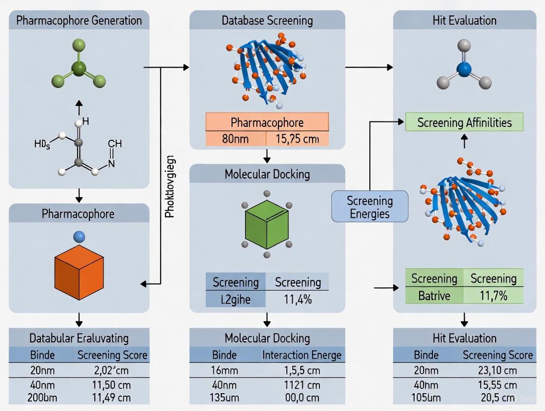
Diagram: Pharmacophore-guided virtual screening workflow integrating multiple computational approaches for hit identification.
Screening Database Preparation
The initial phase involves preparing screening libraries through standardized protocols:
Database Curation: Large compound databases (e.g., ZINC, ChEMBL) are filtered using drug-like property filters such as Lipinski's Rule of Five to focus on chemically relevant space [4].
Conformational Expansion: Each compound in the screening library undergoes conformational analysis to generate a representative set of low-energy conformations, ensuring potential bioactive conformations are available for pharmacophore matching [1] [3].
Feature Annotation: Chemical features relevant to pharmacophore matching (hydrogen bond donors/acceptors, hydrophobic regions, etc.) are identified and annotated for each conformer [3].
Hierarchical Screening Protocol
Modern virtual screening employs a multi-stage filtering approach to efficiently prioritize compounds:
Pharmacophore-Based Screening: The pharmacophore model serves as a 3D search query to identify compounds whose conformations match the essential feature arrangement [3] [4]. This step typically reduces the screening library by 90-99%, dramatically focusing the computational burden for subsequent steps.
Molecular Docking: Compounds matching the pharmacophore hypothesis undergo more computationally intensive molecular docking to assess binding geometry and complementarity with the target protein [4]. Docking scores provide a more refined estimate of binding affinity.
Machine Learning Prioritization: Recent advances integrate machine learning models trained on docking scores to further accelerate screening [4] [9]. These models can predict binding affinities thousands of times faster than molecular docking, enabling ultra-high-throughput virtual screening.
Experimental Validation: The top-ranked compounds from computational screening are selected for experimental testing to confirm biological activity [4].
Advanced Integration with Machine Learning
The field is witnessing rapid advancement through the integration of pharmacophore constraints with deep generative models for molecular design:
Pharmacophore-Guided Molecular Generation: Deep learning approaches like Pharmacophore-Guided deep learning approach for bioactive Molecule Generation (PGMG) use pharmacophore hypotheses as conditional constraints to generate novel molecules with desired bioactivity profiles [9]. These methods introduce latent variables to model the many-to-many relationship between pharmacophores and molecules, enhancing structural diversity while maintaining biological relevance.
Ensemble Machine Learning Models: Predictive models combining multiple types of molecular fingerprints and descriptors can accurately estimate docking scores, enabling rapid virtual screening of billions of compounds [4]. These ensemble models reduce prediction errors and can be generalized across multiple biological targets.
Research Reagent Solutions: Computational Tools for Pharmacophore-Based Screening
The implementation of pharmacophore-based virtual screening requires specialized software tools and computational resources. The following table summarizes key software solutions used in modern pharmacophore workflows.
Table: Essential Computational Tools for Pharmacophore-Based Virtual Screening
| Tool/Software | Type | Key Functionality | Application in Workflow |
|---|---|---|---|
| Catalyst/Discovery Studio | Commercial Software | Pharmacophore model generation (HipHop, HypoGen), 3D database searching | Ligand-based pharmacophore modeling, virtual screening |
| LigandScout | Commercial Software | Structure-based pharmacophore modeling, virtual screening | Protein structure-based pharmacophore development |
| Phase | Commercial Software | Ligand- and structure-based pharmacophore modeling, 3D-QSAR | Pharmacophore model generation, activity prediction |
| MOE | Commercial Software | Comprehensive molecular modeling, pharmacophore modeling | Integrated drug design platform |
| RDKit | Open-Source Library | Cheminformatics, feature detection, molecular generation | Chemical feature annotation, molecular processing |
| ZINC Database | Public Database | Curated compound library for virtual screening | Source of screening compounds |
| ChEMBL Database | Public Database | Bioactivity data, compound structures | Training set selection, model validation |
| Smina | Open-Source Tool | Molecular docking, scoring function optimization | Structure-based screening, binding affinity estimation |
These tools collectively enable the complete pharmacophore-based screening workflow, from model generation through compound prioritization. The selection of specific tools depends on available structural information, computational resources, and the specific objectives of the screening campaign.
The evolution of pharmacophore theory from Paul Ehrlich's initial conceptualization to the modern IUPAC definition represents a remarkable journey of scientific refinement and technological adaptation. What began as a qualitative description of chemical groups responsible for biological activity has matured into a sophisticated, quantitative framework for understanding and predicting molecular recognition. Throughout this evolution, the core insight has remained consistent: that biological activity can be abstracted to essential patterns of chemical features arranged in three-dimensional space.
In contemporary drug discovery, particularly in the context of virtual screening workflows, pharmacophore approaches provide an indispensable strategy for navigating vast chemical spaces and identifying novel bioactive compounds. Their unique strength lies in balancing specificity with generalizability—capturing the essential elements required for binding while enabling scaffold hopping and structural diversity. The integration of pharmacophore modeling with molecular docking and machine learning represents the current state of the art, combining the conceptual clarity of pharmacophore thinking with the predictive power of modern computational methods.
As drug discovery confronts increasingly challenging targets, including protein-protein interactions and novel target classes with limited structural information, pharmacophore-based approaches continue to adapt and evolve. The incorporation of pharmacophore constraints into deep generative models represents particularly promising direction, enabling de novo molecular design guided by fundamental principles of molecular recognition. The continued evolution of pharmacophore theory ensures its enduring relevance in the scientific pursuit of novel therapeutic agents.
A pharmacophore is defined by the International Union of Pure and Applied Chemistry (IUPAC) as "the ensemble of steric and electronic features that is necessary to ensure the optimal supramolecular interactions with a specific biological target structure and to trigger (or to block) its biological response" [10] [11] [12]. This abstract description represents the essential three-dimensional arrangement of chemical functionalities required for a molecule to bind to its biological target, rather than representing a specific molecule or functional group itself [13]. The fundamental principle underpinning pharmacophore modeling is that different molecules sharing common chemical features in a consistent spatial arrangement can elicit similar biological responses by interacting with the same target [11].
Pharmacophore models represent these interaction patterns through abstract chemical features that define interaction types rather than specific functional groups or atoms [10]. The most critical and commonly utilized features include hydrogen bond donors (HBD), hydrogen bond acceptors (HBA), hydrophobic areas (H), positively ionizable groups (PI), negatively ionizable groups (NI), and aromatic rings (AR) [11] [14] [12]. These features are typically represented in three-dimensional space as geometric entities such as spheres (with defined tolerance radii), planes, and vectors that capture the directionality of specific interactions like hydrogen bonding [11] [13].
The primary application of pharmacophore models is in virtual screening (VS) of compound libraries, where they serve as queries to identify novel candidate molecules that match the essential feature arrangement [10] [11] [12]. This approach is particularly valuable for scaffold hopping—identifying structurally diverse compounds with similar biological activity—which has significant implications for overcoming patent restrictions and optimizing drug properties [10] [13]. The following sections provide a detailed examination of the key pharmacophore features, their characteristics, and their roles in molecular recognition.
Detailed Analysis of Core Pharmacophore Features
Hydrogen Bond Donors and Acceptors
Hydrogen bond donors (HBD) and hydrogen bond acceptors (HBA) are among the most crucial features for mediating specific ligand-target interactions [15]. These features represent the capacity of a molecule to form directional hydrogen bonds with complementary residues in the binding pocket.
Hydrogen Bond Donors (HBD): These are typically hydrogen atoms connected to electronegative atoms (most commonly oxygen or nitrogen) that can participate in non-covalent bonding with hydrogen bond acceptors on the target protein. In pharmacophore models, HBD features often include vector constraints that define the preferred directionality of the hydrogen bond formation [13]. Common chemical groups containing HBD features include hydroxyl groups (-OH), primary and secondary amines (-NH₂, -NHR), and sometimes thiol groups (-SH).
Hydrogen Bond Acceptors (HBA): These features represent atoms with lone electron pairs capable of forming hydrogen bonds with donor groups on the target protein. The most common hydrogen bond acceptors are oxygen atoms in carbonyl groups, ethers, and alcohols, as well as nitrogen atoms in amines, amides, and heterocyclic aromatic rings [11] [12]. Some programs further classify hydrogen bond acceptors based on their strength and directionality preferences.
Statistical analyses of protein-ligand complexes reveal that hydrogen bond donors demonstrate high conservation in their interactions, meaning they typically must match identical feature types in the pharmacophore model [15]. The same holds true for hydrogen bond acceptors, though with slightly lower conservation than donors [15]. Notably, exchanges between hydrogen bond donors and acceptors are highly unlikely, occurring barely more frequently than by random chance [15].
Hydrophobic Areas
Hydrophobic features represent regions of the molecule that are non-polar and lipophilic,
capable of engaging in van der Waals interactions and the hydrophobic effect with complementary non-polar regions of the binding pocket [11] [12]. These features are critical for the overall binding affinity, often contributing significantly to the binding energy through the burial of non-polar surface area from the aqueous environment.
Hydrophobic features can be further categorized into:
- Aliphatic hydrophobic groups: Including alkyl chains, alicyclic rings, and other saturated carbon frameworks.
- Aromatic rings (AR): Planar systems with delocalized π-electrons that can engage in stacking interactions with other aromatic systems or amino acid side chains in the binding pocket [14] [12].
- Non-aromatic π-systems: Such as alkenes and alkynes that can participate in weaker π-interactions [15].
Hydrophobic features generally show moderate to low conservation in pharmacophore models, meaning they can sometimes be interchanged or displaced while maintaining biological activity [15]. When ranked by relevance, mutual information analysis places all hydrophobic features as least important, though geometric series ranking assigns higher significance to aromatic features [15].
Ionizable Groups
Ionizable groups are features that can carry formal positive or negative charges under physiological conditions, enabling the formation of strong electrostatic interactions with complementary charged residues in the binding pocket [11].
Positively Ionizable Groups (PI): These are typically basic nitrogen atoms in functional groups such as primary, secondary, or tertiary amines, guanidines, or amidines that can be protonated to form cations. These features can form strong salt bridges with negatively charged acidic residues (aspartate, glutamate) in the target protein [11] [12].
Negatively Ionizable Groups (NI): These are generally acidic functionalities such as carboxylates (-COO⁻), phosphates, phosphonates, sulfates, or sulfonates that can be deprotonated to form anions. These interact strongly with positively charged basic residues (lysine, arginine, histidine) in the binding site [11] [12].
Statistical analysis of feature conservation reveals that negatively ionizable groups (acids) are the most conserved pharmacophore feature, followed by hydrogen bond donors, then positively ionizable groups (basic nitrogens) [15]. This high conservation indicates that these features typically require exact matching in pharmacophore models. The most likely exchanges observed are between carboxylate groups and hydrogen-bond acceptors and similarly between basic nitrogens and hydrogen-bond donors, reflecting the characteristics of Lewis acids and bases [15].
Table 1: Conservation and Exchangeability of Key Pharmacophore Features
| Feature Type | Conservation Rank | Most Likely Exchanges | Common Functional Groups |
|---|---|---|---|
| Negatively Ionizable (NI) | 1 (Most conserved) | Hydrogen Bond Acceptors | Carboxylates, Phosphates, Sulfonates |
| Hydrogen Bond Donor (HBD) | 2 | Positively Ionizable Groups | -OH, -NH₂, -NHR |
| Positively Ionizable (PI) | 3 | Hydrogen Bond Donors | Amines, Guanidines, Amidines |
| Hydrogen Bond Acceptor (HBA) | 4 | Negatively Ionizable Groups | Carbonyl O, Ether O, Amine N |
| Aromatic (AR) | 5 | Other Hydrophobic Groups | Phenyl, Pyridine, Other Heterocycles |
| Other Hydrophobic (H) | 6 (Least conserved) | Aromatic Groups | Alkyl Chains, Alicyclic Systems |
Integration into Virtual Screening Workflow
The core pharmacophore features serve as the fundamental building blocks in comprehensive pharmacophore-based virtual screening workflows, which provide a powerful approach for identifying novel bioactive compounds from extensive chemical libraries [10] [11] [13]. The overall process integrates multiple computational steps that progressively filter compound databases to identify promising candidates for experimental testing.
Diagram 1: Pharmacophore-Based Virtual Screening Workflow. This diagram illustrates the comprehensive process of virtual screening utilizing pharmacophore models, integrating both structure-based and ligand-based approaches.
Pharmacophore Model Generation
The process begins with the creation of a pharmacophore model using either structure-based or ligand-based approaches [10] [11]:
Structure-Based Approach: This method requires three-dimensional structural information about the target protein, typically obtained from X-ray crystallography, NMR spectroscopy, cryo-EM, or homology modeling [10] [11] [14]. The process involves:
- Protein Preparation: Adding hydrogen atoms, correcting protonation states, and addressing missing residues [11].
- Binding Site Identification: Determining the relevant binding pocket using tools like GRID or LUDI that analyze protein surface properties [11].
- Feature Extraction: Identifying key interaction points (HBD, HBA, H, PI, NI, AR) by analyzing the complementarity between the binding site and potential ligands [10] [11].
- Model Optimization: Selecting the most relevant features and defining their spatial relationships with appropriate tolerances [11].
Ligand-Based Approach: When structural data for the target is unavailable, pharmacophore models can be derived from a set of known active compounds [11] [12]. This method involves:
- Training Set Selection: Curating a diverse set of confirmed active molecules with demonstrated binding affinity [10].
- Conformational Analysis: Generating representative low-energy conformations for each molecule [12].
- Common Feature Identification: Aligning the molecules and identifying the essential chemical features shared among active compounds [10] [12].
- Hypothesis Generation: Creating pharmacophore models that capture the common spatial arrangement of key features [12].
Database Screening and Hit Identification
Once a validated pharmacophore model is established, it serves as a query for screening compound databases [10] [13]. This process involves several sophisticated computational steps:
Database Preparation: Large compound libraries (e.g., ZINC, commercial databases, in-house collections) are pre-processed by generating multiple conformers for each compound to account for molecular flexibility [13]. This creates a conformational database that enables efficient 3D searching [13].
Pharmacophore Searching: The actual screening employs a multi-step filtering approach to efficiently identify matches [13]:
- Pre-filtering: Rapid elimination of compounds that lack the necessary feature types or counts using pharmacophore keys or fingerprint methods [13].
- 3D Geometric Matching: More computationally intensive alignment of database conformers to the pharmacophore query using maximum clique detection or sequential buildup algorithms [13].
- Constraint Checking: Verification of additional constraints including vector directions for hydrogen bonds, plane orientations for aromatic systems, and exclusion volumes to prevent steric clashes [10] [13].
Post-Screening Analysis: Compounds that successfully map to the pharmacophore model undergo further computational assessment, which may include molecular docking, ADMET prediction, and similarity analysis to prioritize the most promising candidates for experimental validation [4] [16].
Table 2: Performance Metrics of Pharmacophore-Based Virtual Screening Compared to Alternative Methods
| Screening Method | Typical Hit Rate | Scaffold Diversity | Computational Efficiency | Key Applications |
|---|---|---|---|---|
| Pharmacophore-Based VS | 5-40% [10] | High (scaffold hopping) [13] | Medium to High | Lead identification, Scaffold hopping [10] |
| High-Throughput Experimental Screening | <1% [10] | Variable (library-dependent) | Low (experimental cost) | Primary screening |
| Molecular Docking | 10-30% | Medium | Low (computationally intensive) | Lead optimization, Pose prediction [4] |
| 2D Similarity Search | 1-20% | Low (similar scaffolds) | High | Analog searching |
Experimental Protocols and Validation
Structure-Based Pharmacophore Modeling Protocol
The following detailed protocol outlines the steps for creating a structure-based pharmacophore model, as applied in the identification of PD-L1 inhibitors from marine natural products [16]:
Protein Structure Preparation:
- Obtain the 3D structure of the target protein from the Protein Data Bank (PDB). For example, the PD-L1 structure with PDB ID 6R3K was used in the referenced study [16].
- Remove crystallographic water molecules and extraneous ligands, preserving any essential cofactors (e.g., FAD in dehydrogenase targets) [10] [4].
- Add hydrogen atoms and optimize protonation states of residues using molecular mechanics force fields at physiological pH (7.4) [11].
- Energy minimize the structure to relieve steric clashes and ensure geometric stability [11].
Binding Site Analysis and Feature Mapping:
- Identify the binding pocket either from the location of a co-crystallized ligand or through binding site detection algorithms [11] [16].
- Generate potential interaction points using programs such as LigandScout or Discovery Studio that analyze complementarity between the binding site and potential ligands [10] [16].
- Define pharmacophore features based on the interaction characteristics:
- Hydrogen bond donors complementary to carbonyl oxygen or other HBA in binding site
- Hydrogen bond acceptors complementary to backbone NH or other HBD groups
- Hydrophobic features corresponding to non-polar subpockets
- Ionizable features complementary to charged residues
- Add exclusion volumes to represent steric restrictions of the binding pocket [10].
Model Validation:
Ligand-Based Pharmacophore Generation Protocol
For targets lacking structural information, ligand-based pharmacophore modeling provides an alternative approach, as demonstrated in studies of EGFR inhibitors and monoamine oxidase inhibitors [12] [4]:
Training Set Compilation:
- Curate a set of 3-10 known active compounds with diverse structural scaffolds but similar biological activity from databases such as ChEMBL or BindingDB [10] [4].
- Include confirmed inactive compounds when available to enhance model selectivity [10].
- Define activity thresholds to distinguish between active and inactive molecules (e.g., pIC₅₀ > 4.75 for actives) [14].
Conformational Analysis and Molecular Alignment:
- Generate representative low-energy conformations for each compound using algorithms such as systematic search, random search, or genetic algorithms [12].
- Perform molecular alignment to identify the pharmacophoric pattern common to active compounds using:
- Feature-based alignment that maximizes overlap of key chemical features
- Field-based alignment that matches molecular interaction fields
- Maximum Common Substructure (MCS) approaches [12]
Pharmacophore Hypothesis Generation:
- Identify common chemical features shared by the aligned active compounds [12].
- Define the spatial relationships between features with appropriate distance and angle tolerances [12].
- Generate multiple hypotheses and select the best model based on statistical scoring functions that evaluate how well the model discriminates between active and inactive compounds [12].
Table 3: Essential Software and Databases for Pharmacophore-Based Virtual Screening
| Resource Type | Examples | Key Functionality | Application Context |
|---|---|---|---|
| Pharmacophore Modeling Software | LigandScout [13], Discovery Studio [10], Phase (Schrödinger) [13], MOE [13] | Structure-based and ligand-based model generation, Virtual screening | Core model development and screening |
| Protein Structure Databases | Protein Data Bank (PDB) [10] [11], AlphaFold DB [11] | Source of experimental and predicted protein structures | Structure-based pharmacophore modeling |
| Compound Activity Databases | ChEMBL [10] [4], BindingDB [15], DrugBank [10] | Bioactivity data for known ligands | Training set compilation, Model validation |
| Screening Compound Libraries | ZINC [4], Marine Natural Products Databases [16], Commercial screening collections | Source of compounds for virtual screening | Identification of novel hit compounds |
| Docking Software | AutoDock [16], Smina [4] | Molecular docking to refine and score potential hits | Post-screening analysis, Binding mode prediction |
| Pre-filtering Tools | Directory of Useful Decoys, Enhanced (DUD-E) [10] | Generation of optimized decoy molecules | Model validation and benchmarking |
The core pharmacophore features—hydrogen bond donors and acceptors, hydrophobic areas, and ionizable groups—form the fundamental basis for molecular recognition in drug discovery. These abstract representations of chemical functionality capture the essential elements required for productive binding to biological targets, enabling the development of computational models that can efficiently search chemical space for novel bioactive compounds. The high conservation of ionizable groups and hydrogen bond donors underscores their critical role in specific molecular recognition, while the greater flexibility in hydrophobic features allows for more structural variation in drug design.
The integration of these features into comprehensive virtual screening workflows has demonstrated significant value in drug discovery, with reported hit rates of 5-40% in prospective applications [10]. This represents a substantial enrichment over random screening approaches, which typically yield hit rates below 1% [10]. The ability of pharmacophore models to facilitate scaffold hopping—identifying structurally diverse compounds with similar biological activity—makes them particularly valuable for addressing patent constraints and optimizing drug properties [13].
As virtual screening continues to evolve, the integration of machine learning approaches with traditional pharmacophore methods shows promise for further accelerating the discovery process [4]. These hybrid approaches can reduce computational time by several orders of magnitude while maintaining or improving prediction accuracy [4]. Nevertheless, the fundamental pharmacophore features described in this work will continue to provide the conceptual framework for understanding and exploiting molecular interactions in drug design, serving as the essential building blocks for both traditional and next-generation virtual screening methodologies.
In the field of computer-aided drug discovery (CADD), the efficient identification of novel therapeutic candidates is paramount. Virtual screening (VS) stands as a cornerstone technique for rapidly evaluating vast chemical libraries to pinpoint molecules with promising biological activity against a specific therapeutic target [17]. Pharmacophore-based virtual screening represents one of the most powerful and widely used methodologies within this domain. This approach relies on the fundamental concept of a pharmacophore—defined by the International Union of Pure and Applied Chemistry (IUPAC) as "the ensemble of steric and electronic features that is necessary to ensure the optimal supramolecular interactions with a specific biological target structure and to trigger (or to block) its biological response" [11]. At the heart of pharmacophore development lie two complementary computational strategies: structure-based modeling and ligand-based modeling. These approaches differ primarily in their source of structural information, yet both aim to abstract the essential chemical features required for molecular recognition and biological activity [18] [11]. This technical guide provides an in-depth examination of these two fundamental methodologies, their integration strategies, and their application within modern pharmacophore-based virtual screening workflows for drug development professionals.
Theoretical Foundations
The Pharmacophore Concept
A pharmacophore model consists of a set of chemical features arranged in a specific three-dimensional configuration that collectively confer biological activity against a particular molecular target [18]. These features represent key interaction points rather than specific chemical structures, allowing pharmacophore models to identify structurally diverse compounds that share common activity. The most significant pharmacophoric feature types include:
- Hydrogen bond acceptors (HBA)
- Hydrogen bond donors (HBD)
- Hydrophobic areas (H)
- Positively and negatively ionizable groups (PI/NI)
- Aromatic groups (AR)
- Metal coordinating areas [11]
Additionally, spatial constraints in the form of exclusion volumes can be incorporated to represent steric obstructions within the binding pocket, thereby refining model selectivity [11]. The strength of pharmacophore modeling lies in its scaffold-hopping capability—the ability to identify chemically distinct compounds that nonetheless share the essential functional features required for target binding and activity.
Structure-Based Modeling Approach
Structure-based drug design (SBDD) encompasses methods that rely directly on the three-dimensional structural information of the biological target, typically obtained through experimental techniques such as X-ray crystallography, nuclear magnetic resonance (NMR) spectroscopy, or cryo-electron microscopy (Cryo-EM) [19]. When applied to pharmacophore modeling, the structure-based approach extracts critical chemical features from the analysis of intermolecular interactions between a ligand and its macromolecular target within a complex [18]. This method is particularly valuable when detailed structural knowledge of the binding site is available, as it provides atomic-level insight into the complementarity requirements for ligand binding.
The primary advantage of structure-based pharmacophore modeling lies in its ability to identify novel chemotypes without prior knowledge of active ligands, making it indispensable for targets with limited chemical precedent [11]. Furthermore, by incorporating the spatial and electronic constraints of the actual binding pocket, structure-based models can achieve high selectivity and reduce false positives in virtual screening. However, the quality of these models is heavily dependent on the resolution and accuracy of the experimental protein structure, and they may overlook important ligand conformational preferences that occur during the binding process [17].
Ligand-Based Modeling Approach
Ligand-based drug design (LBDD) approaches are employed when the three-dimensional structure of the target protein is unknown or unavailable. Instead, these methods rely on information derived from known active compounds that bind to the target of interest [19]. Ligand-based pharmacophore modeling identifies common chemical features and their spatial arrangements from a set of active ligands through three-dimensional alignment [18]. The underlying premise is that compounds exhibiting similar biological activity likely share fundamental interaction features necessary for target recognition.
The ligand-based approach offers significant advantages when structural data for the target is lacking, and it inherently incorporates ligand conformational flexibility through multi-conformer analysis [11]. Additionally, by deriving features directly from active compounds, these models implicitly capture key activity-determining elements. However, ligand-based methods are limited by the quality, diversity, and quantity of known actives, with potential bias toward the chemical scaffolds represented in the training set [17]. They also lack explicit information about protein-related constraints, which may reduce their ability to discriminate between true actives and inactive compounds with similar pharmacophoric features.
Table 1: Core Characteristics of Structure-Based and Ligand-Based Modeling Approaches
| Characteristic | Structure-Based Modeling | Ligand-Based Modeling |
|---|---|---|
| Primary Data Source | 3D structure of target protein (from X-ray, NMR, Cryo-EM) | Known active ligands |
| Key Requirements | High-quality protein structure, often with bound ligand | Set of active compounds with diverse structures |
| Feature Identification | Derived from protein-ligand interaction analysis | Extracted from common features of aligned active ligands |
| Advantages | No prior active ligands needed; Direct incorporation of binding site constraints | Target structure not required; Implicit activity correlation |
| Limitations | Dependent on quality and relevance of protein structure; May overlook ligand flexibility | Limited by diversity and quality of known actives; Potential scaffold bias |
Methodological Implementation
Structure-Based Pharmacophore Modeling Workflow
The generation of structure-based pharmacophore models follows a systematic workflow that ensures comprehensive analysis of the binding site and accurate feature identification:
Protein Structure Preparation: The initial step involves obtaining and refining the three-dimensional structure of the target protein, typically from the Protein Data Bank (PDB). Preparation includes adding hydrogen atoms, correcting protonation states, optimizing hydrogen bonding networks, and energy minimization to ensure structural integrity [11] [20]. For example, in a study targeting EGFR, researchers retrieved the crystal structure (PDB ID: 7AEI) and prepared it using Protein Preparation Wizard, assigning bond orders, creating disulfide bonds, and optimizing hydrogen bonds at pH 7.0 [20].
Binding Site Analysis and Characterization: The ligand-binding site is identified through analysis of co-crystallized ligands or computational prediction using tools like GRID or LUDI, which detect regions conducive to molecular interactions based on energetic and geometric considerations [11].
Pharmacophore Feature Generation: Interaction points between the protein and a bound ligand are analyzed to identify key pharmacophoric features. Software such as LigandScout automatically detects and characterizes these features, including hydrogen bond donors/acceptors, hydrophobic regions, and ionizable groups [21]. In the XIAP inhibitor study, researchers used the protein-ligand complex (PDB: 5OQW) to generate a model containing 14 chemical features: four hydrophobic, one positive ionizable, three hydrogen bond acceptors, and five hydrogen bond donors [21].
Feature Selection and Model Validation: The initial feature set is refined by selecting only those features essential for biological activity, followed by validation using known active and inactive compounds to assess model discriminative ability [11] [21]. The XIAP study validated their model using receiver operating characteristic (ROC) analysis, achieving an excellent area under curve (AUC) value of 0.98, confirming strong ability to distinguish true actives from decoys [21].
Ligand-Based Pharmacophore Modeling Workflow
Ligand-based pharmacophore modeling employs a different strategy focused on extracting common features from bioactive molecules:
Ligand Dataset Curation: A structurally diverse set of known active compounds against the target is collected, ensuring representation of various chemotypes while excluding inactive or weakly active molecules to enhance model quality [18].
Conformational Analysis and Molecular Alignment: Multiple low-energy conformations are generated for each active compound, followed by spatial alignment to identify common pharmacophoric features and their three-dimensional arrangement [18] [11]. This step is crucial for capturing the bioactive conformation.
Pharmacophore Hypothesis Generation: The aligned molecules are analyzed to identify conserved chemical features essential for activity. The model may be refined by quantifying the contribution of each feature to biological activity [11].
Model Validation and Refinement: The generated model is validated using a separate test set of active and inactive compounds, with refinement through iterative optimization to improve predictive performance [18]. In a natural product screening study, researchers emphasized that while strict pharmacophore models select compounds with better activity, they may reduce structural diversity, whereas less restrictive models may retrieve more false positives [18].
Table 2: Software Tools for Pharmacophore Modeling and Virtual Screening
| Software | Modeling Approach | License | Key Features |
|---|---|---|---|
| LigandScout | Structure-based & Ligand-based | Commercial | Advanced pharmacophore feature detection, 3D pharmacophore modeling, virtual screening |
| MOE (Molecular Operating Environment) | Structure-based & Ligand-based | Commercial | Comprehensive drug discovery suite with pharmacophore modeling capabilities |
| Pharmer | Ligand-based | Open Source | Efficient pharmacophore search and screening algorithms |
| Align-it | Ligand-based | Open Source | Aligns molecules based on pharmacophore features (formerly Pharao) |
| Pharmit | Structure-based | Free Access Web Server | Online pharmacophore-based virtual screening platform |
| PharmMapper | Structure-based | Free Access Web Server | Reverse pharmacophore screening server for target identification |
Integrated Approaches and Advanced Applications
Hybrid Strategies for Enhanced Screening
Recognizing the complementary strengths and limitations of structure-based and ligand-based approaches, researchers have developed integrated strategies that combine both methodologies to enhance virtual screening performance. These hybrid approaches can be categorized into three main types:
Sequential Approaches: These implement a multi-step VS pipeline where LB and SB techniques are applied consecutively to progressively filter chemical libraries. Typically, faster LB methods perform initial filtering, followed by more computationally intensive SB methods for refined selection [17]. This strategy optimizes the tradeoff between computational efficiency and screening accuracy.
Parallel Approaches: LB and SB methods are run independently on the same compound library, with results combined afterward to select candidates for biological testing. The combination can involve various rank aggregation methods, with studies demonstrating that this approach increases both performance and robustness compared to single-method strategies [17].
Holistic Hybrid Approaches: These represent the most integrated strategy, where LB and SB information is combined into a single, unified model. For example, the CMD-GEN framework utilizes coarse-grained pharmacophore points sampled from a diffusion model conditioned on protein pockets, effectively bridging ligand-protein complexes with drug-like molecules [22]. This method employs a hierarchical architecture that decomposes 3D molecule generation into pharmacophore point sampling, chemical structure generation, and conformation alignment.
Case Study: Integrated EGFR Inhibitor Discovery
A comprehensive drug discovery study targeting the Epidermal Growth Factor Receptor (EGFR) exemplifies the power of integrated approaches [20]. Researchers developed a ligand-based pharmacophore model using the co-crystal ligand (R85) of EGFR (PDB ID: 7AEI) featuring hydrophobic, aromatic, hydrogen bond acceptor, and hydrogen bond donor features. This model screened nine commercial databases, identifying 1,271 hits meeting Lipinski's Rule of Five criteria. Subsequent structure-based molecular docking refined the selection to ten top compounds with binding affinities ranging from -7.691 to -7.338 kcal/mol. Further ADMET analysis and 200 ns molecular dynamics simulations confirmed the stability of protein-ligand complexes for three final candidates: MCULE-6473175764, CSC048452634, and CSC070083626 [20]. This integrated workflow demonstrates how sequentially combining ligand-based and structure-based methods can efficiently identify promising drug candidates.
Emerging AI-Driven Approaches
Recent advances in artificial intelligence are reshaping pharmacophore modeling and virtual screening. The CMD-GEN framework exemplifies this innovation, addressing challenges in structure-based molecular generation by incorporating coarse-grained pharmacophore representations [22]. This approach bridges the gap between limited protein-ligand complex data and extensive chemical compound libraries through a hierarchical process:
- Coarse-grained pharmacophore sampling using diffusion models conditioned on protein pockets
- Chemical structure generation via a gating condition mechanism with pharmacophore constraints
- Conformation alignment based on pharmacophore matching
This method has demonstrated promising results in designing selective PARP1/2 inhibitors, confirmed through wet-lab validation, highlighting the potential of AI-enhanced approaches to tackle challenging drug design problems such as selectivity and polypharmacology [22].
Experimental Protocols
Protocol 1: Structure-Based Pharmacophore Modeling
This protocol outlines the key steps for generating a structure-based pharmacophore model, adapted from studies on XIAP and EGFR targets [20] [21].
Materials and Reagents:
- Experimentally determined 3D protein structure (PDB format)
- Molecular visualization software (e.g., PyMOL)
- Structure-based pharmacophore modeling software (e.g., LigandScout)
- Computer system with adequate processing power and memory
Procedure:
- Protein Preparation:
- Retrieve the target protein structure from the Protein Data Bank (PDB)
- Add hydrogen atoms appropriate for physiological pH (7.4)
- Optimize hydrogen bonding networks using algorithms like PROPKA
- Perform energy minimization using forcefields such as OPLS_2005
- Remove crystallographic water molecules unless functionally important
Binding Site Analysis:
- Identify the binding pocket through analysis of co-crystallized ligands
- Alternatively, use binding site detection tools (GRID, LUDI) for apo structures
- Characterize key interacting residues and their properties
Pharmacophore Feature Generation:
- Load the prepared protein-ligand complex into pharmacophore modeling software
- Automatically detect interaction features between protein and ligand
- Identify hydrogen bond donors/acceptors, hydrophobic regions, charged interactions
- Add exclusion volumes to represent steric constraints of the binding pocket
Feature Selection and Model Refinement:
- Select features critical for binding affinity based on interaction energy and conservation
- Remove redundant or non-essential features to prevent over-constraining the model
- Adjust spatial tolerances based on binding site flexibility
Model Validation:
- Test the model against a set of known active and inactive compounds
- Generate ROC curves and calculate enrichment factors
- Aim for AUC values >0.8 and high early enrichment (EF1% >10) [21]
Protocol 2: Ligand-Based Pharmacophore Modeling
This protocol describes the generation of ligand-based pharmacophore models, following established methodologies from natural product screening studies [18] [11].
Materials and Reagents:
- Set of known active compounds (15-30 molecules with diverse structures)
- Set of known inactive compounds for validation
- Ligand-based pharmacophore modeling software (e.g., MOE, Pharmer)
- Conformational analysis tool
Procedure:
- Ligand Set Preparation:
- Curate a structurally diverse set of confirmed active compounds
- Ensure activity data is consistent and measured under similar conditions
- Include inactive compounds for model validation
- Prepare 3D structures with correct stereochemistry and protonation states
Conformational Analysis:
- Generate multiple low-energy conformers for each active compound
- Use systematic search or stochastic methods for thorough conformational sampling
- Set energy thresholds appropriately (typically 10-15 kcal/mol above global minimum)
Molecular Alignment and Hypothesis Generation:
- Align molecules using flexible alignment algorithms
- Identify common pharmacophoric features across the aligned set
- Develop multiple pharmacophore hypotheses with varying feature compositions
Hypothesis Validation and Selection:
- Test hypotheses against a validation set of active and inactive compounds
- Quantify model performance using statistical measures (ROC-AUC, EF)
- Select the hypothesis with best discrimination ability
- Correlate feature presence with biological activity where quantitative data exists
Table 3: Research Reagent Solutions for Pharmacophore Modeling
| Reagent/Resource | Function/Application | Example Sources |
|---|---|---|
| Protein Data Bank (PDB) | Repository of 3D protein structures | RCSB PDB (www.rcsb.org) |
| ChEMBL Database | Curated database of bioactive molecules | EMBL-EBI ChEMBL |
| ZINC Database | Commercially available compound libraries | ZINC15 (zinc15.docking.org) |
| LigandScout Software | Structure-based & ligand-based pharmacophore modeling | Inte:Ligand |
| Pharmit Server | Online pharmacophore-based virtual screening | http://pharmit.csb.pitt.edu |
| Molecular Operating Environment (MOE) | Comprehensive drug discovery software suite | Chemical Computing Group |
Comparative Analysis and Applications
Strategic Selection Guide
The choice between structure-based and ligand-based modeling approaches depends on available resources and biological knowledge. The following guidelines assist in selecting the appropriate methodology:
Use Structure-Based Methods When: High-resolution protein structures are available (X-ray ≤2.5Å, Cryo-EM ≤3.0Å); The target exhibits conformational stability; Novel chemotypes are desired beyond known ligand scaffolds; Selective targeting of specific binding sites is required.
Use Ligand-Based Methods When: Protein structure is unavailable or of poor quality; Multiple diverse active compounds are known; Understanding structure-activity relationships (SAR) is prioritized; Rapid screening with established chemotypes is sufficient.
Use Integrated Approaches When: Both protein structures and active ligand data are available; Maximizing screening success rate is critical; Resources permit multi-stage virtual screening; Targeting difficult proteins with flexibility or allosteric sites.
Limitations and Future Directions
Despite significant advances, both structure-based and ligand-based approaches face limitations. Structure-based methods grapple with protein flexibility, solvent effects, and accurate scoring functions [17] [19]. Obtaining high-quality structures remains challenging for membrane proteins, large complexes, or highly dynamic targets [19]. Ligand-based methods suffer from training set bias, limited chemical diversity, and the absence of explicit target constraints [17].
Future developments are addressing these challenges through:
- AI-Enhanced Methods: Frameworks like CMD-GEN that combine coarse-grained pharmacophore sampling with deep generative models [22]
- Advanced Solvent Treatments: More sophisticated handling of explicit water molecules and their roles in binding [17]
- Dynamic Pharmacophores: Incorporating protein flexibility and ensemble-based representations [17]
- Fragment-Based Approaches: Methods like FragmentScout that aggregate pharmacophore features from multiple fragment poses [23]
- Multi-Target Profiling: Designing selective or multi-targeted agents through sophisticated pharmacophore matching [22]
These innovations continue to enhance the accuracy and applicability of pharmacophore-based methods in modern drug discovery, solidifying their role as indispensable tools in the quest for novel therapeutics.
The Role of Exclusion Volumes in Representing Binding Pocket Geometry
In the structured workflow of pharmacophore-based virtual screening, the accurate representation of the target's binding site is paramount for success. A pharmacophore model abstractly defines the steric and electronic features necessary for a molecule to interact with a biological target [10] [11]. While features like hydrogen bond donors and hydrophobic areas define favorable interaction points, they do not inherently capture the physical boundaries of the binding pocket. This is where exclusion volumes prove critical. These volumes are steric constraints that geometrically mimic the binding pocket, thereby preventing the mapping of compounds that would be inactive due to steric clashes with the protein surface [10]. Their proper integration significantly enhances the discriminative power of pharmacophore models, leading to higher virtual screening hit rates and more efficient lead identification in computer-aided drug discovery [10] [24].
Theoretical Foundation of Exclusion Volumes
Definition and Core Function
The International Union of Pure and Applied Chemistry (IUPAC) defines a pharmacophore as "the ensemble of steric and electronic features that is necessary to ensure the optimal supra-molecular interactions with a specific biological target structure and to trigger (or to block) its biological response" [10] [11]. Exclusion volumes are integral to this definition, representing the steric component of the model.
In practice, exclusion volumes are three-dimensional constructs, often visualized as spheres or negative space, that define regions inaccessible to a potential ligand [11]. Their primary function is to add a negative image of the binding site's shape, ensuring that any compound which fits the positive pharmacophore features (e.g., hydrogen bond acceptors) but also occupies these forbidden regions is correctly classified as inactive [10]. This directly addresses a key limitation of feature-only models, which might falsely identify overly large molecules as hits simply because they possess the required functional groups, regardless of their overall fit within the binding cavity.
The Underlying Rationale: Mimicking Protein-Ligand Steric Clashes
The theoretical justification for exclusion volumes stems from the fundamental principles of molecular recognition. When a ligand binds to a protein, its favorable interactions are counterbalanced by unfavorable van der Waals repulsions if it penetrates the protein's surface. In a structure-based pharmacophore model, these repulsions are programmatically encoded as exclusion volumes, which are typically placed on atoms lining the binding pocket that are not directly involved in favorable interactions with the ligand [10].
The use of exclusion volumes transforms the pharmacophore query from a purely permissive filter to a more discriminatory one. It refines the virtual screening process by incorporating essential 3D structural information from the target, leading to a significant reduction in false positives and an improved enrichment factor—the metric that quantifies the enrichment of active molecules in a virtual hit list compared to random selection [10] [24].
Methodological Implementation
The generation and application of exclusion volumes follow a systematic process, integrated into the broader pharmacophore modeling workflow. The following diagram illustrates this integrated process, highlighting the key decision points for exclusion volume handling.
Structure-Based Generation of Exclusion Volumes
In the structure-based approach, exclusion volumes are derived directly from the 3D structure of the protein target, often obtained from sources like the Protein Data Bank (PDB) [10] [11].
- Input Data Requirement: The process typically begins with a high-resolution crystal or NMR structure of the target, preferably in a complex with a bound ligand. The quality of this input structure directly influences the accuracy of the resulting exclusion volumes [11].
- Automated Feature Detection: Software tools such as LigandScout and Discovery Studio are commonly used. These programs automatically analyze the binding site and generate an initial set of pharmacophore features and exclusion volumes [10] [25]. For instance, in a study on hydroxysteroid dehydrogenases, exclusion volumes were used to represent the binding pocket geometry and prevent the mapping of sterically clashing compounds [10].
- Advanced Placement Techniques: More sophisticated implementations may add an "exclusion volumes coat," which represents a second shell of steric constraints beyond the immediate binding site surface, providing an even more refined shape definition [25].
Ligand-Based Consideration of Steric Constraints
The ligand-based approach to pharmacophore modeling relies on the alignment of multiple known active molecules to identify their common chemical features [10] [11]. In this scenario, direct structural information about the binding pocket is unavailable.
- Indirect Inference: The spatial arrangement of the ligands themselves implies a complementary volume occupied by the protein. The conserved steric boundaries of the aligned active ligands can be used to infer the general shape of the binding pocket.
- Manual Addition: Based on this inferred volume, exclusion volumes are often added manually around the periphery of the aligned ligand set to prevent overly large compounds from being selected during virtual screening [11]. This process, however, is less precise than the structure-based method and relies heavily on the diversity and quality of the ligand training set.
Refinement and Validation
The initial automated generation of exclusion volumes is typically followed by a refinement stage [10] [26]. This involves:
- Manual Curation: Researchers may add or remove exclusion volumes based on their expert knowledge of the binding site flexibility or specific protein-ligand interaction patterns.
- Model Validation: The final pharmacophore model, including its exclusion volumes, must be rigorously validated. This is done using a dataset of known active and inactive compounds or decoys [10] [21]. Key metrics include:
- Enrichment Factor (EF): Measures the enrichment of active molecules in the virtual hit list versus random selection.
- Receiver Operating Characteristic (ROC) Curve and Area Under the Curve (AUC): Assesses the model's overall ability to distinguish active from inactive compounds. A model with an AUC value of 0.98, as achieved in a study targeting the XIAP protein, indicates excellent predictive power [21].
Impact on Virtual Screening Performance
The strategic use of exclusion volumes has a demonstrable and significant impact on the success of virtual screening campaigns.
Quantitative Performance Metrics
The table below summarizes the performance improvements attributed to well-defined pharmacophore models, which include the proper use of exclusion volumes.
Table 1: Virtual Screening Performance Metrics from Representative Studies
| Target Protein | Virtual Screening Method | Key Performance Metric | Reported Outcome | Reference |
|---|---|---|---|---|
| XIAP | Structure-based pharmacophore (validated with exclusion volumes) | AUC & Enrichment Factor (EF1%) | AUC = 0.98; EF1% = 10.0 | [21] |
| Multiple Targets (ACE, AChE, etc.) | PBVS vs. Docking-Based VS (DBVS) | Average Hit Rate @ 2% & 5% of database | PBVS hit rates "much higher" than DBVS | [24] [27] |
| General HTS vs. VS | Random HTS vs. Pharmacophore-based VS | Typical Hit Rate | HTS: < 1% (e.g., 0.021% for PTP-1B); VS: 5% - 40% | [10] |
Case Studies and Experimental Evidence
- Increased Enrichment Factors: A benchmark study comparing pharmacophore-based virtual screening (PBVS) against docking-based methods (DBVS) across eight diverse protein targets found that PBVS outperformed DBVS in the majority of cases, achieving higher enrichment factors [24] [27]. The presence of exclusion volumes in the pharmacophore queries was a key factor in this superior performance, as it allowed for efficient pre-filtering of molecules that did not fit the binding site geometry.
- Application in SARS-CoV-2 Drug Discovery: In a study targeting the SARS-CoV-2 papain-like protease (PLpro), a structure-based pharmacophore model was developed and used to screen a marine natural product database. The model, which included exclusion volumes, successfully identified a hit compound, aspergillipeptide F, which was subsequently shown via molecular dynamics simulations to form a stable complex with the target, engaging all five binding sites [28]. This demonstrates how a well-defined model can directly lead to the discovery of promising lead candidates.
The Scientist's Toolkit: Essential Reagents and Software
The effective implementation of exclusion volumes in research requires a suite of specialized software tools.
Table 2: Key Software Tools for Pharmacophore Modeling and Virtual Screening
| Tool Name | Type/Function | Role in Handling Exclusion Volumes | Representative Use Case |
|---|---|---|---|
| LigandScout | Software for structure & ligand-based pharmacophore modeling | Automatically generates exclusion volumes from protein structure; allows for manual refinement. | Used in the FragmentScout workflow for SARS-CoV-2 NSP13 helicase to create a joint pharmacophore query [25]. |
| Discovery Studio | Comprehensive modeling and simulation suite | Provides tools for automatic pharmacophore feature and exclusion volume generation from a defined binding site [10]. | Applied in studies on hydroxysteroid dehydrogenases to create models with exclusion volumes [10]. |
| Catalyst/HypoGen | Algorithm and software for pharmacophore generation | Employs exclusion volumes as part of the pharmacophore hypothesis to define unfavorable regions in 3D space. | Used in a benchmark comparison study for pharmacophore-based virtual screening [24] [27]. |
| ZINCPharmer | Online tool for pharmacophore-based screening of the ZINC database | Allows users to define pharmacophore queries, including exclusion volumes, for rapid database filtering. | Utilized to screen the ZINC database for TcaR inhibitors using a pharmacophore model based on Gemifloxacin [29]. |
| Directory of Useful Decoys, Enhanced (DUD-E) | Online resource for benchmarking | Provides optimized decoy molecules used to validate pharmacophore models, testing their ability (including via exclusion volumes) to reject inactive compounds [10]. | Serves as a standard resource for generating decoy sets to test model specificity during validation [10] [21]. |
Exclusion volumes are not merely an optional add-on but a fundamental component of modern, high-fidelity pharmacophore models. By providing an abstract yet accurate representation of binding pocket geometry, they introduce a critical layer of steric discrimination that dramatically improves the efficiency of the virtual screening workflow. Their use leads to higher enrichment factors, reduced false-positive rates, and a greater likelihood of identifying truly active compounds in prospective screening campaigns. As computational methods continue to evolve and integrate with techniques like machine learning [26] and fragment-based screening [25], the precise definition and application of exclusion volumes will remain a cornerstone of successful structure-based drug design.
In the structured workflow of pharmacophore-based virtual screening (PBVS), three technical terms form the foundational pillars: feature mapping, hypothesis generation, and query optimization. A pharmacophore is defined as an abstract description of the structural features of a molecule that are essential for its biological activity [30]. It represents the key molecular interactions—such as hydrogen bonding, charge transfer, or hydrophobic contacts—necessary for a ligand to bind to a macromolecular target. The process of PBVS leverages these concepts to efficiently identify potential hit compounds from vast chemical databases, significantly accelerating the early stages of drug discovery [27] [31]. This guide provides an in-depth technical examination of these core terminologies, framing them within a comprehensive PBVS workflow and detailing the experimental protocols and reagents essential for their successful application.
Feature Mapping: Defining Chemical Interactions
Feature mapping is the process of identifying and spatially locating the essential chemical features on a set of active ligands or within a protein's binding site. These features are the building blocks of any pharmacophore model and represent the specific types of interactions a molecule must be capable of forming to elicit a biological response.
Standard Pharmacophore Feature Types
The table below summarizes the common pharmacophore features used to define molecular interaction patterns.
Table 1: Standard Pharmacophore Features and Their Descriptions
| Feature Type | Abbreviation | Description | Directionality |
|---|---|---|---|
| Hydrogen Bond Acceptor | HA | An atom that can accept a hydrogen bond. | Yes [32] |
| Hydrogen Bond Donor | HD | An atom that can donate a hydrogen bond. | Yes [32] |
| Positively Ionizable | PI | A group that can carry a positive charge. | No [33] |
| Negatively Ionizable | NI | A group that can carry a negative charge. | No [33] |
| Hydrophobic | HY | A non-polar region that engages in van der Waals interactions. | No [32] |
| Aromatic Ring | AR | A pi-system involved in cation-pi or pi-pi stacking. | No [32] [33] |
| Exclusion Volume | EX | A sphere representing sterically forbidden space. | N/A [32] |
Technical Protocols for Feature Mapping
The methodology for feature mapping differs based on the available structural information, leading to two primary approaches.
2.2.1 Structure-Based Feature Mapping When a 3D structure of the protein target (with or without a bound ligand) is available, a structure-based pharmacophore can be developed. The protocol involves:
- Protein Preparation: The protein structure (e.g., from the Protein Data Bank) is prepared by adding hydrogen atoms, assigning correct protonation states, and optimizing hydrogen bonds using tools like the Protein Preparation Wizard in Schrödinger or the "Prepare Protein" module in MOE (Molecular Operating Environment) [34].
- Binding Site Analysis: The active site or a region of interest is defined. This can be the site of a co-crystallized ligand or a predicted binding pocket.
- Feature Generation: Chemical features are mapped onto the binding site, representing potential interaction points complementary to a ligand. For instance, in a study targeting BRAF in melanoma, the binding site was analyzed to identify key amino acids, and features were derived to match them [34]. Software like LigandScout or the "Pharmacophore" module in MOE can automatically generate features from protein-ligand complexes [27].
2.2.2 Ligand-Based Feature Mapping In the absence of a protein structure, features can be mapped from a set of known active ligands. The protocol involves:
- Ligand Preparation and Conformational Analysis: A diverse set of active ligands is energy-minimized, and their multiple low-energy conformations are generated. For example, in developing a model for antimalarial compounds, a maximum of 1000 conformers per ligand were generated using the GB/SA solvent model in PHASE, with an energy cutoff of 10 kcal/mol from the global minimum [33].
- Feature Identification and Alignment: Common chemical features across the active ligand set are identified. The algorithm then attempts to find a common alignment of these molecules that overlays the shared pharmacophore features in 3D space.
Hypothesis Generation: Building the Pharmacophore Model
Hypothesis generation is the process of creating a testable pharmacophore model by synthesizing the information obtained from feature mapping. This model is a spatial arrangement of the essential features required for bioactivity.
Algorithms and Scoring Functions
The generation of a common pharmacophore hypothesis is a computational-intensive process. Software like PHASE employs a tree-based partitioning algorithm to detect common pharmacophores from the aligned conformations of active ligands [33]. It performs an exhaustive analysis of k-point pharmacophore matches based on inter-site distances.
The generated hypotheses are then ranked using a scoring function. The "survival" score (S) in PHASE, for example, includes contributions from [33]:
- The alignment of site points and vectors.
- The volume overlap of the matched ligands.
- The model's selectivity.
- The number of active ligands matched.
- The relative conformational energy of the matched ligands.
- The activity of the matched ligands.
An adjusted survival score (S_I) is often calculated by subtracting the score of any matched inactive molecules, ensuring the model can discriminate between active and inactive compounds [33].
Workflow for Pharmacophore Hypothesis Generation
The following diagram illustrates the logical workflow for generating a pharmacophore hypothesis, integrating both structure-based and ligand-based approaches.
Figure 1: Workflow for generating a pharmacophore hypothesis from either a protein structure or a set of known active ligands.
Query Optimization: Refining for Specificity and Accuracy
Query optimization is the critical process of refining an initial pharmacophore hypothesis to improve its performance in virtual screening. An unoptimized model may retrieve too many false positives (inactive compounds) or miss true positives (active compounds). Optimization tailors the query for a specific screening database and desired outcome.
Optimization Techniques and Genetic Algorithms
A primary method for query optimization is the use of Genetic Algorithms (GA). In this approach, a population of pharmacophore queries is treated as a generation of individuals [35]. Each query is defined by a set of parameters, such as the presence/absence of specific features, their tolerances, and weights. The protocol involves:
- Initialization: Creating an initial population of queries, often variations of the initial hypothesis.
- Fitness Evaluation: Each query is used to screen a training dataset containing known active and inactive compounds. Its "fitness" is calculated based on its ability to correctly identify actives and reject inactives (e.g., a high enrichment factor).
- Selection, Crossover, and Mutation: Queries with high fitness scores are selected to "reproduce." Their parameters are combined (crossover) and randomly altered (mutation) to create a new generation of queries.
- Iteration: Steps 2 and 3 are repeated for multiple generations until a convergence criterion is met, resulting in an optimized query.
A case study on the MC4R system demonstrated the power of this approach, where an optimized query identified 37 agonists with no false positives, a significant improvement over the initial query [35].
Quantitative Benchmarking of Optimized Queries
The success of query optimization is measured by its impact on virtual screening performance. The following table summarizes a benchmark comparison that highlights the efficiency of pharmacophore-based screening (PBVS) versus docking-based screening (DBVS) across eight protein targets.
Table 2: Benchmark Comparison of PBVS vs. DBVS Performance [27]
| Target Protein | Method | Average Hit Rate at 2% | Average Hit Rate at 5% | Key Finding |
|---|---|---|---|---|
| ACE, AChE, AR, etc. (8 targets) | PBVS (Catalyst) | Much Higher | Much Higher | PBVS outperformed DBVS in 14 out of 16 test cases. |
| ACE, AChE, AR, etc. (8 targets) | DBVS (DOCK, GOLD, Glide) | Lower | Lower | PBVS demonstrated superior enrichment in retrieving actives. |
Integrated Virtual Screening Workflow and Applications
The true power of these concepts is realized when they are integrated into a cohesive virtual screening workflow. This workflow enables the identification of novel lead compounds from extensive chemical databases.
End-to-End Screening Protocol
- Hypothesis Establishment: A pharmacophore model is developed and optimized using the methods described in Sections 2-4.
- Database Screening: The optimized query is used as a 3D search filter against a database of small molecules (e.g., ZINC, PubChem). Each compound in the database, in multiple conformations, is checked for its ability to fit the hypothesis.
- Post-Screening Analysis: Hits that match the pharmacophore are subjected to further analysis, which almost always includes molecular docking to refine the binding pose and estimate affinity [31] [34] [30].
- Experimental Validation: The most promising candidates are procured or synthesized and tested in biological assays.
Case Study: Identification of LpxH Inhibitors
A practical application of this workflow is illustrated in a study aiming to find inhibitors for UDP-2,3-diacylglucosamine hydrolase (LpxH) from Salmonella Typhi [36].
- Feature Mapping & Hypothesis Generation: A ligand-based pharmacophore model was built from known LpxH inhibitors.
- Query Optimization & Screening: This model was used to screen a natural product library of 852,445 molecules.
- Downstream Analysis: The resulting hits underwent molecular docking, molecular dynamics simulations (100 ns), and ADMET (Absorption, Distribution, Metabolism, Excretion, Toxicity) analysis.
- Outcome: Two lead compounds (1615 and 1553) were identified with strong predicted binding affinity and favorable drug-like properties, demonstrating the power of the integrated PBVS workflow [36].
The Scientist's Toolkit: Essential Research Reagents and Software
Successful implementation of a PBVS workflow relies on a suite of computational tools and databases.
Table 3: Key Resources for Pharmacophore-Based Virtual Screening
| Resource Type | Name | Function/Brief Description |
|---|---|---|
| Software | Catalyst (e.g., BIOVIA)/Hypogen | A comprehensive suite for developing pharmacophore hypotheses and performing 3D QSAR and virtual screening [27]. |
| PHASE | An algorithm in Schrödinger for pharmacophore perception, 3D QSAR model development, and database screening [33]. | |
| MOE (Molecular Operating Environment) | An integrated software platform that includes modules for pharmacophore modeling, molecular docking, and molecular dynamics [36] [34]. | |
| LigandScout | Specialized software for creating structure- and ligand-based pharmacophore models and performing virtual screening [27]. | |
| Databases | ZINC | A freely available database of commercially available compounds for virtual screening [32] [35]. |
| PubChem | A vast database of chemical molecules and their biological activities [35]. | |
| ChEMBL | A manually curated database of bioactive molecules with drug-like properties [9]. | |
| Protein Data Bank (PDB) | A repository for the 3D structural data of large biological molecules, essential for structure-based pharmacophore modeling [27] [34]. | |
| Computational Engines | AutoDock Vina, GOLD, Glide, DOCK | Molecular docking programs used in conjunction with pharmacophore screening to evaluate binding poses and affinities [27] [34]. |
| GROMACS, AMBER, CHARMM | Software for Molecular Dynamics (MD) simulations, used to validate the stability of protein-ligand complexes identified through screening [30]. |
Implementing PBVS Workflows: From Model Generation to Hit Identification
Structure-based pharmacophore modeling represents a pivotal computational technique in modern computer-aided drug discovery (CADD), serving as a bridge between structural biology and ligand screening. This approach leverages the three-dimensional structural information of macromolecular targets, such as enzymes or receptors, to define the essential steric and electronic features necessary for optimal supramolecular interactions with a biological target structure [11]. In the context of a comprehensive pharmacophore-based virtual screening workflow, structure-based pharmacophore modeling provides a powerful method for rapidly identifying potential lead compounds from extensive chemical libraries by encoding the key interaction patterns required for biological activity [11] [37]. The International Union of Pure and Applied Chemistry (IUPAC) defines a pharmacophore as "the ensemble of steric and electronic features that is necessary to ensure the optimal supra-molecular interactions with a specific biological target structure and to trigger (or to block) its biological response" [11].
The fundamental principle underlying pharmacophore modeling is that compounds sharing common chemical functionalities with similar spatial arrangements typically exhibit biological activity toward the same target [11]. Unlike atom-based approaches, pharmacophore models abstract chemical characteristics into geometric entities such as spheres, planes, and vectors, making them particularly valuable for identifying structurally diverse compounds with similar biological effects—a process known as scaffold hopping [11]. Within the broader framework of pharmacophore-based virtual screening, structure-based approaches offer distinct advantages when experimental protein structures are available, providing direct insights into binding site geometry and complementarity that can guide both hit identification and lead optimization phases of drug discovery campaigns [11] [37].
Theoretical Foundations: Fundamental Concepts and Definitions
Pharmacophore Features and Their Chemical Significance
A pharmacophore model distills molecular interactions into a set of essential chemical features arranged in three-dimensional space. The most significant pharmacophore feature types include [11]:
- Hydrogen Bond Acceptors (HBA): Atoms that can accept hydrogen bonds, typically oxygen or nitrogen with available electron pairs.
- Hydrogen Bond Donors (HBD): Groups that can donate hydrogen bonds, usually featuring a hydrogen atom bonded to oxygen, nitrogen, or sometimes sulfur.
- Hydrophobic Areas (H): Non-polar regions of the molecule that favor interactions with other non-polar surfaces.
- Positively and Negatively Ionizable Groups (PI/NI): Functional groups that can carry positive or negative charges under physiological conditions.
- Aromatic Rings (AR): Planar ring systems with delocalized π-electrons that can participate in cation-π, π-π, and other interactions.
- Metal Coordinating Areas: Atoms with lone electron pairs capable of interacting with metal ions.
Additionally, exclusion volumes (XVOL) can be incorporated to represent steric restrictions and the shape complementarity of the binding pocket, significantly enhancing model selectivity [11]. These features are represented as geometric objects in 3D space: hydrogen bond features as vectors along the expected bond axis, hydrophobic and aromatic features as spheres, and ionizable features as points with associated directionality where appropriate.
Comparison of Structure-Based and Ligand-Based Approaches
Pharmacophore modeling strategies are primarily categorized as structure-based or ligand-based, with the choice dependent on available data, resource constraints, and the intended application [11]. The table below summarizes the key characteristics of each approach:
Table: Comparison of Structure-Based and Ligand-Based Pharmacophore Modeling Approaches
| Aspect | Structure-Based Pharmacophore | Ligand-Based Pharmacophore |
|---|---|---|
| Primary Input Data | 3D structure of target protein (often from PDB) | Set of known active ligands and their biological activities |
| Key Requirements | High-quality protein structure with defined binding site | Series of compounds with diverse structures and known activity data |
| Feature Generation | Derived from analysis of binding site properties and protein-ligand interactions | Inferred from common chemical features among active ligands |
| Advantages | Does not require known active compounds; can identify novel scaffolds; provides structural insights | Applicable when protein structure is unavailable; incorporates SAR data directly |
| Limitations | Dependent on quality and relevance of protein structure; may overlook ligand flexibility | Requires sufficient number of known actives; limited by chemical space of training set |
| Typical Applications | Virtual screening for novel scaffolds; de novo drug design; target identification | Lead optimization; scaffold hopping; QSAR model development |
Structure-based pharmacophore modeling offers the distinct advantage of not requiring known active compounds, making it particularly valuable for novel targets or when ligand information is scarce [11]. Furthermore, it provides direct structural insights into binding mechanisms that can guide rational drug design. However, the quality and biological relevance of the input protein structure significantly influence model accuracy, necessitating careful structure selection and preparation [11] [38].
Computational Methodology: A Step-by-Step Technical Guide
Protein Structure Preparation and Validation
The initial and crucial step in structure-based pharmacophore modeling involves obtaining and preparing a high-quality three-dimensional structure of the target protein. The Protein Data Bank (PDB) serves as the primary repository for experimental structures, typically determined through X-ray crystallography or NMR spectroscopy [11]. When experimental structures are unavailable, computational techniques such as homology modeling or AI-based prediction methods like AlphaFold2 can generate reliable protein models [11] [38].
Protein structure preparation involves several critical steps [11]:
- Protonation State Assignment: Determining the correct protonation states of residues, particularly histidine, aspartic acid, glutamic acid, and lysine, at physiological pH.
- Hydrogen Atom Addition: Adding hydrogen atoms that are typically not resolved in X-ray crystal structures.
- Loop Modeling and Missing Residue Repair: Addressing regions with missing electron density, especially in flexible loops.
- Structure Validation: Assessing stereochemical quality, backbone conformations, and overall structural integrity using tools like MolProbity.
- Water Molecule Evaluation: Deciding which crystallographic water molecules to retain based on their potential functional roles in ligand binding.
For AI-generated models, additional validation is essential. While AlphaFold2 has demonstrated remarkable accuracy for many protein families, including GPCRs, studies indicate that AF2 models may have limitations in extracellular loop conformations and sidechain packing in binding sites [38]. Specifically, for GPCRs, AF2 tends to produce an "average" conformation for class A and an active-like conformation for class B1 GPCRs, reflecting the distribution of structures in the training database [38]. Recent extensions like AlphaFold-MultiState have been developed to generate state-specific GPCR models using activation state-annotated template databases [38].
Binding Site Identification and Analysis
Accurate identification and characterization of the ligand-binding site represents a critical step in structure-based pharmacophore modeling. While binding sites can be manually inferred from experimental data, such as site-directed mutagenesis or structures of protein-ligand complexes, computational tools offer efficient and systematic approaches [11]:
- GRID: A grid-based method that uses different chemical probes to sample protein regions and identify energetically favorable interaction sites, generating molecular interaction fields [11].
- LUDI: A knowledge-based approach that predicts potential interaction sites using distributions of non-bonded contacts from experimental structures or geometric rules [11].
- FPocket: An open-source geometry-based pocket detection algorithm that identifies potential binding pockets based on Voronoi tessellation and alpha spheres.
- SiteMap: A Schrödinger tool that characterizes binding sites according to size, enclosure, hydrophobicity, and hydrogen bonding potential.
The binding site analysis should focus on residues with key functional roles, as identified through sequence conservation analysis, genetic variation data, or experimental mutagenesis studies. For proteins with multiple structures, comparing binding sites across different complexes can reveal conserved interaction patterns essential for molecular recognition [11].
Pharmacophore Feature Generation and Selection
When a protein-ligand complex structure is available, pharmacophore feature generation begins with analyzing the specific interactions between the ligand and binding site residues. The 3D information of the ligand in its bioactive conformation directly guides the identification and spatial arrangement of pharmacophore features corresponding to its functional groups involved in target interactions [11]. Key interaction types and their corresponding pharmacophore features include:
- Hydrogen Bonds: Donor and acceptor features oriented along the hydrogen bond axis.
- Hydrophobic Interactions: Hydrophobic features aligned with aliphatic or aromatic carbon clusters.
- Electrostatic Interactions: Ionizable features positioned at charged groups with appropriate geometry.
- Aromatic Interactions: Aromatic ring features capturing π-π stacking or T-shaped interactions.
- Metal Coordination: Metal binder features positioned at atoms coordinating to metal ions.
In the absence of a bound ligand, the pharmacophore model must be derived solely from the protein structure by analyzing potential interaction points within the binding site. This typically generates numerous features that require careful selection to create a selective yet not overly restrictive model [11]. Feature selection strategies include [11]:
- Energetic Contribution Analysis: Removing features that do not significantly contribute to binding energy.
- Conservation Analysis: Identifying the most conserved interactions across multiple protein-ligand structures.
- Functional Significance: Preserving residues with critical functions based on biochemical data.
- Spatial Constraints: Incorporating exclusion volumes to represent binding site shape and steric restrictions.
Table: Common Pharmacophore Features and Their Geometric Representations
| Feature Type | Geometric Representation | Chemical Groups | Spatial Constraints |
|---|---|---|---|
| Hydrogen Bond Donor | Vector projecting from donor atom | -OH, -NH, -NH2 | Directionality and distance |
| Hydrogen Bond Acceptor | Vector projecting from acceptor atom | C=O, -O-, -N | Directionality and distance |
| Hydrophobic | Sphere | Alkyl chains, aromatic rings | Distance tolerance only |
| Positive Ionizable | Point with directionality | Amines, guanidines | Distance and chemical environment |
| Negative Ionizable | Point with directionality | Carboxylates, phosphates | Distance and chemical environment |
| Aromatic | Ring plane with projection point | Phenyl, heterocycles | Plane orientation and centroid |
| Exclusion Volume | Sphere | N/A | Forbidden regions |
Pharmacophore Model Validation
Validating the generated pharmacophore model is essential before proceeding to virtual screening. Several validation approaches ensure model quality and discrimination capability [37] [21]:
- ROC Curve Analysis: The Receiver Operating Characteristic curve evaluates the model's ability to distinguish between active and inactive molecules. The Area Under the Curve (AUC) quantifies discrimination power, with values closer to 1.0 indicating better performance [37]. For example, in a PD-L1 inhibitor study, the pharmacophore model achieved an AUC of 0.819, demonstrating good separation capability [37].
- Enrichment Factor (EF): Measures the model's ability to enrich active compounds in the early stages of virtual screening. Early enrichment factors (EF1%) values of 10.0 with AUC values of 0.98 have been reported for validated XIAP pharmacophore models [21].
- Decoy Dataset Screening: Testing the model against databases containing known actives and computationally generated decoys with similar physical properties but different biological activities.
- Cross-Validation: Internal validation using leave-one-out or k-fold cross-validation with known active compounds.
These validation methods collectively ensure that the pharmacophore model possesses both the sensitivity to identify true active compounds and the specificity to reject inactive molecules, thereby increasing the success rate of subsequent virtual screening campaigns [37] [21].
Workflow Visualization: Structure-Based Pharmacophore Modeling Process
The following diagram illustrates the comprehensive workflow for structure-based pharmacophore modeling, from initial data collection through model validation:
Diagram: Structure-Based Pharmacophore Modeling and Screening Workflow
Advanced Applications and Case Studies
Identification of PD-L1 Inhibitors from Marine Natural Products
In a comprehensive study targeting the programmed cell death ligand 1 (PD-L1) for cancer immunotherapy, researchers employed structure-based pharmacophore modeling to screen 52,765 marine natural products [37]. The process began with generating a structure-based pharmacophore model based on the PD-L1 crystal structure (PDB ID: 6R3K) complexed with a small molecule inhibitor JQT [37]. The resulting pharmacophore hypothesis contained six key features: two hydrophobic, two hydrogen bond acceptors, and two hydrogen bond donors, along with one positively charged and one negatively charged ion center [37].
Virtual screening with this model identified 12 candidate compounds that matched all pharmacophore features. Subsequent molecular docking revealed two compounds (37080 and 51320) with binding affinities of -6.5 kcal/mol and -6.3 kcal/mol, respectively, superior to the reference PD-L1 inhibitor used in pharmacophore generation (-6.2 kcal/mol) [37]. Compound 51320 formed specific interactions with Ala121 (hydrogen bond), Asp122 (ionic interaction), Ile54 (Pi-Pi interaction), and Tyr123 (Pi-Sigma interaction), suggesting a robust binding mode. ADMET profiling and molecular dynamics simulations further confirmed the potential of this marine-derived compound as a PD-L1 inhibitor candidate [37].
Targeting XIAP for Anti-Cancer Therapy
Another illustrative application involved targeting the X-linked inhibitor of apoptosis protein (XIAP) for cancer treatment [21]. Researchers developed a structure-based pharmacophore model from the XIAP crystal structure (PDB ID: 5OQW) in complex with a known inhibitor [21]. The generated model contained 14 chemical features: four hydrophobic areas, one positive ionizable site, three hydrogen bond acceptors, five hydrogen bond donors, and 15 exclusion volumes representing steric restrictions of the binding pocket [21].
The model demonstrated exceptional discrimination capability with an AUC value of 0.98 at 1% threshold and an early enrichment factor (EF1%) of 10.0 [21]. Virtual screening of natural compound libraries followed by molecular docking and molecular dynamics simulations identified three promising compounds—Caucasicoside A, Polygalaxanthone III, and MCULE-9896837409—as potential XIAP inhibitors with stable binding conformations and favorable pharmacokinetic properties [21].
Targeting Mutant ESR2 for Breast Cancer Therapy
A recent 2024 study demonstrated the application of structure-based pharmacophore modeling for precision oncology in breast cancer targeting mutant estrogen receptor beta (ESR2) proteins [39]. Researchers developed a shared feature pharmacophore (SFP) model by aligning individual pharmacophores from three mutant ESR2 structures (PDB ID: 2FSZ, 7XVZ, and 7XWR) [39]. The comprehensive SFP model incorporated 11 features: two hydrogen bond donors (HBD), three hydrogen bond acceptors (HBA), three hydrophobic interactions (HPho), two aromatic interactions (Ar), and one halogen bond donor (XBD) [39].
To manage the feature complexity, researchers employed an in-house Python script to distribute the 11 features into 336 combinations using a permutation approach, which were then used as queries to screen a library of 41,248 compounds [39]. Virtual screening identified 33 hits, with the top four compounds (ZINC94272748, ZINC79046938, ZINC05925939, and ZINC59928516) showing fit scores exceeding 86% and satisfying Lipinski's rule of five [39]. Molecular docking revealed binding affinities ranging from -5.73 to -10.80 kcal/mol, outperforming the control compound (-7.2 kcal/mol). Subsequent molecular dynamics simulations and MM-GBSA analysis identified ZINC05925939 as the most promising candidate for further development [39].
Integration with Contemporary Computational Methods
Combining Pharmacophore Modeling with AI-Based Structure Prediction
The recent revolution in AI-based protein structure prediction, particularly through AlphaFold2, has significantly expanded the applicability of structure-based pharmacophore approaches [38]. While initial AF2 models showed limitations in extracellular loop conformations and sidechain packing in binding sites, advancements like AlphaFold-MultiState now enable the generation of state-specific models for different functional states of proteins [38]. For GPCRs, which represent important drug targets, these developments have been particularly valuable, as AF2 models now achieve TM domain Cα RMSD accuracy of approximately 1 Å compared to experimental structures [38].
The integration of pharmacophore modeling with AI-predicted structures requires careful validation of binding site geometry. Studies indicate that despite high overall accuracy, AF2 models may still exhibit errors in sidechain conformations critical for ligand binding [38]. Complementing AI-predicted structures with molecular dynamics simulations can enhance binding site sampling and improve pharmacophore model quality for virtual screening [38].
Deep Learning Approaches for Molecular Generation
Recent advances have introduced pharmacophore-guided deep learning for bioactive molecule generation, offering new paradigms for structure-based drug design [40]. The Pharmacophore-Guided deep learning approach for bioactive Molecule Generation (PGMG) uses graph neural networks to encode spatially distributed chemical features and transformer decoders to generate molecules matching specific pharmacophore hypotheses [40]. This approach introduces latent variables to model the many-to-many relationship between pharmacophores and molecules, enhancing generation diversity [40].
In benchmark evaluations, PGMG demonstrated strong performance in generating molecules with desired pharmacophore features while maintaining high validity (0.959), uniqueness (1.000), and novelty (0.912) scores [40]. This integration of pharmacophore constraints with deep learning represents a promising direction for accelerating hit identification and optimization in drug discovery pipelines.
Essential Research Reagents and Computational Tools
Successful implementation of structure-based pharmacophore modeling relies on various specialized software tools and computational resources. The following table summarizes key resources available to researchers:
Table: Essential Research Tools for Structure-Based Pharmacophore Modeling
| Tool Category | Software/Resource | Primary Function | Key Features |
|---|---|---|---|
| Protein Structure Databases | RCSB Protein Data Bank (PDB) [11] | Repository of experimental protein structures | Filtering by resolution, organism, experimental method |
| Structure Preparation | Protein Preparation Wizard (Schrödinger) [11] | Protein structure optimization | Hydrogen addition, protonation state assignment, energy minimization |
| Binding Site Detection | GRID [11], FPocket, SiteMap | Binding site identification and characterization | Interaction energy calculations, pocket geometry analysis |
| Pharmacophore Modeling | LigandScout [21] [39] | Structure-based pharmacophore generation | Feature identification, model validation, virtual screening |
| Virtual Screening | ZINCPharmer [39] | Pharmacophore-based database screening | Large compound library access, feature matching algorithms |
| Molecular Docking | AutoDock [37], Glide [39] | Ligand pose prediction and scoring | Binding affinity estimation, interaction analysis |
| Dynamics & Validation | GROMACS, AMBER | Molecular dynamics simulations | Binding stability assessment, conformational sampling |
| AI-Based Structure Prediction | AlphaFold2 [38], RoseTTAFold [38] | Protein structure prediction | High-accuracy models for targets without experimental structures |
Structure-based pharmacophore modeling represents a powerful and versatile approach in modern computational drug discovery, effectively bridging structural biology and compound screening. By abstracting key interaction patterns from protein-ligand complexes into computable chemical features, this methodology enables efficient virtual screening of large compound libraries while maintaining structural insights crucial for rational drug design. The integration of structure-based pharmacophore modeling with emerging AI technologies, including deep learning-based molecular generation and highly accurate protein structure prediction, continues to expand its capabilities and applications.
As computational methods advance, structure-based pharmacophore approaches are increasingly being integrated into automated drug discovery pipelines, combining with molecular dynamics for flexibility assessment, machine learning for scoring function improvement, and cloud computing for enhanced scalability. These developments promise to further strengthen the role of structure-based pharmacophore modeling as an indispensable tool for accelerating therapeutic development across diverse disease areas, particularly for challenging targets where traditional screening methods have shown limited success.
Ligand-based pharmacophore modeling is a foundational computational technique in modern drug discovery, employed when the three-dimensional structure of a biological target is unknown or uncertain. By analyzing the structural features of known active compounds, this method abstracts the essential chemical elements responsible for biological activity into a three-dimensional map. This map, or pharmacophore, serves as a template for identifying new chemical entities with similar activity, enabling efficient screening of vast chemical libraries [41]. Within a broader pharmacophore-based virtual screening workflow, ligand-based pharmacophore modeling provides a critical starting point for initiating drug discovery campaigns against novel or structurally uncharacterized targets, bridging the gap between known biological data and the pursuit of new therapeutic agents.
A pharmacophore is formally defined as "an abstract representation of molecular features that are responsible for a drug's biological activity" [41]. These features typically include hydrogen bond acceptors (HBA), hydrogen bond donors (HBD), hydrophobic regions (Hy), aromatic rings (Ar), and charged groups, arranged in a specific spatial orientation [41] [42]. Ligand-based pharmacophore modeling specifically extracts these features from a set of active ligands, identifying the common spatial arrangement that correlates with their biological efficacy.
Core Concepts and Methodological Approaches
The fundamental principle of ligand-based pharmacophore modeling is that a set of active compounds targeting the same protein will share common chemical features necessary for molecular recognition and binding. The methodology can be broadly divided into two categories:
- Common Feature Pharmacophore Modeling: This approach identifies the spatial arrangement of features common to a set of highly active molecules. It is ideal for discerning the essential features responsible for high potency without explicit consideration of inactive compounds [42].
- 3D QSAR Pharmacophore Modeling (HypoGen Algorithm): This quantitative approach correlates the spatial arrangement of pharmacophoric features with the biological activity levels (e.g., IC₅₀ values) of a training set containing both active and inactive compounds. It generates a model capable of predicting the activity of new compounds [43] [44].
Table 1: Comparison of Ligand-Based Pharmacophore Modeling Approaches
| Approach | Key Principle | Data Requirements | Primary Output | Best Suited For |
|---|---|---|---|---|
| Common Feature Pharmacophore | Identifies features shared by most active compounds | A set of structurally diverse active compounds | A qualitative model of essential features | Scaffold identification, hit identification |
| 3D QSAR Pharmacophore (HypoGen) | Establishes a quantitative relationship between feature arrangement and biological activity | A training set of compounds with known activity values (e.g., IC₅₀) | A predictive quantitative model | Lead optimization, activity prediction |
The HypoGen algorithm, a widely used 3D QSAR method, operates in three phases [44]:
- Constructive Phase: Identifies all possible pharmacophore configurations from the conformations of the most active compounds.
- Subtractive Phase: Eliminates pharmacophore configurations that are also present in the least active compounds.
- Optimization Phase: Refines the hypothesis by varying features and their locations through a simulated annealing approach to optimize activity prediction.
Detailed Experimental Protocol
Implementing a ligand-based pharmacophore modeling study requires a structured workflow. The following protocol, consolidating methodologies from several studies, provides a detailed, step-by-step guide.
Compound Selection and Dataset Preparation
The first and most critical step is the curation of a high-quality dataset.
- Activity Data: Gather a set of compounds with known biological activities (e.g., IC₅₀, Ki) measured in a consistent assay. The activity range should span several orders of magnitude (e.g., from nM to μM) [43] [44].
- Training Set Selection: From the full dataset, select 15-30 compounds for the training set. This set must include highly active, moderately active, and inactive compounds and represent the structural diversity of the chemical series [43] [44]. For example, a study on Topoisomerase I inhibitors used 29 training set compounds with IC₅₀ values ranging from 0.003 μM to 11.4 μM [43].
- Test Set Selection: The remaining compounds are held back as a test set to validate the predictive power of the generated pharmacophore model [44].
Compound Preparation and Conformation Generation
Each compound in the dataset must be converted into a representative set of three-dimensional conformations.
- 2D to 3D Conversion: Draw 2D structures using tools like ChemDraw or ChemSketch and convert them to 3D structures [43] [44].
- Energy Minimization: Optimize the 3D structures using a force field (e.g., CHARMM or MMFF94) to ensure geometrical stability. A common protocol uses a smart minimizer executing 2000 steps of steepest descent followed by conjugate gradient algorithms [43].
- Conformational Sampling: Generate multiple low-energy conformers for each compound to account for molecular flexibility. Use the "Poling algorithm" or similar methods within software like Discovery Studio to create a representative ensemble, typically with a maximum energy threshold of 20 kcal/mol above the global minimum [44].
Pharmacophore Model Generation
With the prepared dataset, proceed to generate the pharmacophore hypotheses.
- Feature Mapping: Perform an initial analysis to identify the types of chemical features present in the training set compounds (e.g., HBA, HBD, Hy, Ar) [44].
- Model Generation:
- Cost Analysis: Evaluate the generated hypotheses based on cost functions. A good model should have a high correlation coefficient, a low total cost, and a large cost difference between the null cost (cost of a model with no features) and the fixed cost (ideal model) [44].
Model Validation
Rigorous validation is essential before using the model for screening.
- Test Set Prediction: Use the model to estimate the activities of the test set compounds. A robust model will show a high correlation (e.g., R² > 0.8) between experimental and estimated activities [43].
- Fischer Randomization: Assess the statistical significance of the model by running the HypoGen algorithm on randomly scrambled activity data. The original model should have a significantly lower cost than these random models [44].
- Leave-One-Out Method: Validate the model's stability by systematically removing one compound from the training set, rebuilding the model, and predicting the activity of the omitted compound [44].
The following diagram illustrates the complete workflow from dataset preparation to a validated pharmacophore model.
Integration into a Virtual Screening Workflow
A validated pharmacophore model serves as a powerful 3D query for virtual screening. The subsequent steps integrate it into a comprehensive drug discovery pipeline.
- Database Screening: Use the pharmacophore model to screen large chemical databases (e.g., ZINC, NCI). Software like LigandScout or ZINCPharmer can rapidly identify molecules that match the spatial and chemical constraints of the query [43] [45] [4].
- Filtration and Prioritization: The initial "hits" are subjected to sequential filters to prioritize the most promising candidates.
- Drug-Likeness: Apply Lipinski's Rule of Five to filter for compounds with favorable oral bioavailability [43] [45].
- Structural Alerts: Use SMART filtration to remove compounds with undesirable or reactive functional groups [43].
- Estimated Activity: Filter hits based on their estimated activity from the pharmacophore model (e.g., IC₅₀ < 1.0 μM) [43].
- Molecular Docking: Subject the filtered hits to molecular docking into the target's binding site (if the structure is available) to study binding poses and protein-ligand interactions and to refine the selection based on docking scores [43] [45] [46].
- ADMET and Toxicity Prediction: Evaluate the pharmacokinetics and toxicity profiles of the top candidates using tools like TOPKAT to predict absorption, distribution, metabolism, excretion, and toxicity, further narrowing the list to the safest leads [43] [46].
- Molecular Dynamics (MD) Simulations: Perform MD simulations (e.g., for 100 ns) to assess the stability of the protein-ligand complex in a simulated biological environment and confirm that the binding mode is stable over time [43] [46] [42].
Table 2: Key Filtration Steps in a Pharmacophore-Driven Virtual Screening Workflow
| Screening Stage | Filtering Criteria | Purpose | Example from Literature |
|---|---|---|---|
| Primary Pharmacophore Screening | Pharmacophore fit score, RMSD | Identify molecules that match the essential 3D feature arrangement | Screening of 1,087,724 ZINC compounds for Top1 inhibitors [43] |
| Drug-Likeness Filter | Lipinski's Rule of Five, SMART filtration | Prioritize compounds with favorable ADME properties | Application of Lipinski's rule and SMART filtration to virtual screening hits [43] |
| Structural Interaction Analysis | Molecular docking score, key residue interactions | Assess binding mode and affinity within the target's active site | Docking against DNA gyrase (4DDQ) for fluoroquinolone replacements [45] |
| Toxicity & Stability Assessment | TOPKAT prediction, Molecular Dynamics | Eliminate toxic compounds and verify complex stability | TOPKAT and 100 ns MD simulation for Top1 poison candidates [43] |
Advanced Applications and Recent Innovations
Ligand-based pharmacophore modeling continues to evolve, with new applications and integrations enhancing its power.
- Fragment-Based Pharmacophore Screening: The FragmentScout workflow aggregates pharmacophore feature information from multiple experimental fragment poses (e.g., from XChem crystallographic screening). It creates a "joint pharmacophore query" that encapsulates the chemical diversity of fragments binding to a specific site, enabling the discovery of potent inhibitors from weak fragment hits [25].
- Machine Learning Acceleration: ML models are now being trained to predict molecular docking scores based on 2D structures, bypassing the computationally expensive docking process. This approach, when combined with pharmacophore constraints, can accelerate virtual screening by orders of magnitude [4].
- Scaffold Hopping and Drug Repurposing: By focusing on essential features rather than specific chemical scaffolds, pharmacophore models can identify structurally diverse compounds (scaffold hopping) or existing drugs (drug repurposing) that possess the required activity [41].
The diagram below maps the advanced FragmentScout workflow that integrates experimental fragment data.
The Scientist's Toolkit: Essential Research Reagents and Software
Successful implementation of a ligand-based pharmacophore modeling project relies on a suite of software tools and databases.
Table 3: Essential Resources for Ligand-Based Pharmacophore Modeling
| Resource Type | Example Tools / Databases | Primary Function |
|---|---|---|
| Chemical Databases | ZINC Database, NCI Database, ChEMBL | Source of commercially available compounds and bioactivity data for training sets and virtual screening [43] [45] [4] |
| Chemistry Software | ChemDraw, ChemSketch | Drawing and converting 2D chemical structures into 3D formats [43] [44] |
| Pharmacophore Modeling Software | Accelrys Discovery Studio (DS), Molecular Operating Environment (MOE), LigandScout | Platform for the entire workflow: compound preparation, conformational analysis, pharmacophore generation (HypoGen), validation, and screening [43] [44] [42] |
| Conformation Generation | DS Diverse Conformation Generation, CONFORGE | Algorithm for generating a representative ensemble of low-energy 3D conformers for each molecule [44] |
| Virtual Screening & Docking | LigandScout XT, ZINCPharmer, Glide, Smina | Tools for screening compound databases with a pharmacophore query and for subsequent molecular docking studies [25] [45] [4] |
| Dynamics & ADMET | GROMACS/AMBER (MD), TOPKAT | Assessing stability of protein-ligand complexes (MD) and predicting pharmacokinetic and toxicity profiles (ADMET) [43] |
Pharmacophore-based virtual screening is a foundational technique in modern computer-aided drug discovery (CADD), serving as a powerful filter to identify promising lead compounds from extensive chemical libraries [11]. This methodology leverages the abstract representation of steric and electronic features necessary for a molecule to trigger a specific biological response—the pharmacophore [10]. The execution of a successful virtual screening campaign hinges on two critical, interconnected phases: the meticulous preparation of the chemical database and the strategic prioritization of resulting hits. When performed correctly, this process can yield hit rates typically between 5% to 40%, significantly outperforming random selection strategies [10]. This guide details the core technical procedures for these phases, framed within a comprehensive pharmacophore-based workflow essential for researchers and drug development professionals.
Database Preparation Protocols
The initial phase of virtual screening involves creating a refined, search-ready database. The quality of the input database directly influences the success of the entire campaign.
Database Sourcing and Initial Curation
The first step involves sourcing compounds from commercial or proprietary databases. Common sources include ZINC, PubChem, Enamine, Chemspace, and specialized databases like the Vitas-M Laboratory library [47] [48] [20]. One study screened 200,000 compounds from a total of 1.4 million available in the Vitas-M database [47] [48]. Initial curation involves applying Lipinski's Rule of Five as a primary filter to focus on drug-like molecules. Standard criteria include [20]:
- Molecular Weight (MW) < 500
- Number of Hydrogen Bond Donors (HBD) < 5
- Number of Hydrogen Bond Acceptors (HBA) < 10
- LogP (calculated octanol-water partition coefficient) < 5
For natural product libraries or specialized targets, a chronological index or other pharmacology filters may be applied first to narrow down the candidate pool [49].
Conformational Expansion and Tautomer Generation
To account for ligand flexibility during the pharmacophore mapping process, multiple low-energy conformers must be generated for each molecule in the database. Studies often generate 10 conformers per ligand to adequately explore the chemical space [47] [48]. Software tools like Schrödinger's LigPrep or MOE are typically used for this purpose [20]. Simultaneously, diverse likely protonation and tautomeric states are generated at a physiological pH (e.g., 7.0 ± 2.0), often using tools like Epik [47] [48]. High-energy tautomeric states are typically eliminated from the database to maintain relevance to biological conditions.
ADMET Property Filtering
Absorption, Distribution, Metabolism, Excretion, and Toxicity (ADMET) prediction is a crucial step to eliminate compounds with undesirable properties early in the pipeline. Filtered compounds are subjected to ADMET analysis using tools such as QikProp, SwissADME, or ADMETlab 2.0 [47] [48] [20]. Key properties to predict include:
- CYP2C9/CYP2D6 inhibition (to avoid metabolic issues)
- hERG pIC50 (to flag potential cardiotoxicity)
- Human Intestinal Absorption (HIA)
- Blood-Brain Barrier (BBB) penetration (critical for CNS targets)
- Aqueous Solubility (LogS)
Compounds that pass these filters are considered for the virtual screening step [47] [48].
Table 1: Key Software Tools for Database Preparation
| Software Tool | Primary Function | Application in Workflow |
|---|---|---|
| Schrödinger LigPrep | 3D structure generation & minimization | Ligand preparation, conformer generation [20] |
| Epik | Tautomer and protonation state generation | Generating diverse states at pH 7.0 [47] [48] |
| QikProp | ADMET property prediction | Predicting pharmacokinetic and toxicity profiles [47] [20] |
| SwissADME/ADMETlab | Web-based ADMET screening | Evaluating drug-likeness and toxicity parameters [47] [48] |
Compound Prioritization Strategy
Following pharmacophore-based screening and molecular docking, a robust strategy is required to prioritize the resulting hits for further experimental validation.
Core Interaction and Pose Assessment
Prioritization begins with a visual and computational assessment of the binding poses of the top-ranking compounds. The following 3D parameters should be evaluated [50]:
- Hydrogen Bond Network: Quality and geometry of H-bonds with key residues (e.g., with catalytic aspartates in BACE1) [47] [48].
- Key Interactions: Specific contacts with essential residues (e.g., π-π interactions with phenylalanine in HPPD inhibitors) [51].
- Shape Complementarity: How well the ligand's shape fits the binding pocket.
- Ligand Strain: Identification of high-energy conformations or unfavorable molecular torsions.
- Absence of Clashes: Checking for intra- and intermolecular steric clashes.
Software like SeeSAR can highlight unusual torsion angles and clashes, facilitating rapid visual assessment [50].
Efficiency Metrics and Drug-Likeness
Beyond raw docking scores, ligand efficiency metrics help identify compounds that provide maximal binding affinity per atom. Key metrics include [50]:
- Ligand Efficiency (LE): Estimated binding affinity per heavy atom.
- Lipophilic Ligand Efficiency (LLE): A measure that combines potency and lipophilicity (LLE = pIC50 - LogP).
These metrics can be used alongside predefined filters to group compounds:
- Drug-likeness: Adherence to Lipinski's Rule of Five [50].
- Lead-likeness: More stringent criteria (e.g., lower LogP, fewer rotatable bonds) for superior starting points [50].
- Fragment-likeness: For FBDD campaigns (MW <300, lower complexity) [50].
Advanced Prioritization Techniques
- Binding Affinity Estimation: Tools like SeeSAR's HYDE assessment can visualize the contribution of individual heavy atoms to the overall binding affinity, quickly identifying poorly interacting groups [50].
- Pharmacophore Constraints as 3D Filters: Pre-defined pharmacophore features (e.g., essential H-bond donors/acceptors, hydrophobic areas) can be applied post-docking to filter results and ensure desired binding modes [50] [52].
- Structural Diversity: To avoid over-representing a single chemotype, the final selection should encompass a structurally diverse set of scaffolds [50].
Table 2: Key Parameters for Compound Prioritization
| Category | Parameter | Description & Rationale |
|---|---|---|
| 3D Pose & Interactions | H-bond Network | Essential for specific binding; check geometry [47] [50] |
| Interaction with Key Residues | Confirms expected mechanism of action [47] [51] | |
| Ligand Strain / Torsion Quality | Flags high-energy, unrealistic conformations [50] | |
| Efficiency & Properties | Ligand Efficiency (LE) | Normalizes affinity by size; identifies optimal fragments [50] |
| Lipophilic Efficiency (LLE) | Balances potency and lipophilicity; improves developability [50] | |
| ADMET Profile | Ensures favorable pharmacokinetics and low toxicity [47] [20] | |
| Chemical Appeal | Structural Novelty / Scaffold | Identifies new chemotypes, avoids patent issues [50] |
| Synthetic Accessibility | Considers ease and cost of synthesis for follow-up |
Integrated Workflow Visualization
The following diagram synthesizes the database preparation and compound prioritization stages into a cohesive workflow, illustrating their role within the broader pharmacophore-based virtual screening pipeline.
Virtual Screening Execution Workflow
Experimental Protocols for Key Steps
Protocol: Database Preparation and Conformer Generation
This protocol is adapted from methodologies used in recent studies targeting BACE1 and EGFR [47] [48] [20].
- Source Compound Library: Download or compile a library of compounds in a standard format (e.g., SDF, SMILES). Example databases include ZINC, PubChem, or commercial vendor libraries.
- Apply Initial Filters: Using a tool like Schrödinger's Canvas or a custom script, filter the library based on Lipinski's Rule of Five (MW < 500, HBD < 5, HBA < 10, LogP < 5).
- Prepare Ligands with LigPrep:
- Input the filtered list into LigPrep.
- Select a force field for energy minimization (e.g., OPLS_2005).
- Generate possible states at pH 7.0 ± 2.0 using the Epik module.
- Specify the desired number of output conformations per structure (e.g., 10).
- Predict ADMET Properties:
- Process the prepared 3D structures with QikProp.
- Set filters for key properties relevant to the target (e.g., QPlogPo/w, QPPCaco, #metab).
- Export the final, curated database for virtual screening.
Protocol: Post-Docking Compound Prioritization
This protocol leverages best practices outlined in commercial software and successful case studies [50] [20] [51].
- Visual Inspection of Top Ranks:
- Load the top 100-500 docking hits into a visualization platform (e.g., SeeSAR, Maestro).
- Manually verify the plausibility of the binding pose, ensuring key functional groups interact with target residues as expected by the pharmacophore model.
- Apply Interaction and Strain Filters:
- Use software capabilities to flag poses with intramolecular clashes or high-energy torsions.
- Confirm the presence of critical interactions identified in the original pharmacophore hypothesis.
- Calculate Efficiency Metrics:
- For each candidate, calculate Ligand Efficiency (LE = 1.37 * pIC50 / Heavy Atom Count) and Lipophilic Efficiency (LLE = pIC50 - LogP).
- Prioritize compounds with LE > 0.3 and LLE > 5.
- Enforce Drug-Likeness and Diversity:
- Apply lead-like or drug-like filters to the candidate list.
- Cluster the remaining compounds by molecular scaffold (e.g., using Bemis-Murcko scaffolds) and select the top-ranked compounds from each major cluster to ensure structural diversity in the final hit list for experimental testing.
The Scientist's Toolkit: Essential Research Reagents & Software
Table 3: Essential Resources for Virtual Screening Execution
| Category / Item | Specific Examples | Function in Workflow |
|---|---|---|
| Commercial Compound Databases | ZINC, Vitas-M Laboratory, Enamine, ChemDiv, MCULE | Source of millions of purchasable compounds for screening [47] [4] [20] |
| Bioactivity Databases | ChEMBL, PubChem Bioassay, DrugBank | Source of known active/inactive compounds for model training & validation [10] [4] |
| Structure Preparation Suites | Schrödinger Suite (LigPrep, Protein Prep Wizard), MOE | Preparation of ligands and protein targets for computation [47] [20] |
| Pharmacophore Modeling & Screening | Schrödinger Phase, MOE, Catalyst, Pharmit | Creation of pharmacophore models and database screening [47] [20] [52] |
| Molecular Docking Software | Glide (Schrödinger), AutoDock Vina, Smina, FlexX | Predicting binding poses and affinities of hits [47] [4] [20] |
| Visualization & Analysis Platforms | SeeSAR, Maestro (Schrödinger), Discovery Studio | Interactive analysis of docking poses, interactions, and efficiency metrics [50] |
| ADMET Prediction Tools | QikProp, SwissADME, ADMETlab | In silico prediction of pharmacokinetic and toxicity properties [47] [48] [20] |
This case study examines the application of a novel fragment-based pharmacophore screening workflow, termed FragmentScout, for the rapid identification of potent inhibitors targeting the SARS-CoV-2 NSP13 helicase. The method addresses a critical bottleneck in fragment-based drug discovery by efficiently evolving millimolar fragment hits into micromolar leads. We detail the workflow's implementation, which leverages public structural data from high-throughput crystallographic fragment screens to construct aggregated pharmacophore queries. The result was the successful discovery of 13 novel micromolar potent inhibitors of SARS-CoV-2 NSP13, validated in both cellular antiviral and biophysical assays. This approach demonstrates significant potential for accelerating the development of broad-spectrum antiviral therapeutics.
The SARS-CoV-2 non-structural protein 13 (NSP13) is a multifunctional enzyme essential for viral replication and transcription, making it a promising target for antiviral drug development. As a member of the helicase superfamily 1B, NSP13 utilizes the energy from nucleotide triphosphate hydrolysis to unwind double-stranded DNA or RNA in a 5′ to 3′ direction [53]. Beyond its helicase activity, NSP13 also possesses RNA 5′ triphosphatase activity within the same active site, suggesting an additional essential role in the formation of the viral 5′ mRNA cap [53].
The strategic importance of NSP13 as a drug target is underscored by its high sequence conservation across coronavirus species. It differs from SARS-CoV-1 in only a single amino acid (V570I) and shares approximately 70% identity with MERS-CoV NSP13 [54]. This remarkable conservation makes it an ideal target for the development of broad-spectrum antiviral agents capable of addressing current and future coronavirus threats [53]. Furthermore, structural analyses have revealed two key "druggable" pockets on NSP13 that are among the most conserved sites in the entire SARS-CoV-2 proteome [53].
The FragmentScout Workflow: Methodology and Implementation
The FragmentScout workflow was developed to systematically address the challenge of converting weak fragment hits into potent leads. Traditional fragment-based drug discovery often identifies low-molecular-weight fragments with millimolar affinity through techniques like XChem high-throughput crystallographic fragment screening. The FragmentScout workflow enhances this process by aggregating pharmacophore feature information from multiple experimental fragment poses into a single, powerful screening query [23] [25].
This approach leverages the extensive structural data generated by facilities like the XChem facility at the Diamond Light Source, which has been particularly impactful in drug discovery against SARS-CoV-2 [25]. By combining information from multiple fragments that bind to the same site, the method creates a comprehensive map of the chemical features essential for binding, enabling more effective virtual screening of large compound databases.
Key Experimental Procedures
Data Collection and Preparation
The workflow commenced with the collection of 51 XChem PanDDA NSP13 fragment screening crystallographic coordinate files from the RCSB Protein Data Bank [25]. These structures included accessions 5RL6 through 5RMM, providing a comprehensive set of fragment-bound NSP13 structures for analysis. Additionally, the 6XEZ cryo-EM structure of the SARS-CoV-2 replication-transcription complex was included, with the coordinates of the E chain NSP13 molecule extracted along with its bound ATP-mimetic ligand [25].
Pharmacophore Feature Detection and Query Generation
The generation of the joint pharmacophore query was performed interactively using LigandScout 4.5 software. The process involved importing each pre-aligned Protein Data Bank (PDB) structure into the structure-based perspective of the software [25]. For each structure, the software automatically performed:
- Pharmacophore feature assignment identifying key chemical features
- Addition of exclusion volumes representing steric constraints
- Addition of exclusion volumes coat creating a second shell of exclusion volumes
The generated pharmacophore queries were stored in the alignment perspective of the software, and this process was repeated for all structures of a given binding site. Within the alignment perspective, all queries were selected, aligned, and merged using the "based-on reference points" option. The final step involved interpolating all features within a distance tolerance, resulting in the joint pharmacophore query for each binding site [25].
Virtual Screening with LigandScout XT
The joint pharmacophore query was used for virtual screening of chemical compound libraries using Inte:ligand's LigandScout XT software. This implementation employs a Greedy 3-Point Search algorithm that identifies fitting molecules through a new alignment method without requiring pre-filtering steps [25]. This approach is particularly valuable for ultra-large libraries where file space presents challenges. The algorithm finds optimal alignments by using a matching-feature-pair maximizing search strategy, making it both faster and more accurate than previous methods [25].
Complementary Docking-Based Virtual Screening
For performance comparison, the researchers implemented more traditional docking-based virtual screening using Glide docking software. Two high-resolution NSP13 protein structures were used for docking: PDB entry 5RL7 (1.89 Å resolution) for the nucleotide pocket and PDB entry 5RLZ (1.97 Å resolution) for the 5′-RNA pocket [25]. Protein and ligands were prepared with the Protein Preparation Wizard and LigPrep using default settings, with water molecules within the 5 Å contact sphere of the ligand retained. Glide was run in Standard Precision (SP) mode with specific hydrogen bond constraints defined for each binding pocket [25].
Workflow Visualization
Key Research Findings and Experimental Outcomes
Screening Results and Compound Identification
The FragmentScout workflow demonstrated remarkable efficiency in identifying potent NSP13 inhibitors. Through the application of this method, researchers discovered 13 novel micromolar potent inhibitors of the SARS-CoV-2 NSP13 helicase [23] [25]. These compounds were validated in both cellular antiviral assays and biophysical ThermoFluor assays, confirming their biological activity and binding affinity [23].
The performance of the FragmentScout approach was compared with more classical docking-based virtual screening using Glide docking software. This comparative analysis provided insights into the relative strengths and weaknesses of each method for targeting specific binding sites on the NSP13 protein [25].
Structural Insights into NSP13 and Inhibitor Binding
Complementary structural studies have provided crucial insights into NSP13's conformational states and inhibitor binding modes. Recent research has elucidated the myricetin-bound crystal structure of SARS-CoV-2 NSP13 at 2.0 Å resolution, revealing a conserved allosteric binding site for this natural flavonoid inhibitor [54]. This structural information has facilitated the discovery of additional natural inhibitors, including caffeic acid derivatives such as rosmarinic acid and chlorogenic acid [54].
Additionally, nucleotide-bound crystal structures of SARS-CoV-2 NSP13 in both ADP- and ATP-bound states have been resolved to high resolutions (1.8 Å and 1.9 Å, respectively) [55]. These structures capture different states of the ATP hydrolysis cycle, with the ADP-bound model representing a state immediately following ATP hydrolysis, with both ADP and orthophosphate present in the active site [55]. These structural insights are invaluable for understanding the mechanism of inhibition and guiding further optimization of NSP13 inhibitors.
Orthogonal Screening Approaches
Other research groups have implemented complementary screening strategies to identify NSP13 inhibitors. One study performed an NMR-based fragment screening using approximately 500 fragments from their internal collection, employing Saturation Transfer Difference (STD), WaterLOGSY, and relaxation-based experiments (T2 and T1ρ) [56]. This approach led to the identification of 40 high-confidence fragment hits, which were further validated using Affinity Selection Mass Spectrometry (ASMS) and Surface Plasmon Resonance (SPR) techniques [56].
Another large-scale effort implemented a high-throughput screening (HTS)-compatible assay to measure SARS-CoV-2 NSP13 helicase activity in a 1,536-well plate format [57]. This campaign screened approximately 650,000 compounds and identified 7,009 primary hits, with 1,763 compounds confirming upon retesting. Through subsequent orthogonal assays and titration studies, researchers identified 674 compounds with IC50 values below 10 μM [57].
Table 1: Summary of Key Experimental Results from NSP13 Inhibitor Screening Campaigns
| Screening Method | Library Size | Primary Hits | Confirmed Hits | Potent Inhibitors (IC50) | Reference |
|---|---|---|---|---|---|
| FragmentScout (Pharmacophore) | Not specified | Not specified | Not specified | 13 compounds (micromolar) | [23] [25] |
| NMR Fragment Screening | ~500 fragments | 40 fragments | Not specified | Not specified | [56] |
| High-Throughput Screening | ~650,000 compounds | 7,009 compounds | 1,763 compounds | 674 compounds (<10 μM) | [57] |
| Orthogonal Assay Validation | Various | Various | Compound C1 | Low micromolar KD | [56] |
Table 2: Key Structural Studies Informing NSP13 Inhibitor Design
| Structural Study | Resolution | Ligand/Bound State | Key Insights | Reference |
|---|---|---|---|---|
| Nucleotide-bound structures | 1.8-1.9 Å | ADP- and ATP-bound states | Captured states post-ATP hydrolysis; influence of crystal packing on nucleotide-binding site | [55] |
| Myricetin-bound structure | 2.0 Å | Myricetin (flavonoid) | Revealed conserved allosteric binding site | [54] |
| Fragment screening structures | 1.89-1.97 Å | Multiple fragments | Identified two druggable pockets; conformational changes in catalytic cycle | [25] [53] |
The Scientist's Toolkit: Essential Research Reagents and Solutions
Table 3: Key Research Reagent Solutions for NSP13 Inhibitor Screening
| Reagent/Resource | Function/Application | Specific Examples/Details |
|---|---|---|
| XChem Fragment Libraries | High-throughput crystallographic screening of fragments | Publicly accessible structural data of SARS-CoV-2 NSP13 generated at Diamond LightSource [23] |
| LigandScout Software | Pharmacophore feature detection and virtual screening | Versions 4.5 and XT; used for generating joint pharmacophore queries and database screening [25] |
| Glide Docking Software | Complementary docking-based virtual screening | Standard Precision (SP) mode with defined hydrogen bond constraints [25] |
| ThermoFluor Assay | Biophysical binding validation | Used to confirm compound binding to NSP13 [23] [25] |
| Cellular Antiviral Assays | Functional validation of inhibitor activity | Confirmed antiviral activity in cellular models [23] [25] |
| NMR Spectroscopy | Fragment screening and binding validation | STD, WaterLOGSY, T2/T1ρ experiments for fragment binding assessment [56] |
| Surface Plasmon Resonance (SPR) | Binding affinity determination | Used AMP-NP (non-hydrolyzable ATP analog) as positive control; determined KD values [56] |
| Affinity Selection Mass Spectrometry (ASMS) | High-throughput binding confirmation | Identified binders based on response ratio >3; enabled KD determination [56] |
The FragmentScout workflow represents a significant advancement in fragment-based drug discovery, effectively bridging the gap between initial fragment hits and potent lead compounds. By systematically aggregating pharmacophore information from multiple fragment structures, this approach enables more efficient mining of the growing collection of XChem datasets [23] [25].
The successful application of this method to SARS-CoV-2 NSP13 has yielded multiple promising inhibitors with demonstrated activity in both biochemical and cellular assays. These findings, coupled with structural insights from complementary studies, provide a strong foundation for the development of novel antiviral therapeutics targeting this essential viral enzyme [54].
Future directions in this field will likely focus on optimizing the identified hit compounds through structure-guided design, exploring combination therapies targeting multiple viral enzymes, and extending these approaches to other pathogens with pandemic potential. The integration of artificial intelligence and machine learning with pharmacophore-based methods may further enhance the efficiency and success rate of virtual screening campaigns.
The pursuit of effective cancer immunotherapies has identified Indoleamine 2,3-dioxygenase 1 (IDO1) as a pivotal therapeutic target due to its crucial role in promoting tumor immune escape [58]. This case study examines the application of pharmacophore-guided structural simplification for discovering novel apo-IDO1 inhibitors, framed within a comprehensive pharmacophore-based virtual screening workflow. This approach addresses critical limitations of traditional IDO1 inhibitors by targeting the heme-free apo-form of the enzyme, yielding compounds with superior sustained target engagement and pharmacodynamic profiles [59].
The clinical setbacks of first-generation IDO1 inhibitors, particularly the failure of the Epacadostat Phase III trial, underscored the need for innovative inhibition strategies [58]. Concurrently, structural simplification has emerged as a powerful strategy in lead optimization to counter "molecular obesity" – the trend toward designing increasingly complex molecules that often exhibit poor drug-like properties and high attrition rates [60]. This case study explores the convergence of these two paradigms through the development of XW-032, a simplified thienopyrimidine derivative exhibiting remarkable potency against apo-IDO1 [59].
Theoretical Background and Key Concepts
IDO1 as an Immunotherapeutic Target
IDO1 is a heme-containing enzyme that catalyzes the initial, rate-limiting step in the degradation of the essential amino acid L-tryptophan (L-Trp) into N-formylkynurenine (NFK) [61]. This catalytic activity initiates the kynurenine pathway, which orchestrates potent immunosuppressive effects through three primary mechanisms:
- Local Tryptophan Depletion: IDO1-mediated L-Trp depletion in the tumor microenvironment creates a starved environment that suppresses T cell proliferation and activation, as T cell function is critically impaired when extracellular L-Trp concentrations fall below 0.5–1.0 mM [61].
- Kynurenine Metabolite Accumulation: Immunosuppressive metabolites including NFK and downstream kynurenines inhibit T cell function, induce T cell apoptosis, and promote the differentiation of regulatory T cells (Tregs) that further suppress anti-tumor immunity [61] [58].
- Establishment of Immune Tolerance: Collectively, these processes enable IDO1 to maintain an immune-privileged niche within the tumor microenvironment, facilitating tumor immune escape [61].
The therapeutic rationale for IDO1 inhibition is further strengthened by clinical correlative studies consistently linking its overexpression to poor prognosis across multiple malignancies [58].
Apo-IDO1 Inhibition: A Paradigm Shift
Traditional IDO1 inhibitors primarily targeted the heme-bound (holo) form of the enzyme, often relying on direct coordination with the heme iron center [61]. Recent strategic innovation has shifted focus toward inhibitors that displace heme to target the heme-free apo-form of IDO1 [59]. This approach offers significant pharmacological advantages:
- Sustained Target Engagement: Apo-IDO1 inhibitors exhibit slow dissociation rates reminiscent of irreversible inhibitors, providing prolonged pharmacodynamic effects [59].
- Reduced Substrate Competition: Apo-form targeting circumvents competition with the Trp substrate, enhancing cellular potency [62].
- Favorable Target Distribution: Quantitative analyses reveal that >85% of tumor-associated IDO1 exists in the apo-conformation, suggesting enhanced target coverage in malignant tissues [58].
- Delayed Heme Rebinding: Following inhibitor dissociation, delayed heme rebinding to the apoenzyme extends the duration of enzymatic inhibition [62].
These advantages position apo-IDO1 inhibitors as promising candidates for overcoming the limitations of previous therapeutic approaches.
Pharmacophore Modeling in Drug Discovery
A pharmacophore is defined by IUPAC as "the ensemble of steric and electronic features that is necessary to ensure the optimal supramolecular interactions with a specific biological target and to trigger (or block) its biological response" [11] [63]. Pharmacophore modeling provides an abstract representation of molecular interactions independent of specific scaffold constraints, making it particularly valuable for:
- Virtual Screening: Rapid identification of potential hit compounds from large chemical databases [11] [63].
- Scaffold Hopping: Discovery of novel chemotypes with similar interaction capabilities [63].
- Lead Optimization: Guidance for structural modifications to enhance potency and drug-like properties [60].
Pharmacophore models can be generated through structure-based approaches (using 3D protein structures to identify key interaction features) or ligand-based approaches (extracting common features from known active compounds) [11].
Structural Simplification Strategy
Structural simplification is a powerful lead optimization strategy that involves the judicious truncation of non-essential molecular components from complex lead compounds [60]. This approach counteracts "molecular obesity" by:
- Improving synthetic accessibility and reducing molecular complexity
- Enhancing pharmacokinetic profiles and reducing side effects
- Maintaining or improving target potency through focus on essential pharmacophoric elements
The strategy requires careful analysis of structure-activity relationships (SAR) to identify and retain critical binding elements while eliminating redundant structural features [60].
Case Study: Discovery of XW-032
Identification of Initial Hit XW-001
The discovery campaign began with structure-based virtual screening of compound libraries against the apo-IDO1 structure [59]. This initial screening identified the thienopyrimidine derivative XW-001 as a promising hit compound with moderate inhibitory activity. XW-001 served as the founding complex structure for subsequent simplification efforts.
Pharmacophore-Based Structural Simplification
Researchers implemented a systematic pharmacophore-guided structural simplification approach to optimize XW-001 [59]. The workflow involved:
- Pharmacophore Feature Identification: Critical molecular features essential for apo-IDO1 binding were delineated through analysis of the XW-001-IDO1 interaction pattern.
- SAR Analysis: Structure-activity relationship studies determined which structural elements were indispensable for maintaining binding affinity.
- Iterative Truncation: Non-essential groups were systematically removed while preserving the core pharmacophoric features.
- Potency Optimization: Simplified analogs were synthesized and evaluated to refine inhibitory activity.
This iterative design-synthesis-test cycle ultimately yielded XW-032, a simplified analog with significantly improved potency and drug-like properties [59].
Experimental Validation of XW-032
Comprehensive biological evaluation demonstrated the success of this simplification approach:
- In Vitro Potency: XW-032 exhibited remarkable inhibitory activity against apo-IDO1 with an IC50 value of 21 ± 5 nM, representing a substantial improvement over the original hit compound [59].
- In Vivo Efficacy: In the CT26 syngeneic mouse model, XW-032 demonstrated potent antitumor efficacy, achieving 63% tumor growth inhibition (TGI) [59].
- Sustained Target Engagement: Consistent with the apo-IDO1 inhibition paradigm, XW-032 exhibited prolonged target residence time, contributing to its robust in vivo efficacy [59].
The following table summarizes the quantitative outcomes of the structural simplification campaign:
Table 1: Experimental Results of Apo-IDO1 Inhibitor Development
| Compound | Structural Features | Apo-IDO1 IC50 | In Vivo Efficacy (TGI) | Key Advantages |
|---|---|---|---|---|
| XW-001 (Initial hit) | Complex thienopyrimidine derivative | Not specified (moderate activity) | Not reported | Founding structure for optimization |
| XW-032 (Optimized compound) | Simplified structure retaining pharmacophore | 21 ± 5 nM | 63% in CT26 mouse model | Improved potency, sustained target engagement |
Integrated Pharmacophore-Based Virtual Screening Workflow
The discovery of XW-032 exemplifies a comprehensive virtual screening workflow that integrates multiple computational and experimental approaches. The following diagram illustrates this multi-stage process:
Structure-Based Pharmacophore Modeling
The initial phase employed structure-based pharmacophore modeling utilizing the 3D structure of apo-IDO1 [59] [11]. The protocol encompassed:
- Protein Structure Preparation: The IDO1 structure (obtained from PDB database) underwent critical preparation steps including protonation state assignment, hydrogen atom addition, and energy minimization to ensure biological relevance [11].
- Binding Site Analysis: The heme-binding pocket was characterized using computational tools like GRID and LUDI to identify potential interaction points [11].
- Feature Mapping: Key pharmacophoric features including hydrogen bond donors/acceptors, hydrophobic areas, and aromatic rings were identified within the binding site [11] [63].
- Exclusion Volumes: Spatial constraints representing forbidden areas where ligand atoms would cause steric clashes were incorporated to refine the model [11].
Virtual Screening and Hit Identification
The validated pharmacophore model served as a query for screening large compound databases [59] [25]. The screening protocol employed advanced alignment algorithms such as the Greedy 3-Point Search to identify molecules matching the pharmacophore features without requiring pre-filtering steps [25]. This approach successfully identified the thienopyrimidine derivative XW-001 as a promising initial hit compound [59].
Advanced Fragment-Based Screening Approaches
Complementary to traditional pharmacophore screening, the FragmentScout workflow represents an innovative fragment-based approach that aggregates pharmacophore feature information from multiple experimental fragment poses [25]. This methodology:
- Aggregates Fragment Information: Generates a joint pharmacophore query by combining features from all experimental fragment poses within a binding site cluster [25].
- Enables Micromolar Hit Identification: Facilitates the evolution of primary fragment hits with millimolar potency to lead candidates with micromolar potency [25].
- Systematically Mines XChem Data: Enhances data mining of the growing collection of XChem fragment screening datasets [25].
This approach was successfully applied to SARS-CoV-2 NSP13 helicase, discovering 13 novel micromolar potent inhibitors validated in cellular antiviral assays [25].
Experimental Protocols and Methodologies
Virtual Screening Protocol
The virtual screening methodology followed a standardized protocol [25] [11]:
- Database Preparation: Compound libraries (e.g., Enamine REAL, ZINC, or corporate collections) were converted into 3D conformational databases using CONFORGE or similar conformer generation software [25].
- Pharmacophore Screening: The LigandScout software platform was employed with the Greedy 3-Point Search algorithm to identify compounds matching the pharmacophore query [25].
- Molecular Docking: Selected hits underwent molecular docking using Glide or similar software to refine binding pose predictions and score interactions [25].
- Visual Inspection: Top-ranking compounds were visually examined for binding mode rationality and synthetic accessibility.
Table 2: Virtual Screening Parameters and Software Tools
| Screening Step | Software Tools | Key Parameters | Purpose |
|---|---|---|---|
| Structure Preparation | Protein Preparation Wizard (Schrödinger) | Protonation states, hydrogen bonding optimization | Ensure biological relevance of target structure |
| Pharmacophore Generation | LigandScout, MOE | Feature tolerance angles, exclusion volumes | Create query for database screening |
| Conformer Generation | CONFORGE, OMEGA | Maximum conformers per compound, energy window | Represent compound flexibility |
| Database Screening | LigandScout XT, Catalyst | Fit value cutoff, maximum omitted features | Identify potential hit compounds |
| Molecular Docking | Glide (SP/XP mode), GOLD | Hydrogen bond constraints, docking score threshold | Refine binding pose predictions |
Biological Evaluation Methods
Comprehensive experimental validation was essential for confirming compound activity [59] [62]:
In Vitro Apo-IDO1 Inhibition Assay
- Objective: Quantify direct enzymatic inhibition of apo-IDO1.
- Protocol: Recombinant apo-IDO1 was incubated with test compounds followed by addition of L-tryptophan substrate. Reaction products were measured via spectrophotometric or HPLC-based methods.
- Key Parameters: IC50 values determined from dose-response curves (XW-032 IC50 = 21 ± 5 nM) [59].
Cellular IDO1 Activity Assay
- Objective: Evaluate compound activity in cellular context.
- Protocol: IDO1-expressing cells (e.g., SKOV3 for IDO1, SW48 for TDO2) were treated with compounds, and kynurenine accumulation in supernatant was quantified [62].
- Key Parameters: Cellular IC50 values; counter-screening for cytotoxicity (e.g., MTT assay) [62].
Differential Scanning Fluorimetry (DSF)
- Objective: Confirm direct binding to apo-IDO1 through thermal stabilization.
- Protocol: Apo-IDO1 was incubated with compounds and Sypro orange dye, then subjected to temperature ramp while monitoring fluorescence [62].
- Key Parameters: Melting temperature (Tm) shift; Cpd-1 and Cpd-2 increased Tm by 11.2°C and 14.5°C, respectively [62].
In Vivo Efficacy Studies
- Objective: Evaluate antitumor activity in immunocompetent models.
- Protocol: CT26 syngeneic mouse model treated with compounds; tumor volume measured over time [59].
- Key Parameters: Tumor growth inhibition (TGI); XW-032 achieved 63% TGI [59].
The Scientist's Toolkit: Essential Research Reagents and Materials
Successful implementation of this workflow requires specific reagents, software tools, and experimental systems:
Table 3: Essential Research Reagents and Tools for Apo-IDO1 Inhibitor Discovery
| Category | Specific Items | Function/Purpose | Example Sources/References |
|---|---|---|---|
| Computational Tools | LigandScout, MOE, Schrӧdinger Suite | Pharmacophore modeling, virtual screening, molecular docking | [25] [11] |
| Compound Databases | Enamine REAL, ZINC, corporate screening collections | Source of potential hit compounds | [25] |
| Protein Production | Recombinant human IDO1, E. coli or insect cell expression systems | Source of protein for biochemical assays and structural studies | [62] |
| Biological Assays | Kynurenine detection kits, cell lines (SKOV3, SW48), assay buffers | Evaluation of enzymatic inhibition in biochemical and cellular contexts | [62] |
| Structural Biology | Crystallization screens, X-ray diffraction facilities, cryo-EM | Determination of inhibitor binding modes | [59] [62] |
| Animal Models | CT26 syngeneic mouse model, other immunocompetent tumor models | In vivo efficacy evaluation | [59] |
This case study demonstrates the powerful synergy between pharmacophore-based virtual screening and structural simplification in advancing novel therapeutic agents. The successful development of XW-032 from initial hit XW-001 validates this integrated approach for addressing challenging drug targets like apo-IDO1.
The pharmacophore-guided structural simplification strategy proved particularly effective for optimizing both potency and drug-like properties simultaneously – a critical consideration in contemporary drug discovery where molecular complexity often compromises developability [60]. The resulting compound XW-032 embodies the optimal balance of simplified structure and enhanced potency, achieving low nanomolar inhibition of apo-IDO1 and significant tumor growth suppression in vivo [59].
Looking forward, several emerging trends are poised to enhance this workflow:
- Artificial Intelligence Integration: Machine learning algorithms are increasingly being applied to pharmacophore modeling, enabling more accurate feature identification and activity prediction [31].
- Advanced Fragment Screening: Methodologies like FragmentScout that systematically aggregate pharmacophore information from multiple fragment poses will accelerate hit identification [25].
- PROTAC Applications: Proteolysis-targeting chimeras (PROTACs) that degrade IDO1 rather than merely inhibiting it represent a promising complementary strategy, with molecules like NU223612 demonstrating potent cellular degradation and in vivo efficacy [58].
The continued evolution of pharmacophore-based approaches, coupled with strategic simplification paradigms, holds significant promise for delivering the next generation of immuno-oncology therapeutics targeting the tryptophan-kynurenine-aryl hydrocarbon receptor pathway.
Pharmacophore-based virtual screening (PBVS) serves as a powerful initial filter to rapidly identify potential hit compounds from vast chemical libraries. However, the true strength of a modern virtual screening workflow lies in the strategic integration of PBVS with subsequent, more computationally intensive methods. This multi-tiered approach refines the list of candidates by evaluating atomic-level interactions, pharmacokinetic properties, and binding affinity with increasing accuracy. This guide details the protocols for integrating molecular docking, ADMET profiling, and binding free energy calculations into a pharmacophore-driven discovery pipeline, ensuring the selection of high-quality leads for experimental validation.
Core Methodologies and Protocols
Molecular Docking
Molecular docking predicts the preferred orientation and conformation of a small molecule (ligand) when bound to a target protein's active site, providing a qualitative and semi-quantitative assessment of binding.
2.1.1 Detailed Protocol:
Protein Preparation:
- Obtain the 3D structure of the target protein from the PDB (Protein Data Bank). Prefer a high-resolution structure co-crystallized with a native ligand.
- Using a tool like UCSF Chimera or the Protein Preparation Wizard (Schrödinger):
- Remove all water molecules, except those involved in critical bridging interactions.
- Add missing hydrogen atoms and assign correct protonation states for ionizable residues (e.g., Asp, Glu, His, Lys) at the target pH (typically 7.4) using PROPKA.
- Add missing side chains or loops using homology modeling if necessary.
- Perform energy minimization to relieve steric clashes and optimize hydrogen bonding.
Ligand Preparation:
- Convert the hit compounds from PBVS from 2D (SDF, SMILES) to 3D formats.
- Using a tool like LigPrep (Schrödinger) or Open Babel:
- Generate possible tautomers and stereoisomers at the target pH.
- Assign correct bond orders and formal charges.
- Perform a conformational search to generate low-energy 3D conformers.
Grid Generation:
- Define the docking search space. For a targeted approach, generate a grid box centered on the pharmacophore-mapped region or the native ligand from the crystal structure. For blind docking, the grid may encompass the entire protein surface. A typical box size is 20x20x20 Å.
Docking Execution:
- Select a docking algorithm (e.g., Glide SP/XP, AutoDock Vina, GOLD).
- Run the docking simulation. Key parameters include the number of poses to generate per ligand (e.g., 10-50) and the search algorithm's exhaustiveness.
Pose Analysis and Scoring:
- Analyze the top-ranked poses based on the docking score (e.g., GlideScore, Vina score) and visual inspection.
- Prioritize poses that satisfy the key pharmacophore features (e.g., hydrogen bonds, hydrophobic contacts) and form specific interactions with critical amino acids.
2.1.2 Research Reagent Solutions
| Reagent / Software | Function |
|---|---|
| Schrödinger Suite | Integrated platform for protein prep (Protein Prep Wizard), ligand prep (LigPrep), and docking (Glide). |
| AutoDock Vina | Open-source, efficient docking software for predicting ligand binding modes and affinities. |
| UCSF Chimera | Visualization and analysis tool for molecular structures; used for protein cleanup and visualization of docking results. |
| Open Babel | Open-source chemical toolbox for format conversion and descriptor calculation. |
| PDB Protein File | The source file containing the 3D atomic coordinates of the target macromolecule. |
ADMET Profiling
ADMET (Absorption, Distribution, Metabolism, Excretion, Toxicity) profiling computationally predicts the pharmacokinetic and safety profiles of compounds, essential for dismissing compounds with poor drug-likeness early.
2.2.1 Detailed Protocol:
Descriptor Calculation:
- Use software like RDKit or PaDEL-Descriptor to calculate key physicochemical properties from the ligand's structure.
- Core descriptors include: Molecular Weight (MW), Log P (lipophilicity), Topological Polar Surface Area (TPSA), number of Hydrogen Bond Donors (HBD), and Acceptors (HBA).
Rule-Based Filters:
- Apply established rules like Lipinski's Rule of Five (for oral bioavailability) and Veber's rules to flag compounds with a high probability of poor absorption or permeability.
Predictive Model Application:
- Utilize pre-built QSAR/QSPR models within platforms like SwissADME or admetSAR to predict:
- Absorption: Caco-2 permeability, Human Intestinal Absorption (HIA).
- Metabolism: Interaction with Cytochrome P450 enzymes (e.g., CYP2D6 inhibition).
- Toxicity: Ames test (mutagenicity), hERG channel inhibition (cardiotoxicity).
- Utilize pre-built QSAR/QSPR models within platforms like SwissADME or admetSAR to predict:
Data Integration:
- Compounds passing the docking filter are evaluated against ADMET criteria. A scoring system can be implemented to rank compounds based on a balanced profile of binding and drug-likeness.
2.2.2 Quantitative ADMET Criteria Table
| Property | Optimal Range / Criteria | Tool / Model Example |
|---|---|---|
| Lipinski's Rule of Five | MW ≤ 500, Log P ≤ 5, HBD ≤ 5, HBA ≤ 10 | RDKit, SwissADME |
| Veber's Rules | Rotatable bonds ≤ 10, TPSA ≤ 140 Ų | RDKit, SwissADME |
| Solubility (Log S) | > -4 log mol/L | SwissADME, ADMET Predictor |
| Caco-2 Permeability | > -5.15 log cm/s (High) | admetSAR |
| hERG Inhibition | pIC50 < 5 (Low risk) | ProTox-II, admetSAR |
| Ames Mutagenicity | Non-mutagen | ProTox-II, admetSAR |
Binding Free Energy Calculations
For the final, top-ranked compounds, binding free energy (ΔG) calculations provide a more rigorous and quantitative estimate of binding affinity, helping to prioritize the very best candidates for synthesis and testing.
2.3.1 Detailed Protocol: Thermodynamic Integration (TI) / Free Energy Perturbation (FEP)
System Setup:
- Take the top docking pose for a ligand and solvate it in a pre-equilibrated water box (e.g., TIP3P) with a minimum 10 Å buffer from the protein.
- Add counterions to neutralize the system's charge.
Molecular Dynamics (MD) Equilibration:
- Using a software like AMBER, GROMACS, or Desmond (Schrödinger):
- Minimize the energy of the system to remove bad contacts.
- Heat the system to 310 K under NVT conditions.
- Equilibrate the density under NPT conditions (1 atm) for at least 1 ns.
- Using a software like AMBER, GROMACS, or Desmond (Schrödinger):
Alchemical Transformation:
- In FEP/TI, one ligand is "alchemically" transformed into another in a series of discrete steps (λ windows). This is ideal for evaluating a congeneric series.
- For absolute binding free energy, the ligand is "annihilated" from the binding site. This is more computationally demanding.
Production and Analysis:
- Run MD simulations at each λ window to sample the conformational space.
- The free energy difference is calculated by integrating the derivative of the Hamiltonian with respect to λ (for TI) or using the Bennett Acceptance Ratio (BAR) method between windows (for FEP).
2.3.2 Research Reagent Solutions
| Reagent / Software | Function |
|---|---|
| Desmond (Schrödinger) | High-performance MD simulator with integrated FEP+ workflows for relative binding free energy calculations. |
| GROMACS | Open-source, highly optimized MD simulation package for running TI and other free energy methods. |
| AMBER | Suite of biomolecular simulation programs with extensive tools for force field application (e.g., GAFF) and FEP/TI. |
| Force Fields (e.g., OPLS4, ff19SB) | Empirical potential energy functions that define the interactions between atoms in the system. |
| TP3P Water Model | A commonly used water model to simulate the solvation environment in MD simulations. |
Integrated Workflow Visualization
Integrated VS Workflow
Binding Free Energy Concept
The sequential integration of pharmacophore modeling, molecular docking, ADMET profiling, and binding free energy calculations creates a robust and powerful computational pipeline for drug discovery. This tiered strategy efficiently navigates from millions of compounds to a handful of high-probability leads by progressively applying more discerning and computationally expensive filters. By leveraging these complementary methods, researchers can significantly de-risk the early stages of drug development, saving substantial time and resources.
Optimizing PBVS Performance: Strategies for Enhanced Accuracy and Efficiency
Within a comprehensive pharmacophore-based virtual screening workflow, the step of curating reliable datasets of active and inactive compounds is not merely preliminary; it is a foundational determinant of the entire project's success [11] [10]. The principle of "garbage in, garbage out" is acutely relevant in computer-aided drug design, where the predictive power and real-world utility of a pharmacophore model are directly contingent on the quality of the data used for its generation and validation [10]. A model built on flawed or non-representative data will likely fail during prospective screening, leading to a wasteful expenditure of time and resources in subsequent experimental testing [21].
This guide details the critical methodologies for assembling and assessing high-quality datasets, framing this process as an essential component of a robust pharmacophore-based virtual screening research thesis.
The Critical Role of Data Curation in Pharmacophore Modeling
A pharmacophore model is an abstract representation of the ensemble of steric and electronic features necessary for a molecule to interact with a specific biological target and trigger its pharmacological response [11] [10]. The quality of this model is intrinsically linked to the data from which it is derived.
The impact of data quality permeates every stage of the workflow. In structure-based approaches, where models are generated from protein-ligand complexes, the quality of the input data is paramount [11]. For ligand-based models, which rely on the physicochemical properties of known active molecules, the model's ability to identify novel leads is almost entirely dependent on the quality and representativeness of the training set compounds [11] [26]. Errors, biases, or noise in the underlying data will be encoded into the model, compromising its performance in virtual screening by increasing false positives and false negatives [10] [21]. Therefore, rigorous data assessment is not a preliminary step but a continuous and integral part of the model development cycle.
Defining and Sourcing Active Compounds
Key Criteria for Active Compounds
Active compounds are molecules with confirmed, direct, and potent interaction with the target of interest. When curating a set of actives, the following criteria are essential:
- Direct and Specific Binding: Activity should be demonstrated through target-based assays on isolated or recombinant proteins, such as receptor binding or enzyme activity assays [10]. Cell-based assays should be avoided for model building as their results can be confounded by factors like membrane permeability, efflux, and metabolism, making it unclear if the observed effect is due to direct target interaction [10].
- Potency Thresholds: Establish a clear activity cutoff based on binding affinity (Ki) or inhibitory concentration (IC50/EC50) to exclude weakly binding compounds that may introduce noise [10]. For example, only compounds with IC50 values in the nanomolar or low micromolar range might be included.
- Structural Diversity: The set should contain structurally diverse molecules to ensure the pharmacophore model captures the essential features for binding and is not biased toward a specific chemical scaffold [10]. This improves the model's ability to identify novel chemotypes.
Public Data Repositories for Active Compounds
Several public repositories provide curated bioactivity data suitable for sourcing active compounds:
- ChEMBL: A manually curated database of bioactive molecules with drug-like properties, containing information on targets and measured potencies [10] [4].
- PubChem Bioassay: Provides bioactivity data from high-throughput screening (HTS) initiatives, including both active and inactive results for a wide range of targets [10].
- DrugBank: A comprehensive resource containing detailed drug and drug-target data [10].
- Protein Data Bank (PDB): An essential source for structure-based design, providing experimentally determined structures of protein-ligand complexes that can be used to extract interaction patterns for pharmacophore generation [11] [21].
Table 1: Key Criteria for Active and Inactive Compound Sets
| Criterion | Active Compounds | Inactive/Decoy Compounds |
|---|---|---|
| Primary Requirement | Direct, target-specific binding confirmed in biochemical assays [10] | Assay-confirmed inactivity, or carefully matched decoys with unknown activity [10] |
| Assay Type | Target-based (e.g., enzyme inhibition, receptor binding) [10] | Same as actives (for true inactives); not applicable for decoys |
| Potency | High potency (e.g., IC50/Ki < 1 µM) with a defined cutoff [10] | Demonstratable lack of activity at relevant concentrations |
| Structural Consideration | Chemically diverse scaffolds representing multiple chemotypes [10] | Similar 1D physicochemical properties but distinct 2D topologies compared to actives [10] |
| Data Sources | ChEMBL, PubChem Bioassay, PDB, scientific literature [10] [21] | PubChem Bioassay (for true inactives), DUD-E (for decoys) [10] |
Constructing Inactive and Decoy Sets
A set of known inactive compounds or carefully designed decoys is crucial for validating a model's ability to discriminate and avoid identifying too many false positives [10].
Known Inactive Compounds
Ideal inactive compounds are those that have been experimentally tested in the same target-based assay as the actives but showed no significant activity at relevant concentrations [10]. Sources like PubChem Bioassay often provide data for such compounds [10]. The main advantage of using true inactives is the high confidence that they do not bind to the target, providing a robust benchmark for model specificity.
Decoy Compounds
When known inactive compounds are scarce, decoy sets are used. Decoys are molecules with unknown activity against the target but are assumed to be inactive [10]. They are not randomly selected; they must be matched to active compounds based on similar one-dimensional (1D) physicochemical properties while being topologically dissimilar to ensure they are not accidentally active [10]. Key properties for matching include:
- Molecular weight
- Calculated LogP
- Number of hydrogen bond donors (HBD)
- Number of hydrogen bond acceptors (HBA)
- Number of rotatable bonds [10]
The Directory of Useful Decoys, Enhanced (DUD-E) is a widely used resource that provides optimized decoy sets generated based on the submitted active molecules, following these principles [10]. A typical recommended ratio is approximately 1 active to 50 decoys to mimic a prospective screening scenario where active compounds are rare [10].
A Practical Protocol for Dataset Curation
The following workflow provides a step-by-step methodology for curating reliable compound sets, from initial sourcing to final validation.
Diagram Title: Compound Set Curation Workflow
Step-by-Step Instructions
- Define Target and Scope: Clearly define the biological target and the desired potency range for active compounds. This guides all subsequent sourcing and filtering steps.
- Source Actives: Query databases like ChEMBL and PubChem Bioassay using the target name or identifier. Extract compounds with reported IC50 or Ki values. Apply a predetermined potency cutoff (e.g., < 1 µM) to select for highly active molecules [10].
- Apply Filters:
- Remove compounds tested only in cell-based assays to ensure data reflects direct target binding [10].
- Perform chemical clustering to ensure the final set encompasses diverse scaffolds and avoids over-representation of a single chemotype [10].
- Filter out compounds with pan-assay interference structures (PAINS) and other undesirable sub-structures that can produce false positives in assays.
- Source/Build Inactives:
- Search PubChem Bioassay for compounds tested against your target that showed no activity.
- If insufficient true inactives are available, use the DUD-E website (http://dude.docking.org) to generate a matched decoy set by uploading the SMILES codes of your curated active compounds [10].
- Finalize Dataset: Split the final set of actives and inactives/decoys into training and test sets, typically using an 80/20 ratio. The test set should be held back and used only for the final validation of the generated pharmacophore model to ensure an unbiased performance assessment [21].
Validation and Quality Metrics for Compound Sets
After a pharmacophore model is generated using the training set, its quality must be quantitatively assessed using the test set. Key metrics include:
- Enrichment Factor (EF): Measures the model's ability to "enrich" the top portion of a screened database with active compounds compared to a random selection [10]. For example, an EF of 10 at the 1% level means the model found ten times more actives in the top 1% of the ranked database than would be expected by chance [21].
- Receiver Operating Characteristic (ROC) Curve & AUC: The ROC curve plots the true positive rate against the false positive rate at various classification thresholds. The Area Under the Curve (AUC) provides a single measure of overall discriminative power, where an AUC of 1.0 represents perfect classification and 0.5 represents random performance [10] [21]. An AUC value of 0.9-1.0 is typically considered excellent [21].
- Yield of Actives and Specificity: The yield of actives (also known as recall or sensitivity) is the percentage of known active compounds successfully retrieved by the model. Specificity is the model's ability to correctly exclude inactive compounds [10].
Table 2: Essential Research Reagents and Computational Tools
| Tool/Resource Category | Name | Primary Function in Curation |
|---|---|---|
| Bioactivity Databases | ChEMBL [10] [4] | Source for curated bioactivity data (IC50, Ki) of small molecules. |
| PubChem Bioassay [10] | Source for both active and inactive results from HTS campaigns. | |
| Structural Databases | Protein Data Bank (PDB) [11] [21] | Source for 3D protein-ligand complex structures for structure-based modeling. |
| Decoy Set Generators | DUD-E (Directory of Useful Decoys, Enhanced) [10] | Generates property-matched decoy molecules for virtual screening validation. |
| Cheminformatics Software | Schrödinger Suite [47] | Integrated platform for pharmacophore modeling, virtual screening, and molecular docking. |
| Discovery Studio [64] | Software for pharmacophore generation, QSAR modeling, and macromolecule analysis. | |
| Validation Metrics | ROC-AUC [10] | Quantitative metric for evaluating model discrimination performance. |
| Enrichment Factor (EF) [10] [21] | Metric for evaluating early recognition capability of a model. |
In the structured workflow of pharmacophore-based virtual screening, model validation is not merely a final step but a critical determinant of prospective success. It provides the quantitative foundation to distinguish a predictive model from a mere conceptual hypothesis. Validation answers a fundamental question: How well can the computational tool discriminate active compounds from inactive ones in a large, diverse chemical library? Within the context of a comprehensive pharmacophore screening pipeline, rigorous validation directly follows pharmacophore model generation and precedes costly experimental testing. It ensures that the virtual hits proposed for further study have a statistically significant likelihood of being true actives, thereby optimizing the use of resources and increasing the efficiency of drug discovery campaigns [10] [13].
The core challenge that validation addresses is model generalization. A pharmacophore model that perfectly fits the training set of known active compounds is of little value if it fails to identify new active chemotypes from a database. Therefore, validation techniques simulate this real-world scenario by testing the model against an independent set of known actives and decoys (assumed inactives). The three pillars of this process are Enrichment Factor (EF) analysis, Receiver Operating Characteristic-Area Under the Curve (ROC-AUC) analysis, and the careful use of specialized decoy databases. Together, these metrics provide a robust, multi-faceted assessment of a model's performance, with each offering a unique perspective on its strengths and weaknesses [10] [65].
Core Validation Metrics and Their Interpretation
Enrichment Factor (EF) Analysis
The Enrichment Factor (EF) is a straightforward and intuitive metric that measures the concentration of active compounds in a virtual screening hit list compared to a random selection. It directly answers the question, "How much better is my model at finding needles in a haystack than blind chance?"
Calculation and Interpretation: The EF is calculated as the ratio of the hit rate from the virtual screening to the hit rate from a random selection. Formally, EF = (Hitssampled / Nsampled) / (Hitstotal / Ntotal), where Hitssampled is the number of active compounds found in the top-ranked subset of the database, Nsampled is the size of that subset (e.g., the top 1% of the database), Hitstotal is the total number of active compounds in the entire database, and Ntotal is the total number of compounds in the database [10]. An EF of 1 indicates performance equivalent to random selection. The higher the EF value, the greater the enrichment power of the model. For example, in a study targeting the Brd4 protein, an excellent EF result was indicated by values ranging from 11.4 to 13.1, significantly greater than 1 [66]. The EF is often reported at different early enrichment levels, such as EF1% (top 1% of the database), EF5%, and EF10%, as early enrichment is particularly valuable in practical screening where only a limited number of top-ranked compounds are selected for experimental testing [67].
Limitations: While highly practical, the EF is sensitive to the ratio of actives to inactives in the database and the chosen cutoff for the top-ranked fraction. Therefore, it should not be used in isolation but rather alongside other metrics like ROC-AUC [65].
ROC-AUC Analysis
The Receiver Operating Characteristic (ROC) curve and the Area Under this Curve (AUC) provide a more comprehensive evaluation of model performance across all possible classification thresholds, offering a single measure of overall discriminative ability.
Methodology: A ROC curve is a probability plot that illustrates the performance of a classification model. It is created by plotting the True Positive Rate (TPR or Sensitivity) against the False Positive Rate (FPR or 1-Specificity) at various threshold settings. Sensitivity is the ability to correctly identify active compounds, while Specificity is the ability to correctly reject inactive compounds [10] [21].
AUC Interpretation: The Area Under the ROC Curve (AUC) quantifies the overall performance. The AUC value ranges from 0 to 1, and its interpretation is as follows:
- AUC = 0.5: The model has no discriminative power, equivalent to random guessing.
- 0.7 < AUC < 0.8: The model has acceptable discriminative ability.
- 0.8 < AUC < 0.9: The model has excellent discriminative ability.
- AUC > 0.9: The model has outstanding discriminative ability [66] [21].
For instance, a validated pharmacophore model for XIAP protein achieved an outstanding AUC value of 0.98, while another for Brd4 showed a perfect AUC of 1.0, indicating an exceptional ability to distinguish true actives from decoys [66] [21]. The major advantage of ROC-AUC is that it is threshold-independent, providing a global view of model performance.
Table 1: Comparison of Key Validation Metrics
| Metric | What It Measures | Interpretation | Key Advantage | Key Limitation |
|---|---|---|---|---|
| Enrichment Factor (EF) | Concentration of actives in a hit list vs. random. | Higher values are better. EF=1 is random. | Intuitive; highly relevant for practical compound selection. | Depends on the chosen cutoff and database composition. |
| ROC-AUC | Overall ability to discriminate actives from inactives. | 0.5 (random) to 1.0 (perfect). >0.7 is acceptable. | Threshold-independent; gives a global performance measure. | Less sensitive to early enrichment, which is critical in VS. |
| Early Enrichment (e.g., EF1%) | Enrichment at the very top of the ranked list. | Critical for real-world screening where resources are limited. | Focuses on the most practically relevant part of the list. | Does not reflect performance in the rest of the database. |
The Role and Selection of Decoy Databases
The Critical Importance of Decoys
Decoys are molecules assumed to be inactive that are used to mimic the vast background of non-binders in a real screening database. The selection of decoys is not a trivial task; it is arguably the most significant source of bias in virtual screening validation. The core principle is that decoys should be physicochemically similar to the active compounds (making them challenging to distinguish) but structurally dissimilar to reduce the probability that they are actually active [65]. Using simple random compounds from general chemical databases as decoys is problematic because they often differ systematically from active drugs in properties like molecular weight and polarity. This can make actives trivially easy to distinguish, leading to over-optimistic performance metrics and a false sense of model quality [65].
Evolution and Current Standards of Decoy Selection
The approach to decoy selection has evolved significantly to minimize bias:
- Early Random Selection: Initially, decoys were selected randomly from commercial directories like the ACD (Available Chemicals Directory) or MDDR (MDL Drug Data Report), sometimes with basic property filters [65].
- Property-Matched Decoys: A major advancement was the introduction of the Directory of Useful Decoys (DUD) database. DUD created decoys for each target by matching the physicochemical properties (e.g., molecular weight, logP, number of hydrogen bond donors/acceptors) of its known actives but ensuring topological dissimilarity. This made the discrimination task more challenging and realistic [65].
- Enhanced and Customizable Tools: Subsequent databases and generators like DUD-E (Enhanced DUD) and DEKOIS further refined this process. DUD-E improved the chemical diversity and quality of decoys, while tools like the DUD-E website and DEKOIS allow scientists to generate customized, target-specific decoy sets. This is crucial because VS performance is highly target-dependent [65] [67]. A recommended ratio of decoys to active molecules is approximately 50:1, reflecting the low hit rates expected in prospective screening [10].
Table 2: Overview of Publicly Available Decoy Databases and Tools
| Database/Tool | Key Features | Application Context | Access/URL |
|---|---|---|---|
| DUD-E (Directory of Useful Decoys, Enhanced) | An enhanced version of DUD; includes more targets and better property-matched decoys. | General purpose benchmarking for a wide range of targets. | http://dude.docking.org |
| DEKOIS | Provides challenging decoy sets with a focus on minimizing latent actives. | Benchmarking for targets where avoiding false negatives is critical. | Publicly available |
| Custom DUD-E Generator | Allows users to generate decoys for their own set of active compounds. | For validating models against novel targets not in standard databases. | http://dude.docking.org/generate |
Experimental Protocols for Model Validation
Standard Workflow for Pharmacophore Model Validation
The following protocol outlines a standard procedure for validating a pharmacophore model using decoy databases and standard metrics, as exemplified in recent studies [66] [21] [67].
- Preparation of the Active Set: Curate a set of known active compounds for the target. These should be molecules with experimentally confirmed activity (e.g., from receptor binding or enzyme activity assays) and should be structurally diverse. This set should be distinct from the training set used to generate the pharmacophore model. The active set can be gathered from public repositories like ChEMBL or the primary literature [10] [21].
- Preparation of the Decoy Set: Obtain a corresponding set of decoy molecules for the active set. This can be done by downloading a pre-built set for a common target from DUD-E or DEKOIS, or by generating a custom set by uploading the SMILES codes of the active compounds to the DUD-E website. The tool will return a list of property-matched decoys [10] [65].
- Validation Screening Run: Screen the combined set of actives and decoys against the pharmacophore model using screening software (e.g., LigandScout). The software will rank the compounds based on their fit value to the pharmacophore query.
- Data Analysis and Metric Calculation:
- ROC Curve and AUC: Use the screening results (the fit values and the known classification of each compound as active or decoy) to generate a ROC curve and calculate the AUC. Most pharmacophore software includes built-in functions for this. Alternatively, the data can be exported and processed with statistical software like R or Python.
- Enrichment Factor: From the ranked list, calculate the EF at various thresholds (e.g., 1%, 5%). For example, to calculate EF1%, determine how many of the top 1% of compounds are known actives, and divide this by the number of actives you would expect to find in a random 1% of the database.
- Model Refinement (Optional): If the validation metrics are unsatisfactory, the pharmacophore model may need refinement. This can involve adjusting feature tolerances, making some features optional, or adding/removing exclusion volume spheres to better define the binding site geometry. The validation process is then repeated [67].
Machine Learning-Accelerated Validation
Emerging methodologies are integrating machine learning (ML) to drastically accelerate the virtual screening process. In one approach, ML models are trained to predict molecular docking scores based on 2D chemical structures, bypassing the computationally expensive docking simulation. These ML models can be trained on docking results, allowing for a target-specific and highly efficient screening process. The performance of such ML models is itself validated using standard metrics like ROC-AUC, demonstrating their ability to retain the discriminative power of the original docking method while being orders of magnitude faster. This approach is particularly powerful when combined with an initial pharmacophore-based filter to define a constrained chemical space for the ML model to explore [4].
The Scientist's Toolkit: Essential Research Reagents
Table 3: Key Software, Databases, and Resources for Pharmacophore Validation
| Resource Name | Type | Primary Function in Validation | Key Characteristic |
|---|---|---|---|
| LigandScout | Software | Used to create structure- and ligand-based pharmacophores and perform virtual screening. | Provides integrated tools for model validation, including ROC-AUC analysis [66] [67]. |
| DUD-E | Database/Tool | Provides pre-computed and custom-generated property-matched decoy sets. | The current gold standard for minimizing bias in decoy selection [65]. |
| ZINC Database | Compound Library | A source of commercially available compounds for prospective screening; also used for decoy generation. | Contains over 230 million purchasable compounds in ready-to-dock 3D formats [21] [4]. |
| ChEMBL | Bioactivity Database | A repository of curated bioactive molecules with experimental data. | Used to gather sets of known active compounds for validation [10] [4]. |
| Protein Data Bank (PDB) | Structure Database | The primary source for 3D structures of biological macromolecules. | Essential for generating structure-based pharmacophore models [10] [67]. |
Workflow and Decision Pathway
The following diagram illustrates the logical sequence and decision points in a comprehensive pharmacophore model validation workflow, integrating the concepts of decoy selection, metric calculation, and model refinement.
Robust validation using Enrichment Factors, ROC-AUC analysis, and carefully selected decoys is the cornerstone of a reliable pharmacophore-based virtual screening campaign. These techniques transform a theoretical model into a quantitatively vetted tool for drug discovery. By adhering to rigorous validation protocols and utilizing modern, unbiased decoy databases, researchers can significantly increase the probability of identifying novel and potent lead compounds, thereby streamlining the path from computational prediction to experimental confirmation. As the field evolves, the integration of machine learning promises to further accelerate this process while maintaining, and even enhancing, the predictive power of these in silico methods.
In the structured workflow of pharmacophore-based virtual screening, three technical challenges consistently emerge as critical bottlenecks that can determine the success or failure of a campaign: intelligent feature selection, comprehensive conformational sampling, and the accurate representation of pharmacophore flexibility. These challenges are particularly pronounced when targeting proteins with high binding pocket flexibility, such as the Liver X Receptor β (LXRβ), where differences in ligand binding poses complicate the identification of consistent interaction features [68]. Similarly, the fragment-based discovery of SARS‐CoV‐2 NSP13 helicase inhibitors highlights the difficulty of evolving millimolar fragment hits into micromolar leads—a process that depends critically on these foundational elements [25].
This technical guide provides an in-depth examination of these three core challenges, offering detailed methodologies and advanced computational frameworks to address them. By integrating traditional approaches with cutting-edge artificial intelligence (AI) and deep learning (DL) techniques, we present a comprehensive strategy to enhance the efficacy and accuracy of pharmacophore-guided drug discovery.
Challenge I: Feature Selection in Pharmacophore Modeling
Feature selection forms the foundational step in pharmacophore model development, where the goal is to identify the essential steric and electronic features necessary for optimal supramolecular interactions with a specific biological target [11]. The challenge lies in distinguishing critical features from redundant ones, especially when dealing with flexible binding sites or multiple ligand poses.
Structure-Based Feature Selection Protocols
The structure-based approach relies on the three-dimensional structure of the target protein, typically obtained from sources like the RCSB Protein Data Bank. The quality of the input protein structure directly influences the quality of the resulting pharmacophore model. The following protocol outlines a robust methodology for structure-based feature selection:
Protein Structure Preparation: Begin by evaluating and refining the protein structure. Critical steps include:
- Assigning proper protonation states to residues using tools like REDUCE [69].
- Adding hydrogen atoms, which are typically absent in X-ray crystal structures.
- Checking for missing residues or atoms and addressing any stereochemical or energetic irregularities.
Ligand-Binding Site Detection: Identify the binding pocket using computational tools such as GRID or LUDI [11]. GRID uses a grid-based method with various molecular probes to sample protein regions and identify energetically favorable interaction points, while LUDI predicts interaction sites based on distributions of non-bonded contacts from experimental structures and geometric rules.
Pharmacophore Feature Generation and Selection:
- If a protein-ligand complex structure is available, the ligand's bioactive conformation directly guides the placement of pharmacophore features corresponding to its functional groups involved in target interactions [11].
- In the absence of a bound ligand, the binding site is analyzed to detect all possible ligand interaction points, generating a large set of potential pharmacophore features. The final feature selection should prioritize:
- Features involved in strong, energetically favorable interactions.
- Interactions conserved across multiple protein-ligand complex structures, if available.
- Residues with key functional roles, as identified through sequence alignments or mutational analysis.
Advanced and AI-Enhanced Workflows
Fragment-Based Pharmacophore Screening (FragmentScout): This novel workflow addresses feature selection by aggregating information from multiple fragment poses. Applied successfully to SARS‐CoV‐2 NSP13 helicase, the protocol involves [25]:
- Utilizing high-throughput crystallographic fragment screening data (e.g., from XChem facilities).
- Generating a joint pharmacophore query for each binding site by combining the pharmacophore feature information from every experimental fragment pose using software like LigandScout.
- Using this consolidated query for virtual screening of large 3D conformational databases.
Knowledge-Guided Diffusion Framework (DiffPhore): This deep learning approach incorporates explicit pharmacophore-ligand mapping knowledge, including rules for pharmacophore type matching and directional alignment [32] [70]. It encodes a pharmacophore model and ligand conformation as a geometric heterogeneous graph, integrating pharmacophore fingerprints, orientations, and feature directions to robustly represent the alignment essence.
Table 1: Quantitative Performance Comparison of Feature Selection and Screening Workflows
| Workflow/Method | Target Application | Key Metric | Performance Outcome |
|---|---|---|---|
| FragmentScout [25] | SARS‐CoV‐2 NSP13 Helicase | Hit Potency | Discovery of 13 novel micromolar potent inhibitors from millimolar fragments |
| O-LAP Modeling [69] | Docking Rescoring (General Targets) | Enrichment Factor | Massive improvement over default docking enrichment in benchmark testing |
| PGMG [9] | De Novo Molecule Generation | Novelty/Availability | Highest novelty score and 6.3% improvement in available molecule ratio |
Figure 1: Feature Selection and Integration Workflow. This diagram outlines the convergence of structure-based and ligand-based approaches, followed by advanced aggregation and AI-guided encoding, to form a refined pharmacophore model.
Challenge II: Conformational Sampling for Ligand-Pharmacophore Mapping
Accurately predicting the binding conformation of a ligand that matches a pharmacophore model is a central challenge in virtual screening. Traditional methods often struggle with the vastness of conformational space and the precise geometric alignment required.
Traditional Pharmacophore Screening Protocol
The standard protocol for conformational sampling in pharmacophore screening involves:
Conformational Database Generation:
- Use a conformer generator (e.g., CONFGENX in MAESTRO, CONFORGE) to create a diverse set of low-energy 3D conformations for each compound in the screening library [25] [69].
- Ensure coverage of the molecule's conformational space by generating a sufficient number of conformers per compound (often 10-100s, depending on rotatable bond count).
Pharmacophore Search:
- Use the pharmacophore model as a query to screen the conformational database.
- Software like LigandScout XT employs alignment algorithms (e.g., Greedy 3-Point Search) to identify conformers that match the spatial arrangement of pharmacophore features [25].
- The match is typically scored based on the root-mean-square deviation (RMSD) of feature alignment and the volume overlap.
Deep Learning Approaches for Binding Conformation Prediction
AI-based methods are revolutionizing conformational sampling by directly generating conformations that align with a given pharmacophore.
DiffPhore Framework Protocol: This knowledge-guided diffusion model is designed for "on-the-fly" 3D ligand-pharmacophore mapping [32] [70]. The key steps are:
Data Preparation and Training:
Conformation Generation:
- Given a target pharmacophore model, the framework's modules work together:
- The LPM Encoder represents the pharmacophore and the initial (noised) ligand conformation as a geometric graph.
- The Diffusion-Based Generator iteratively denoises the ligand conformation, estimating translation ((\Delta r)), rotation ((\Delta R)), and torsion ((\Delta \theta)) transformations at each step to improve the match to the pharmacophore.
- The Calibrated Sampler adjusts the perturbation strategy to reduce the discrepancy between training and inference, enhancing sampling efficiency and final pose accuracy.
- Given a target pharmacophore model, the framework's modules work together:
Performance Advantage: DiffPhore has demonstrated state-of-the-art performance in predicting ligand binding conformations, surpassing traditional pharmacophore tools and several advanced docking methods in independent evaluations [32] [70].
Challenge III: Accounting for Pharmacophore Flexibility
Proteins are dynamic entities, and their binding sites can adopt different shapes upon binding to various ligands. Accounting for this flexibility and the resulting variability in potential pharmacophore models is a significant challenge.
Multi-Structure and Multi-Ligand Consensus Approaches
A practical strategy to incorporate flexibility is to develop pharmacophore models based on multiple protein structures or multiple aligned active ligands.
Case Study: LXRβ Nuclear Receptor Protocol [68]:
- Data Collection: Gather several X-ray structures of the target (LXRβ) bound to different ligands.
- Binding Mode Analysis: Analyze differences in the ligands' binding poses and their interactions within the flexible binding pocket.
- Consensus Model Generation: Generate a pharmacophore model that represents the general elements of ligand binding common across multiple structures and ligands. This model may include alternative features or be a merged model capturing the essential interactions tolerated by the flexible binding site.
Shape-Focused and Cluster-Based Modeling
The O-LAP algorithm introduces a graph-clustering approach to create shape-focused pharmacophore models that implicitly account for flexibility [69].
O-LAP Workflow Protocol:
- Input Preparation: Fill the target protein's binding cavity with the top-ranked flexibly docked poses of known active ligands.
- Data Preprocessing: Remove non-polar hydrogen atoms and covalent bonding information from the docked ligands, leaving a cloud of atomic points filling the binding space.
- Graph Clustering:
- Use pairwise distance-based graph clustering to clump together overlapping atoms with matching types.
- Apply atom-type-specific radii for distance measurements.
- This process generates representative centroids, creating a consolidated, shape-focused model that aggregates information from many diverse ligand poses.
- Optional Optimization: If a training set is available, perform a greedy search optimization (e.g., BR-NiB) to refine the model's atomic content for optimal enrichment in virtual screening [69].
This method generates a model that represents the "consensus shape" and interaction potential of the binding site as sampled by multiple flexible ligands, making it highly effective for docking rescoring.
Table 2: Experimental Protocols for Addressing Key Pharmacophore Challenges
| Challenge | Core Protocol | Key Software/Tools | Primary Outcome |
|---|---|---|---|
| Feature Selection | Generate joint pharmacophore query from multiple fragment poses; Select conserved features from multi-structure analysis [25] [68] | LigandScout, FragmentScout, GRID, LUDI | A selective pharmacophore hypothesis with essential binding features |
| Conformational Sampling | Generate multi-conformer database; Apply AI diffusion model (DiffPhore) for on-the-fly pose generation [25] [32] | CONFORGE, LigandScout XT, DiffPhore | Bioactive ligand conformations aligned with the pharmacophore model |
| Model Flexibility | Cluster overlapping atoms from multiple docked poses; Build consensus model from multiple protein-ligand structures [68] [69] | O-LAP, R-NiB/BR-NiB optimization | A flexibility-integrating model capturing binding site variability |
Figure 2: Incorporating Pharmacophore Flexibility. The process involves using multiple structural inputs to represent flexibility, which is then consolidated through clustering or consensus-building to create a final model that accounts for binding site variability.
The Scientist's Toolkit: Essential Research Reagents and Computational Solutions
Successfully implementing the protocols described above requires a suite of specialized software tools and data resources. The following table catalogs key solutions relevant to addressing the core challenges in pharmacophore-based screening.
Table 3: Research Reagent Solutions for Advanced Pharmacophore Modeling
| Tool/Resource Name | Primary Function | Application in Challenge Resolution |
|---|---|---|
| LigandScout [25] [11] | Structure & ligand-based pharmacophore model generation and virtual screening | Core platform for creating joint pharmacophore queries and performing feature-based screening. |
| FragmentScout [25] | Fragment-based pharmacophore virtual screening workflow | Addresses feature selection by aggregating pharmacophore information from multiple fragment poses. |
| DiffPhore [32] [70] | Knowledge-guided diffusion model for 3D ligand-pharmacophore mapping | Solves conformational sampling by generating binding poses that match a pharmacophore on-the-fly. |
| O-LAP [69] | Graph clustering software for generating shape-focused pharmacophore models | Addresses pharmacophore flexibility by creating consensus models from multiple docked ligand poses. |
| PGMG [9] | Pharmacophore-guided deep learning approach for bioactive molecule generation | Uses pharmacophores as a conditional input for de novo molecular generation, bridging activity data and molecule design. |
| CpxPhoreSet & LigPhoreSet [32] [70] | Curated datasets of 3D ligand-pharmacophore pairs | Provides essential training data for developing AI/ML models in pharmacophore-guided drug discovery. |
| PLANTS [69] | Flexible molecular docking software | Used to generate initial ligand poses for O-LAP modeling and other structure-based workflows. |
The challenges of feature selection, conformational sampling, and pharmacophore flexibility are interconnected and pivotal to the success of any pharmacophore-based virtual screening campaign. By moving beyond rigid, single-structure approaches and embracing methods that integrate information from multiple fragments, structures, and ligands, researchers can create more robust and effective pharmacophore models. Furthermore, the integration of advanced AI and deep learning frameworks, such as diffusion models and graph neural networks, is setting a new standard for accuracy and efficiency in tackling the complex conformational sampling problem. The protocols and tools detailed in this guide provide a roadmap for scientists to navigate these challenges systematically, ultimately enhancing the hit identification and lead optimization processes in drug discovery.
The escalating size of make-on-demand chemical libraries, which now encompass tens to hundreds of billions of compounds, presents an unprecedented challenge for structure-based virtual screening (SBVS) [71] [72]. Traditional molecular docking, while successful, is computationally intensive, often requiring substantial resources to screen even million-compound libraries [4] [73]. Machine learning (ML) now offers a transformative approach by creating predictive models that estimate docking scores orders of magnitude faster than conventional docking procedures [4] [73]. This technical guide details the integration of ML-based docking score prediction into pharmacophore-guided virtual screening workflows, providing researchers with methodologies to dramatically accelerate early drug discovery campaigns.
Core Methodology and Workflow Integration
Machine learning models for docking score prediction operate by learning the complex relationships between a compound's molecular representation and its computed docking score from a pre-docked training set.
Fundamental Workflow Architecture
The typical ML-powered virtual screening workflow integrates multiple computational components into a cohesive pipeline as illustrated below:
This integrated framework demonstrates how pharmacophore filtering initially reduces the chemical space, followed by ML-based scoring to rapidly identify top candidates without exhaustively docking the entire constrained library [4].
Molecular Representations for ML Models
The performance of ML models heavily depends on how molecular structures are encoded. The table below summarizes common representation types used in docking score prediction:
Table 1: Molecular Representations for Docking Score Prediction
| Descriptor Type | Description | Key Advantages | Example Algorithms |
|---|---|---|---|
| Morgan Fingerprints | RDKit implementation of circular fingerprints (e.g., ECFP4) [73] | High performance in virtual screening benchmarks; computationally efficient [73] | Morgan2 fingerprints [73] |
| Continuous Descriptors | Dense latent representations from autoencoders [73] | Captures continuous chemical space; lower dimensionality [73] | CDDD (Continuous Data-Driven Descriptors) [73] |
| Transformer-Based Features | Molecular representations from pretrained chemical language models [73] | Leverages chemical context from large unlabeled datasets [73] | RoBERTa-based encoders [73] |
Experimental Protocols and Implementation
Training Data Generation and Curation
The foundation of accurate ML models is a robust training dataset generated through systematic docking:
- Protein Preparation: Obtain high-resolution crystal structures from the PDB (e.g., 2Z5Y for MAO-A, 2V5Z for MAO-B) [4]. Remove crystallographic water molecules and add hydrogens. Assign appropriate charges to protein residues and co-factors (e.g., FAD for MAO enzymes) [4].
- Binding Site Definition: Identify the binding cavity using known ligand coordinates from co-crystal structures or computational pocket detection tools like GRID [11].
- Training Library Docking: Screen a diverse subset (∼1 million compounds) from large libraries (e.g., ZINC, Enamine REAL) using docking software such as Smina or AutoDock Vina [4] [73]. Ensure chemical diversity by including various scaffolds and rule-of-four compliant compounds (MW <400 Da, cLogP <4) [73].
- Data Splitting Strategies: Implement scaffold-based splitting to assess model generalization to novel chemotypes [4]. Random splits may overestimate performance. Use Kolmogorov-Smirnov test to ensure consistent docking score distributions across training, validation, and test sets [4].
Machine Learning Model Development
The model architecture and training process significantly impact prediction reliability:
- Algorithm Selection: Gradient boosting machines (e.g., CatBoost) provide an optimal balance between speed and accuracy [73]. Deep neural networks and transformer models offer alternatives with potentially higher capacity but greater computational demands [73].
- Model Training: Train on 1 million compounds with 80% for proper training and 20% for calibration [73]. Use five independent models with median aggregation to reduce prediction variance [4].
- Conformal Prediction Framework: Implement Mondrian conformal predictors to control error rates and generate valid predictions for both majority and minority classes [73]. This is particularly valuable for the imbalanced datasets typical in virtual screening where actives are rare [73].
Table 2: Performance Metrics for ML-Based Docking Score Prediction
| Metric | Target A2AR [73] | Target D2R [73] | MAO Inhibitors [4] |
|---|---|---|---|
| Sensitivity | 0.87 | 0.88 | High (precise value not reported) |
| Library Reduction | 234M to 25M compounds | 234M to 19M compounds | ~1000-fold faster than docking |
| Computational Speedup | >1000-fold | >1000-fold | 1000-fold |
| Error Rate Control | ≤12% (ε=0.12) | ≤8% (ε=0.08) | Strong correlation with docking |
Advanced Implementation Strategies
Ensemble Methods and Model Robustness
Ensemble approaches combining multiple fingerprint types and descriptors significantly reduce prediction errors [4]. This strategy mitigates the limitations of individual representation methods and provides more reliable docking score estimates. For critical applications, implement ensemble models with Morgan fingerprints combined with continuous or transformer-based descriptors [4] [73].
Integration with Pharmacophore Screening
The synergy between pharmacophore filtering and ML-based scoring creates a powerful hierarchical screening protocol:
- Structure-Based Pharmacophore Development: Create a pharmacophore model from the target binding site, identifying key features including hydrogen bond acceptors (HBA), hydrogen bond donors (HBD), hydrophobic areas (H), and aromatic rings (AR) [11].
- Constrained Library Creation: Screen ultra-large libraries using the pharmacophore model to generate a focused subset enriched with compounds matching the essential steric and electronic features [4].
- ML-Based Prioritization: Apply the trained docking score prediction model to the pharmacophore-constrained library instead of performing exhaustive docking [4].
The Scientist's Toolkit
Table 3: Essential Research Reagents and Computational Tools
| Tool/Resource | Type | Function/Purpose | Application Notes |
|---|---|---|---|
| Smina | Docking Software | Generates training data with customized scoring [4] | Used for docking score calculation in training set generation [4] |
| AutoDock Vina | Docking Software | Molecular docking with empirical scoring function [74] | Alternative for training data generation; good balance of speed and accuracy [74] |
| CatBoost | ML Algorithm | Gradient boosting implementation optimized for categorical features [73] | Provides optimal speed-accuracy balance for classification [73] |
| RDKit | Cheminformatics | Molecular descriptor calculation and fingerprint generation [73] | Open-source platform for chemical informatics; generates Morgan fingerprints [73] |
| ZINC/Enamine REAL | Compound Libraries | Source of screening compounds with billions of entries [4] [73] | Make-on-demand libraries expand accessible chemical space [73] |
| PharmacoNet | Deep Learning Tool | Deep pharmacophore modeling for rapid pre-screening [71] [72] | Frame pharmacophore modeling as instance segmentation problem [72] |
ML Model Architecture and Implementation
The technical implementation of ML models for docking score prediction involves specific architectural considerations:
This architecture demonstrates the pathway from molecular structures to predicted docking scores, highlighting the critical role of molecular representations and model selection in prediction accuracy [73].
Validation and Experimental Confirmation
Rigorous validation is essential before deploying ML models in production screening:
- Pose Reproduction: Validate docking protocols by re-docking known ligands and comparing RMSD to crystal structures [74].
- Enrichment Studies: Confirm that ML models prioritize known active compounds over decoys in benchmark datasets [74].
- Experimental Verification: Synthesize and test top-ranked compounds to validate predictions. In MAO inhibitor studies, this approach identified compounds with up to 33% enzyme inhibition [4].
The integration of machine learning for docking score estimation within pharmacophore-based virtual screening represents a paradigm shift in early drug discovery. By combining the strategic filtering of pharmacophore models with the rapid evaluation of ML-based scoring, researchers can now effectively navigate chemical spaces of unprecedented size, accelerating the identification of novel therapeutic agents.
In modern computer-aided drug discovery (CADD), the integration of multiple computational techniques has emerged as a powerful paradigm for identifying and optimizing novel therapeutic compounds. Pharmacophore-based virtual screening and molecular dynamics (MD) simulations represent two cornerstone methodologies that, when combined, create a robust pipeline for efficient drug discovery. This integrated approach addresses critical limitations of standalone methods by leveraging the complementary strengths of each technique: pharmacophore models provide an abstract representation of steric and electronic features necessary for molecular recognition, while MD simulations offer dynamic insights into protein-ligand complex stability and binding mechanisms over time [11].
The fundamental premise of pharmacophore modeling lies in its ability to distill the essential steric and electronic features required for a molecule to interact with a specific biological target and trigger its pharmacological response. According to the International Union of Pure and Applied Chemistry (IUPAC) definition, a pharmacophore represents "the ensemble of steric and electronic features that is necessary to ensure the optimal supra-molecular interactions with a specific biological target structure and to trigger (or to block) its biological response" [11]. This abstract representation enables researchers to efficiently screen vast chemical libraries while focusing on functional compatibility rather than structural similarity alone.
When coupled with MD simulations, which provide temporal resolution of molecular interactions, this combination offers a powerful framework for prioritizing compounds with not only good complementarity to the target but also stable binding characteristics under biologically relevant conditions. This multi-step workflow has demonstrated significant value across various drug discovery programs, including those targeting cancer, neurological disorders, and infectious diseases [75] [37] [76].
Theoretical Foundation and Key Concepts
Pharmacophore Modeling Approaches
Pharmacophore modeling strategies are primarily categorized into two distinct methodologies based on available input data: structure-based and ligand-based approaches. Structure-based pharmacophore modeling relies on three-dimensional structural information of the target protein, typically obtained from X-ray crystallography, NMR spectroscopy, or cryo-electron microscopy. This approach extracts crucial interaction features directly from the binding site of the target, either in its apo form or in complex with a ligand [11]. The process involves critical steps including protein preparation, binding site detection, and identification of key interaction points that contribute to binding energy and specificity. When a protein-ligand complex structure is available, the resulting pharmacophore model benefits from precise spatial arrangement of features corresponding to the ligand's functional groups directly involved in target interactions [11].
In contrast, ligand-based pharmacophore modeling is employed when the three-dimensional structure of the target protein is unavailable. This approach utilizes the structural and chemical features of known active compounds to infer the essential elements required for biological activity. The underlying principle assumes that compounds sharing similar pharmacophore features and spatial orientation are likely to exhibit similar biological effects [11] [77]. These models often incorporate quantitative structure-activity relationship (QSAR) data to weight the importance of different pharmacophoric features based on their contribution to biological activity [77].
Table 1: Comparison of Pharmacophore Modeling Approaches
| Feature | Structure-Based Approach | Ligand-Based Approach |
|---|---|---|
| Data Requirement | 3D protein structure | Set of known active ligands |
| Key Advantages | Direct incorporation of receptor information | No need for protein structural data |
| Limitations | Dependent on quality and resolution of protein structure | Limited by diversity and quality of known actives |
| Feature Selection | Based on protein-ligand interaction analysis | Based on common features among active ligands |
| Spatial Constraints | Derived from binding site geometry | Inferred from ligand alignment |
Essential Pharmacophore Features and Molecular Dynamics
The most critical pharmacophoric feature types include hydrogen bond acceptors (HBA), hydrogen bond donors (HBD), hydrophobic areas (H), positively and negatively ionizable groups (PI/NI), aromatic rings (AR), and metal-coordinating areas [11]. These features are represented as geometric entities such as spheres, planes, and vectors in three-dimensional space, defining the chemical functionality required for optimal interaction with the biological target. Additionally, exclusion volumes (XVOL) can be incorporated to represent steric hindrances and shape constraints of the binding pocket, enhancing the selectivity of pharmacophore queries [11].
Molecular dynamics simulations provide the dynamic context for evaluating how these pharmacophore features maintain their interactions under simulated physiological conditions. While pharmacophore models typically represent a static snapshot of interactions, MD simulations reveal the persistence and stability of these interactions over time, offering insights into the kinetic stability of protein-ligand complexes [75] [37]. This temporal dimension is crucial for understanding whether critical hydrogen bonds, hydrophobic contacts, or other interactions remain stable throughout the simulation trajectory or dissociate rapidly, indicating potentially weak binding.
Integrated Workflow Design
The combination of pharmacophore screening and MD simulations follows a sequential workflow that progressively filters and validates potential drug candidates through multiple computational tiers. This integrated approach maximizes efficiency by rapidly eliminating unsuitable compounds in early stages while applying more computationally intensive methods to a refined subset of promising candidates.
Diagram 1: Integrated workflow combining pharmacophore screening with molecular dynamics simulations
Workflow Rationale and Strategic Considerations
The sequential design of this workflow implements a multistage filtering strategy that balances computational efficiency with predictive accuracy. In the initial stages, pharmacophore-based virtual screening rapidly reduces the chemical search space from millions of compounds to a manageable number of hits (typically hundreds to thousands) that match the essential pharmacophoric features [75] [37]. This approach is significantly faster than molecular docking of entire compound libraries, making it ideal for initial screening phases.
The subsequent application of molecular docking provides a more refined assessment of binding modes and affinities, leveraging the atomic-level detail of the protein target. Following docking, ADMET (Absorption, Distribution, Metabolism, Excretion, and Toxicity) prediction filters compounds based on drug-likeness and pharmacokinetic properties, ensuring that only candidates with favorable physicochemical and ADMET profiles advance to more computationally demanding MD simulations [75] [20].
The final stages employ explicit-solvent MD simulations to evaluate the temporal stability of protein-ligand complexes and calculate binding free energies using methods such as MM/PBSA (Molecular Mechanics/Poisson-Boltzmann Surface Area) or MM/GBSA (Molecular Mechanics/Generalized Born Surface Area) [78] [76]. This hierarchical approach ensures that computational resources are allocated efficiently, with the most intensive methods reserved for the most promising candidates.
Detailed Methodologies
Pharmacophore Model Development and Validation
Structure-based pharmacophore generation begins with careful preparation of the protein structure, which involves adding hydrogen atoms, assigning proper bond orders, optimizing hydrogen bonding networks, and energy minimization [75]. The binding site is then defined, either through identification of a co-crystallized ligand or using computational binding site detection algorithms such as GRID or LUDI [11]. Pharmacophore features are generated based on complementary chemical features in the binding site, with particular attention to conserved interactions critical for biological activity.
For ligand-based models, a set of known active compounds with diverse chemical scaffolds but similar biological activity is collected. Using tools like Schrödinger's Phase or Discovery Studio, common pharmacophore hypotheses are generated by identifying overlapping chemical features in energetically favorable conformations of these active compounds [77]. The model quality is enhanced by including both active and inactive compounds to refine feature selection based on their ability to discriminate between them.
Pharmacophore validation is a critical step before virtual screening. Common validation methods include:
- Receiver Operating Characteristic (ROC) curve analysis: Evaluates the model's ability to distinguish known active compounds from decoys or inactive molecules. The Area Under the Curve (AUC) value quantifies model performance, with values above 0.7 indicating acceptable discrimination power [37].
- Enrichment factor analysis: Measures the model's ability to retrieve active compounds early in the screening process compared to random selection.
- Fisher's validation: Assesses the statistical significance of the pharmacophore hypothesis [77].
Table 2: Pharmacophore Validation Metrics from Recent Studies
| Target Protein | Validation Method | Result | Reference |
|---|---|---|---|
| PD-L1 | ROC Curve Analysis | AUC = 0.819 at 1% threshold | [37] |
| MMP-9 | 3D-QSAR Model | R² = 0.9076, Q² = 0.8170 | [77] |
| PAD2 | ROC Curve Analysis | AUC = 0.972 | [76] |
| HDAC3 | 3D-QSAR Model | R² = 0.89, Q² = 0.88 | [78] |
Virtual Screening and Compound Selection
Virtual screening using validated pharmacophore models involves screening large compound libraries such as ZINC, PubChem, Enamine, DrugBank, or commercial databases [75] [37]. Compounds that match the pharmacophore features within a specified tolerance (typically 1-2 Å) are selected as hits. Additional drug-likeness filters are often applied concurrently, including Lipinski's Rule of Five (molecular weight < 500, H-bond donors < 5, H-bond acceptors < 10, logP < 5) and Veber's rules for oral bioavailability [75].
The molecular docking phase involves preparing the protein target by defining a grid around the binding site and processing hit compounds through geometry optimization and conformer generation [75]. Docking simulations are performed using tools such as Glide, AutoDock, or GOLD, with compounds ranked based on their docking scores and binding poses analyzed for key interactions with the target protein.
ADMET prediction provides preliminary assessment of absorption, distribution, metabolism, excretion, and toxicity properties using tools like QikProp or admetSAR [75] [20]. Key parameters include:
- QPPCaco: Predicts Caco-2 cell permeability, indicating intestinal absorption
- QPlogBB: Predicts blood-brain barrier penetration
- QPlogHERG: Predicts hERG channel inhibition (cardiac toxicity risk)
- QPlogKhsa: Predicts human serum albumin binding
Molecular Dynamics Simulations
MD simulations provide atomic-level insights into the dynamic behavior of protein-ligand complexes under conditions mimicking the biological environment. The standard protocol includes:
System Preparation:
- The protein-ligand complex is solvated in an explicit water model (typically TIP3P)
- The system is neutralized by adding counterions
- Physiological salt concentration (e.g., 0.15 M NaCl) is added to mimic biological conditions [75]
Energy Minimization and Equilibration:
- Energy minimization removes steric clashes using steepest descent or conjugate gradient algorithms
- The system is gradually heated to the target temperature (typically 300 K) over 100-500 ps
- Density equilibration is performed under constant pressure (1 atm) [75] [76]
Production Simulation:
- Unrestrained MD simulations are conducted for timescales ranging from 50 ns to 500 ns or longer
- Trajectories are saved at regular intervals (typically 1-100 ps) for subsequent analysis [75] [76]
Trajectory Analysis:
- Root Mean Square Deviation (RMSD): Measures structural stability of the protein and ligand
- Root Mean Square Fluctuation (RMSF): Identifies flexible regions in the protein
- Hydrogen bond analysis: Quantifies persistence of critical interactions
- Binding free energy calculations: MM/PBSA or MM/GBSA methods estimate binding affinity [76]
Principal Component Analysis (PCA) and Free Energy Landscape (FEL) analysis can further reveal conformational changes and dominant motion patterns in the protein-ligand complex [76].
Research Applications and Case Studies
The integrated pharmacophore-MD approach has been successfully applied to numerous drug discovery targets across therapeutic areas. The following case studies illustrate the practical implementation and outcomes of this methodology.
Epidermal Growth Factor Receptor (EGFR) Inhibitors
A 2024 study demonstrated this workflow to identify novel EGFR inhibitors for cancer therapy [75] [20]. Researchers developed a ligand-based pharmacophore model using the co-crystallized ligand R85 from the EGFR structure (PDB ID: 7AEI). The model incorporated four features: hydrophobic, aromatic, hydrogen bond acceptor, and hydrogen bond donor. Virtual screening of nine commercial databases identified 1,271 hits matching these features [75].
Molecular docking refined these hits to the top ten compounds with binding affinities ranging from -7.691 to -7.338 kcal/mol. ADMET analysis prioritized three compounds (MCULE-6473175764, CSC048452634, and CSC070083626) based on favorable QPPCaco values indicating good intestinal absorption [75]. Finally, 200 ns MD simulations confirmed the stability of these complexes, with stable RMSD profiles and persistent key interactions with critical EGFR residues, validating them as promising lead compounds for experimental development [75].
Programmed Death-Ligand 1 (PD-L1) Inhibitors
In a study targeting the PD-1/PD-L1 immune checkpoint pathway, researchers employed structure-based pharmacophore modeling using the PD-L1 crystal structure (PDB ID: 6R3K) [37]. From 52,765 marine natural products, virtual screening identified 12 compounds matching the pharmacophore features. Molecular docking selected two top compounds with binding affinities of -6.5 kcal/mol and -6.3 kcal/mol, better than the reference inhibitor.
After ADMET evaluation, the top compound (51320) underwent MD simulations, which confirmed its stable binding mode with key interactions maintained throughout the simulation trajectory [37]. Specifically, the compound formed a stable hydrogen bond with Ala121, ionic interaction with Asp122, and π-π interaction with Ile54, explaining its favorable binding affinity and suggesting its potential as a PD-L1 inhibitor for cancer immunotherapy.
Protein Arginine Deiminase 2 (PAD2) Inhibitors
A 2024 study addressed the challenge of developing selective PAD2 inhibitors for neurological disorders and cancer [76]. Researchers developed a structure-based pharmacophore model using the PAD4 structure as a template (due to high PAD2 structural similarity). The best model (Pharm_01) featured three hydrogen bond donors and two hydrophobic features (DDDHH) with excellent ROC curve quality (AUC = 0.972) [76].
Virtual screening of approximately 9.2 million compounds yielded 2,575 hits, with the top 10 proceeding to molecular docking and MD simulations. The simulations revealed that two DrugBank compounds (Leads 1 and 2) showed potential for drug repurposing, while one ZINC compound (Lead 8) emerged as a novel PAD2 inhibitor [76]. MM-PBSA calculations, Principal Component Analysis, and Free Energy Landscape analysis provided comprehensive validation of binding stability and conformational properties.
Table 3: Summary of Case Study Results
| Target | Initial Library Size | Pharmacophore Hits | Final Candidates | MD Simulation Time |
|---|---|---|---|---|
| EGFR | 9 databases | 1,271 | 3 | 200 ns |
| PD-L1 | 52,765 | 12 | 1 | Not specified |
| PAD2 | ~9.2 million | 2,575 | 3 | 100-200 ns |
| HDAC3 | Not specified | 10 | 4 | 100 ns |
Successful implementation of integrated pharmacophore-MD workflows requires access to specialized software tools, databases, and computational resources. The following table summarizes key components of the technology stack used in referenced studies.
Table 4: Essential Research Reagents and Computational Tools
| Resource Category | Specific Tools | Primary Function | Application Example |
|---|---|---|---|
| Pharmacophore Modeling | Pharmit, Discovery Studio, Schrödinger Phase | Pharmacophore generation, virtual screening | Ligand-based EGFR pharmacophore [75] |
| Molecular Docking | Glide, AutoDock, CDOCKER | Binding pose prediction, affinity estimation | Docking screening of EGFR hits [75] |
| MD Simulation Software | Desmond, GROMACS, AMBER | Molecular dynamics simulations | 200 ns simulations of EGFR complexes [75] |
| Compound Databases | ZINC, PubChem, Enamine, DrugBank | Source compounds for virtual screening | Screening of 9.2 million compounds for PAD2 [76] |
| ADMET Prediction | QikProp, admetSAR | Pharmacokinetic and toxicity profiling | ADMET analysis of EGFR candidates [75] |
| Protein Data Resources | RCSB PDB, AlphaFold2 | Protein structure retrieval | EGFR structure (7AEI) [75] |
Diagram 2: Computational tools workflow in integrated pharmacophore-MD simulations
The integration of pharmacophore-based virtual screening with molecular dynamics simulations represents a powerful paradigm in modern computational drug discovery. This multi-step workflow effectively balances computational efficiency with predictive accuracy by leveraging the complementary strengths of each method. The hierarchical filtering approach rapidly narrows large compound libraries to a manageable number of high-quality candidates through successive stages of increasing computational intensity and predictive power.
As demonstrated across multiple case studies targeting therapeutically relevant proteins including EGFR, PD-L1, and PAD2, this integrated approach consistently identifies promising lead compounds with validated binding stability and favorable drug-like properties [75] [37] [76]. The continuous advancement of computational resources, simulation algorithms, and chemical biology insights will further enhance the accuracy and applicability of this workflow, solidifying its role as a cornerstone methodology in rational drug design.
Validating PBVS Efficacy: Benchmark Studies and Real-World Applications
Virtual screening (VS) has become a cornerstone of modern drug discovery, enabling researchers to computationally prioritize compounds with the highest likelihood of biological activity from extensive chemical libraries. Two primary methodologies dominate the field: pharmacophore-based virtual screening (PBVS) and docking-based virtual screening (DBVS). PBVS relies on identifying compounds that match a three-dimensional arrangement of steric and electronic features essential for biological activity, an concept dating back to Paul Ehrlich [10]. In contrast, DBVS predicts the binding pose and affinity of a compound within a target's binding site using molecular docking algorithms [24].
The selection between PBVS and DBVS strategies remains a critical decision point in designing virtual screening workflows. This whitepaper presents an in-depth benchmark comparison of these methodologies across eight structurally diverse protein targets, providing a rigorous, data-driven framework to guide their application within pharmacophore-based virtual screening workflow research.
Core Concepts and Methodologies
Pharmacophore-Based Virtual Screening (PBVS)
A pharmacophore is defined by IUPAC as "the ensemble of steric and electronic features that is necessary to ensure the optimal supra-molecular interactions with a specific biological target structure and to trigger (or to block) its biological response" [10]. It abstracts specific functional groups into generalized interaction features, such as hydrogen bond donors/acceptors, charged regions, hydrophobic zones, and aromatic contacts.
Model Generation Approaches:
- Structure-Based Modeling: Pharmacophore features are derived directly from analysis of experimentally determined protein-ligand complexes (e.g., from X-ray crystallography or NMR), extracting the essential interaction pattern between the ligand and target binding site [10].
- Ligand-Based Modeling: When structural target information is unavailable, models are generated by identifying common 3D pharmacophore features shared by multiple known active compounds through molecular alignment [10].
High-quality pharmacophore models often incorporate exclusion volumes to sterically define the binding pocket geometry and prevent mapping of compounds that would clash with the protein [10].
Docking-Based Virtual Screening (DBVS)
DBVS directly simulates the physical binding process of a ligand to a protein target. It involves two main computational challenges: pose prediction (sampling possible ligand orientations and conformations in the binding site) and scoring (ranking these poses based on estimated binding affinity using scoring functions) [24] [79]. DBVS has gained popularity as structural biology resources like the Protein Data Bank (PDB) have expanded, providing more high-quality target structures [4].
Benchmark Study Design
Target Selection and Dataset Preparation
This benchmark evaluation was conducted across eight pharmaceutically relevant targets representing diverse functions and disease areas: Angiotensin Converting Enzyme (ACE), Acetylcholinesterase (AChE), Androgen Receptor (AR), D-Alanyl-D-Alanine Carboxypeptidase (DacA), Dihydrofolate Reductase (DHFR), Estrogen Receptor α (ERα), HIV-1 Protease (HIV-pr), and Thymidine Kinase (TK) [24] [27].
Dataset Construction:
- Active Compounds: Experimentally validated active molecules were compiled for each target from scientific literature and bioactivity databases [24].
- Decoy Molecules: For each target, two sets of decoy molecules (Decoy I and Decoy II) were generated. Decoys possess similar physicochemical properties (e.g., molecular weight, logP, hydrogen bond donors/acceptors) to active compounds but different 2D topology, making them presumed inactives [24] [80]. This creates a realistic screening scenario where active compounds must be distinguished from chemically similar but non-binding molecules [10] [80].
Virtual Screening Protocols
PBVS Protocol:
- Pharmacophore Model Generation: Multiple X-ray crystal structures of protein-ligand complexes were used for each target to construct comprehensive pharmacophore models using LigandScout software [24] [27].
- Screening: Virtual screening was performed using the Catalyst program (version 4.10) to identify database compounds matching the pharmacophore hypotheses [24] [27].
DBVS Protocol:
- Docking Programs: Three widely used docking programs with different algorithms and scoring functions were employed: DOCK, GOLD, and Glide [24] [27].
- Structure Preparation: A single high-resolution crystal structure of a ligand-protein complex was selected for each target to define the binding site and prepare the protein structure for docking [24].
Performance Metrics
To quantitatively compare screening performance, two key metrics were employed:
- Enrichment Factor (EF): Measures how much more concentrated active compounds are in the hit list compared to a random selection. Higher EF indicates better performance, particularly at early stages of screening [24] [79].
- Hit Rate: The percentage of active compounds retrieved within a specified top fraction (e.g., 2% or 5%) of the ranked database [24] [27].
Table 1: Key Research Reagents and Computational Tools
| Category | Name | Function in Benchmark Study |
|---|---|---|
| Pharmacophore Software | LigandScout | Generation of structure-based pharmacophore models from protein-ligand complexes [24] |
| Catalyst (v4.10) | Performance of pharmacophore-based database searching and virtual screening [24] [27] | |
| Docking Software | DOCK | Grid-based docking algorithm employing shape matching and force field scoring [24] |
| GOLD | Genetic algorithm-based docking with a scoring function considering hydrogen bonding, lipophilic contacts, and ligand torsion strain [24] | |
| Glide | Hierarchical docking approach with systematic search of conformational space and sophisticated scoring [24] | |
| Benchmark Datasets | Active Compounds | Experimentally validated actives (14-32 compounds per target) to assess true positive identification [24] |
| Decoy Sets | Structurally similar but presumed inactive molecules (Decoy I & II) to create realistic screening scenario [24] | |
| Data Resources | Protein Data Bank (PDB) | Source of 3D protein structures for structure-based pharmacophore modeling and docking [24] [10] |
Diagram 1: Benchmarking workflow for comparing PBVS and DBVS methodologies across eight protein targets.
Results and Performance Analysis
The benchmark study revealed a significant performance advantage for PBVS over DBVS across most targets and datasets [24] [27]. Of the sixteen sets of virtual screens conducted (eight targets versus two testing databases), PBVS demonstrated higher enrichment factors in fourteen cases compared to DBVS methods [24] [27] [81].
When examining early enrichment—a critical factor for practical virtual screening where only the top-ranked compounds are selected for experimental testing—PBVS showed substantially higher average hit rates across all eight targets. At the top 2% of the ranked database, PBVS consistently retrieved more active compounds, with similar superiority observed at the 5% cutoff level [24] [27].
Table 2: Performance Comparison of PBVS versus DBVS Across Eight Targets
| Target | PBVS Enrichment | Best DBVS Enrichment | Performance Advantage |
|---|---|---|---|
| Angiotensin Converting Enzyme (ACE) | Higher | Lower | PBVS outperformed all three docking programs [24] |
| Acetylcholinesterase (AChE) | Higher | Lower | PBVS demonstrated superior enrichment [24] |
| Androgen Receptor (AR) | Higher | Lower | PBVS more effective at retrieving actives [24] |
| D-Alanyl-D-Alanine Carboxypeptidase (DacA) | Higher | Lower | PBVS showed better performance [24] |
| Dihydrofolate Reductase (DHFR) | Higher | Lower | PBVS achieved higher hit rates [24] |
| Estrogen Receptor α (ERα) | Higher | Lower | PBVS more successful in 14 of 16 screen sets [24] [27] |
| HIV-1 Protease (HIV-pr) | Higher | Lower | Consistent PBVS advantage across datasets [24] |
| Thymidine Kinase (TK) | Higher | Lower | PBVS demonstrated superior enrichment [24] |
Critical Analysis of Methodological Strengths
The superior performance of PBVS in this comprehensive benchmark can be attributed to several methodological advantages:
Handling of Target Flexibility: By integrating information from multiple protein-ligand complexes during model generation, structure-based pharmacophores implicitly account for protein flexibility and different binding modes, creating more versatile screening queries [24] [10].
Pre-filtering of Chemical Space: Pharmacophore models efficiently eliminate compounds lacking essential interaction features early in the screening process, reducing false positives from molecules that might score well in docking due to force field artifacts but lack critical binding elements [24].
Reduced Conformational Sampling Burden: While both methods must address ligand flexibility, PBVS typically uses pre-computed conformer libraries, whereas DBVS must simultaneously optimize ligand conformation and orientation within the binding site—a more computationally complex search problem [24].
Implementation Protocols
Structure-Based Pharmacophore Modeling
Protocol for Model Generation from Multiple Structures:
- Collect Multiple Protein-Ligand Complexes: Gather several high-quality X-ray structures for the target from the PDB, ensuring diverse chemotypes are represented where possible [24] [10].
- Extract Interaction Patterns: For each complex, use software such as LigandScout to automatically identify key protein-ligand interactions (hydrogen bonds, hydrophobic contacts, ionic interactions) [24] [10].
- Generate and Align Pharmacophores: Create individual pharmacophore models for each complex and align them based on protein structure superposition to identify conserved interaction features across different ligand complexes [24].
- Define Consensus Model: Integrate features from individual models into a consensus pharmacophore, prioritizing features that recur across multiple complexes as essential for binding [24] [10].
- Add Exclusion Volumes: Incorporate exclusion volumes based on the protein binding site geometry to prevent steric clashes [10].
- Validate with Known Actives/Inactives: Test the model's ability to retrieve known active compounds while excluding inactives before proceeding to full virtual screening [10].
Virtual Screening Execution
PBVS Screening Protocol:
- Database Preparation: Convert screening database into 3D multi-conformer format compatible with the pharmacophore screening software [24] [10].
- Screening Run: Perform the pharmacophore search using flexible fitting algorithms to identify database compounds matching the pharmacophore features [24].
- Result Ranking: Rank hits based on pharmacophore fit value, which quantifies how well the compound matches the hypothesized interaction pattern [24] [10].
- Visual Inspection: Manually review top-ranking hits to verify chemical reasonable-ness and remove compounds with potential reactivity or undesirable motifs [10].
DBVS Screening Protocol:
- Protein Preparation: Add hydrogen atoms, assign protonation states, and optimize hydrogen bonding network for the protein structure [79].
- Binding Site Definition: Delineate the search space for docking based on known ligand binding location or binding site prediction algorithms [79].
- Docking Run: Perform docking calculations using selected program(s) with appropriate sampling parameters [24] [79].
- Pose Clustering and Selection: Analyze multiple poses for each compound, cluster similar binding modes, and select representative poses for scoring [79].
- Result Ranking: Rank compounds based on docking scores from the selected scoring function [24] [79].
Diagram 2: Decision framework for selecting between PBVS and DBVS approaches based on available data and project goals.
Advanced Applications and Emerging Trends
Machine Learning-Enhanced Workflows
Recent advances integrate machine learning (ML) with both PBVS and DBVS to address limitations of traditional methods:
ML-Accelerated Docking: ML models trained on docking results can predict binding scores directly from 2D molecular structures, achieving speed increases of up to 1000× compared to classical docking while maintaining similar enrichment performance [4]. This enables ultra-large virtual screening campaigns previously considered computationally infeasible.
ML Scoring Functions: Traditional docking scoring functions show limited accuracy in binding affinity prediction. ML-based scoring functions (e.g., CNN-Score, RF-Score-VS v2) significantly improve enrichment when used to re-score docking outputs, with studies reporting >3× higher hit rates at the top 1% of ranked compounds compared to classical scoring functions [79].
Application to Drug-Resistant Targets
PBVS demonstrates particular utility for targeting mutant protein variants associated with drug resistance. Benchmark studies on resistant dihydrofolate reductase (PfDHFR) variants show that structure-based pharmacophores can effectively identify inhibitors effective against both wild-type and resistant forms by focusing on conserved essential interactions [79].
This comprehensive benchmark analysis demonstrates that pharmacophore-based virtual screening outperforms docking-based approaches in retrieving active compounds across eight diverse protein targets. PBVS achieved superior enrichment factors in 14 of 16 virtual screening scenarios and higher hit rates at critical early enrichment cutoffs (2% and 5% of the database) [24] [27].
The performance advantage of PBVS stems from its ability to integrate structural information from multiple complexes, efficiently pre-filter chemical space based on essential interaction features, and reduce the conformational sampling burden. These findings position PBVS as a powerful standalone method for virtual screening, particularly when high-quality protein-ligand complex structures are available for pharmacophore model generation.
For optimal results in drug discovery workflows, researchers should consider an integrated approach that leverages the complementary strengths of both methodologies: using PBVS for rapid filtering of large chemical databases followed by DBVS with ML rescoring for detailed analysis of prioritized compounds. This hybrid strategy maximizes enrichment while providing structural insights for lead optimization, representing a robust framework for modern structure-based drug discovery.
In the structured pipeline of pharmacophore-based virtual screening (PBVS), success is not merely defined by the identification of computational hits but by the experimental confirmation of their biological activity. This phase transforms a theoretical model into a validated tool for drug discovery. For researchers and drug development professionals, understanding and applying rigorous success metrics is paramount for evaluating the performance of a screening campaign and for justifying further investment in lead optimization. This guide details the core metrics—hit rates and enrichment factors—and integrates them with the essential experimental protocols required for confirmation, providing a comprehensive framework for validating your pharmacophore screening workflow within a broader research thesis.
Defining and Calculating Key Performance Metrics
Hit Rate: The Primary Indicator of Success
The hit rate (HR) is the most direct measure of a virtual screening campaign's success. It quantifies the proportion of tested compounds that demonstrate confirmed activity above a predefined threshold.
- Definition: The number of active compounds identified divided by the total number of compounds tested experimentally, expressed as a percentage [24] [82].
- Calculation: ( \text{Hit Rate (\%)} = \left( \frac{\text{Number of Confirmed Active Compounds}}{\text{Total Number of Compounds Tested}} \right) \times 100 )
- Performance Benchmark: Prospective PBVS campaigns typically achieve hit rates in the 5% to 40% range [10]. This is a substantial enrichment over random high-throughput screening (HTS), where hit rates are often below 1% (e.g., 0.075% for PPARγ and 0.021% for protein tyrosine phosphatase-1B) [10].
Enrichment Factor: Measuring Performance Against Random Selection
The Enrichment Factor (EF) evaluates the ability of your pharmacophore model to prioritize active compounds early in the screening process compared to a random selection.
- Definition: A metric that quantifies the fold-increase in the hit rate at a specific fraction of the screened database over the hit rate from a random selection process [24] [10].
- Calculation: ( \text{EF} = \left( \frac{\text{Hit Rate}{\text{from VS}}}{\text{Hit Rate}{\text{from random screening}}} \right) )
- Interpretation: An EF of 1 indicates performance no better than random. A higher EF signifies superior model performance and early enrichment of actives.
Table 1: Benchmark Performance of PBVS versus DBVS
| Target Protein | PBVS Enrichment Factor | DBVS Enrichment Factor | Reference |
|---|---|---|---|
| Average across 8 targets | Significantly Higher | Lower | [24] |
| Cyclooxygenase (COX) | High | Variable | [83] |
| Glycogen Synthase Kinase-3β (GSK-3β) | ~5% Hit Rate (vs. 0.55% HTS) | Not Reported | [10] |
| Protein Tyrosine Phosphatase-1B | ~5-40% Hit Rate (vs. 0.021% HTS) | Not Reported | [10] |
Experimental Confirmation: From Virtual Hits to Confirmed Actives
A computational hit only becomes a validated hit through experimental testing. The following protocols describe the standard cascade for confirmation.
Primary Biochemical Assays
Objective: To determine the concentration-dependent activity of virtual hits against the purified target protein.
Protocol Details:
- Assay Type: Conduct enzyme inhibition or receptor binding assays [82] [10].
- Key Parameters:
- Concentration-Response: Test compounds across a range of concentrations (e.g., from nM to μM).
- Endpoint Measurement: Determine half-maximal inhibitory/effective concentration (IC₅₀ or EC₅₀) or inhibition constant (Kᵢ) [82].
- Hit Identification Criteria: Define activity thresholds beforehand. For lead-like compounds, a cutoff in the low- to mid-micromolar range (e.g., 1-25 μM) is common. For fragments, higher thresholds may be acceptable [82].
- Controls: Always include a known active control (e.g., PF-06835919 for KHK-C [84]) and a negative control (DMSO vehicle).
Orthogonal and Counter-Screens
Objective: To verify that the observed activity results from a specific interaction with the target and is not an artifact.
Protocol Details:
- Orthogonal Binding Assays: Use biophysical methods like Surface Plasmon Resonance (SPR) or Isothermal Titration Calorimetry (ITC) to confirm direct target binding [82].
- Counter-Screens: Test compounds against related but off-target proteins (e.g., both MAO-A and MAO-B isoforms to assess selectivity [4] [85]) or for general assay interference (e.g., fluorescence quenching, aggregation) [83].
- Cellular Assays: Move to cell-based models to confirm activity in a more physiologically relevant environment and assess cell permeability. For example, immunofluorescence confocal microscopy was used to confirm tubulin inhibition and microtubule disruption in human HeLa cells [86].
Table 2: Key Research Reagents and Computational Tools
| Item Name | Function / Application | Example from Literature |
|---|---|---|
| Purified Target Protein | Essential for primary biochemical assays (enzyme inhibition/binding). | KHK-C [84], Tubulin [86], MAO-B [85] |
| Known Active Inhibitors | Serve as positive controls in assays to validate experimental setup. | PF-06835919 (KHK-C) [84], Harmine (MAO-A) [4] |
| Compound Libraries | Source of molecules for virtual and experimental screening. | ZINC database [86] [4], NCI library [84] |
| Pharmacophore Modeling Software | Used to build and run virtual screening hypotheses. | Catalyst/LigandScout [24], PharmaGist [87] [85] |
| Decoy Datasets | Used for theoretical validation of pharmacophore models. | Directory of Useful Decoys, Enhanced (DUD-E) [10] |
Integrated Workflow for Model Validation and Hit Confirmation
The following diagram illustrates the standard workflow from model validation to experimental hit confirmation, showing how computational and experimental phases integrate.
Workflow for Model Validation and Hit Confirmation
The rigorous assessment of hit rates and enrichment factors, followed by a tiered experimental confirmation protocol, forms the bedrock of a successful pharmacophore-based virtual screening project. By integrating these quantitative metrics and robust experimental designs into your research workflow, you can objectively evaluate model performance, translate computational predictions into biologically active leads, and firmly establish the value of your work within the broader context of drug discovery research.
Fragment-Based Drug Discovery (FBDD) has emerged as a powerful paradigm in modern drug development, offering a strategic alternative to traditional high-throughput screening (HTS). By focusing on small, low-molecular-weight chemical fragments (typically <300 Da), FBDD provides an efficient approach to identifying novel therapeutic agents with high ligand efficiency and the ability to access cryptic binding pockets [88]. These fragments bind weakly to target proteins but serve as ideal starting points for rational elaboration into potent and selective lead compounds [89]. The integration of pharmacophore modeling with FBDD has created innovative workflows that significantly enhance the efficiency and success rate of early-stage drug discovery.
A pharmacophore is defined as a description of the structural features of a compound that are essential to its biological activity, representing the essential components of molecular recognition in either two or three dimensions [30]. When combined with fragment-based approaches, pharmacophore models provide a powerful framework for virtual screening and lead optimization. The fusion of these methodologies has proven particularly valuable for tackling challenging drug targets, including protein-protein interactions and previously considered "undruggable" targets [89]. This technical guide explores the recent advances in fragment-based pharmacophore workflows, their applications, and implementation protocols within the broader context of pharmacophore-based virtual screening research.
Foundations of Fragment-Based Drug Discovery
Core Principles and Advantages
FBDD operates on the fundamental principle that small, low-molecular-weight fragments (typically <300 Da) can bind weakly but efficiently to specific regions of a target protein. Despite their lower binding affinities (usually in the micromolar to millimolar range), fragments exhibit high ligand efficiency, making them excellent starting points for drug development [88]. Their smaller size enables broader coverage of chemical space with smaller libraries, often consisting of only hundreds to a few thousand compounds compared to the millions required for HTS [89].
The success of any FBDD campaign hinges critically on the quality and design of its fragment library. These libraries are meticulously curated with an emphasis on rational design strategies guided by computational methods to ensure broad chemical space coverage and diversity [88]. Fragments are selected to represent a broad spectrum of key chemical functionalities essential for molecular recognition, including various hydrogen bond donors and acceptors, hydrophobic centers, aromatic rings, and ionizable groups. This ensures the library can probe diverse interaction types within a binding site [88]. Additionally, fragments are designed with "growth vectors" – specific, synthetically tractable sites or functional groups that can be readily elaborated without disrupting initial binding interactions [88].
Table 1: Key Characteristics of Fragment Libraries in FBDD
| Parameter | Typical Range | Significance |
|---|---|---|
| Molecular Weight | <300 Da | Ensures high ligand efficiency and better absorption |
| cLogP | <3 | Maintains favorable solubility and permeability |
| Hydrogen Bond Donors | <3 | Optimizes pharmacokinetic properties |
| Hydrogen Bond Acceptors | <3 | Balances polarity and cell permeability |
| Rotatable Bonds | <3 | Reduces conformational flexibility for better binding |
| Polar Surface Area | Varies | Influences membrane permeability |
Historical Context and Success Stories
Since its conception in 1980, FBDD has evolved into a well-established approach with demonstrated success in drug development. The first FDA-approved FBDD-derived drug, Zelboraf (vemurafenib, PLX4032), a BRAF inhibitor for melanoma developed by Plexxikon, was initiated in 2005 and approved in 2011, demonstrating the efficiency of this approach in accelerating drug discovery timelines [89]. To date, FBDD has led to eight FDA-approved drugs and more than 50 compounds in clinical stages, validating its effectiveness as a drug discovery strategy [89].
The historical development of FBDD parallels advances in structural biology and computational chemistry. Early FBDD campaigns relied heavily on biophysical techniques like X-ray crystallography and NMR spectroscopy for fragment screening and validation. Over time, the integration of computational methods, including pharmacophore modeling and virtual screening, has enhanced the efficiency and rational design aspects of FBDD [88] [89]. Recent innovations have further accelerated this trend through the incorporation of machine learning and artificial intelligence workflows [90] [4].
Integrated Fragment-Based Pharmacophore Workflow
Comprehensive Workflow Architecture
The integration of fragment-based screening with pharmacophore modeling creates a systematic workflow that leverages the strengths of both approaches. This unified workflow encompasses multiple stages, from initial library design to lead optimization, with computational methods enhancing each step [88].
Diagram 1: Integrated Fragment-Based Pharmacophore Workflow. This architecture shows the synergy between experimental and computational phases in modern FBDD.
Fragment Library Design and Screening
The FBDD process begins with the careful selection or design of a diverse library of small molecule fragments. These libraries are typically smaller than those used in HTS (ranging from hundreds to a few thousand compounds) but provide better chemical space coverage due to the smaller size and simplicity of fragments [89]. Library design follows stringent criteria, including the "Rule of 3" (molecular weight <300 Da, cLogP <3, hydrogen bond donors <3, hydrogen bond acceptors <3, rotatable bonds <3) to ensure good aqueous solubility, chemical stability, and synthetic accessibility [88].
Following library design, initial fragment hits are identified via highly sensitive biophysical screening techniques capable of detecting weak binding interactions. Key technologies include Surface Plasmon Resonance (SPR), which provides comprehensive kinetic data; MicroScale Thermophoresis (MST), which requires minimal sample consumption; Isothermal Titration Calorimetry (ITC), considered the gold standard for thermodynamic characterization; NMR Spectroscopy, which provides detailed structural insights; and Differential Scanning Fluorimetry (DSF) or Thermal Shift Assays, which are cost-effective for initial hit identification [88]. These orthogonal methods ensure robust hit validation before proceeding to structural characterization.
Structural Elucidation and Pharmacophore Modeling
Critical structural characterization follows fragment hit identification, as precise atomic-level understanding of each fragment's binding mode is paramount for rational optimization [88]. X-ray Crystallography (XRC) remains the gold standard for elucidating atomic-level fragment-protein interactions, providing an unambiguous three-dimensional map of the binding site [88]. Recent advancements in Cryo-EM resolution are also making it increasingly viable for structural determination of protein-ligand complexes, particularly for challenging targets that are difficult to crystallize [88].
The structural information obtained from these techniques directly informs pharmacophore model development. A pharmacophore model captures the essential chemical features responsible for biological activity, including hydrogen bond donors and acceptors, charged groups, hydrophobic regions, and aromatic interactions [30]. In structure-based pharmacophore design, these features are derived from analysis of the target's binding site and key interactions observed in fragment complexes [30]. This model then serves as a template for virtual screening of larger compound databases to identify novel scaffolds that match the pharmacophoric pattern.
Advanced Computational Methodologies
Machine Learning-Accelerated Virtual Screening
Recent advances have integrated machine learning (ML) with pharmacophore-based virtual screening to dramatically accelerate the identification of potential lead compounds. ML approaches can predict docking scores without time-consuming molecular docking procedures, enabling rapid screening of ultra-large chemical libraries [4]. One recently developed methodology uses an ensemble of ML models that learn from docking results, allowing researchers to choose their preferred docking software while achieving prediction speeds 1000 times faster than classical docking-based screening [4].
This approach employs multiple types of molecular fingerprints and descriptors to construct an ensemble model that reduces prediction errors and delivers highly precise docking score values for target ligands [4]. Unlike traditional QSAR models that rely on scarce and sometimes incoherent experimental activity data, this methodology learns directly from docking results, making it more versatile and applicable to targets with limited experimental data. The methodology has been successfully applied to identify monoamine oxidase inhibitors, discovering weak inhibitors of MAO-A with percentage efficiency indices close to known drugs at the lowest tested concentration [4].
Diagram 2: Machine Learning-Accelerated Pharmacophore Screening Workflow. This approach combines traditional docking with ML models for accelerated virtual screening.
FragmentScout: A Novel Workflow for Systematic Data Mining
A recently developed workflow called FragmentScout represents a significant advancement in fragment-based pharmacophore screening. This approach uses publicly accessible structural data of protein targets, such as the SARS-CoV-2 NSP13 helicase data previously generated at the Diamond LightSource by XChem high-throughput crystallographic fragment screening [23]. The workflow generates a joint pharmacophore query for each binding site, aggregating the pharmacophore feature information present in each experimental fragment pose [23].
The joint pharmacophore query is then used to search 3D conformational databases using the Inte:ligand LigandScout XT software [23]. This approach offers a novel tool for identifying micromolar hits from millimolar fragments in fragment-based lead discovery, systematically mining the growing collection of XChem datasets [23]. In practice, this methodology has led to the discovery of 13 novel micromolar potent inhibitors of the SARS-CoV-2 NSP13 helicase, validated in cellular antiviral and biophysical ThermoFluor assays [23]. This demonstrates the power of integrating fragment screening data with pharmacophore-based virtual screening for identifying potent inhibitors against challenging targets.
Molecular Dynamics and Enhanced Pharmacophore Modeling
Beyond static structural approaches, molecular dynamics (MD) simulations provide additional insights for refining pharmacophore models. MD simulations study the dynamics of atoms and molecules over time, providing information on solvent effects, dynamic features, and the free energy associated with protein/ligand binding [30]. This dynamic perspective is crucial for understanding the flexibility and adaptability of both the target protein and potential ligands.
The integration of MD with pharmacophore modeling addresses the limitation of static crystal structures, which may not represent the full range of conformational states accessible to a protein [30]. By sampling multiple conformational states, MD simulations can help identify persistent pharmacophoric features that remain stable throughout the simulation, leading to more robust pharmacophore models that account for protein flexibility [30]. This approach is particularly valuable for targets with known conformational heterogeneity or those that undergo significant structural changes upon ligand binding.
Experimental Protocols and Implementation
Protocol: Fragment-Based Pharmacophore Virtual Screening
The following protocol outlines the key steps for implementing a fragment-based pharmacophore virtual screening campaign, based on recently published methodologies [23] [4] [91]:
Target Preparation and Fragment Library Screening
- Obtain the crystal structure of the target protein from the PDB database
- Prepare the protein structure by removing ligands and water molecules, adding hydrogen atoms, and optimizing hydrogen bonding networks
- Screen a diverse fragment library (500-2000 compounds) using high-sensitivity biophysical methods (SPR, MST, ITC, or NMR)
- Identify initial hits based on binding affinity and ligand efficiency metrics
Structural Characterization and Pharmacophore Model Generation
- Conduct co-crystallization experiments or Cryo-EM studies for promising fragment hits
- Analyze binding modes and key protein-fragment interactions
- Generate a structure-based pharmacophore model using software such as LigandScout or Schrödinger's Phase
- Define key pharmacophoric features: hydrogen bond donors/acceptors, hydrophobic regions, charged groups, and exclusion volumes
Virtual Screening and Hit Identification
- Apply the pharmacophore model to screen large compound databases (e.g., ZINC, ChEMBL)
- Use the model as a 3D search query to identify compounds matching the pharmacophore pattern
- Apply drug-like filters (Lipinski's Rule of Five) and target-specific constraints
- Prioritize compounds for experimental validation based on pharmacophore fit score and chemical novelty
Experimental Validation and Iterative Optimization
- Acquire or synthesize top-ranking virtual screening hits
- Evaluate binding affinity using biophysical methods (SPR, ITC)
- Assess functional activity in biochemical or cell-based assays
- Use structural information to guide iterative optimization of hit compounds
Protocol: Machine Learning-Accelerated Screening
For ML-accelerated pharmacophore screening, the following specialized protocol has been developed [4]:
Training Data Preparation
- Collect known active and inactive compounds for the target from public databases (ChEMBL, BindingDB)
- Generate molecular descriptors and fingerprints (ECFP, MACCS, topological descriptors) for all compounds
- Perform molecular docking using preferred software (Smina, AutoDock, Glide) to generate docking scores
- Split data into training, validation, and test sets (70/15/15 ratio) using scaffold-based splitting to ensure chemical diversity
Model Training and Validation
- Train multiple ML models (random forest, gradient boosting, neural networks) using docking scores as target values
- Optimize hyperparameters using cross-validation on the training set
- Evaluate model performance on the validation set using metrics like RMSE, MAE, and R²
- Create an ensemble model combining predictions from multiple individual models
Pharmacophore-Constrained Virtual Screening
- Apply pharmacophore constraints to define essential binding features
- Use the trained ML model to rapidly predict docking scores for millions of compounds in databases
- Prioritize compounds that match the pharmacophore model and have favorable predicted docking scores
- Select top candidates for experimental validation based on predicted activity, chemical novelty, and synthetic accessibility
Applications and Case Studies
SARS-CoV-2 NSP13 Helicase Inhibitors
The FragmentScout workflow was successfully applied to identify potent inhibitors of SARS-CoV-2 NSP13 helicase [23]. Researchers used publicly accessible structural data generated by XChem high-throughput crystallographic fragment screening to develop a joint pharmacophore query that aggregated feature information from multiple experimental fragment poses [23]. This query was then used to screen 3D conformational databases, leading to the identification of 13 novel micromolar potent inhibitors that were validated in cellular antiviral and biophysical assays [23]. This case study demonstrates how systematic mining of fragment screening data can efficiently identify promising lead compounds against challenging viral targets.
Monoamine Oxidase Inhibitors
Machine learning-accelerated pharmacophore screening was employed to discover novel monoamine oxidase inhibitors (MAOIs) [4]. Researchers developed an ensemble ML model trained on docking scores to predict binding affinities for MAO ligands, achieving a 1000-fold acceleration compared to classical docking-based screening [4]. Pharmacophore-constrained screening of the ZINC database led to the selection of 24 compounds that were synthesized and evaluated biologically. The campaign discovered weak inhibitors of MAO-A with percentage efficiency indices close to known drugs at the lowest tested concentration [4]. This approach demonstrated the power of combining pharmacophore constraints with ML-based affinity predictions for efficient lead discovery.
Antidiabetic Agents
Integrated fragment-based design and virtual screening techniques were applied to explore the antidiabetic potential of thiazolidine-2,4-dione derivatives [91]. Researchers created a diverse set of 1000 fragments based on literature surveys, filtered them using Rule of Three criteria, and performed molecular docking studies [91]. The top twelve compounds were synthesized and evaluated for their antidiabetic potential. Molecular docking analysis revealed that compounds SP4e and SP4f showed high docking scores of -9.082 and -10.345, respectively, with binding free energies of -19.9 and -16.1 kcal/mol calculated using the Prime MM/GBSA approach [91]. In vivo studies in Swiss albino mice models demonstrated significant hypoglycemic effects comparable to the reference drug pioglitazone, highlighting the potential of these compounds as antidiabetic agents [91].
Table 2: Performance Metrics of Advanced Fragment-Based Pharmacophore Workflows
| Application Area | Workflow | Screening Efficiency | Key Results |
|---|---|---|---|
| SARS-CoV-2 NSP13 Helicase Inhibition | FragmentScout | High-throughput screening of 3D databases | 13 novel micromolar inhibitors identified |
| Monoamine Oxidase Inhibition | ML-Accelerated Screening | 1000x faster than classical docking | 24 compounds synthesized, weak MAO-A inhibitors discovered |
| Antidiabetic Agent Development | Integrated FBDD & Virtual Screening | Rule of Three filtering of 1000 fragments | Compounds SP4e & SP4f with docking scores -9.082 & -10.345 |
Essential Research Reagents and Tools
The Scientist's Toolkit
Successful implementation of fragment-based pharmacophore workflows requires specialized reagents, software tools, and experimental systems. The following table details key resources referenced in recent studies:
Table 3: Essential Research Reagents and Tools for Fragment-Based Pharmacophore Workflows
| Category | Specific Tool/Reagent | Function/Application | Key Features |
|---|---|---|---|
| Fragment Libraries | Rule of Three-Compliant Libraries | Initial screening hits | MW <300 Da, cLogP <3, HBD <3, HBA <3 |
| Biophysical Screening | Surface Plasmon Resonance (SPR) | Fragment binding detection | Label-free, real-time kinetic data (KD, kon, koff) |
| MicroScale Thermophoresis (MST) | Fragment binding detection | Minimal sample consumption, solution-based | |
| Isothermal Titration Calorimetry (ITC) | Thermodynamic characterization | Gold standard for complete thermodynamic profile | |
| Structural Biology | X-ray Crystallography | Atomic-level binding mode elucidation | Unambiguous 3D interaction mapping |
| Cryo-Electron Microscopy | Structural determination | Suitable for challenging targets | |
| Computational Software | Schrödinger Maestro | Integrated drug design platform | Molecular modeling, docking, and visualization |
| LigandScout | Pharmacophore modeling and screening | 3D pharmacophore generation and virtual screening | |
| Smina Docking Software | Molecular docking and scoring | Customizable scoring functions | |
| Database Resources | Protein Data Bank (PDB) | Source of protein structures | Structural information for target preparation |
| ZINC Database | Virtual compound screening | Commercially available compounds for screening | |
| ChEMBL Database | Bioactivity data source | Known active and inactive compounds for ML training |
Fragment-based pharmacophore workflows represent a powerful synergy between experimental structural biology and computational drug design. The integration of FBDD with pharmacophore modeling has created efficient pipelines for lead discovery that leverage the advantages of both approaches: the high ligand efficiency and novel chemical space exploration of fragments, combined with the predictive power and screening efficiency of pharmacophore models [88] [30]. Recent advances, including machine learning acceleration and automated workflows like FragmentScout, have further enhanced the efficiency and success rates of these approaches [23] [4].
Looking forward, several emerging trends are poised to shape the future of fragment-based pharmacophore workflows. The growing application of artificial intelligence and agentic workflows in quantitative clinical pharmacology offers promising avenues for further automation and enhancement of drug discovery processes [90]. These systems, where specialized AI agents work together to perform complex tasks while keeping "human in the loop," have the potential to streamline processes such as data collection, analysis, modeling, and simulation, leading to greater efficiency and consistency [90]. Additionally, the continued expansion of structural databases and fragment screening data will provide richer datasets for training more accurate predictive models and developing more comprehensive pharmacophore queries.
As these methodologies continue to evolve, fragment-based pharmacophore workflows are expected to play an increasingly important role in addressing challenging drug targets and accelerating the discovery of novel therapeutic agents across a wide range of disease areas.
Pharmacophore-based virtual screening is a foundational computational technique in modern drug discovery. A pharmacophore is defined as "a set of points that represents areas of interactions between a protein and a ligand," capturing the essential steric and electronic features necessary for molecular recognition [92]. This methodology provides a resource-efficient alternative to molecular docking, as "pharmacophore search can be done in sub-linear time, allowing the search of millions of compounds at speeds orders of magnitude faster than traditional virtual screening" [92]. The utility of pharmacophore screening results is heavily dependent on the quality of the pharmacophore model, which can be generated from known active ligands or protein structures [85] [92].
This technical guide explores the application of pharmacophore-based virtual screening across four major therapeutic areas: central nervous system (CNS) disorders, metabolic diseases, antivirals, and oncology. For each area, we present specific case studies, detailed methodologies, and key findings to provide researchers with practical insights for implementing these approaches in their drug discovery pipelines.
Core Concepts and Workflow
Fundamental Components of a Pharmacophore
A pharmacophore model consists of several key components that define the spatial and chemical constraints required for biological activity:
- Hydrogen Bond Donor/Acceptor: Represents capacity for hydrogen bonding interactions
- Hydrophobic Features: Identifies regions favoring hydrophobic contacts
- Aromatic Rings: Defines centers for π-π interactions and cation-π bonding
- Charged/Ionizable Groups: Captures electrostatic interaction potential
- Exclusion Volumes: Represents sterically forbidden regions
The arrangement of these features in three-dimensional space creates a query that can be used to screen compound databases for molecules with complementary interaction potential.
The standard pharmacophore-based virtual screening workflow integrates multiple computational approaches to identify and optimize potential drug candidates. The following diagram illustrates this integrated process:
Figure 1: Integrated pharmacophore-based drug discovery workflow showing key computational stages from target identification to experimental validation.
Therapeutic Area Applications
Central Nervous System (CNS) Disorders
Case Study: Monoamine Oxidase B (MAO-B) Inhibitors for Parkinson's Disease
Parkinson's disease is a neurodegenerative disorder characterized by the degeneration of dopaminergic neurons. Monoamine oxidase B (MAO-B) has emerged as a key therapeutic target because it "is directly associated with dopamine metabolism" and contributes to oxidative stress through free radical production during dopamine degradation [85].
Experimental Protocol:
- Ligand Selection and Preparation: 50 benzamide-based HDAC3 selective inhibitors were used for pharmacophore modeling [78]. Molecules from alkaloid and flavonoid groups with potential antiparkinsonian activity were identified from literature and obtained from PubChem in 2D SDF format [85].
- Structure Optimization: Molecular structures were optimized using HyperChem v. 8.0.8 with the semi-empirical method Recife Model 1 (RM1), followed by correction of partial charges in Discovery Studio Visualizer [85].
- Pharmacophore Generation: Aligned molecules were processed through PharmaGist webserver to identify common chemical features with feature weighting parameters: aromatic ring = 3.0; hydrogen bond donor/acceptor = 1.5; hydrophobic = 3.0; and charge (anion/cation) = 1.0 [85].
- Virtual Screening: ZINCPharmer online platform was used for pharmacophore-based screening with parameters set to RMSD 1.5, molecular weight <400 g/mol, Max Hits per Conf = 1, and Max Hits per Mol = 1 [85].
- ADMET Evaluation: Drug-likeness was assessed based on blood-brain barrier penetration capability, lipophilicity, molecular weight, and number of hydrogen bond donors/acceptors [85].
Key Findings: The virtual screening identified several promising MAO-B inhibitors, including palmatine, genistein, and compounds ZINC00597214 and ZINC72342127, which demonstrated superior performance across all evaluated criteria including pharmacophore fit, binding affinity, and drug-likeness properties [85].
Advanced Approaches: Machine Learning-Accelerated Screening
Recent advances have integrated machine learning with pharmacophore screening to dramatically improve efficiency. One study demonstrated that "ML models can predict docking scores 1000 times faster than classical docking-based screening" by learning from docking results and using molecular fingerprints and descriptors to construct ensemble models [4]. This approach is particularly valuable for CNS targets where specific properties like blood-brain barrier penetration must be optimized.
Metabolic Diseases
Case Study: HDAC3 Inhibitors for Diabetes and Cancer
Histone deacetylase 3 (HDAC3) is an epigenetic regulator that has emerged as a promising therapeutic target for metabolic diseases and cancer. HDAC3 "expresses in the β cells of the pancreatic cells which are key cells in regulating insulin resistance, as well as the formation of diabetes," making it a valuable target for both type 1 and type 2 diabetes [78]. Additionally, HDAC3 overexpression is implicated in various cancers including colon cancer, non-small cell lung cancer, breast cancer, and prostate cancer [78].
Experimental Protocol:
- Pharmacophore Modeling: 50 benzamide-based HDAC3 selective inhibitors were used to develop pharmacophore models [78].
- 3D-QSAR Model Development: The dataset inhibitors were used to build a 3D QSAR model with excellent predictive ability (regression coefficient R² = 0.89, predictive coefficient Q² = 0.88) [78].
- Virtual Screening and Docking: PHASE ligand screening was performed to retrieve hits with similar pharmacophore features, followed by docking against HDAC3 to identify potential inhibitors [78].
- Binding Free Energy Calculations: Prime MM/GBSA and AutoDock binding free energies were calculated for lead optimization [78].
- MD Simulations: Top leads were subjected to molecular dynamics simulations to evaluate complex stability with HDAC3 [78].
Key Findings: The study identified four potential leads (M1, M2, M3, and M4) with high affinity against HDAC3. Newly designed leads M11 and M12 were confirmed as potential HDAC3 inhibitors through MD simulation studies, showing improved selectivity and potential activity against diabetes and various cancers [78].
Antiviral Applications
Case Study: Kyasanur Forest Disease Virus (KFDV) NS1 Protein Inhibitors
Kyasanur forest disease virus (KFDV) remains a significant public health challenge with 400-500 new cases annually and a mortality rate of 3-5% [93]. The nonstructural protein 1 (NS1), which "plays crucial roles in host cell interactions, immune evasion, and viral replication," represents a promising target for antiviral drug development [93].
Experimental Protocol:
- Protein Structure Prediction: The 3D structure of KFDV NS1 protein was predicted using homology modeling through I-Tasser-MTD, Robetta, and Swiss Model servers [93].
- Model Validation: The minimized model from I-Tasser achieved an ERRAT score of 94.37 and was validated using Ramachandran plot analysis [93].
- Virtual Screening: 11,530 phytochemicals from the Indian Medicinal Plants, Phytochemistry And Therapeutics (IMPPAT) database were screened [93].
- Binding Affinity Assessment: Top compounds were selected based on binding affinities, with L2, L3, and L5 demonstrating notable values of -9.34, -9.12, and -9.08 kcal/mol, respectively, compared to FDA-approved antiviral dasabuvir (-8.0 kcal/mol) [93].
- Molecular Dynamics Simulations: Simulations were conducted in duplicates for 200 ns to evaluate ligand-NS1 complex stability, with additional independent simulation with randomized initial velocities for statistical robustness [93].
- Binding Free Energy Calculations: MM-GBSA method was used to calculate binding free energies [93].
Key Findings: Compounds L2 (IMPHY010294) and L3 (IMPHY001281) showed strong binding affinities with free-energy binding values of -62.97 ± 4.0 and -77.22 ± 4.71 kcal/mol, respectively, comparable to dasabuvir (-87.68 ± 4.31 kcal/mol), indicating their potential as pharmacological inhibitors of KFDV NS1 protein [93].
Case Study: SARS-CoV-2 Inhibitors with CNS Safety
For antiviral therapies targeting SARS-CoV-2, researchers have emphasized CNS safety alongside efficacy. One study evaluated seven flavone-derived analogues (M1-M7) using "a fully in-silico workflow that linked ADME filtering, ProTox-III neuro-toxicity prediction, multi-target docking, density functional theory (DFT) and 100 ns atomistic molecular-dynamics (MD) simulations" [94]. All analogues demonstrated favorable safety profiles, "remained outside blood-brain-barrier risk space" with "≥84% probability of neuro-inactivity" according to ProTox-III classification [94].
Oncology Applications
HDAC3 Inhibition in Cancer Therapy
As previously discussed in section 3.2.1, HDAC3 represents a significant target in oncology due to its role in epigenetic regulation of gene expression, apoptosis, and cell cycle progression. The overexpression of HDAC3 "results in hypoacetylation of histones that are responsible for the pathophysiological consequences leading to carcinogenic mutations" across various cancer types [78].
The pharmacophore-based approach to HDAC3 inhibitor development has yielded selective inhibitors that avoid the toxicity associated with pan-HDAC inhibitors. The benzamide-based inhibitors identified through virtual screening show promise for targeted cancer therapy with reduced side effects [78].
Key Research Reagents and Computational Tools
Table 1: Essential Research Reagent Solutions for Pharmacophore-Based Virtual Screening
| Resource Category | Specific Tools/Databases | Key Functionality | Therapeutic Application |
|---|---|---|---|
| Chemical Databases | IMPPAT [93], ZINC [4], PubChem [85] | Source of compounds for virtual screening | All therapeutic areas |
| Pharmacophore Modeling | PharmaGist [85], ZINCPharmer [85], PharmacoForge [92] | Pharmacophore generation and screening | CNS, Metabolic diseases |
| Molecular Docking | Smina [4], AutoDock [78] | Binding pose prediction and scoring | Antivirals, Oncology |
| MD Simulation | GROMACS, AMBER, CHARMM | Complex stability assessment | All therapeutic areas |
| ADMET Prediction | SwissADME [94], ADMETlab 3 [94], ProTox-III [94] | Drug-likeness and toxicity prediction | CNS-specific safety |
| Machine Learning | Scikit-learn, Deep Neural Networks | Docking score prediction, QSAR modeling | Accelerated screening |
Advanced Methodologies and Emerging Trends
Machine Learning Integration in Virtual Screening
Traditional virtual screening methods face limitations in handling increasingly large chemical spaces. Machine learning approaches now offer significant advantages: "ML models can outperform single-conformation docking when trained with docking scores from protein conformation ensembles" [4]. These methods use various molecular fingerprints and descriptors to construct ensemble models that reduce prediction errors and enable faster identification of promising compounds.
Innovative Pharmacophore Generation Methods
Recent advances in pharmacophore generation include deep learning approaches such as PharmacoForge, "a diffusion model for generating 3D pharmacophores conditioned on a protein pocket" [92]. This method represents a significant improvement over traditional approaches by generating pharmacophore candidates of any desired size conditioned on a protein pocket of interest, with the advantage that "screening with generated pharmacophores results in matching ligands that are guaranteed to be valid and commercially available" [92].
Multi-Target Screening Strategies
For complex diseases, multi-target screening approaches have gained prominence. In the SARS-CoV-2 flavone study, researchers performed "multi-target docking (main protease Mpro: 7RN1, 9ARQ, 9ART; ACE2: 7UFL)" to identify compounds with dual inhibitory activity [94]. This approach increases the likelihood of identifying broad-spectrum therapeutics with potential activity against multiple viral targets.
Pathway Diagrams for Key Therapeutic Targets
MAO-B in Parkinson's Disease Pathology
The role of MAO-B in Parkinson's disease involves multiple interconnected pathways that contribute to neurodegeneration:
Figure 2: MAO-B role in Parkinson's disease pathology showing multiple pathways leading to neuronal cell death.
HDAC3 Signaling in Metabolic Disease and Cancer
HDAC3 participates in complex epigenetic regulatory pathways that influence both metabolic diseases and cancer development:
Figure 3: HDAC3 signaling pathways in metabolic disease and cancer showing epigenetic regulation mechanisms.
Pharmacophore-based virtual screening has established itself as an indispensable methodology in modern drug discovery across multiple therapeutic areas. The integration of advanced computational approaches—including homology modeling, machine learning, molecular dynamics simulations, and multi-target docking—has significantly enhanced the efficiency and predictive power of virtual screening workflows. As demonstrated through the case studies in CNS disorders, metabolic diseases, antivirals, and oncology, this methodology enables rapid identification of novel therapeutic candidates with optimized properties while reducing reliance on costly experimental screening. Emerging trends, particularly the integration of deep learning models like PharmacoForge for pharmacophore generation and ML-based docking score prediction, promise to further accelerate the drug discovery process and expand the accessible chemical space for therapeutic development.
Pharmacophore-based virtual screening has matured into an indispensable tool in modern computer-aided drug discovery, defined by the International Union of Pure and Applied Chemistry (IUPAC) as "the ensemble of steric and electronic features that is necessary to ensure the optimal supra-molecular interactions with a specific biological target structure and to trigger (or to block) its biological response" [11] [10]. This approach abstracts molecular interactions into a three-dimensional arrangement of chemical features including hydrogen bond acceptors (HBAs), hydrogen bond donors (HBDs), hydrophobic areas (H), positively and negatively ionizable groups (PI/NI), and aromatic groups (AR) [11] [10]. Traditionally, pharmacophore models have been generated through either structure-based approaches (using protein-ligand complex structures) or ligand-based approaches (using aligned active molecules) [11] [10]. However, the field is currently undergoing a transformative shift driven by the convergence of artificial intelligence/machine learning (AI/ML) and the exponential growth of high-throughput structural biology data. This integration is systematically addressing key bottlenecks in pharmacophore screening, particularly the computational demands of screening ultra-large chemical libraries and the evolution of weak fragment hits into potent lead compounds [25] [4]. This whitepaper examines these emerging trends, providing technical insights and methodologies that are reshaping pharmacophore-based virtual screening workflows for researchers and drug development professionals.
AI/ML Acceleration of Pharmacophore-Based Screening
The application of AI/ML represents a paradigm shift in the execution and optimization of pharmacophore-based virtual screening. Traditional molecular docking procedures, while valuable, become computationally prohibitive when applied to libraries containing billions of compounds [4]. Machine learning approaches now circumvent this bottleneck by learning to predict docking scores directly from molecular structures, bypassing the need for explicit, time-consuming docking simulations.
Machine Learning Methodologies for Docking Score Prediction
Recent advancements have demonstrated that ensemble ML models trained on docking results can achieve binding energy predictions approximately 1000 times faster than classical docking-based screening [4]. This dramatic acceleration enables the practical screening of ultra-large chemical spaces that were previously inaccessible. The methodology employs multiple types of molecular fingerprints and descriptors to construct a predictive model that learns directly from docking results, allowing researchers to choose their preferred docking software without relying on potentially scarce or incoherent experimental activity data [4]. This approach differs from traditional QSAR models, which are limited by their dependence on available bioactivity data and often struggle to generalize to novel chemotypes.
The technical workflow for implementing this ML-accelerated screening involves several critical steps. First, a dataset of known active compounds is collected from sources like the ChEMBL database, retaining only compounds with reliable IC₅₀ or Kᵢ values [4]. These compounds then undergo molecular docking using standard software (e.g., Smina) to generate docking scores for training. The dataset is strategically split into training, validation, and testing subsets, with scaffold-based splitting recommended to ensure the model generalizes to new chemotypes rather than merely memorizing known structures [4]. Multiple molecular representations (fingerprints, descriptors) are used to train an ensemble of models, whose predictions are aggregated to reduce errors and enhance robustness.
Performance and Validation Metrics
The performance of these ML models is rigorously evaluated using standard information retrieval metrics. In application to monoamine oxidase (MAO) inhibitors, the described ensemble model achieved high precision in retrieving active compounds from screening databases [4]. When combined with pharmacophore constraints to define relevant chemical subspaces, this approach enabled the rapid identification of 24 synthesized compounds, with subsequent biological validation revealing MAO-A inhibitors with percentage efficiency indices comparable to known drugs at the lowest tested concentrations [4].
Table 1: Performance Comparison of Virtual Screening Methods
| Screening Method | Speed | Key Advantage | Limitation | Application Example |
|---|---|---|---|---|
| Traditional Docking | Baseline | Detailed pose analysis | Computationally intensive (hours-days for large libraries) | Structure-based lead optimization [4] |
| ML-Accelerated Screening | ~1000x faster than docking | Ultra-large library screening | Requires quality training data | MAO inhibitor discovery [4] |
| Fragment-Based Pharmacophore (FragmentScout) | Varies by implementation | Aggregates fragment information into joint queries | Dependent on quality of fragment screening data | SARS-CoV-2 NSP13 helicase inhibitors [25] |
Leveraging High-Throughput Structural Biology Data
The increasing availability of high-throughput structural biology data has created unprecedented opportunities for enhancing pharmacophore model quality and applicability. Structural databases have expanded dramatically, with the Protein Data Bank (PDB) now containing over one million protein structures, further augmented by AlphaFold DB's release of 214 million predicted structures [95]. This wealth of structural information provides the foundation for more comprehensive and accurate pharmacophore modeling.
Fragment-Based Pharmacophore Modeling with XChem Data
A novel methodology termed FragmentScout has been developed specifically to leverage high-throughput crystallographic fragment screening data from facilities like the XChem platform at Diamond Light Source [25]. This approach systematically aggregates pharmacophore feature information from multiple experimental fragment poses within a target binding site, generating a joint pharmacophore query that captures the essential interaction landscape. The workflow begins with importing multiple structurally pre-aligned Protein Data Bank files into pharmacophore modeling software such as LigandScout 4.5 [25]. For each structure, pharmacophore features, exclusion volumes, and exclusion volume coats (a second shell of exclusion volumes) are automatically assigned. All generated queries for a given binding site are then aligned and merged using reference points, with a final interpolation step consolidating features within a defined distance tolerance to produce the joint pharmacophore query [25].
This methodology directly addresses the critical bottleneck in fragment-based lead discovery: the evolution of primary fragment hits with millimolar potency to lead candidates with micromolar potency [25]. When applied to the SARS-CoV-2 NSP13 helicase, FragmentScout enabled the discovery of 13 novel micromolar potent inhibitors that were subsequently validated in cellular antiviral and biophysical ThermoFluor assays [25]. The success of this approach demonstrates how systematic data mining of growing XChem datasets can accelerate the identification of promising drug candidates.
Advanced Structural Search Algorithms
The explosion of structural data has necessitated the development of more efficient search and alignment algorithms. SARST2 represents a next-generation protein structural alignment algorithm that integrates primary, secondary, and tertiary structural features with evolutionary statistics to perform accurate and rapid alignments [95]. Employing a filter-and-refine strategy enhanced by machine learning, SARST2 implements a diagonal shortcut for word-matching, a weighted contact number-based scoring scheme, and a variable gap penalty based on substitution entropy [95]. In large-scale benchmarks, SARST2 achieved an alignment search accuracy of 96.3%, outperforming state-of-the-art methods including FAST (95.3%), TM-align (94.1%), and Foldseek (95.9%) while completing AlphaFold Database searches significantly faster and with substantially less memory than BLAST and Foldseek [95]. This efficiency enables researchers to search hundreds of millions of structures using ordinary personal computers, dramatically expanding accessibility to structural bioinformatics resources.
Integrated Workflows in Action: Case Studies and Applications
The integration of AI/ML with high-throughput structural biology is transitioning from theoretical promise to practical application across the drug discovery landscape. Several case studies illustrate the power and versatility of these integrated approaches.
BiortusAI: End-to-End Integrated Platform
Biortus has launched an integrated Structural Biology and AI/ML computational platform that combines protein design, antibody optimization, and lead molecule discovery into a unified workflow [96]. This platform leverages high-resolution structural determination through X-ray crystallography and Cryo-EM alongside advanced AI/ML prediction models, creating an intelligent, end-to-end pipeline from sequence generation to experimental validation. In one demonstration, the platform completed the design and experimental validation of bdSENP1 mutants in just 14 days, achieving a 30°C improvement in thermal stability and over 500% increase in enzyme activity [96]. For drug discovery applications, the platform identified high-affinity fragment molecules for GPCR targets with K_D values reaching 16.4 nM within just four weeks from initial docking to hit validation [96].
Superluminal Medicines: Conformational Ensemble Screening
Superluminal Medicines has developed a distinctive approach that emphasizes protein dynamics rather than static structures [97]. Their Hyperloop platform generates ensembles of conformations and screens massive virtual libraries (containing tens of billions of compounds) in parallel across these multiple conformations. This conformation-specific targeting has proven particularly valuable for GPCR drug discovery, where they have identified specific conformations that yield biased signaling toward desired pharmacology [97]. By combining this approach with generative AI for de novo compound design and high-throughput experimentation, Superluminal has achieved hit-to-lead timelines of under five months for challenging GPCR targets, including class B GPCRs [97].
Table 2: Key Research Reagent Solutions for Integrated Pharmacophore Screening
| Reagent/Resource | Type | Function in Workflow | Example Sources/Platforms |
|---|---|---|---|
| Fragment Libraries | Chemical Library | Provides starting points for fragment-based pharmacophore modeling | XChem fragment libraries, proprietary fragment collections [25] |
| Structural Databases | Data Resource | Source of protein structures for structure-based pharmacophore modeling | PDB, AlphaFold DB [11] [95] |
| Bioactivity Databases | Data Resource | Provides experimental data for model training and validation | ChEMBL, DrugBank, PubChem Bioassay [4] [10] |
| Virtual Compound Libraries | Chemical Library | Source of compounds for virtual screening | ZINC, Enamine REAL, proprietary screening collections [25] [4] |
| Pharmacophore Modeling Software | Computational Tool | Generation and application of pharmacophore models | LigandScout, Discovery Studio [25] [10] |
| Docking Software | Computational Tool | Binding pose prediction and scoring for structure-based approaches | Glide, Smina [25] [4] |
Experimental Protocols and Methodologies
Protocol: FragmentScout for Joint Pharmacophore Query Generation
The FragmentScout methodology enables the creation of comprehensive pharmacophore queries from multiple fragment structures [25]:
Data Acquisition: Download multiple XChem PanDDA fragment screening crystallographic coordinate files from the RCSB Protein Data Bank. For SARS-CoV-2 NSP13 helicase, 51 structures with accession codes 5RL6-5RMM were utilized [25].
Structure Preparation: Import all 3D structurally pre-aligned PDB files into pharmacophore modeling software (e.g., LigandScout 4.5 structure-based perspective).
Feature Detection: For each structure, automatically assign pharmacophore features, exclusion volumes, and exclusion volume coats using software algorithms.
Query Storage: Store each generated pharmacophore query in the alignment perspective of the software.
Query Alignment and Merging: Select all queries for a given binding site, align them, and merge using the based-on reference points option.
Feature Interpolation: Perform final interpolation of all features within a defined distance tolerance to generate the joint pharmacophore query for the binding site.
Virtual Screening: Use the joint pharmacophore query to search 3D conformational databases using advanced search algorithms like the Greedy 3-Point Search in LigandScout XT, which enables screening with a minimum number of required features despite the large model size [25].
Protocol: ML-Accelerated Virtual Screening with Pharmacophore Constraints
This protocol describes the integration of machine learning with pharmacophore-based screening [4]:
Dataset Curation: Collect known active compounds from databases like ChEMBL, retaining only those with reliable IC₅₀ or Kᵢ values. Filter compounds by molecular weight (e.g., excluding >700 Da) and structural complexity.
Docking Score Generation: Perform molecular docking for all curated compounds using preferred docking software (e.g., Smina) to generate docking scores for training.
Data Splitting: Split the dataset into training, validation, and testing subsets using scaffold-based splitting to minimize scaffold overlap between subsets and ensure model generalization to new chemotypes.
Model Training: Train multiple machine learning models using different molecular fingerprints and descriptors as input features to predict docking scores.
Ensemble Model Construction: Combine predictions from multiple models to reduce errors and improve robustness.
Pharmacophore-Constrained Screening: Apply pharmacophore models to define relevant chemical subspaces within large screening databases like ZINC.
ML-Based Prioritization: Use the trained ensemble model to rapidly score compounds in the pharmacophore-constrained subspace, prioritizing those with predicted high affinity.
Experimental Validation: Synthesize or acquire top-ranked compounds for biological testing in relevant assay systems.
Visualization of Integrated Workflows
The following diagram illustrates the integrated workflow combining AI/ML with high-throughput structural biology data for enhanced pharmacophore-based screening:
Integrated AI/ML and Structural Biology Workflow
The complementary workflow below details the specific steps in the FragmentScout approach for generating pharmacophore models from fragment screening data:
FragmentScout Pharmacophore Generation
The integration of AI/ML with high-throughput structural biology data represents a fundamental shift in pharmacophore-based virtual screening, transitioning the approach from a valuable but limited tool to a powerful, predictive technology capable of navigating ultra-large chemical spaces. The emerging trends detailed in this whitepaper—including ML-accelerated docking score prediction, fragment-based pharmacophore modeling, and dynamic conformational ensemble screening—are collectively addressing long-standing challenges in virtual screening efficiency and effectiveness. As structural databases continue to expand and machine learning algorithms become increasingly sophisticated, this integrated approach promises to further accelerate the drug discovery process, reducing timelines from years to months while improving success rates. For researchers and drug development professionals, mastery of these integrated methodologies will be essential for remaining at the forefront of computational drug discovery. The future of pharmacophore-based screening lies not in choosing between structure-based and AI-driven approaches, but in leveraging their synergistic potential to advance therapeutic development.
Conclusion
Pharmacophore-based virtual screening represents a sophisticated and highly effective approach in modern computational drug discovery, consistently demonstrating superior performance in retrieving active compounds compared to docking-based methods. By abstracting key molecular interaction patterns, PBVS enables efficient exploration of vast chemical spaces while maintaining focus on essential bioactivity determinants. The methodology's versatility is evidenced by successful applications across diverse therapeutic areas, from discovering SARS-CoV-2 NSP13 helicase inhibitors to identifying novel human hepatic ketohexokinase inhibitors for metabolic disorders. Future directions point toward increased integration with machine learning algorithms for accelerated screening, systematic mining of growing structural datasets from initiatives like XChem, and enhanced predictive accuracy through multi-method workflows combining pharmacophore screening with molecular dynamics and free energy calculations. As structural biology and computational power continue to advance, PBVS is poised to play an increasingly pivotal role in addressing complex biomedical challenges and accelerating the development of novel therapeutics.
References
- 1. Pharmacophore [https://en.wikipedia.org/wiki/Pharmacophore]
- 2. IUPAC - pharmacophore (11485) [https://goldbook.iupac.org/terms/view/11485]
- 3. Applications of the Pharmacophore Concept in Natural ... [https://pmc.ncbi.nlm.nih.gov/articles/PMC7685156/]
- 4. Machine learning accelerates pharmacophore-based ... [https://www.nature.com/articles/s41598-024-58122-7]
- 5. Setting the record straight: the origin of the pharmacophore ... [https://pubmed.ncbi.nlm.nih.gov/24745881/]
- 6. Pharmacophore - an overview [https://www.sciencedirect.com/topics/medicine-and-dentistry/pharmacophore]
- 7. History of 3D pharmacophore searching: commercial ... [https://www.sciencedirect.com/science/article/abs/pii/S1740674910000545]
- 8. Pharmacophore - an overview | ScienceDirect Topics [https://www.sciencedirect.com/topics/immunology-and-microbiology/pharmacophore]
- 9. A pharmacophore-guided deep learning approach for ... [https://www.nature.com/articles/s41467-023-41454-9]
- 10. Concepts and Applications Exemplified on Hydroxysteroid ... [https://pmc.ncbi.nlm.nih.gov/articles/PMC6332202/]
- 11. Drug Design by Pharmacophore and Virtual Screening ... [https://pmc.ncbi.nlm.nih.gov/articles/PMC9145410/]
- 12. T009 · Ligand-based pharmacophores [https://projects.volkamerlab.org/teachopencadd/talktorials/T009_compound_ensemble_pharmacophores.html]
- 13. Strategies for 3D pharmacophore-based virtual screening [https://www.sciencedirect.com/science/article/abs/pii/S1740674910000375]
- 14. Pharmacophore – Knowledge and References [https://taylorandfrancis.com/knowledge/Medicine_and_healthcare/Pharmaceutical_medicine/Pharmacophore/]
- 15. Conservation and Relevance of Pharmacophore Point Types [https://pubmed.ncbi.nlm.nih.gov/30807146/]
- 16. Structure-Based Pharmacophore Modeling, Virtual ... [https://www.mdpi.com/1660-3397/20/1/29]
- 17. Merging Ligand-Based and Structure-Based Methods in ... [https://pmc.ncbi.nlm.nih.gov/articles/PMC7587536/]
- 18. Ligand-Based and Structure-Based Pharmacophore ... [https://bio-protocol.org/exchange/minidetail?id=9587520&type=30]
- 19. Structure-Based vs Ligand-Based Drug Design [https://www.iaanalysis.com/structure-based-vs-ligand-based-drug-design.html]
- 20. A comprehensive drug discovery study using commercial ... [https://journals.plos.org/plosone/article?id=10.1371/journal.pone.0311527]
- 21. Structure based pharmacophore modeling, virtual ... [https://www.nature.com/articles/s41598-021-83626-x]
- 22. A structure-based framework for selective inhibitor design ... [https://www.nature.com/articles/s42003-025-07840-3]
- 23. A New Fragment-Based Pharmacophore Virtual Screening ... [https://pubmed.ncbi.nlm.nih.gov/40910569/]
- 24. Pharmacophore-based virtual screening versus docking ... [https://pmc.ncbi.nlm.nih.gov/articles/PMC4007494/]
- 25. A New Fragment‐Based Pharmacophore Virtual Screening ... [https://pmc.ncbi.nlm.nih.gov/articles/PMC12412281/]
- 26. Applications of the Novel Quantitative Pharmacophore Activity ... [https://pmc.ncbi.nlm.nih.gov/articles/PMC9504690/]
- 27. Pharmacophore-based virtual screening versus docking ... [https://www.nature.com/articles/aps2009159]
- 28. Pharmacophore model-aided virtual screening combined ... [https://www.sciencedirect.com/science/article/pii/S1878535222006505]
- 29. A Combination of Pharmacophore-Based Virtual Screening ... [https://pmc.ncbi.nlm.nih.gov/articles/PMC9146354/]
- 30. Pharmacophore modeling in drug design [https://www.sciencedirect.com/science/article/abs/pii/S1054358925000109]
- 31. Pharmacophore modeling in drug design [https://pubmed.ncbi.nlm.nih.gov/40175047/]
- 32. Knowledge-guided diffusion model for 3D ligand ... [https://www.nature.com/articles/s41467-025-57485-3]
- 33. Pharmacophore modeling and 3D quantitative structure ... [https://pmc.ncbi.nlm.nih.gov/articles/PMC3645363/]
- 34. Computational and pharmacophore-based study of ... [https://www.nature.com/articles/s41598-025-22320-8]
- 35. A Case Study Using the MC4R System [https://pubmed.ncbi.nlm.nih.gov/17555310/]
- 36. Pharmacophore-based approach for the identification of ... [https://www.sciencedirect.com/science/article/pii/S266702242500012X]
- 37. Structure-Based Pharmacophore Modeling, Virtual ... [https://pmc.ncbi.nlm.nih.gov/articles/PMC8777599/]
- 38. AI meets physics in computational structure-based drug ... [https://www.nature.com/articles/s44386-025-00019-0]
- 39. Structure‐based pharmacophore modeling for precision ... [https://pmc.ncbi.nlm.nih.gov/articles/PMC11299079/]
- 40. A pharmacophore-guided deep learning approach for ... [https://pmc.ncbi.nlm.nih.gov/articles/PMC10558534/]
- 41. What is pharmacophore modeling and its applications? [https://synapse.patsnap.com/article/what-is-pharmacophore-modeling-and-its-applications]
- 42. Ligand based pharmacophore modelling and integrated ... [https://pubs.rsc.org/en/content/articlehtml/2024/ra/d3ra08618f]
- 43. Ligand-based Pharmacophore Modeling, Virtual Screening ... [https://pmc.ncbi.nlm.nih.gov/articles/PMC6396084/]
- 44. Development, evaluation and application of 3D QSAR ... [https://bmcbioinformatics.biomedcentral.com/articles/10.1186/1471-2105-12-S14-S4]
- 45. Ligand-based pharmacophore modeling targeting the ... [https://www.sciencedirect.com/science/article/pii/S2950363924000218]
- 46. Pharmacophore-based virtual screening and in silico ... [https://www.frontiersin.org/journals/pharmacology/articles/10.3389/fphar.2025.1531512/full]
- 47. Pharmacophore-based virtual screening of commercial ... [https://www.frontiersin.org/journals/chemistry/articles/10.3389/fchem.2024.1412349/full]
- 48. Pharmacophore-based virtual screening of commercial ... [https://pmc.ncbi.nlm.nih.gov/articles/PMC11263123/]
- 49. Pharmacophore-based virtual screening for identification of ... [https://www.sciencedirect.com/science/article/pii/S2667022424003499]
- 50. Compound Prioritization [https://www.biosolveit.de/drug-discovery-solutions/compound-prioritization/]
- 51. Virtual screening based on pharmacophore model for ... [https://www.sciencedirect.com/science/article/abs/pii/S0048357522000761]
- 52. 3.2. Pharmacophore-Based Virtual Screening [https://bio-protocol.org/exchange/minidetail?id=7580127&type=30]
- 53. Structure, mechanism and crystallographic fragment ... [https://www.nature.com/articles/s41467-021-25166-6]
- 54. Myricetin-bound crystal structure of the SARS-CoV-2 helicase ... [https://journals.iucr.org/d/issues/2025/06/00/ud5057/]
- 55. Nucleotide-bound crystal structures of the SARS-CoV-2 ... [https://pubmed.ncbi.nlm.nih.gov/40638074/]
- 56. Discovering SARS-CoV-2 NSP13 helicase inhibitors [https://www.news-medical.net/health/Discovering-SARS-CoV-2-NSP13-helicase-inhibitors-through-integrated-screening.aspx]
- 57. High throughput screening for SARS-CoV-2 helicase ... [https://www.sciencedirect.com/science/article/pii/S247255522400042X]
- 58. Recent update on the discovery of indoleamine-2,3- ... [https://www.sciencedirect.com/science/article/abs/pii/S0223523425007822]
- 59. Discovery and Optimization of Novel Apo-IDO1 Inhibitors by... [https://pubmed.ncbi.nlm.nih.gov/40042617/]
- 60. : an efficient strategy in lead optimization... Structural simplification [https://pmc.ncbi.nlm.nih.gov/articles/PMC6804494/]
- 61. Action mechanism and molecular design of indolepyrrodione ... [https://pmc.ncbi.nlm.nih.gov/articles/PMC12588868/]
- 62. Discovery and binding mode of small molecule inhibitors ... [https://www.nature.com/articles/s41598-024-78981-4]
- 63. Pharmacophore modeling and applications in drug discovery [https://www.sciencedirect.com/science/article/pii/S135964461000111X]
- 64. Ligand-based Pharmacophore Modeling, Virtual Screening ... [https://www.sciencedirect.com/science/article/pii/S2001037018302617]
- 65. Decoys Selection in Benchmarking Datasets [https://www.frontiersin.org/journals/pharmacology/articles/10.3389/fphar.2018.00011/full]
- 66. Pharmacophore-Model-Based Virtual-Screening Approaches ... [https://pmc.ncbi.nlm.nih.gov/articles/PMC9600652/]
- 67. Pharmacophore model-aided virtual screening combined with ... [https://pmc.ncbi.nlm.nih.gov/articles/PMC9554199/]
- 68. Pharmacophore modeling for biological targets with high ... [https://www.sciencedirect.com/science/article/pii/S2590098622000161]
- 69. Building shape-focused pharmacophore models for effective ... [https://jcheminf.biomedcentral.com/articles/10.1186/s13321-024-00857-6]
- 70. Knowledge-guided diffusion model for 3D ligand ... [https://pmc.ncbi.nlm.nih.gov/articles/PMC11885826/]
- 71. PharmacoNet: Accelerating Large-Scale Virtual Screening ... [https://arxiv.org/abs/2310.00681]
- 72. PharmacoNet: Accelerating Large-Scale Virtual by Deep... Screening [https://arxiv.org/html/2310.00681v3]
- 73. Rapid traversal of vast chemical space using machine ... [https://www.nature.com/articles/s43588-025-00777-x]
- 74. for Drug Discovery... | Technology Networks Virtual Screening [https://www.technologynetworks.com/tn/articles/what-is-virtual-screening-in-drug-discovery-407347]
- 75. Integration of pharmacophore-based virtual screening ... [https://pmc.ncbi.nlm.nih.gov/articles/PMC11627428/]
- 76. Identification of novel PAD2 inhibitors using ... [https://www.nature.com/articles/s41598-024-78330-5]
- 77. Ligand-based pharmacophore modeling and molecular ... - PMC [https://pmc.ncbi.nlm.nih.gov/articles/PMC9037691/]
- 78. Pharmacophore-based virtual screening, 3D QSAR ... [https://www.sciencedirect.com/science/article/abs/pii/S0010482523009460]
- 79. Benchmarking the Structure-Based Virtual Screening ... [https://pmc.ncbi.nlm.nih.gov/articles/PMC12363558/]
- 80. 2.3. Data Sets and Benchmarks in Virtual Screening [https://bio-protocol.org/exchange/minidetail?id=9605214&type=30]
- 81. Pharmacophore-based virtual screening versus docking ... [https://hero.epa.gov/hero/index.cfm/reference/details/reference_id/5919847]
- 82. Hit Identification and Optimization in Virtual Screening [https://pmc.ncbi.nlm.nih.gov/articles/PMC3772997/]
- 83. Prospective performance evaluation of selected common ... [https://www.sciencedirect.com/science/article/pii/S0223523415002664]
- 84. Pharmacophore-based virtual screening and in silico ... [https://pmc.ncbi.nlm.nih.gov/articles/PMC12009819/]
- 85. Pharmacophore-Based Virtual Screening of Alkaloids and ... [https://www.mdpi.com/2076-3417/15/18/10162]
- 86. Application of ensemble pharmacophore-based virtual ... [https://www.sciencedirect.com/science/article/pii/S2001037021003299]
- 87. A Novel Approach for Efficient Pharmacophore-based ... [https://pmc.ncbi.nlm.nih.gov/articles/PMC2767445/]
- 88. Fragment-based drug discovery: A unified workflow for ... [https://www.ddw-online.com/fragment-based-drug-discovery-a-unified-workflow-for-efficient-lead-generation-38290-202510/]
- 89. Fragment-Based Drug Discovery (FBDD): Workflow, Benefits ... [https://obn.org.uk/news/fragment-based-drug-discovery-fbdd-workflow-benefits-and-how-its-shaping-the-future-of-drug-research/]
- 90. Agents for Change: Artificial Intelligent Workflows for ... [https://pmc.ncbi.nlm.nih.gov/articles/PMC11889410/]
- 91. Integrated fragment-based drug design and virtual ... [https://www.sciencedirect.com/science/article/abs/pii/S0223523423007936]
- 92. PharmacoForge: pharmacophore generation with diffusion ... [https://www.frontiersin.org/journals/bioinformatics/articles/10.3389/fbinf.2025.1628800/full]
- 93. PMC - PubMed Central [https://pmc.ncbi.nlm.nih.gov/articles/PMC12515432/]
- 94. CNS-safe flavone analogues as dual SARS-CoV-2 inhibitors [https://www.sciencedirect.com/science/article/pii/S2772571525000683]
- 95. SARST2 high-throughput and resource-efficient protein ... [https://www.nature.com/articles/s41467-025-63757-9]
- 96. Biortus Launches an AI /ML-Powered... — TradingView News [https://www.tradingview.com/news/reuters.com,2025-08-25:newsml_NFCbz3lM9:0-biortus-launches-an-ai-ml-powered-r-d-platform-for-protein-antibody-hit-discovery-design/]
- 97. Drug Discovery at Superluminal Speeds: Accelerating GPCR Programs... [https://www.collaborativedrug.com/cdd-blog/drug-discovery-at-superluminal-speeds-accelerating-gpcr-programs-with-ai-structure-high-throughput-integration]