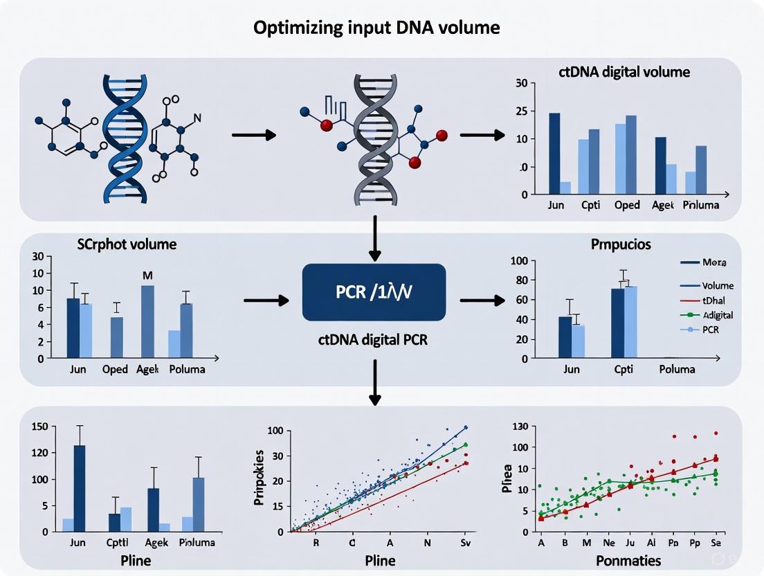
Optimizing Input DNA Volume for ctDNA Digital PCR: A Guide for Enhanced Sensitivity in Cancer Research
This article provides a comprehensive guide for researchers and drug development professionals on optimizing input DNA volume for circulating tumor DNA (ctDNA) digital PCR (dPCR).
Optimizing Input DNA Volume for ctDNA Digital PCR: A Guide for Enhanced Sensitivity in Cancer Research
Abstract
This article provides a comprehensive guide for researchers and drug development professionals on optimizing input DNA volume for circulating tumor DNA (ctDNA) digital PCR (dPCR). Effective input DNA volume is a critical pre-analytical factor that directly impacts the sensitivity, accuracy, and reliability of liquid biopsy assays for cancer detection and monitoring. We explore the foundational principles of ctDNA biology and dPCR technology, detail methodological approaches for volume optimization, address common troubleshooting and optimization challenges, and discuss validation strategies against other technologies. The goal is to establish best practices for input DNA volume to maximize the detection of low-frequency mutations, crucial for applications in early cancer detection, minimal residual disease (MRD) assessment, and therapy monitoring.
The Fundamentals of ctDNA and Digital PCR: Why Input Volume Matters
Frequently Asked Questions (FAQs)
FAQ 1: What are the core characteristics of ctDNA that impact its detection? Circulating tumor DNA (ctDNA) is a subset of cell-free DNA (cfDNA) released into the bloodstream by tumor cells through processes including apoptosis, necrosis, and active release [1] [2]. Its key characteristics are:
- Short Half-Life: ctDNA clears from circulation rapidly, with a half-life ranging from 16 minutes to 2.5 hours, making it suitable for real-time monitoring [2].
- Low Abundance: In early-stage cancer patients, ctDNA can constitute as little as 0.01% - 1.0% of the total cfDNA, which is predominantly derived from normal hematopoietic cells [3] [4] [2].
- Fragment Size: ctDNA fragments are typically short, ranging from 70–200 base pairs, and are often shorter than non-tumor cfDNA fragments [1] [3].
FAQ 2: What are the most critical pre-analytical steps to ensure reliable ctDNA detection? The pre-analytical phase is fundamental to the success of ctDNA testing, as it directly impacts the quality, integrity, and quantity of the extracted DNA [5]. Key steps include:
- Blood Collection: A minimum of 10 mL of blood is recommended, collected using butterfly needles to reduce shear stress [5]. Plasma is the preferred sample type over serum, as serum can have higher cfDNA concentrations due to leukocyte lysis during clotting, which increases background DNA [5] [6].
- Collection Tubes: The choice of blood collection tubes is critical.
- Centrifugation: A two-step protocol is recommended: an initial low-speed spin (800–1,900 g for 10 min) to separate plasma, followed by a high-speed spin (14,000–16,000 g for 10 min) to remove residual debris and platelets [5] [6].
- Storage: Plasma should be aliquoted and stored immediately at -80 °C to prevent nucleic acid degradation. Avoid more than three freeze-thaw cycles [5] [6].
FAQ 3: How does input DNA volume and quality affect digital PCR results for low-frequency variants? The quantity and quality of input ctDNA are critical determinants for the sensitivity of digital PCR (dPCR) assays [5].
- Sample Volume: For routine cfDNA applications, extracting DNA from at least 4 mL of plasma is recommended. However, for minimal residual disease (MRD) detection or in early-stage disease where ctDNA fraction is extremely low, higher plasma volumes (8–20 mL) are required to ensure a sufficient number of genome copies are analyzed [5].
- Tumor Fraction Assessment: Accurately quantifying the tumor fraction (TF) in the total cfDNA is essential, particularly when reporting negative results. A negative result may be a false negative if the tumor-derived ctDNA in the sample is insufficient [5]. TF can be estimated from the variant allele frequency (VAF) of somatic mutations or through copy number variations [5].
- Impact on Sensitivity: The extremely low concentration and short fragment length of ctDNA demand highly sensitive detection methods. dPCR is well-suited for this as it can detect mutant allele frequencies as low as 0.1% by partitioning samples into thousands of individual reactions [7] [2].
FAQ 4: What are the main technological approaches for ctDNA detection, and when should they be used? ctDNA detection methods can be broadly divided into targeted and untargeted (or hypothesis-free) approaches [6]. The choice depends on the clinical or research question.
Table 1: Comparison of Key ctDNA Detection Technologies
| Technology | Key Principle | Sensitivity | Best Use Cases | Key Limitations |
|---|---|---|---|---|
| Digital PCR (dPCR/ddPCR) [4] [7] [2] | Partitions sample into thousands of nano-reactions for absolute quantification of known mutations. | ~0.1% VAF (can be lower with barcoding) | - Monitoring known mutations/MRD [8]- Longitudinal therapy response [4] | - Limited to known, pre-defined targets- Low multiplexing capability |
| BEAMing [2] | Combines dPCR with magnetic beads and flow cytometry for highly sensitive detection. | ~0.02% VAF | - Ultra-sensitive detection of rare variants | - Complex workflow- Limited multiplexing |
| Next-Generation Sequencing (NGS) [9] [1] | High-throughput parallel sequencing of multiple genomic regions. | Varies; ~0.1%-1% with error suppression | - Untargeted discovery- Assessing tumor heterogeneity- Large gene panel screening | - Higher cost and longer turnaround- Requires complex bioinformatics |
| CAPP-Seq [2] | Targeted NGS method using selector probes to enrich for recurrently mutated regions in cancer. | High sensitivity with error correction | - Cost-effective targeted profiling- Sensitivity-focused NGS applications | - Requires design of selector probes |
Troubleshooting Common Experimental Issues
Problem: Inconsistent or low yield of ctDNA from plasma.
- Potential Cause 1: Improper blood processing or use of inappropriate collection tubes leading to white blood cell lysis and contamination with genomic DNA [5] [6].
- Solution: Use specialized cfDNA BCTs if processing delays are expected. For EDTA tubes, ensure plasma separation via two-step centrifugation is completed within 2-4 hours of blood draw [5] [6].
- Potential Cause 2: Inefficient DNA extraction method.
- Solution: Validate the extraction kit for your specific application. Studies have shown that silica-based manual kits (e.g., QIAamp Circulating Nucleic Acid Kit) and magnetic bead-based automated systems can provide high cfDNA recovery rates, though performance may vary [5]. Magnetic bead-based systems are particularly efficient at recovering smaller DNA fragments typical of ctDNA [6].
Problem: High background noise or false positives in dPCR/NGS.
- Potential Cause 1: Errors introduced during library preparation (for NGS) or non-specific amplification [1] [2].
- Solution: For NGS, employ error-suppression strategies such as molecular barcoding (unique molecular identifiers, UMIs) to distinguish true somatic mutations from PCR or sequencing errors [1] [2]. For dPCR, optimize probe design and annealing temperatures.
- Potential Cause 2: Detection of clonal hematopoiesis of indeterminate potential (CHIP). CHIP are age-related mutations in blood cells that are not of tumor origin [2].
- Solution: When possible, compare ctDNA variants with a matched normal sample (e.g., peripheral blood mononuclear cells) to filter out CHIP-related mutations [2].
Problem: Failure to detect ctDNA in patients with confirmed cancer.
- Potential Cause: The tumor may be a "low-shedder," or the tumor fraction in the blood is below the limit of detection (LOD) of the assay [1].
- Solution:
- Increase Plasma Input: Process a larger volume of plasma (e.g., 8-20 mL) to increase the number of template molecules for analysis [5].
- Use Ultra-Sensitive Assays: Employ more sensitive technologies like BEAMing dPCR or use NGS methods with molecular barcoding that can achieve a lower LOD [2].
- Multi-Modal Analysis: Supplement mutation analysis with other features like methylation status or fragmentomics (fragmentation patterns), which can provide an alternative signal for tumor detection [1] [10].
Essential Research Reagent Solutions
Table 2: Key Materials and Reagents for ctDNA Analysis
| Item | Function | Examples & Notes |
|---|---|---|
| Blood Collection Tubes | Preserves cfDNA and prevents white blood cell lysis during transport/storage. | Streck, PAXgene, Cell-Free DNA BCT [5] [6] |
| Nucleic Acid Extraction Kits | Isolates and purifies cfDNA from plasma. | Silica-membrane columns (QIAamp CNA Kit) or magnetic bead-based automated systems (Maxwell, QIAsymphony) [5] |
| Digital PCR Systems | Absolute quantification of rare mutations in ctDNA. | QuantStudio Absolute Q with pre-designed TaqMan Liquid Biopsy Assays for known hotspots [7] |
| Next-Generation Sequencers | Broad, hypothesis-free profiling of mutations and other genomic alterations. | Platforms enabling high-depth, targeted sequencing; requires molecular barcoding kits for error correction [9] [1] |
| DNA Quantification Kits | Accurately measure the concentration and quality of extracted cfDNA. | Fluorometric methods (e.g., Qubit dsDNA HS Assay); qPCR-based methods can assess amplifiable DNA [5] |
Workflow and Decision-Making Diagrams
The following diagram illustrates the complete workflow for ctDNA analysis, from sample collection to data interpretation, highlighting critical pre-analytical and analytical steps.
Figure 1: Comprehensive ctDNA Analysis Workflow.
This decision pathway helps researchers select the most appropriate detection technology based on their experimental goals and available resources.
Figure 2: Decision Pathway for ctDNA Detection Technology Selection.
Digital PCR (dPCR) is a powerful molecular biology technique that enables the absolute quantification of nucleic acids without the need for a standard curve. By partitioning a sample into thousands of individual reactions, dPCR allows for the precise detection and quantification of rare targets, making it particularly valuable for applications such as circulating tumor DNA (ctDNA) analysis in cancer research. This technical support center provides comprehensive troubleshooting guides and FAQs to assist researchers in optimizing their dPCR experiments, with a specific focus on input DNA volume for ctDNA research.
Core Principles of Digital PCR
Partitioning and Poisson Statistics
The fundamental principle of dPCR involves partitioning a PCR reaction into numerous individual partitions, such as microchambers or droplets. Partitioning is a critical step that allows for the detection of single molecules. According to Poisson statistics, when a sample is sufficiently diluted and randomly distributed, each partition will contain zero, one, or a few target molecules. After endpoint PCR amplification, partitions are analyzed as positive (containing the target) or negative (no target). The absolute quantity of the target in the original sample is then calculated using Poisson distribution models to account for the fact that some partitions may have contained more than one molecule [11].
This partitioning approach effectively enriches low-level targets and enables the detection of rare sequences with mutation allele frequencies (MAFs) as low as 0.1%, a level of sensitivity crucial for detecting ctDNA in liquid biopsies [7].
Absolute Quantification
Unlike quantitative real-time PCR (qPCR), dPCR provides absolute quantification without requiring a standard curve. Quantification is based on the binary readout (positive/negative) of thousands of individual partitions, allowing direct calculation of the target concentration in units of copies per microliter [7] [11]. This eliminates uncertainties associated with standard curve preparation and improves measurement accuracy and reproducibility.
Enhanced Sensitivity
The sensitivity of dPCR stems from its ability to detect very low fractions of mutant targets against a high background of wild-type sequences. This makes it a technology of choice for cancer researchers studying rare somatic mutations and for genetic disease researchers detecting low-frequency variants [7]. dPCR has demonstrated superior sensitivity compared to qPCR, particularly for detecting low bacterial loads in complex clinical samples [12].
Optimizing Input DNA for ctDNA dPCR Research
Calculating Optimal Input DNA
Determining the correct input DNA amount is crucial for accurate dPCR results. The optimal template concentration should yield an average of 0.5 to 3 copies per partition to remain within the "digital range" [13]. Excessive template can lead to multiple targets per partition, violating Poisson statistics and causing quantification inaccuracies.
For human genomic DNA, the copy number calculation is based on genome size. The haploid human genome is approximately 3.3 × 10^9 bp, with a mass of about 3.3 pg per diploid copy [13]. The table below provides copy number calculations for 10 ng of gDNA from various organisms:
| Organism | Genome Size (bp) | Gene Copies in 10 ng gDNA |
|---|---|---|
| Homo sapiens | 3.3 × 10^9 | 3,000 |
| Zebrafish | 1.7 × 10^9 | 5,400 |
| Saccharomyces cerevisiae | 1.2 × 10^7 | 760,500 |
| Escherichia coli | 4.6 × 10^6 | 2,000,000 |
| Standard plasmid DNA | 3.5 × 10^3 | 2,600,000,000 |
When preparing samples, researchers must account for all dilution factors. For example, if adding 1 µL of a sample diluted 1:10 from stock to a 16 µL reaction, the total dilution factor is 0.00625 (1:160). Properly accounting for these dilutions in analysis software ensures accurate calculation of the target concentration in the starting material [14].
Special Considerations for ctDNA
ctDNA fragments are typically short and present in very low concentrations in blood samples. The sensitivity of dPCR makes it an ideal tool for ctDNA studies, enabling researchers to detect cancer early, measure therapeutic response, quantify residual tumor burden, and monitor emerging resistance to therapies [7]. Studies have shown that ctDNA is present at significantly higher levels in cerebrospinal fluid compared to plasma, highlighting the importance of sample selection [15].
Diagram 1: dPCR Workflow for ctDNA Analysis
Troubleshooting Common dPCR Issues
FAQ: Addressing Specific Experimental Challenges
Q: What are the primary causes of poor sensitivity in ctDNA dPCR assays?
A: Poor sensitivity can result from several factors:
- Insufficient template input: For rare targets like ctDNA, ensure adequate sample volume is processed to capture sufficient mutant molecules.
- Suboptimal partitioning: Verify partition quality and integrity. Incomplete partitioning reduces effective reactions.
- Inhibitors in sample: Substances like heparin, hemoglobin, or ionic detergents can inhibit polymerase activity. Additional purification steps or sample dilution may be necessary.
- Poor primer/probe efficiency: Redesign suboptimal assays and validate efficiency. Higher primer (0.5-0.9 µM) and probe (0.25 µM) concentrations than qPCR are often needed in dPCR to increase fluorescence intensity for better cluster separation [13].
Q: How can I resolve quantification inaccuracies in my dPCR experiments?
A: Quantification issues often relate to:
- Template overloading: Maintain template concentration between 0.5-3 copies per partition to stay within the optimal digital range. Excessive template leads to underestimation of concentration [13] [14].
- Incorrect dilution factor calculation: Account for all dilution steps when calculating final concentration. Software like AnalysisSuite requires accurate dilution factors to report correct stock concentrations [14].
- Poor partition quality: Ensure uniform partition size and integrity. For droplet-based systems, check droplet generator performance and oil quality.
Q: What sample preparation issues most commonly affect dPCR results?
A: Common sample issues include:
- Sample purity: Contaminants like salts, EDTA, alcohols, proteins, and chaotropic agents can inhibit PCR. Humic acids can quench fluorescence in dsDNA-binding dyes like EvaGreen. Use high-purity nucleic acid extraction methods and consider additional purification if needed [13].
- Sample integrity: Degraded templates (common in FFPE samples or cfDNA) may require larger input amounts. Keep amplicons short (especially for degraded samples) and use restriction digestion for complex templates [13].
- Inappropriate template structure: For high-molecular-weight DNA, linked gene copies, or supercoiled plasmids, use restriction digestion to ensure random distribution. Select enzymes that do not cut within the amplicon sequence [13].
Essential Research Reagent Solutions
The table below outlines key reagents and materials essential for successful dPCR experiments, particularly in ctDNA research:
| Reagent/Material | Function | Application Notes |
|---|---|---|
| Digital PCR Master Mix | Provides optimized buffer, dNTPs, and polymerase for partitioning and amplification | Use hot-start enzymes to prevent non-specific amplification; select compatible with your detection system [16] |
| TaqMan Probe Assays | Sequence-specific detection with fluorophore-quencher system | Pre-designed liquid biopsy assays available; higher concentrations (0.25 µM) often needed vs. qPCR [7] [13] |
| Partitioning Oil/Matrix | Creates stable partitions for reaction segregation | Quality critical for partition integrity; ensure compatibility with your dPCR system |
| cfDNA Extraction Kits | Isolves cell-free DNA from plasma, CSF, or other biofluids | Use specialized kits (e.g., QIAamp circulating nucleic acid kit) for optimal cfDNA recovery [15] |
| Restriction Enzymes | Digests complex DNA structures for even distribution | Essential for high-molecular-weight DNA, linked copies, or supercoiled plasmids; avoid cutting within amplicon [13] |
Advanced Methodologies for ctDNA Detection
Droplet Digital PCR for ctDNA Analysis
Droplet digital PCR (ddPCR) has been successfully applied to detect tumor-specific mutations in ctDNA from various biofluid sources. The methodology typically involves:
Sample Collection: Collect blood in specialized cell-free DNA collection tubes (e.g., Streck tubes) to prevent genomic DNA contamination and preserve ctDNA integrity [15].
Plasma Processing: Two-step centrifugation (first at 1,600 × g, then at up to 16,000 × g) to remove cellular components and debris [15].
cfDNA Extraction: Use commercial circulating nucleic acid kits (e.g., QIAamp circulating nucleic acid kit) for optimal recovery of short cfDNA fragments [15].
ddPCR Assay Setup: Custom TaqMan-based genotyping assays designed against known tumor mutations, with validation using serial dilutions of mutant DNA in wild-type DNA [15].
Partitioning and Amplification: Generate droplets followed by endpoint PCR amplification (typically 40-45 cycles) [15].
Data Analysis: Quantify mutant and wild-type alleles using Poisson statistics, with sensitivity down to 0.1% variant allele frequency achievable in optimized assays [7].
Comparison with Quantitative NGS
While dPCR offers exceptional sensitivity for detecting known mutations, quantitative next-generation sequencing (qNGS) methods are emerging that enable absolute quantification of multiple variants simultaneously without prior knowledge of tumor genetics. These approaches incorporate unique molecular identifiers (UMIs) and quantification standards (QSs) to address the semi-quantitative limitations of conventional NGS [17]. For focused analysis of specific mutations in ctDNA monitoring, dPCR remains the gold standard due to its sensitivity, precision, and relatively simple workflow.
Diagram 2: dPCR Troubleshooting Guide
Digital PCR provides researchers with a powerful tool for absolute quantification of nucleic acids, offering exceptional sensitivity for detecting rare targets like ctDNA mutations. Understanding the core principles of partitioning, Poisson statistics, and optimal input DNA volume is essential for successful experimental outcomes. By addressing common troubleshooting scenarios and implementing optimized methodologies, researchers can leverage the full potential of dPCR in cancer research and drug development programs. The continued refinement of dPCR technologies and methodologies promises to further enhance its applications in precision oncology and liquid biopsy development.
Frequently Asked Questions (FAQs)
FAQ 1: Why is input DNA volume so critical for ctDNA dPCR assay sensitivity? The input DNA volume directly determines the number of haploid genome equivalents (GEs) analyzed, which is the fundamental factor limiting the detection of rare mutant molecules. Circulating tumor DNA (ctDNA) fragments often exist at very low variant allele frequencies (VAFs), sometimes below 0.1% [18]. If the total number of DNA fragments in a sample is too low, the specific mutant molecules may be so scarce that their detection becomes statistically improbable [18]. For example, a sample with only 8,000 total GEs and a ctDNA fraction of 0.1% contains merely 8 mutant GEs, making reliable detection unlikely even with a highly sensitive dPCR assay [18].
FAQ 2: How do I calculate the required input DNA volume for my experiment? First, quantify your extracted cell-free DNA (cfDNA) concentration. The required input is then calculated based on the number of genome equivalents needed for your desired sensitivity. Use the formula that 1 ng of human genomic DNA corresponds to approximately 300 haploid genome equivalents [18] [13]. The table below shows copy numbers from a standard input of 10 ng of gDNA [13]:
| Organism | Genome Size | Gene Copies in 10 ng gDNA |
|---|---|---|
| Homo sapiens | 3.3x10⁹ bp | 3000 |
| Escherichia coli | 4.6x10⁶ bp | 2,000,000 |
For dPCR, the average number of copies per partition should ideally be between 0.5 and 3 to ensure optimal partitioning and accurate quantification [13].
FAQ 3: What are the consequences of using insufficient input DNA?
- Increased False Negatives: The primary risk is failing to detect a low-frequency mutant present in the patient's plasma due to an inadequate number of mutant GEs in the analyzed sample [18] [15].
- Reduced Precision and Accuracy: Low input amounts can lead to higher quantification variability and less reliable results, especially for low-abundance targets [13] [19].
- Poor Assay Performance: Reactions with very low DNA input may exhibit reduced PCR efficiency, lower fluorescence amplitude, and impaired separation between positive and negative partitions [20] [13].
FAQ 4: My sample volume is limited and I cannot obtain sufficient DNA. What are my options?
- Maximize Sample Collection: Ensure you collect an adequate volume of blood. Studies have shown that using mean plasma volumes as low as 0.49 mL can severely limit utility, while larger volumes (e.g., 10 mL) are recommended [15].
- Alternative Biofluids: For certain cancers, such as brain tumors, cerebrospinal fluid (CSF) has been shown to contain significantly higher levels of ctDNA than plasma and may be a more informative biofluid when available [15].
- Technical Replicates: If the total sample volume is low, running analytical replicates (duplicate or triplicate) can help pool data and increase the effective number of measured events, improving precision [13].
Troubleshooting Common Input DNA Issues
Problem: Inconsistent or Erratic dPCR Results
| Possible Cause | Recommendation |
|---|---|
| Low sample purity with PCR inhibitors (e.g., salts, EDTA, alcohols, phenol). | Re-purify DNA, precipitate with 70% ethanol, or use inhibitor-tolerant polymerases. Ensure high nucleic acid purity for optimal fluorescence detection [20] [13]. |
| Sample integrity issues (e.g., degraded DNA, residual crosslinks from FFPE). | Keep amplicons short for degraded samples like cfDNA. Use dedicated kits for FFPE DNA recovery to repair damage and improve quality [13]. |
| Pipetting inaccuracies with low-volume samples. | Use calibrated pipettes and master mixes. Perform reactions in duplicate or triplicate to mitigate the impact of pipetting errors [13]. |
| Inaccurate DNA quantification prior to dPCR setup. | Use fluorometric methods (e.g., Qubit) for quantifying cfDNA, as they are more accurate for low-concentration and fragmented DNA compared to spectrophotometry [15]. |
Problem: Failing to Detect Known Low-Frequency Variants
This problem often stems from the absolute limit of detection dictated by input mass. If a 10 mL blood draw from a lung cancer patient yields only ~8,000 GEs, a 0.1% VAF mutant is represented by only ~8 molecules [18]. Detection is statistically challenging.
- Solution: The most direct solution is to increase the input DNA mass. This may require larger blood collection volumes (e.g., 10 mL into cell-free DNA collection tubes) or more efficient cfDNA extraction methods to maximize yield from the available plasma [15].
Experimental Protocols & Data Presentation
Protocol: Determining the Limit of Detection (LoD) for a ctDNA dPCR Assay
This protocol outlines how to empirically establish the LoD, considering input DNA volume.
- Prepare DNA Dilutions: Serially dilute a known mutant DNA (e.g., synthetic DNA or cell line DNA) into a background of wild-type DNA (e.g., from healthy donor plasma) to create samples with mutant allele frequencies ranging from 1:10 to 1:10,000 (0.01%) [15].
- Use Constant Input Mass: Use a fixed, high-input mass of DNA (e.g., 5-10 ng per reaction) for each dilution point to ensure the number of GEs is not the limiting factor during validation [15].
- Run Replicates: Perform each dilution point in a minimum of duplicate reactions to assess reproducibility and precision [13].
- Include Controls: Run negative controls (wild-type DNA only) and non-template controls (NTCs) to monitor for false positives and contamination [13].
- Data Analysis: Calculate the mean and standard deviation of the measured mutant copies for each dilution. The LoD is the lowest mutant allele frequency at which the mutant signal is consistently and significantly distinguishable from the negative controls with a high degree of confidence (e.g., >95%) [19].
Quantitative Data: The Relationship Between Input, Coverage, and Detection
The following table summarizes key quantitative relationships derived from NGS principles that underscore the importance of input DNA in detecting low-frequency variants [18].
| Target VAF | Approx. Depth of Coverage Required for 99% Detection Probability | ~Equivalent Input DNA* |
|---|---|---|
| 1.0% | 1,000x | ~3.3 ng |
| 0.5% | 2,000x | ~6.6 ng |
| 0.1% | 10,000x | ~33 ng |
*Calculation based on ~300 Haploid Genome Equivalents (GEs) per ng of human DNA [18] [13].
The Scientist's Toolkit: Essential Reagents and Materials
| Item | Function in ctDNA dPCR |
|---|---|
| Cell-Free DNA Blood Collection Tubes (e.g., Streck) | Preserves blood sample integrity by preventing white blood cell lysis and release of genomic DNA during transport and storage, stabilizing the native cfDNA population [15]. |
| cfDNA Extraction Kit (e.g., QIAamp Circulating Nucleic Acid Kit) | Specialized silica-membrane technology optimized for efficient isolation of short, low-concentration cfDNA fragments from large-volume plasma samples [15]. |
| Fluorometer (e.g., Qubit with dsDNA HS Assay) | Accurately quantifies the low concentrations of double-stranded cfDNA obtained from plasma, which is critical for calculating input GEs [15]. |
| Digital PCR System & Reagents | Platform (e.g., droplet-based or nanoplate-based systems) and associated master mixes containing DNA polymerase, dNTPs, and buffers tailored for robust amplification from fragmented cfDNA templates [15] [13]. |
| TaqMan Probe-based Assays | Sequence-specific hydrolysis probes labeled with a fluorophore and quencher provide high specificity for discriminating mutant from wild-type sequences, which is essential for low-VAF detection [15] [13]. |
The reliable detection of circulating tumor DNA (ctDNA) using digital PCR (dPCR) is fundamentally dependent on the quality of the pre-analytical phase. For researchers in drug development and cancer diagnostics, variations in blood collection, processing, and storage can significantly impact the yield, quality, and subsequent analysis of cell-free DNA (cfDNA), potentially leading to false negatives or inaccurate quantification. This technical support center provides troubleshooting guides and FAQs, framed within the broader thesis of optimizing pre-analytical workflows to ensure the integrity of input DNA for ctDNA dPCR research. Standardizing these initial steps is crucial for achieving reproducible, sensitive, and reliable results in liquid biopsy applications.
Troubleshooting Guides & FAQs
Frequently Asked Questions
FAQ 1: What is the maximum time delay allowed for processing blood samples collected in standard K3EDTA tubes?
- Answer: For K3EDTA tubes, plasma should be separated as soon as possible, ideally within 1 to 6 hours of venipuncture [21]. If immediate processing is not feasible, storing the blood at 4°C can help stabilize the sample for a short period. However, studies show that cfDNA levels in K3EDTA tubes increase gradually with time at room temperature due to leukocyte lysis, which dilutes the mutant allele fraction and can compromise the detection of low-abundance ctDNA [22] [23].
FAQ 2: We need to ship samples from a clinical site to our central lab. What is the recommended collection method?
- Answer: For studies involving sample shipment or delays in processing, cell-stabilizing blood collection tubes (BCTs), such as Cell-free DNA BCT (Streck), are strongly recommended. These tubes contain a preservative that stabilizes nucleated blood cells, preventing the release of genomic DNA and maintaining the integrity of the ctDNA profile. Research has demonstrated that cfDNA levels remain stable in BCTs for up to 3 to 7 days at ambient temperatures, making them ideal for multi-center trials [22] [21].
FAQ 3: Our centrifugation protocol is inconsistent across sites. What is a validated, double-centrifugation protocol for obtaining cell-free plasma?
Answer: A validated double-centrifugation protocol is critical to remove cells and cellular debris. The following protocol is widely cited and can be adapted based on equipment availability. The key is consistency across all samples.
- Protocol A (from [22]):
- First Spin: 820 × g for 10 minutes at room temperature. Carefully transfer the supernatant (plasma) to a new tube, avoiding the buffy coat.
- Second Spin: 14,000 × g for 10 minutes at room temperature. Transfer the final supernatant (cell-free plasma) to a new tube for storage or DNA extraction.
- Alternative Protocol (from [22]): A second spin at 3,000 × g for 10 minutes has been shown to yield similar cfDNA results as higher-speed centrifugation, which may be a consideration for labs without high-speed micro-centrifuges.
- Protocol A (from [22]):
FAQ 4: How can we improve the recovery of fragmented cfDNA, especially from early-stage cancer patients with low ctDNA levels?
- Answer: The choice of DNA extraction method significantly impacts recovery. While silica-membrane columns (e.g., QIAamp Circulating Nucleic Acid Kit) are common, novel liquid-phase extraction methods have demonstrated superior recovery of cfDNA. One study comparing seven kits found that the PHASIFY MAX method (Phase Scientific) recovered 91% more of a 145 bp dsDNA fragment than a common solid-phase method, leading to a 171% increase in mutant copy recovery and the positive conversion of samples previously deemed negative [24].
Troubleshooting Common Problems
Problem: High and variable wild-type cfDNA background, obscuring ctDNA detection.
- Potential Cause: Inadequate sample processing leading to contamination from genomic DNA of lysed white blood cells.
- Solutions:
- Switch Tubes: Use cell-stabilizing BCTs instead of K3EDTA, especially if processing delays are anticipated [22] [23].
- Control Temperature: If using K3EDTA, store blood at 4°C and process within 6 hours [22].
- Optimize Centrifugation: Ensure the double-centrifugation protocol is strictly followed to completely remove cellular debris [22] [25].
Problem: Low overall cfDNA yield from plasma samples.
- Potential Cause: Inefficient DNA extraction or loss of small DNA fragments during isolation.
- Solutions:
- Evaluate Kits: Compare the performance of your current extraction kit against alternatives. Liquid-phase extraction (e.g., PHASIFY) or kits specifically optimized for small fragments (e.g., Quick cfDNA Serum & Plasma Kit) may offer higher yields [26] [24].
- Increase Input Volume: If yield is consistently low, consider increasing the starting volume of plasma for extraction, provided it is within the kit's specifications.
- Verify Protocols: Ensure that no steps in the extraction protocol, such as washing or elution, are leading to inadvertent DNA loss.
Problem: Inconsistent dPCR results between sample batches.
- Potential Cause: Pre-analytical variability in sample handling, storage, or extraction.
- Solutions:
- Standardize SOPs: Implement and rigorously adhere to Standard Operating Procedures (SOPs) for every step from phlebotomy to DNA elution.
- Implement QC Measures: Introduce a quality control step for extracted cfDNA. A multiplex ddPCR assay that assesses cfDNA concentration and fragment size can identify suboptimal samples before proceeding to costly dPCR assays [27] [25].
- Audit Storage Conditions: Ensure plasma is stored at -80°C and that freeze-thaw cycles are minimized.
Summarized Data for Experimental Planning
Table 1: Impact of Blood Collection Tubes and Processing Delays on cfDNA
The following table summarizes key quantitative findings from the literature on how collection devices and processing delays affect cfDNA levels, which is critical for designing robust experiments.
| Collection Tube | Processing Delay | Storage Temperature | Key Impact on cfDNA | Source |
|---|---|---|---|---|
| K3EDTA | 0 - 6 hours | Room Temperature | Baseline cfDNA levels | [22] |
| K3EDTA | 24 - 96 hours | Room Temperature | Gradual increase in cfDNA levels due to cell lysis | [22] [23] |
| K3EDTA | 24 - 96 hours | 4°C | Less variation than RT, but levels still elevated compared to BCT | [22] |
| Cell-free DNA BCT | Up to 24 hours | Room Temperature | cfDNA levels remain stable, no significant increase | [22] [25] |
| Cell-free DNA BCT | Up to 72 hours / 1 week | Room Temperature | cfDNA levels remain stable, no significant difference in yield or fragment size | [22] [25] [21] |
Table 2: Comparison of Centrifugation Protocols for Plasma Preparation
This table compares different centrifugation protocols evaluated in published studies, aiding in the selection and standardization of this critical step.
| Protocol Name | First Centrifugation | Second Centrifugation | Reported Outcome | Source |
|---|---|---|---|---|
| Protocol A | 820 × g for 10 min | 14,000 × g for 10 min | Standard protocol used for comparative studies | [22] |
| Protocol B | 1,600 × g for 10 min | 14,000 × g for 10 min | Comparable cfDNA yield to other protocols | [22] |
| Protocol C | 1,600 × g for 10 min | 3,000 × g for 10 min | Similar cfDNA yields compared to higher-speed protocols | [22] |
| Streck BCT Protocol | 1,600 × g for 10 min | 3,000 × g for 10 min | Recommended protocol for specific tube types | [28] |
Essential Experimental Protocols
Protocol 1: Plasma Separation from Whole Blood Using Double Centrifugation
This is a detailed methodology for obtaining high-quality, cell-free plasma from blood drawn in K3EDTA or BCTs, based on protocols from [22] and [28].
Materials:
- Whole blood in K3EDTA or Cell-free DNA BCT.
- Centrifuge capable of swing-bucket rotor (for first spin).
- Micro-centrifuge (for second spin).
- Sterile polypropylene tubes (e.g., 15 mL and 2 mL).
Procedure:
- First Centrifugation (to separate plasma from cells):
- Centrifuge whole blood tubes at 1,600 - 2,000 × g for 10 minutes at room temperature.
- Using a sterile pipette, carefully transfer the supernatant (plasma) to a new 15 mL tube. Critical Step: Avoid disturbing the buffy coat (the white layer of white blood cells) to prevent genomic DNA contamination. Leave about 0.5 cm of plasma above the buffy coat.
- Second Centrifugation (to remove residual cells and platelets):
- Transfer the harvested plasma to 2 mL microcentrifuge tubes.
- Centrifuge at 14,000 × g for 10 minutes at room temperature. Alternatively, a force of 3,000 × g for 10 minutes can be used effectively [22].
- Carefully pool the resulting cell-free plasma into a fresh tube, again avoiding the pellet at the bottom.
- Storage:
- Aliquot the cell-free plasma to avoid freeze-thaw cycles.
- Store at -80°C until DNA extraction.
- First Centrifugation (to separate plasma from cells):
Protocol 2: Assessing cfDNA Quality and Quantity using Multiplex ddPCR
This protocol, adapted from [27] and [25], describes a quality control assay to evaluate cfDNA extracts before downstream dPCR analysis.
Principle: A multiplex ddPCR assay that simultaneously amplifies short and long genomic targets to estimate total amplifiable cfDNA concentration and assess the degree of high molecular weight (gDNA) contamination based on fragment size distribution.
Materials:
- Extracted cfDNA.
- ddPCR Supermix for Probes (No dUTP).
- Primer/Probe mix for short amplicon targets (e.g., ~70 bp, FAM-labeled).
- Primer/Probe mix for long amplicon targets (e.g., ~400 bp, HEX/VIC-labeled).
- Droplet generator, reader, and consumables.
Procedure:
- Prepare the ddPCR reaction mix according to manufacturer's instructions, including the multiplexed primers and probes.
- Generate droplets.
- Perform PCR amplification with a standardized thermal cycling protocol.
- Read the plate on the droplet reader.
- Analysis:
- The concentration of short amplicons reflects the total amplifiable cfDNA.
- The ratio of short to long amplicons indicates the fragment size profile. A high ratio suggests a sample enriched for short, mononucleosomal cfDNA, while a low ratio suggests gDNA contamination [27] [25].
- Samples with significant gDNA contamination can be flagged or excluded from sensitive ctDNA assays.
Workflow Visualization
The following diagram illustrates the critical decision points in the pre-analytical workflow and their impact on DNA yield for ctDNA research.
The Scientist's Toolkit: Research Reagent Solutions
Table 3: Key Materials and Kits for Pre-analytical Workflow
| Item Category | Example Product | Primary Function in Workflow | Key Consideration |
|---|---|---|---|
| Blood Collection Tubes | Cell-free DNA BCT (Streck) | Stabilizes blood cells for up to 7 days at room temperature, preventing gDNA release. | Essential for multi-site trials or when processing delays are unavoidable. [22] [21] |
| cfDNA Extraction Kits | QIAamp Circulating Nucleic Acid Kit (Qiagen) | Solid-phase extraction of cfDNA using a silica membrane. | Common industry standard; provides consistent results. [26] [22] |
| cfDNA Extraction Kits | Quick cfDNA Serum & Plasma Kit (Zymo Research) | Solid-phase extraction designed for serum and plasma. | Cited as yielding high cfDNA concentrations. [26] |
| cfDNA Extraction Kits | PHASIFY MAX / ENRICH (Phase Scientific) | Liquid-phase extraction using aqueous two-phase systems (ATPS). | Demonstrates significantly higher cfDNA and mutant copy recovery vs. solid-phase. [24] |
| Quality Control Assay | Multiplex ddPCR Size Assay | Quantifies amplifiable cfDNA and assesses fragment size/profile to detect gDNA contamination. | Critical for pre-screening samples to ensure dPCR assay validity. [27] [25] |
Strategic Approaches to Input DNA Volume Optimization in dPCR Workflows
## Frequently Asked Questions (FAQs)
FAQ 1: Why is plasma volume so critical for detecting ctDNA, especially in low-fraction scenarios? The concentration of ctDNA in a patient's blood can be extremely low, often constituting less than 0.1% of the total cell-free DNA (cfDNA), particularly in early-stage disease or minimal residual disease (MRD) [29] [2] [30]. The absolute quantity of mutant molecules available for analysis is directly proportional to the plasma volume processed [31] [15]. Using a larger plasma volume increases the number of tumor DNA molecules in the input material, thereby improving the statistical confidence and sensitivity of the assay for detecting rare mutations [31].
FAQ 2: What is the minimum plasma volume recommended for a reliable ddPCR assay? While the optimal volume is context-dependent, several studies indicate that volumes of 2-4 mL of plasma are often used and provide a practical starting point [30] [32]. One study explicitly highlighted limitations when using a mean plasma volume of only 0.49 mL, suggesting this is generally insufficient for reliable detection in plasma from patients with certain cancers [15]. For applications requiring the highest sensitivity, such as MRD detection, collecting additional blood tubes is recommended to increase the total plasma volume available [31].
FAQ 3: How does plasma volume relate to input DNA and the limit of detection (LOD) in digital PCR? Digital PCR assays are fundamentally dependent on the absolute number of DNA molecules partitioned. The relationship between plasma volume and LOD can be summarized as follows:
- Absolute Input: The total amount of cfDNA extracted is a function of the plasma volume and its cfDNA concentration (typically 0-100 ng/mL in healthy individuals, and higher in cancer patients) [29] [2].
- LOD Calculation: The theoretical LOD is a function of the total number of genome equivalents analyzed. For example, 20 ng of input cfDNA is equivalent to approximately 6,000 copies of the haploid human genome, which theoretically allows for the detection of a mutant allele at a frequency as low as 0.02% (i.e., 1 mutant molecule in 6,000 wild-type molecules) [30].
- Volume Requirement: To achieve this 20 ng input, the required plasma volume will vary based on the individual's cfDNA concentration. A patient with a low cfDNA concentration of 5 ng/mL would require 4 mL of plasma, whereas a patient with 10 ng/mL would require only 2 mL to obtain the same input mass and, therefore, the same theoretical LOD.
FAQ 4: What are the consequences of using an insufficient plasma volume? Insufficient plasma volume is a primary contributor to false-negative results [2]. If the volume is too low, the number of mutant DNA molecules present in the sample may be below the detection threshold of the assay, even if the tumor fraction in the patient's blood is theoretically detectable with a larger input. This can lead to an underestimation of tumor burden and incorrect clinical conclusions.
FAQ 5: How does sample type (e.g., plasma vs. CSF) influence volume requirements? The required volume is highly dependent on the biofluid's ctDNA concentration. For central nervous system tumors, cerebrospinal fluid (CSF) often contains significantly higher levels of ctDNA than plasma [15]. Consequently, a smaller volume of CSF (e.g., 1-2 mL) may be sufficient for detection, whereas a larger volume of plasma is needed for the same patient.
## Troubleshooting Guides
Problem: Consistently Low cfDNA Yield from Plasma
Potential Cause: The initial volume of plasma processed is too low, or the plasma separation protocol is suboptimal, leading to cellular contamination.
Solutions:
- Increase Plasma Volume: Standardize sample collection to obtain a larger volume of blood, thereby yielding more plasma. For high-sensitivity applications, plan for processing ≥4 mL of plasma per extraction [30].
- Optimize Blood Collection and Processing:
- Use blood collection tubes containing K2- or K3-EDTA and process plasma within 4-6 hours of draw to prevent leukocyte lysis and contamination of the cfDNA with genomic DNA [31].
- If a longer processing delay is unavoidable, use specialized cell preservation tubes, which can stabilize blood for 5-7 days at room temperature [31].
- Employ a two-step centrifugation protocol: a first centrifugation at 800–1,600×g for 10 minutes at 4°C, followed by a second centrifugation of the supernatant at 14,000–16,000×g for 10 minutes at 4°C to ensure cell-free plasma [31].
Problem: High Variability in Mutation Quantification Between Replicates
Potential Cause: Stochastic sampling error due to a low absolute number of target mutant molecules in the reaction.
Solutions:
- Input More DNA: Increase the amount of cfDNA loaded into the ddPCR reaction by extracting from a larger volume of plasma. This increases the number of mutant molecules, improving quantification accuracy and reproducibility [30].
- Verify Input DNA Mass: Accurately quantify the extracted cfDNA using a fluorescence-based method (e.g., Qubit) rather than spectrophotometry to ensure a sufficient and consistent mass of DNA is used in each assay [32] [15].
Problem: Inability to Detect Low-Frequency Mutations (e.g., <0.1%) in Known Positive Samples
Potential Cause: The combination of plasma volume and assay sensitivity is insufficient for the very low tumor fraction.
Solutions:
- Maximize Plasma Input: Process the maximum feasible volume of plasma for DNA extraction to "fish in a bigger pond" for the rare mutant molecules [31] [30].
- Consider Pre-Amplification: For multi-target assays, a limited-cycle, non-selective pre-amplification step (e.g., 5-7 cycles using a method like TOP-PCR) can increase the amount of available template. However, this requires careful optimization and inclusion of negative controls, as it can introduce PCR errors [30].
- Re-evaluate Assay LOD: Re-validate the ddPCR assay's Limit of Detection (LoD) using serially diluted mutant DNA in a wild-type background with the same plasma volume and extraction protocol used for patient samples [15].
Table 1: Quantitative Guidelines for Plasma Volume and Input DNA
| Clinical / Analytical Context | Recommended Plasma Volume | Target Input cfDNA | Theoretical LOD (VAF) | Key Supporting Evidence |
|---|---|---|---|---|
| Minimal Residual Disease (MRD) / Ultra-Sensitive Detection | ≥ 4 mL [30] | ~20 ng [30] | ~0.02% [30] | 20 ng input provides ~6000 haploid genomes, enabling detection of a single mutant molecule. |
| Standard Mutation Detection & Monitoring | 2 - 4 mL [32] | 10 - 20 ng | 0.1% - 0.01% | Studies successfully detected TP53 mutations using ddPCR with 2 mL plasma inputs [32]. |
| Challenging Cases (e.g., CNS Tumors in Plasma) | ≥ 3 mL [15] | As much as possible | Varies | One study found 0.49 mL plasma inadequate; ≥3 mL recommended for reliable plasma-based detection in glioma [15]. |
Table 2: Key Reagents and Materials for Plasma Processing and cfDNA Analysis
| Item | Function / Description | Example Product / Note |
|---|---|---|
| Cell-Free DNA Blood Collection Tubes | Preserves blood cells and stabilizes cfDNA for up to 5-7 days at room temperature, critical for multi-center trials. | Streck Cell-Free DNA BCT, PAXgene Blood ccfDNA Tubes [31] |
| K2/K3-EDTA Blood Collection Tubes | Standard anticoagulant tube; requires plasma separation within 4-6 hours to prevent gDNA release from lysed cells. | BD Vacutainer K2EDTA [31] [32] |
| cfDNA Extraction Kit | Optimized for efficient recovery of short, fragmented cfDNA from plasma. | QIAamp Circulating Nucleic Acid Kit [30] [32] [15] |
| Fluorometric DNA Quantification Assay | Accurately quantifies double-stranded DNA concentration in low-concentration samples. Essential for standardizing input. | Qubit dsDNA HS Assay Kit [30] [32] [15] |
| Droplet Digital PCR System | Platform for partitioning samples and performing absolute quantification of rare alleles. | Bio-Rad QX200/600 systems [33] [32] [15] |
Workflow: From Blood Draw to Reliable Data
The following diagram illustrates the critical steps for optimal plasma volume utilization in a ctDNA ddPCR workflow.
Core Principles of cfDNA Pre-Analytics
The success of circulating tumor DNA (ctDNA) analysis in liquid biopsy hinges on the quality and quantity of the extracted cell-free DNA (cfDNA). This is particularly critical for detecting low-abundance ctDNA in early-stage cancers or minimal residual disease (MRD), where ctDNA can constitute less than 0.1% of total cfDNA [34] [35]. The following pre-analytical variables are fundamental to maximizing cfDNA yield and purity.
- Sample Type Selection: Plasma is the preferred sample type over serum. During the coagulation process, serum is contaminated by genomic DNA released from leukocytes, resulting in cfDNA concentrations 1 to 8 times higher than in plasma, which compromises assay sensitivity and specificity [6].
- Blood Collection Tubes: The use of specialized blood collection tubes (BCTs) containing cell-stabilizing agents is recommended. Tubes from manufacturers like Streck, Roche, and Norgen inhibit leukocyte lysis and preserve ctDNA integrity, allowing for sample stability for up to 48 hours or longer at room temperature. This is superior to standard EDTA tubes, which require processing within 4-6 hours to prevent genomic DNA contamination [6].
- Centrifugation Protocols: A two-step centrifugation protocol is essential for obtaining high-purity plasma. An initial low-speed spin (e.g., 800–1,900 ×g for 10 minutes) pellets blood cells. This is followed by a high-speed spin (e.g., 14,000–16,000 ×g for 10 minutes) to remove remaining cellular debris and platelets [6]. Adherence to this protocol minimizes contamination by long genomic DNA fragments.
- Sample Storage and Handling: After processing, plasma should be aliquoted to avoid repeated freeze-thaw cycles and stored at -80 °C. While one freeze-thaw cycle has minimal impact, more than three cycles can significantly degrade cfDNA, reducing detection efficiency. For mutation detection, samples can be stored for up to 9 months, but quantification and fragmentation analysis are best performed within 3 months of storage [6].
Table 1: Optimal Pre-Analytical Conditions for cfDNA Collection and Storage
| Pre-Analytical Factor | Recommended Protocol | Rationale |
|---|---|---|
| Sample Type | Plasma | Avoids false elevation of cfDNA from leukocyte lysis during serum preparation [6] |
| Collection Tube | Cell-stabilizing BCTs (e.g., Streck) | Preserves ctDNA integrity and prevents gDNA contamination for up to 48-72 hours [6] |
| Initial Centrifugation | 800–1,900 ×g for 10 min | Pellets blood cells without causing cellular lysis [6] |
| Secondary Centrifugation | 14,000–16,000 ×g for 10 min | Removes residual cellular debris and platelets, enhancing cfDNA purity [6] |
| Long-Term Storage | -80 °C in small aliquots | Preserves cfDNA integrity; avoids degradation from multiple freeze-thaw cycles [6] |
Advanced cfDNA Extraction and Concentration Techniques
Efficient extraction is critical for concentrating the low amounts of cfDNA present in plasma. The choice of method directly impacts the recovery of the short, fragmented cfDNA molecules that are characteristic of tumor-derived DNA.
- Magnetic Bead-Based Isolation: This method is highly efficient for recovering the small DNA fragments (typically 160-180 bp) that constitute cfDNA. Silica-coated magnetic beads have a high binding capacity for DNA and allow for full automation, leading to higher recovery rates, shorter processing times, and lower costs compared to other methods [6]. This makes them particularly suitable for concentrating low-abundance cfDNA from large volume plasma samples.
- Spin Column-Based Isolation: While also widely used, spin columns can be less efficient at recovering the shortest cfDNA fragments compared to magnetic bead-based techniques. They are generally more reliable for recovering variable-sized DNA, including high molecular weight fragments (>600 bp) [6].
- Novel Enrichment Technologies: Emerging methods show great promise for superior cfDNA concentration.
- Magnetic Ionic Liquids (MILs): MIL-based dispersive liquid-liquid microextraction (DLLME) has demonstrated significantly higher enrichment factors for multiple DNA fragments from human plasma compared to conventional silica-based or magnetic bead methods [6].
- Magnetic Nanowire Networks: These nanostructures, with their high surface area and magnetization, facilitate highly efficient cfDNA capture while minimizing sample loss and degradation [6].
- Microfluidic Devices: Integrated and automated chips use solid-phase or liquid-phase isolation to process minimal sample volumes rapidly, reducing DNA degradation and enabling high-throughput concentration with high yield and specificity [6].
The following diagram illustrates the complete workflow from blood draw to concentrated cfDNA, integrating the key strategies discussed.
Troubleshooting & FAQs: Addressing Common cfDNA Yield Issues
FAQ 1: My cfDNA yields are consistently low. What are the primary factors I should investigate?
Low cfDNA yield can often be traced to pre-analytical variables. Focus on these areas:
- Verify Centrifugation Protocol: Inadequate removal of cellular components during centrifugation is a common culprit. Ensure you are using a validated two-step protocol. A single centrifugation step may fail to remove all platelets and nucleated cells, which can both degrade cfDNA and contaminate the sample with genomic DNA during storage [6].
- Check Blood Storage Conditions: If using standard EDTA tubes, process blood samples within 4 hours of collection. While specialized BCTs allow for longer storage times, exceeding the manufacturer's recommended timeframe (e.g., >5 days for some tubes) can still lead to reduced yield [6].
- Evaluate Extraction Method Efficiency: Confirm that your extraction kit is optimized for low-abundance, short-fragment DNA. Magnetic bead-based systems generally outperform spin columns for this specific application. Consider cross-validating your results with a different, highly sensitive extraction kit [6].
- Confirm Plasma Volume: The absolute amount of cfDNA is low. Increasing the starting volume of plasma processed (e.g., from 1 mL to 4-5 mL) can directly increase the total yield of cfDNA, thereby improving the chances of detecting low-abundance ctDNA [6].
FAQ 2: How can I determine if my low cfDNA yield is due to poor extraction efficiency or truly low sample concentration?
Implement the following controls to diagnose the issue:
- Spike-in Controls: Add a known quantity of synthetic DNA oligos or DNA from a different species (e.g., lambda phage DNA) to your plasma or lysis buffer before the extraction begins. By quantifying the recovery of this spike-in control after extraction (e.g., via dPCR or qPCR), you can directly calculate the percentage recovery efficiency of your extraction process [14].
- Analyze Fragment Profile: Use a high-sensitivity instrument (e.g., Bioanalyzer, TapeStation) to analyze the extracted cfDNA. A healthy cfDNA profile should show a dominant peak at ~167 bp (mononucleosomal fragment). A significant proportion of high molecular weight DNA (>1000 bp) indicates contamination from cellular genomic DNA due to lysis during sample handling, not a lack of cfDNA [6] [36].
- Use Digital PCR for Direct Quantification: Digital PCR (dPCR) provides absolute quantification without the need for a standard curve and is highly sensitive. Use a reference assay (e.g., targeting a housekeeping gene) to precisely measure the copies/µL of total cfDNA in your eluate. This will confirm if the concentration is truly low [14] [37].
FAQ 3: What specific strategies can I use to concentrate my cfDNA sample for downstream dPCR?
- Maximize Input Plasma Volume: The most straightforward strategy is to process a larger volume of plasma. Instead of the typical 2-4 mL, process 6-10 mL if possible, splitting the volume across multiple extraction columns or a single, scalable magnetic bead-based protocol. This pools the cfDNA from a larger blood volume into a single, final elution [6].
- Reduce Elution Volume: After extraction, elute the purified cfDNA in a smaller volume of buffer or nuclease-free water. For example, if your kit suggests eluting in 50-100 µL, eluting in 20-25 µL will effectively double the concentration of your sample. Ensure the elution buffer is thoroughly pipetted over the entire membrane or bead surface to maximize recovery [37].
- Vacuum Centrifugation: After extraction, the eluted cfDNA can be concentrated using a vacuum concentrator (e.g., SpeedVac). This carefully evaporates the solvent, leaving the DNA behind in a smaller volume. Note: This method can be harsh, potentially shearing DNA or leading to salt precipitation, so it requires optimization [37].
- Ethanol Precipitation: A traditional but effective method. Add salts (e.g., sodium acetate) and absolute ethanol to the cfDNA eluate, incubate at -20°C, and centrifuge at high speed to pellet the DNA. The pellet is then washed with 70% ethanol, dried, and resuspended in a significantly smaller volume of buffer [37].
Table 2: Troubleshooting Guide for Low cfDNA Yield
| Problem | Possible Cause | Solution |
|---|---|---|
| Consistently Low Yield | Inefficient extraction method; insufficient plasma volume. | Switch to a magnetic bead-based extraction kit; increase the starting plasma volume to 5-10 mL [6] |
| High Genomic DNA Background | Delayed processing; use of serum; inefficient centrifugation. | Process EDTA blood within 4 hours or use BCTs; use plasma & two-step centrifugation [6] |
| Poor Recovery Efficiency | Suboptimal elution technique; bead or column overloading. | Elute in a smaller volume with multiple incubations; split sample across multiple columns [6] |
| Inaccurate Quantification | Use of fluorometers insensitive to low concentrations/fragments. | Use dPCR for absolute quantification or high-sensitivity fragment analyzers [14] [36] |
The Scientist's Toolkit: Essential Reagents and Kits
Table 3: Key Research Reagent Solutions for cfDNA Concentration
| Reagent / Kit | Function / Application | Key Feature |
|---|---|---|
| Cell-Free DNA BCTs (Streck, Roche) | Blood collection; stabilizes nucleated blood cells for up to 14 days. | Prevents release of genomic DNA, preserving the true cfDNA profile [6] |
| QIAamp MinElute ccfDNA Kit (Qiagen) | Silica-membrane based spin column extraction of cfDNA. | Optimized for purification of fragments <500 bp; allows low elution volumes [36] |
| Magnetic Bead Kits (e.g., MagMAX) | Automated, high-throughput cfDNA extraction using magnetic particles. | High recovery of short fragments; scalable for large plasma volumes [6] |
| Magnetic Ionic Liquids (MILs) | Novel extraction & enrichment via dispersive liquid-liquid microextraction. | Superior enrichment factors for multiple DNA targets vs. traditional methods [6] |
| KAPA HyperPrep Kit (Roche) | Library construction for next-generation sequencing from low-input DNA. | Designed for high performance with fragmented DNA, ideal for cfDNA WGS [36] |
| Digital PCR Assays (ddPCR, Naica) | Absolute quantification of ctDNA mutations & total cfDNA load. | High sensitivity and precision for low-abundance targets without standard curves [14] |
Precision medicine approaches, especially in cancer research, rely on the accurate detection of somatic variants from often limited biological samples. A critical, yet often overlooked, variable in these genomic workflows is the precise quantification of the DNA input. Inaccurate DNA measurement can lead to false negatives or skewed data, compromising research outcomes and drug development. This technical support guide explores the use of multiplexed digital PCR (dPCR) reference gene panels as a robust solution for total DNA quantification and precise copy number variation (CNV) analysis, providing researchers with the tools to enhance the reliability of their ctDNA experiments.
Frequently Asked Questions (FAQs)
1. Why is a multiplexed approach for reference genes superior to a single reference gene in dPCR?
Using a panel of multiple reference genes is superior because it mitigates bias that can arise from genomic instability, which is a hallmark of cancer [38] [39]. In cancer samples, a single-copy reference gene locus itself may be amplified or deleted due to genome instability, leading to inaccurate CNV calculations for your biomarker of interest (e.g., ERBB2/HER2). A pentaplex (five-gene) panel provides a more stable baseline by averaging measurements across several genomic locations, thereby reducing measurement uncertainty and improving the accuracy of total DNA quantification [38] [39].
2. What are the common causes of false negatives in multiplex dPCR assays?
False negatives, which reduce the sensitivity of your assay, can be caused by several factors [40]:
- Target Secondary Structure: The folding of the DNA template can physically block primers from binding to their target sequence.
- Formation of Primer Dimers: Primers can accidentally anneal to each other, depleting the reagents needed for the actual target amplification.
- Primer-Amplicon Interactions: A primer designed for one target can bind non-specifically to an amplicon from a different target, interfering with amplification.
- Sequence Variation: Natural variation in the target sequence can prevent primer binding if not accounted for during assay design.
3. How can I minimize measurement uncertainty in my DNA quantification?
A key strategy is to use a multiplex reference gene panel. One study demonstrated that a five-gene multiplex approach afforded lower measurement uncertainty compared to using a single reference gene [39]. The expanded relative measurement uncertainty for this multiplex method was reported to be 12.1–19.8% for healthy genomic DNA and 9.2–25.2% for cell-free DNA, providing a more reliable foundation for calibration [39].
4. What probe chemistries are available for multiplex dPCR, and how do they compare?
Multiple probe chemistries are suitable for multiplex dPCR. A recent development validated a pentaplex reference gene panel using two different chemistries: traditional hydrolysis probes (e.g., TaqMan) and a novel universal probe assay chemistry (Rainbow probes) where sequence-specific probes are not required [38] [39]. The study found that both chemistries demonstrated robust performance, with comparable results and a wide dynamic range [38].
Troubleshooting Guide
The table below outlines common issues, their potential causes, and recommended solutions for your multiplex dPCR experiments.
| Problem | Possible Causes | Recommended Solutions |
|---|---|---|
| False Negatives / Low Sensitivity | Target DNA secondary structure [40]; Primer-dimer formation [40] [41]; Low primer binding efficiency [41] | Use software to predict & avoid structured regions [40]; Check for primer dimers/hairpins with tools like OligoAnalyzer [42]; Add DMSO or other enhancers [41] |
| High Measurement Uncertainty | Use of a single, unstable reference gene [39]; Poor assay precision [39] | Switch to a multiplex reference gene panel [38] [39]; Validate assay linearity and dynamic range [39] |
| Uneven Amplification | Differing primer melting temperatures (Tm) [41]; Varying levels of target secondary structure [40] | Design primers with matched Tm (within 5°C) [41]; Use sophisticated software to solve for coupled equilibria [40] |
| Non-Specific Amplification | Primer cross-hybridization to non-targets [40]; Low annealing temperature [41] | Perform BLAST analysis to ensure specificity [42] [41]; Optimize annealing temperature and Mg²⁺ concentration [41] |
| Inaccurate CNV Ratios | Genomic instability affecting reference gene [38] [39] | Employ a multiplexed reference gene panel to average out instability-related biases [38] [39] |
Experimental Protocols & Data
Protocol: Implementing a Pentaplex Reference Gene Panel for DNA Quantification
This protocol is adapted from a study that developed a five-gene multiplex dPCR reference gene panel for genomic and cell-free DNA analysis [39].
1. Sample Preparation:
- Genomic DNA: Digest 1 µg of gDNA with a restriction enzyme (e.g., HindIII) to reduce viscosity and ensure uniform amplification. Confirm fragment profile by gel electrophoresis [39].
- Cell-free DNA: Isolate cfDNA from plasma using a commercially available kit. Quantify the extract using a fluorescence-based method (e.g., Qubit) for an initial estimate [39].
- Synthetic Controls: Prepare gBlocks gene fragments for each reference gene target. Mix them volumetrically at a 1:1 ratio to create a synthetic control [39].
2. dPCR Reaction Setup:
- Assay Chemistry: Select your probe chemistry (e.g., Hydrolysis probes or Universal Rainbow probes) [39].
- Reaction Mix: Prepare a master mix containing the dPCR supermix, the pentaplex primer/probe set, and your DNA template.
- Partitioning: Load the reaction mix into a dPCR chip or cartridge to generate thousands of individual partitions.
- Thermal Cycling: Run the appropriate cycling protocol for your enzyme and probe chemistry.
3. Data Analysis:
- Calculate the copy number concentration for each of the five reference genes individually.
- The total DNA concentration (in genome equivalents) can be derived from the aggregate or average of these five measurements.
- For CNV analysis, the ratio of your target gene (e.g., HER2) to the averaged reference gene count provides the copy number.
The following table summarizes key performance data from the validation of a pentaplex dPCR reference gene panel, providing benchmarks for your own experiments [39].
| Parameter | Sample Type | Result / Value | Implication |
|---|---|---|---|
| Expanded Relative Measurement Uncertainty | Healthy gDNA | 12.1 - 19.8% | Multiplexing reduces uncertainty vs. single reference |
| Plasma cfDNA | 9.2 - 25.2% | Robust performance for liquid biopsy applications | |
| Reference Gene Ratio | Healthy Samples | ~1:1 (Expected) | Validates panel stability in non-cancerous DNA |
| Dynamic Range | gBlocks & gDNA | Robust linearity (Two-fold serial dilutions) | Suitable for quantifying samples of varying concentrations |
| Probe Chemistry Comparison | Hydrolysis vs. Universal Probe | Comparable performance | Flexibility in assay chemistry selection |
Workflow Visualization
The Scientist's Toolkit: Essential Research Reagents
This table details key materials and reagents used in the development and execution of multiplex dPCR reference gene panels, as featured in the cited research [39].
| Item | Function / Description |
|---|---|
| Restriction Endonuclease (e.g., HindIII) | Digests long genomic DNA strands to reduce viscosity and improve amplification uniformity [39]. |
| dPCR Supermix | The core reaction buffer containing DNA polymerase, dNTPs, and optimized salts for partitioning and amplification. |
| Hydrolysis Probes (TaqMan) | Sequence-specific probes with a fluorophore-quencher pair that cleaves during amplification, generating a fluorescent signal [39]. |
| Universal Probe Chemistry (e.g., Rainbow) | A probe technology that does not require sequence-specific probes, simplifying multiplex assay design [38] [39]. |
| Synthetic gBlocks Gene Fragments | Double-stranded DNA fragments used as quantitative standards for assay validation and calibration [39]. |
| Fluorometric Quantification Kit (e.g., Qubit) | Provides initial DNA concentration measurements using dye-based fluorescence, complementary to dPCR [39]. |
FAQs: Sample Volume and Input DNA in ctDNA Studies
Q1: Why is plasma sample volume critical for detecting ctDNA in early-stage cancers? In early-stage tumors, the concentration of circulating tumor DNA (ctDNA) can be very low, often representing ≤ 0.1% of the total cell-free DNA (cfDNA) [43]. Using a larger starting volume of plasma ensures that a sufficient amount of total cfDNA is extracted for analysis, thereby increasing the probability of capturing the rare mutant DNA molecules. A study on pediatric brain tumors highlighted that analysis was of limited use when only small plasma volumes (mean = 0.49 mL) were available, underscoring the necessity for adequate sample collection [15].
Q2: What is a typical recommended plasma volume for ctDNA analysis in early-stage disease? While the optimal volume can depend on the specific cancer type and assay sensitivity, a study on early-stage breast cancer successfully detected ctDNA by analyzing cfDNA extracted from 5 mL of baseline plasma [43]. For context, other research efforts have used plasma volumes ranging from 1 mL to 10 mL [15] [44].
Q3: What are the consequences of using an insufficient sample volume? Insufficient sample volume can lead to:
- False Negative Results: The number of mutant DNA molecules in the reaction may fall below the assay's limit of detection [4].
- High Variability: Quantification becomes less precise and more susceptible to random sampling error [44].
- Limited Utility: As noted in a pediatric glioma trial, small volumes can severely restrict the ability to monitor disease progression [15].
Q4: How can I improve my detection rate without increasing blood draw volume?
- Maximize Extraction Efficiency: Use a spike-in control (like a synthetic DNA gBlock) to monitor and correct for DNA losses during the extraction process, allowing for more accurate quantification [44].
- Use Highly Sensitive Assays: Employ robust digital PCR assays that are optimized for very low false-positive rates [44].
- Analyze Alternative Biofluids: Where applicable, consider cerebrospinal fluid (CSF) or other fluids closer to the tumor, as ctDNA can be significantly more concentrated in these than in plasma [15] [45].
Troubleshooting Guide: Low ctDNA Detection Sensitivity
| Observation | Possible Cause | Recommended Solution |
|---|---|---|
| No mutant molecules detected | Sample volume too low / insufficient input DNA | Increase plasma starting volume (e.g., to 5-10 mL). Concentrate extracted cfDNA if eluted in a large volume [15] [43]. |
| Mutant allele frequency below assay's Limit of Detection (LOD) | Validate LOD using serial dilutions of mutant DNA. Use techniques with single-molecule sensitivity like ddPCR [15] [44]. | |
| High technical variation in replicates | Input DNA concentration is too low for precise quantification | Ensure a minimum number of target DNA copies are added to each reaction (e.g., >10,000 copies for reliable quantification) [44]. |
| Unexpectedly low cfDNA yield | Inefficiency in cfDNA extraction process | Spike plasma with a known quantity of synthetic control DNA (e.g., XenT gBlock) before extraction to calculate and correct for recovery efficiency [44]. |
| False positive/negative droplets in ddPCR | Suboptimal assay or threshold setting | Systematically optimize singleplex and multiplex assays to ensure low false positives and clear fluorescence amplitude separation [44]. |
Experimental Protocol: Volume Optimization and Extraction Efficiency
Objective: To accurately detect and quantify low-frequency ctDNA in a 5 mL plasma sample from an early-stage breast cancer patient, while controlling for pre-analytical variables.
Materials and Reagents
- Blood Collection Tubes: Cell-Free DNA BCT (Streck tubes) [15] [44].
- cfDNA Extraction Kit: QIAamp Circulating Nucleic Acid Kit (Qiagen) or Maxwell RSC ccfDNA Plasma Kit (Promega) [15] [44].
- Spike-in Control: XenT gBlock Gene Fragment (Integrated DNA Technologies) [44].
- Digital PCR System: QX200 Droplet Digital PCR System (Bio-Rad) or QuantStudio Absolute Q (Thermo Fisher) [15] [43].
- Assays: Validated ddPCR assays for tumor-specific mutation (e.g., PIK3CA) and a reference gene (e.g., RPP30) [44].
Workflow Diagram for ctDNA Analysis
Step-by-Step Method
- Plasma Processing: Collect peripheral blood into Streck Cell-Free DNA BCTs. Centrifuge twice (first at 1,600 g for 10 min, then at 16,000 g for 10 min) to isolate plasma [15].
- Efficiency Control Spike-in: Prior to cfDNA extraction, spike 5 mL of plasma with 20,000 copies of the XenT gBlock synthetic DNA fragment [44].
- cfDNA Isolation: Extract cfDNA from the spiked plasma using the QIAamp Circulating Nucleic Acid Kit or equivalent, following the manufacturer's protocol. Elute in a defined, small volume (e.g., 20-50 µL) to maximize concentration [15] [44].
- Droplet Digital PCR:
- Reaction Setup: Set up 22 µL ddPCR reactions using 2x ddPCR SuperMix for Probes (no dUTP). Include primers and probes for the tumor mutation (FAM-labeled), the reference gene RPP30 (HEX-labeled), and the XenT control [44].
- Controls: Include no-template controls (NTC) and positive controls for both wild-type and mutant sequences in each run [44].
- Droplet Generation & PCR: Generate droplets using the QX200 AutoDG system. Perform PCR amplification on a thermal cycler using optimized cycling conditions [44].
- Data Analysis:
- Read the plate on the QX200 droplet reader and analyze using the manufacturer's software.
- Calculate Extraction Efficiency: (Recovered XenT copies / 20,000) * 100.
- Calculate Mutant Allele Frequency (MAF): (Mutant copies per µL / (Wild-type [RPP30] copies per µL * 2)) * 100. Use the extraction efficiency factor to correct the initial plasma volume if needed for absolute quantification [44].
The table below summarizes key parameters and findings from a relevant study that successfully detected ctDNA in early-stage cancer using dPCR.
Table: Volume and Performance in Early-Stage Breast Cancer ctDNA Study [43]
| Parameter | Detail |
|---|---|
| Cancer Type | Early-Stage Breast Cancer |
| Patients (n) | 46 |
| Plasma Input Volume | 5 mL (baseline) |
| dPCR Platforms Compared | Bio-Rad QX200 (ddPCR) vs. Thermo Fisher Absolute Q (pdPCR) |
| Key Finding on Volume | 5 mL plasma input was sufficient for ctDNA detection using both dPCR platforms in a research setting. |
| Concordance | > 90% in ctDNA positivity between platforms. |
| ctDNA Level & Correlation | MAF ≤ 0.1%. Higher levels correlated with aggressive clinicopathological features (e.g., Ki67 > 20%, ER-negative, TNBC). |
The Scientist's Toolkit: Key Research Reagents
Table: Essential Materials for ctDNA Volume Optimization Studies
| Reagent / Kit | Function in the Protocol |
|---|---|
| Cell-Free DNA BCT (Streck) | Preserves blood sample and prevents release of genomic DNA from white blood cells, ensuring accurate cfDNA background [15] [44]. |
| QIAamp Circulating Nucleic Acid Kit (Qiagen) | Manual extraction for high yield and purity of cfDNA from plasma [15]. |
| Maxwell RSC ccfDNA Plasma Kit (Promega) | Automated extraction platform for standardized cfDNA isolation [44]. |
| gBlock Gene Fragments (IDT) | Synthetic double-stranded DNA used as an exogenous spike-in control to calculate and correct for cfDNA extraction efficiency [44]. |
| QX200 ddPCR System (Bio-Rad) | Droplet-based digital PCR system for absolute quantification of mutant alleles with high sensitivity [15] [43]. |
| PrimeTime qPCR Probes (IDT) | Hydrolysis probes, often with LNA modifications, designed for high specificity to discriminate single-nucleotide variants in ddPCR [44]. |
Troubleshooting Input Volume Challenges and Advanced Optimization Techniques
Troubleshooting Guides
Table 1: Troubleshooting Common Issues in Ultra-Low ctDNA dPCR Experiments
| Problem | Possible Causes | Recommended Solutions |
|---|---|---|
| Low Amplification Yield [16] | Poor DNA integrity, insufficient template, PCR inhibitors, suboptimal thermal cycling. | - Assess DNA integrity via gel electrophoresis; minimize shearing during isolation [16].- Increase input DNA amount or number of PCR cycles for very low copies [16].- Re-purify DNA to remove inhibitors (e.g., phenol, EDTA) using 70% ethanol precipitation [16].- Optimize denaturation/annealing/extension times and temperatures [16]. |
| Non-Specific Amplification [16] | Excess primers/DNA polymerase, high Mg2+ concentration, low annealing temperature. | - Use hot-start DNA polymerases to suppress non-specific product formation [16].- Optimize primer concentrations (typically 0.1–1 µM) to prevent primer-dimer formation [16].- Titrate Mg2+ concentration downward [16].- Increase annealing temperature in 1-2°C increments [16]. |
| Inconsistent Quantification [7] | Non-homogeneous reagents, suboptimal partitioning, assay design. | - Mix all reagent stocks and reaction mixes thoroughly before use [16].- Use predesigned, validated dPCR assays for specific mutations [7]. |
| False Positive Results [35] | Clonal hematopoiesis (CH), PCR errors. | - Sequence matched white blood cells to identify and filter CH-associated variants [35].- Employ unique molecular identifiers (UMIs) to distinguish true mutations from amplification artifacts [46] [47]. |
Frequently Asked Questions (FAQs)
Q1: What is the typical limit of detection for dPCR in ctDNA analysis, and can it be improved? dPCR can consistently detect mutant alleles at a Variant Allele Frequency (VAF) of 0.1% (1 mutant molecule in 1,000 wild-type molecules) and, with optimal conditions, can detect as few as three mutation-bearing genome equivalents in a single reaction [48] [7]. Improvement strategies include using tumor-informed assays, optimizing primer design for small amplicons (<150 bp) to match fragmented ctDNA, and ensuring high-quality, inhibitor-free DNA input [48] [35].
Q2: How does the "dilution effect" impact ctDNA detection, and what are the primary strategies to overcome it? The "dilution effect" refers to the very low concentration of ctDNA (sometimes <0.1% of total cell-free DNA) in a high background of wild-type DNA, making detection challenging [35] [47]. Key strategies to overcome this include:
- Assay Sensitivity: Using highly sensitive techniques like dPCR or error-corrected NGS [7] [47].
- Tumor-Informed Approach: Creating a personalized assay based on mutations found in the patient's tumor tissue, which dramatically increases specificity and sensitivity for tracking Minimal Residual Disease (MRD) [35] [49].
- Molecular Barcoding: Using UMIs to tag original DNA molecules before amplification, allowing bioinformatic filtering of PCR and sequencing errors [46] [47].
Q3: What is the difference between tumor-informed and tumor-agnostic approaches for ctDNA detection?
- Tumor-Informed (Non-agnostic): This approach requires prior sequencing of the patient's tumor tissue to identify specific mutations. The ctDNA assay is then custom-designed to track those particular alterations. It is highly sensitive and specific for MRD monitoring but is more time-consuming and requires a tissue sample [35] [49].
- Tumor-Naïve (Agnostic): This approach directly screens plasma cfDNA for common mutations associated with specific cancer types (e.g., KRAS in pancreatic cancer) without prior tissue analysis. It is faster and simpler but generally less sensitive, and results can be confounded by clonal hematopoiesis [35].
Q4: How can clonal hematopoiesis confound ctDNA results, and how can this be mitigated? Clonal hematopoiesis (CH) is an age-related process where blood cells acquire mutations, which are then shed into the bloodstream. These mutations can be detected in cfDNA and mistakenly classified as tumor-derived, leading to false positives [35]. Mitigation strategies include sequencing a matched white blood cell sample in parallel to identify and filter out CH-derived variants, though this increases costs. Alternatively, in silico algorithms can help infer the origin of the DNA fragments [35].
Experimental Protocols for Key Cited Studies
Protocol 1: Single-Color dPCR for ctDNA Mutation Detection
This protocol is adapted from the study demonstrating high-performance detection of mutations like BRAF V600E and KRAS G12D with a sensitivity of 0.10% [48].
1. Sample Preparation and DNA Extraction:
- Collect blood in EDTA tubes and process plasma within 2 hours.
- Perform a double centrifugation: first at 2000 × g for 10 minutes, then transfer the plasma for a second spin at 2000 × g for 10 minutes.
- Extract cell-free DNA from 0.5–1.0 mL of plasma using a commercial circulating DNA plasma kit (e.g., Promega Maxwell 16). Quantify DNA using a fluorescence assay (e.g., Qubit dsDNA BR Assay Kit) [48].
2. Assay Design:
- Design two sets of PCR primers for each mutation: a common forward primer and two allele-specific reverse primers (one for the wild-type sequence and one for the mutant sequence).
- The mutant-specific primer should include a configurable extension tail (e.g., 20-40 extra nucleotides) so that the resulting amplicon is a different size than the wild-type amplicon.
- Aim for small amplicons (<150 bp) to accommodate fragmented ctDNA [48].
3. dPCR Reaction Setup:
- Use a reaction mix containing a double-stranded DNA intercalator dye (e.g., EvaGreen).
- Use an input of 1 ng of non-amplified cell-free DNA (approximately 300 genome equivalents).
- Partition the reaction mixture into thousands of nanodroplets using a commercial droplet generator [48].
4. Thermal Cycling and Data Analysis:
- Perform PCR amplification according to optimized cycling conditions.
- After cycling, measure the fluorescence in each droplet. Droplets containing amplified DNA will show higher fluorescence.
- Use the difference in amplicon size (from the extension tail) to separate the fluorescence clusters of wild-type and mutant droplets on a 2D plot.
- The concentration of mutant and wild-type DNA is calculated by counting the positive droplets using Poisson statistics [48].
Protocol 2: Tumor-Informed dPCR for MRD Monitoring in TNBC (TRICIA Trial)
This protocol is based on the TRICIA trial, which used a tumor-informed ddPCR assay to risk-stratify triple-negative breast cancer patients with residual disease after chemotherapy [8].
1. Tumor Sequencing and Assay Design:
- First, sequence the patient's primary tumor tissue (e.g., via Whole Exome or Genome Sequencing) to identify patient-specific somatic mutations.
- Select several (e.g., 2-3) high-confidence mutations to design a personalized, multiplex dPCR assay [8].
2. Longitudinal Plasma Collection:
- Collect plasma at multiple time points:
- T1: After neoadjuvant chemotherapy but before surgery.
- T2: After surgery.
- T3: During adjuvant chemotherapy.
- T4: After completion of all treatments [8].
3. dPCR Analysis and Interpretation:
- Extract cfDNA from each plasma sample.
- Run each sample using the patient-specific dPCR assay.
- A sample is considered ctDNA-positive if the mutant molecules are significantly above the background of a non-template control.
- In the TRICIA trial, patients with undetectable ctDNA post-NAC (T1) had a 95% distant-disease relapse-free survival, demonstrating the high prognostic value of this method [8].
Research Reagent Solutions
Table 2: Essential Materials for ctDNA dPCR Research
| Item | Function/Benefit | Example Product/Catalog |
|---|---|---|
| Digital PCR System | Partitions samples for absolute quantification of rare targets; essential for ctDNA analysis. | QuantStudio Absolute Q Digital PCR System [7] |
| Liquid Biopsy dPCR Assays | Pre-validated, mutation-specific assays for sensitive detection (down to 0.1% VAF). | Absolute Q Liquid Biopsy dPCR Assays [7] |
| Hot-Start DNA Polymerase | Reduces non-specific amplification by remaining inactive until a high-temperature activation step. | Recommended in general PCR troubleshooting [16] |
| Circulating DNA Extraction Kit | Optimized for low-yield cfDNA isolation from plasma. | Promega Maxwell 16 Circulating DNA Plasma Kit [48] |
| DNA Quantification Kit | Accurate quantification of low-concentration DNA samples. | Qubit dsDNA BR Assay Kit [48] |
| Unique Molecular Identifiers (UMIs) | Molecular barcodes for error correction in NGS-based ctDNA assays. | Discussed in NGS optimization reviews [46] [47] |
Workflow and Strategy Diagrams
Diagram 1: dPCR Workflow for Ultra-Low ctDNA Analysis
Diagram 2: Strategy for Overcoming the Dilution Effect
How does increasing sample volume help mitigate PCR inhibition, and what are the key trade-offs?
Increasing the volume of extracted DNA in a digital PCR (dPCR) reaction is a common strategy to introduce more target DNA templates into the assay. This can help overcome the dilution effect caused by PCR inhibitors, which are substances that co-extract with the DNA and can partially or completely block amplification [50]. In a inhibited reaction, a higher absolute number of target molecules increases the probability that a sufficient number will be amplified to yield a reliable positive signal, thereby improving detection sensitivity [51] [48].
However, this approach involves critical trade-offs that must be carefully managed:
- Increased Inhibitor Load: Adding more sample volume concurrently introduces a higher total amount of PCR inhibitors into the reaction mix. If the inhibitor concentration is high, this can exacerbate the problem rather than alleviate it [50].
- Altered Reaction Chemistry: Excessive sample volume can dilute critical PCR components, such as primers, DNA polymerase, and dNTPs, potentially reducing the overall amplification efficiency [16] [52].
- Impact on Partitioning: In droplet digital PCR (ddPCR), maintaining the correct water-in-oil emulsion chemistry is vital. Excess sample volume can compromise droplet stability and integrity, leading to assay failure or inaccurate quantification [53].
The table below summarizes the primary considerations for this mitigation strategy.
Table 1: Trade-offs of Increasing Sample Volume in dPCR
| Aspect | Potential Benefit | Potential Risk |
|---|---|---|
| Target Template Quantity | Increases absolute number of target molecules, improving detection probability for low-concentration targets [48]. | - |
| Inhibitor Concentration | - | Concurrently increases the absolute amount of inhibitors in the reaction, potentially worsening suppression [50]. |
| Reaction Chemistry | - | Dilutes essential PCR reagents (polymerase, dNTPs, salts), reducing amplification efficiency [16]. |
| Droplet Integrity (ddPCR) | - | Can destabilize the water-in-oil emulsion, leading to droplet coalescence or failure [53]. |
What specific experimental protocols can I follow to optimize sample volume?
A systematic, experimental approach is required to determine the optimal sample volume for your specific samples and assay conditions.
Protocol 1: Determining the Optimal Input Volume
This protocol involves testing a range of sample volumes to find the one that provides the best data quality without causing reaction failure.
- Sample Preparation: Serially dilute your extracted DNA sample in the same elution buffer used for extraction (e.g., TE buffer or nuclease-free water). Do not use pure water as it can be acidic and damage DNA [16].
- Reaction Setup: Prepare a series of dPCR reactions containing a fixed final volume. Systematically vary the volume of the DNA extract added to each reaction. A suggested starting range is 1 μL to 10 μL per 20-25 μL reaction [53].
- Data Analysis: Process the plates and analyze the results. The optimal volume is typically identified by:
- Maximizing Target Concentration: The volume that yields the highest calculated concentration (copies/μL) for your target.
- Assessing Data Quality: The volume that produces clear, well-separated positive and negative droplet clusters with minimal "rain" (droplets with intermediate fluorescence) [51] [53].
- Maintaining Reference Gene Ratio: For assays using a reference gene for normalization, the ratio between the target and reference should remain consistent across volumes, indicating robust amplification [53].
Protocol 2: Validating the Optimized Volume with a Spike-In Control
Once a candidate volume is identified, confirm that it effectively mitigates inhibition by using a known control.
- Control Selection: Use a non-target DNA sequence at a known, low concentration (e.g., a synthetic oligonucleotide or control plasmid).
- Spike-In Experiment: Split your sample extract into two aliquots. Spike the control DNA into one aliquot. Leave the other aliquot unspiked. Run both aliquots at your optimized sample volume.
- Interpretation: Compare the measured concentration of the spike-in control in your sample to the concentration measured in a non-inhibited control (e.g., water). A recovery of 90–110% indicates successful inhibition mitigation. Significantly lower recovery suggests inhibitors are still affecting the reaction [50].
Figure 1: Experimental workflow for optimizing sample volume in dPCR to mitigate inhibition.
What other methods can be combined with adjusted sample volume to combat inhibition?
Adjusting sample volume is often most effective when used as part of a comprehensive strategy. The following table compares alternative or complementary approaches for managing PCR inhibition.
Table 2: Comparison of Inhibition Mitigation Strategies
| Mitigation Strategy | Protocol Summary | Key Advantages | Key Limitations |
|---|---|---|---|
| Sample Dilution | Perform a 10-fold dilution of the DNA extract and re-run the dPCR assay [50]. | Simple, fast, and cost-effective. | Dilutes the target DNA along with inhibitors, risking loss of sensitivity for rare targets [50]. |
| Additive: BSA | Add Bovine Serum Albumin to the PCR mix (e.g., 0.2–0.5 μg/μL final concentration) [50]. | Binds to inhibitors, is inexpensive, and easy to implement. | May not be effective against all inhibitor types; requires concentration optimization. |
| Additive: T4 gp32 | Add T4 gene 32 protein to the PCR mix (e.g., 0.2 μg/μL final concentration) [50]. | Shown to be highly effective for removing inhibition in complex samples like wastewater. | More expensive than BSA; requires optimization. |
| Inhibitor Removal Kits | Use a commercial kit designed to remove inhibitors (e.g., humic acids, tannins) from DNA extracts post-extraction. | Specifically designed for potent inhibitor removal. | Adds extra cost and processing time; potential for loss of target DNA during clean-up [50]. |
| Alternative Polymerases | Use DNA polymerases engineered for high tolerance to common inhibitors carried over from blood, soil, or plants [16]. | Can be a "built-in" solution without changing protocol. | May still be overwhelmed by high inhibitor loads; cost may be higher. |
How should data analysis be adjusted when inhibition is suspected?
Inhibition can distort data analysis in dPCR by reducing the fluorescence amplitude of positive droplets, making it challenging to set a threshold between positive and negative populations [51].
- Double Threshold Method: For ddPCR, a double threshold method can improve accuracy. This approach accounts for both very high fluorescence droplets (potential artifacts) and the main positive cloud, which may shift due to inhibition. By approximating droplet distributions as normal, this method allows for consistent droplet calling across samples with varying inhibitor levels [51].
- Normalization: Normalize your target concentration to the measured concentration of a reference gene assay run in duplex. This corrects for variations in the actual amount of amplifiable nucleic acid present in the reaction, providing a more reliable relative quantification [53].
Research Reagent Solutions
Table 3: Essential Reagents for Inhibition Mitigation Protocols
| Reagent | Function in Mitigation Protocol | Example Usage |
|---|---|---|
| T4 Gene 32 Protein (gp32) | Binds to single-stranded DNA and inhibitors, stabilizing DNA and preventing polymerase blockage [50]. | Add at 0.2 μg/μL final concentration to the PCR mix. |
| Bovine Serum Albumin (BSA) | Acts as a competitive binding protein for common PCR inhibitors, shielding the polymerase [50]. | Add at 0.2–0.5 μg/μL final concentration to the PCR mix. |
| Inhibitor Removal Kits | Silica-based or chemical matrices designed to adsorb inhibitory compounds from nucleic acid extracts [50]. | Used post-extraction and prior to PCR setup, following manufacturer's instructions. |
| Inhibitor-Tolerant Polymerase | Enzyme blends with enhanced resistance to inhibitors commonly found in clinical or environmental samples [16]. | Can be used as a direct replacement for standard polymerases in the reaction mix. |
| Digital PCR Supermix | Optimized reaction buffers specifically formulated for partitioning and stability in dPCR/ddPCR [53]. | Serves as the base for the reaction; use the supermix recommended for your platform. |
Frequently Asked Questions (FAQs)
FAQ 1: What is the single most critical step in blood collection to ensure ctDNA quality? The most critical step is choosing the correct blood collection tube and adhering to its processing timeline. For standard EDTA tubes, plasma must be separated via centrifugation within 1 hour of draw to prevent white blood cell lysis and genomic DNA contamination, which drastically dilutes the rare ctDNA signal. Using specialized cell-stabilizing Blood Collection Tubes (BCTs) extends this window to up to 48-72 hours, facilitating more flexible logistics for multi-center trials [6] [25].
FAQ 2: Why does my extracted cfDNA yield vary even when using the same protocol? Variability in cfDNA yield is often traced to pre-analytical factors rather than the extraction itself. Key influences include:
- Biological Factors: Tumor type, stage, and total tumor burden can significantly affect the absolute concentration of cfDNA in the bloodstream [18] [25].
- Sample Handling: Differences in time-to-processing, storage temperature, or the number of freeze-thaw cycles can degrade DNA and impact yield [54] [6].
- Extraction Kit Efficiency: Different kits have varying efficiencies in recovering the small, fragmented cfDNA from plasma. Silica-based column kits often show higher yields compared to some magnetic bead-based methods [55] [25].
FAQ 3: How can I quickly assess the quality of my plasma DNA extract before proceeding to expensive dPCR or NGS? A multiplexed droplet digital PCR (ddPCR) assay can be used for rapid quality control. This assay simultaneously targets short (~70 bp) and long (~470 bp) amplicons within single-copy genomic loci. The ratio of short-to-long amplicon concentrations gives a reliable estimate of the low-molecular-weight cfDNA fraction and can detect contamination from high-molecular-weight genomic DNA from lysed blood cells, providing a snapshot of sample quality [25].
FAQ 4: My sample has a very low ctDNA fraction. What extraction method maximizes my chance of detection? For ultra-rare variant detection, the recovery efficiency of the extraction method is paramount. Studies show that methods based on silica spin columns, such as the QIAamp Circulating Nucleic Acid Kit, consistently provide high yields of low-molecular-weight cfDNA, which is crucial when the absolute number of mutant molecules is low [55] [25]. Furthermore, ensure you start with a sufficient plasma volume (recommended ≥ 2 mL) to maximize the number of mutant genome equivalents input into your dPCR assay [18].
Troubleshooting Guides
Problem: Low DNA Yield After Extraction
| Possible Cause | Recommended Action | Preventive Measures for Future |
|---|---|---|
| Insufficient input plasma volume. | Increase the starting plasma volume to 3-4 mL if possible. Concentrate the eluted DNA if volume is low. | Standardize on a minimum of 2-4 mL of plasma for ctDNA studies, especially for low-shedding tumors. |
| Inefficient extraction kit for cfDNA. | Compare your kit's performance against a high-yield alternative (e.g., a different silica-column based kit) using a control plasma sample. | Select extraction kits specifically validated for cfDNA and reviewed in comparative studies [55] [25]. |
| Incomplete elution from column/beads. | Ensure elution buffer is warmed and pipetted directly onto the membrane. Let it incubate for 5 minutes before centrifugation. | Use a smaller elution volume (e.g., 20-30 µL) to increase final concentration, but ensure it fully covers the membrane. |
| Improper blood processing, leading to high background genomic DNA. | Use the ddPCR size-assay to check for high-molecular-weight DNA contamination [25]. | Use stabilizer tubes if processing delays are expected. Adhere strictly to centrifugation protocols: initial slow spin (800-1,900 g) to pellet cells, followed by a high-speed spin (14,000-16,000 g) to clear debris [6]. |
Problem: High Background Noise in dPCR/Inconsistent Results
| Possible Cause | Recommended Action | Preventive Measures for Future |
|---|---|---|
| Cellular genomic DNA contamination. | Re-check sample quality with the ddPCR size-assay. If contaminated, re-process a new aliquot with stricter centrifugation. | Implement a standardized, dual-centrifugation protocol immediately upon plasma separation. Use cell-stabilizing BCTs. |
| Pipetting errors with viscous liquids. | Use the reverse pipetting technique for master mixes and other viscous solutions to improve accuracy [56]. | Calibrate pipettes regularly. Train staff on forward vs. reverse pipetting techniques and use filter tips to prevent aerosol contamination. |
| Sample degradation due to improper storage. | Avoid multiple freeze-thaw cycles. If the sample has been thawed more than 3 times, results may be unreliable. | Aliquot plasma into single-use volumes before the first freeze. Store at -80°C for long-term preservation [6] [54]. |
| Inadequate mixing during extraction. | Ensure all buffers are thoroughly mixed with the plasma sample during the lysis and binding steps. | Follow manufacturer's instructions precisely for vortexing and incubation times. |
| Specimen Type | Target | Storage Temperature | Maximum Recommended Duration |
|---|---|---|---|
| Whole Blood (EDTA) | DNA | Room Temperature | Up to 24 hours |
| Whole Blood (EDTA) | DNA | 2-8°C | Up to 72 hours (optimal) |
| Whole Blood (Cell-Stabilizing BCT) | DNA | Room Temperature | Up to 48-72 hours |
| Plasma | DNA | Room Temperature | 24 hours |
| Plasma | DNA | 2-8°C | 5 days |
| Plasma | DNA | -20°C | Longer than 5 days |
| Plasma | DNA | -80°C | 9 to 41 months |
| Extraction Method | Principle | Relative Yield | Key Characteristics |
|---|---|---|---|
| QIAamp Circulating Nucleic Acid Kit | Silica Spin Column | High | High recovery of LMW cfDNA; considered a benchmark; manual or semi-automated. |
| Magnetic Bead-Based Kits | Magnetic Silica Beads | Variable | Amenable to full automation; processing time can be faster; efficiency varies by brand. |
| Magnetic Bead with Crosslinker [57] | Magnetic Beads with Homobifunctional Crosslinker | Higher (in one study) | Rapid processing (~10 min); reported 56% higher efficiency than a commercial spin column in a specific study. |
Detailed Experimental Protocols
Protocol 1: Standardized Plasma Processing from Whole Blood
This protocol minimizes cellular genomic DNA contamination, a major pre-analytical error.
Materials:
- EDTA tubes or Cell-stabilizing BCTs (e.g., Streck, Roche)
- Refrigerated centrifuge
- Microcentrifuge tubes
- Pipettes and sterile tips
Procedure:
- Collection: Draw blood into appropriate tubes. Invert gently 8-10 times to mix.
- First Centrifugation: Spin blood tubes in a refrigerated centrifuge at 800-1,900 g for 10 minutes at 4°C (or room temperature if using BCTs as per manufacturer's instructions).
- Plasma Transfer: Carefully transfer the upper plasma layer to a new tube using a pipette, avoiding the buffy coat and cell pellet.
- Second Centrifugation: Spin the plasma in a microcentrifuge at 14,000-16,000 g for 10 minutes at 4°C.
- Aliquot and Store: Transfer the supernatant (cleared plasma) into fresh microcentubes. Aliquot to avoid repeated freeze-thaw cycles. Store at -80°C until DNA extraction [6] [25].
Protocol 2: Assessing cfDNA Quality and Quantity by Multiplex ddPCR
This quality control assay quantifies amplifiable DNA and estimates the fraction of fragmented cfDNA.
Materials:
- Multiplex ddPCR assay with short (~67-75 bp) and long (~439-522 bp) amplicons [25]
- ddPCR Supermix for Probes (no dUTP)
- Droplet generator and reader
- Extracted cfDNA sample
Procedure:
- Prepare Reaction Mix: Combine ddPCR supermix, multiplexed primer/probe assays (FAM for short amplicons, TET/HEX for long amplicons), and cfDNA sample.
- Generate Droplets: Use the droplet generator to create an emulsion of thousands of nanodroplets.
- Amplify: Run the PCR with standard thermal cycling conditions.
- Read and Analyze: Use the droplet reader to count positive and negative droplets for each channel. The concentration of short and long fragments is calculated using Poisson statistics. The Low Molecular Weight (LMW) DNA fraction indicates sample quality [25].
The Scientist's Toolkit: Essential Research Reagents & Materials
| Item | Function in ctDNA Analysis | Key Consideration |
|---|---|---|
| Cell-Stabilizing BCTs (e.g., Streck) | Prevents white blood cell lysis during transport/storage, preserving the true cfDNA profile and preventing gDNA contamination. | Essential for clinical trials with central labs; extends processing window. |
| Silica-Membrane Spin Columns | Efficiently binds and purifies cfDNA from plasma lysates, often providing high yield and purity. | Kits like QIAamp Circulating Nucleic Acid Kit are widely used as a benchmark [55] [25]. |
| Magnetic Bead-Based Kits | Automatable method for DNA extraction; silica-coated beads bind DNA in high-salt conditions. | Efficiency varies; some newer methods use crosslinkers for enhanced recovery [57]. |
| Unique Molecular Indices | Short nucleotide tags added to each DNA molecule before PCR to correct for amplification biases and errors, improving quantification accuracy. | Critical for NGS to distinguish true low-frequency variants from artifacts [18]. |
| Droplet Digital PCR | Enables absolute quantification of target DNA sequences without a standard curve, with high sensitivity down to <0.1% VAF. | Gold standard for validating low-frequency mutations and monitoring MRD [33]. |
Workflow Visualization
Frequently Asked Questions (FAQs)
FAQ 1: What are the primary methods for inducing transient ctDNA release, and when should I use them? Inducing transient ctDNA release is a strategy to increase the concentration of circulating tumor DNA in a patient's bloodstream immediately before a blood draw, thereby improving the sensitivity of detection for liquid biopsy. The following table summarizes the primary methods, their protocols, and key application notes [58].
| Method | Experimental Protocol | Optimal Blood Draw Timing | Key Application Notes |
|---|---|---|---|
| Localized Irradiation | Apply a single, low-dose fraction (e.g., 2-8 Gy) to a known tumor mass. | 6 to 24 hours post-irradiation. | A modest, transient spike in ctDNA concentration is observed. Ideal for accessible, discrete tumors. |
| Ultrasound (Sonobiopsy) | Apply focused ultrasound (parameters vary by device and target) to the tumor region. | Within 1 hour post-procedure. | An emerging technique, particularly promising for brain tumors (e.g., with focused ultrasound under MRI guidance). |
| Mechanical Stress | Perform standardized mechanical compression (e.g., mammography for breast cancer, digital rectal examination for prostate cancer). | Immediately after the procedure. | The effect is less well-quantified than irradiation. Best suited for tumors where standard clinical palpation is feasible. |
FAQ 2: My ctDNA yields are consistently low, even with large blood volumes. What novel extraction technologies can improve recovery? Traditional silica-based and magnetic bead methods can miss the short, fragmented nature of ctDNA. Novel technologies focus on improving the efficiency of capturing these fragments from plasma [6].
| Technology | Principle | Protocol Overview | Key Advantage |
|---|---|---|---|
| Magnetic Nanowire Networks | Use high-aspect-ratio, magnetic nanowires to create a dense network that physically entraps cfDNA fragments with minimal loss. | 1. Add nanowires to plasma lysate.2. Incubate with mixing.3. Apply a magnetic field to capture nanowires-bound DNA.4. Wash and elute DNA. | Superior capture efficiency for short fragments due to elongated morphology and high saturation magnetization, leading to higher yields. |
| Magnetic Ionic Liquids (MILs) | Utilize paramagnetic ionic liquids for dispersive liquid-liquid microextraction (DLLME), concentrating DNA directly from the aqueous phase. | 1. Disperse MIL into the plasma sample.2. Induce phase separation via a magnetic field.3. Discard the aqueous phase.4. Elute DNA from the MIL phase. | Enables simultaneous enrichment of multiple DNA fragments; demonstrated significantly higher enrichment factors than conventional kits. |
| Integrated Microfluidic Devices | Miniaturized, automated chips or discs that perform solid- or liquid-phase DNA isolation on a single device. | 1. Load plasma into the microfluidic device.2. The device automates lysis, binding, washing, and elution. | Minimizes processing time and sample handling, reducing DNA degradation. Enables high-throughput, automated sample preparation. |
Troubleshooting Guides
Issue 1: Low ctDNA Yield After Induced Release
- Potential Cause: The timing of the blood draw post-induction is suboptimal.
- Solution: Adhere to the recommended time windows for blood collection. For irradiation, a draw at 6, 12, and 24 hours can help establish the optimal peak for a specific tumor type [58].
- Potential Cause: Inefficient DNA extraction method for low-concentration samples.
- Solution: Transition from a standard silica-column kit to a magnetic bead-based system or a novel method like magnetic nanowires, which are more efficient at recovering low-abundance, short-fragment DNA [6]. Ensure you are processing the recommended minimum plasma volume (4 mL for routine applications, 8-20 mL for MRD detection) [5].
Issue 2: High Background Noise in Digital PCR Following Novel Extraction
- Potential Cause: Co-purification of PCR inhibitors from the plasma sample or novel extraction reagents.
- Solution:
- Re-purify DNA: Precipitate the DNA using ethanol and wash thoroughly with 70% ethanol to remove salts and ionic contaminants [16].
- Assay Optimization: Increase the number of PCR cycles (e.g., to 40 cycles) for very low copy number input. Use DNA polymerases with high processivity and tolerance to inhibitors [16].
- Verify DNA Integrity: Check the fragment size profile of the extracted cfDNA via gel electrophoresis or a Bioanalyzer to ensure it is not degraded, which can cause smearing and high background [16].
The Scientist's Toolkit: Research Reagent Solutions
This table details essential materials for implementing the featured advanced methods [5] [58] [6].
| Item | Function | Specific Examples / Notes |
|---|---|---|
| cfDNA BCT Tubes | Preserves blood sample integrity by preventing white blood cell lysis during transport/storage. | Streck Cell-Free DNA BCT, PAXgene Blood ccfDNA Tube (Qiagen), Roche cfDNA Tube. |
| Novel Extraction Kits | Isolate ctDNA with high efficiency and yield from plasma. | Magnetic Nanowire Kits (emerging), Magnetic Ionic Liquid (MIL) kits, Automated magnetic bead systems (e.g., Promega Maxwell RSC LV). |
| High-Sensitivity DNA Assays | Accurately quantify the low concentrations of DNA obtained from novel extraction methods. | Fluorometric assays (Qubit dsDNA HS Assay), qPCR-based assays. Avoid spectrophotometers for quantification. |
| Hot-Start DNA Polymerases | Increase specificity and yield in digital PCR by preventing non-specific amplification during reaction setup. | Essential for detecting low-VAF variants. Reduces primer-dimer formation and false positives [16]. |
Experimental Workflows
Workflow for Inducing and Analyzing Transient ctDNA Release
Workflow for Novel ctDNA Extraction Technologies
Validating Your Method: dPCR Performance, Comparison with NGS, and Standardization
FAQs: Core Concepts for Volume-Optimized ctDNA dPCR Assays
Q1: Why is establishing precision particularly challenging for volume-optimized ctDNA assays? Precision validation for volume-optimized ctDNA assays is challenging due to the exceptionally low variant allele frequencies (VAF) often encountered, sometimes less than 0.1% and even down to 0.001% in early-stage disease or minimal residual disease (MRD) monitoring [59]. At these low concentrations, the impact of pre-analytical variables, partitioning efficiency, and molecular sampling effects is magnified. Volume optimization must therefore ensure that reduced reaction volumes do not compromise the statistical power needed for reliable low-VAF detection [60].
Q2: How does input DNA volume affect the dynamic range of a dPCR assay? The input DNA volume directly influences the absolute number of DNA molecules partitioned for amplification, which defines the effective dynamic range. Smaller volumes risk under-sampling the target molecules, potentially raising the lower limit of detection (LOD) and limit of quantification (LOQ). Conversely, volume-optimized protocols must ensure that the upper limit of quantification is not prematurely reached due to over-concentration of the template. The Poisson statistics governing dPCR assume a random distribution of molecules, which can be violated if the reaction volume is too small or the template concentration is too high [60] [61].
Q3: What are the key parameters to define for a linearity experiment in this context? A linearity experiment for a volume-optimized assay must confirm that the measured copy concentration remains accurate and proportional to the expected concentration across the intended working range. Key parameters to define and report include [62]:
- Input Material: A well-quantified standard (e.g., synthetic oligonucleotide or reference plasmid).
- Concentration Range: A series of dilutions spanning the entire expected dynamic range, from below the anticipated LOQ to above the upper limit of quantification.
- Replicates: A sufficient number of technical replicates (e.g., n=5 or more) at each dilution level to properly assess precision and variance.
- Statistical Model: The model used to assess linearity (e.g., ordinary least squares regression) and the acceptance criteria for the coefficient of determination (R²), slope, and y-intercept.
Q4: Our volume-optimized assay shows high variability. What are the primary factors to troubleshoot? High variability (poor precision) in a volume-optimized dPCR assay can stem from several factors. A systematic troubleshooting approach should investigate the following, informed by recent dPCR validation studies [60] [61]:
- Pipetting Accuracy: Small volumes magnify pipetting errors. Ensure the use of calibrated pipettes and appropriate techniques for viscous liquids.
- Master Mix Homogeneity: The choice of ddPCR master mix has been shown to be a critical factor affecting accuracy and robustness [61]. Incomplete mixing of the reaction master mix can lead to inconsistent partitioning and amplification.
- Inhibition: Check for PCR inhibitors in the sample matrix, which may not be diluted out in volume-optimized protocols.
- Partitioning Quality: For droplet-based systems, assess droplet generation quality and evidence of droplet coalescence or rain.
- Template Integrity: Assess the quality and fragment length of the input DNA/cfDNA.
Troubleshooting Guide for Validation Parameters
Table 1: Troubleshooting Precision, Linearity, and Dynamic Range
| Observation | Potential Cause | Solution(s) | Reference |
|---|---|---|---|
| High CV (%) across replicates | Inaccurate pipetting of small volumes; inefficient mixing of master mix. | Use calibrated, positive-displacement pipettes; vortex and centrifuge master mix thoroughly before partitioning. | [61] |
| Non-linear standard curve | PCR inhibition at high template concentrations; inaccurate standard quantification. | Dilute the sample to overcome inhibition; re-quantify the standard using fluorometry (e.g., PicoGreen) rather than spectrophotometry. | [62] |
| Reduced dynamic range | Input volume too small, leading to poor sampling of low-abundance targets; partition number too low. | Optimize input volume to balance sensitivity with upper limit; use platforms or settings that maximize partition count. | [60] |
| 'Rain' in droplet digital PCR | Suboptimal annealing temperature; enzymatic inhibition. | Perform a temperature gradient to optimize annealing; use a restriction enzyme (e.g., HaeIII) to improve template accessibility, which can enhance precision [60]. | [60] [61] |
| Poor inter-assay precision | Variable pre-analytical conditions (e.g., centrifugation); reagent lot-to-lot variability. | Standardize sample processing protocols; validate new reagent lots against the previous lot. | [62] |
Experimental Protocols for Key Validation Experiments
Protocol 1: Determining Limits of Detection (LOD) and Quantification (LOQ)
This protocol is adapted from validation studies for nucleic acid quantification [60] [62].
1. Principle The LOD is the lowest copy concentration at which a target can be detected in ≥95% of replicates. The LOQ is the lowest concentration at which the target can be quantified with acceptable precision (e.g., CV ≤ 25%) and accuracy (e.g., 80-120% of expected value).
2. Materials
- A well-characterized, synthetic DNA standard (e.g., gBlock, plasmid) matching the target sequence.
- dPCR system (e.g., QX200 Droplet Digital PCR System or QIAcuity One nanoplate system).
- Assay-specific primers and probes.
3. Procedure
- Serially Dilute the DNA standard in a background of wild-type (non-target) DNA to mimic the ctDNA in total cfDNA context. Create a dilution series spanning from an expected concentration of ~10 copies/µL down to ~0.1 copies/µL.
- Run dPCR with a minimum of 10-12 technical replicates for each dilution level.
- Analyze Data: Calculate the measured copy number concentration for each replicate.
4. Calculations
- LOD: Determine the lowest concentration where ≥95% of replicates show a positive signal.
- LOQ: Determine the lowest concentration where the coefficient of variation (CV) is ≤25% and the mean measured concentration is within 80-120% of the expected value. A polynomial model (e.g., 3rd degree) can be used to determine the LOQ from precision profiles [60].
Protocol 2: Establishing Precision (Repeatability & Intermediate Precision)
1. Principle Assess the variation under repeatable (same run, same operator, same instrument) and intermediate (different runs, different days, possibly different operators) conditions.
2. Materials
- Two distinct quality control (QC) samples: a "Low" QC (near the LOQ) and a "High" QC (in the middle of the dynamic range).
3. Procedure
- Repeatability: Analyze each QC sample in at least 5 replicates within a single dPCR run.
- Intermediate Precision: Analyze each QC sample in at least 3 replicates over at least three separate runs on different days.
4. Calculations
- Calculate the mean, standard deviation (SD), and coefficient of variation (CV%) for the measured copy number concentration for each level under both conditions.
- Acceptance criteria should be pre-defined (e.g., CV < 10-15% for the High QC, CV < 20-25% for the Low QC) [61] [62].
Protocol 3: Assessing Linearity and Dynamic Range
1. Principle Verify that the assay response is linear and the measured concentration is directly proportional to the expected concentration across the claimed working range.
2. Procedure
- Prepare a 5- to 10-point dilution series of the DNA standard, covering the entire dynamic range from below the LOQ to the upper limit.
- Run each dilution level in triplicate in a single dPCR run.
- Plot the mean measured concentration against the expected concentration.
3. Calculations
- Perform a linear regression analysis.
- Report the slope (should be close to 1.0), y-intercept (should be close to 0), and coefficient of determination (R²). An R² value ≥ 0.98 is typically considered excellent [62].
Experimental Workflow and Pathway Diagrams
Assay Validation Workflow
dPCR Precision Optimization Pathway
Research Reagent Solutions
Table 2: Essential Materials for dPCR Assay Validation
| Item | Function / Role in Validation | Example / Consideration |
|---|---|---|
| Quantified DNA Standard | Serves as the reference material for creating calibration curves for linearity, LOD, and LOQ studies. | Synthetic oligonucleotides (gBlocks) or cloned plasmids. Quantify via fluorometry (PicoGreen) for accuracy [62]. |
| Restriction Enzymes | Enhance precision by digesting genomic DNA to improve template accessibility, especially for targets in tandem repeats. | HaeIII or EcoRI. The choice of enzyme can significantly impact precision results [60]. |
| dPCR Master Mix | The core biochemical environment for amplification. Critical for achieving accurate and precise results. | Supermix for Probes (no dUTP) was identified as a critical factor for accuracy in one systematic validation [61]. |
| Wild-type DNA | Provides a biologically relevant background for diluting standards to mimic the low VAF of ctDNA in total cfDNA. | Sheared human genomic DNA or pooled cfDNA from healthy donors. |
| Droplet Generation Oil / Nanoplates | The physical medium for creating partitions. Lot-to-lot consistency is important for robust performance. | Use manufacturer-recommended consumables; validate new lots as part of intermediate precision studies. |
| Reference Plasmid for Copy Number | Allows for conversion of mass-based concentration (ng/µL) to molecular copy number (copies/µL), which is essential for dPCR quantification. | Plasmid containing a single copy of the target sequence. |
Technical Specifications at a Glance
The core technical differences between digital PCR (dPCR) and Next-Generation Sequencing (NGS) directly influence their input requirements and detection sensitivity for circulating tumor DNA (ctDNA) analysis. The following table summarizes these key parameters based on current literature and commercial assays.
Table 1: Comparative Technical Specifications of dPCR and NGS for ctDNA Analysis
| Parameter | Digital PCR (dPCR) | Next-Generation Sequencing (NGS) |
|---|---|---|
| Typical Input DNA Mass | Can work with low input; as little as 1 ng demonstrated in some assays [63]. | Requires higher input; ~60 ng recommended for optimal sensitivity to achieve 20,000x coverage after deduplication [18]. |
| Limit of Detection (LoD) | Can detect mutant allele frequencies (MAF) as low as 0.001% to 0.1% [63] [64]. | Typical LoD for commercial therapy selection panels is ~0.5% MAF; can be pushed to ~0.1% with ultra-deep sequencing [18] [65]. |
| Sensitivity in Clinical Studies | Detected ctDNA in 58.5% of baseline plasma samples vs. 36.6% for NGS in a rectal cancer study [66]. | Approximately 30% less sensitive than tissue-based testing in some comparisons; altered detection influenced by sequencing depth [18]. |
| Quantification Method | Absolute quantification without standard curves, based on Poisson statistics [33] [64]. | Relative quantification; requires calibration and bioinformatic analysis based on sequencing read depth [18] [46]. |
| Key Strength | High sensitivity for tracking known, low-frequency mutations [63] [67]. | High-throughput, broad panel for discovering unknown and novel variants [18] [68]. |
Troubleshooting Guides & FAQs
Frequently Asked Questions
Q1: My ctDNA levels are very low. Which technology is more likely to yield a detectable result? A: For known mutations, dPCR is generally superior for detecting ultra-low frequency variants. Its partitioning and direct counting method allow it to reliably detect mutant allele frequencies below 0.1%, whereas standard NGS panels have a limit of detection around 0.5% [18] [63]. In a direct comparison for localized rectal cancer, ddPCR detected a significantly higher proportion of baseline ctDNA-positive patients (58.5%) than an NGS panel (36.6%) [66].
Q2: Why does NGS require more input DNA than dPCR to achieve high sensitivity? A: The requirement stems from the need for high sequencing depth and the inefficiency of library preparation. To detect a variant at a 0.1% allele frequency with 99% confidence, a coverage depth of approximately 10,000x is theoretically required [18]. Furthermore, during the NGS library preparation process, the use of Unique Molecular Identifiers (UMIs) for error correction involves deduplication, which can reduce the final usable read depth to about 10% of the raw sequencing reads. Therefore, to achieve an effective depth of 2000x, a very high initial input of DNA molecules is necessary to ensure sufficient mutant molecules are present for statistically robust detection [18].
Q3: When should I choose NGS over dPCR for my ctDNA experiment? A: Choose NGS when your goal is hypothesis-free discovery or you need to screen a broad panel of genes without prior knowledge of the specific mutation. This is crucial for comprehensive molecular profiling, identifying novel resistance mechanisms, or detecting complex alteration types like gene fusions [18] [68]. Choose dPCR for high-sensitivity monitoring of known, specific mutations, such as tracking minimal residual disease (MRD) or resistance mutations like EGFR T790M or BTK C481S during targeted therapy [63] [67].
Q4: How can I improve the sensitivity of my NGS assay for ctDNA? A: Several methodological improvements can enhance NGS sensitivity:
- Increase Sequencing Depth: Moving from a 0.5% LoD to a 0.1% LoD can increase alteration detection rates from 50% to approximately 80% [18].
- Utilize UMIs: Incorporating Unique Molecular Identifiers during library preparation is essential to correct for PCR amplification errors and sequencing artifacts, reducing false positives [18] [47].
- Optimize Bioinformatics: Employ sophisticated bioinformatics pipelines with "allowed" and "blocked" lists to enhance accuracy while minimizing false positives [18].
- Ensure Sufficient Input DNA: Verify DNA quantity and quality, as the ultimate constraint on sensitivity is the absolute number of mutant DNA fragments in the sample [18].
Troubleshooting Common Experimental Issues
Table 2: Troubleshooting Guide for Input-Related Challenges
| Problem | Potential Cause | Solution |
|---|---|---|
| Low mutant signal in dPCR despite positive control. | Input DNA mass is too low, resulting in too few mutant molecules being partitioned. | Concentrate the extracted cfDNA. Use the maximum recommended input volume for the dPCR reaction. Ensure accurate DNA quantification using fluorometry. |
| High background noise or false positives in NGS. | Inadequate error correction and high duplicate read rates due to low input or inefficient library prep. | Incorporate a robust UMI-based deduplication pipeline. Optimize library preparation protocols to minimize PCR duplicates. Increase input DNA if possible. |
| Inconsistent results between technical replicates. | Stochastic sampling due to very low input, meaning mutant molecules are not evenly distributed across aliquots. | Increase the input DNA volume to raise the number of genome equivalents. Perform a higher number of replicates and use the consensus result. |
| NGS fails to detect a mutation previously found by dPCR. | The variant allele frequency is below the LoD of the NGS panel and/or sequencing depth was insufficient. | Confirm the NGS assay's validated LoD. Consider using an ultra-deep sequencing approach (>10,000x coverage) for high-sensitivity applications. Use a tumor-informed (personalized) NGS assay if available. |
Experimental Protocols for Key Applications
A. Highly Sensitive Detection of EGFR T790M via dPCR
This protocol is adapted from a large-scale, multicenter clinical trial that demonstrated the high sensitivity of dPCR for detecting the EGFR T790M resistance mutation in non-small cell lung cancer (NSCLC) patients [63].
1. Sample Collection & DNA Purification:
- Collect 20 mL of peripheral blood in cell-free DNA blood collection tubes (e.g., PAXgene Blood cfDNA Tubes).
- Double-centrifuge plasma: first at 1,600 g for 10 minutes, then subject the supernatant to a second spin at 1,600 g for 10 minutes to pellet debris.
- Purify cfDNA from the final supernatant using a specialized kit (e.g., QIAamp Circulating Nucleic Acid Kit).
- Elute DNA in a small volume (e.g., 20-100 µL) and quantify using a high-sensitivity fluorometric method (e.g., Qubit dsDNA HS Assay).
2. dPCR Reaction Setup:
- Prepare a 14.5 µL reaction mixture containing:
- 7.25 µL of dPCR reaction mix.
- 0.72 µL of a mutation-specific assay mix (containing primers and FAM-labeled probe for T790M, and VIC-labeled probe for wild-type sequence).
- 6.53 µL of DNA sample, aiming for an input of 20-80 ng of cfDNA.
- Load the reaction mixture onto a dPCR chip (e.g., using a QuantStudio 3D Digital PCR Chip Loader).
3. PCR Amplification:
- Perform thermal cycling with the following conditions:
- 96 °C for 10 minutes (enzyme activation).
- 60 °C for 2 minutes.
- 39 cycles of: 98 °C for 30 seconds (denaturation), 60 °C for 2 minutes (annealing/extension).
- Final hold at 10 °C.
4. Data Analysis:
- Read the chip on a compatible instrument (e.g., QuantStudio 3D Digital PCR Instrument).
- The fraction of positive and negative partitions is used to absolutely quantify the mutant and wild-type DNA concentrations using Poisson statistics.
- A result is typically considered positive if the mutant allele frequency is above the validated LoD (e.g., 0.1%) and supported by a minimum number of mutant positive signals (e.g., ≥5) [63].
B. Multiplexed Detection of Resistance Mutations via mdPCR
For monitoring resistance in therapies like BTK inhibitors, a multiplex dPCR (mdPCR) approach can be used to screen several key mutations simultaneously, offering a sensitive and rapid alternative to NGS [67].
1. Panel Design:
- Design primer and probe sets for the most frequent resistance mutations (e.g., for BTK inhibitors: BTK C481S, C481F, C481R, and PLCG2 R665W).
- Optimize assays individually and in multiplex by testing annealing temperatures and primer/probe concentrations. Use metrics like separability scores to determine optimal conditions.
2. mdPCR Assay Performance:
- Use a platform capable of multiplexed analysis (e.g., Naica system).
- The optimal DNA input was found to be 100 ng per reaction to balance sensitivity and sample conservation.
- Determine the Limit of Blank (LOB) and Limit of Detection (LOD) for each assay using negative controls (DNA from healthy donors) and positive controls (synthetic DNA fragments) to establish reliable thresholds for variant calling.
This mdPCR approach was shown to be more sensitive than targeted NGS, detecting a greater number of mutations, particularly those at low allelic frequencies [67].
Technology Selection and Experimental Workflow
The following diagram illustrates the decision-making pathway for selecting between dPCR and NGS based on experimental goals, and their generalized workflows.
The Scientist's Toolkit: Essential Research Reagents
Table 3: Key Reagents and Materials for ctDNA Detection Experiments
| Reagent / Material | Function | Example Products / Notes |
|---|---|---|
| cfDNA Blood Collection Tubes | Stabilizes nucleated cells in blood to prevent genomic DNA contamination and preserve cfDNA profile. | PAXgene Blood cfDNA Tube, Cell-Free DNA BCT tubes. |
| cfDNA Extraction Kits | Isolves high-purity, short-fragment cfDNA from plasma. | QIAamp Circulating Nucleic Acid Kit. |
| dPCR Mutation Detection Assays | Target-specific primer and probe sets for absolute quantification of mutant and wild-type alleles. | Commercially available or custom-designed TaqMan assays (FAM-labeled for mutant, VIC for wild-type). |
| NGS Library Preparation Kits | Prepares cfDNA for sequencing by end-repair, adapter ligation, and incorporation of UMIs. | Kits designed for low-input cfDNA, such as those from ArcherDX or commercial providers. |
| Unique Molecular Identifiers (UMIs) | Short random nucleotide sequences added to each DNA molecule pre-amplification to tag and track original molecules for error correction. | Integrated into modern NGS library prep kits. Essential for distinguishing true low-frequency variants from sequencing errors [18]. |
| High-Sensitivity DNA Quantification Kits | Accurately measures low concentrations and small volumes of extracted cfDNA. | Fluorometric assays like Qubit dsDNA HS Assay. |
Frequently Asked Questions (FAQs)
Q1: What is the core advantage of using a multiplexed approach over a single reference gene for DNA quantification?
Using multiple reference genes in a single reaction (multiplexing) mitigates bias caused by genomic instability, which is a common feature in cancer samples. While a single reference gene might itself be amplified or deleted, leading to inaccurate quantification, simultaneously measuring several stable reference genes located on different chromosomes provides a more robust and reliable estimate of total DNA content. This approach effectively averages out any individual gene bias, resulting in lower overall measurement uncertainty compared to using a single reference gene target [69].
Q2: How does multiplex digital PCR specifically reduce measurement uncertainty?
Measurement uncertainty arises from both technical (e.g., pipetting) and biological (e.g., genomic instability) variations. Multiplex dPCR reduces technical errors by quantifying multiple targets from the same sample aliquot in a single reaction, eliminating accumulated pipetting inaccuracies that would occur if targets were measured in separate wells [70]. Biologically, it avoids the large errors that occur if a single, unstable reference gene is used for normalization, especially in cancer samples. Studies have demonstrated that a pentaplex (five-gene) reference panel achieves significantly lower measurement uncertainty compared to single-reference assays [69].
Q3: What are the key considerations when designing a multiplex dPCR assay?
Successful multiplex dPCR requires careful optimization to ensure accurate and precise quantification of all targets. Key considerations include:
- Primer and Probe Compatibility: Primers and probes must be specific, have similar melting temperatures, and not form dimers or secondary structures. Probes for different targets must be labeled with distinguishable fluorophores [70] [71].
- Balanced Amplification: Reactions must be optimized so that all targets amplify with similar efficiency to prevent "preferential amplification" of one target over another, which can skew quantification [44] [71].
- Reaction Optimization: Buffer composition, polymerase, magnesium, and dNTP concentrations, as well as cycling parameters, often require systematic optimization to achieve clear separation between positive and negative partitions for all targets [44] [71].
Q4: What are common issues in multiplex dPCR and how can I troubleshoot them?
Common issues and their solutions are summarized in the table below.
| Problem | Possible Causes | Potential Solutions |
|---|---|---|
| No or low amplification | Insufficient/degraded template, incorrect primer concentration or annealing temperature, reaction inhibition [71]. | Increase template amount/quality, adjust primer concentration or annealing temperature, use a different buffer or extraction method [71]. |
| Non-specific amplification or primer dimers | Non-specific or incompatible primers, too high primer concentration or annealing temperature [71]. | Redesign primers, reduce primer concentration or annealing temperature, shorten extension time [71]. |
| Unequal amplification of targets | Unbalanced or incompatible primers, too low primer concentration or annealing temperature [71]. | Re-balance primer concentrations, increase primer concentration or annealing temperature, lengthen extension time [71]. |
| High false-positive rate | Polymerase errors during early cycling phases, especially critical for rare variant detection [44]. | Systematically optimize assay conditions, use unique molecular identifiers (UMIs), and incorporate stringent bioinformatics filters [18] [44]. |
Q5: How is input DNA quantity linked to reliable variant detection in ctDNA analysis?
The absolute quantity of input DNA is a critical limiting factor for sensitivity in ctDNA analysis. The number of mutant DNA fragments in a sample is determined by the total input DNA and the variant allele frequency (VAF). For example, a 10 mL blood draw from a lung cancer patient might yield only ~8,000 haploid genome equivalents (GEs). If the ctDNA fraction is 0.1%, this provides only about eight mutant GEs for the entire assay, making detection statistically improbable. Accurate quantification of total DNA via multiplex dPCR is therefore essential to ensure sufficient input material is used to achieve the desired limit of detection [18].
Key Experimental Protocols
Protocol 1: Implementing a Pentaplex Reference Gene Panel for Total DNA Quantification
This protocol is adapted from a study that developed a five-gene multiplex dPCR panel for precise DNA quantification [69].
1. Principle: A panel of five reference genes (e.g., DCK, HBB, PMM1, RPS27A, RPPH1) located on different chromosomes is amplified simultaneously in a single dPCR reaction. This multi-target approach provides a robust estimate of haploid genome equivalent (GE) concentration, minimizing bias from potential genomic instability in any single locus [69].
2. Reagents and Equipment:
- dPCR system (e.g., QX200 Droplet Digital PCR System).
- dPCR Supermix for Probes (no dUTP).
- Hydrolysis probes (e.g., TaqMan) or universal probes (e.g., Rainbow) for each of the five reference genes, labeled with distinct fluorophores.
- Restriction endonuclease (e.g., HindIII) for digesting genomic DNA.
- Commercially available human genomic DNA or patient-derived cfDNA samples.
3. Step-by-Step Procedure:
- Step 1: Sample Preparation. Digest 1 µg of human genomic DNA with a restriction enzyme (e.g., 10 units of HindIII) at 37°C for 1 hour to fragment the DNA and prevent topological constraints that affect partitioning. Confirm fragmentation by gel electrophoresis [69].
- Step 2: Serial Dilution. Prepare a ten-fold dilution of the digested DNA in TE buffer. Subsequently, create a two-fold serial dilution series for assessing linearity and dynamic range [69].
- Step 3: dPCR Reaction Setup. Assemble the multiplex dPCR reaction containing the primer-probe mix for all five reference genes. The final reaction should contain optimized concentrations of each primer and probe. Load the reaction mix and sample into the dPCR partitioning device (e.g., droplet generator) [69].
- Step 4: PCR Amplification. Perform PCR cycling with standard conditions: 95°C for 10 minutes, followed by 40 cycles of 94°C for 30 seconds and 60°C for 60 seconds, with a final signal stabilization step [69].
- Step 5: Data Analysis. Read the plate on the dPCR reader. Analyze the data using the manufacturer's software, setting thresholds to distinguish positive and negative partitions for each channel. The concentration (copies/µL) for each target is calculated automatically using Poisson statistics. The total DNA concentration can be derived from the average or sum of the stable reference genes [69] [70].
4. Data Interpretation: In healthy donor samples, the ratios between the five reference genes should be close to the expected 1:1 ratio. Significant deviations in one or more genes in cancer samples may indicate local genomic instability, highlighting the advantage of the multiplex panel over a single reference. The expanded measurement uncertainty for this method has been reported to be 12.1–19.8% for healthy gDNA and 9.2–25.2% for cfDNA, which is lower than that achievable with a single reference gene [69].
Protocol 2: Optimizing a Multiplex ddPCR Assay for Low-Abundance Targets
This protocol provides a generalized workflow for optimizing singleplex and multiplex ddPCR assays, with a focus on ensuring low false positives and high sensitivity, as required for ctDNA analysis [44].
1. Principle: Systematic optimization of primer and probe concentrations, as well as thermal cycling conditions, is performed to achieve a balanced and efficient multiplex reaction with minimal background signal.
2. Reagents and Equipment:
- Bio-Rad QX200 ddPCR system.
- ddPCR SuperMix for Probes (no dUTP).
- Primers and probes (e.g., LNA-modified probes to enhance discrimination).
- Positive controls (gDNA reference standards or synthetic gBlocks containing the target mutations).
- Negative controls (water, TE buffer, elution buffer).
3. Step-by-Step Procedure:
- Step 1: Initial Singleplex Optimization. Develop and optimize each assay in a singleplex format first. Determine the optimal primer and probe concentrations that yield a strong, clear cluster separation with minimal background [44].
- Step 2: Combine into Multiplex. Once each singleplex assay is optimized, combine them into a single multiplex reaction. It is critical to re-optimize primer and probe concentrations in the multiplex, as performance can differ from singleplex [44].
- Step 3: Thermal Cycling Optimization. Test different annealing/extension temperatures using a thermal gradient to find the temperature that provides the best cluster resolution and amplitude for all targets simultaneously [44].
- Step 4: Validate with Controls. Include multiple negative template controls (NTCs) to establish the background (false-positive) rate and positive template controls (PTCs) to confirm assay sensitivity and specificity in every run [44].
4. Data Interpretation: A well-optimized multiplex assay will show four distinct, tight clusters in a 2D plot (for a duplex): double-negative, two single-positive, and one double-positive cluster. A high number of false-positive droplets in the NTCs indicates a need for further optimization of probe specificity or thermal cycling conditions [70] [44].
Data Presentation
Table 1: Performance Comparison of DNA Quantification Methods
This table summarizes key performance characteristics of different DNA quantification methods, highlighting the advantages of multiplex dPCR.
| Method | Principle | Key Advantage | Major Limitation | Reported Uncertainty / CV |
|---|---|---|---|---|
| UV Spectrophotometry | Absorption of UV light by nucleic acids | Fast, inexpensive | Does not distinguish between DNA, RNA, or contaminants; poor sensitivity | Not typically reported for bioanalysis |
| Fluorescent Binding Dyes | Fluorescence upon DNA binding | More DNA-specific than UV | Does not distinguish between human and non-human DNA; affected by fragmentation | Not typically reported for bioanalysis |
| Single Reference Gene qPCR/dPCR | Amplification of a single-copy gene | Estimates haploid genome equivalents | Susceptible to bias from genomic instability of the single reference | Higher than multiplex dPCR [69] |
| Multiplex Reference Gene dPCR | Simultaneous amplification of multiple reference genes | Mitigates bias from genomic instability; lower measurement uncertainty | Requires complex assay design and optimization | 12.1-19.8% (gDNA); 9.2-25.2% (cfDNA) [69] |
| Methylation-based Multiplex Assay | Quantification of multiple methylated loci | High precision for ctDNA; tumor-naive application | Requires bisulfite conversion, which reduces DNA yield | <10% CV at 1% tumor fraction [72] |
Table 2: Research Reagent Solutions for Multiplex dPCR
A list of essential reagents and their functions for setting up a multiplex dPCR experiment.
| Reagent / Kit | Function | Consideration for Multiplexing |
|---|---|---|
| dPCR Supermix for Probes | Provides core components for probe-based PCR (polymerase, dNTPs, buffer) | Must be validated for compatibility with multiple primer-probe sets. |
| Hydrolysis Probes (TaqMan) | Sequence-specific probes that release a fluorescent signal upon cleavage. | Each target requires a unique fluorophore; common dyes are FAM, HEX/VIC, CY5, ROX. |
| Universal Probes (Rainbow) | Probe chemistry that does not require sequence-specific fluorescent probes. | Can simplify multiplex assay design; performance is comparable to hydrolysis probes [69]. |
| Restriction Enzymes (e.g., HindIII) | Fragment high-molecular-weight genomic DNA. | Prevents topological constraints that can lead to inaccurate partitioning in dPCR [69]. |
| Synthetic DNA Fragments (gBlocks) | Double-stranded DNA fragments used as positive controls and for standard curves. | Essential for validating assay specificity and quantifying performance without using precious samples [69] [44]. |
| Blood Collection Tubes with Stabilizers | Preserve blood samples and prevent leukocyte lysis. | Critical for pre-analytical stability of cfDNA; enables longer transport times (e.g., Streck, Roche tubes) [6]. |
| Silica-Membrane/Magnetic Bead Kits | Extract and purify cfDNA from plasma. | Magnetic bead-based methods are often more efficient at recovering small, fragmented cfDNA [6]. |
Workflow Diagrams
Multiplex dPCR DNA Quantification Workflow
FAQ: DNA Quantification and Quality Control
Q1: Why is accurate DNA quantification critical for ctDNA dPCR experiments? Accurate DNA quantification is the foundational step for reliable digital PCR (dPCR) results. Inaccurate measurements of input DNA volume or concentration can lead to suboptimal partitioning in dPCR, causing inaccurate absolute quantification and reducing the assay's ability to detect low-frequency variants [73]. For circulating tumor DNA (ctDNA) applications, where mutant alleles can be present at frequencies as low as 0.1%, precise input DNA quantification is essential to achieve the required analytical sensitivity [7].
Q2: What are the common methods for DNA quantification, and which is recommended for ctDNA work? The two primary methods are spectrophotometry (e.g., NanoDrop) and fluorometry (e.g., Qubit Fluorometer).
- Spectrophotometry measures the absorbance of light at 260nm by nucleic acids. It provides a quick assessment of concentration and purity through the 260/280 ratio, where a value of 1.8-2.0 is generally considered pure for DNA [73]. However, it can overestimate concentration due to interference from RNA, free nucleotides, or salts.
- Fluorometry uses dyes that bind specifically to DNA, providing a more accurate concentration measurement, especially for low-concentration samples like extracted ctDNA [74]. For ctDNA research, fluorometric quantification is strongly recommended as it provides the accuracy needed for downstream dPCR setup.
Q3: How does the choice of dPCR platform influence input DNA requirements? Different dPCR platforms have varying partition counts and reaction volumes, which can influence the optimal input DNA copy number. The table below summarizes a comparative study of two common platforms [60].
Table 1: Comparison of dPCR Platform Performance Parameters
| Parameter | Bio-Rad QX200 ddPCR | QIAGEN QIAcuity ndPCR |
|---|---|---|
| Partitioning Method | Droplet (oil-water emulsion) | Nanoplate (microchambers) |
| Typical Partition Count | ~20,000 | ~26,000 [75] |
| Reaction Volume | 20 µL [60] | 40 µL [60] |
| Limit of Detection (LOD) | ~0.17 copies/µL input [60] | ~0.39 copies/µL input [60] |
| Limit of Quantification (LOQ) | ~4.26 copies/µL input [60] | ~1.35 copies/µL input [60] |
| Key Finding | Precision improved with choice of restriction enzyme (HaeIII) [60] | High precision across a wide concentration range [60] |
Both platforms demonstrated high precision and are suitable for GMO quantification, a field with regulatory requirements similar to clinical diagnostics [75]. The choice of platform may depend on specific workflow preferences, such as the integrated nature of the nanoplate system versus the droplet-based workflow.
Troubleshooting Guide: Input DNA and dPCR Analysis
Table 2: Common Issues and Solutions Related to Input DNA
| Problem | Potential Cause | Solution |
|---|---|---|
| High variation between technical replicates. | Inaccurate DNA quantification; pipetting errors; inadequate mixing of DNA sample. | Use fluorometric quantification for DNA; calibrate pipettes; vortex and spin down DNA samples before use. |
| Low positive partition count. | Input DNA concentration too low; poor partitioning efficiency. | Increase the amount of input DNA, ensuring it is above the platform's LOQ; check partitioning step (e.g., droplet generation for ddPCR) for errors. |
| Saturated positive partition count. | Input DNA concentration too high, leading to multiple target molecules per partition. | Dilute the DNA sample to bring the target concentration within the optimal dynamic range of the dPCR assay. |
| Inconsistent copy number estimates. | Inhibition from DNA extraction reagents; suboptimal restriction enzyme choice for complex genomes. | Purify DNA to remove inhibitors; test different restriction enzymes (e.g., HaeIII showed higher precision than EcoRI in one study [60]). |
| Discrepancy between expected and measured DNA concentration. | Use of spectrophotometry for quantified DNA with contaminants; DNA dissolved in water instead of buffer. | Re-quantify DNA using a fluorometric method; always resuspend or dilute DNA in a buffered solution (e.g., TE buffer) for stable pH and accurate readings [73]. |
Standardized Experimental Protocol for Input DNA Validation
This protocol provides a methodology for in-house validation of input DNA volume and quality for a dPCR assay, based on guidelines for validating quantitative methods [75] [76].
Objective: To determine the optimal input DNA volume and validate key performance parameters for a specific dPCR assay on your chosen platform.
Materials:
- Certified Reference Material (CRM) or a well-characterized positive control sample with known genotype [75].
- DNA extraction kit (e.g., Maxwell RSC Instrument kits [75]).
- Fluorometer for DNA quantification (e.g., Qubit Fluorometer [74]).
- dPCR system (e.g., Bio-Rad QX200 or QIAGEN QIAcuity) and associated reagents.
- Target-specific dPCR assay (e.g., TaqMan probe-based [7]).
Procedure:
- DNA Extraction and Quantification:
- Extract DNA from your CRM or control sample using a standardized protocol. Document any deviations from the standard procedure [77].
- Quantify the DNA concentration using a fluorometer. Perform serial dilutions to create a dilution series covering a range of expected copy numbers (e.g., from 1 to 500 copies/µL input).
dPCR Run:
- Prepare dPCR reaction mixes according to the manufacturer's instructions for your platform, using the dilution series as template.
- Include a no-template control (NTC) to check for contamination.
- Load samples and run the dPCR protocol.
Data Analysis and Validation Parameters:
- Calculate the following performance characteristics for each dilution level:
- Accuracy/Trueness: Compare the measured copy number to the expected copy number (based on your CRM). The geometric mean of absolute fold-differences (gmAFD) should be close to 1 [76].
- Precision: Calculate the coefficient of variation (CV%) between technical replicates. A qmCV (quadratic mean of CV%) of less than 10-15% is typically acceptable [76] [60].
- Linearity and Dynamic Range: Assess the linear relationship between the input DNA quantity and the measured copy number across the dilution series.
- Limit of Blank (LoB) / Limit of Detection (LOD): Determine using the NTC and low-copy samples, as demonstrated in platform comparisons [60].
- Calculate the following performance characteristics for each dilution level:
Interpretation: The optimal input DNA volume is the one that falls within the linear dynamic range of your assay and yields copy number measurements with high accuracy and precision. This validated volume should then be documented in your Standard Operating Procedure (SOP) [77].
Workflow Diagram: Standardized Pathway for Input DNA Protocol
The following diagram illustrates the logical workflow for establishing a standardized input DNA protocol.
Research Reagent Solutions
Table 3: Essential Materials for ctDNA dPCR Research
| Item Category | Example Product | Function/Benefit |
|---|---|---|
| Nucleic Acid Purification | RSC PureFood GMO Kit / CTAB-based methods [75] | Efficient isolation of high-quality DNA from complex samples, including formalin-fixed paraffin-embedded (FFPE) tissue and blood. Maximizes yield and purity. |
| Fluorometric Quantification | Qubit Fluorometer [74] | Provides highly specific and sensitive quantification of DNA concentration, superior to spectrophotometry for low-abundance samples like ctDNA. |
| Digital PCR System | QuantStudio Absolute Q Digital PCR System [7] | Enables absolute quantification of rare mutations without a standard curve. Features a simple workflow and can detect mutant allele frequencies as low as 0.1%. |
| Digital PCR System | QIAcuity dPCR System [75] | A fully integrated nanoplate-based system that combines partitioning, thermocycling, and imaging. Offers high partition counts for precise quantification. |
| Digital PCR System | QX200 Droplet Digital PCR System [60] | A droplet-based dPCR system that partitions samples into nanoliter-sized droplets. A widely used platform for sensitive detection and quantification. |
| Assay Kits | Absolute Q Liquid Biopsy dPCR Assays [7] | Pre-formulated, validated assays for detecting known somatic mutations from liquid biopsies. Offer high sensitivity (0.1% VAF) and reduce development time. |
| Reference Materials | Certified Reference Materials (CRMs) [75] | Essential for method validation, calibration, and quality control. Provides a ground truth for assessing the accuracy and trueness of the dPCR assay. |
Disclaimer: The products listed are examples based on cited literature and are for research purposes only. They are not endorsed for clinical diagnostics.
Conclusion
Optimizing input DNA volume is not a standalone step but a fundamental component that intersects with every phase of the ctDNA dPCR workflow, from blood collection to data analysis. A meticulously optimized input volume is paramount for pushing the boundaries of detection sensitivity, which is especially critical for monitoring minimal residual disease and detecting early-stage cancers where ctDNA levels are vanishingly low. The future of ctDNA analysis in precision oncology hinges on the standardization of these pre-analytical and analytical protocols. As dPCR technologies continue to evolve—with advancements in multiplexing, partitioning density, and automated microfluidic systems—the principles of robust input volume optimization will remain central. Widespread adoption of these best practices will ensure data reproducibility, enhance the comparability of results across laboratories, and ultimately accelerate the integration of liquid biopsy into routine clinical decision-making and drug development.
References
- 1. Clinical relevance of blood-based ctDNA analysis: mutation detection and beyond | British Journal of Cancer [https://www.nature.com/articles/s41416-020-01047-5]
- 2. Clinical Circulating Tumor DNA Testing for Precision Oncology - PMC [https://pmc.ncbi.nlm.nih.gov/articles/PMC10101787/]
- 3. Liquid biopsy in cancer: current status, challenges and ... [https://www.nature.com/articles/s41392-024-02021-w]
- 4. Circulating Tumor DNA Detection by Digital-Droplet PCR in ... [https://pmc.ncbi.nlm.nih.gov/articles/PMC7956845/]
- 5. International society of liquid biopsy (ISLB) perspective on ... [https://pmc.ncbi.nlm.nih.gov/articles/PMC12164225/]
- 6. Circulating tumor DNA laboratory processes and clinical ... [https://www.frontiersin.org/journals/oncology/articles/10.3389/fonc.2025.1520733/full]
- 7. Digital PCR for Rare Mutation Detection [https://www.thermofisher.com/us/en/home/life-science/pcr/digital-pcr/mutation-detection.html]
- 8. Clinical Validation of Digital PCR-based ctDNA detection ... [https://www.medrxiv.org/content/10.1101/2025.07.02.25330747v1]
- 9. “Liquid biopsy”—ctDNA detection with great potential and ... [https://pmc.ncbi.nlm.nih.gov/articles/PMC4598453/]
- 10. A Review of Circulating Tumor DNA (ctDNA) and the Liquid Biopsy in Cancer Diagnosis, Screening, and Monitoring Treatment Response - PMC [https://pmc.ncbi.nlm.nih.gov/articles/PMC12032849/]
- 11. Digital Assays Part I: Partitioning Statistics and Digital PCR [https://www.sciencedirect.com/science/article/pii/S2472630322013334]
- 12. Digital PCR outperforms quantitative real-time PCR for the ... [https://pmc.ncbi.nlm.nih.gov/articles/PMC12288185/]
- 13. dPCR assay development | Digital PCR troubleshooting [https://www.qiagen.com/us/applications/digital-pcr/beginners/dpcr-guide/setup-and-troubleshooting]
- 14. Digital PCR Support—Troubleshooting [https://www.thermofisher.com/us/en/home/technical-resources/technical-reference-library/real-time-digital-PCR-applications-support-center/digital-pcr-support/digital-pcr-support-troubleshooting.html]
- 15. Droplet digital PCR-based detection of circulating tumor DNA ... [https://pmc.ncbi.nlm.nih.gov/articles/PMC8218704/]
- 16. PCR Troubleshooting Guide | Thermo Fisher Scientific - US [https://www.thermofisher.com/us/en/home/life-science/cloning/cloning-learning-center/invitrogen-school-of-molecular-biology/pcr-education/pcr-reagents-enzymes/pcr-troubleshooting.html]
- 17. Absolute Quantification of Nucleotide Variants in Cell-Free ... [https://www.mdpi.com/2072-6694/17/5/783]
- 18. Real-World Technical Hurdles of ctDNA NGS Analysis [https://pmc.ncbi.nlm.nih.gov/articles/PMC12564474/]
- 19. Mastering assay validation strategies for qPCR and ddPCR [https://www.drugdiscoverynews.com/mastering-assay-validation-strategies-for-qpcr-and-ddpcr-16307]
- 20. PCR Troubleshooting Guide | Thermo Fisher Scientific - US [https://www.thermofisher.com/ar/en/home/life-science/cloning/cloning-learning-center/invitrogen-school-of-molecular-biology/pcr-education/pcr-reagents-enzymes/pcr-troubleshooting.html]
- 21. Improvement of the sensitivity of circulating tumor DNA- ... [https://www.explorationpub.com/Journals/etat/Article/1002333]
- 22. Effects of Collection and Processing Procedures on Plasma ... [https://pmc.ncbi.nlm.nih.gov/articles/PMC6197164/]
- 23. Effects of Collection and Processing Procedures on ... [https://www.sciencedirect.com/science/article/pii/S1525157817306256]
- 24. A novel method for liquid-phase extraction of cell-free DNA ... [https://www.nature.com/articles/s41598-021-98815-x]
- 25. Evaluation of pre-analytical factors affecting plasma DNA ... [https://www.nature.com/articles/s41598-018-25810-0]
- 26. Preanalytical blood sample workup for cell-free DNA ... [https://pubmed.ncbi.nlm.nih.gov/28940814/]
- 27. Evaluating the quantity, quality and size distribution of cell ... [https://www.nature.com/articles/s41598-020-69432-x]
- 28. Sensitive and Specific Droplet Digital PCR Assays for ... [https://pmc.ncbi.nlm.nih.gov/articles/PMC11512825/]
- 29. Techniques of using circulating tumor DNA as a liquid ... [https://pmc.ncbi.nlm.nih.gov/articles/PMC6197739/]
- 30. Balancing Amplification Errors with Enhanced Sensitivity - PMC [https://pmc.ncbi.nlm.nih.gov/articles/PMC12191314/]
- 31. Clinical Practice Guideline for Blood-based Circulating Tumor ... [https://pmc.ncbi.nlm.nih.gov/articles/PMC10813828/]
- 32. Droplet digital PCR for detection and quantification of ... [https://bmccancer.biomedcentral.com/articles/10.1186/s12885-017-3424-0]
- 33. Digital PCR: from early developments to its future application ... [https://pubs.rsc.org/en/content/articlehtml/2025/lc/d5lc00055f]
- 34. Advances and challenges in circulating tumor DNA-based ... [https://pmc.ncbi.nlm.nih.gov/articles/PMC12605629/]
- 35. Circulating Tumor DNA detection in cancer - PubMed Central [https://pmc.ncbi.nlm.nih.gov/articles/PMC12450317/]
- 36. Low-coverage whole genome sequencing of cell-free DNA to ... [https://jeccr.biomedcentral.com/articles/10.1186/s13046-025-03348-0]
- 37. Understand and Troubleshoot PCR with The BitesizeBio ... [https://bitesizebio.com/21840/understand-and-troubleshoot-pcr-with-the-bitesizebio-guide-to-pcr/]
- 38. Multiplexed Digital PCR Reference Gene Measurement for ... [https://pubmed.ncbi.nlm.nih.gov/41090772/]
- 39. Multiplexed Digital PCR Reference Gene Measurement for ... [https://www.mdpi.com/2073-4409/14/19/1544]
- 40. The Four Most Common Problems in Multiplex pcr Design [https://www.dnasoftware.com/common-problems-in-multiplex-panel-design/]
- 41. Polymerase Chain Reaction: Basic Protocol Plus ... [https://pmc.ncbi.nlm.nih.gov/articles/PMC4846334/]
- 42. Designing multiplex qPCR assays | IDT [https://eu.idtdna.com/pages/support/faqs/what-tools-does-idt-provide-to-design-multiplex-qpcr-assays-]
- 43. Comparative study of droplet-digital PCR and absolute Q ... [https://www.sciencedirect.com/science/article/pii/S0009898123004758]
- 44. Optimisation of robust singleplex and multiplex droplet ... [https://www.nature.com/articles/s41598-019-49043-x]
- 45. Optimizing ctDNA: An Updated Review of a Promising ... [https://pmc.ncbi.nlm.nih.gov/articles/PMC11394595/]
- 46. Strategies for improving detection of circulating tumor DNA ... [https://www.sciencedirect.com/science/article/pii/S0305737223000889]
- 47. Circulating tumor DNA to monitor treatment response in ... [https://www.nature.com/articles/s41698-025-00876-y]
- 48. Single-Color Digital PCR Provides High-Performance ... [https://pmc.ncbi.nlm.nih.gov/articles/PMC6593258/]
- 49. Bespoke ctDNA for longitudinal detection of molecular ... [https://www.sciencedirect.com/science/article/pii/S2059702924017484]
- 50. Evaluation of PCR-enhancing approaches to reduce ... [https://www.sciencedirect.com/science/article/abs/pii/S0048969724069250]
- 51. Mitigating inhibition effects on ddPCR data analysis ... [https://pmc.ncbi.nlm.nih.gov/articles/PMC9668142/]
- 52. PCR Basic Troubleshooting Guide [https://www.creative-biogene.com/blog/pcr-basic-troubleshooting-guide]
- 53. Mitigation of the effect of variability in digital PCR assays ... [https://pmc.ncbi.nlm.nih.gov/articles/PMC6424105/]
- 54. Pre-analytical Practices in the Molecular Diagnostic Tests ... [https://pmc.ncbi.nlm.nih.gov/articles/PMC7691716/]
- 55. Extraction of Cell-Free DNA: Evaluation of Efficiency, ... [https://www.sciencedirect.com/science/article/pii/S152515782400014X]
- 56. Guide to Pipetting [https://pipette.com/blog/guide-to-pipetting?srsltid=AfmBOooavQQJ_J4997g-qNLuUdrk_FHYC2qWFh1Sk9JEVcZPdzGgbWo3]
- 57. Rapid and Efficient Extraction of Cell-Free DNA Using ... [https://pmc.ncbi.nlm.nih.gov/articles/PMC9405790/]
- 58. Improvement of the sensitivity of circulating tumor DNA-based ... [https://pmc.ncbi.nlm.nih.gov/articles/PMC12332530/]
- 59. Monitoring and Assessment of Circulating Tumor DNA in ... [https://pmc.ncbi.nlm.nih.gov/articles/PMC12550271/]
- 60. Comparing the precision of two digital PCR applications for ... [https://www.nature.com/articles/s41598-025-13143-8]
- 61. In-house validation of a droplet digital PCR system using ... [https://www.sciencedirect.com/science/article/pii/S0003267025005665]
- 62. Validating quantitative PCR assays for cfDNA detection ... [https://www.nature.com/articles/s41598-021-92826-4]
- 63. A large-scale, multicentered trial evaluating the sensitivity and ... [https://tlcr.amegroups.org/article/view/57174/html]
- 64. Revolution of Circulating Tumor DNA: From Bench ... [https://www.mdpi.com/1467-3045/47/6/428]
- 65. Validation of a next-generation sequencing assay for the ... [https://www.sciencedirect.com/science/article/abs/pii/S001448002100085X]
- 66. Performance Comparison of Droplet Digital PCR and Next- ... [https://pubmed.ncbi.nlm.nih.gov/40346007/]
- 67. Multiplex digital PCR enables sensitive detection of ... [https://link.springer.com/article/10.1007/s00277-025-06200-9]
- 68. Next-generation sequencing in liquid biopsy - Human Genomics [https://humgenomics.biomedcentral.com/articles/10.1186/s40246-019-0220-8]
- 69. Multiplexed Digital PCR Reference Gene Measurement for ... [https://pmc.ncbi.nlm.nih.gov/articles/PMC12523864/]
- 70. Fundamentals of multiplexing with digital PCR - PMC [https://pmc.ncbi.nlm.nih.gov/articles/PMC5154634/]
- 71. Troubleshooting Sample Preparation Issues in Multiplex PCR [https://www.linkedin.com/advice/0/how-do-you-troubleshoot-sample-preparation-issues]
- 72. Molecular counting enables accurate and precise ... [https://www.nature.com/articles/s41598-025-90013-3]
- 73. DNA Quantification [https://www.addgene.org/protocols/dna-quantification/]
- 74. Isolation and Quantification of Cancer Cell DNA & RNA [https://www.thermofisher.com/us/en/home/life-science/cancer-research/cancer-genomics/isolation-and-quantification-of-cancer-cell-dna-and-rna.html]
- 75. Comparison of two digital PCR platforms for quantification ... [https://pmc.ncbi.nlm.nih.gov/articles/PMC12622311/]
- 76. Validation and standardization of DNA extraction and library ... [https://microbiomejournal.biomedcentral.com/articles/10.1186/s40168-021-01048-3]
- 77. Standardization and quality management in next-generation ... [https://pmc.ncbi.nlm.nih.gov/articles/PMC5025460/]