Navigating the Gray Zone: A Research-Focused Framework for Managing Variants of Uncertain Significance in Cancer Genetics
This article provides a comprehensive analysis of Variants of Uncertain Significance (VUS) management in cancer genetics for researchers and drug development professionals.
Navigating the Gray Zone: A Research-Focused Framework for Managing Variants of Uncertain Significance in Cancer Genetics
Abstract
This article provides a comprehensive analysis of Variants of Uncertain Significance (VUS) management in cancer genetics for researchers and drug development professionals. It explores the foundational challenges of VUS, including their high prevalence in extended multigene panels and genetically diverse populations. The review details cutting-edge methodological approaches for VUS resolution, from functional assays like CRISPR-based characterization to updated clinical interpretation guidelines. It further examines the real-world impact of VUS reclassification on clinical trials and patient care, and discusses the limitations of current genomic-focused paradigms. Finally, the article synthesizes strategies for validating VUS management protocols and underscores the imperative for collaborative, evidence-based frameworks to translate genomic uncertainty into actionable insights for precision oncology.
Understanding the VUS Landscape: Origins, Prevalence, and Impact on Precision Oncology
FAQs: Core Concepts and Clinical Impact
What is a Variant of Uncertain Significance (VUS)?
A Variant of Uncertain Significance (VUS) is a genetic variant identified through genetic testing whose impact on health and disease risk is currently unknown [1] [2]. It is a finding where it is unclear whether the variant is connected to a health condition or is simply a benign difference [1]. A VUS should not be used for clinical decision-making regarding patient management or treatment [3] [4].
Why are VUS findings so common in genetic test reports?
VUS are common for several key reasons:
- Rarity of the Variant: Many variants are so rare in the population that there is little information available about them [1].
- Limitations of Population Databases: Genomic databases have historically lacked diversity, being overrepresented by individuals of European ancestry. This leads to a higher number of VUS in underrepresented populations because the frequency of a variant cannot be accurately determined [1] [5].
- Scope of Testing: The use of large multi-gene panels or whole exome/genome sequencing increases the probability of finding a VUS compared to single-gene tests that look for specific, well-known variants [3].
What is the potential clinical impact of a VUS result?
The primary impact is diagnostic uncertainty. A VUS result cannot confirm or rule out a genetic diagnosis [6]. Management and treatment decisions must be based on the patient's personal and family history of cancer, not on the VUS finding [3] [4]. Misinterpretation of a VUS as a disease-causing (pathogenic) variant can lead to unnecessary medical procedures, such as preventive surgeries, which are potentially harmful if the variant is later reclassified as benign [3].
How frequently are VUS reclassified, and what is the trend?
Reclassification is an ongoing process as more evidence is gathered. Studies show that a significant proportion of VUS are eventually reclassified. One study focusing on hereditary breast and ovarian cancer in a Middle Eastern cohort found that 32.5% of VUS were reclassified upon reassessment [5]. The vast majority of reclassified variants are downgraded to "benign" or "likely benign." A study from MD Anderson Cancer Center indicated that about 91% of reclassified VUS were downgraded to benign, while only 9% were upgraded to pathogenic [3]. Reclassification can take months, years, or even decades [3].
FAQs: Troubleshooting VUS in the Research Lab
What methodologies are critical for VUS reassessment?
A combination of computational, clinical, and functional data is required to resolve a VUS. The following table summarizes the core experimental approaches and their objectives.
Table 1: Key Methodologies for VUS Reclassification
| Method Category | Specific Method | Primary Objective | Key Outcome |
|---|---|---|---|
| Bioinformatic Analysis | Population Frequency Analysis (gnomAD) [5] [7] | Determine if the variant is too common to be causative of a rare disease. | Application of PM2 (absent from controls) or BS1/BA1 (too frequent) criteria [7]. |
| In silico Prediction Tools (REVEL, Sift, Polyphen, SpliceAI) [5] [7] | Computational prediction of the variant's impact on protein function or splicing. | Application of PP3 (deleterious prediction) or BP4 (benign prediction) criteria [7]. | |
| Clinical Data Analysis | Phenotype Specificity Assessment (PP4) [7] | Correlate the patient's clinical features with the known disease spectrum of the gene. | Strong evidence for pathogenicity if the phenotype is highly specific to the gene. |
| Co-segregation Studies (PP1) [1] [4] | Track the variant and disease through multiple family members. | Evidence for pathogenicity if the variant tracks with the disease in a family. | |
| Functional Studies | In vitro or in vivo Assays [1] [4] | Directly test the functional impact of the variant on protein activity, cell growth, or other relevant pathways. | Provides direct evidence (PS3/BS3 criteria) for or against a damaging effect. |
How can we leverage new guidelines to improve VUS resolution?
Recent updates to classification guidelines are designed to reduce the number of VUS by allowing for more nuanced use of existing evidence. A key development is the new ClinGen guidance for PP1/PP4 criteria [7]. This guidance introduces a points-based system that assigns greater weight to phenotype specificity (PP4) for genes associated with highly characteristic diseases. For example, in a 2025 study, applying these new criteria to VUS in tumor suppressor genes like NF1 and STK11 resulted in the reclassification of 31.4% of the remaining VUS to "Likely Pathogenic" [7]. This demonstrates that systematic application of updated, gene-specific rules can significantly enhance VUS resolution.
What is a detailed protocol for conducting a family segregation study?
Family segregation studies are a powerful method for gathering evidence to reclassify a VUS [4]. The following workflow outlines the key steps.
Key Considerations:
- Family Structure: Ideal families for segregation analysis have multiple living relatives, both affected and unaffected by the relevant cancer [4].
- Age Considerations: Testing unaffected, young relatives is generally not informative for adult-onset diseases, as they may still develop the disease later in life [4].
- Causality: Perfect co-segregation provides strong evidence that the VUS is the causative variant, especially in large families [4].
The Scientist's Toolkit: Essential Research Reagent Solutions
Successfully navigating VUS challenges requires a suite of key resources. The following table details essential databases, tools, and consortiums critical for VUS interpretation and reclassification efforts.
Table 2: Key Research Reagents and Resources for VUS Investigation
| Resource Name | Type | Primary Function in VUS Research |
|---|---|---|
| ClinVar [8] | Public Database | Archives aggregate reports of variant pathogenicity from clinical and research labs, providing a consensus view. |
| gnomAD [5] [7] | Population Database | Provides allele frequencies across diverse populations, critical for assessing variant rarity (ACMG criterion PM2). |
| In silico Predictors (REVEL, SIFT, Polyphen, SpliceAI) [5] [7] | Computational Tool | Predicts the functional consequence of a variant on the protein or mRNA splicing, informing ACMG criteria PP3/BP4. |
| ClinGen [8] [7] | Expert Consortium | Develops refined, gene-specific guidelines for variant interpretation and hosts expert-curated gene-disease validity resources. |
| ENIGMA [5] | International Consortium | Focused specifically on the evidence-based classification of variants in genes associated with breast and ovarian cancer (e.g., BRCA1/2). |
| American College of Medical Genetics and Genomics (ACMG) [8] [3] | Professional Society | Establishes and updates the foundational standards and guidelines for variant classification used globally. |
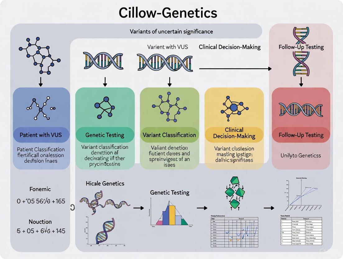
Visualizing the VUS Reclassification Pathway
The journey from a finding of "uncertain significance" to a definitive classification is a structured, evidence-driven process. The pathway below integrates data from clinical, bioinformatic, and functional sources to resolve a VUS.
The rapid adoption of large multigene panels in clinical and research genetics has created a critical paradox: while expanded testing increases the detection of clinically actionable pathogenic variants, it simultaneously generates more variants of uncertain significance (VUS)—genetic alterations with unclear implications for disease risk. This phenomenon presents substantial challenges for genetic counselors, researchers, and drug development professionals who must interpret these ambiguous results while providing clear guidance to patients and research participants.
The VUS classification means that at the time of interpretation, there is insufficient evidence to determine whether the variant is disease-causing or benign [4]. According to ACMG guidelines, VUS should not be used in clinical decision-making, which instead should be based on personal and family history [4]. This creates a complex landscape where the technological capacity to generate data has outpaced our ability to interpret it, particularly for populations underrepresented in genomic databases.
Quantitative Evidence: The Direct Relationship Between Panel Size and VUS Rates
Comparative Analysis of Panel Performance
Multiple large-scale studies have demonstrated a clear correlation between the number of genes tested and VUS detection rates. The following table summarizes key findings from recent research:
| Panel Size (Number of Genes) | VUS Detection Rate | Study Population | Key Findings |
|---|---|---|---|
| 20-gene panel [9] | 23.9% | 364 Brazilian high-risk patients | Significant difference in VUS detection compared to larger panels |
| 23-gene panel [9] | 31.0% | Same Brazilian cohort | Intermediate VUS detection rate |
| 144-gene panel [9] | 56.3% | Same Brazilian cohort | 2.4× higher VUS rate than 20-gene panel without substantial PV/LPV improvement |
| 2-10 genes [10] | 6.0% | 1,463,812 MGP tests | Lowest VUS rate in large North American study |
| >200 genes [10] | 76.2% | 84,316 MGP tests | 12.7× higher VUS rate than smallest panels |
| Exome/Genome Sequencing [10] | 22.5% | 48,494 ES/GS tests | Lower VUS rate than large MGPs despite more genes interrogated |
This data reveals a consistent pattern: as panel size increases, so does the VUS detection rate. The Brazilian study specifically noted that while the 144-gene panel significantly increased VUS detection compared to smaller panels, it did not substantially improve the identification of pathogenic or likely pathogenic variants (PV/LPV) [9]. This highlights the diminishing returns of panel expansion without concomitant improvements in variant interpretation capabilities.
Population-Specific Disparities in VUS Rates
The challenges of VUS interpretation are particularly pronounced for underrepresented populations. Research has consistently demonstrated that individuals of non-European ancestry experience higher VUS rates due to less representation in genomic databases [11] [12] [13]. A comprehensive analysis of genetic testing results showed that in 2019, VUS were found nearly twice as often as pathogenic variants in White patients (25% vs. 14%), but approximately four times as often in Asian and Black patients (39-40% vs. 10-11%) [12].
These disparities have significant implications for both clinical care and research, as they can exacerbate existing health inequities and complicate the recruitment of diverse populations into genetic studies and drug development trials.
Mechanisms and Methodologies: Understanding VUS Classification and Reclassification
Variant Classification Systems and Evidence Requirements
Genetic variants are classified according to standardized guidelines that evaluate multiple types of evidence:
The American College of Medical Genetics and Genomics (ACMG) guidelines establish a five-tier classification system for genetic variants: pathogenic, likely pathogenic, variant of uncertain significance (VUS), likely benign, and benign [14] [4]. Classification relies on integrating multiple evidence types, including population frequency data, computational predictions, functional evidence, segregation studies, and clinical observations [11]. Variants are classified as VUS when the available evidence is contradictory or insufficient to support definitive classification [14].
VUS Reclassification Protocols and Timelines
The reclassification of VUS represents a critical process for resolving uncertainty in genetic test results. Research indicates that approximately 10-15% of reclassified VUS are upgraded to likely pathogenic/pathogenic, while the remainder are downgraded to likely benign/benign [11]. A multicenter retrospective analysis focusing on breast cancer susceptibility genes found that 20% of VUS were reclassified over the study period, with the vast majority (92%) being downgraded to benign/likely benign [13].
The timeline for VUS resolution remains challenging. One study found that only 7.7% of unique VUS were resolved over a 10-year period in cancer-related testing at a major laboratory [11]. The mean time to VUS reclassification was approximately 2.8 years in a study of breast cancer susceptibility genes, with no significant difference in reclassification time across racial and ethnic groups [13].
Experimental Approaches for VUS Resolution
Family Segregation Studies
Protocol Overview: Family studies examine whether a VUS co-segregates with disease in multiple affected family members across generations.
Key Considerations:
- Family Structure: Ideal families have multiple affected individuals across generations with confirmed disease status
- Age of Onset: For adult-onset conditions, testing asymptomatic young relatives may not provide informative data [4]
- Informed Consent: All participants should understand the research nature of VUS investigation and potential implications
Evidence Strength: Finding the same VUS in distantly related affected individuals provides strong evidence for pathogenicity, while absence in affected relatives suggests the variant may be benign [11] [4].
Functional Assays Using Advanced Genome Editing
Emerging Technologies: Prime editing platforms enable scalable functional assessment of variants in their endogenous cellular context [15]. This approach allows high-throughput screening of variant effects without exogenous expression systems.
Experimental Workflow:
Validation: In one implementation, researchers tested over 7,500 pegRNAs targeting SMARCB1 and 65.3% of all possible single nucleotide variants in a 200-bp region of MLH1, demonstrating the platform's accuracy for discriminating pathogenic variants [15].
Computational and Predictive Modeling
Methodology: Advanced computational tools use machine learning and artificial intelligence to predict variant pathogenicity based on evolutionary conservation, protein structure, and existing variant databases [11].
Implementation:
- Multiple Algorithm Integration: Compare predictions across different algorithms considering cross-species conservation, protein folding, critical domains, and amino acid substitutions [11]
- Database Integration: Incorporate data from population databases (gnomAD), disease-specific databases (ClinVar), and functional prediction scores
- Continuous Re-evaluation: Establish systems for automatic reassessment as new data becomes available
Research Reagent Solutions for VUS Investigation
The following table outlines essential research reagents and methodologies for comprehensive VUS analysis:
| Research Reagent/Methodology | Primary Function | Application in VUS Resolution |
|---|---|---|
| Prime Editing Systems [15] | Precise genome editing without double-strand breaks | Functional assessment of VUS in endogenous context at scale |
| Next-Generation Sequencing [9] [14] | High-throughput DNA sequencing | Identification and confirmation of variants in multi-gene panels |
| Multiplex Ligation-dependent Probe Amplification (MLPA) [9] [16] | Detection of copy number variations | Validation of deletion/duplication variants |
| Sanger Sequencing [16] | Targeted DNA sequencing | Confirmatory testing of specific variants in patients and relatives |
| Functional Cell-Based Assays [14] | Assessment of protein function and impact | Direct measurement of variant effects on protein activity |
| Bioinformatic Prediction Tools [11] [14] | Computational assessment of pathogenicity | Integration of multiple evidence types for classification |
Frequently Asked Questions: Troubleshooting VUS Challenges
Q: How should I proceed when a patient receives a VUS result? A: Clinical management should be based on personal and family history, not the VUS result [4]. Document the result systematically and establish a plan for periodic re-evaluation. Consider which evidence-generation approaches (family studies, functional assays) might be most productive for resolution.
Q: What factors contribute to higher VUS rates in larger gene panels? A: The increase stems from two primary factors: (1) inclusion of genes with less established disease associations and lower penetrance variants, and (2) investigation of more genetic regions where rare population variants are more likely to be encountered and initially classified as VUS due to insufficient evidence [9] [10].
Q: How can we address disparities in VUS rates across different ancestral populations? A: Implement several complementary strategies: prioritize inclusion of diverse populations in genomic research; support development of population-specific reference databases; utilize testing approaches that minimize uninformative results; and advocate for research funding specifically dedicated to variant interpretation in underrepresented groups [11] [12] [13].
Q: What is the typical timeframe for VUS reclassification? A: Timeframes vary significantly, with studies reporting mean times of approximately 2.8 years for some cancer gene VUS [13], while other data shows only 7.7% of unique VUS resolved over a 10-year period [11]. The timeline depends on gene-specific research activity, variant frequency, and available resources for functional studies.
Q: When should family studies be pursued for VUS resolution? A: Family studies are most informative when: (1) multiple affected and unaffected relatives are available and willing to participate; (2) the disease has clear inheritance patterns; (3) affected relatives are old enough to have manifested symptoms; and (4) the VUS is in a gene with established disease association [4].
The panel size paradox represents a fundamental challenge in modern genetic medicine and research. While expanded genetic testing offers undeniable benefits for identifying actionable variants, the concomitant increase in VUS detection necessitates sophisticated approaches to variant interpretation and classification. Strategic panel design—focusing on genes with established clinical validity and utility—can help balance comprehensive assessment with manageable result interpretation.
Moving forward, researchers and clinicians must collaborate to develop more efficient pathways for VUS resolution through shared data, standardized classification systems, and innovative functional assays. The implementation of robust reclassification protocols and commitment to diverse population representation in genomic databases will be essential for maximizing the clinical utility of genetic testing while minimizing the burden of uncertain results.
A Variant of Uncertain Significance (VUS) is a genetic variant for which the association with disease risk is unclear. Unlike pathogenic or benign variants, a VUS is not clinically actionable, and its identification should not directly influence patient management decisions [17] [3]. The central challenge is that a VUS result provides no clarification of an individual's cancer risk, creating a state of uncertainty for both researchers and patients [18].
A critical driver of VUS rates is the severe underrepresentation of non-European populations in genomic databases [19] [1]. With over 80% of genomic data coming from individuals of European ancestry, the interpretation of genetic variants in underrepresented groups is fundamentally hampered [20] [1]. This lack of diverse reference data means that variants commonly found in non-European populations are more likely to be flagged as "uncertain" simply because they are observed less frequently in the existing, skewed datasets.
Quantitative Evidence of Disparities
The following tables summarize key quantitative findings from large-scale studies, highlighting the persistent disparities in VUS rates and reclassification.
Table 1: Disparities in VUS and Pathogenic/Likely Pathogenic (P/LP) Variant Rates by Race/Ethnicity (Multi-Gene Panel Testing) [19]
| Race/Ethnicity | VUS Rate | Pathogenic/Likely Pathogenic (P/LP) Variant Rate |
|---|---|---|
| African American/Black (AA/B) | 46% | 9% |
| Non-Hispanic White (NHW) | 32% | 13% |
Note: Data based on a retrospective cohort of 48,684 AA/B and 444,831 NHW individuals. All differences were statistically significant (p<0.0001).
Table 2: VUS Reclassification Rates in Early-Onset Colorectal Cancer by Self-Identified Race/Ethnicity [21]
| Race/Ethnicity | Percentage of Individuals with a Reclassified VUS |
|---|---|
| Ashkenazi Jewish | 4.8% |
| Asian | 18.2% |
| Black | 12.2% |
| Hispanic | 7.6% |
| White | 6.7% |
Note: Disparities in reclassification rates were statistically significant (P < 0.0001).
Experimental Protocols for VUS Investigation
Protocol: Reclassification Using Expert Panel Specifications
Aim: To reclassify VUS in clinically relevant genes (e.g., BRCA1 and BRCA2) using gene-specific specifications to reduce ambiguity.
Background: The standard 2015 ACMG/AMP guidelines provide a general framework for variant classification. However, gene-specific specifications, such as those developed by the ENIGMA (Evidence-based Network for the Interpretation of Germline Mutant Alleles) Variant Curation Expert Panel (VCEP), are superior for reducing VUS rates [22].
Methodology: [22]
- Variant Set: Compile a set of VUS from your cohort or database.
- Data Annotation: Gather the latest annotation data for each variant.
- Dual-Track Classification:
- Reclassify variants using the standard ACMG/AMP system.
- Reclassify the same variants using the ENIGMA VCEP specifications, which provide gene-specific adjustments to the weight of different types of evidence (e.g., population data, computational predictions, functional data).
- Comparison: Compare the percentage of VUS successfully reclassified to a definitive category (Benign/Likely Benign or Pathogenic/Likely Pathogenic) between the two methods.
Workflow Visualization:
Diagram 1: Experimental workflow for VUS reclassification using expert panel specifications.
Protocol: Functional Studies to Assess VUS Impact
Aim: To determine the biochemical and functional consequences of a VUS to provide evidence for its pathogenicity classification.
Background: Functional assays provide direct evidence of a variant's effect on protein function, which is a strong criterion for classification under ACMG/AMP guidelines [23].
- In Silico Analysis: Use computational tools to predict the impact of a missense VUS on protein structure and function (e.g., based on sequence conservation, physicochemical properties).
- Functional Assay Development: Design an experiment that tests the core function of the wild-type protein.
- For tumor suppressor genes (e.g., BRCA1, TP53): This may include assays for transcription activation, centrosome duplication, or cell cycle regulation.
- For DNA repair genes (e.g., ATM, BRCA2): Consider assays for homology-directed repair proficiency, phosphorylation of downstream targets, or sensitivity to DNA-damaging agents.
- Experimental Expression: Introduce the VUS into an appropriate cell line model (e.g., using site-directed mutagenesis) and express it alongside the wild-type protein and known pathogenic controls.
- Phenotypic Assessment: Quantify the functional output of the assay. A result similar to wild-type supports benign classification, while a result similar to known pathogenic variants supports pathogenic classification.
Workflow Visualization:
Diagram 2: High-level workflow for functional characterization of a VUS.
The Scientist's Toolkit: Research Reagent Solutions
Table 3: Essential Materials for VUS Research
| Research Reagent / Tool | Function in VUS Investigation |
|---|---|
| Multi-Gene Panels (MGPT) | Allows simultaneous sequencing of dozens to hundreds of cancer susceptibility genes. Larger panels increase VUS detection but are necessary for comprehensive risk assessment [18] [19]. |
| CLIA-Certified Laboratory | A Clinical Laboratory Improvement Amendments (CLIA)-certified lab ensures the analytical validity of genetic testing. Results from non-CLIA labs should not be used for clinical decision-making [20]. |
| ClinVar Database | A public archive that aggregates information about genomic variants and their relationship to human health. It is critical for comparing local VUS findings with global data [20] [23]. |
| ENIGMA VCEP Specifications | Gene-specific guidelines for classifying variants in BRCA1 and BRCA2. Using these specifications over the standard ACMG/AMP framework has been shown to dramatically reduce VUS rates [22]. |
| Functional Assay Kits | Commercial kits (e.g., for measuring DNA repair proficiency, protein phosphorylation, or transcriptional activity) are used to determine the biochemical impact of a VUS in a controlled laboratory setting [23]. |
Troubleshooting Guides & FAQs
FAQ 1: Our research has identified a VUS that is prevalent in our underrepresented cohort. What are the immediate next steps to resolve its significance?
- Answer: A multi-faceted approach is required:
- Data Sharing: Immediately submit the VUS to public databases like ClinVar. This contributes to the global knowledge pool and can help identify other individuals with the same variant [20].
- Segregation Analysis: If possible, test other family members in the cohort, particularly those with and without cancer, to see if the VUS co-segregates with the disease. This provides key genetic evidence [1] [3].
- Case-Control Studies: Check the frequency of this VUS in large, population-specific control databases. A frequency similar to or higher in controls than in cases is evidence for benign classification [23].
- Initiate Functional Studies: Begin designing experiments, as outlined in Protocol 3.2, to characterize the variant's functional impact [20] [23].
FAQ 2: We are observing high VUS rates in our study of a non-European population. Is this a technical error, and how can we mitigate this in our analysis?
- Answer: High VUS rates in underrepresented populations are an established disparity, not a technical error [19] [21]. To mitigate its impact on your research:
- Benchmark Against Known Rates: Compare your VUS rate to published rates for the same racial/ethnic group (e.g., ~46% for African American/Black individuals) to contextualize your findings [19].
- Prioritize Clinical History: Emphasize that clinical management for individuals with a VUS must be based on personal and family history of cancer, not the VUS result itself [17] [3].
- Focus on Reclassification: Dedicate research resources to the reclassification protocols described in Section 3, specifically for the high-frequency VUS in your cohort.
FAQ 3: How should we handle patient and provider anxiety when a VUS is reported in a high-risk gene like BRCA1?
- Answer:
- Clear Communication: Reinforce that a VUS is an "uncertain" finding, not a positive one. Guidelines state it should not be used for clinical decision-making [17] [3].
- Contextualize with History: Redirect the focus to the established, history-based risk management guidelines (e.g., NCCN guidelines). For a patient with a VUS in BRCA1 but no strong family history, risk management would be based on population guidelines, not enhanced screening for BRCA1 carriers [17].
- Cite Empirical Evidence: Inform providers that meta-analyses show no significant difference in surgery rates between patients with VUS and those with benign results, suggesting that, at a population level, inappropriate management based on a VUS is not prevalent [17].
FAQ 4: What is the typical timeframe for VUS reclassification, and how can we track it?
- Answer: Reclassification can be a lengthy process, taking from months to years, and some VUS may never be reclassified due to insufficient data [3]. To track reclassifications:
- Establish a process for periodic re-analysis of unresolved VUS in your cohort.
- Maintain contact with the clinical laboratory that performed the testing, as they are responsible for issuing revised reports when a VUS is reclassified [3].
- Monitor public databases like ClinVar for updated classifications submitted by other labs and researchers [20].
The Patient and Clinical Burden of Uncertainty in Genomic Results
Clinical Context: The Impact of Uncertainty on Patients and Clinical Practice
The integration of genomic testing into oncology, while clinically transformative, inherently generates significant uncertainty for patients and clinicians. This uncertainty stems from the complexity of genomic technology and the biological ambiguity it can reveal [24].
The Patient Experience of Genomic Uncertainty
A systematic review of patient experiences in cancer genomics identified four central themes [24]:
- Coexisting Uncertainties: Patients navigate multiple, simultaneous uncertainties.
- Factors Influencing Uncertainty: Genetic literacy, types of results, and trust in science significantly shape the experience of uncertainty [24].
- Outcomes of Uncertainty: Uncertainty can act as either a motivator or a barrier to pursuing genomic testing.
- Coping with Uncertainty: How a patient appraises uncertain results influences the coping strategies they employ.
This review found that while genomic results can reduce uncertainty regarding treatment decisions, they often do not reduce uncertainty in the context of future cancer risk [24].
The Clinical and Systemic Burden of VUS
Variants of Uncertain Significance (VUS) represent a major challenge in clinical genomics. They are equivocal, non-actionable results that require active monitoring for potential future reclassification [13]. This creates a significant burden:
- Clinical Management: Patients with VUS are generally not eligible for intensive surveillance, chemoprevention, or surgical risk reduction options that are available to those with clearly pathogenic variants [13].
- Psychological Impact: VUS results can cause patient distress and confusion regarding result interpretation [13].
- Healthcare System Workflow: VUS require ordering providers and genetic counselors to implement additional follow-up procedures to manage reclassification notifications from testing laboratories over time [13].
Technical Support: Resolving Variants of Uncertain Significance
Troubleshooting Guide: VUS Reclassification
| Problem Scenario | Potential Root Cause | Recommended Solution | Key Considerations |
|---|---|---|---|
| High VUS rate in specific patient populations. | Lack of diverse ancestral representation in genomic databases [13]. | Utilize labs that employ large, diverse datasets and population-specific frequency data. | One study found no significant association between race/ethnicity/ancestry and VUS reclassification rate or time [13]. |
| Inability to apply ACMG/AMP PS4 criterion (prevalence in affecteds vs. controls). | Limited availability of robust, matched case-control genomic and phenotypic data [25]. | Integrate Real-World Evidence (RWE) from large-scale clinicogenomic datasets. | An RWE approach reclassified 32% of VUS carriers across 20 hereditary cancer and cardiovascular genes [25]. |
| VUS persists in a tumor suppressor gene with a highly specific phenotype. | Classic ACMG/AMP criteria may fail to assign sufficient weight to highly specific phenotypes [7]. | Apply new ClinGen guidance for PP1/PP4 criteria that quantitatively scores phenotype specificity [7]. | In one study, this method reclassified 31.4% of remaining VUS in tumor suppressor genes as Likely Pathogenic [7]. |
| Patient anxiety and clinical inertia following a VUS result. | Lack of actionable clinical guidance and misunderstanding of VUS meaning. | Provide clear counseling that most VUS are downgraded and schedule periodic reassessment. | 92% of reclassified VUS in a breast cancer risk cohort were downgraded to Benign/Likely Benign [13]. |
Frequently Asked Questions (FAQs)
Q1: What is the typical timeframe for VUS reclassification? A: Evidence suggests the mean time to VUS reclassification is approximately 2.8 years, though this can vary based on the gene and the evidence available [13].
Q2: Are VUS more common or persistent in certain racial or ethnic groups? A: While individuals of non-European ancestry may have a higher initial prevalence of VUS, a multi-center study on breast cancer risk genes found that race, ethnicity, and ancestry were not significantly associated with the likelihood or timing of VUS reclassification. The majority of VUS were downgraded across all groups [13].
Q3: What percentage of VUS are eventually reclassified, and to what? A: Reclassification rates vary. One study focusing on hereditary cancer and cardiovascular genes using Real-World Evidence (RWE) reclassified 32% of VUS carriers, with 99.7% of those downgraded to Benign/Likely Benign (B/LB) and 0.3% upgraded to Pathogenic/Likely Pathogenic (P/LP) [25]. Another study on breast cancer risk genes found 92% of reclassified VUS were downgraded to B/LB [13].
Q4: What new methods are improving VUS resolution? A: Key advancements include:
- New ClinGen PP1/PP4 Criteria: Provides a systematic method to assign higher pathogenicity scores based on supporting evidence from highly specific phenotypes [7].
- Real-World Evidence (RWE): Leveraging longitudinal clinical data from large biobanks enables rigorous case-control analyses to apply the ACMG/AMP PS4 criterion more effectively [25].
- AI and Machine Learning: Tools like DeepVariant use deep learning to identify genetic variants with greater accuracy, aiding initial interpretation [26].
Experimental Protocols for VUS Reclassification
Protocol 1: Reassessment of VUS Using Updated ClinGen Guidance
This protocol details the reassessment of VUS in tumor suppressor genes using the updated ClinGen criteria for cosegregation (PP1) and phenotype specificity (PP4) [7].
1. Variant Selection and Data Annotation
- Retrieve VUS from clinical database for target genes (e.g., NF1, TSC1, TSC2, RB1, PTCH1, STK11, FH).
- Annotate variants using tools like ANNOVAR with population frequency databases (gnomAD), clinical databases (ClinVar), and in silico prediction tools (REVEL, SpliceAI) [7].
2. Phenotype and Family History Review
- Systematically collect clinical information from electronic medical records, including detailed phenotype and family history.
- For PP4, define the specific phenotypic criteria for the gene-disease relationship (e.g., multiple café-au-lait spots and neurofibromas for NF1) [7].
3. Point-Based Pathogenicity Assessment
- Conduct a baseline classification using the classic ACMG/AMP criteria within a quantitative point-based framework [7].
- Perform a secondary classification using the new ClinGen PP1/PP4 guidance. This involves:
- a. Determining the gene's diagnostic yield from resources like GeneReviews.
- b. Translating the diagnostic yield into points for the PP4 criterion based on a predefined transition table [7].
- c. Applying updated PP1 criteria using a Bayes point system.
4. Evidence Integration and Final Classification
- Combine points from all applicable evidence criteria (PM2, PP3, PP4, etc.).
- Assign a final classification based on the aggregated points: ≥10 (Pathogenic), 6–9 (Likely Pathogenic), 0–5 (VUS), -1 to -6 (Likely Benign), ≤-6 (Benign) [7].
Protocol 2: Reclassification Using Multi-Institutional Real-World Evidence
This protocol employs RWE from longitudinal clinicogenomic datasets to resolve VUS [25].
1. Dataset Curation and Integration
- Assemble large-scale datasets linking exome or genome sequence data with de-identified, longitudinal clinical records (e.g., from the Helix Research Network, UK Biobank, All of Us) [25].
- For a target gene (e.g., BRCA2), curate phenotypes for established gene-disease associations from the medical records of sequenced individuals.
2. Case-Control Analysis
- Define cases as individuals with the phenotype and controls as those without.
- For each variant, perform a variant-specific case-control analysis, comparing the frequency of the variant in cases versus controls [25].
3. Application of RWE Code
- Apply a new evidence code, RWE, within the ACMG/AMP framework.
- Use statistically significant associations from the case-control analysis to provide evidence for pathogenicity (RWEP) or, if no association is found, evidence for benignity (RWEB) [25].
4. Variant Re-evaluation
- Re-score the variant by integrating the new RWE evidence with existing evidence.
- Reclassify the variant if the total evidence meets the threshold for a new classification category (e.g., Likely Benign or Likely Pathogenic) [25].
Visualizing the VUS Reclassification Workflow
The following diagram illustrates the logical workflow and decision points in the VUS reassessment process.
VUS Reassessment and Reclassification Workflow
The following table details key resources and their functions in the management and reclassification of VUS.
| Research Reagent / Resource | Function in VUS Management |
|---|---|
| ACMG/AMP-ClinGen Framework | Provides the standardized international system for variant classification, including disease-specific refinements [7]. |
| Real-World Evidence (RWE) Datasets (e.g., UK Biobank, All of Us, Helix) | Enables case-control analyses by linking genomic data with longitudinal clinical records to assess variant-disease associations [25]. |
| Population Frequency Databases (e.g., gnomAD) | Provides allele frequency data across diverse populations to filter common polymorphisms and apply frequency-based evidence codes (PM2, BS1, BA1) [7]. |
| In Silico Prediction Tools (e.g., REVEL, SpliceAI) | Computational algorithms that predict the functional impact of missense variants and splice-altering variants, informing the application of PP3/BP4 criteria [7]. |
| Variant Annotation Tools (e.g., ANNOVAR) | Software that automates the annotation of genetic variants with information from multiple genomic databases, streamlining the interpretation pipeline [7]. |
| Point-Based Classification System | A quantitative framework that abstracts ACMG/AMP evidence criteria into points, facilitating transparent and consistent variant assessment [7]. |
Resolving Uncertainty: Advanced Methodologies for VUS Interpretation and Reclassification
Technical Support Center: FAQs & Troubleshooting Guides
Frequently Asked Questions
Q1: What are the main types of functional readouts I can measure with CRISPR-based VUS characterization, and how do I choose?
Modern CRISPR-based functional assays have moved beyond simple cell viability to offer multiparametric readouts. You can choose the assay based on the biological question you want to answer about your variant [27].
- CRISPR-SelectTIME: Tracks variant frequency over time to determine effects on cell proliferation and survival.
- CRISPR-SelectSPACE: Tracks variant frequency across a spatial dimension (e.g., through a membrane) to assay effects on cell migration or invasiveness.
- CRISPR-SelectSTATE: Tracks variant frequency as a function of a cell state measurable by flow cytometry (FACS), allowing you to link the variant to any physiological state (e.g., apoptosis, differentiation, specific marker expression) [27].
Q2: My negative selection screen shows positive log-fold changes (LFC) for some sgRNAs. Is this normal?
Yes, this can occur and is often a result of how the data is processed. When using algorithms like Robust Rank Aggregation (RRA), the gene-level LFC is calculated as the median of its sgRNA-level LFCs. Extreme values from individual, poorly performing sgRNAs can skew the median, resulting in a positive LFC in a negative screen (or vice versa). To mitigate this, ensure you design and use at least 3-4 sgRNAs per gene to provide robustness against individual sgRNA performance variability [28].
Q3: How can I determine if my CRISPR screen was successful, especially if I lack well-characterized positive controls?
The most reliable method is to include known positive-control sgRNAs in your library. If these controls show significant enrichment or depletion as expected, your screen conditions are likely effective [28]. In the absence of such controls, you can evaluate:
- Cellular Response: Assess the degree of cell killing or survival under your selection pressure.
- Data Quality: Examine bioinformatics outputs, including the distribution and log-fold change of sgRNA abundance. High reproducibility between biological replicates (Pearson correlation coefficient > 0.8) is a good indicator of a successful screen [28].
Q4: What should I do if my sequencing results show a large number of lost sgRNAs?
The troubleshooting steps depend on when the loss occurs [28]:
- Loss in the initial library pool: This indicates insufficient initial library coverage. You should re-establish the CRISPR library cell pool, ensuring you use a sufficient number of cells to maintain >99% library representation.
- Loss after selection in the experimental group: This may reflect that the selection pressure applied was too severe, causing the loss of sgRNAs beyond those specifically targeting essential genes.
Q5: When analyzing data from multiple replicates, should I analyze them together or in pairs?
The best approach depends on the reproducibility of your replicates [28]:
- High Reproducibility (Pearson R > 0.8): Perform a combined analysis across all replicates to increase statistical power.
- Low Reproducibility: It is more appropriate to perform pairwise comparisons first. You can then use a meta-analysis (e.g., Venn diagrams) to identify candidate genes that consistently overlap across the different comparisons, improving the reliability of your hit identification.
Troubleshooting Common Experimental Issues
Issue: No Significant Gene Enrichment in Screening Results
- Potential Cause: Insufficient selection pressure during the screening process. When pressure is too low, the experimental group may fail to exhibit a strong enough phenotype, weakening the signal-to-noise ratio [28].
- Solution: Optimize your screening conditions by increasing the selection pressure and/or extending the duration of the screen. This allows for greater enrichment of positively selected cells.
Issue: High False Positive/Negative Rate in FACS-Based Screens
- Potential Cause: FACS-based screening often allows for only a single round of enrichment and is subject to significant technical noise [28].
- Solutions:
- Increase the initial number of cells used for sorting.
- Perform multiple independent rounds of sorting where experimentally feasible.
- These steps help to dilute out technical noise and improve the robustness of your results [28].
Issue: Low Mapping Rate in Sequencing Data
- Potential Cause: While a low mapping rate itself does not necessarily compromise result reliability, it can be a symptom of underlying issues [28].
- Solution: The critical factor is the absolute number of mapped reads. Ensure that the number of successfully mapped reads is sufficient to maintain the recommended sequencing depth (typically ≥200x coverage). The focus should be on achieving sufficient data volume rather than the mapping rate percentage alone [28].
Quantitative Data & Methodologies
The table below summarizes key quantitative findings from recent studies using functional assays to characterize VUS.
| Gene / Study | Total VUS Tested | Reclassified/Oncogenic | Key Finding |
|---|---|---|---|
| BRCA2 (Mayo Clinic, 2025) | ~7,000 variants in DNA-binding domain | 91% clinically classified | Comprehensive functional assessment allowed precise risk assessment [29]. |
| PALB2 & Other Actionable Genes (MD Anderson, 2023) | 438 VUS | 106 (24.2%) oncogenic | Rule-based pre-classification of VUS as "Potentially actionable" successfully enriched for true oncogenic variants (37% vs 13% in "Unknown" group) [30]. |
| PALB2 (Various, 2020) | Not Specified | Multiple VUS characterized | The Coiled-Coil and WD40 domains are hotspots for VUS that impair PALB2's function in Homologous Recombination [31]. |
Detailed Experimental Protocol: CRISPR-Select Assay
The CRISPR-Select protocol is a powerful, population-based method for functional analysis of genetic variants. Below is a step-by-step guide [27].
1. Assay Design and Cassette Preparation
- CRISPR-Cas9 Reagent: Design a gRNA to elicit a double-strand break close to the genomic site of the variant of interest (VOI). The gRNA should be chosen so that the VOI and a neutral control mutation are located in the seed or PAM region to minimize post-knock-in recutting.
- Repair Templates: Synthesize two single-stranded oligodeoxynucleotides (ssODNs):
- VOI ssODN: Contains the variant of interest to be knocked in.
- WT' ssODN: Contains a synonymous, internal normalization mutation (WT') at the same (or nearly the same) position as the VOI. This template is otherwise identical to the VOI ssODN and serves as an internal control.
2. Cell Preparation and Transfection
- Engineer your chosen cell line (e.g., MCF10A for breast cancer studies) for doxycycline-inducible Cas9 expression if not already available.
- Pre-treat cells with doxycycline to induce Cas9 expression.
- Co-transfect the cell population with the synthetic gRNA and the two ssODN repair templates using your preferred method (e.g., lipofection).
3. Tracking and Sampling
- After transfection, aliquot the cell population for your chosen readout (TIME, SPACE, or STATE).
- For CRISPR-SelectTIME, culture the cells under relevant selective conditions and collect samples at multiple time points (e.g., Day 2, 4, 6...).
4. Genomic DNA Extraction and Amplification
- Isolate genomic DNA from each cell sample.
- Perform PCR amplification of the target locus using primers that anneal to sequences outside the region covered by the ssODNs. This ensures unbiased amplification of all editing outcomes.
5. Next-Generation Sequencing (NGS) and Data Analysis
- Subject the PCR amplicons to NGS.
- Analyze the sequencing data to determine the types and frequencies of all editing outcomes.
- Key Calculation: Based on the known amount of genomic DNA used for PCR, calculate the absolute numbers of knock-in alleles for both the VOI and the WT' control. This allows you to track the ratio of VOI to WT' over time or across conditions, directly revealing the functional impact of the variant.
Signaling Pathways & Experimental Workflows
Homologous Recombination (HR) Repair Pathway
The following diagram illustrates the key pathway in which genes like BRCA1, PALB2, and BRCA2 operate, and where their VUS can disrupt normal function [31].
CRISPR-Select Experimental Workflow
This diagram outlines the logical flow of the CRISPR-Select assay, from design to functional interpretation [27].
The Scientist's Toolkit: Essential Research Reagents
This table lists key materials and reagents essential for setting up CRISPR-based functional characterization of VUS.
| Reagent / Material | Function / Explanation | Example/Note |
|---|---|---|
| CRISPR-Cas9 System | Core gene-editing machinery. Creates a double-strand break at the target genomic locus. | Can be delivered as plasmid, ribonucleoprotein (RNP) complex, or via viral vectors. |
| ssODN Repair Templates | Serves as the donor template for Homology-Directed Repair (HDR) to introduce the specific VUS or control mutation. | Must contain the variant and homologous arms. A synonymous "WT'" template is critical for internal normalization [27]. |
| iPSCs (Induced Pluripotent Stem Cells) | Patient-specific cells that can be differentiated into relevant cell types (e.g., cardiomyocytes) for disease modeling. | Allows creation of isogenic lines; crucial for studying variants in hard-to-access tissues [32]. |
| Validated Positive Control sgRNAs | sgRNAs targeting genes with known strong phenotypes (e.g., essential genes for depletion, oncogenes for enrichment). | Vital for assessing the technical success of a CRISPR screen [28]. |
| PARP Inhibitors / Cisplatin | Chemicals used for functional validation. Sensitivity to these drugs indicates a deficient Homologous Recombination pathway (a common VUS impact) [31]. | Useful for confirming loss-of-function variants in HR genes like BRCA1, BRCA2, and PALB2. |
| FACS Marker Antibodies | Enable cell sorting based on protein expression levels for CRISPR-SelectSTATE or FACS-based screens. | Used to isolate cell populations based on a physiological state of interest [28] [27]. |
Frequently Asked Questions (FAQs)
Q1: What is the core innovation of the new ClinGen PP1/PP4 guidance? The guidance introduces a point-based Bayesian framework that quantitatively links the co-segregation evidence (PP1/BS4) and phenotype specificity (PP4) criteria. It recognizes that these criteria are inseparably coupled. The key advancement is allowing the assignment of higher pathogenicity scores based on phenotype specificity, especially for genes where the phenotype is highly specific to that single gene (locus homogeneity), enabling more VUS reclassifications [7] [33].
Q2: When applying the new PP4 criteria, how is the strength of phenotypic evidence determined? The strength of evidence is primarily determined by the degree of locus heterogeneity for the phenotype. The more a single gene is responsible for a specific phenotype (high locus homogeneity), the more points can be assigned from the PP4 criterion alone. The diagnostic yield values for each gene, often available from resources like GeneReviews, are transformed into points using a predefined transition table [7].
Q3: What is a common reason for VUS reclassification failures, and how can it be addressed? A common reason for failing to reclassify a VUS is insufficient or non-specific phenotypic data. This can be addressed by ensuring patient phenotypes are meticulously documented and aligned with the highly specific characteristics of the disease associated with the gene. Collaborative data sharing through databases like ClinVar is also crucial for accumulating evidence [7] [11] [20].
Q4: How should a VUS result be used in clinical decision-making for patient care? According to guidelines from the American College of Medical Genetics and Genomics (ACMG), a VUS should not be used for clinical decision-making. Clinical management should be based on the patient's personal and family history of cancer. Unnecessary procedures based on a VUS are discouraged, as the vast majority are eventually reclassified as benign [3].
Q5: What strategies can research teams employ to manage VUS findings in a clinical trial setting? Effective strategies include [20]:
- Engaging genetic counsellors to help participants understand the result and to guide researchers.
- Implementing long-term monitoring of participants to accumulate data on the variant over time.
- Conducting functional studies to assess the variant's biological impact.
- Prioritizing diversity in genomic databases to reduce the disproportionate rate of VUS in under-represented populations.
Performance Data: VUS Reclassification Using the New Framework
The table below summarizes quantitative data on the effectiveness of the new ClinGen PP1/PP4 criteria from a recent study on tumor suppressor genes [7].
Table 1: VUS Reclassification Outcomes after Applying New ClinGen PP1/PP4 Criteria
| Metric | Study Data | Additional Context from Other Studies |
|---|---|---|
| Overall VUS Reclassified as Likely Pathogenic (LPV) | 31.4% (32/101 VUS) [7] | About 10-15% of all reclassified VUS are upgraded to (Likely) Pathogenic; ~90% are downgraded to Benign/Likely Benign [11] [3] [13]. |
| Highest Reclassification Rate by Gene | STK11: 88.9% [7] | Reclassification rates can vary significantly by gene and clinical context. |
| Reclassification Timeframe | Not specified in the study. | A mean time of 2.8 years to reclassification has been reported in breast cancer gene studies, though the process can take years or even decades [3] [13]. |
Table 2: Gene-Specific Diagnostic Yields Informing PP4 Strength [7]
| Gene | Associated Syndrome | Reported Diagnostic Yield (from GeneReviews) |
|---|---|---|
| NF1 | Neurofibromatosis type 1 | ≥ 95% |
| TSC1/TSC2 | Tuberous Sclerosis Complex | 85% |
| STK11 | Peutz-Jeghers Syndrome | 82% |
| FH | Hereditary Leiomyomatosis and Renal Cell Cancer | ~100% for FH-deficient tumors |
Experimental Protocol: Reassessing VUS with the ClinGen PP1/PP4 Framework
This protocol outlines the methodology for reassessing Variants of Uncertain Significance (VUS) in tumor suppressor genes using the updated ClinGen guidelines, as applied in recent research [7].
1. Variant Selection and Data Annotation
- Selection: Identify VUS from your database for the genes of interest (e.g., NF1, TSC1, TSC2, RB1, PTCH1, STK11, FH).
- Exclusion: Filter out cases where a separate known Pathogenic/Likely Pathogenic variant is identified.
- Annotation: Annotate the selected VUS using bioinformatic tools (e.g., ANNOVAR) with data from population frequency databases (gnomAD), in-silico prediction tools (REVEL, SpliceAI), and public variant databases (ClinVar) [7].
2. Phenotype and Segregation Data Collection
- Clinical Review: Systematically review electronic medical records to document patient phenotypes.
- Specificity Assessment: Align the documented phenotypes with the core clinical features of the syndrome associated with the gene (see Table 2 for diagnostic yields).
- Family History: Collect co-segregation data from family pedigrees where available.
3. Pathogenicity Assessment Using Point-Based System Apply the evidence criteria within the quantitative point-based system, where evidence strengths are assigned points [7] [34]:
- Pathogenic Evidence: Supporting (1 point), Moderate (2 points), Strong (4 points), Very Strong (8 points)
- Benign Evidence: Supporting (-1 point), Moderate (-2 points), Strong (-4 points)
Perform two separate classifications for each variant:
- Baseline Classification: Use the classic ACMG/AMP PP1/PP4 criteria.
- New Framework Classification: Use the new ClinGen PP1/PP4 guidance, incorporating diagnostic yield to determine the strength of PP4.
4. Classification and Reporting
- Finalize Classification: Sum the points from all applied criteria and assign the final classification based on the predefined ranges:
- Report and Update: Document the revised classification and update internal databases and ClinVar entries to share the new evidence.
VUS Reassessment Workflow
Table 3: Essential Resources for Variant Interpretation and Reclassification
| Resource / Tool | Type | Primary Function in VUS Reassessment |
|---|---|---|
| ACMG/AMP Guidelines [35] | Classification Framework | Foundational international standard for variant interpretation. |
| ClinGen PP1/PP4 Guidance [33] | Specific Interpretation Rule | Provides the updated, quantitative framework for co-segregation and phenotype criteria. |
| gnomAD [7] | Population Database | Provides allele frequency data to apply PM2 (absent from controls), BS1/BA1 (too common for disease). |
| REVEL & SpliceAI [7] | In-silico Prediction Tool | Computational prediction of variant impact for applying PP3 (deleterious) or BP4 (benign) evidence. |
| ClinVar [7] [20] | Public Variant Archive | Repository for aggregating and sharing clinical interpretations of variants from multiple labs. |
| GeneReviews [7] | Clinical Reference | Source for gene-specific information, including diagnostic yields and clinical criteria, essential for PP4. |
Implementing Computational and In-Silico Prediction Tools
Frequently Asked Questions (FAQs) and Troubleshooting
FAQ 1: What are the most accurate in-silico prediction tools for variant classification?
Recent independent evaluations highlight several top-performing tools. When focusing on genes with high structural and functional similarity, such as the CHD chromatin remodeler family, BayesDel (specifically its addAF component), ClinPred, AlphaMissense, ESM-1b, and SIFT have demonstrated high accuracy [36]. For general use, a combination of tools is recommended to improve reliability [37].
FAQ 2: Why do different in-silico tools sometimes provide conflicting predictions, and how should this be resolved?
Lack of concordance, especially for benign variants, is a known challenge. Analyses show that even among five commonly used algorithms, concordance can be as low as 33% for benign variants and 79% for pathogenic ones [37]. This occurs because tools are trained on different datasets and use distinct underlying algorithms.
- Troubleshooting Guide: Adopt a tiered approach. First, use a robust combination of high-performing algorithms (like those identified above). Second, do not use in-silico evidence if predictions disagree, as per ACMG/AMP guidelines [37]. Third, leverage meta-predictors like BayesDel that integrate multiple sources of evidence for a more reliable score [36].
FAQ 3: How should we handle the validation and reclassification of Variants of Uncertain Significance (VUS)?
VUS reclassification is an ongoing process. Foundational and specialized methodologies from expert groups like InSiGHT and ClinGen recommend a multi-modal approach [38]:
- Functional Data: Prioritize direct functional assays, such as in vitro MMR assays or deep mutational scanning.
- Computational Evidence: Integrate robust in-silico meta-predictors with experimental data.
- Enhanced Detection: Use whole-genome or long-read sequencing to ensure the actual causal variant is identified. Most VUS are eventually downgraded to benign, a trend observed across diverse racial and ethnic groups in genes related to breast cancer risk [13].
FAQ 4: What are the best practices for predicting the impact of non-coding variants?
Non-coding variants can affect splicing, transcription factor binding, or chromatin architecture. Specialized tools are required:
- Splicing Impact: Use SpliceAI, a deep learning model integrated into the Ensembl Variant Effect Predictor (VEP), to predict disruptions to normal splicing [39].
- Transcriptional Regulation: Tools like Enformer can predict the effect of variants on a wide range of functional genomic tracks (e.g., chromatin accessibility) across different cell contexts by analyzing the reference genome sequence [39]. The choice of cell context is critical due to the cell-type specificity of regulatory programs.
Performance Comparison of In-Silico Prediction Tools
The table below summarizes the performance characteristics of selected in-silico tools as reported in recent studies.
Table 1: Tool Performance and Application
| Tool Name | Type / Category | Key Strengths / Applications | As Reported In |
|---|---|---|---|
| BayesDel_addAF | Meta-score (ensemble) | Overall most robust tool for CHD variant prediction [36]. | [36] |
| AlphaMissense | AI-based (evolutionary & structure) | High predictive power; shows promise for future pathogenicity prediction [36]. | [39] [36] |
| SpliceAI | AI-based (splicing) | Accurately identifies splicing sites and the potential impact of variants on splicing [39]. | [39] |
| SIFT | Homology-based | Most sensitive categorical classifier for pathogenic CHD variants (93% correct) [36]. | [36] |
| Enformer | AI-based (non-coding) | Predicts effects of sequence changes on thousands of functional genomics tracks across cell types [39]. | [39] |
| Combination of 3-5 tools | Ensemble/Varied | Required by ACMG/AMP guidelines; but full concordance is rare, especially for benign variants [37]. | [37] |
Experimental Protocol: A Framework for VUS Assessment and Reclassification
The following workflow outlines a recommended procedure for the assessment and reclassification of VUS, synthesizing methodologies from expert panels [38] [13].
Objective: To systematically classify a VUS using computational, phenotypic, and functional evidence to enable clinical decision-making.
Materials: See "Research Reagent Solutions" below.
Methodology:
Foundational Evidence Gathering
- Phenotypic Data: Collect detailed personal and family cancer history. For hereditary cancer syndromes, apply established criteria (e.g., NCCN guidelines) to assess gene-disease congruence [13].
- Population Data: Query population frequency databases (e.g., gnomAD) to assess variant rarity.
- Computational Prediction (In-Silico Analysis): Run a curated set of high-performance prediction tools.
Specialized/Functional Assays
- If the VUS remains uncertain after foundational analysis, proceed to functional assays.
- Direct Functional Assays: For specific gene families, employ direct measures of protein function. For MMR genes, this includes in vitro MMR assays or cell-based MMR proficiency tests [38].
- Deep Mutational Scanning: Where available, consult data from high-throughput functional assays that systematically characterize the functional impact of thousands of variants in a gene of interest [39] [38].
- Transcriptomic/Proteomic Analysis: Use RNA sequencing to validate splicing defects or assess allele-specific expression. Proteomic approaches can check for aberrant protein expression or stability [38].
Evidence Integration and Classification
Periodic Re-review
Research Reagent Solutions
Table 2: Essential Materials and Databases for VUS Interpretation
| Item Name | Type / Category | Function in Experiment |
|---|---|---|
| AlphaMissense | AI Prediction Tool | Provides pathogenicity scores for missense variants using evolutionary and structural information [39] [36]. |
| SpliceAI | AI Prediction Tool | Predicts the likelihood that a variant alters mRNA splicing, crucial for interpreting non-coding and synonymous variants [39]. |
| BayesDel | Meta-Predictor | Integrates scores from multiple other tools to provide a more robust, consolidated estimate of variant deleteriousness [36]. |
| Ensembl VEP (Variant Effect Predictor) | Annotation Suite | Integrates multiple in-silico algorithms (like SIFT, PolyPhen) and functional annotations to provide a comprehensive overview of a variant's potential impact [39] [40]. |
| ClinVar | Clinical Database | A public archive of reports of the relationships between human variants and phenotypes, with supporting evidence; used for benchmarking and evidence gathering [37]. |
| dbNSFP | Database | A comprehensive collection of pre-computed predictions and functional annotations for all potential human non-synonymous variants, facilitating batch analysis [37]. |
| GENCODE / GTEx | Functional Genomics Resource | Provides reference gene annotations and tissue-specific gene expression and QTL data, essential for interpreting non-coding variants [39]. |
Troubleshooting Guides and FAQs
FAQ: My analysis revealed a BRCA2 variant classified as a "Variant of Uncertain Significance" (VUS). What does this mean for clinical interpretation? A VUS is a genetic change for which there is not enough information to definitively classify it as pathogenic (disease-causing) or benign. The clinical utility of the test result is therefore limited, and it should not be used for clinical decision-making [41]. The primary goal of high-throughput functional studies is to reclassify these VUS into more definitive categories.
FAQ: What is the gold-standard framework for classifying sequence variants? The joint consensus recommendation from the American College of Medical Genetics and Genomics (ACMG) and the Association for Molecular Pathology (AMP) provides the standard framework. It specifies a five-tier classification system for variants: Pathogenic, Likely Pathogenic, Uncertain Significance, Likely Benign, and Benign [42] [43].
FAQ: How confident can I be in the "Likely Pathogenic" or "Likely Benign" classifications? The ACMG-AMP guidelines propose that the "likely" categories should be used when the evidence supports a greater than 90% certainty of a variant being either disease-causing or benign [42].
FAQ: What is the difference between a germline and a somatic variant in cancer genetics? A germline variant is inherited and present in every cell of the body, indicating a hereditary cancer predisposition. A somatic (acquired) variant occurs in specific body cells (but not germ cells) and is not inherited; these variants are typically the genetic changes found within a tumor itself [43].
Troubleshooting: Our functional assay results for a set of BRCA2 variants conflict with existing clinical data. How should we resolve this? Begin by validating your functional assay against known pathogenic and benign control variants. In the landmark BRCA2 saturation genome editing study, the functional scores were calibrated using known nonsense (assumed pathogenic) and silent (assumed benign) variants. The assay was further validated against established benchmarks from ClinVar and the ENIGMA consortium, achieving >99% sensitivity and specificity, which lends high credibility to its findings [41]. Ensure your assay's performance meets similar standards before re-evaluating conflicting evidence.
Troubleshooting: Our high-throughput sequencing has generated numerous BRCA2 VUS. What is the most efficient path to functional characterization? Employ a multiplex assay of variant effect (MAVE) approach, such as saturation genome editing (SGE). This method uses CRISPR-Cas9 to knock in thousands of individual single-nucleotide variants (SNVs) into the endogenous gene in a haploid cell line. The functional impact is then assessed based on cell viability, allowing for the simultaneous functional characterization of nearly all possible SNVs in a target region [41].
Data Presentation: Functional Classification of BRCA2 Variants
The following tables summarize the quantitative results from the high-throughput functional evaluation of single-nucleotide variants (SNVs) in the BRCA2 DNA-binding domain (exons 15-26) [41].
Table 1: Summary of BRCA2 Variant Classification by Functional Assay
| Variant Category | Number of SNVs | Percentage of Total | Key Composition |
|---|---|---|---|
| Combined Benign | 5,680 | 81.6% | 3,661 missense, 1,326 silent, 434 intronic |
| Combined Pathogenic | 1,155 | 16.6% | 502 missense, 339 nonsense, 119 canonical splice |
| Uncertain Significance (VUS) | 124 | 1.8% | Variants not meeting classification thresholds |
Table 2: Validation of Functional Assay Performance Against Standards
| Benchmark Set | Number of Variants | Sensitivity | Specificity |
|---|---|---|---|
| ClinVar Missense Variants | 70 | 94% | 95% |
| Homology-Directed Repair (HDR) Assay | 417 | 93% | 95% |
| ENIGMA & ClinGen VCEP Standards | 71 | 93% | 96% |
Table 3: Clinical Implications of BRCA2 Pathogenic/Likely Pathogenic Variants
| Associated Cancer Type | Risk Increase (for carriers of P/LP variants) | Clinical Management Implications |
|---|---|---|
| Breast Cancer | 69% lifetime risk [41] | Enhanced screening (e.g., MRI), risk-reducing medication or surgery [43] |
| Ovarian Cancer | 15% lifetime risk [41] | Risk-reducing salpingo-oophorectomy [43] |
| Other Cancers | Increased risk for pancreatic and prostate cancer [41] | Consider tailored screening protocols |
Experimental Protocols
Detailed Methodology: Saturation Genome Editing (SGE) of BRCA2
This protocol details the CRISPR-Cas9-based method for high-throughput functional characterization of BRCA2 variants [41].
1. Target Region Selection
- Gene and Transcript: BRCA2 (MANE transcript ENST00000380152.8; genomic coordinates hg38, 32356418–32396954).
- Targeted Exons: Exons 15 through 26, which encode the DNA-binding domain (DBD), a known hotspot for pathogenic missense variants.
- Additional Sequence: Include 10 base pairs of adjacent intronic sequence on either side of each exon. Large exons (18 and 25) were divided into two separate target regions.
2. Library Generation (Saturation Mutagenesis)
- Method: Perform site-saturation mutagenesis using NNN-tailed PCR primers to generate libraries containing all possible single-nucleotide variants (SNVs).
- Coverage: Create libraries covering 6,959 out of 6,960 possible SNVs (99.9% completeness) across the 14 target regions.
- Controls: The library design inherently includes nonsense variants (as presumed pathogenic controls) and silent variants (as presumed benign controls), except those with predicted splice effects.
3. Cell Culture and Transfection
- Cell Line: Use the near-haploid human HAP1 cell line. The essential nature of BRCA2 in this cell line provides a strong selection pressure based on cell viability.
- Transfection: Co-transfect the library plasmids with a plasmid containing a single guide RNA (sgRNA) and Cas9, optimized for each specific target region, into HAP1 cells.
- Replication: Perform all transfections in triplicate to ensure statistical robustness.
4. Sample Collection and Sequencing
- Time Points: Collect genomic DNA (gDNA) at Day 0 (D0, baseline), Day 5 (D5), and Day 14 (D14) post-transfection.
- Sequencing: Subject gDNA samples to amplicon-based deep paired-end sequencing.
- Read Depth: Achieve an average sequencing depth of ~3,500-3,900 reads per variant per replicate at each time point.
5. Data Analysis and Variant Classification
- Variant Frequency Calculation: Calculate the frequency of each variant in the population at D0, D5, and D14 based on read counts.
- Position Adjustment: Adjust for variant position-dependent effects on frequency using replicate-level generalized additive models.
- Functional Score: Calculate a log2-transformed fold change (LFC) of the D14/D0 frequency ratio as a raw functional score.
- VarCall Model: Apply a Bayesian hierarchical model (VarCall) to the adjusted LFC values. This model uses a two-component Gaussian mixture to probabilistically assign each variant to a pathogenic or benign status, using nonsense and silent variants for calibration.
- Final Classification: Assign variants to seven distinct categories based on posterior probability of pathogenicity, which are then mapped to the standard ACMG-AMP clinical classifications (e.g., Pathogenic Strong, Benign Strong, VUS).
Mandatory Visualization
Diagram: Saturation Genome Editing Workflow for BRCA2
Diagram: BRCA2 Variant Classification and Clinical Integration
The Scientist's Toolkit: Research Reagent Solutions
Table 4: Essential Materials for Saturation Genome Editing Experiments
| Research Reagent | Function and Application |
|---|---|
| Haploid HAP1 Cell Line | A near-haploid human cell line used for SGE. The essentiality of BRCA2 in these cells creates a strong viability-based selection pressure, allowing functional assessment of variants [41]. |
| CRISPR-Cas9 System | A plasmid system expressing both Cas9 nuclease and a target-specific single guide RNA (sgRNA). It is used to create double-strand breaks at the endogenous BRCA2 locus for precise knock-in of variant libraries [41]. |
| Saturation Mutagenesis Library | A plasmid library containing all possible single-nucleotide variants (SNVs) within the targeted exonic regions, generated via NNN-tailed primer PCR. This is the source of genetic variation for the functional screen [41]. |
| HGVS Nomenclature | The international standard for the unambiguous description of DNA, RNA, and protein sequence variants. It is critical for consistent reporting and sharing of variant data in publications and clinical reports [44]. |
| ACMG-AMP Classification Framework | The standardized set of criteria and terminology for interpreting and classifying sequence variants. It integrates functional data, population data, computational predictions, and clinical evidence into a final pathogenicity assessment [42]. |
| VarCall Bayesian Model | A class of Bayesian hierarchical model used to analyze the functional scores from SGE data. It uses a Gaussian mixture model to assign a posterior probability of pathogenicity to each variant, calibrated using known control variants [41]. |
Navigating Practical Challenges and Optimizing VUS Management in Research and Clinical Development
Addressing the Diversity Gap in Genomic Databases
FAQs: Variant Interpretation and Diversity
Why does the diversity gap in genomic databases affect the interpretation of Variants of Uncertain Significance (VUS)?
Over 80% of genomic data comes from people of European ancestry [45]. This creates a biased foundation for medical research, meaning that genetic variants common in non-European populations are more likely to be classified as VUS due to a lack of population frequency data. In one study, while 20% of VUS were reclassified, the majority were downgraded to benign across all racial, ethnic, and ancestry (REA) groups [13].
What are the clinical implications of VUS reclassification for patient care?
Reclassifying a VUS to pathogenic or likely pathogenic enables healthcare professionals to offer more precise cancer risk assessments and personalized management plans, such as enhanced screening or preventive measures. Conversely, reclassifying a VUS to benign or likely benign can alleviate patient anxiety and prevent unnecessary medical interventions [29]. One study found that 92% of reclassified VUS in breast cancer susceptibility genes were downgraded to benign/likely benign [13].
How can researchers improve VUS classification in underrepresented populations?
Strategies include supporting diverse genomic initiatives (e.g., the GenomeIndia Project [45]), implementing more detailed ethnolinguistic categorization in studies, and adopting new classification guidelines that leverage phenotype-specificity, which have been shown to significantly improve VUS reclassification rates in tumor suppressor genes [7].
Troubleshooting Guides
Issue: High VUS Rate in Understudied Populations
Problem: A high number of variants are reported as VUS when analyzing genomic data from a South Asian cohort, complicating clinical interpretation.
Solution:
- Contextualize Frequency: Compare the variant's frequency against population-specific databases (e.g., GenomeIndia) rather than only general databases like gnomAD, which are heavily weighted toward European ancestry [45].
- Leverage Phenotype Data: Apply updated guidance like the ClinGen PP1/PP4 criteria. These criteria allow for a higher evidence score when a patient's phenotype is highly specific to the gene in question, aiding VUS reclassification [7].
- Utilize Functional Data: Incorporate data from functional studies. For instance, a comprehensive study of BRCA2 variants classified 91% of VUS in the gene's DNA-binding domain, drastically reducing uncertainty [29].
Verification: A variant's pathogenicity assessment should now include evidence from population-specific frequency data and/or functional studies, leading to a more definitive classification (Pathogenic, Likely Pathogenic, Benign, or Likely Benign).
Issue: Inconsistent Ethnicity Data Recording
Problem: Inconsistent recording of patient ethnicity and ancestry in clinical and research databases impedes the analysis of population-specific genetic risks.
Solution:
- Implement Standardized Categories: Move beyond broad labels like "South Asian" and adopt more granular, ethnolinguistic categories (e.g., Indo-European, Dravidian) to capture the deep genetic diversity within large populations [45].
- Collect Detailed Family History: Systematically document detailed self-reported ancestry and family origins, as this provides critical context for interpreting genetic variants [13].
Verification: The database should record detailed, structured ethnicity and ancestry information, improving the ability to identify population-specific genetic risks and variant frequencies.
Experimental Protocols for VUS Reclassification
Protocol 1: Reassessment of VUS using Updated ClinGen Criteria
This protocol outlines the reassessment of VUS in tumor suppressor genes using the new ClinGen guidance for cosegregation (PP1) and phenotype-specificity (PP4) criteria [7].
1. Objective: To reclassify VUS in genes with characteristic phenotypes (e.g., NF1, STK11, FH) by systematically applying updated evidence criteria.
2. Materials and Reagents:
- In-house variant database containing all germline variants detected via sequencing.
- Clinical data from electronic medical records, including detailed patient phenotypes and family history.
- Bioinformatics tools for variant annotation (e.g., ANNOVAR).
- Population frequency databases (e.g., gnomAD).
- In-silico prediction tools (e.g., REVEL, SpliceAI).
3. Methodology:
- Step 1: Variant Selection. Retrieve VUS from the target genes (e.g., NF1, TSC1, TSC2, RB1) from your database.
- Step 2: Data Annotation. Annotate variants with information from ClinVar, gnomAD, REVEL, and SpliceAI.
- Step 3: Phenotype Review. Manually review patient phenotypes and family histories from medical records.
- Step 4: Point-Based Pathicity Assessment.
- Baseline Assessment: Classify variants using a point-based adaptation of the classic ACMG/AMP criteria [7].
- New Criteria Assessment: Reclassify variants using the new ClinGen PP1/PP4 criteria. This involves using diagnostic yield values for each gene (often available from resources like GeneReviews) to assign points based on the specificity of the patient's phenotype to the gene [7].
- Step 5: Reclassification. Compare the results from both assessments. Variants scoring 6-9 points in the point-based system are classified as "Likely Pathogenic" [7].
4. Expected Outcomes: A significant portion of VUS (in one study, 31.4% of remaining VUS) can be reclassified as Likely Pathogenic, with the highest reclassification rates often observed in genes with very specific phenotypes, such as STK11 [7].
Protocol 2: Functional Characterization of VUS in a Key Gene Domain
This protocol describes a large-scale functional approach to characterize VUS, as demonstrated in a study on the BRCA2 gene [29].
1. Objective: To determine the functional impact of nearly all possible variants within a crucial domain of a cancer susceptibility gene (e.g., the DNA-binding domain of BRCA2) to resolve VUS.
2. Materials and Reagents:
- CRISPR-Cas9 gene-editing system for introducing variants into model cell lines.
- Cell culture materials and appropriate cell lines.
- Functional assay reagents to test DNA repair proficiency or other relevant pathways.
- Next-generation sequencing equipment and reagents for analyzing outcomes.
3. Methodology:
- Step 1: Saturation Genome Editing. Use CRISPR-Cas9 to generate a library of cell lines, each harboring a different single-nucleotide variant in the target gene domain.
- Step 2: Functional Screening. Subject the cell library to a functional assay that measures the gene's core activity (e.g., homology-directed DNA repair for BRCA2).
- Step 3: Deep Sequencing. Sequence the cell population before and after the functional assay to quantify the relative abundance of each variant. Variants that impair function will be depleted.
- Step 4: Data Analysis. Calculate a functional score for each variant based on its depletion. Variants with scores similar to known pathogenic variants are classified as pathogenic; those similar to known benign variants are classified as benign.
4. Expected Outcomes: Creation of a comprehensive catalog of variant function. In the BRCA2 study, this approach allowed for the clinical classification of 91% of VUS in the DNA-binding domain, dramatically improving cancer risk assessment [29].
Quantitative Data on VUS Reclassification
Table 1: VUS Reclassification Outcomes in Tumor Suppressor Genes Using New ClinGen Criteria [7]
| Gene | Initial VUS Count | VUS Reclassified as Likely Pathogenic (Count) | VUS Reclassified as Likely Pathogenic (%) |
|---|---|---|---|
| STK11 | 9 | 8 | 88.9% |
| NF1 | 43 | 15 | 34.9% |
| FH | 12 | 4 | 33.3% |
| TSC2 | 20 | 4 | 20.0% |
| Total | 101 | 32 | 31.4% |
Table 2: VUS Reclassification by Self-Reported Race, Ethnicity, and Ancestry in Breast Cancer Risk Genes [13]
| Race, Ethnicity, and Ancestry (REA) | Proportion of Reclassified VUS | Mean Time to Reclassification (Years) | Most Common Reclassification Outcome |
|---|---|---|---|
| White | 19% | 2.8 | Downgrade to Benign/Likely Benign (92%) |
| Black or African American | 23% | 2.8 | Downgrade to Benign/Likely Benign (92%) |
| Asian | 27% | 2.8 | Downgrade to Benign/Likely Benign (92%) |
The Scientist's Toolkit: Research Reagent Solutions
Table 3: Essential Materials for VUS Reclassification and Functional Studies
| Item | Function / Application |
|---|---|
| CRISPR-Cas9 System | Gene-editing technology used for saturation mutagenesis to create comprehensive variant libraries for functional testing [29]. |
| Next-Generation Sequencing (NGS) | Platform for detecting germline variants and performing deep sequencing in functional screens to quantify variant enrichment/depletion [7] [29]. |
| Bioinformatics Annotation Tools (e.g., ANNOVAR) | Software for functional annotation of genetic variants from sequencing data, integrating information from population and disease databases [7]. |
| In-silico Prediction Tools (REVEL, SpliceAI) | Computational algorithms that provide supporting evidence for variant pathogenicity by predicting impact on protein function and RNA splicing [7]. |
| Point-Based Classification Framework | A quantitative system that translates ACMG/AMP evidence criteria into points, enabling more standardized and refined variant classification [7]. |
Balancing Comprehensive Testing with Clinically Actionable Output
Troubleshooting Guide: Resolving Common VUS Challenges
This section addresses frequent operational hurdles researchers face when managing variants of uncertain significance (VUS) in genomic data.
Problem: High VUS Rates Complicating Data Interpretation
- Root Cause: Multi-gene panel testing, expanded access to testing in non-European populations, and analysis of rare or newly described genes significantly increase VUS detection [17]. Roughly 20% of genetic tests identify a VUS, with this number rising in understudied populations [20] [3].
- Solution: Implement a tiered evidence framework for VUS prioritization. Focus initially on VUS found in individuals with strong personal or family histories of the associated cancer, or those located in critical functional domains of the gene (e.g., the DNA-binding domain of BRCA2) [29].
Problem: Inconsistent Clinical Management Based on VUS
- Root Cause: Healthcare provider inexperience or misinterpretation of guidelines can lead to both over-management and under-management of patients with VUS results [17].
- Solution: Adhere strictly to professional guidelines stating that "a variant of uncertain significance should not be used in clinical decision making" [3]. Base clinical decisions on the patient's personal and family history while the VUS is under investigation. Evidence indicates that when guidelines are followed, significant differences in surgical management are not observed between patients with VUS and those with benign results [17].
Problem: Functional Validation Bottlenecks
- Root Cause: Traditional functional studies are low-throughput and time-consuming, creating a backlog of uncharacterized VUS.
- Solution: Leverage high-throughput functional techniques. Recent studies have successfully used CRISPR-Cas9 gene-editing to analyze the impact of nearly 7,000 BRCA2 variants in a single study, leading to the classification of 91% of VUS in that gene region [29].
Problem: Data Sharing and Reclassification Delays
- Root Cause: VUS reclassification is an ongoing process that can take years. Lack of data sharing between laboratories slows the accumulation of evidence needed for definitive classification [20] [3].
- Solution: Proactively share anonymized VUS data and associated phenotypic findings with central, public databases such as ClinVar [20]. A study found that 91% of reclassified VUS were downgraded to "benign," while only 9% were upgraded to "pathogenic" [3]. Establish a lab protocol for periodically reviewing VUS classifications and notifying researchers of updates.
Frequently Asked Questions (FAQs)
Q1: What exactly defines a Variant of Uncertain Significance (VUS)? A VUS is a genetic alteration for which there is insufficient evidence to classify it as clearly disease-causing (pathogenic) or harmless (benign) [20] [3]. The American College of Medical Genetics and Genomics (ACMG) categorizes genetic variants on a five-tier spectrum: Pathogenic, Likely Pathogenic, VUS, Likely Benign, and Benign [3]. A VUS is not a definitive medical finding but a starting point for further research.
Q2: How should a VUS result influence clinical trial patient stratification? A VUS should generally not be used as a deterministic criterion for inclusion or exclusion in clinical trials. Patient management and trial stratification should be guided by the patient's personal and family history of cancer, not by the VUS itself [17] [3]. Ignoring VUS entirely, however, can exclude valuable genetic information. The recommended strategy is to document the VUS, monitor for future reclassifications, and use other validated biomarkers for primary stratification.
Q3: What are the key steps to take when a VUS is identified in a research participant?
- Collaborate with Experts: Engage genetic counselors and clinical geneticists to interpret the finding in the context of the participant's clinical and family history [20].
- Investigate the Variant: Utilize population frequency databases (e.g., gnomAD), disease-specific mutation databases (e.g., ClinVar), and computational prediction tools to gather existing evidence [20].
- Consider Functional Studies: For high-priority VUS, design experiments to assess the variant's impact on protein function, such as in vitro assays or model organism studies [20].
- Communicate Clearly: When reporting to participants, clearly explain the meaning of a VUS, its current limitations for clinical decision-making, and the plan for future follow-up if the variant is reclassified [46] [47].
Q4: What does "medically actionable" truly mean in a genomic context? In genomic medicine, "medically actionable" specifically means that upon identifying a pathogenic variant, there are known clinical actions that can be taken to prevent, screen for, or reduce the symptoms of the associated health condition [48] [47]. It is crucial to clarify that "actionable" does not mean "curable" [47]. These actions often involve a multi-step pathway, such as surveillance (e.g., imaging) that guides downstream interventions (e.g., surgery or pharmacotherapy) [48].
Quantitative Data on Clinical Management and VUS
The following table summarizes key quantitative findings from a systematic review and meta-analysis on clinical management actions following different types of genetic test results, highlighting that inappropriate management based on VUS is not prevalent when guidelines are followed [17].
Table: Surgical Management Comparisons by Genetic Test Result
| Surgical Procedure | Comparison (P/LP vs. VUS) | Odds Ratio (95% CI) | Statistical Significance |
|---|---|---|---|
| Therapeutic Mastectomy with Contralateral Prophylactic Mastectomy | P/LP vs. VUS | OR = 7.35 (4.14 - 13.64) | Significant |
| Prophylactic Mastectomy | P/LP vs. VUS | OR = 3.05 (1.5 - 6.19) | Significant |
| Oophorectomy | P/LP vs. VUS | OR = 6.46 (3.64 - 11.44) | Significant |
| Therapeutic Mastectomy | P/LP vs. VUS | Not Significant | |
| Breast Conservation / Lumpectomy | P/LP vs. VUS | Not Significant | |
| All listed outcomes | VUS vs. B/LB | Not Significant |
Abbreviations: P/LP: Pathogenic/Likely Pathogenic; VUS: Variant of Uncertain Significance; B/LB: Benign/Likely Benign; OR: Odds Ratio; CI: Confidence Interval. Adapted from [17].
Experimental Protocols for VUS Resolution
Protocol 1: High-Throughput Functional Characterization of VUS
This methodology is based on a landmark study that classified VUS in the BRCA2 gene [29].
- VUS Selection & Library Design: Compile all possible missense variants within a specific functional domain of a gene of interest. Synthesize an oligonucleotide library encoding these variants.
- CRISPR-Cas9 Delivery: Use a CRISPR-Cas9 system to integrate the variant library into a haploid cell line (e.g., HAP1) that is dependent on the gene of interest for survival.
- Functional Selection: Culture the cells and apply selective pressure. Cells with variants that disrupt gene function (pathogenic) will not survive, while cells with functional variants (benign) will proliferate.
- Deep Sequencing & Analysis: At multiple time points, harvest cell DNA and use deep sequencing to quantify the abundance of each variant. Pathogenic variants will drop out over time.
- Data Interpretation: Calculate a functional score for each variant based on its change in frequency. Establish thresholds to classify variants as functionally competent (benign) or deficient (pathogenic) [29].
Protocol 2: Evidence-Based Framework for VUS Assessment
A structured approach for assessing VUS in a clinical research setting.
- Population Frequency Check: Query population databases (e.g., gnomAD). Variants with high allele frequencies are unlikely to be highly penetrant for severe diseases.
- Computational Prediction: Run in silico prediction tools (e.g., SIFT, PolyPhen-2) to assess the variant's potential impact on protein structure and function.
- Literature and Database Mining: Search disease-specific databases (e.g., BRCA Exchange, ClinVar) and published literature for any existing functional or clinical data on the variant.
- Segregation Analysis: If possible, test for the variant in other affected and unaffected family members. Co-segregation of the variant with disease in the family supports pathogenicity.
- Clinical Correlation: Analyze the phenotype of individuals carrying the variant. Is it consistent with the known disease spectrum of the gene?
- Final Classification: Weigh all evidence according to ACMG/AMP guidelines to reach a final classification: Benign, Likely Benign, VUS, Likely Pathogenic, or Pathogenic.
Workflow Diagram: VUS Resolution Pathway
Table: Essential Resources for VUS Investigation
| Resource / Reagent | Function in VUS Research |
|---|---|
| ClinVar Database | A public archive of reports of the relationships among human variations and phenotypes, with supporting evidence. Critical for comparing your VUS against existing submissions [20]. |
| CRISPR-Cas9 Systems | Gene-editing technology used to precisely introduce specific VUS into cell lines for high-throughput functional studies, as demonstrated in the BRCA2 study [29]. |
| CLIA-Certified Laboratory | A clinical laboratory certified under the Clinical Laboratory Improvement Amendments (CLIA). Essential for performing clinical confirmation testing of research findings before they can guide patient care [20] [49]. |
| Population Genomic Databases (e.g., gnomAD) | Provide frequency data of genetic variants in large, general populations. A high frequency of a variant makes it less likely to be pathogenic for a rare, severe disease. |
| Genetic Counselors / Clinical Geneticists | Healthcare professionals vital for interpreting genetic results in a clinical context, communicating with patients and families, and guiding appropriate medical management [50] [20] [46]. |
Integrating Multimodal Data Beyond Genomics for True Personalization
FAQs: Multimodal Data Integration in Cancer Research
General Concepts
What is multimodal data integration in oncology? Multimodal data integration combines diverse data types for personalized cancer care and predictive modeling. It encompasses various scales, modalities, and resolutions—from screening and diagnostic imaging to digitized histopathology slides, various molecular data, and clinical records. This integration aims to enhance the accuracy and reliability of cancer screening, diagnosis, and treatment by capturing the complex and heterogeneous nature of cancer that traditional unimodal methods often miss [51] [52].
Why is multimodal integration crucial for managing Variants of Uncertain Significance (VUS)? A VUS is a genetic alteration for which there is insufficient or conflicting evidence regarding its role in disease. Multimodal integration provides additional layers of contextual biological and clinical data beyond the genomic sequence itself. This additional evidence—from transcriptomics, proteomics, histopathology, and clinical outcomes—can help resolve conflicting evidence and reclassify VUS as either pathogenic or benign. This is essential because clinical decision-making should not be based on a VUS alone [20] [53] [3].
Technical Implementation
What are the primary fusion strategies for combining multimodal data? The taxonomy of multimodal learning includes various fusion strategies [54] [51]:
- Early Fusion: Data from different modalities (e.g., genomics, imaging) are combined at the input level before being fed into a model.
- Intermediate Fusion: Data is integrated within the model's internal layers, allowing for complex interactions to be learned.
- Late Fusion: Models are trained separately on each modality, and their predictions are combined at the final stage.
What computational tools are best suited for multimodal integration? Deep neural networks, particularly Graph Neural Networks (GNNs) and Transformers, have emerged as powerful tools. GNNs are excellent at modeling relationships in heterogeneous data, while Transformers are effective at capturing long-range dependencies and correlations across disparate data types [54] [51].
What are common data heterogeneity challenges and their solutions? Data heterogeneity is a key challenge in multimodal learning [51]. The table below summarizes common issues and recommended solutions.
| Challenge | Description | Solution / Mitigation Strategy |
|---|---|---|
| Data Sparsity | Medical datasets are often too small for modern machine learning. | Use of large-scale, multi-institutional data sources (e.g., AACR Project GENIE, The Cancer Genome Atlas) [52]. |
| Technical Variability | Differences in data generation across institutions and platforms. | Implement standardized quantitative radiomics (Image Biomarker Standardization Initiative) and histopathology pipelines [52]. |
| Integration Complexity | Difficulty in aligning data of different structures and scales. | Employ emerging foundation models pretrained on vast amounts of data as a flexible backbone for downstream tasks [54]. |
Experimental & Clinical Application
What key data modalities are used to contextualize a VUS? Beyond the DNA sequence containing the VUS, researchers should integrate multiple data types to build a comprehensive evidence profile [54] [52].
| Modality | Role in VUS Interpretation | Common Analytical Techniques |
|---|---|---|
| Multi-omics | Provides functional context (e.g., is the variant expressed? Does it affect protein function?). | RNA sequencing, DNA methylation arrays, metabolomics. |
| Histopathology | Reveals phenotypic consequences in tissue architecture and cell morphology. | Whole-slide imaging with deep learning for mutation prediction. |
| Clinical Data | Correlates the variant with patient outcomes, family history, and treatment response. | Electronic health record (EHR) analysis, survival analysis. |
What is an experimental workflow for VUS resolution using multimodal data? The following diagram outlines a logical workflow for investigating a VUS.
What are the key reagent solutions for multimodal experiments? The table below lists essential materials and tools for building a multimodal resource.
| Research Reagent / Tool | Function in Multimodal Integration |
|---|---|
| Multi-gene Panels (NGS) | Cost-effective and informative casting of a wide net to capture relevant genetic variation, including VUS [53]. |
| CLIA-approved Laboratory | Ensures clinical-grade molecular genetic testing and accurate variant classification [20]. |
| ClinVar Database | A public archive for reporting and sharing information on genetic variants, crucial for VUS reclassification [20]. |
| Digital Biobanks (e.g., Isabl) | Platforms for processing and managing multimodal patient data across entire cohorts [52]. |
| Spatial Transcriptomics | Allows for the exploration of tissue architecture and gene expression in situ, linking morphology to molecular data [52]. |
Troubleshooting Guides
Data Quality and Preprocessing
Problem: Inconsistent data formats hinder integration.
- Solution: Establish a standardized data engineering pipeline at the outset. Use tools like the Image Biomarker Standardization Initiative (IBSI) for radiomics and common genomic data formats (e.g., VCF) to ensure consistency across datasets [52].
Problem: Data is sparse, leading to poor model performance.
- Solution:
- Leverage large-scale, multi-institutional data sources like The Cancer Genome Atlas (TCGA) and AACR Project GENIE for initial model pretraining [52].
- Apply data augmentation techniques specific to each modality (e.g., image rotations for histology, synthetic minority over-sampling for genomic data).
- Use foundation models (FMs) pretrained on extensive datasets as a starting point, and fine-tune them on your specific, smaller dataset [54].
Model Training and Interpretation
Problem: Model fails to learn meaningful correlations across modalities.
- Solution:
- Revisit Fusion Strategy: Shift from simple early or late fusion to more complex intermediate fusion using architectures like GNNs or Transformers, which are designed to model intricate relationships [51].
- Check Data Alignment: Ensure samples are correctly paired across modalities (e.g., the genomic data and the histopathology image are from the same patient and tumor region).
- Simplify the Task: Start by trying to predict a known, clinically actionable genetic alteration from histopathology images to validate the integration pipeline [52].
Problem: Difficulty interpreting the model's decision-making process for a VUS.
- Solution: Employ explainable AI (XAI) techniques.
- For imaging data, use saliency maps to highlight which image regions most influenced the prediction.
- For graph-based models (GNNs), analyze which data nodes and connections had the highest importance scores.
- This can generate biologically plausible hypotheses about why a VUS might be pathogenic, which can then be validated through functional studies [51].
Clinical and Functional Validation
Problem: How to validate a computational prediction that a VUS is pathogenic?
- Solution: A multi-pronged validation strategy is required, as outlined below.
Problem: Communicating multimodal evidence for a VUS to clinicians and patients.
- Solution:
- Collaborate with Genetic Experts: Engage genetic counselors and clinical geneticists from the beginning. They are critical for translating complex computational findings into actionable clinical insights [20] [53].
- Contextualize Results: Frame the findings within the patient's personal and family history of cancer. A VUS found in an individual with a strong family history of the associated cancer is more concerning than one found in an individual with no such history [3].
- Emphasize Caution: Clearly state that management should not be based on a VUS alone. Clinical decision-making must be disciplined to avoid unnecessary procedures or false reassurance [53].
Designing Clinical Trials for Tumors with VUS and Agnostic Targets
Troubleshooting Guides
Guide 1: Resolving Insufficient Evidence for VUS Pathogenicity
Problem: A Variant of Uncertain Significance (VUS) is identified in a trial, but there is not enough evidence to determine if it is disease-causing or not, creating uncertainty for clinical action [1].
Solution: Implement a multi-faceted evidence-gathering strategy.
- Action 1: Leverage Family Studies. Trace the variant in other family members who have or do not have the same health condition. Co-segregation of the variant with the disease in a family can provide strong evidence for pathogenicity [1].
- Action 2: Initiate Functional Studies. Conduct in vitro or in vivo experiments to assess the functional impact of the variant on the gene's protein product (e.g., enzyme activity, protein expression, or cellular growth assays) [1] [53].
- Action 3: Utilize Broad Genomic Databases. Aggregate data from large, diverse population databases to determine the variant's frequency. A variant that is too common in the general population is unlikely to be highly pathogenic for a rare disease [1] [3].
Guide 2: Addressing Low Accrual for Rare Biomarker-Tumor Combinations
Problem: A trial for a tissue-agnostic therapy is not enrolling enough patients because the actionable molecular alteration (e.g., NTRK fusion) is very rare across many cancer types [55].
Solution: Adopt innovative trial designs and operational changes to enhance recruitment efficiency.
- Action 1: Implement a Basket Trial Design. Open a single, master protocol (a "basket" trial) that allows patients with any tumor type harboring the specific molecular alteration of interest to enroll. This pools patients from multiple rare disease cohorts into one study [56].
- Action 2: Establish a Centralized Genomic Screening Protocol. Partner with multiple institutions to implement widespread genomic testing (e.g., using MSK-IMPACT or similar assays) to proactively identify eligible patients rather than waiting for referrals [56] [57].
- Action 3: Utilize Real-World Evidence (RWE). Collect and analyze data from patients treated off-trial or in expanded access programs. RWE can supplement traditional clinical trial data and provide insights into effectiveness across a broader patient population [55].
Guide 3: Managing Variable Treatment Response in Tissue-Agnostic Trials
Problem: A therapy targeting a specific molecular alteration (e.g., BRAF V600E) shows dramatic efficacy in some tumor types but minimal response in others (e.g., colorectal cancer), complicating the tissue-agnostic claim [55].
Solution: Systematically investigate and account for tumor-specific resistance mechanisms.
- Action 1: Profile Tumor Microenvironment & Resistance Pathways. Perform comprehensive molecular profiling of responsive and non-responsive tumors to identify co-alterations or tumor microenvironment factors that confer resistance. For example, in BRAF V600E colorectal cancer, EGFR activation was identified as a key resistance mechanism [55].
- Action 2: Design Adaptive Combination Therapy Arms. Amend the trial protocol to include combination therapies that target identified resistance mechanisms for specific tumor types. The VE-Basket trial, for instance, was amended to add an EGFR inhibitor for colorectal cancer patients [55].
- Action 3: Adopt a Refined Taxonomy. Classify therapies not simply as "tumor-agnostic" but using more precise categories such as "tumor-agnostic," "tumor-modulated" (effective across many tumors but may require combinations), or "tumor-restricted," as proposed by ESMO [55].
Frequently Asked Questions (FAQs)
Q1: What is the fundamental difference between a VUS and an agnostic target? A1: A VUS (Variant of Uncertain Significance) is a genetic variant for which the clinical significance is unknown; it is a finding from a diagnostic test that cannot yet be used for clinical decision-making [1] [3]. An agnostic target (e.g., NTRK fusions, MSI-H) is a well-validated molecular biomarker with proven clinical actionability, for which a therapy is effective regardless of the tumor's tissue of origin [56] [58]. The goal of research is to move a VUS in a relevant gene into a category of understood pathogenic or benign significance, potentially revealing a new agnostic target.
Q2: Should clinical management be changed based on a VUS result? A2: No. Current guidelines from the American College of Medical Genetics and Genomics and the National Comprehensive Cancer Network (NCCN) explicitly state that a VUS result should not be used for clinical decision-making [17] [3]. Medical management should be based on the patient's personal and family history of cancer. A meta-analysis of 22 studies confirmed that patients with a VUS do not have significantly different rates of risk-reducing surgery compared to those with benign results, indicating that, in aggregate, inappropriate management is not widespread [17].
Q3: How can basket trials validate a tissue-agnostic therapy without a control arm? A3: Regulatory approvals for tissue-agnostic therapies are often based on single-arm basket trials that use historically controlled response rates as a comparator [56] [58]. For aggressive, rare cancers, the magnitude and durability of the objective response rate (ORR) in a pooled population across tumor types are considered compelling evidence. For example, larotrectinib for NTRK fusion-positive cancers and pembrolizumab for MSI-H/dMMR tumors achieved high ORRs that were unprecedented for those advanced, treatment-refractory diseases [56] [58].
Q4: What are the key reagents and tools for developing trials in this field? A4: The table below outlines essential research reagents and their functions.
| Research Reagent Solution | Function in Trial Design |
|---|---|
| Next-Generation Sequencing (NGS) Panels (e.g., MSK-IMPACT) | Identifies a wide spectrum of genomic alterations (SNVs, indels, fusions, copy number changes) in hundreds of cancer genes from tumor or blood samples, enabling patient screening [57]. |
| Immunohistochemistry (IHC) Assays | Detects protein expression and loss (e.g., for MMR deficiency) in tumor tissue; often used as a complementary or companion diagnostic [58]. |
| Cell-Free DNA (cfDNA) Liquid Biopsy (e.g., MSK-ACCESS) | Enables non-invasive tumor genotyping and monitoring of treatment response via a blood draw, crucial for serial assessment in trials [57]. |
| OncoKB Database | A precision oncology knowledge base that curates the biological and clinical implications of genetic variants, helping to match trial therapies to molecular alterations [57]. |
Detailed Experimental Protocols
Protocol 1: Family Segregation Study for VUS Reclassification
Objective: To determine if a VUS co-segregates with a disease phenotype within a family.
Methodology:
- Pedigree Construction: Map a detailed family history, identifying all relatives with and without the disease in question.
- Sample Collection: Obtain DNA samples (via blood or saliva) from multiple affected and unaffected family members.
- Genetic Analysis: Perform targeted genetic testing for the specific VUS in all collected samples.
- Statistical Analysis: Calculate a LOD (logarithm of the odds) score to statistically assess the likelihood of the variant co-segregating with the disease by chance. A high LOD score provides evidence for pathogenicity.
Protocol 2: Basket Trial Design for a Tissue-Agnostic Target
Objective: To evaluate the efficacy of a single therapeutic agent across multiple tumor types that share a common molecular alteration.
Methodology:
- Master Protocol Development: Create a single protocol that defines the molecular eligibility criterion (e.g., NTRK gene fusion) and a primary efficacy endpoint (e.g., Objective Response Rate per RECIST 1.1).
- Patient Screening: Implement a centralized genomic screening program using an NGS panel to identify eligible patients from a broad population.
- Cohort Assignment: Enroll all eligible patients into the trial, regardless of their tumor histology. They may be analyzed as one pooled cohort or pre-specified sub-cohorts based on tumor type.
- Statistical Analysis: Analyze efficacy data for the overall population and for individual tumor-type cohorts. A high ORR in the overall population is the primary basis for a pan-cancer claim, while responses within specific cohorts provide supportive data.
Pathways and Workflows
VUS Clinical Management Pathway
Tissue-Agnostic Trial Screening Logic
Quantitative Data Tables
Table 1: FDA-Approved Tissue-Agnostic Therapies and Trial Evidence
| Molecular Target / Biomarker | Approved Therapy | Key Trial(s) | Pooled Objective Response Rate (ORR) |
|---|---|---|---|
| MSI-H/dMMR [58] | Pembrolizumab | KEYNOTE-016, -164, -012, -028, -158 | 39.6% (149 patients; 11 CR, 48 PR) [58] |
| NTRK gene fusions [56] | Larotrectinib, Entrectinib | LOXO-TRK-14001 (Vitrakvi), STARTRK-1/2 | ~75% for larotrectinib (across multiple cancer types) [56] |
| TMB-High [55] | Pembrolizumab | KEYNOTE-158 | ~29% (TMB ≥10 mut/Mb) [55] |
| BRAF V600E [56] | Dabrafenib + Trametinib | ROAR, NCI-MATCH | ~80% in non-melanoma cancers (e.g., anaplastic thyroid cancer) [56] |
Table 2: Analysis of Clinical Management by Genetic Test Result
| Surgical Procedure | Comparison: P/LP vs. VUS (Odds Ratio) | Comparison: VUS vs. B/LB (Odds Ratio) | Clinical Implication |
|---|---|---|---|
| Therapeutic Mastectomy with CPM | OR = 7.35 (95% CI, 4.14–13.64) [17] | Not Significant [17] | P/LP results drive surgery; VUS management aligns with benign results. |
| Prophylactic Mastectomy | OR = 3.05 (95% CI, 1.5–6.19) [17] | Not Significant [17] | |
| Oophorectomy | OR = 6.46 (95% CI, 3.64–11.44) [17] | Not Significant [17] |
Evaluating Efficacy and Impact: Validation of VUS Reclassification and Its Consequences
In clinical genomics, the interpretation of genetic variants is a cornerstone of precision medicine. A significant proportion of these variants, particularly in cancer predisposition genes, are classified as variants of uncertain significance (VUS), creating challenges for clinical decision-making. For researchers, scientists, and drug development professionals, managing and reclassifying these VUS is critical for advancing diagnostic accuracy and therapeutic development. This technical support center provides troubleshooting guidance and frameworks for benchmarking reclassification rates, with a specific focus on tumor suppressor genes where loss-of-function mechanisms are paramount.
Understanding VUS Reclassification
What are the primary drivers of VUS reclassification? VUS reclassification is primarily driven by the accumulation of evidence across multiple domains:
- Clinical data: Phenotype specificity and family segregation studies
- Functional data: Experimental validation of variant impact
- Population data: Frequency in general populations
- In silico predictions: Computational assessments of pathogenicity
- Transcript analysis: Direct assessment of splicing and expression effects
Why is benchmarking reclassification rates important for research consistency? Standardized benchmarking allows laboratories to:
- Measure the effectiveness of new classification guidelines
- Compare performance across different genes and variant types
- Identify systematic gaps in evidence collection
- Establish quality metrics for variant interpretation pipelines
- Provide transparent metrics for drug development decisions
Quantitative Benchmarks in VUS Reclassification
Table 1: Reclassification Rates of VUS in Tumor Suppressor Genes Using Updated ClinGen PP1/PP4 Criteria
| Gene | Initial VUS Count | Reclassified as Likely Pathogenic | Reclassification Rate |
|---|---|---|---|
| STK11 | 9 | 8 | 88.9% |
| NF1 | 34 | 10 | 29.4% |
| TSC2 | 28 | 7 | 25.0% |
| FH | 9 | 2 | 22.2% |
| PTCH1 | 8 | 2 | 25.0% |
| TSC1 | 10 | 2 | 20.0% |
| RB1 | 3 | 1 | 33.3% |
| Overall | 101 | 32 | 31.4% |
Data adapted from a study reassessing 128 unique VUS across seven tumor suppressor genes using updated ClinGen guidance [59] [7].
Table 2: Performance of Computational Prediction Methods in Identifying Cancer Drivers
| Method Category | Representative Tools | Average AUROC (Oncogenes) | Average AUROC (Tumor Suppressors) |
|---|---|---|---|
| Deep Learning | AlphaMissense | 0.98 | 0.98 |
| Ensemble Methods | VARITY, REVEL | 0.85-0.95 | 0.90-0.97 |
| Evolution-based | EVE | 0.83 | 0.92 |
| Cancer-Specific | CHASMplus, BoostDM | Varies by cancer type | Varies by cancer type |
AUROC = Area Under the Receiver Operating Characteristic curve; Performance data from validation against OncoKB-annotated pathogenic variants [60].
Experimental Protocols for VUS Reclassification
Protocol 1: RNA Splicing Analysis Without NMD Inhibition
Application: Determining the impact of potential splice-altering variants on transcript structure.
Detailed Methodology:
- RNA Extraction: Isolate RNA from patient peripheral blood samples using TRIzol methods [61].
- cDNA Synthesis: Reverse transcribe 1 μg of RNA using random hexamer primers and RevertAid First Strand cDNA Synthesis Kit.
- Target Amplification: Perform PCR amplification using gene-specific primers and a thermal cycler.
- Product Analysis:
- Analyze PCR products by electrophoresis for size discrepancies
- Perform Sanger sequencing of purified products
- Compare sequence traces to healthy controls using sequence analysis software
- Interpretation: Consider peaks with height <15% of main peaks as background noise; assess for aberrant splicing patterns including exon skipping, intron retention, or cryptic splice site usage.
Troubleshooting:
- No aberrant product detected: If NMD is suspected based on canonical rules (PTC >50 nucleotides upstream of final exon-exon junction), consider literature review for the same variant or employ NMD inhibition protocols [61].
- Multiple PCR products: Clone products before sequencing to isolate individual transcripts.
- Weak amplification: Optimize primer design and annealing temperatures; ensure RNA quality.
Protocol 2: Computational Validation Framework for Cancer Driver Prediction
Application: Prioritizing VUS for functional studies using computational predictions.
Detailed Methodology:
- Variant Annotation: Compile multisource annotation for each VUS using tools like ANNOVAR.
- Multiple Method Application: Apply diverse computational methods including:
- Deep learning-based predictors (AlphaMissense)
- Ensemble methods (REVEL, VARITY)
- Evolution-based methods (EVE)
- Cancer-specific predictors (CHASMplus) [60]
- Benchmarking: Validate predictions using:
- Known driver mutations from OncoKB
- Binding site enrichment analyses
- Survival associations in patient cohorts
- Mutual exclusivity with known oncogenic pathways
- Integration: Use random forest models to combine predictions from multiple methods for improved accuracy.
Troubleshooting:
- Conflicting predictions: Prioritize methods with highest AUROC for specific gene type (oncogene vs. tumor suppressor).
- Limited validation data: Utilize structural enrichment analyses (binding site disruption) as orthogonal validation.
- Tumor type specificity: Apply cancer-specific models when available.
Visualization of Experimental Workflows
Diagram 1: Computational VUS Reclassification Validation
Diagram 2: Experimental RNA Splicing Analysis Workflow
The Scientist's Toolkit: Essential Research Reagents
Table 3: Key Research Reagents for VUS Reclassification Studies
| Reagent / Resource | Function / Application | Example Products / Sources |
|---|---|---|
| RNA Extraction Kits | Isolation of high-quality RNA from patient samples | TRIzol-based methods [61] |
| cDNA Synthesis Kits | Reverse transcription of RNA to cDNA for splicing analysis | RevertAid First Strand cDNA Synthesis Kit [61] |
| PCR Reagents | Target amplification for sequencing | Standard PCR master mixes with gene-specific primers |
| Sanger Sequencing Kits | Sequence determination of PCR products | BigDye Terminator v3.1 Cycle Sequencing Kit [61] |
| NMD Inhibitors | Block nonsense-mediated decay to detect unstable transcripts | Cycloheximide, puromycin, or other translational inhibitors |
| Computational Tools | In silico prediction of variant impact | AlphaMissense, REVEL, SpliceAI, CADD [59] [60] |
| Variant Databases | Evidence aggregation for classification | ClinVar, gnomAD, OncoKB, COSMIC [62] [60] [63] |
Frequently Asked Questions
What is the typical reclassification rate for VUS in tumor suppressor genes? Based on recent studies applying updated ClinGen criteria, approximately 31.4% of VUS in tumor suppressor genes can be reclassified as likely pathogenic, with significant variation between genes (ranging from 20% to 88.9%) [59] [7]. These rates demonstrate the substantial potential for resolving uncertainty through systematic reassessment.
How reliable are computational predictions for cancer driver identification? Modern computational methods show high reliability, with top-performing tools like AlphaMissense achieving AUROCs of 0.98 for distinguishing known oncogenic mutations from benign polymorphisms [60]. However, performance varies by method type and gene context, with ensemble approaches often outperforming individual methods.
When should RNA sequencing be performed without NMD inhibition? RNA sequencing without NMD inhibition is appropriate as a first-line investigation for potential splicing variants, as studies show approximately 90% of cases are interpretable despite theoretical NMD effects due to incomplete NMD efficiency or allele-specific expression [61].
What evidence strength does phenotype specificity provide in VUS classification? Under new ClinGen guidance, phenotype specificity (PP4 criterion) can provide substantial evidence points—up to 5 points in genes with high locus homogeneity—significantly impacting classification outcomes, particularly for tumor suppressor genes with characteristic phenotypes [59] [7].
How can structural variant detection be optimized in targeted NGS panels? Random-forest decision models applied to outputs from multiple SV callers (Delly, Manta, Lumpy) can improve true positive rates to >90% accuracy, significantly enhancing detection of clinically relevant structural variants in cancer [64].
Key Insights for Successful VUS Reclassification
- Multi-method approaches consistently outperform reliance on single prediction methods or evidence types
- Gene-specific characteristics significantly impact reclassification rates and appropriate methods
- Standardized frameworks for benchmarking enable meaningful comparison across studies and institutions
- Iterative reassessment is essential as new evidence and methods emerge
- Clinical correlation remains indispensable for validating computational predictions
The ongoing refinement of variant interpretation guidelines, coupled with advanced computational and functional methods, continues to improve our ability to resolve variants of uncertain significance, directly impacting patient care and therapeutic development in cancer genetics.
Assessing the Clinical Utility of VUS Resolution in Patient Management
Core Concepts: VUS in Cancer Genetics
What is a Variant of Uncertain Significance (VUS)?
A Variant of Uncertain Significance (VUS) is a genetic alteration for which the association with disease risk is unclear. It is a classification of exclusion used when available evidence is insufficient to classify a variant as either pathogenic or benign. Current professional guidelines from the American College of Medical Genetics and Genomics (ACMG) and the Association for Molecular Pathology (AMP) categorize variants into five groups: Pathogenic (P), Likely Pathogenic (LP), Variant of Uncertain Significance (VUS), Likely Benign (LB), and Benign (B) [65].
Why are VUS a Major Challenge in Clinical Practice and Research?
VUS present a multi-faceted challenge for several reasons:
- Clinical Ambiguity: VUS create uncertainty for oncologists and patients, complicating decisions about cancer screening, preventive measures, and familial risk assessment [66]. It is recommended that only P/LP variants be used for clinical decision-making, creating an "actionability threshold" between LP and VUS [65].
- Therapeutic Implications: A VUS in a therapeutically actionable gene (e.g., BRCA1/2, ATM) may obscure whether a patient would benefit from targeted therapies like PARP inhibitors [66] [30].
- High Prevalence: In genomic testing, a significant number of variants are reported as VUS. One study noted that VUS accounted for 48% of variant annotations in genomic reports, and they are found in a large proportion of major cancer genes [66] [30].
Frequently Asked Questions (FAQs) for Researchers
Q1: What is the realistic probability that a VUS will be reclassified as pathogenic?
Most VUS are eventually reclassified as benign. However, the reclassification rate to pathogenic is not zero. One retrospective study found that over a 10-year period, approximately 7.7% of individual variants were reclassified, with 8.7% of those (about 0.67% of the original total) being reclassified as more pathogenic [66]. The likelihood of reclassification is higher for variants in genes with a "Definitive" gene-disease relationship (GDR) and for those subjected to new functional studies [67].
Q2: How does the strength of the Gene-Disease Relationship (GDR) impact VUS classification?
The GDR is fundamental. If the evidence linking a gene to a specific disease is weak, accurately classifying variants within that gene is impossible. Genes are categorized based on the strength of evidence supporting their relationship to a disease (e.g., Definitive, Strong, Moderate, Limited). VUS in genes with "Limited" evidence do not contribute to diagnostic yield and only increase uncertainty. One study showed that including Limited evidence genes on a multi-gene panel test increased the VUS frequency by 13.7 percentage points without identifying any pathogenic variants [67].
Q3: What are the most promising high-throughput methods for VUS resolution?
- Functional Genomics Platforms: High-throughput cell-based assays (e.g., using MCF10A or Ba/F3 cell lines) can systematically test hundreds of variants for oncogenic activity by measuring impacts on cell viability or growth factor independence [30].
- Saturation Genome Editing: Technologies like CRISPR-Cas9 can be used to create and functionally assess every possible single-nucleotide change within a critical genomic domain, as demonstrated in a study that classified nearly 7,000 variants in the BRCA2 DNA-binding domain [29] [68].
- Episignature Analysis: This method identifies unique, disease-specific genome-wide DNA methylation patterns. It is particularly useful for genes involved in epigenetic regulation and can help resolve VUS in neurodevelopmental disorders, with potential applications in cancer [69].
Q4: How should we handle discrepancies between germline and somatic VUS classifications?
There is no standardized process for this. Discrepancies can arise because fewer genes are typically analyzed in germline testing than in somatic testing. One study found that 5% of somatic variants had a different classification from germline results, and some of these differences altered treatment choices. It is crucial to evaluate both reports carefully and consider the technical and biological contexts of each test [66].
Troubleshooting Common VUS Scenarios
| Scenario | Challenge | Recommended Action |
|---|---|---|
| VUS in a candidate gene with Limited evidence | The gene-disease link itself is weak, making variant interpretation unreliable. | Prioritize genes with Definitive/Strong GDRs. Flag results from Limited evidence genes as highly speculative for research only. |
| VUS in an actionable gene (e.g., BRCA2) | Uncertainty regarding treatment with PARP inhibitors or platinum therapy. | Utilize emerging functional data catalogs. Implement rule-based actionability frameworks to stratify VUS as "Potentially Actionable" or "Unknown". |
| Strong family history with a VUS finding | Pressure to use the VUS for familial risk assessment despite its uncertainty. | Base clinical management on the strong family history, not the VUS. Refer the family for genetic counseling and consider further testing or research studies. |
| Conflicting interpretations between labs | Different laboratories may assign different classifications to the same variant. | Review the evidence tracks from each lab. Submit the variant to expert panels (e.g., ClinVar BRCA1/2 Expert Panel) for consensus review. |
Quantitative Data on VUS and Reclassification
Table 1: VUS Prevalence and Reclassification Metrics
| Metric | Value | Context / Source |
|---|---|---|
| VUS in clinical reports | 48% of variants | Found in therapeutically actionable genes [30] |
| VUS in major cancer genes (TCGA) | ATM: 62% (474/766)BRCA1: 70% (215/307)BRCA2: 75% (513/683)CHEK2: 68% (101/148) | Proportion of variants classified as VUS in The Cancer Genome Atlas samples [66] |
| 10-year reclassification rate | 7.7% of variants | Any reclassification (benign or pathogenic) [66] |
| Reclassification as more pathogenic | 8.7% of reclassified variants | Subset of reclassified variants that were upgraded to pathogenic/likely pathogenic [66] |
| VUS with functional impact | 24% (106/438) | Proportion of VUS in actionable genes confirmed as oncogenic in functional assays [30] |
Table 2: Predictive Value of a Rule-Based VUS Actionability Framework
| PODS Actionability Classification | Likelihood of Being Functionally Oncogenic | Odds Ratio |
|---|---|---|
| "Potentially Actionable" VUS | 37% (76/204) | 3.94 (p = 4.08e-09) |
| "Unknown Actionability" VUS | 13% (30/230) | Reference |
| Independent Validation Cohort | ||
| "Potentially Actionable" VUS | 44% (290/659) | 9.50 (p = 4.719e-16) |
| "Unknown Actionability" VUS | 8% (9/118) | Reference |
Data sourced from MD Anderson Precision Oncology Decision Support (PODS) study [30]. Variants were classified as "Potentially Actionable" if located in key functional domains or near known oncogenic variants.
Experimental Protocols for VUS Resolution
Protocol: A Rule-Based Framework for Preliminary VUS Actionability Assessment
This methodology, derived from the MD Anderson PODS system, allows for the triage of VUS for further functional testing [30].
- Principle: Classify VUS based on genomic features correlated with functional impact, such as proximity to known pathogenic variants or location within critical protein domains.
- Workflow:
- Input: A VUS in a therapeutically actionable gene.
- Step 1 - Gene Actionability Check: Confirm the gene has at least preclinical evidence of being a therapeutic target (e.g., sensitivity to an FDA-approved or investigational agent).
- Step 2 - Literature & Database Curation: Research the variant in knowledgebases (OncoKB, ClinVar) and published literature for any known functional or therapeutic data.
- Step 3 - Functional Domain & Proximity Analysis:
- Is the variant located within a known functional domain (e.g., kinase domain, DNA-binding domain)?
- Is the variant in close amino acid proximity (±2 residues) to a known oncogenic alteration?
- Step 4 - Classification:
- If the answer to either question in Step 3 is Yes, classify the VUS as "Potentially Actionable."
- If the answer is No, classify the VUS as "Unknown" for actionability.
- Application: This classification enriches for variants more likely to be functionally significant, guiding resource allocation for further functional studies.
VUS Triage Workflow: A rule-based pathway for initial VUS actionability assessment.
Protocol: Functional Characterization of VUS using a Cell Viability Assay
This protocol outlines a high-throughput method for functionally testing VUS for oncogenic potential [30].
- Principle: Introduce the VUS into a relevant cell line and measure its impact on cell viability/growth compared to wild-type and known oncogenic controls.
- Key Reagents & Cell Lines:
- Cell Lines: MCF10A (non-tumorigenic mammary epithelial cells) and Ba/F3 (interleukin-3-dependent murine pro-B cells). The use of two lines helps assess cell-type-specific effects.
- Mutant Generation: Tools for site-directed mutagenesis (e.g., CRISPR-Cas9) to create the specific VUS expression construct.
- Viability Assay: A robust assay (e.g., CellTiter-Glo) to quantify the number of viable cells.
- Methodology:
- Generation of Mutants: Create expression constructs harboring the VUS, wild-type, and known pathogenic control variants.
- Cell Transduction: Stably transduce the Ba/F3 and MCF10A cell lines with the constructs.
- Viability Measurement:
- For Ba/F3 cells, culture them without interleukin-3. Oncogenic variants will confer growth factor independence.
- For MCF10A cells, measure baseline proliferation and/or anchorage-independent growth in soft agar.
- Data Analysis: Normalize viability data to wild-type controls. A statistically significant increase in viability indicates an oncogenic (gain-of-function) effect. Variants with no effect or decreased viability are classified as not oncogenic.
Functional Assay Workflow: Key steps for assessing VUS oncogenic potential via cell viability.
The Scientist's Toolkit: Essential Research Reagents & Platforms
| Tool / Reagent | Function in VUS Research | Key Considerations |
|---|---|---|
| CRISPR-Cas9 Gene Editing | Enables saturation genome editing and precise introduction of VUS into cell models for functional study. | Critical for high-throughput functional mapping of protein domains, as demonstrated in the BRCA2 study [29]. |
| Illumina EPIC Methylation Array | Profiles >850,000 CpG sites to identify disease-specific DNA methylation "episignatures." | Useful for resolving VUS in epigenetic regulator genes. Independent validation of signature performance is essential [69]. |
| Cell Viability Assays (e.g., MCF10A, Ba/F3) | High-throughput platforms to test if a VUS confers oncogenic phenotypes like growth factor independence. | A study using this platform found 24% of VUS in actionable genes were functionally oncogenic [30]. |
| CLIA-Certified Genetic Testing | Provides clinically validated genetic testing results. Essential for correlating research findings with clinical-grade data. | Performing testing in CLIA-approved labs with qualified interpreters is a recommended standard [20]. |
| Public Databases (ClinVar, gnomAD) | Provide population frequency data and aggregate clinical interpretations from multiple labs. | ClinVar is a primary repository for sharing clinical variant interpretations. gnomAD provides allele frequencies in broad populations [65] [70]. |
Comparative Analysis of VUS Prevalence and Spectra Across Global Populations
FAQs: Understanding VUS in Global Populations
1. What is a Variant of Uncertain Significance (VUS) and why is it a challenge in cancer genetics? A Variant of Uncertain Significance (VUS) is a genetic alteration for which the impact on gene function and disease risk is unknown—it cannot be classified as clearly pathogenic (disease-causing) or benign. In cancer genetics, VUS pose significant challenges for clinical management because their uncertain nature complicates genetic counseling, risk assessment, and decisions regarding preventive measures or targeted treatments [66].
2. How does VUS prevalence vary across different global populations? Research consistently shows that VUS rates are substantially higher in populations that are underrepresented in genomic databases, such as Middle Eastern, Latin American, and East Asian cohorts, compared to those of European descent.
Table 1: Documented VUS Prevalence in Underrepresented Populations
| Population Group | Study/Region | VUS Prevalence | Key Observations |
|---|---|---|---|
| Middle Eastern | Levantine (Lebanese-based) Cohort [5] | 40% of participants | Median of 4 total VUS per patient |
| Latin American | Brazilian Public Health System [9] | 86% of patients had alterations (VUS & PV/LPV) | High VUS prevalence in the ATM gene |
| Latin American | Colombian Cohort [9] | 42% had one or more VUS | 26% carried pathogenic/likely pathogenic variants |
3. What are the primary factors driving the disparities in VUS rates? The major factors include:
- Lack of Diversity in Reference Databases: Global population databases (like gnomAD) are predominantly composed of data from individuals of European ancestry. This limits the accurate assessment of variant frequency in other populations [5] [71].
- Size of Gene Panels: Larger multi-gene panels, while casting a wider net, linearly increase the detection of VUS. A 144-gene panel identified significantly more VUS (56.3%) compared to smaller 20- or 23-gene panels (23.9% and 31%) [9].
- Population-Specific Founder Variants: Some variants are common and benign in certain populations but may be misclassified as pathogenic if population-specific data is lacking. Conversely, some high-frequency variants in underrepresented groups can be genuine, lower-penetrance founder mutations, as seen in a Korean MYO7A variant [71].
4. A VUS in my patient's report was reclassified as pathogenic. How does this happen and how often does it impact management? VUS reclassification is an ongoing process as more evidence accumulates. In a hereditary cancer study, 32.5% of VUS were reclassified upon reassessment, with 2.5% of those upgraded to Pathogenic/Likely Pathogenic [5]. However, a different study found that fewer than 5% of initial hereditary cancer reports were updated, and only a small fraction of those reclassifications (3 out of 40 in that cohort) significantly altered medical management [72]. This underscores the importance of open communication with testing laboratories for updates.
5. What tools and strategies are available to help resolve VUS?
- Computational & Machine Learning (ML) Tools: ML models like Random Forest (RF) can accurately prioritize VUS for further investigation. Tools like VusPrize use models trained on conservation scores and allele frequency data to classify VUS [73].
- Population-Specific Data Pipelines: Leveraging data from underrepresented populations itself is a powerful strategy. One study used Korean population data to reclassify 24 variants from pathogenic to benign/likely benign and 3,736 VUS as benign [71].
- Functional Assays: While not covered in detail here, experimental validation of a VUS's effect on protein function remains a gold standard for reclassification.
Troubleshooting Guides
Issue: High VUS Rate in a Genetically Diverse Study Cohort
Problem: Your research involving a non-European population is yielding an unexpectedly high number of VUS, making data interpretation difficult.
Solution: Implement a multi-faceted reassessment strategy.
Table 2: Troubleshooting High VUS Rates
| Step | Action | Rationale & Technical Notes |
|---|---|---|
| 1. Data Enrichment | Procure and utilize population-specific allele frequency data from local genomic databases or public repositories like gnomAD. | Variants common in a specific population but rare in general databases are more likely to be benign. The BA1/BS1 criteria from ACMG guidelines can be applied using these frequencies for filtering [71]. |
| 2. In-silico Re-evaluation | Re-classify VUS using the latest ACMG/AMP guidelines and the ClinGen ENIGMA methodology for specific genes like BRCA1/2 [5]. | Standardized frameworks ensure consistent application of evidence. The ENIGMA methodology has been shown to dramatically reduce VUS compared to standard ACMG/AMP classification [5]. |
| 3. Computational Prioritization | Employ machine learning-based prioritization tools. | Models like Random Forest have demonstrated superior accuracy in classifying VUS, helping to focus resources on the most likely pathogenic variants [73]. |
| 4. Segregation Analysis | If possible, perform familial segregation studies for persistent VUS. | Co-segregation of the variant with the disease phenotype in a family provides strong evidence for pathogenicity (ACMG PP1 criterion) [71]. |
The following workflow diagram outlines this troubleshooting process:
Issue: Reconciling a Somatic VUS with a Germline VUS for Treatment Decisions
Problem: A patient's tumor (somatic) sequencing reveals a VUS in a gene like BRCA2, while germline testing is negative or shows a different VUS, creating uncertainty for using PARP inhibitors.
Solution:
- Understand the Context: Recognize that somatic and germline testing have different scopes and classifications. There is no standardized process for interpreting them together, and discrepancies occur [66].
- Seek Functional Clues: Look for additional evidence in the tumor, such as bi-allelic inactivation (a second hit) or genomic signatures like homologous recombination deficiency (HRD), which might imply the VUS is functionally damaging.
- Refer to a Molecular Tumor Board: Discuss the case in a multidisciplinary setting to weigh the evidence for potential therapeutic actionability [66].
- Consider Genetic Counseling: If the germline VUS is in a gene associated with hereditary cancer (e.g., MLH1), advise genetic counseling for the patient and family, regardless of the somatic findings [66].
The Scientist's Toolkit: Key Research Reagent Solutions
Table 3: Essential Resources for VUS Reclassification Studies
| Resource / Tool | Function in VUS Research | Example / Note |
|---|---|---|
| Population Databases | Provides allele frequency data to filter common, likely benign variants. | gnomAD [5] [71]; Population-specific databases (e.g., KOVA2 for Koreans [71]). |
| Variant Classification Guidelines | Standardized framework for interpreting variant pathogenicity. | ACMG/AMP 2015 criteria [5]; ClinGen ENIGMA expert panel specifications for genes like BRCA1/2 [5]. |
| In-silico Prediction Tools | Computational assessment of a variant's potential impact on protein function. | Variant Effect Predictor (VEP), Polyphen, SIFT [5]. |
| Machine Learning Models | Prioritizes VUS by predicting pathogenicity from multiple data features. | Random Forest models (e.g., VusPrize) using conservation scores and allele frequency [73]. |
| Variant Archives | Repository of peer-reported evidence and classifications. | ClinVar [5]; Deafness Variation Database (DVD) for hearing loss genes [71]. |
| Functional Assay Kits | In vitro or in vivo testing of variant impact (beyond scope of results, but critical). | Custom kits for assessing protein function, splicing, or cell growth. |
Validating AI and Machine Learning Tools for VUS Interpretation
Frequently Asked Questions (FAQs)
General Tool Validation
Q1: What are the key limitations of current AI tools for VUS interpretation? Current AI tools demonstrate significant limitations despite their advanced capabilities. They achieve high accuracy for clearly pathogenic or benign variants but struggle significantly with variants of uncertain significance (VUS) where evidence is sparse or conflicting [74]. Key limitations include:
- Lack of contextual understanding: AI models primarily rely on statistical correlations and cannot integrate complex clinical nuances like patient phenotype, inheritance patterns, and environmental influences [75].
- Data quality dependency: AI tools are only as effective as the data they're trained on, and public databases may contain incomplete, conflicting, or outdated information [75].
- Inability to handle novel variants: AI models struggle with rare or novel VUS that don't have sufficient supporting evidence in existing datasets [75].
Q2: How can I assess the clinical readiness of an AI interpretation tool? Clinical readiness requires demonstrating performance across multiple dimensions. A comprehensive assessment should evaluate the tool's concordance with expert panels, with particular attention to VUS classification accuracy [74]. Consider these critical factors:
- Explainability: The tool must provide transparent reasoning that clinicians can understand and verify, not just black-box predictions [76] [77].
- Regulatory compliance: Tools should align with FDA and EMA requirements for transparency and clinical validity [77].
- Integration capabilities: The tool must fit seamlessly into existing clinical workflows, including electronic health records and reporting systems [77].
Technical and Methodological Questions
Q3: What validation framework is most effective for assessing AI tool performance with VUS? An active learning framework that combines iterative machine learning with functional validation provides a robust approach [78]. This methodology involves:
- Initial model training using available biochemical, evolutionary, and functional annotations
- Targeted selection of the most challenging VUS for experimental validation
- Iterative model refinement by incorporating new functional data
- Performance comparison against traditional learning approaches Studies demonstrate that active learning yields significant improvement in VUS classification over traditional methods [78].
Q4: How do AI-driven interpretations compare to expert curation? Recent evidence indicates that expert curation remains superior for complex cases. An NIH study comparing literature mining tools found that expert-curated databases demonstrated a 126% higher precision score than AI-derived databases for classifying RYR1 variants [75]. While AI offers superior speed and scalability for processing large datasets, expert human curators excel at:
- Complex contextual interpretation involving gene function and clinical presentation
- Critical evaluation of source quality and relevance
- Nuanced decision-making for ambiguous or borderline cases [75]
Q5: What experimental approaches best validate AI predictions for VUS? Functional characterization using quantitative high-throughput cell-based assays provides robust validation. For cancer-related VUS, consider these approaches:
- Functional activity assays: For DNA repair genes like XPA, use fluorescence-based multiplex flow-cytometric host cell reactivation (FM-HCR) assays to measure protein function [78].
- Loss of heterozygosity (LOH) analysis: In tumor samples, high mutant allele fraction suggests LOH and supports pathogenic classification [79].
- Segregation analysis: Study genotype-phenotype correlations within families to assess variant penetrance [79].
- Co-occurrence evidence: Identify if VUS co-exists with known pathogenic mutations in the same gene, which may indicate neutrality [79].
Troubleshooting Guides
Problem: Inconsistent VUS Classifications Between Tools
Symptoms:
- Different pathogenicity predictions for the same variant across multiple AI tools
- Fluctuating classifications when using different algorithm versions
- Discrepancies between AI tools and expert panel interpretations
Solution: Table 1: Troubleshooting Inconsistent VUS Classifications
| Issue | Diagnostic Steps | Resolution Actions |
|---|---|---|
| Data source discrepancies | Audit the reference databases and training data used by each tool | Standardize on manually curated databases like HGMD Professional; verify data versions and sources [75] |
| Algorithmic approach differences | Analyze the underlying model architecture (knowledge graphs vs. random forests vs. deep learning) | Implement ensemble approaches that combine multiple methods; prioritize tools with explainable AI features [76] [80] |
| Feature weighting variations | Examine how different types of evidence are weighted in each model | Apply gene- or disease-specific guideline adaptations; customize weighting based on clinical context [74] [65] |
| Version control issues | Document exact tool versions and reference genome builds used | Establish standardized operating procedures with fixed tool versions; implement regular update protocols with revalidation [74] |
VUS Classification Troubleshooting Workflow
Problem: Poor Performance with Novel or Rare VUS
Symptoms:
- High rates of false positives/negatives with novel variants
- Inadequate evidence for rare population-specific variants
- Inability to classify variants absent from major databases
Solution: Implement an active learning framework that combines computational predictions with targeted functional validation [78]:
Initial Assessment Phase:
- Compile available evidence from population databases (gnomAD, 1000 Genomes)
- Run in silico predictors (SIFT, PolyPhen-2, Align-GVGD)
- Extract features from dbNSFP and other annotation databases
Priority Selection:
- Use uncertainty sampling to identify VUS most challenging for current models
- Apply diversity sampling to ensure representation across protein domains
- Balance selection based on clinical urgency and prevalence
Functional Validation:
- Deploy high-throughput functional assays (e.g., FM-HCR for DNA repair genes)
- Conduct targeted experiments for top-priority VUS
- Incorporate clinical evidence from family studies and tumor characteristics
Model Retraining:
- Integrate new functional data into training sets
- Update prediction algorithms with expanded evidence
- Revalidate performance against expanded gold standard sets
Table 2: Active Learning Implementation Framework
| Phase | Key Activities | Validation Metrics |
|---|---|---|
| Initial Model Training | Feature compilation from dbNSFP; Principal component analysis; Logistic regression modeling | AUC-ROC; Precision-recall; Cross-validation accuracy [78] |
| VUS Prioritization | Uncertainty sampling; Diversity sampling; Clinical impact assessment | Selection efficiency; Domain coverage; Functional validation yield [78] |
| Experimental Validation | High-throughput functional assays; Clinical correlation studies; Family segregation analysis | Assay reproducibility; Clinical concordance; Segregation LOD scores [79] [78] |
| Iterative Improvement | Model retraining with new labels; Feature weight optimization; Performance re-evaluation | Improvement in AUC; Reduction in uncertainty; Increase in classification confidence [78] |
Problem: Lack of Explainability in AI Predictions
Symptoms:
- Black-box predictions without supporting evidence
- Inability to understand reasoning for clinical decision-making
- Difficulty reconciling conflicting predictions from different tools
Solution: Implement explainable AI (XAI) frameworks that provide transparent reasoning:
Explainable AI Framework for VUS Interpretation
Knowledge Graph Construction:
- Integrate diverse databases (ClinVar, dbNSFP, COSMIC, dbscSNV) using RDF-style ontologies
- Establish hub nodes to connect genes and variants across databases
- Apply biomedical ontologies (Med2rdf, SIO, HCO) for computational understanding [76]
Explanation Mechanism:
- Calculate node importance (X-Impact) based on clinical relevance
- Generate human-readable explanations using predefined rules (X-Rules)
- Sort evidence by combined contribution scores (X-Factor) [76]
ACMG Guideline Alignment:
The Scientist's Toolkit: Research Reagent Solutions
Table 3: Essential Research Reagents for VUS Validation
| Reagent/Resource | Function | Application in VUS Validation |
|---|---|---|
| HGMD Professional | Expert-curated mutation database | Provides comprehensively curated germline mutations with clinical significance annotations; essential for benchmarking AI tools [75] |
| VarSome | Variant interpretation platform | Integrates multiple databases and in silico prediction tools for ACMG-based classification; useful for initial variant assessment [79] |
| dbNSFP | Functional prediction database | Compiles diverse in silico scores (SIFT, PolyPhen, CADD) for machine learning feature extraction [76] [78] |
| FM-HCR Assay | Functional activity measurement | Quantitative high-throughput cell-based assay for measuring protein function (e.g., DNA repair capacity) [78] |
| ClinVar | Public variant archive | Community-contributed variant interpretations; useful for identifying interpretation discrepancies and consensus [74] [79] |
| BRCA Exchange | Gene-specific database | Curated BRCA1/2 variants with expert classifications; model for gene-specific interpretation resources [79] |
| gnomAD | Population frequency database | Assesses variant prevalence in control populations; critical for filtering common polymorphisms [65] [79] |
Conclusion
The effective management of VUS is a pivotal frontier in realizing the full promise of precision oncology. This synthesis demonstrates that while technological advances in sequencing have amplified the VUS challenge, concurrent progress in functional characterization and refined clinical guidelines offers powerful tools for resolution. The high reclassification rates achieved by new methodologies underscore a dynamic landscape where continuous reassessment is critical. Future progress hinges on overcoming significant barriers, including the profound lack of genetic diversity in reference databases and the need for research that moves beyond a purely genomic focus to integrate other layers of biological and clinical data. For researchers and drug developers, this necessitates a committed shift toward collaborative data sharing, the development of population-specific resources, and the design of robust clinical trials that can validate the therapeutic implications of VUS reclassification. By systematically addressing these areas, the field can transform VUS from a source of clinical ambiguity into a wellspring of discovery for novel therapeutic targets and improved cancer risk assessment.
References
- 1. Variant of Uncertain Significance (VUS) [https://www.genome.gov/genetics-glossary/Variant-of-Uncertain-Significance-VUS]
- 2. Variant of uncertain significance [https://en.wikipedia.org/wiki/Variant_of_uncertain_significance]
- 3. Genetic testing found a variant of uncertain significance. ... [https://www.mdanderson.org/cancerwise/genetic-testing-found-a-variant-of-uncertain-significance--now-what.h00-159464001.html]
- 4. VUS – The Most Maligned Result in Genetic Testing [https://www.blueprintgenetics.com/resources/vus-the-most-maligned-result-in-genetic-testing/]
- 5. The Frequency and Reclassification of Variants Uncertain ... [https://pmc.ncbi.nlm.nih.gov/articles/PMC12577467/]
- 6. Variants of uncertain significance (VUS) — Knowledge Hub [https://www.genomicseducation.hee.nhs.uk/genotes/knowledge-hub/visual-communication-aid-variants-of-uncertain-significance-vus/]
- 7. Reassessment of variants of uncertain significance in ... [https://www.nature.com/articles/s41431-025-01911-z]
- 8. Clinical Genetics Toolkit with NCBI Resource Research Tips [https://www.nlm.nih.gov/ncbi/workshops/2024-08_GWUMS-molecular-pathology/clinicalgenetics-toolkit.html]
- 9. Impact of germline panel size on hereditary cancer [https://www.frontiersin.org/journals/cancer-control-and-society/articles/10.3389/fcacs.2025.1549584/full]
- 10. The landscape of reported VUS in multi-gene panel and ... [https://pmc.ncbi.nlm.nih.gov/articles/PMC10825061/]
- 11. The challenge of genetic variants of uncertain clinical ... [https://pmc.ncbi.nlm.nih.gov/articles/PMC10555957/]
- 12. Most cancer patients do not have genetic testing that may ... [https://www.facingourrisk.org/XRAY/genetic-testing-uptake-among-eligible-cancer-patients]
- 13. Reclassification of variants of uncertain significance by ... [https://www.frontiersin.org/journals/oncology/articles/10.3389/fonc.2025.1455509/full]
- 14. Variants of uncertain significance in the era of high-throughput ... [https://pmc.ncbi.nlm.nih.gov/articles/PMC7055088/]
- 15. High-throughput screening of human genetic variants by ... [https://www.sciencedirect.com/science/article/pii/S2666979X25000709]
- 16. Clinical application of expanded carrier screening based ... [https://pmc.ncbi.nlm.nih.gov/articles/PMC12374860/]
- 17. Clinical management among individuals with variant of ... [https://pmc.ncbi.nlm.nih.gov/articles/PMC8672382/]
- 18. Experiences of individuals with a variant on... of uncertain significance [https://pmc.ncbi.nlm.nih.gov/articles/PMC9314499/]
- 19. Disparate rates of germline variants in cancer ... [https://pmc.ncbi.nlm.nih.gov/articles/PMC12085080/]
- 20. Handling variants of uncertain significance (VUS) in clinical ... [https://sanogenetics.com/resources/blog/handling-variants-of-uncertain-significance-vus-in-clinical-trials]
- 21. Variant of Uncertain Significance Patterns among Patients ... [https://pmc.ncbi.nlm.nih.gov/articles/PMC11824576/]
- 22. Reclassification of VUS in BRCA1 and BRCA2 using the ... [https://www.sciencedirect.com/science/article/pii/S2949774424011075]
- 23. in the era of high-throughput... Variants of uncertain significance [https://jeccr.biomedcentral.com/articles/10.1186/s13046-020-01554-6]
- 24. experience of Patient in cancer uncertainty : a systematic... genomics [https://pmc.ncbi.nlm.nih.gov/articles/PMC7462749/]
- 25. From Uncertain to Actionable: Significant Reduction in ... [https://www.medrxiv.org/content/10.1101/2025.08.12.25333547v1]
- 26. and Beyond: The Future of 2025 Data Analysis and... Genomic [https://blog.crownbio.com/2025-and-beyond-the-future-of-genomic-data-analysis-and-innovations-in-genomics-services]
- 27. Multiparametric and accurate functional analysis of genetic ... [https://www.nature.com/articles/s41588-022-01224-7]
- 28. CRISPR Screen Analysis: Expert Troubleshooting Q&A [https://www.ubigene.us/application/crispr-screening-data-analytics-faq.html]
- 29. Researchers resolve uncertainty in BRCA2 testing ... [https://cancerblog.mayoclinic.org/2025/02/06/researchers-resolve-uncertainty-in-brca2-testing-improving-cancer-risk-assessment-and-patient-care/]
- 30. Actionability classification of variants of unknown ... [https://www.nature.com/articles/s41698-023-00420-w]
- 31. Functional Characterization of PALB2 Variants of Uncertain ... [https://pmc.ncbi.nlm.nih.gov/articles/PMC7525363/]
- 32. Deciphering Pathogenicity of Variants of Uncertain ... [https://pmc.ncbi.nlm.nih.gov/articles/PMC8578372/]
- 33. ClinGen guidance for use of the PP1/BS4 co-segregation ... [https://clinicalgenome.org/docs/clingen-guidance-for-use-of-the-pp1-bs4-co-segregation-and-pp4-phenotype-specificity-criteria-for-sequence-variant/]
- 34. Sequence Variant Interpretation [https://clinicalgenome.org/working-groups/sequence-variant-interpretation/]
- 35. Specifications of the ACMG/AMP variant curation guidelines ... [https://pubmed.ncbi.nlm.nih.gov/40967221/]
- 36. Assessing In Silico Tools for Accurate Pathogenicity ... [https://www.sciencedirect.com/science/article/pii/S0022283625004796]
- 37. Evaluation of in silico algorithms for use with ACMG/AMP ... [https://genomebiology.biomedcentral.com/articles/10.1186/s13059-017-1353-5]
- 38. A series of reviews in familial cancer : genetic risk in context... cancer [https://pubmed.ncbi.nlm.nih.gov/40317406/]
- 39. Understanding variants of unknown significance [https://pmc.ncbi.nlm.nih.gov/articles/PMC11299926/]
- 40. Genome interpretation using in silico predictors of variant ... [https://pmc.ncbi.nlm.nih.gov/articles/PMC9055222/]
- 41. Functional evaluation and clinical classification of BRCA2 ... [https://www.nature.com/articles/s41586-024-08388-8]
- 42. Standards and Guidelines for the Interpretation of Sequence ... [https://pmc.ncbi.nlm.nih.gov/articles/PMC4544753/]
- 43. Cancer Genetics Overview (PDQ®) - NCI [https://www.cancer.gov/publications/pdq/information-summaries/genetics/overview-hp-pdq]
- 44. HGVS Nomenclature [https://hgvs-nomenclature.org/]
- 45. Closing the genomic data diversity gap: lessons from South ... [https://www.phgfoundation.org/blog/closing-the-genomic-data-diversity-gap-lessons-from-south-asia/]
- 46. Clinical and Family Conversations Regarding Genetic ... [https://pubmed.ncbi.nlm.nih.gov/41102875/]
- 47. Interpretations of the term “actionable” when discussing ... [https://pmc.ncbi.nlm.nih.gov/articles/PMC10558004/]
- 48. Establishing the Medical Actionability of Genomic Variants [https://pmc.ncbi.nlm.nih.gov/articles/PMC10184682/]
- 49. An electronic review of clinical outcomes after return ... [https://pubmed.ncbi.nlm.nih.gov/41241316/]
- 50. ARCHIVED Genetic Testing for Inherited Conditions 2025-03 ... [https://guidelines.carelonmedicalbenefitsmanagement.com/genetic-testing-for-inherited-conditions-2025-03-23-updated-2025-07-01/]
- 51. for oncology in the era of deep neural... Multimodal data integration [https://pubmed.ncbi.nlm.nih.gov/39118787/]
- 52. Harnessing multimodal to... | Nature Reviews data integration Cancer [https://www.nature.com/articles/s41568-021-00408-3?error=cookies_not_supported&code=3a94f25f-6486-4e6b-9aff-5c4c4249668c]
- 53. Variants of Uncertain Significance: Should We Revisit How ... [https://pmc.ncbi.nlm.nih.gov/articles/PMC5999032/]
- 54. [2507.09028] From Classical Machine Learning to Emerging... [https://arxiv.org/abs/2507.09028]
- 55. Tissue-agnostic cancer therapies: promise, reality, and the ... [https://www.nature.com/articles/s41467-025-60369-1]
- 56. Tumor-Agnostic Therapies in Practice - PubMed Central - NIH [https://pmc.ncbi.nlm.nih.gov/articles/PMC11899253/]
- 57. Next-Generation Tumor Genetic Testing [https://www.mskcc.org/cancer-care/diagnosis-treatment/diagnosing/next-generation-tumor-genetic-testing]
- 58. Agnostic Cancer Therapies (PDQ®) - NCI [https://www.cancer.gov/about-cancer/treatment/types/agnostic-cancer-therapies-hp-pdq]
- 59. Reassessment of variants of uncertain significance in ... [https://pmc.ncbi.nlm.nih.gov/articles/PMC12480980/]
- 60. AI cancer driver mutation predictions are valid in real-world ... [https://pmc.ncbi.nlm.nih.gov/articles/PMC12474978/]
- 61. Reclassification of variants of tumor suppressor genes ... [https://www.frontiersin.org/journals/genetics/articles/10.3389/fgene.2023.1283611/full]
- 62. Standards and Guidelines for the Interpretation and Reporting ... [https://pmc.ncbi.nlm.nih.gov/articles/PMC5707196/]
- 63. A Practical Guide to Clinical Variant Interpretation [https://www.euformatics.com/blog-post/a-practical-guide-to-clinical-variant-interpretation]
- 64. A benchmarking framework for the accurate and cost-effective ... [https://translational-medicine.biomedcentral.com/articles/10.1186/s12967-024-04865-w]
- 65. Understanding variants of unknown significance and ... [https://pmc.ncbi.nlm.nih.gov/articles/PMC11299939/]
- 66. Variants of uncertain significance in precision oncology [https://pmc.ncbi.nlm.nih.gov/articles/PMC11299927/]
- 67. Understanding how gene-disease relationships can impact ... [https://pmc.ncbi.nlm.nih.gov/articles/PMC12220650/]
- 68. Researchers resolve uncertainty in BRCA2 testing, ... [https://www.sciencedaily.com/releases/2025/01/250108143449.htm]
- 69. Episignatures in practice: independent evaluation of ... [https://www.nature.com/articles/s41431-023-01474-x]
- 70. Variants of uncertain significance: At the crux of diagnostic ... [https://www.sciencedirect.com/science/article/abs/pii/S0378111925003762]
- 71. Leveraging underrepresented population data improves ... [https://www.nature.com/articles/s41598-025-18852-8]
- 72. Observed frequency and challenges of variant ... [https://www.sciencedirect.com/science/article/pii/S1098360021018827]
- 73. Machine learning models for accurate prioritization of ... [https://pubmed.ncbi.nlm.nih.gov/35143088/]
- 74. The promises and pitfalls of automated variant interpretation [https://pmc.ncbi.nlm.nih.gov/articles/PMC12513165/]
- 75. Expert-curation vs. Artificial intelligence for variant ... [https://digitalinsights.qiagen.com/news/blog/clinical/expert-curation-vs-artificial-intelligence-for-variant-classification-which-approach-is-better-for-clinical-genetic-testing-labs/]
- 76. Explainable AI for Estimating Pathogenicity of Genetic ... [https://pmc.ncbi.nlm.nih.gov/articles/PMC9953952/]
- 77. AI in Genetic Variant Interpretation: From Data to Meaning [https://www.technolynx.com/post/ai-in-genetic-variant-interpretation-from-data-to-meaning]
- 78. An active learning framework improves tumor variant ... [https://pmc.ncbi.nlm.nih.gov/articles/PMC9357215/]
- 79. How does re-classification of variants of unknown ... [https://pmc.ncbi.nlm.nih.gov/articles/PMC9158111/]
- 80. A machine learning approach based on ACMG/AMP ... [https://www.nature.com/articles/s41598-022-06547-3]