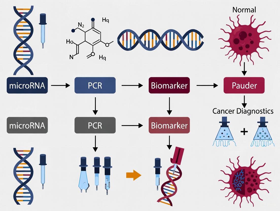
MicroRNAs as Biomarkers in Cancer Diagnostics: PCR-Based Approaches, Clinical Challenges, and Emerging Alternatives
This article comprehensively examines the role of microRNAs (miRNAs) as biomarkers in PCR-based cancer diagnostics for research and drug development professionals.
MicroRNAs as Biomarkers in Cancer Diagnostics: PCR-Based Approaches, Clinical Challenges, and Emerging Alternatives
Abstract
This article comprehensively examines the role of microRNAs (miRNAs) as biomarkers in PCR-based cancer diagnostics for research and drug development professionals. It explores the foundational biology of miRNAs and their deregulation in cancer, details established PCR methodologies like RT-qPCR and ddPCR for miRNA detection, and analyzes key technical and translational challenges limiting clinical adoption. The content further evaluates validation strategies and performance comparisons with other technologies, while discussing emerging amplification-free platforms that aim to overcome PCR limitations. By integrating current research and future perspectives, this review provides a critical resource for scientists advancing miRNA-based cancer detection and therapeutic development.
The Biology of microRNAs and Their Fundamental Role in Cancer Pathogenesis
MicroRNAs (miRNAs) are a class of small, non-coding RNA molecules, approximately 19-24 nucleotides in length, that play fundamental roles in post-transcriptional gene regulation. Since the landmark discovery of the first miRNA, lin-4, in Caenorhabditis elegans in 1993, our understanding of miRNA biogenesis and function has expanded tremendously [1] [2]. This technical guide provides a comprehensive overview of canonical and non-canonical miRNA biogenesis pathways, mechanistic actions in gene silencing, and their dynamic roles in cellular regulation. Framed within cancer research, this article further explores the immense diagnostic potential of circulating miRNAs as stable, sensitive biomarkers in PCR-based liquid biopsies, highlighting innovative detection technologies and experimental protocols driving precision oncology forward.
miRNA Biogenesis: From Transcription to Maturation
MiRNA biogenesis involves a meticulously coordinated multi-step process that transforms primary transcripts into mature, functional miRNAs, with both canonical and non-canonical pathways contributing to the rich diversity of the miRNA repertoire [1].
The Canonical Biogenesis Pathway
The canonical pathway represents the dominant mechanism for miRNA processing and involves two crucial RNase III enzymes, Drosha and Dicer [1] [2].
Transcription and Nuclear Processing: miRNA genes are predominantly transcribed by RNA polymerase II into primary miRNAs (pri-miRNAs), which are 5'-capped and polyadenylated [2]. These pri-miRNAs harbor one or more hairpin structures and are processed in the nucleus by the microprocessor complex, consisting of the RNase III enzyme Drosha and its cofactor DGCR8 (DiGeorge Syndrome Critical Region 8). This complex cleaves the pri-miRNA to release a ~70 nucleotide precursor miRNA (pre-miRNA) featuring a 2-nucleotide 3' overhang [1].
Nuclear Export and Cytoplasmic Processing: The pre-miRNA is exported to the cytoplasm via Exportin-5 (XPO5) in a RanGTP-dependent manner [2]. Within the cytoplasm, the RNase III enzyme Dicer, in partnership with TAR RNA-binding protein (TRBP), cleaves the terminal loop of the pre-miRNA, generating a transient ~22 nucleotide miRNA duplex [1] [3].
RISC Loading and Strand Selection: The mature miRNA duplex is loaded into the Argonaute (AGO) family of proteins (AGO1-4 in humans) to form the core of the miRNA-induced silencing complex (miRISC) [1]. Strand selection determines the functional guide strand; typically, the strand with lower thermodynamic stability at its 5' end is preferentially selected as the guide, while the passenger strand is degraded [1].
Non-Canonical Biogenesis Pathways
Several non-canonical pathways diversify miRNA biogenesis, functioning independently of Drosha or Dicer [1]:
Mirtrons: Originate from short introns that mimic pre-miRNA structural features. After debranching of the spliced intron, they are directly exported by Exportin-5 and processed by Dicer, bypassing Drosha cleavage [1].
Dicer-Independent miRNAs: Processed by Drosha from endogenous short hairpin RNA (shRNA) transcripts but are too short to be Dicer substrates. Instead, their maturation is completed by AGO2-mediated slicing of the passenger strand followed by 3'-5' trimming [1].
m7G-Capped pre-miRNAs: Bypass Drosha processing and are exported via Exportin-1. The m7G cap causes a strong strand bias, typically preventing the 5p strand from loading into Argonaute [1].
Table 1: Core Protein Complexes in miRNA Biogenesis
| Protein Complex/Component | Function | Localization |
|---|---|---|
| Drosha-DGCR8 Complex | Cleaves pri-miRNA to release pre-miRNA | Nucleus |
| Exportin-5 (XPO5) | Exports pre-miRNA to the cytoplasm | Nuclear Membrane |
| Dicer-TRBP Complex | Cleaves pre-miRNA to generate miRNA duplex | Cytoplasm |
| Argonaute (AGO1-4) | Loads mature miRNA guide strand to form miRISC | Cytoplasm |
Diagram 1: Canonical miRNA Biogenesis Pathway
Mechanisms of miRNA-Mediated Gene Regulation
MiRNAs fine-tune gene expression through sequence-specific interactions with target mRNAs, primarily leading to translational repression or mRNA destabilization [1] [2].
miRNA-mRNA Interaction and Target Recognition
The minimal functional unit is miRISC, comprising the guide strand and AGO protein [1]. Target specificity is determined by interaction with complementary sequences on target mRNAs, called miRNA response elements (MREs). The degree of MRE complementarity dictates the silencing mechanism [1]:
Perfect Complementarity: Leads to AGO2 endonuclease activity and direct cleavage of target mRNA, common in plants but rare in animals [1].
Partial Complementarity: Characteristic of animal miRNAs, where the "seed region" (nucleotides 2-8) provides primary specificity. This interaction recruits effector proteins that mediate translational repression and mRNA decay without cleavage [1].
Although MREs are predominantly located in the 3' untranslated region (3' UTR) of mRNAs, interactions with coding sequences and 5' UTRs have also been reported [1].
Silencing Mechanisms
For partially complementary targets, gene silencing occurs through a multi-step process [1]:
GW182 Recruitment: AGO recruits GW182 family proteins, which provide scaffolding for effector complexes.
Deadenylation: GW182 recruits PAN2-PAN3 and CCR4-NOT deadenylase complexes, removing the poly(A) tail.
Decapping: The decapping complex DCP1-DCP2 is recruited, removing the 5' cap.
Exonucleolytic Degradation: The decapped and deadenylated mRNA undergoes 5'-to-3' exonucleolytic degradation.
While translational repression can occur independently, mRNA decay is considered the predominant mechanism of miRNA-mediated silencing in animals [1].
miRNAs as Biomarkers in Cancer Diagnostics
The remarkable stability of circulating miRNAs (cmiRNAs) in biofluids, their tissue-specific expression patterns, and their involvement in carcinogenesis make them exceptionally attractive biomarkers for non-invasive cancer detection and monitoring [4] [5] [6].
Circulating miRNAs: Origin and Stability
CmiRNAs are released from cells into extracellular fluids through various mechanisms, including active secretion via exosomes, microvesicle shedding, and protein-mediated export (e.g., bound to AGO2 or high-density lipoproteins) [6]. Their association with these carriers protects them from endogenous RNase activity, conferring exceptional stability in circulation, even under harsh conditions like multiple freeze-thaw cycles [6].
miRNA Dysregulation in Cancer
Cancer cells exhibit widespread miRNA dysregulation, influenced by multiple mechanisms [2]:
- Genetic Alterations: Deletions, amplifications, or mutations in miRNA genes or their promoters.
- Epigenetic Modifications: Aberrant DNA methylation or histone modifications silencing tumor-suppressive miRNA genes.
- Transcription Factor Dysregulation: Oncogenic or tumor-suppressive transcription factors altering miRNA expression.
- Defects in Biogenesis Machinery: Impaired expression or function of Drosha, Dicer, or other processing proteins.
These alterations lead to two fundamental functional categories in cancer [5]:
OncomiRs: Oncogenic miRNAs that are overexpressed in cancer, promoting tumor development by targeting tumor suppressor genes (e.g., miR-21, which targets PTEN) [2].
Tumor-Suppressor miRNAs: Downregulated in cancer, whose normal function is to restrain tumor growth by targeting oncogenes (e.g., miR-15a/16-1 cluster targeting BCL2) [6] [2].
Table 2: Clinically Relevant miRNA Biomarkers in Cancer
| miRNA | Expression in Cancer | Proposed Function | Cancer Type(s) |
|---|---|---|---|
| miR-21 | Upregulated | OncomiR (targets PTEN, LZTFL1) | Breast, Colorectal, Gastric, DLBCL [5] [2] |
| miR-15a/16-1 | Downregulated | Tumor Suppressor (targets BCL2) | Chronic Lymphocytic Leukemia [6] [2] |
| miR-205-5p | Upregulated | Diagnostic Biomarker | Pancreatic Cancer [6] |
| miR-4488 | Upregulated | OncomiR, Response Biomarker | Metastatic Melanoma [4] |
| miR-579-3p | Downregulated | Tumor Suppressor, Response Biomarker | Metastatic Melanoma [4] |
| miR-141-3p | Upregulated | Diagnostic Biomarker | Prostate Cancer [7] |
| let-7 family | Downregulated | Tumor Suppressor | Lung, Ovarian Cancers [5] |
Advanced PCR-Based Methodologies for miRNA Detection
The short length of mature miRNAs presents unique challenges for detection that have been overcome by specialized molecular techniques, with PCR-based methods forming the cornerstone of sensitive and specific miRNA quantification in clinical research [4] [3].
Reverse Transcription Quantitative PCR (RT-qPCR)
RT-qPCR remains the gold standard for targeted miRNA quantification due to its sensitivity, specificity, and quantitative capabilities [3]. Key adaptations enable precise miRNA measurement:
Stem-Loop Reverse Transcription: Specialized stem-loop primers with a complementary 3' end bind specifically to the mature miRNA's 3' region. This creates an extended template that facilitates efficient reverse transcription and subsequent amplification, overcoming the challenge posed by the short miRNA sequence [3].
TaqMan Probe Chemistry: Fluorogenic probes provide sequence-specific detection, minimizing false positives from non-specific amplification. Universal reverse transcription and pre-amplification steps enable profiling of multiple miRNAs from limited sample material [4].
Digital PCR (dPCR) for Absolute Quantification
Digital PCR represents a technological advancement for absolute quantification of low-abundance miRNAs without requiring standard curves [4]. The principle involves:
Partitioning: The reaction mixture is partitioned into thousands of nanoliter-scale reactions.
Endpoint Amplification: Each partition undergoes PCR amplification.
Absolute Quantification: Partitions are scored as positive or negative based on fluorescence, and miRNA concentration is calculated using Poisson statistics.
Recent innovations include duplex dPCR assays that enable simultaneous detection of two miRNAs in a single reaction, conserving precious patient samples and improving throughput for biomarker ratios like the miRatio (miR-4488/miR-579-3p) used in metastatic melanoma management [4].
Diagram 2: miRNA Detection Workflow for Cancer Diagnostics
Experimental Protocol: Duplex dPCR for Circulating miRNAs
The following protocol, adapted from metastatic melanoma research, details the simultaneous quantification of two miRNAs from patient serum [4]:
Sample Preparation and RNA Extraction
- Collect peripheral blood into EDTA-coated tubes and separate serum by centrifugation.
- Extract total RNA (including miRNAs) from 200 μL serum using miRNeasy Mini Kit or Trizol reagent.
- For Trizol-based extraction: Add 750μL Trizol to 400μL blood, mix with 200μL chloroform, separate phases, and precipitate RNA from aqueous phase with isopropanol.
- Elute RNA in 20μL nuclease-free water. Note: RNA concentration is often below detection limits of spectrophotometers; use fixed input volume for reverse transcription.
Reverse Transcription and Preamplification
- Perform reverse transcription using TaqMan Advanced miRNA cDNA Synthesis Kit.
- Use 2μL total RNA as input for universal reverse transcription of all miRNAs.
- Include a preamplification step to enrich target sequences prior to detection.
Duplex Digital PCR Setup
- Prepare dPCR reaction mix containing:
- TaqMan probes for miR-4488 and miR-579-3p with different fluorophores
- Advanced miRNA Assay reagents
- Preamplified cDNA template
- Partition reaction into approximately 20,000 nanoliter-scale reactions using a droplet generator or chip-based system.
- Perform PCR amplification with the following cycling conditions:
- Enzyme activation: 95°C for 10 minutes
- 40 cycles of: Denature at 95°C for 30 seconds, Anneal/Extend at 60°C for 1 minute
- Signal stabilization: 98°C for 10 minutes
- Read droplets/chips on a droplet reader to determine positive and negative partitions for each target.
Data Analysis and MiRatio Calculation
- Calculate absolute concentrations (copies/μL) of each miRNA based on Poisson distribution of positive partitions.
- Compute miRatio = [miR-4488] / [miR-579-3p]
- Establish clinical cutoff values for prognostic stratification (e.g., high vs. low miRatio).
The Scientist's Toolkit: Essential Research Reagents
Table 3: Key Research Reagents for miRNA Studies in Cancer Diagnostics
| Reagent/Technology | Function | Example Products |
|---|---|---|
| RNA Extraction Kits | Isolation of high-quality total RNA including small RNAs from serum, plasma, or tissues | miRNeasy Mini Kit (Qiagen), Trizol reagent |
| Stem-Loop RT Primers | miRNA-specific reverse transcription for mature miRNAs; provides specificity and enables amplification of short targets | TaqMan Advanced miRNA cDNA Synthesis Kit, miScript System |
| TaqMan Probes | Sequence-specific fluorescent probes for highly specific detection in qPCR and dPCR | TaqMan Advanced miRNA Assays |
| Digital PCR Systems | Absolute quantification of miRNA copies without standard curves by partitioning samples | QIAcuity, QuantStudio, Bio-Rad Droplet Digital PCR |
| Pre-amplification Kits | Signal enhancement for profiling multiple miRNAs from limited RNA inputs | TaqMan PreAmp Master Mix |
| miRNA Inhibitors/Mimics | Functional studies to suppress (antagomiRs) or restore (miRNA mimics) miRNA activity in cell models | Anti-miR inhibitors, miRNA mimics |
Future Perspectives and Challenges
The integration of miRNA biomarkers into clinical oncology presents both tremendous opportunities and significant challenges that must be addressed through continued innovation [6] [3].
Standardization Hurdles: Technical variability in RNA extraction, normalization strategies, and platform differences create barriers to clinical implementation. Establishing standardized protocols across institutions is critical for reproducible results [7].
Multiplexing and Signature Panels: While single miRNAs provide valuable information, multi-miRNA signatures or ratios (like miRatio) often demonstrate superior diagnostic and prognostic accuracy, reflecting the complex biological networks in cancer [4]. Emerging technologies that enable cost-effective multiplexing will be essential for clinical adoption.
Integration with Machine Learning: The analysis of complex miRNA expression data is increasingly leveraging machine learning algorithms to identify subtle patterns that distinguish cancer types, predict treatment response, and improve diagnostic sensitivity and specificity beyond traditional statistical methods [7].
Therapeutic Development: Beyond diagnostics, miRNAs represent promising therapeutic targets. miRNA mimics to restore tumor-suppressor function and antagomiRs to inhibit oncomiR activity are under active investigation, with challenges including targeted delivery and minimizing off-target effects [2].
As detection technologies continue to advance—with developments in biosensors, enhanced bioinformatics, and more sophisticated computational approaches—miRNA-based liquid biopsies are poised to become integral components of precision oncology, enabling earlier detection, improved monitoring, and personalized treatment strategies for cancer patients.
- Introduction: Overview of miRNA biogenesis and significance in cancer diagnostics.
- Mechanisms of miRNA dysregulation: Genomic alterations, epigenetic changes, and processing defects.
- Diagnostic approaches: PCR-based profiling, machine learning integration, and emerging technologies.
- Therapeutic strategies: miRNA mimics, inhibitors, and delivery systems.
- Methodology: Detailed protocols for miRNA analysis, reagents, and computational tools.
MicroRNA Dysregulation in Oncogenesis: Dual Roles as Oncogenes and Tumor Suppressors
MicroRNAs (miRNAs) have emerged as critical regulators of gene expression in cancer biology, functioning as both oncogenic drivers and tumor suppressors in various malignancies. These small non-coding RNA molecules, approximately 20-24 nucleotides in length, regulate gene expression through post-transcriptional mechanisms by binding to complementary sequences on messenger RNAs (mRNAs), leading to mRNA degradation or translation inhibition. Through this precise regulatory control, miRNAs influence fundamental biological processes including cell proliferation, differentiation, apoptosis, and metabolism, thereby maintaining cellular homeostasis. Dysregulation of miRNA expression can disrupt these critical processes, contributing significantly to oncogenesis by either promoting oncogenic pathways or impairing tumor-suppressive functions [8]. The stability of miRNAs in various biological fluids, including blood, and their differential expression patterns in diseased versus healthy subjects make them promising candidates for non-invasive cancer diagnostics [7]. Their detection via polymerase chain reaction (PCR)-based methods offers a viable approach for clinical translation, particularly when integrated with advanced computational approaches like machine learning to enhance diagnostic accuracy.
The biogenesis of miRNAs involves multiple complex steps, beginning with transcription by RNA polymerase II or III, which produces primary miRNA transcripts (pri-miRNAs). These pri-miRNAs undergo sequential processing by the RNase III enzymes Drosha and Dicer to form mature miRNA molecules that are incorporated into the RNA-induced silencing complex (RISC) to regulate target mRNAs [9]. Understanding this biogenesis pathway is crucial for comprehending how disruptions at various stages can contribute to miRNA dysregulation in cancer. The intricate involvement of miRNAs in carcinogenesis, coupled with their stability in circulating blood, positions them as ideal biomarkers for PCR-based cancer diagnostics within precision medicine frameworks [7] [8].
Mechanisms of miRNA Dysregulation in Cancer
Genomic and Epigenetic Alterations
miRNA genes are frequently located in genomic regions susceptible to cancer-associated alterations, including copy number variations (CNVs), translocations, and deletions. These genomic instabilities can lead to either amplification of oncogenic miRNAs (oncomiRs) or deletion of tumor-suppressive miRNAs, significantly impacting miRNA expression profiles. For instance, the frequent amplification of chromosome 13q31, which contains the miR-17-92 cluster, is observed in several lymphoma types and contributes to enhanced cell proliferation and tumor development [9]. Similarly, deletions in chromosomal regions harboring tumor-suppressive miRNAs like miR-15a and miR-16-1 are commonly observed in chronic lymphocytic leukemia and various solid tumors [8].
Epigenetic modifications, particularly DNA methylation of promoter regions and histone modifications, represent another crucial mechanism of miRNA dysregulation in cancer. Hypermethylation of CpG islands in promoter regions associated with tumor-suppressive miRNAs leads to their transcriptional silencing. For example, miR-127, miR-124-1, and miR-129-2 are frequently inactivated through promoter hypermethylation in various solid cancers [9]. Additionally, histone deacetylation and other chromatin modifications can contribute to the suppression of miRNA expression, further exacerbating the oncogenic phenotype. The interplay between genetic and epigenetic mechanisms creates a complex regulatory network that profoundly influences miRNA expression patterns during carcinogenesis.
Defects in miRNA Processing Machinery
Aberrations in the core miRNA processing machinery represent another significant mechanism contributing to miRNA dysregulation in cancer. Components such as Drosha, DGCR8, Exportin-5, and Dicer are essential for the proper maturation of miRNAs, and defects in any of these components can lead to global alterations in miRNA expression profiles. Mutations in the DICER1 gene are associated with DICER1 syndrome, which increases susceptibility to various cancers, including pulmonary pleuroblastoma, cystic nephroma, and thyroid gland neoplasia [9]. These mutations interfere with normal miRNA biogenesis and alter gene expression patterns, driving oncogenesis.
Similarly, aberrant expression of Drosha or DGCR8 has been documented in various malignancies, though their precise roles in carcinogenesis remain somewhat controversial. Despite the ongoing debate, it is well-established that disruptions in miRNA processing machinery correlate strongly with widespread alterations in miRNA expression patterns observed in cancer cells [9]. Additionally, post-translational modifications of processing proteins and interactions with regulatory RNA-binding proteins can further modulate miRNA biogenesis and activity, adding another layer of complexity to the regulation of miRNA expression in cancer.
Table 1: Common miRNA Dysregulation Patterns in Solid Tumors
| Cancer Type | Oncogenic miRNAs (Upregulated) | Tumor-Suppressive miRNAs (Downregulated) |
|---|---|---|
| Breast Cancer | miR-21, miR-155, miR-10b, miR-210, miR-27a | miR-125b, miR-205, let-7, miR-31, miR-34a |
| Colorectal Cancer | miR-21, miR-17-5p, miR-155, miR-126 | miR-34a, miR-143, miR-145, miR-137-3p |
| Lung Cancer | miR-21, miR-31, miR-155, miR-210, miR-17-92 | let-7, miR-34a, miR-126, miR-200b, miR-124a |
| Prostate Cancer | miR-21, miR-221, miR-222, miR-141 | miR-34a, miR-125b, miR-145, let-7c |
| Hepatocellular Carcinoma | miR-21, miR-221, miR-222, miR-224 | miR-122, miR-125b, miR-199a, let-7 |
| Glioblastoma | miR-21, miR-10b, miR-221, miR-17-92 | miR-34a, miR-128, miR-137, miR-124 |
Transcriptional Regulation and miRNA Sponging
Transcriptional regulation of miRNA genes by oncogenic or tumor-suppressive transcription factors represents another layer of control that is frequently disrupted in cancer. For instance, the tumor suppressor p53 transcriptionally activates miR-34 family members, which are involved in cell cycle arrest and apoptosis, while Myc activates the oncogenic miR-17-92 cluster. Mutations in these transcription factors can therefore indirectly alter miRNA expression patterns. Additionally, the phenomenon of miRNA sponging through competitive endogenous RNAs (ceRNAs) has emerged as a significant mechanism of miRNA regulation. Long non-coding RNAs (lncRNAs), circular RNAs (circRNAs), and other RNA transcripts containing miRNA response elements can sequester miRNAs, preventing them from binding to their natural mRNA targets. This mechanism effectively reduces the available concentration of functional miRNAs within the cell, contributing to dysregulation of their target genes [9].
miRNA Dysregulation in Cancer Pathways and Diagnostics
Oncogenic miRNAs (OncomiRs) and Their Pathways
Oncogenic miRNAs, commonly referred to as oncomiRs, are frequently overexpressed in various cancers and promote tumorigenesis by targeting and repressing critical tumor suppressor genes and pathways. Among the most prominently upregulated oncomiRs is miR-21, which is overexpressed in breast, lung, gastric, colorectal, brain, and many other cancers. miR-21 exerts its oncogenic effects by inhibiting key tumor suppressors such as PTEN, PDCD4, and TPM1, thereby enhancing tumor growth, metastasis, and chemoresistance [8]. Another significant oncomiR, miR-221, is highly expressed in hepatocellular carcinoma, melanoma, colon cancer, and renal cell carcinoma, where it promotes uncontrolled proliferation by targeting cell cycle inhibitors like p27 and p57 [8].
Other notable oncomiRs include miR-155, which plays a crucial role in lymphomagenesis and solid tumors; miR-182, involved in melanoma progression; and members of the miR-17-92 cluster, which are amplified in several lymphoma types and solid tumors. These oncomiRs typically target multiple components of tumor-suppressive pathways, creating a coordinated oncogenic program that drives cancer progression. The ability of individual oncomiRs to regulate multiple targets within oncogenic networks makes them attractive therapeutic targets, as their inhibition could simultaneously restore multiple tumor-suppressive pathways. Furthermore, the detection of these oncomiRs in circulation provides valuable opportunities for non-invasive cancer diagnosis and monitoring using PCR-based approaches [7] [8].
Tumor-Suppressive miRNAs and Their Mechanisms
Tumor-suppressive miRNAs are frequently downregulated in cancer, leading to the loss of their inhibitory effects on oncogenic pathways and subsequent tumor progression. The let-7 family represents one of the most significant tumor-suppressive miRNA families, frequently suppressed in lung cancer where its loss leads to unchecked activity of oncogenes such as RAS and HMGA2 [8]. Similarly, the miR-34 family functions as a critical mediator of p53 tumor suppressor activity and is downregulated in diverse cancer types, making it a promising candidate for therapeutic replacement strategies.
Other important tumor-suppressive miRNAs include miR-143 and miR-145, which are consistently downregulated in bladder, breast, lung, colorectal, and pancreatic cancers [8]. miR-126 and miR-125b are reduced in breast, lung, and gastric cancers, highlighting their importance in regulating apoptosis and cellular differentiation. The restoration of these tumor-suppressive miRNAs represents a promising therapeutic approach, as it can simultaneously target multiple oncogenic pathways. From a diagnostic perspective, the decreased expression of specific tumor-suppressive miRNAs in tissue or circulation can serve as valuable biomarkers for early cancer detection using sensitive PCR-based methods [7] [8].
Table 2: Key miRNA Biomarkers in Cancer Diagnostics and Their Clinical Significance
| miRNA | Role in Cancer | Primary Cancer Types | Detection Method | Clinical Significance |
|---|---|---|---|---|
| miR-21-5p | Oncogenic | Prostate, Breast, Lung, Colorectal | RT-PCR, NGS | Discriminates PCa from BPH; associated with poor prognosis |
| miR-141-3p | Oncogenic | Prostate, Colorectal | RT-PCR | Elevated in metastatic disease; biomarker for aggressive PCa |
| miR-221-3p | Oncogenic | Prostate, Hepatocellular, Melanoma | RT-PCR, NGS | Promotes proliferation; miR-141-3p/miR-221-3p ratio has diagnostic value |
| miR-375-3p | Context-dependent | Prostate, Head & Neck | RT-PCR | Associated with treatment resistance |
| let-7 family | Tumor-suppressive | Lung, Breast, Gastric | RT-PCR, Microarray | Loss correlates with RAS activation; prognostic marker |
| miR-34a | Tumor-suppressive | Various solid tumors | RT-PCR, NGS | Mediates p53 function; therapeutic candidate |
miRNA Dysregulation in Hematological Malignancies
miRNA dysregulation is not limited to solid tumors but also plays a crucial role in hematological malignancies. In multiple myeloma (MM), for example, miR-21 and miR-221 are frequently upregulated, enhancing cell survival, proliferation, and drug resistance, while tumor-suppressive miRNAs like miR-29b and miR-34a are downregulated, contributing to disease aggressiveness and poor prognosis [8]. Similarly, in acute lymphoblastic leukemia (ALL), upregulated miRNAs such as miR-21, miR-221, miR-155, and miR-181a promote leukemogenesis by enhancing cell proliferation and shortening survival, whereas the downregulation of miR-34a and miR-29b is associated with disease progression [8].
The patterns of miRNA dysregulation in hematological malignancies often reflect those observed in solid tumors, with overlapping oncomiRs and tumor-suppressive miRNAs across different cancer types. This conservation of miRNA dysregulation patterns highlights the fundamental role these molecules play in oncogenesis and suggests that diagnostic and therapeutic approaches targeting these miRNAs could have broad applications across cancer types. The detection of dysregulated miRNAs in peripheral blood or bone marrow samples from patients with hematological malignancies using PCR-based methods offers promising avenues for diagnosis, prognosis, and treatment monitoring [7] [8].
PCR-Based Diagnostic Approaches and Emerging Technologies
miRNA Profiling Using Reverse Transcription PCR
Reverse transcription polymerase chain reaction (RT-PCR) has become the primary method for validating miRNA biomarkers in clinical research settings due to its sensitivity, specificity, and accessibility compared to more complex high-throughput approaches like next-generation sequencing (NGS). The process typically begins with RNA extraction from clinical samples (e.g., whole blood, plasma, serum, or tissue), followed by reverse transcription using miRNA-specific stem-loop primers that enhance specificity and detection efficiency. Quantitative PCR is then performed using systems such as the Applied Biosystem QuantStudio, with SYBR Green or TaqMan chemistry for detection [7].
The use of whole blood for miRNA profiling offers distinct advantages, including higher miRNA yield, reduced susceptibility to pre-analytical variability, and a more comprehensive representation of systemic disease states compared to plasma or serum [7]. In a prospective cohort study on prostate cancer diagnosis, whole blood miRNA profiling identified miR-21-5p, miR-141-3p, and miR-221-3p as significant discriminators between prostate cancer (PCa) and benign prostatic hyperplasia (BPH). The expression ratios of these miRNAs, particularly miR-141-3p/miR-221-3p, demonstrated superior sensitivity and specificity compared to traditional prostate-specific antigen (PSA) testing [7]. However, challenges remain in the clinical application of miRNA biomarkers using RT-PCR, including technical variability introduced by differences in devices, methodologies, and sample handling, which can result in data variations that do not accurately reflect the biological state of the patient [7].
Integration of Machine Learning for Enhanced Diagnostics
The integration of machine learning (ML) algorithms with miRNA expression data represents a transformative approach to enhancing diagnostic accuracy in cancer detection. ML models, particularly ensemble methods like random forests, can capture complex, non-linear relationships in multiple miRNA expression patterns that may go undetected by traditional analysis methods. In a study on prostate cancer diagnosis, a random forest ML model trained on miRNA expression data achieved an accuracy of 77.42% with an AUC of 0.78 during verification, and 74.07% accuracy with 0.75 AUC in validation [7].
These models excel in analyzing high-dimensionality, non-linear, and noisy biological datasets, making them particularly suited for miRNA expression data from RT-PCR. By processing data from multiple miRNAs simultaneously, ML algorithms can identify subtle patterns that distinguish cancer from benign conditions, significantly improving both sensitivity and specificity compared to individual miRNA biomarkers or traditional diagnostic tests like PSA for prostate cancer [7]. The bioinformatics analysis supporting these models can confirm the association of selected miRNAs with critical cancer pathways, including PD-L1/PD-1 checkpoint and androgen receptor signaling, validating the biological relevance of the findings and providing insights into potential therapeutic targets [7].
Emerging Amplification-Free Detection Technologies
While PCR-based methods currently dominate miRNA diagnostics, amplification-free or "PCR-free" technologies represent an emerging frontier that addresses several limitations of conventional PCR approaches. These innovative methods, including bead-based assays and sensor detection platforms, offer streamlined workflows, reduced error rates, and enhanced compatibility with various clinical sample types [10]. Crucially, they enable absolute quantification without the need for pre-nucleic acid extraction, reverse transcription, or amplification, while also allowing simultaneous detection of multiple miRNAs within a single assay [10].
These emerging technologies provide cost-effective and scalable solutions for comprehensive biomarker profiling, potentially overcoming long-standing technical barriers that have hindered the clinical translation of miRNA biomarkers. The transition from PCR-based to PCR-free technologies represents a significant step forward in miRNA diagnostics, supporting the advancement of precision medicine and offering promise for improving early cancer detection in routine clinical settings [10]. However, despite these advances, broader population studies and standardization of protocols are still needed to ensure scalability and clinical applicability of miRNA-based diagnostics [7].
Therapeutic Strategies and Clinical Translation
miRNA-Based Therapeutic Approaches
miRNA-based therapeutic strategies primarily involve two fundamental approaches: miRNA inhibition for oncogenic miRNAs and miRNA replacement for tumor-suppressive miRNAs. For oncogenic miRNAs (oncomiRs) that are overexpressed in cancer, synthetic inhibitors such as antimiRs or antagomiRs are designed to sequester or degrade the target miRNA, thereby restoring the expression of repressed tumor suppressor genes. These inhibitors are typically chemically modified oligonucleotides with enhanced stability and binding affinity, such as locked nucleic acid (LNA)-modified anti-miRNAs [8]. Conversely, for tumor-suppressive miRNAs that are downregulated in cancer, miRNA mimics are used to restore their lost function. These synthetic double-stranded RNA molecules mimic the endogenous mature miRNA and are designed to reconstitute the tumor-suppressive activity of the natural miRNA [8].
Both therapeutic approaches have shown promise in preclinical models and are gradually advancing to clinical trials. For instance, MRX34, a liposomal formulation of a miR-34 mimic, became the first miRNA-based therapy to enter human clinical trials for cancer treatment, highlighting the translational potential of miRNA replacement strategies. Similarly, LNA-based inhibitors targeting miR-155 have shown efficacy in preclinical models of lymphoma and are advancing toward clinical development. The ability of individual miRNAs to regulate multiple genes within biological networks offers the advantage of simultaneously targeting multiple pathways involved in oncogenesis, potentially reducing the likelihood of resistance development compared to single-target agents [8].
Delivery Systems and Challenges
The effective delivery of miRNA-based therapeutics to tumor sites remains a significant challenge in the field. Current delivery strategies include lipid nanoparticles, viral vectors, polymeric nanoparticles, and natural delivery vehicles such as exosomes or cell-based systems. Lipid nanoparticles, similar to those used in mRNA COVID-19 vaccines, have shown promise in protecting miRNA therapeutics from degradation and facilitating their cellular uptake [8]. Viral vectors, particularly adeno-associated viruses (AAV), offer efficient gene transfer but face challenges related to immunogenicity and payload capacity.
Despite these advances, several obstacles remain in the clinical translation of miRNA therapeutics, including off-target effects, immune activation, delivery inefficiencies, and potential toxicity. Strategies to improve specificity include optimizing chemical modifications to enhance target affinity and reduce off-target effects, as well as developing tissue-specific delivery systems to minimize exposure to healthy tissues. Additionally, the complex regulatory networks governed by miRNAs pose challenges in predicting and managing potential compensatory mechanisms and network perturbations following therapeutic intervention [8]. Ongoing research focuses on addressing these challenges through advanced formulation design, combination therapies, and patient stratification based on miRNA profiling to identify those most likely to benefit from miRNA-based treatments.
Experimental Methodology and Research Toolkit
Sample Preparation and miRNA Profiling Protocol
Sample collection and RNA isolation represent critical initial steps in miRNA biomarker research. For blood-based miRNA profiling, peripheral venous blood is typically collected into EDTA-coated tubes prior to biopsy or treatment initiation. RNA extraction commonly utilizes Trizol reagent-based methods, starting with 400 μL of blood mixed with 750 μL of Trizol reagent. Following chloroform addition and phase separation, the upper aqueous phase is combined with isopropanol to precipitate RNA, and the resulting pellet is resuspended in DEPC-treated water [7]. RNA concentration and quality are assessed using spectrophotometric methods such as NanoDrop.
For reverse transcription, miRNA-targeted stem-loop primers are employed to enhance specificity and detection efficiency. The RevertAid First Strand cDNA Synthesis Kit provides a reliable approach for cDNA synthesis [7]. Quantitative PCR is then performed in triplicate reactions using systems like the Applied Biosystem QuantStudio with Maxima SYBR Green/ROX RT-PCR Master Mix. Delta Ct values are calculated by subtracting the Ct value of an endogenous control (commonly RNU6) for each sample, enabling normalization and comparative analysis of miRNA expression levels [7]. This standardized protocol ensures reproducible and reliable miRNA quantification for diagnostic applications.
Research Reagent Solutions and Computational Tools
Table 3: Essential Research Reagents and Computational Tools for miRNA Analysis
| Category | Specific Product/Tool | Application/Function |
|---|---|---|
| RNA Isolation | Trizol Reagent | Total RNA extraction from blood, plasma, tissue |
| Reverse Transcription | RevertAid First Strand cDNA Synthesis Kit | cDNA synthesis with stem-loop primers for miRNA detection |
| Quantitative PCR | Maxima SYBR Green/ROX RT-PCR Master Mix | Fluorescence-based detection of amplified miRNA products |
| PCR Instrument | Applied Biosystem QuantStudio 6 Flex | High-throughput real-time PCR analysis |
| Endogenous Control | RNU6 | Reference gene for normalization of miRNA expression data |
| Data Analysis | Random Forest ML Models | Pattern recognition in miRNA expression data for classification |
| Genomic Visualization | Gviz Bioconductor Package | Plotting genomic data and annotation features along coordinates |
| Pathway Analysis | Bioinformatics Tools (KEGG, GO) | Association of dysregulated miRNAs with cancer pathways |
The Gviz package in Bioconductor provides specialized capabilities for visualizing genomic data along with miRNA annotation features. This flexible framework allows researchers to plot various types of genomic data in the context of diverse genome annotation features, integrating publicly available genomic annotation data from sources like UCSC or ENSEMBL [11] [12]. The package uses a track-based visualization approach, where individual types of genomic features or data are represented by separate tracks, all sharing the same genomic coordinate system. This enables clear visualization of miRNA genes in their genomic context, along with expression data and other relevant annotations [12].
For machine learning analysis, random forest models and other ensemble methods are particularly valuable for analyzing complex miRNA expression datasets. These models can be implemented using various R or Python libraries and are effective at capturing non-linear relationships and interactions between multiple miRNA expressions that might be missed by traditional statistical methods [7]. The integration of these computational tools with experimental data enables comprehensive analysis of miRNA dysregulation patterns and their clinical implications in cancer diagnostics and therapeutics.
Pathway Diagrams and Experimental Workflows
Diagram 1: miRNA Biogenesis Pathway. This diagram illustrates the multi-step process of miRNA maturation, from transcription to functional RISC complex formation, highlighting key processing enzymes.
Diagram 2: miRNA Diagnostic Workflow. This diagram outlines the integrated experimental-computational pipeline for miRNA-based cancer diagnostics, from sample collection to machine learning classification.
Diagram 3: miRNA Therapeutic Strategies. This diagram illustrates the two primary approaches for miRNA-based cancer therapy: inhibition of oncogenic miRNAs and replacement of tumor-suppressive miRNAs, highlighting delivery challenges.
Circulating microRNAs (miRNAs) have emerged as a revolutionary class of biomarkers in molecular diagnostics, offering a non-invasive window into human health and disease. These small non-coding RNAs demonstrate remarkable stability in biofluids, are actively involved in critical oncogenic pathways, and can be quantitatively detected using PCR-based technologies. This technical guide synthesizes current evidence on the discovery pipelines, stability mechanisms, and analytical protocols for circulating miRNAs, with a specific focus on their application in cancer diagnostics. We further explore how integration with high-throughput profiling and machine learning is poised to overcome existing challenges and solidify the role of miRNAs in precision oncology.
MicroRNAs (miRNAs) are short (∼18–25 nucleotides), non-coding RNA molecules that function as post-transcriptional regulators of gene expression [6]. Their discovery in circulation has fundamentally altered the landscape of biomarker discovery, positioning them as powerful tools for non-invasive liquid biopsies. The profound significance of circulating miRNAs stems from their dual role as both passive indicators of disease state and active participants in intercellular communication and disease pathogenesis [13] [14].
In the context of cancer, miRNA dysregulation is a hallmark of tumorigenesis. These molecules orchestrate complex oncogenic networks, including PI3K/AKT signaling, Wnt/β-catenin, epithelial-mesenchymal transition (EMT), and immune regulation [15]. For instance, miR-21, one of the most frequently upregulated miRNAs in cancer, promotes cell survival by targeting tumor suppressors like PTEN and PDCD4, and its high serum levels are strongly associated with advanced-stage disease and poor survival [14]. Conversely, the let-7 family acts as a tumor suppressor by regulating critical oncogenes such as RAS and HMGA2, and its consistent downregulation is observed throughout carcinogenesis [15].
A pivotal biological feature of circulating miRNAs is their exceptional stability in biofluids like plasma, serum, and saliva—an environment rich in RNases. This stability is conferred through their packaging into extracellular vesicles (EVs) such as exosomes, or through complex formation with proteins like Argonaute 2 (AGO2) and lipoproteins [13] [6]. This protection allows them to withstand routine clinical handling, including multiple freeze-thaw cycles, making them exceptionally suitable for clinical assay development [16].
Stability and Protection Mechanisms in Biofluids
The utility of circulating miRNAs as robust biomarkers is fundamentally rooted in their stability. Understanding the mechanisms behind this stability is crucial for developing reliable assays.
Mechanisms of Stability
Circulating miRNAs are protected from degradation by RNases through several well-characterized mechanisms:
- Association with RNA-Binding Proteins: miRNAs can be bound to Argonaute (AGO) proteins, key components of the RNA-induced silencing complex (RISC), which provide a protective shield [6].
- Packaging into Extracellular Vesicles: A significant proportion of circulating miRNAs are encapsulated within membrane-bound vesicles, primarily exosomes and microvesicles, which are secreted by cells into the extracellular environment [13] [14]. The lipid bilayer of these vesicles physically protects the miRNA cargo from enzymatic degradation.
- Association with Lipoproteins: miRNAs have also been found to be associated with high-density lipoproteins (HDL), which further contributes to their longevity in the circulation [6].
Empirical Evidence of Stability
Recent studies have systematically quantified the stability of miRNAs under various pre-analytical conditions, confirming their resilience and informing sample handling protocols.
- A 2024 study analyzed the longitudinal stability of plasma miRNAs in healthy adults over a 3-month period. The research identified 74 miRNAs that exhibited high test-retest reliability and low percentage level drift, demonstrating their inherent stability within an individual over time. The study also highlighted that technical variance could be effectively corrected using calibration strategies, such as spike-in controls (e.g., cel-miR-39-3p) [17].
- A 2025 study specifically tested the stability of miRNA profiles in serum and plasma under different storage temperatures (4°C or 25°C) for varying periods (0–24 h). The results showed that mean Cq values for specific miRNAs (e.g., miR-15b, miR-16, miR-21, miR-24, and miR-223) remained consistent over 24 hours. Small-RNA sequencing detected approximately 650 different miRNA signals in plasma, with over 99% of the miRNA profile unchanged even when blood draw tubes were left at room temperature for 6 hours prior to processing [16].
Table 1: Key Findings from miRNA Stability Studies
| Study Focus | Key Findings | Implications for Protocol Development |
|---|---|---|
| Longitudinal Stability (3 months) [17] | 74 miRNAs showed high test-retest reliability. Spike-in controls (cel-miR-39-3p) are effective for technical calibration. | Enables longitudinal disease monitoring. Supports use of calibration for robust data. |
| Short-Term Stability (24 hours) [16] | Cq values for key miRNAs (miR-15b, miR-16, etc.) were stable. 99% of plasma miRNAome unchanged after 6h at RT. | Assays can withstand routine clinical processing delays. |
| Impact of Hemolysis [17] | Hemolysis and tobacco use have the greatest impact on miRNA levels and variance. | Highlights critical pre-analytical variables to control during sample collection. |
Circulating miRNAs as Non-Invasive Biomarkers in Cancer
The application of circulating miRNAs for the early detection, prognosis, and therapeutic monitoring of cancer represents a paradigm shift in precision oncology, moving away from invasive tissue biopsies.
Diagnostic and Prognostic Utility
Multi-miRNA panels have consistently demonstrated superior diagnostic performance compared to single miRNAs, reflecting the multifactorial nature of cancer.
- A 2025 meta-analysis of colorectal cancer (CRC) diagnostics, which included 29 studies and 5,497 participants, found that multi-miRNA panels had a pooled sensitivity of 0.85 and specificity of 0.84, with an area under the curve (AUC) of 0.90 [15].
- Panels derived from plasma samples showed the highest balanced performance (sensitivity 0.88; specificity 0.87), and three-miRNA panels exhibited the best diagnostic trade-offs [15].
- In lung cancer, EV-associated miRNAs show distinct expression signatures that correlate with tumour stage, progression, and overall survival. Diagnostic models based on plasma EV-miRNA signatures can distinguish between malignant and benign pulmonary nodules, with some studies suggesting potential for predicting malignancy in nodules smaller than 1 cm [13].
Predictive Biomarkers for Therapy
Circulating miRNAs also show great promise in predicting response to therapy.
- In a phase II trial (T1219) for advanced biliary tract cancer, a three-miRNA signature (hsa-miR-16-5p, hsa-miR-93-5p, and hsa-miR-126-3p) was identified. High baseline levels of these miRNAs were associated with response to chemoimmunotherapy. Specifically, high hsa-miR-16-5p expression was correlated with longer progression-free survival and overall survival [18].
- Tracking dynamic changes in EV-miRNA profiles during treatment may provide predictive insights into responsiveness to immunotherapy and targeted therapy in lung cancer and other malignancies [13].
Table 2: Clinically Significant Multi-miRNA Panels in Cancer Detection and Monitoring
| Cancer Type | miRNA Signature | Clinical Utility & Performance | Source |
|---|---|---|---|
| Colorectal Cancer | Various panels (e.g., miR-15b, miR-21, miR-31) | Pooled Sensitivity: 0.85; Specificity: 0.84; AUC: 0.90 [15] | Plasma/Serum |
| Biliary Tract Cancer | hsa-miR-16-5p, hsa-miR-93-5p, hsa-miR-126-3p | Predictive of chemoimmunotherapy response; high miR-16-5p associated with longer survival (HR=0.44 for PFS) [18] | Plasma |
| Non-Small Cell Lung Cancer | EV-associated signatures (e.g., miR-1247-5p, miR-301b-3p) | Distinguishes malignant from benign nodules; correlates with tumour stage and survival [13] [6] | Plasma EVs |
| Pancreatic Cancer | miR-205-5p | Discerns pancreatic cancer from chronic pancreatitis with 91.5% accuracy [6] | Serum |
Experimental Workflows: From Sample to Signal
Robust and standardized experimental protocols are the bedrock of reliable circulating miRNA biomarker discovery and validation. The following workflow details a method for exosomal miRNA profiling from serum samples.
Sample Collection and Processing
Blood sample processing has a substantial impact on the results of miRNA profiling and must be controlled to minimize pre-analytical variation.
- Collection: Draw whole blood into collection tubes (e.g., red-top vacutainer for serum, K2EDTA tubes for plasma).
- Clotting (for serum): Incubate samples at room temperature for 30 minutes to allow complete coagulation [14].
- Centrifugation: Centrifuge coagulated samples or plasma at 1,500 × g for 10 minutes at room temperature. Carefully transfer the supernatant (serum/plasma) to a new tube.
- Additional Clearance: Perform a second centrifugation of the supernatant at a higher speed (e.g., 2,500 × g for 5-10 minutes) to remove residual platelets, microparticles, and debris [14]. Aliquot and store the final supernatant at -80°C.
Exosome and EV Isolation
While multiple methods exist, precipitation-based kits offer a balance of simplicity and efficiency.
- Principle: Use a kit (e.g., miRCURY Exosome Isolation Kit) based on capturing water molecules to reduce the hydrate envelope of particles, leading to their precipitation.
- Protocol: Thaw serum on ice. Mix with a precipitation solution and incubate at 4°C for 1 hour. Centrifuge at low speed to pellet the exosomes. Resuspend the pellet in a provided resuspension buffer [14].
- Characterization: Isolated vesicles should be characterized for size distribution using Nanoparticle Tracking Analysis (NTA) and for presence of exosome markers (e.g., CD63, CD9) using flow cytometry or Western blot, as per guidelines from the International Society for Extracellular Vesicles (ISEV) [14].
RNA Purification and Quality Control
- Extraction: Use column-based kits designed for small RNA isolation from biofluids (e.g., miRCURY RNA Isolation Kit). The process involves lysis, protein precipitation, binding of RNA to a column, washing, and elution in RNase-free water [14].
- Spike-in Control: To monitor RNA isolation efficiency and correct for technical variation, spike a known amount of exogenous synthetic miRNA (e.g., cel-miR-39) into each sample during the denaturation step [17] [14].
- Hemolysis Assessment: Evaluate hemolysis, a major confounder, by measuring the absorbance of free hemoglobin or using a miRNA-based signature (e.g., ratio of miR-451a to miR-23a-3p). Samples with appreciable hemolysis should be noted or excluded [19] [17].
miRNA Profiling and Quantification
- Reverse Transcription: Synthesize cDNA using a High-Capacity RNA-to-cDNA kit. For genome-wide profiling, systems requiring only a single cDNA synthesis reaction for the full miRNome are preferred.
- Quantification:
- RT-qPCR: The gold standard for targeted validation. Use TaqMan MicroRNA Assays or SYBR Green-based systems for precise quantification of specific miRNAs. All reactions should be performed in technical replicates [16] [14].
- High-Throughput Methods: For discovery phases, microarray or small RNA-sequencing can profile hundreds to thousands of miRNAs simultaneously, identifying differentially expressed signatures [6] [14].
- Data Analysis: Normalize Cq data using global mean normalization, reference miRNAs (e.g., miR-16-5p), or spike-in controls (cel-miR-39) [17]. Statistical analysis (e.g., Student's t-test, ANOVA) and machine learning models can then be applied to identify diagnostic signatures and build classifiers.
Diagram 1: Experimental workflow for circulating miRNA analysis from blood collection to data analysis.
The Scientist's Toolkit: Essential Research Reagents
Successful discovery and validation of circulating miRNA biomarkers depend on a suite of reliable reagents and instruments.
Table 3: Essential Reagents and Kits for Circulating miRNA Research
| Category / Reagent | Specific Product Examples | Function & Application Notes |
|---|---|---|
| RNA Isolation Kits | miRCURY RNA Isolation Kit (Exiqon), miRNeasy Serum/Plasma Kit (Qiagen) | Purify total RNA including the small RNA fraction (<200 nt) from biofluids; critical for miRNA recovery. |
| Exosome/EV Isolation | miRCURY Exosome Isolation Kit (Exiqon), Total Exosome Isolation Kits (Thermo Fisher) | Precipitate or immuno-capture extracellular vesicles from serum/plasma to analyze vesicle-encapsulated miRNAs. |
| Spike-in Controls | cel-miR-39 (synthetic) | Exogenous RNA added during lysis to normalize for variations in RNA extraction efficiency and qPCR inhibition. |
| cDNA Synthesis Kits | High-Capacity RNA-to-cDNA Kit (Thermo Fisher), miRCURY LNA RT Kit (Qiagen) | Convert RNA into cDNA; specialized kits are optimized for miRNA templates. |
| qPCR Assays | TaqMan MicroRNA Assays (Thermo Fisher), miRCURY LNA miRNA PCR Assays (Qiagen) | Gene-specific primers and probes for highly sensitive and specific quantification of individual miRNAs by RT-qPCR. |
| Profiling Platforms | Affymetrix GeneChip miRNA Array, Illumina Small RNA-Seq | High-throughput platforms for unbiased discovery of miRNA signatures across the whole miRNome. |
| Characterization Instruments | NanoSight LM10 (NTA), Attune NxT Flow Cytometer | Characterize the size, concentration, and surface markers (CD63, CD9) of isolated extracellular vesicles. |
The field of circulating miRNAs is rapidly evolving, driven by technological advancements and a deeper understanding of their biology. Key future directions include:
- Integration with Multi-Omics and AI: Combining miRNA signatures with other molecular data (e.g., genomics, proteomics) and analyzing them with machine learning algorithms will enhance the accuracy of diagnostic and predictive models [13] [6]. AI-driven diagnostics are already being used to identify complex miRNA patterns associated with difficult-to-diagnose cancers.
- Single-EV Analysis: Emerging technologies for single extracellular vesicle detection will help unravel the heterogeneity of EVs and identify the most disease-relevant vesicle subpopulations and their miRNA cargo [13].
- Standardization and Clinical Translation: The major hurdle remains the lack of standardized protocols for sample processing, EV isolation, and miRNA quantification. Concerted efforts to establish standard operating procedures (SOPs) are essential for translating these promising biomarkers into routine clinical practice [13] [15] [14].
In conclusion, circulating miRNAs represent a cornerstone of the liquid biopsy paradigm. Their stability, disease-specific expression, and functional relevance make them exceptionally strong candidates for non-invasive biomarkers. When leveraged within robust PCR-based workflows and integrated with modern computational approaches, they hold the undeniable potential to revolutionize early cancer detection, risk stratification, and personalized treatment monitoring, ultimately improving patient outcomes in precision oncology.
Key MicroRNA Signatures in Solid and Hematological Malignancies
MicroRNAs (miRNAs) have emerged as pivotal regulators of gene expression and play a fundamental role in the pathogenesis of both solid and hematological malignancies. Their ability to fine-tune cellular processes such as proliferation, differentiation, and apoptosis positions them as critical molecules in carcinogenesis [8] [9]. Widespread dysregulation of specific miRNAs serves as a molecular signature of various cancers, offering immense potential for PCR-based diagnostic applications [20]. This technical guide provides a comprehensive overview of key miRNA signatures, their roles in oncogenic pathways, and detailed methodological frameworks for their detection and analysis in cancer research and diagnostic development.
miRNA Biogenesis and Functional Mechanisms
The maturation of miRNAs is a multi-step process essential for their gene regulatory function. Understanding this biogenesis pathway is crucial for developing detection methodologies and therapeutic interventions.
Diagram 1: miRNA Biogenesis and Function Pathway
The canonical miRNA biogenesis pathway begins with RNA polymerase II-mediated transcription of miRNA genes, producing primary miRNAs (pri-miRNAs) [9]. The microprocessor complex, comprising Drosha and DGCR8, then processes these pri-miRNAs into precursor miRNAs (pre-miRNAs) approximately 60-100 nucleotides in length [21] [9]. After export to the cytoplasm via Exportin-5, Dicer cleaves pre-miRNAs into mature miRNA duplexes of ~22 nucleotides [21]. The guide strand incorporates into the RNA-induced silencing complex (RISC), while the passenger strand is typically degraded [21] [9]. The mature miRNA-RISC complex binds complementary sequences on target mRNAs, leading to translational repression or mRNA degradation [9]. Non-canonical pathways such as mirtrons bypass certain steps but converge on the formation of functional RISC complexes [21].
Comprehensive miRNA Signatures Across Malignancies
miRNA Dysregulation in Solid Tumors
Solid tumors demonstrate characteristic miRNA expression profiles that vary by tissue origin while sharing common dysregulated miRNAs. Research has identified a solid cancer miRNA signature comprising predominantly overexpressed miRNAs, including miR-17-5p, miR-20a, miR-21, miR-92, miR-106a, and miR-155 [22]. Among these, miR-21 is significantly overexpressed across all six major solid tumor types (breast, colon, lung, pancreas, prostate, and stomach) and functions as an oncomiR by targeting tumor suppressors including PTEN, PDCD4, and TPM1 [8] [22].
Table 1: Key miRNA Signatures in Major Solid Tumors
| Solid Tumor | Upregulated miRNAs (OncomiRs) | Downregulated miRNAs (Tumor Suppressors) |
|---|---|---|
| Breast Cancer | miR-21, miR-155, miR-10b, miR-210, miR-141 | miR-125b, miR-205, let-7, miR-31, miR-34a |
| Colorectal Cancer | miR-21, miR-17-5p, miR-92, miR-155 | miR-34a, miR-143, miR-145, miR-137-3p |
| Lung Cancer | miR-21, miR-31, miR-155, miR-17-92 cluster | let-7, miR-34a, miR-126, miR-200b |
| Prostate Cancer | miR-21, miR-221, miR-222, miR-141 | miR-34a, miR-125b, miR-145, let-7c |
| Pancreatic Cancer | miR-21, miR-155, miR-210, miR-200b | miR-34a, miR-101, miR-124, miR-142 |
| Hepatocellular Carcinoma | miR-21, miR-221, miR-222, miR-224 | miR-122, miR-125b, miR-199a, let-7 |
miRNA Dysregulation in Hematological Malignancies
Hematological malignancies exhibit distinct miRNA profiles that contribute to pathogenesis, progression, and treatment response. miR-155 represents a particularly significant oncomiR, demonstrating overexpression in acute lymphoblastic leukemia (ALL), chronic lymphocytic leukemia (CLL), diffuse large B-cell lymphoma (DLBCL), and other hematologic disorders [21]. In multiple myeloma, miR-21 and miR-221 are frequently upregulated, enhancing cell survival and drug resistance, while tumor-suppressive miRNAs including miR-29b and miR-34a are commonly downregulated [8] [21].
Table 2: Key miRNA Signatures in Hematological Malignancies
| Hematological Malignancy | Upregulated miRNAs | Downregulated miRNAs | Functional Targets |
|---|---|---|---|
| Acute Lymphoblastic Leukemia (ALL) | miR-21, miR-155, miR-181a | miR-34a, miR-29b | Cell proliferation, survival pathways |
| Chronic Lymphocytic Leukemia (CLL) | miR-150, miR-155, miR-21 | miR-15a, miR-16-1, miR-29b | TCL1, BCL2, TP53INP1 |
| Diffuse Large B-Cell Lymphoma (DLBCL) | miR-155, miR-21, miR-221 | miR-143, miR-145 | PTEN, c-KIT, p27KIP1 |
| Multiple Myeloma (MM) | miR-21, miR-221 | miR-29b, miR-34a | Cell survival, drug resistance |
| Cutaneous T-cell Lymphoma (CTCL) | miR-155 | miR-203, miR-205 | Disease progression markers |
miRNA Detection Methodologies and Protocols
PCR-Based miRNA Detection Workflow
Reverse transcription quantitative PCR (RT-qPCR) remains the gold standard for sensitive and specific miRNA detection in clinical research settings [20]. The methodology, however, presents unique challenges due to the short length of mature miRNAs (~22 nucleotides), high sequence similarity among family members, and lack of poly-A tails [20].
Diagram 2: PCR-Based miRNA Detection Workflow
Detailed Experimental Protocol
Sample Collection and RNA Extraction
- Collect peripheral blood in EDTA-coated tubes or tissue samples in RNA-stabilizing solutions [7].
- Extract total RNA using Trizol reagent (400μL blood + 750μL Trizol) [7].
- Add chloroform (200μL) and separate phases by centrifugation [7].
- Precipitate RNA from aqueous phase with isopropanol (500μL) [7].
- Resuspend RNA pellet in 20μL DEPC-treated water [7].
- Assess RNA concentration and quality using NanoDrop spectrophotometry [7].
Reverse Transcription
- Use stem-loop primers specifically designed for mature miRNA targets for reverse transcription [7].
- Employ the RevertAid First Strand cDNA Synthesis Kit with miRNA-targeted stem-loop primers [7].
- Reaction conditions: 25°C for 5 minutes (annealing), 42°C for 60 minutes (extension), 70°C for 5 minutes (enzyme inactivation) [20].
Quantitative PCR Amplification
- Prepare reactions in triplicate using Maxima SYBR Green/ROX RT-PCR Master Mix (2X) [7].
- Use appropriate miRNA-specific forward primers and universal reverse primers.
- Perform amplification in Applied Biosystem QuantStudio 6 Flex Real-Time PCR System with cycling parameters: 95°C for 10 minutes (initial denaturation), followed by 40 cycles of 95°C for 15 seconds and 60°C for 60 seconds [20] [7].
- Include appropriate endogenous controls (e.g., RNU6, SNORD44) for normalization [7].
Data Analysis
- Calculate ΔCt values: Ct(target miRNA) - Ct(endogenous control) [7].
- Apply the ΔΔCt method for relative quantification between experimental groups.
- Use statistical analyses (e.g., Mann-Whitney U test) to determine significance between sample groups [7].
Advanced Detection Strategies
While RT-qPCR remains dominant, emerging technologies aim to address its limitations. Amplification-free detection platforms are being developed to streamline workflows, reduce technical variability, and enable absolute quantification without reverse transcription or amplification steps [20]. These approaches include bead-based assays and sensor detection platforms that offer potential for multiplexed miRNA analysis directly from clinical samples [20].
Integration of Machine Learning for miRNA Signature Analysis
Machine learning (ML) approaches significantly enhance the diagnostic and prognostic utility of miRNA signatures by identifying complex patterns in expression data. In prostate cancer research, random forest models trained on miRNA expression data achieved 77.42% accuracy and AUC of 0.78 in distinguishing cancer from benign conditions, outperforming individual miRNA markers [7]. For colorectal cancer, Boruta feature selection combined with random forest and XGBoost models identified a 146-miRNA signature with AUC exceeding 95% on external validation datasets [23].
Table 3: The Scientist's Toolkit - Essential Research Reagents and Platforms
| Research Tool Category | Specific Products/Platforms | Application in miRNA Research |
|---|---|---|
| RNA Extraction | Trizol reagent, column-based small RNA kits | Total RNA isolation with small RNA enrichment |
| Reverse Transcription | RevertAid First Strand cDNA Synthesis Kit, miRNA-specific stem-loop primers | cDNA synthesis from mature miRNAs |
| qPCR Amplification | Maxima SYBR Green/ROX RT-PCR Master Mix, TaqMan miRNA assays | Quantitative amplification of miRNA targets |
| Detection Platforms | Applied Biosystem QuantStudio systems, droplet digital PCR | Sensitive detection and quantification |
| Data Analysis Software | R/Bioconductor packages, Python scikit-learn | Differential expression analysis, machine learning modeling |
| Feature Selection Algorithms | Boruta wrapper method, random forest importance | Identification of most relevant miRNA biomarkers |
Therapeutic Targeting of miRNA Pathways
Therapeutic strategies leveraging miRNA biology include two primary approaches: miRNA inhibition for overexpressed oncomiRs and miRNA replacement for downregulated tumor suppressors [8]. miRNA inhibitors (antimiRs), such as locked nucleic acid (LNA)-modified oligonucleotides, effectively neutralize oncogenic miRNAs like miR-21 and miR-155 [8]. Conversely, miRNA mimics restore the function of tumor-suppressive miRNAs such as miR-34 and let-7 families [8]. Delivery systems including lipid nanoparticles and exosomes are being optimized to enhance stability and tissue specificity while minimizing off-target effects [8].
Comprehensive miRNA signatures provide valuable insights into cancer pathogenesis and offer promising avenues for diagnostic and therapeutic development. The integration of robust PCR-based detection methods with advanced machine learning analytics creates a powerful framework for biomarker discovery and validation. As research progresses, standardization of protocols and amplification-free detection technologies will further enhance the clinical translation of miRNA signatures, ultimately advancing precision oncology and improving patient outcomes in both solid and hematological malignancies.
PCR-Based Methodologies for microRNA Detection and Quantification in Cancer
MicroRNAs (miRNAs) are short, non-coding RNA molecules, typically 18-25 nucleotides in length, that function as crucial post-transcriptional regulators of gene expression [24] [6]. Their significance in cancer biology stems from their role as master regulators of numerous physiological and pathological processes, where they can function either as oncogenes or tumor suppressors [25] [24]. The remarkable stability of miRNAs in biofluids such as blood, urine, and saliva—protected from degradation by incorporation into extracellular vesicles or complexation with proteins—makes them exceptionally promising non-invasive biomarkers for cancer detection, prognosis, and therapeutic monitoring [26] [24] [6].
Among the various technological platforms available for miRNA quantification, reverse transcription quantitative polymerase chain reaction (RT-qPCR) has emerged as the reference standard for validation studies in both research and clinical settings [26] [24]. This technical guide examines the position of RT-qPCR within the miRNA detection landscape, detailing its experimental workflows, technical advancements, and critical applications in PCR-based cancer diagnostics research.
The miRNA Detection Technology Landscape
The selection of a methodology for miRNA profiling represents a critical decision point in experimental design, balancing factors such as throughput, sensitivity, specificity, and cost. The three primary technologies employed in miRNA research are microarrays, next-generation sequencing (NGS), and RT-qPCR, each with distinct advantages and limitations [25] [24].
Next-generation sequencing provides unparalleled discovery power, enabling comprehensive profiling of known and novel miRNAs without prior sequence knowledge, and is particularly valuable for identifying miRNA isoforms (isomiRs) [25] [26]. However, NGS suffers from high per-sample costs, complex workflows, and relatively poor quantification precision for low-abundance miRNAs [26]. Microarray technology offers a cost-efficient alternative for high-throughput screening of large sample numbers but is limited by lower sensitivity and dynamic range compared to PCR-based methods [25] [24].
In contrast, RT-qPCR excels in applications requiring precise quantification of a defined set of miRNAs, especially when sample material is limited [26]. Its superior sensitivity and specificity, simple workflow, and cost-effectiveness for targeted analyses make it the preferred method for validation studies in both research and clinical environments [26] [24]. This positioned RT-qPCR as an indispensable tool in the miRNA researcher's arsenal, particularly for biomarker validation and clinical assay development.
Table 1: Comparison of Primary miRNA Profiling Technologies
| Technology | Throughput | Sensitivity | Discovery Capability | Best Application |
|---|---|---|---|---|
| RT-qPCR | Low to medium | High (detects down to 10 copies) | Low (requires prior sequence knowledge) | Targeted validation, clinical diagnostics |
| Next-Generation Sequencing | High | Medium (poor for low-abundant miRNAs) | High (discovers novel miRNAs) | Discovery screening, isomiR identification |
| Microarray | High | Low to medium | Medium (limited by probe design) | High-throughput screening |
RT-qPCR Methodologies for miRNA Quantification
The technical workflow for RT-qPCR-based miRNA detection involves multiple critical steps, each requiring optimization to ensure accurate and reproducible results. The fundamental challenge stems from the short length of mature miRNAs, which approximates the length of a conventional PCR primer itself [26]. This limitation has spurred the development of several innovative methodological approaches.
Core Technical Principles and Workflows
The basic RT-qPCR process for miRNA detection consists of three fundamental stages: (1) RNA isolation, (2) reverse transcription, and (3) quantitative PCR amplification and detection [24]. The reverse transcription step converts miRNA to complementary DNA (cDNA), which is subsequently amplified and quantified using sequence-specific primers [24]. Detection typically employs either fluorescent DNA-binding dyes (e.g., SYBR Green) or sequence-specific fluorescent probes (e.g., TaqMan) [26] [27].
Commercially Available Platforms
Several commercial platforms have been developed to address the technical challenges of miRNA quantification. The TaqMan MicroRNA Assays (Thermo Fisher Scientific) utilize stem-loop reverse transcription primers that provide high specificity and sensitivity, though they require expensive hydrolytic probes and may not efficiently detect isomiR variants [26] [27]. The miRCURY LNA system (Qiagen) employs locked nucleic acid (LNA)-enhanced primers that increase melting temperature and improve discrimination between highly similar miRNA sequences, using universal cDNA synthesis with polyadenylation and reverse transcription [26]. The Two-tailed RT-qPCR method represents a novel approach with structured primers containing two hemiprobes complementary to different parts of the target miRNA, connected by a hairpin structure, enabling high specificity and the ability to capture the full isomiR repertoire while using cost-effective SYBR Green chemistry [26].
Table 2: Performance Characteristics of Commercial RT-qPCR Platforms
| Platform/Method | Chemistry | Dynamic Range | Key Advantage | Reported Sensitivity |
|---|---|---|---|---|
| Stem-Loop (TaqMan) | Hydrolytic probes | 7-8 logs | High specificity | ~10 copies [26] |
| Polyadenylation (miRCURY) | SYBR Green | 6-7 logs | Flexible primer design | Not specified |
| Two-Tailed RT-qPCR | SYBR Green | 7 logs | Excellent isomiR detection | 10 target molecules [26] |
| Digital PCR | Probe-based | 5 logs | Absolute quantification | 8.8 fg/μL [28] |
Advanced Applications and Technical Considerations
Addressing the IsomiR Challenge
A significant technical challenge in miRNA quantification arises from the natural occurrence of isomiRs—miRNA isoforms that differ from their canonical sequences in length, sequence, or both [27]. Conventional RT-qPCR assays designed against canonical miRNA sequences may fail to detect or accurately quantify relevant isomiRs, potentially leading to incomplete or misleading biological interpretations [27]. For example, a 2022 study demonstrated that while a commercial TaqMan assay failed to detect canonical miR-100-5p in plasma samples, a customized RT-qPCR assay targeting the predominant miR-100-5piso3p:−2 variant successfully quantified this biomarker, with results highly concordant with sequencing data (r=0.55, p<0.0001) [27]. This highlights the importance of isoform-aware assay design for accurate miRNA quantification.
Integration with Novel Sampling Technologies
Recent technological innovations have combined RT-qPCR with novel sampling approaches to enhance the non-invasive nature of miRNA-based diagnostics. A 2025 study described a platform integrating hydrogel microneedles with droplet digital PCR (ddPCR—an advanced form of qPCR) for lung cancer miRNA detection [28]. This system extracted interstitial fluid containing lung cancer biomarkers (miR-21, -155, and -210) with 88% efficiency and achieved a detection limit of 8.8 fg/μL, demonstrating 114-fold greater sensitivity than conventional qPCR [28]. While this approach utilizes digital PCR rather than traditional qPCR, it illustrates how sampling innovations can expand the application of PCR-based miRNA detection in cancer diagnostics.
The Scientist's Toolkit: Essential Research Reagents
Table 3: Key Research Reagent Solutions for miRNA RT-qPCR
| Reagent/Category | Function | Example Products |
|---|---|---|
| miRNA Isolation Kits | Purify intact small RNAs from various sample matrices | miRNeasy (Qiagen), mirVana (Thermo Fisher) |
| Reverse Transcription Kits | Convert miRNA to cDNA with high efficiency | TaqMan MicroRNA Reverse Transcription Kit, miScript Reverse Transcription Kit |
| qPCR Master Mixes | Provide optimized buffers, enzymes, and detection chemistry for amplification | TaqMan Universal Master Mix, miScript SYBR Green PCR Kit |
| Reference Genes | Enable normalization of technical variations | miR-16-5p, miR-93-5p, SNORD RNAs |
| Synthetic miRNA Spikes | Monitor extraction and reverse transcription efficiency | cel-miR-39-3p, ath-miR-159a |
Application in Cancer Biomarker Research
RT-qPCR has proven instrumental in validating miRNA biomarkers across various cancer types, particularly through multi-miRNA panels that enhance diagnostic performance beyond single miRNA measurements.
In lung cancer, analysis of exhaled breath condensate (EBC) using RT-qPCR has identified distinct miRNA dysregulation patterns, with several panels achieving diagnostic sensitivity and specificity exceeding 75% [25]. Notably, oncogenic miRNAs (e.g., miR-21) are consistently upregulated while tumor-suppressor miRNAs (e.g., miR-486) are downregulated in lung cancer patients [25]. A plasma-based study identified a three-miRNA signature (miR-1247-5p, miR-301b-3p, and miR-105-5p) that accurately distinguished NSCLC patients from healthy individuals, with AUC values of 0.769, 0.761, and 0.777, respectively [6].
In colorectal cancer, a 2025 study demonstrated that a stool-based miRNA panel (miR-21-5p, miR-199a-5p, and age) achieved 88% sensitivity for cancer detection, while a more comprehensive panel (miR-451a, miR-21-5p, miR-199a-5p, age, and gender) showed 91% sensitivity for identifying high-grade dysplasia lesions [29]. When combined, these panels reached 96% sensitivity for detecting high-grade dysplasia, significantly outperforming traditional fecal occult blood testing [29].
For breast cancer prognosis, the BREMIR study utilized RT-qPCR to validate a two-miRNA signature (miR-3916 and miR-3613-5p) in an extended cohort of 223 patients [30]. Higher miR-3916 expression was associated with reduced metastasis risk (OR=0.42, 95%CI 0.23-0.70, p=0.002), while higher miR-3613-5p expression increased risk (OR=2.06, 95%CI 1.27-3.50, p=0.005) [30]. Incorporating these miRNAs with clinicopathological features improved discrimination (AUC=0.85 vs. 0.76, p=0.001) for predicting distant metastasis [30].
RT-qPCR maintains its position as the gold standard for sensitive miRNA detection in cancer biomarker research due to its exceptional sensitivity, specificity, and practical utility in validation studies. While NGS excels in discovery applications and microarrays in high-throughput screening, RT-qPCR remains unmatched for targeted quantification of defined miRNA signatures, particularly when sample material is limited [26] [24]. The ongoing development of improved methodologies, such as Two-tailed RT-qPCR and digital PCR integration, continues to address historical limitations related to isomiR detection and absolute quantification [26] [28].
For the research and clinical community, RT-qPCR provides a technologically accessible, cost-effective, and highly reliable platform for translating miRNA discoveries into clinically actionable biomarkers. As the field advances toward liquid biopsy applications and personalized cancer diagnostics, the rigorous validation enabled by RT-qPCR will remain essential for bringing miRNA-based tests from the research laboratory to clinical practice.
Droplet Digital PCR (ddPCR) represents a third-generation PCR technology that enables absolute quantification of nucleic acids without the need for a standard curve, a significant advancement over quantitative real-time PCR (qPCR) [31]. This technology is revolutionizing the detection and quantification of microRNAs (miRNAs), which are short (18-25 nucleotide) non-coding RNA molecules that have emerged as promising biomarkers for cancer diagnostics [6]. miRNAs regulate gene expression by binding to complementary messenger RNA (mRNA) sequences, leading to translational repression or mRNA degradation [32]. The remarkable stability of circulating miRNAs in biofluids like blood, serum, and saliva, where they are protected from degradation by proteins or extracellular vesicles, makes them ideal candidates for non-invasive liquid biopsy applications in oncology [6] [32] [33].
The integration of ddPCR into miRNA-based cancer research addresses critical limitations of conventional PCR methods, particularly for detecting low-abundance miRNAs that often serve as significant biomarkers for early-stage cancers [10]. By providing unmatched sensitivity and precision for rare allele detection, ddPCR enables researchers to quantify miRNA expression patterns with digital precision, supporting the development of minimally invasive diagnostic tests for difficult-to-detect cancers such as pancreatic, non-small cell lung cancer (NSCLC), liver, and central nervous system tumors [6]. This technical guide explores the fundamental principles, methodologies, and applications of ddPCR technology within the context of miRNA biomarker research, providing researchers with comprehensive protocols and analytical frameworks to advance precision oncology initiatives.
Fundamental Principles of ddPCR
Core Technological Mechanism
The ddPCR workflow fundamentally differs from traditional PCR methods through its implementation of a sample partitioning system. This process involves dividing a single PCR reaction into thousands to millions of nanoliter-sized water-in-oil droplets, effectively creating numerous individual microreactors [34] [31]. Each droplet functions as an independent PCR amplification vessel, containing the template DNA/RNA, primers, and fluorescent probes randomly distributed according to Poisson statistics. Following thermal cycling, the droplets are analyzed individually for fluorescence using a flow-based droplet reader [31]. Droplets containing the target miRNA generate a positive fluorescence signal, while those without the target remain negative. The concentration of the target nucleic acid is then calculated based on the ratio of positive to total droplets, applying Poisson statistical analysis to determine the absolute copy number concentration in copies per microliter of the original sample [34] [31].
This partitioning mechanism provides ddPCR with several analytical advantages over qPCR. By converting a continuous measurement of concentration into a digital binary readout (positive or negative), ddPCR achieves absolute quantification without requiring standard curves, eliminating potential variability introduced by reference samples or external calibration [31]. The massive sample partitioning also enhances sensitivity for rare targets because individual target molecules are concentrated within discrete partitions, making them more readily detectable against background noise. Additionally, the technology demonstrates superior tolerance to PCR inhibitors, as these inhibitors are diluted across thousands of partitions, minimizing their impact on amplification efficiency in any single droplet [31].
Comparative Advantages for miRNA Quantification
For miRNA biomarker research specifically, ddPCR offers critical advantages that make it particularly suitable for analyzing circulating miRNAs from liquid biopsy samples. The technology's ability to precisely quantify low-abundance miRNAs is crucial, as these molecules often exist at minimal concentrations in biological fluids yet provide significant diagnostic information [10]. The absolute quantification capability ensures consistent measurement of miRNA expression levels across different experiments and laboratories, facilitating the development of standardized diagnostic panels [35]. Furthermore, ddPCR's robustness against inhibitors is especially valuable when working with complex clinical samples like plasma, serum, or saliva, which may contain substances that interfere with conventional PCR amplification [31].
Figure 1: Core ddPCR workflow depicting the process from sample partitioning to absolute quantification.
ddPCR Performance Metrics and Comparative Analysis
Quantitative Performance Assessment
The analytical performance of ddPCR in miRNA detection can be evaluated through several key metrics that demonstrate its superiority for biomarker research. Sensitivity refers to the minimum number of target molecules that can be reliably detected, with ddPCR capable of identifying rare mutations at frequencies as low as 0.1% in a background of wild-type sequences [35]. This exceptional sensitivity is crucial for detecting low-abundance miRNA biomarkers in early-stage cancers. Precision is reflected in the low coefficient of variation (CV) values achieved through the analysis of tens of thousands of individual partitions, providing significantly higher reproducibility compared to qPCR [31]. The dynamic range of ddPCR extends from single copies to high concentrations of nucleic acids, enabling both rare target detection and high-load quantification within the same experimental framework [31].
Clinical validation studies have demonstrated the practical impact of these performance characteristics. In diagnostic applications, ddPCR has achieved sensitivity of 91.22% with 100% specificity for detecting hepatic alveolar echinococcosis through circulating DNA detection, outperforming conventional antibody tests [36]. Similar advantages have been documented in oncology research, where ddPCR's precision enables reliable monitoring of treatment response and disease progression through serial liquid biopsy analysis [34] [35].
Comparative Analysis with qPCR
Table 1: Performance comparison between ddPCR and qPCR for miRNA biomarker analysis
| Parameter | ddPCR | Traditional qPCR |
|---|---|---|
| Quantification Method | Absolute quantification without standard curves | Relative quantification requiring standard curves |
| Sensitivity | Detection of rare targets at 0.1% mutation frequency [35] | Limited sensitivity for rare variants (<1-5%) |
| Precision | High precision with low CV due to massive partitioning [31] | Moderate precision dependent on standard curve quality |
| Inhibitor Tolerance | High tolerance due to inhibitor dilution across partitions [31] | Susceptible to inhibition effects in complex samples |
| Dynamic Range | Linear from single copies to high concentrations [31] | Limited by standard curve and amplification efficiency |
| Data Output | Copy number per volume (absolute) | Cycle threshold (Ct) relative to standards |
| miRNA Application | Ideal for low-abundance circulating miRNAs [10] | Challenging for rare miRNA detection in biofluids |
The comparative advantages of ddPCR become particularly significant when analyzing miRNA biomarkers from liquid biopsy samples, where target molecules are often scarce and sample quality varies. The absolute quantification capability eliminates inter-assay variability associated with standard curve preparation in qPCR, while the partitioning approach provides more reliable detection of minimally expressed miRNAs that might serve as critical early cancer biomarkers [10] [31].
miRNA Biomarkers in Cancer Diagnostics
Biological Significance and Clinical Relevance
MicroRNAs have emerged as powerful regulators of gene expression in cancer biology, functioning as either oncogenes or tumor suppressors depending on their specific targets and cellular context [32]. The discovery that circulating miRNAs remain remarkably stable in blood and other biofluids has positioned them as ideal biomarkers for non-invasive cancer detection [6] [32]. Their stability derives from sophisticated protective mechanisms, including packaging within extracellular vesicles (exosomes, microvesicles, apoptotic bodies) or complex formation with RNA-binding proteins such as Argonaute 2 (AGO2) and nucleophosmin [32]. This protection allows miRNAs to withstand rigorous sample processing conditions, including multiple freeze-thaw cycles, making them particularly suitable for clinical diagnostic applications [6].
The clinical significance of miRNA biomarkers is especially pronounced for cancers that are typically detected at late stages due to the absence of early symptoms or limitations in current screening methods. For pancreatic cancer, which often presents with non-specific symptoms and lacks reliable screening tests, miR-205-5p has demonstrated 91.5% accuracy in distinguishing between patients with pancreatitis and pancreatic cancer [6]. In non-small cell lung cancer (NSCLC), a panel of three miRNAs (miR-1247-5p, miR-301b-3p, and miR-105-5p) has shown significant diagnostic potential, with area under the curve (AUC) values of 0.769, 0.761, and 0.777, respectively, for distinguishing NSCLC patients from healthy individuals [6]. For melanoma, the MEL38 miRNA signature has achieved 93% sensitivity and 98% specificity for detecting invasive melanoma in plasma samples, demonstrating utility across different melanoma types including those with challenging diagnostic features [32].
Analytical Considerations for miRNA Detection
The quantification of circulating miRNAs presents unique technical challenges that ddPCR is particularly well-suited to address. miRNA molecules are significantly shorter than messenger RNAs, requiring specialized detection methods that accommodate their small size. Their typically low abundance in circulation necessitates highly sensitive detection technologies, especially for early-stage cancers where miRNA signatures may be minimal but clinically most meaningful [10]. Additionally, the sequence similarity among miRNA family members demands exceptional assay specificity to distinguish between closely related isoforms that may have distinct biological functions [6].
The integration of ddPCR with miRNA analysis directly addresses these challenges through its single-molecule sensitivity, precise absolute quantification, and robust performance across diverse sample types [10] [31]. These characteristics make ddPCR an enabling technology for developing miRNA-based liquid biopsy tests that can detect cancers at earlier, more treatable stages, ultimately improving patient outcomes through timely intervention.
Experimental Protocols for miRNA Analysis Using ddPCR
Sample Preparation and miRNA Isolation
Proper sample preparation is critical for successful miRNA quantification using ddPCR. The protocol begins with collection of biofluids (plasma, serum, or saliva) using standardized methods to minimize pre-analytical variability. For blood samples, collection in EDTA or citrate tubes followed by plasma separation within 2-4 hours of collection is recommended. Serum samples should be allowed to clot completely before centrifugation. All samples should be aliquoted and stored at -80°C to preserve miRNA integrity until analysis [33].
miRNA Extraction Protocol:
- Sample Input: Use 200-500 μL of plasma, serum, or saliva per extraction
- Lysis: Add 1mL of Qiazol or TRIzol LS reagent to the sample, vortex thoroughly, and incubate at room temperature for 5 minutes
- Phase Separation: Add 200μL of chloroform, shake vigorously for 15 seconds, incubate at room temperature for 2-3 minutes, then centrifuge at 12,000 × g for 15 minutes at 4°C
- RNA Precipitation: Transfer the upper aqueous phase to a new tube and add 1.5 volumes of 100% ethanol
- Column Purification: Transfer the mixture to a silica-based spin column, centrifuge at 8,000 × g for 15 seconds, and discard flow-through
- Washing: Perform two wash steps using appropriate wash buffers (typically provided with commercial miRNA isolation kits)
- Elution: Elute miRNA in 30-50μL of nuclease-free water, pre-heated to 95°C to enhance elution efficiency
- Quality Assessment: Measure RNA concentration using a spectrophotometer and assess purity (A260/280 ratio >1.8)
For difficult samples with low miRNA content, additional concentration steps using speed vacuum systems or carrier molecules may be incorporated. The inclusion of spike-in synthetic miRNAs (e.g., cel-miR-39) during the lysis step enables normalization of extraction efficiency and identification of potential inhibition in downstream applications [33].
Reverse Transcription and ddPCR Setup
Following miRNA isolation, the experimental workflow proceeds to cDNA synthesis and droplet generation. The small size of mature miRNAs necessitates specialized reverse transcription approaches, typically using stem-loop primers that provide enhanced specificity over linear primers.
Reverse Transcription Protocol:
- Reaction Setup: Combine 1-10μL of extracted miRNA with stem-loop RT primers (250nM final concentration), dNTPs (500μM), reverse transcriptase (10U), and appropriate reaction buffer
- Thermal Cycling: Incubate at 16°C for 30 minutes, 42°C for 30 minutes, and 85°C for 5 minutes to inactivate the enzyme
- cDNA Storage: Store synthesized cDNA at -20°C or proceed directly to ddPCR reaction setup
ddPCR Reaction Setup and Droplet Generation:
- Reaction Mixture Preparation: Combine cDNA template (up to 10μL) with ddPCR supermix (1× final concentration), miRNA-specific TaqMan assay (1× final concentration), and nuclease-free water to a total volume of 20-22μL
- Droplet Generation: Transfer the reaction mixture to a DG8 cartridge together with droplet generation oil, place into the droplet generator, and process according to manufacturer instructions
- Transfer to PCR Plate: Carefully transfer the generated droplets (approximately 40μL) to a 96-well PCR plate and seal with a pierceable foil heat seal
- Thermal Cycling: Perform PCR amplification using the following conditions:
- Initial denaturation: 95°C for 10 minutes
- 40 cycles of:
- Denaturation: 95°C for 30 seconds
- Annealing/Extension: 60°C for 1 minute
- Enzyme deactivation: 98°C for 10 minutes
- Hold at 4°C
- Droplet Reading: Transfer the plate to the droplet reader for automated analysis of positive and negative droplets
This protocol typically generates 20,000 droplets per sample, enabling precise absolute quantification of target miRNAs. Appropriate controls including no-template controls, positive controls with synthetic miRNA targets, and inter-plate calibrators should be included in each run to ensure assay validity and reproducibility [31].
Data Analysis and Interpretation
Following droplet reading, data analysis proceeds through several methodical steps to ensure accurate quantification. The ddPCR software automatically identifies positive and negative droplets based on fluorescence amplitude thresholds, which should be manually verified for each assay. The application of Poisson statistics accounts for the random distribution of targets across droplets, including droplets that may contain more than one target molecule [31].
Concentration Calculation: The absolute concentration of the target miRNA is calculated using the formula: [ \text{Concentration (copies/μL)} = \frac{-\ln(1 - p)}{V} \times D ] Where:
- ( p ) = fraction of positive droplets
- ( V ) = volume of each droplet (nL)
- ( D ) = dilution factor of the original sample
For miRNA expression analysis, normalization to reference genes is essential to account for technical variability. Suitable reference miRNAs (e.g., miR-16-5p, miR-93-5p) should be selected based on stable expression across sample cohorts. Normalized expression values are then calculated as: [ \text{Normalized Expression} = \frac{\text{Target miRNA concentration (copies/μL)}}{\text{Reference miRNA concentration (copies/μL)}} ]
Statistical analysis should include assessment of technical reproducibility through calculation of coefficients of variation, with well-optimized ddPCR assays typically achieving CVs <10% even for low-abundance targets. For clinical application development, receiver operating characteristic (ROC) analysis should be performed to establish diagnostic cut-off values that optimize sensitivity and specificity for the intended use case [33].
Essential Research Reagents and Materials
Table 2: Essential research reagents for miRNA analysis using ddPCR
| Reagent Category | Specific Examples | Function and Application Notes |
|---|---|---|
| Nucleic Acid Extraction | Qiazol, TRIzol LS, silica-based spin columns, miRNA enrichment kits | Isolation of high-quality miRNA from biofluids; specialized methods required for small RNAs |
| Reverse Transcription | Stem-loop RT primers, dNTPs, reverse transcriptase, RNAse inhibitors | cDNA synthesis with high specificity for mature miRNAs; stem-loop primers enhance detection specificity |
| ddPCR Reagents | ddPCR supermix, miRNA-specific TaqMan assays, droplet generation oil, DG8 cartridges | Partitioning and amplification; validated assays ensure optimal performance and reproducibility |
| Reference Assays | miR-16-5p, miR-93-5p, U6 snRNA, cel-miR-39 (spike-in control | Normalization for technical variability; spike-in controls monitor extraction efficiency |
| Quality Control | Synthetic miRNA standards, no-template controls, inter-plate calibrators | Assay validation and quality assurance; essential for reproducible results across experiments |
| Consumables | 96-well PCR plates, pierceable foil seals, pipette tips, collection plates | Laboratory supplies specifically validated for ddPCR workflows to prevent droplet disruption |
The selection of appropriate reagents is critical for obtaining reliable ddPCR results, particularly for miRNA analysis where assay specificity and sensitivity are paramount. Using validated, platform-compatible reagents ensures optimal droplet generation, efficient amplification, and reproducible quantification across experiments [35]. Researchers should prioritize reagents specifically designed for miRNA detection and ddPCR applications to maximize data quality and experimental success.
Research Applications in Cancer Biomarker Development
Diagnostic and Prognostic Applications
The combination of ddPCR and miRNA analysis has enabled significant advances in cancer biomarker development across multiple cancer types. In colorectal cancer (CRC), circulating miRNAs have demonstrated impressive diagnostic performance, with a recent meta-analysis of 37 studies reporting a pooled area under the curve (AUC) of 0.87 for combined blood- and saliva-derived miRNAs, with sensitivity of 0.76 and specificity of 0.83 [33]. The absolute quantification capability of ddPCR enhances the reliability of such miRNA signatures, facilitating their translation into clinical practice.
For melanoma, the MEL38 miRNA signature has shown remarkable diagnostic utility, achieving 93% sensitivity and 98% specificity for detecting invasive melanoma in plasma samples [32]. This signature not only serves diagnostic purposes but also demonstrates prognostic value, with continuous expression levels predicting melanoma-specific survival [32]. The precision of ddPCR is particularly valuable for monitoring such longitudinal changes in miRNA expression patterns, enabling dynamic assessment of disease progression and treatment response.
In the context of treatment resistance, ddPCR enables sensitive detection of miRNA signatures associated with therapy response. For example, in melanoma patients treated with targeted therapies (BRAF and MEK inhibitors) or immune checkpoint inhibitors (PD-1 and CTLA-4 inhibitors), specific miRNA profiles can indicate emerging resistance mechanisms before clinical manifestation [32]. This early warning system provides critical opportunities for treatment modification before overt disease progression occurs.
Emerging Research Directions
The application of ddPCR in miRNA biomarker research continues to evolve, with several emerging directions showing significant promise. Multiplexed detection of miRNA panels is advancing through the use of fluorescent probes with different emission spectra, allowing simultaneous quantification of multiple miRNAs in a single reaction [10]. This approach conserves precious patient samples while providing comprehensive biomarker profiles. Integration with machine learning represents another frontier, where ddPCR-generated miRNA quantification data serves as input for sophisticated algorithms that improve diagnostic and prognostic accuracy [6]. These computational approaches can identify complex patterns in miRNA expression data that might elude conventional analysis methods.
The development of PCR-free detection platforms that incorporate ddPCR principles for direct miRNA analysis is also underway, potentially simplifying workflows by eliminating reverse transcription and amplification steps [10]. Such technologies could further enhance quantification accuracy while reducing processing time and potential amplification biases. As these methodological advances mature, they are expected to accelerate the clinical translation of miRNA biomarkers, ultimately expanding the role of liquid biopsy in cancer management across the diagnostic, prognostic, and treatment monitoring spectrum.
Figure 2: miRNA biomarker research workflow from sample collection to clinical applications.
Digital Droplet PCR represents a transformative technology for miRNA quantification in cancer biomarker research, offering unparalleled sensitivity, precision, and absolute quantification capabilities. The integration of ddPCR with miRNA analysis addresses critical challenges in molecular diagnostics, particularly for detecting low-abundance biomarkers in liquid biopsy samples from patients with early-stage or difficult-to-diagnose cancers. As research continues to validate specific miRNA signatures across cancer types, and as technological advances further enhance the efficiency and accessibility of ddPCR platforms, this synergistic combination is poised to significantly impact oncology practice. By enabling non-invasive, sensitive, and reproducible miRNA quantification, ddPCR facilitates the development of robust liquid biopsy tests that can detect cancers earlier, monitor treatment response more precisely, and ultimately improve patient outcomes through personalized cancer management strategies.
In the evolving field of molecular diagnostics, the workflow from RNA extraction to amplification represents a foundational process in PCR-based cancer research. This technical guide provides an in-depth analysis of this core methodology, framed within the context of utilizing microRNAs (miRNAs) as biomarkers for cancer diagnostics. miRNAs are short (~22 nucleotides), non-coding RNA molecules that regulate gene expression and show remarkable stability in bodily fluids such as blood, urine, and saliva [6] [37]. Their altered expression profiles in various cancers, combined with their accessibility via liquid biopsy, position them as promising tools for non-invasive early cancer detection, prognosis, and therapeutic monitoring [38] [6]. The following sections detail the essential procedures for accurately capturing these miRNA signatures, from initial sample preparation to final amplification, providing researchers with the technical foundation required for robust biomarker analysis.
The Critical Role of microRNAs in Cancer Diagnostics
MicroRNAs function as master regulators of gene expression, influencing key biological processes including cell division, differentiation, and apoptosis [38]. Their dysregulation is implicated in numerous pathological conditions, particularly cancer, where they can act as either oncogenes (oncomiRs) or tumor suppressors [37]. For instance, miR-21 promotes tumor growth by inhibiting PTEN, leading to PI3K/AKT pathway activation, while miR-34a functions as a tumor suppressor by targeting SIRT1 and BCL2 to induce apoptosis and cell cycle arrest [37].
The exceptional stability of circulating miRNAs in biofluids, despite the presence of RNases, is a key property that makes them viable biomarkers [6]. This stability is conferred through their association with various carriers, including exosomes, microvesicles, and RNA-binding proteins like Argonaute 2 (AGO2), which protect them from degradation [6] [37]. This robustness facilitates their detection and quantification in clinical settings.
Research has demonstrated the significant diagnostic potential of specific miRNA panels. For colorectal cancer, multi-miRNA panels have demonstrated a pooled sensitivity of 0.85 and specificity of 0.84, with an area under the curve (AUC) of 0.90 [15]. Studies on non-small cell lung cancer (NSCLC) have identified miR-1247-5p, miR-301b-3p, and miR-105-5p as effective discriminators, with AUCs of 0.769, 0.761, and 0.777, respectively [6]. Furthermore, longitudinal studies have revealed that miRNA trajectories, such as the decreasing levels of miR-99a-5p observed in lung, breast, and colon cancers, can provide valuable diagnostic information years before clinical diagnosis [39].
Core Workflow: From RNA to Amplification
The standard workflow for miRNA analysis involves sequential steps to isolate, convert, and amplify the target molecules from a biological sample. The diagram below illustrates this integrated process.
RNA Extraction
The process begins with the extraction of total RNA, including miRNAs, from biological samples such as serum, plasma, or other biofluids [38] [40]. This step is critical, as the purity and integrity of the isolated RNA directly impact the accuracy and reliability of all subsequent analyses [38]. Specialized RNA extraction kits designed to enrich small RNAs (less than 200 nucleotides) are often employed to improve the yield of target miRNAs, though careful controls are necessary as enrichment steps can sometimes lead to partial loss of targets [38]. Protocols typically involve using reagents like TRIzol for cell lysis and stabilization, followed by purification with kits such as the PureLink RNA Mini Kit, which includes an on-column DNase digestion step to remove genomic DNA contamination [40].
Reverse Transcription (RT)
Following extraction, the RNA template is reverse transcribed into complementary DNA (cDNA) [38] [41]. This conversion is essential because the enzymes used in the subsequent amplification step are DNA polymerases. Reverse transcription can be performed using sequence-specific primers, random hexamers, or oligo-dT primers, though the latter is less effective for miRNAs due to their lack of poly-A tails [38] [41]. The reaction relies on reverse transcriptase enzymes, such as Moloney Murine Leukemia Virus (M-MLV) Reverse Transcriptase, which are capable of synthesizing cDNA even through difficult secondary structures [41]. The RT step is typically performed at 48°C for 30 minutes [40].
Quantitative PCR (qPCR) Amplification
The synthesized cDNA is then amplified and quantified using quantitative PCR (qPCR) [41] [42]. This process involves repeated thermal cycles that denature the double-stranded DNA, allow primers to anneal to their complementary sequences, and extend the primers using a thermostable DNA polymerase [43]. The most common detection chemistries are:
- SYBR Green: A fluorescent dye that intercalates into double-stranded DNA, used for low-throughput, singleplex assays [41] [40].
- Hydrolysis Probes (e.g., TaqMan): Sequence-specific probes that provide higher specificity and are preferred for multiplex assays [41].
A typical one-step RT-qPCR reaction uses a master mix containing buffer, reverse transcriptase, hot-start Taq polymerase, dNTPs, and SYBR Green or probe, to which the user adds primers, and RNA template [41] [40]. The cycling conditions often include an initial RT step (48°C for 30 min), polymerase activation/initial denaturation (95°C for 10 min), followed by 40 cycles of denaturation (95°C for 15 sec), and combined annealing/extension (60°C for 1 min) [40].
Technical Parameters and Optimization
The efficiency of the amplification workflow is highly dependent on the careful optimization of several technical parameters.
Table 1: Key PCR Cycling Parameters and Optimization Guidelines
| Parameter | Typical Conditions | Optimization Considerations | Impact on Results |
|---|---|---|---|
| Initial Denaturation | 94–98°C for 1–3 min [43] | Longer for GC-rich templates or complex genomic DNA [43] | Ensures complete strand separation; activates hot-start polymerases [43] |
| Denaturation | 94–98°C for 15 sec–2 min [43] [40] | Higher/longer for long or GC-rich targets [43] | Incomplete denaturation leads to poor amplification yield [43] |
| Annealing | 55–72°C for 15 sec–2 min [43] [40] | Start 3–5°C below primer Tm; adjust for specificity [43] | Critical for specificity; low temperature causes nonspecific binding; high temperature reduces yield [43] |
| Extension | 70–75°C; 1 min/kb for Taq [43] | Two-step PCR if annealing Tm is within 3°C of extension Tm [43] | "Slow" enzymes require longer times; insufficient time causes incomplete products [43] |
| Cycle Number | 25–40 cycles [43] [40] | Use minimum cycles needed (e.g., 25–35); up to 40 for low copy number [43] | Too many cycles (>45) increases nonspecific products and leads to plateau [43] [42] |
| Final Extension | 72°C for 5–15 min [43] | Essential for full-length products and A-tailing for TA cloning [43] | Ensures complete synthesis of all amplicons [43] |
Primer Annealing and Design
The annealing temperature is one of the most critical parameters to optimize. It is determined based on the melting temperature (Tm) of the primers, which can be calculated using various methods [43]. A simple formula is Tm = 4(G + C) + 2(A + T), though more accurate calculations account for salt concentration: Tm = 81.5 + 16.6(log[Na+]) + 0.41(%GC) – 675/primer length [43]. The presence of additives like DMSO can lower the effective Tm, requiring temperature adjustment [43]. For miRNA-specific quantification, systems like the MystiCq MicroRNA RT-qPCR System utilize pre-designed primer pairs that target only the mature miRNA forms [41].
Reaction Efficiency and Quantification
In qPCR, the amplification is monitored in real-time by fluorescence. The quantification cycle (Cq) is the cycle number at which the fluorescence crosses a predefined threshold [42]. PCR efficiency, ideally at 100% (a fold increase of 2 per cycle), directly impacts the accuracy of quantification [42]. Low efficiency requires more cycles to reach the threshold, leading to a higher Cq value [42]. Data are often normalized to a reference gene (e.g., 5S rRNA) and analyzed using the comparative Cq (ΔΔCq) method to determine the relative change in gene expression [40].
The Scientist's Toolkit: Essential Research Reagents
Table 2: Key Research Reagent Solutions for miRNA RT-qPCR
| Reagent / Kit | Primary Function | Key Features | Example Use Cases |
|---|---|---|---|
| TRIzol Reagent | Lysis and stabilization of RNA in samples [40] | Maintains RNA integrity during sample storage [40] | Initial homogenization of cell pellets prior to RNA extraction [40] |
| PureLink RNA Mini Kit | Purification of total RNA, including small RNAs [40] | Includes DNase treatment step to remove genomic DNA [40] | RNA extraction for downstream RT-qPCR analysis [40] |
| KiCqStart One-Step Probe RT-qPCR ReadyMix | Combined reverse transcription and probe-based qPCR [41] | Ready-to-use master mix; tolerant of PCR inhibitors [41] | High-throughput, sequence-specific miRNA detection [41] |
| SYBR Green Master Mix | Detection of amplified double-stranded DNA [41] [40] | Cost-effective for singleplex assays; requires melting curve analysis for specificity [41] | Gene expression analysis with one-step or two-step RT-qPCR [40] |
| MystiCq MicroRNA System | miRNA-specific cDNA synthesis and quantification [41] | Pre-designed primers for mature miRNAs; validated workflow [41] | Specific quantification of mature miRNA expression levels [41] |
| M-MLV Reverse Transcriptase | Synthesis of cDNA from an RNA template [41] | Robust enzyme capable of transcribing through difficult secondary structures [41] | First-strand cDNA synthesis in two-step RT-qPCR workflows [41] |
Challenges and Emerging Amplification-Free Technologies
Despite being the gold standard, the PCR-based workflow for miRNA analysis faces significant challenges. The short length of miRNAs makes primer and probe design difficult, while the high sequence similarity among family members increases the risk of cross-reactivity and reduces specificity [38]. Furthermore, the reliance on multiple steps—RNA extraction, reverse transcription, and amplification—introduces technical variability, potential bias, and is labor-intensive, delaying the transition of miRNA research into clinical practice [38].
These limitations have spurred the development of amplification-free ('PCR-free') technologies. These innovative approaches, including bead-based assays and sensor detection platforms, aim to enable the direct detection and absolute quantification of native miRNAs without the need for preprocessing steps [38]. Such technologies offer streamlined workflows, reduced error rates, and the potential for multiplexing within a single assay, providing more scalable and cost-effective solutions for comprehensive biomarker profiling in clinical settings [38].
The workflow from RNA extraction to amplification is a sophisticated yet foundational process in miRNA-based cancer diagnostics. While RT-qPCR remains the benchmark method, its successful application requires meticulous attention to technical details, from sample preparation to data analysis. The emergence of amplification-free technologies promises to overcome many current limitations, potentially paving the way for broader clinical adoption. As the field advances, the integration of robust miRNA profiling with bioinformatics and machine-learning techniques will further enhance the accuracy and reliability of these powerful biomarkers, ultimately improving early cancer detection and patient outcomes in precision medicine.
Multiplexing Strategies for Parallel Analysis of microRNA Panels
MicroRNAs (miRNAs) have emerged as a novel class of molecules with significant potential as biomarkers for human diseases, particularly in cancer diagnostics [44]. These small non-coding RNAs, typically 18-24 nucleotides in length, regulate gene expression post-transcriptionally and demonstrate remarkable stability in various bodily fluids, including blood, making them ideal candidates for liquid biopsy applications [10] [7]. Their differential expression patterns in cancer versus normal tissues provide valuable signatures for early detection, prognosis, and monitoring of therapeutic responses. However, the transition of miRNA research into clinical practice has been hampered by limitations of conventional polymerase chain reaction (PCR)-based methods, including labour-intensive workflows, exposure to errors, and difficulties in detecting and quantifying low-abundance miRNAs [10].
The analysis of miRNA panels rather than individual miRNAs significantly enhances diagnostic accuracy, as patterns of multiple miRNAs can provide more robust biological signatures than single biomarkers [7]. This comprehensive technical guide explores current multiplexing strategies that enable parallel analysis of miRNA panels, focusing on their applications within PCR-based cancer diagnostics research. By enabling the simultaneous quantification of multiple targets from limited sample material, multiplexing technologies address critical challenges in biomarker validation and clinical translation, ultimately supporting the advancement of precision medicine and improving early cancer detection capabilities [10].
Conventional Multiplexing Approaches in qPCR
Fundamental Principles of Multiplex qPCR
Multiplex quantitative PCR (qPCR) represents the foundational approach for parallel nucleic acid analysis, allowing researchers to amplify and detect two or more target genes simultaneously in a single reaction using the same reagent mix [45]. The simplest and most commonly used form of multiplexing is duplexing, where two genes are amplified in a single reaction, typically a target gene of interest and an endogenous control [45]. This approach conserves valuable sample material, reduces reagent costs, minimizes pipetting errors, and improves precision by ensuring that genes being compared are amplified under identical reaction conditions [45].
The technical implementation of multiplex qPCR relies on the use of target-specific probes labeled with distinct fluorophores that have minimal spectral overlap. Common dye combinations include FAM and VIC, whose emission spectra peak at 517nm and 551nm respectively, making them easily distinguishable by most real-time PCR instruments [45]. Under carefully optimized conditions, multiplex qPCR can be extended to measure the expression of three or four genes simultaneously, though this increased multiplexing capacity comes with greater technical complexity and more demanding validation requirements [45].
Technical Considerations and Optimization Strategies
The reliability of multiplex PCR assays is influenced by several critical factors, including competition or inhibition between assays through interactions among various primer pairs, probes, targets, and amplicons, as well as the relative expression levels of targets and their dynamic range [45]. To ensure successful assay performance, several parameters must be carefully optimized:
Primer and Probe Design: Primers must be specific and should not bind elsewhere in the template DNA, to the probe, or to each other. The Tm of TaqMan probes should be approximately 10°C higher than the Tm of the primers (approximately 68-70°C) [45].
Amplicon Characteristics: Amplicons should not overlap and should be approximately the same size. Researchers should verify amplicon coordinates using tools like the UCSC Genome Browser In Silico PCR utility and check for primer dimer formation using multiple primer analyzer tools [45].
Dye Selection and Compatibility: Dyes with little to no overlap in their emission spectra should be selected. For optimal sensitivity, the brightest dye should be paired with low-abundance targets, while the dimmest dye should be paired with high-abundance targets [45].
A particularly effective optimization strategy for addressing imbalances in target abundance is primer limitation, where the assay for the more abundant gene is run with strictly limited primer amounts. This prevents the highly expressed gene from depleting reaction reagents before less abundant genes have amplified, ensuring accurate Ct values for all targets [45]. In a typical singleplex TaqMan reaction, primer concentrations are 900nM each, but in a primer-limited assay, primers are typically reduced to 150nM each while probe concentrations remain unchanged at 250nM [45].
Advanced Digital PCR Multiplexing Platforms
Higher-Order Multiplexing in dPCR
Digital PCR (dPCR) represents a significant technological advancement for nucleic acid quantification, enabling absolute quantification without the need for standard curves by partitioning a PCR reaction into thousands to millions of individual partitions [46]. The fundamental principle of dPCR involves distributing template molecules across these partitions such that some contain no template molecules, followed by endpoint PCR amplification and counting of positive versus negative partitions using Poisson statistics [46]. This partitioning approach provides unprecedented precision and sensitivity for quantifying multiple targets simultaneously, especially for rare allele detection and copy number variation analysis [46].
Multiplexing with dPCR can be achieved through several strategies, with traditional multicolor multiplexing utilizing different fluorophore-labeled probes to distinguish targets. Most dPCR instruments offer detection in at least two dedicated optical channels, but advanced systems like the QIAcuity Digital PCR System enable clear discrimination of up to 12 targets in the same reaction through a combination of color and amplitude multiplexing [47]. The QIAcuity system offers six standard colors plus two hybrid channels for Long Stokes-Shift (LSS) dyes, facilitating analysis of up to 8-plex reactions [47]. For even higher multiplexing, amplitude multiplexing simultaneously quantifies two targets in the same color channel by introducing three adjustable thresholds within the channel to distinguish between target one, target two, and double positives, effectively doubling the data output per reaction [47].
Applications in Cancer Biomarker Analysis
The enhanced multiplexing capabilities of dPCR platforms offer particular advantages for cancer biomarker research, where simultaneous assessment of multiple genetic alterations is often necessary for comprehensive molecular profiling. In copy number variation (CNV) assays, multiplex dPCR enables pairing of target and reference genes to determine their ratio using a duplex approach [47]. This application has been successfully demonstrated in neuroblastoma, where simultaneous copy number assessment of two major oncogenes with two normal diploid reference genes was achieved, preserving precious patient samples [47]. The multiplexed dPCR assays displayed 100% specificity and sensitivity in the simultaneous detection of gene mutations, fusion, and duplication [46].
Similarly, multiplex dPCR panels have been developed for quantitative analysis of somatic mutations in cell-free DNA (cfDNA) and plasma, supporting liquid biopsy applications for cancer monitoring and treatment response assessment [47]. The exceptional sensitivity and partitioning nature of dPCR make it particularly suitable for detecting rare mutation events in complex biological samples, where traditional qPCR may lack the necessary precision [46]. Additionally, dPCR demonstrates superior resistance to PCR inhibitors compared to qPCR, a significant advantage when working with challenging clinical samples [47].
Table 1: Comparison of Multiplex PCR Platforms for miRNA Analysis
| Platform | Multiplexing Capacity | Detection Principle | Key Advantages | Limitations |
|---|---|---|---|---|
| Conventional qPCR | 2-4 targets [45] | Fluorescent probes with distinct emission spectra | Wide availability; established protocols; cost-effective | Limited multiplexing; susceptible to amplification bias |
| Digital PCR | Up to 12 targets [47] | Partitioning + multi-color fluorescence/amplitude multiplexing | Absolute quantification; high sensitivity; resistant to inhibitors | Higher instrument cost; lower throughput |
| STEM-Mi-PCR | 4-plex demonstrated [48] | Specific terminal-mediated PCR with universal primers | Equivalent amplification efficiency (95.67%); high specificity | Limited validation in clinical settings |
| LEM-PIN Particle PCR | Potentially >1000 targets [49] | Primer-immobilized hydrogel particles with optical encoding | Extremely high multiplexing capacity; minimal primer-dimer formation | Specialized reagents required; complex workflow |
Emerging PCR-Based Multiplexing Technologies
Specific Terminal-Mediated miRNA PCR (STEM-Mi-PCR)
A novel approach addressing the challenge of equivalent amplification efficiency across multiple miRNA targets is Specific Terminal-Mediated miRNA PCR (STEM-Mi-PCR) [48]. This method employs a unique design comprising a linear reverse transcription step using tailored-designed target-specific capture primers, followed by an exponential amplification process using two universal primers to execute the multiplex assay [48]. The strategy demonstrates remarkable performance characteristics, with the 4-plex assay achieving sensitivity of approximately 100 aM with equivalent amplification efficiency of 95.67 ± 8.58%, and no cross-reactivity between different miRNA targets [48].
The STEM-Mi-PCR method has shown exceptional capability in discriminating single nucleotide mutations among different let-7 family members, with nonspecific detection signals not exceeding 0.7% [48]. This specificity is particularly valuable for miRNA profiling, as many miRNA family members differ by only one or two nucleotides yet may have distinct biological functions. In practical applications, the technology has successfully quantified different miRNAs in twenty patients' tissues at concentrations ranging from approximately pM to fM, demonstrating its potential for clinical translation [48].
Lithographically Encoded Microparticle PCR (LEM-PIN)
For ultrahigh-plex miRNA analysis, lithographically encoded microparticles of primer-immobilized networks (LEM-PIN) represent a groundbreaking technological platform [49]. This system utilizes novel particles with two-dimensional codes engraved into their structure, serving as discrete reactors for individual amplification reactions. The non-fluorescent encoding allows high-fidelity acquisition of signal in real-time PCR, while the polyethylene glycol (PEG) composition creates a highly porous and hydrophilic environment that maintains amplification efficiency exceeding 95% [49].
The LEM-PIN platform offers unprecedented multiplexing capacity, with a theoretical encoding capability exceeding 3^12 unique identifiers, far surpassing the number of known human miRNAs [49]. During PCR, amplicons accumulate within the volume of the particles, with fluorescence from SYBR green I confined to the particles containing the specific target. This spatial confinement creates virtually isolated reaction environments without the need for physical compartmentalization, enabling limited sample amounts to be analyzed simultaneously for dozens of targets in a single chamber [49]. In demonstration experiments, the system successfully profiled 10 kinds of miRNAs from purified extracellular vesicles (EVs), with significant suppression of no-template control signals compared to conventional qPCR [49].
Experimental Protocols for Multiplex miRNA Analysis
Sample Preparation and RNA Isolation
Proper sample preparation is fundamental to successful multiplex miRNA analysis, particularly when working with challenging clinical specimens. For blood-based miRNA profiling, studies have demonstrated that whole blood offers distinct advantages, including higher miRNA yield, reduced susceptibility to technical errors, and a more comprehensive representation of the systemic profile compared to plasma or serum [7]. For blood collection and RNA isolation:
- Collect peripheral venous blood into EDTA-coated collection tubes prior to biopsy or treatment [7].
- Extract total RNA from 400 μL of blood using 750 μL of Trizol reagent [7].
- Add 200 μL of chloroform and partition the sample into three distinct phases [7].
- Carefully separate the upper aqueous phase and combine with 500 μL of isopropanol to precipitate RNA [7].
- Resuspend the pellet in 20 μL of DEPC-treated water and assess RNA concentration and quality using NanoDrop or similar instrumentation [7].
For formalin-fixed, paraffin-embedded (FFPE) tissues, specialized kits like the RecoverAll Kit enable quantitative recovery of RNA, including miRNA, from these challenging sample types [50]. The MagMAX-96 for Microarrays Total RNA Isolation Kit, with minor protocol modifications, can also effectively isolate miRNAs while combining TRI Reagent Solution and bead-based technology for high-quality RNA recovery [50].
Reverse Transcription and Multiplex Amplification
The reverse transcription step is critical for miRNA detection due to the small size of mature miRNAs. Specific protocols vary depending on the multiplexing platform employed:
For STEM-Mi-PCR:
- Perform linear reverse transcription using tailored-designed target-specific capture primers [48].
- Execute exponential amplification using two universal primers [48].
- Utilize the specific terminal-mediated process to maintain equivalent amplification efficiency across multiple targets [48].
For LEM-PIN Particle PCR:
- Conduct poly(A)-tailing reverse transcription favored for multiplex miRNA analysis [49].
- Generate complementary DNAs (cDNAs) en masse through RT with one universal RT primer binding to common polyadenylated tails of miRNAs [49].
- For PCR of specific cDNA, use LEM-PIN particles containing specific forward primers while providing universal reverse primer from the master mix solution [49].
- During PCR cycling, monitor accumulation of amplicons within individual particles via real-time fluorescence detection [49].
For Conventional Multiplex qPCR:
- Use the RevertAid First Strand cDNA Synthesis Kit with miRNA-targeted stem-loop primers for reverse transcription [7].
- Perform quantitative PCR reactions in triplicate using systems like the Applied Biosystem QuantStudio with SYBR Green/ROX RT-PCR Master mix [7].
- Calculate ΔCt values by subtracting the Ct value of the endogenous control (e.g., RNU6) for each sample [7].
Diagram: Generalized Workflow for Multiplex miRNA Analysis
Research Reagent Solutions for miRNA Studies
Table 2: Essential Research Reagents for Multiplex miRNA Analysis
| Reagent/Category | Specific Examples | Function/Application | Technical Notes |
|---|---|---|---|
| RNA Isolation Kits | MagMAX-96 for Microarrays Total RNA Isolation Kit [50] | Recovery of high-quality RNA including miRNA | Combines TRI Reagent Solution with bead-based technology |
| RecoverAll Kit [50] | RNA recovery from FFPE samples | Optimized for challenging clinical samples | |
| mirVana miRNA Isolation Kit [50] | Specialized small RNA isolation | Effective for plant and animal tissues | |
| Reverse Transcription Kits | RevertAid First Strand cDNA Synthesis Kit [7] | cDNA synthesis from miRNA | Used with miRNA-targeted stem-loop primers |
| qPCR Master Mixes | Maxima SYBR Green/ROX RT-PCR Master mix [7] | Fluorescence-based detection | Compatible with various real-time PCR systems |
| TaqMan Multiplex Master Mix [45] | Optimized for multiplex reactions | Formulated with Mustang Purple dye as passive reference | |
| Specialized Assays | TaqMan MicroRNA Assays [50] | Specific miRNA quantification | Available pre-loaded in array formats for profiling |
| Pre-miR miRNA Precursor Molecules [50] | miRNA functional studies | Positive controls for transfection and overexpression | |
| Anti-miR miRNA Inhibitors [50] | miRNA inhibition studies | Tools for loss-of-function experiments |
Integration with Machine Learning and Clinical Translation
The complexity of multiplex miRNA data necessitates advanced analytical approaches for optimal clinical interpretation. Recent research has demonstrated the powerful synergy between multiplex miRNA profiling and machine learning (ML) algorithms for enhancing diagnostic accuracy in cancer detection [7]. In a prospective cohort study focused on prostate cancer diagnosis, a random forest ML model trained on miRNA expression data achieved notable performance metrics, with an accuracy of 77.42% and AUC of 0.78 during verification, and 74.07% accuracy and 0.75 AUC in validation [7].
The integration of ML addresses a fundamental challenge in miRNA-based diagnostics: the establishment of precise cutoff values for differentiation based on Ct values that can reliably distinguish malignant from benign conditions [7]. By leveraging expression ratios of multiple miRNAs, such as miR-141-3p/miR-221-3p, ML models can achieve superior sensitivity and specificity compared to traditional single-marker approaches like PSA testing for prostate cancer [7]. Bioinformatics analysis further strengthens the biological relevance of these models by confirming associations between selected miRNAs and cancer pathways, including PD-L1/PD-1 checkpoint and androgen receptor signaling in prostate cancer [7].
For successful clinical translation, standardization of protocols across different platforms and laboratories is essential [7]. The inherent variability introduced by differences in devices, methodologies, and sample handling can generate technical artifacts that obscure true biological signals [7]. Amplification-free ("PCR-free") technologies represent a promising direction for improving the reliability, scalability, and practicality of miRNA diagnostics in clinical settings [10]. These emerging approaches eliminate the need for pre-nucleic acid extraction, reverse transcription, or amplification, potentially enabling absolute quantification and simultaneous detection of multiple miRNAs within a single assay [10].
Multiplexing strategies for parallel analysis of miRNA panels have revolutionized the field of cancer biomarker research, enabling comprehensive molecular profiling from limited clinical samples. From conventional duplex qPCR to advanced dPCR platforms with 12-plex capability and emerging technologies like STEM-Mi-PCR and LEM-PIN particles, the evolution of multiplexing platforms continues to address critical challenges in sensitivity, specificity, and quantification accuracy. The integration of these technological advances with machine learning analytics and standardized protocols creates a powerful framework for translating miRNA signatures into clinically actionable diagnostic tools. As these technologies mature and validation studies expand across diverse patient populations, multiplex miRNA profiling promises to significantly enhance precision medicine approaches in oncology, ultimately improving early cancer detection and personalized treatment strategies.
MicroRNAs (miRNAs) are short, non-coding RNA molecules, typically 18–25 nucleotides in length, that regulate gene expression post-transcriptionally [6]. Their discovery revolutionized understanding of gene regulation, and their dysregulation is now recognized as a hallmark of various cancers. MiRNAs can function as either oncogenes (oncomiRs) or tumor suppressors, making them powerful biomarkers for cancer diagnostics [51]. The stability of circulating miRNAs in biofluids like blood, saliva, and exhaled breath condensate (EBC), often encapsulated in exosomes or bound to proteins, further enhances their clinical utility for non-invasive liquid biopsies [25] [6] [33]. This technical guide details the application of miRNA biomarkers within PCR-based frameworks, covering their roles from early detection to therapy monitoring, and provides essential methodologies for researchers and drug development professionals.
The integration of miRNA profiling with advanced technologies like Next-Generation Sequencing (NGS) and machine learning is pushing the boundaries of molecular diagnostics, enabling the development of highly sensitive and specific miRNA signatures for various cancer types [25] [6] [7]. The subsequent sections will explore these applications in detail, supported by quantitative data, standardized protocols, and visual workflows.
microRNAs in Early Detection and Diagnosis
Early detection of cancer is critical for improving patient survival rates. Circulating miRNAs offer a promising approach for identifying tumors at their initial stages, even when they are imperceptible by other methods [6]. Distinct miRNA expression patterns, or "miRNA signatures," can differentiate cancer patients from healthy individuals with high accuracy.
Diagnostic miRNA Panels Across Cancers
Research has consistently identified specific miRNA panels for various difficult-to-detect cancers. The table below summarizes key diagnostic miRNA signatures and their performance metrics.
Table 1: Diagnostic miRNA Signatures for Early Cancer Detection
| Cancer Type | miRNA Signature | Biofluid | AUC | Sensitivity / Specificity | Citation |
|---|---|---|---|---|---|
| Lung Cancer | miR-21, miR-486 | Exhaled Breath Condensate (EBC) | - | >75% / >75% | [25] |
| Non-Small Cell Lung Cancer (NSCLC) | miR-1247-5p, miR-301b-3p, miR-105-5p | Plasma | 0.76 - 0.77 | - | [6] |
| Pancreatic Cancer | miR-205-5p | Serum | - | 91.5% Accuracy | [6] |
| Prostate Cancer (PCa) | miR-21-5p, miR-141-3p, miR-221-3p | Whole Blood | 0.75 - 0.78 | 74.07% - 77.42% Accuracy | [7] |
| Colorectal Cancer (CRC) | Various Panels | Blood & Saliva | 0.86 - 0.87 | 76% / 83% | [33] |
| High-Grade Glioma | miR-181a, miR-181b, miR-410 (down), miR-155 (up) | Plasma | ≥ 0.75 | - | [52] |
Experimental Protocol: miRNA Profiling from Whole Blood for Cancer Detection
The following protocol, adapted from a prostate cancer study [7], outlines a robust methodology for miRNA biomarker discovery and validation using RT-PCR.
Objective: To isolate, profile, and validate differentially expressed miRNAs from whole blood for discriminating between cancer and benign conditions.
Materials and Reagents:
- Patient Cohort: Recruit treatment-naïve patients and healthy controls. Collect peripheral venous blood in EDTA-coated tubes.
- RNA Isolation: Trizol reagent (e.g., Cat No. 15596026), chloroform, isopropanol, DEPC-treated water.
- Reverse Transcription: RevertAid First Strand cDNA Synthesis Kit with miRNA-targeted stem-loop primers.
- Quantitative PCR (RT-PCR): Maxima SYBR Green/ROX RT-PCR Master Mix (2X), platform such as Applied Biosystem QuantStudio 6 Flex.
Procedure:
- Sample Collection and Storage: Collect whole blood via venipuncture into EDTA tubes. Process samples promptly; plasma can be separated by centrifugation and stored at -80°C until RNA extraction.
- RNA Isolation:
- Mix 400 µL of blood with 750 µL of Trizol reagent.
- Add 200 µL of chloroform, vortex vigorously, and centrifuge to separate phases.
- Transfer the upper aqueous phase to a new tube and add 500 µL of isopropanol to precipitate RNA.
- Centrifuge, wash the pellet, and resuspend in 20 µL of DEPC-treated water.
- Assess RNA concentration and purity using a spectrophotometer (e.g., NanoDrop).
- Reverse Transcription (cDNA Synthesis): Use a stem-loop primer-based RT kit to convert specific miRNAs into cDNA. This method increases specificity and efficiency for short miRNA templates.
- Quantitative PCR (RT-PCR):
- Perform reactions in triplicate using a SYBR Green-based master mix.
- Use the following cycling conditions: initial denaturation (95°C for 10 min), followed by 40 cycles of denaturation (95°C for 15 sec) and annealing/extension (60°C for 60 sec).
- Include a negative control (no-template) for each assay.
- Data Analysis:
- Calculate the cycle threshold (Ct) value for each reaction.
- Normalize Ct values using a stable endogenous control (e.g., RNU6): ΔCt = Ct(miRNA of interest) - Ct(Endogenous Control).
- For relative quantification between groups (e.g., Cancer vs. BPH), calculate ΔΔCt and the fold change (2^(-ΔΔCt)).
- Employ statistical tests (e.g., Mann-Whitney U test) to identify significantly dysregulated miRNAs.
- For enhanced diagnostic power, train a machine learning model (e.g., Random Forest) on the ΔCt values from a verification cohort and validate its accuracy on an independent cohort [7].
microRNAs in Prognostic Stratification
Beyond diagnosis, miRNA expression levels provide significant prognostic information, correlating with tumor aggressiveness, metastatic potential, and overall survival (OS). The ability to stratify patients based on risk is crucial for personalizing treatment plans.
Key Prognostic miRNAs and Their Targets
Dysregulation of specific miRNAs is strongly associated with clinical outcomes across multiple cancers.
Table 2: Prognostic miRNAs and Their Clinical Significance
| miRNA | Role | Cancer Type | Prognostic Value | Key Gene Targets |
|---|---|---|---|---|
| miR-21 | OncomiR | Breast, Lung, Ovarian, Colon, etc. | Upregulation associated with poor prognosis, drug resistance [51]. | PTEN, PDCD4, TPM1 [51]. |
| miR-155 | OncomiR | High-Grade Glioma | Higher expression linked to worse OS; independent prognostic predictor [52]. | - |
| miR-181a/b | Tumor Suppressor | High-Grade Glioma | Lower expression associated with worse OS; independent prognostic predictor [52]. | MET [52] |
| miR-410 | Tumor Suppressor | High-Grade Glioma | Lower expression associated with worse OS; independent prognostic predictor [52]. | MET [52] |
| Let-7 family | Tumor Suppressor | Lung Cancer | Ectopic expression induces cell death; targets Rasa and Myc oncogenes [51]. | Rasa, Myc, HMGA2 [51] |
| miR-17-92 cluster | OncomiR | Small-Cell Lung Cancer, Colon Cancer | Overexpression inhibits E2F1, HIF1α, PTEN; promotes proliferation [25] [51]. | PTEN, E2F1, HIF1α [25] |
Signaling Pathways in Prognosis
The prognostic power of miRNAs stems from their regulation of core cancer pathways. The diagram below illustrates how a key oncomiR, miR-21, and a tumor-suppressor miRNA, miR-34, exert their effects.
microRNAs in Therapy Monitoring
Circulating miRNAs can serve as dynamic biomarkers to monitor treatment response and disease recurrence. Their levels can change rapidly following therapeutic intervention, providing real-time feedback on efficacy.
Application in Clinical Monitoring
A study on high-grade glioma demonstrated this application effectively. Plasma levels of miR-181a, miR-181b, miR-410, and miR-155 were measured in patients 3 days before and 10 days after surgical resection [52]. The results showed that the post-operative miRNA profile shifted significantly, moving towards the expression pattern observed in healthy controls. This approach can be extended to monitor response to chemotherapy and radiation, offering a non-invasive alternative to repeated imaging or biopsies. Furthermore, specific miRNAs are implicated in drug resistance; for example, downregulation of miR-29 is associated with cisplatin resistance in ovarian cancer, while downregulation of miR-34 is linked to proliferation in various cancers post-therapy [51].
The Scientist's Toolkit: Essential Research Reagents and Platforms
Successfully translating miRNA research requires a suite of reliable tools and technologies. The table below catalogues key solutions for developing PCR-based miRNA diagnostics.
Table 3: Research Reagent Solutions for miRNA Biomarker Studies
| Category | Item / Technology | Function & Application | Example Use Case |
|---|---|---|---|
| Sample Collection | EDTA Blood Collection Tubes | Prevents coagulation for plasma preparation; preserves miRNA integrity. | Standard for whole blood miRNA studies [7]. |
| RNA Isolation | Trizol Reagent | Monophasic solution for simultaneous liquid/RNA/protein isolation from complex samples. | RNA extraction from whole blood, cells, tissues [7]. |
| Reverse Transcription | Stem-loop RT Primers | Increases specificity and efficiency of cDNA synthesis for short miRNA templates. | Targeted RT-PCR profiling of specific miRNAs [7]. |
| qPCR Detection | SYBR Green Master Mix | Fluorescent dye that binds double-stranded DNA for real-time PCR quantification. | Cost-effective detection of miRNA amplification [7]. |
| High-Throughput Profiling | Next-Generation Sequencing (NGS) | Unbiased, comprehensive discovery of known and novel miRNAs; high sensitivity. | Identification of novel miRNA signatures in EBC and plasma [25] [6]. |
| Multiplexed Analysis | miRNA-Switch Cocktail Platform | Reports multiple miRNA activities at single-cell level in real-time using subcellular tags. | Tracking miRNA levels during cell differentiation [53]. |
| Data Analysis & Modeling | Machine Learning (e.g., Random Forest) | Identifies complex patterns in miRNA expression data to build diagnostic classifiers. | Improving diagnostic accuracy of RT-PCR miRNA panels [6] [7]. |
The integration of miRNA biomarkers into PCR-based cancer diagnostics represents a paradigm shift in clinical oncology. Their stability in circulation, tissue-specific expression, and fundamental role in tumorigenesis make them exceptionally suited for non-invasive liquid biopsies. As detailed in this guide, miRNAs demonstrate robust utility across the entire clinical spectrum—from early detection of imperceptible cancers to prognostic risk stratification and real-time therapy monitoring.
The future of this field hinges on overcoming challenges related to standardization of collection protocols, analytical platforms, and data analysis to ensure reproducibility [25] [6]. Furthermore, large-scale, multicenter validation studies are essential to establish robust, clinically applicable diagnostic algorithms [25] [33]. The convergence of miRNA biology with advancing technologies like multiplexed sensing [53], sophisticated bioinformatics, and machine learning [6] [7] promises to unlock the full potential of these molecular fingerprints, ultimately paving the way for more personalized and effective cancer management.
Navigating Technical Challenges and Optimization Strategies in PCR-Based microRNA Analysis
Polymerase chain reaction (PCR) is a foundational technology in molecular biology, yet its inherent limitations present significant challenges in sensitive applications like microRNA (miRNA) biomarker research. In cancer diagnostics, the accurate detection and quantification of circulating miRNAs are crucial for early detection and patient monitoring [6] [54]. However, the unique characteristics of miRNAs—including their short length (18-25 nucleotides), low abundance in biofluids, and high sequence similarity among family members—make them particularly vulnerable to PCR-induced artifacts [54]. This technical review examines the core limitations of PCR technology within the context of miRNA-based cancer biomarker research, addressing workflow complexity, amplification bias, and error introduction, while providing actionable solutions for the research community.
Workflow Complexity in miRNA Analysis
The multi-step nature of PCR-based miRNA analysis introduces significant operational challenges that can compromise experimental outcomes. Conventional miRNA detection requires extensive manual preparation, including RNA extraction, reverse transcription to complementary DNA (cDNA), and subsequent amplification [54]. This labor-intensive process ties up skilled personnel with repetitive tasks like reagent gathering, volume calculations, and aliquoting, which may not fully utilize their expertise [55].
The complexity is further compounded by the need to optimize unique conditions for different miRNA assays, including specific annealing temperatures, enzyme formulations, and cycling parameters [55]. This diversity increases the risk of protocol misapplication and requires substantial troubleshooting efforts when results are inconsistent. Even minor pipetting errors during manual preparation can have dramatic effects on downstream data due to PCR's exponential amplification nature [55].
Additionally, the requirement for multiple preprocessing steps before actual PCR amplification introduces significant opportunities for contamination and human error. Frequent handling of reagents raises the likelihood of cross-contamination, which can jeopardize sensitive experiments, particularly when dealing with low-abundance miRNA targets that may be present at concentrations as low as femtomolar to picomolar ranges in blood plasma [54].
Table 1: Common Workflow Bottlenecks in PCR-based miRNA Analysis
| Workflow Stage | Specific Challenges | Impact on Results |
|---|---|---|
| Assay Preparation | Manual reagent mixing, volume calculations, master mix preparation | Pipetting errors, reagent waste, personnel time allocation |
| Sample Processing | RNA extraction, reverse transcription to cDNA, quality control | Variable recovery efficiency, gDNA contamination, RNA degradation |
| Assay Diversity | Different annealing temperatures, enzyme requirements, cycling conditions | Misapplied protocols, difficult troubleshooting, reproducibility issues |
| Post-Amplification | Data analysis, interpretation of amplification curves | Inconsistent thresholds, false positives/negatives, quantification inaccuracies |
Amplification Bias in Multi-Template PCR
In miRNA profiling for cancer diagnostics, researchers frequently must amplify multiple miRNA targets simultaneously to generate comprehensive biomarker signatures. However, this multi-template PCR approach suffers from fundamental biases that can dramatically skew abundance data and compromise quantitative accuracy [56].
Sequence-Specific Efficiency Variations
The core issue lies in sequence-specific amplification efficiencies between different templates. Even minimal efficiency differences—as small as 5% below the population average—can cause substantial under-representation of specific sequences after just 12 PCR cycles, a common cycle number in library preparation for sequencing [56]. This effect is exponentially compounded with each additional cycle, potentially rendering low-efficiency templates virtually undetectable in later amplification stages.
Recent research utilizing synthetic DNA pools has demonstrated that approximately 2% of sequences exhibit severely poor amplification efficiency (as low as 80% relative to the population mean), leading to their effective disappearance from sequencing data after 60 cycles [56]. Crucially, this phenomenon persists even when controlling for GC content, suggesting the involvement of other sequence-specific factors beyond traditional explanations.
Mechanistic Insights into Amplification Bias
Deep learning approaches have identified specific sequence motifs adjacent to adapter priming sites as major contributors to poor amplification efficiency. The 1D-CNN model developed to predict sequence-specific amplification efficiencies achieved a predictive performance with AUROC of 0.88, enabling the identification of problematic sequences directly from their sequence information [56].
Further analysis revealed adapter-mediated self-priming as a key mechanism causing uneven amplification during PCR. This finding challenges long-standing PCR design assumptions and provides a mechanistic explanation for the observed efficiency drops in specific sequence subsets. The identification of this phenomenon enables more informed primer and adapter design to minimize such biases in future miRNA panel development.
Table 2: Quantitative Analysis of Amplification Bias in Multi-Template PCR
| Parameter | Impact on Amplification | Experimental Evidence |
|---|---|---|
| 5% Efficiency Reduction | ~2x under-representation after 12 cycles | Synthetic DNA pool analysis [56] |
| Poorly Amplifying Sequences | ~2% of sequences with 80% relative efficiency | Characterization of 12,000 random sequences [56] |
| GC Content Effect | Limited impact on amplification skewing | Comparison of GCall vs. GCfix pools (50% GC constrained) [56] |
| Cycle Dependency | Progressive broadening of coverage distribution | Serial amplification over 90 PCR cycles [56] |
Experimental Protocol: Quantifying Amplification Bias
To systematically evaluate amplification bias in miRNA panels, researchers can employ the following protocol adapted from studies on synthetic DNA pools [56]:
Pool Design: Synthesize a defined pool of 12,000 random DNA sequences with common terminal primer binding sites (truncated TruSeq adapters). Include both standard GC distribution and GC-constrained (50% GC) variants.
Serial Amplification: Perform six consecutive PCR reactions with 15 cycles each, collecting samples for sequencing after each iteration to track amplicon composition throughout the amplification trajectory.
Efficiency Calculation: Fit sequencing coverage data to an exponential PCR amplification model using two parameters per sequence: initial coverage bias and PCR-induced bias from individual amplification efficiency (εi).
Validation: Select sequences with high, medium, and low attributed amplification efficiencies for orthogonal validation using dilution curves in single-template qPCR.
This protocol enables the identification of systematically poorly amplifying sequences that could compromise miRNA biomarker panels and provides quantitative efficiency metrics for assay optimization.
Beyond quantitative biases, PCR introduces specific artifacts and errors that directly impact the reliability of miRNA detection in cancer diagnostics. These artifacts pose particular challenges for applications requiring high accuracy, such as distinguishing between closely related miRNA family members that may differ by only a single nucleotide.
Chimeric Read Formation
Long-range PCR, sometimes employed in sequencing library preparation, demonstrates significant challenges with chimeric read formation—PCR artifacts derived from two different biological sequences. Under standard conditions, the median proportion of chimeric reads can reach 2.80%, with some experiments showing rates as high as 16.12% [57]. These chimeric products form when partially extended fragments from one template prime on another template in subsequent cycles, creating artificial hybrid sequences that can be misinterpreted as novel biomarkers or genetic variants.
The impact of chimeric artifacts is particularly pronounced in miRNA sequencing, where they can generate apparent novel miRNA species or obscure the true relationships between expressed sequences. Efficient A-tailing of PCR products has been reported as a universal procedure to prevent chimera formation across different library construction techniques [58].
Reverse Transcription Limitations
For miRNA detection specifically, the requirement for reverse transcription (RT) before PCR introduces additional error sources. The short length of mature miRNAs (18-24 nucleotides) makes optimal primer and probe design exceptionally difficult [54]. Furthermore, the stable hairpin structures of precursor miRNAs (pre-miRNAs) complicate differentiation between precursor and mature forms, potentially leading to quantification inaccuracies.
Unlike mRNAs, miRNAs lack poly-A tails, rendering them incompatible with the poly-T priming method employed in numerous traditional RT reactions [54]. This limitation necessitates specialized adapter ligation approaches that can themselves introduce bias through sequence-dependent ligation efficiencies. The high sequence similarity among miRNA family members, sometimes differing by just one nucleotide, increases the risk of cross-reactivity and reduces assay specificity in both RT and PCR steps.
Table 3: Common PCR Artifacts and Their Impact on miRNA Quantification
| Artifact Type | Formation Mechanism | Impact on miRNA Analysis |
|---|---|---|
| Chimeric Reads | Incomplete extension followed by mispriming | False novel miRNA sequences, inaccurate variant calling |
| PCR Duplicates | Over-amplification of identical fragments | Skewed abundance measurements, uneven coverage |
| Sequence-Dependent Bias | Variation in amplification efficiency between templates | Distorted miRNA expression profiles |
| RT Inefficiency | Poor adapter ligation or primer extension | Underestimation of low-abundance miRNAs |
| Base Misincorporation | Polymerase errors during amplification | False single-nucleotide variant calls |
Advanced Solutions and Alternative Approaches
Computational Correction Methods
Emerging computational approaches offer promising pathways to mitigate PCR-derived biases in miRNA analysis. Deep learning models, particularly one-dimensional convolutional neural networks (1D-CNNs), can predict sequence-specific amplification efficiencies based solely on sequence information, achieving high predictive performance (AUROC: 0.88) [56]. These models enable the identification of problematic sequences before experimental implementation, allowing for targeted redesign of amplification panels.
For chimeric read detection, specialized bioinformatic pipelines can identify and filter these artifacts by recognizing reads that map to multiple genomic locations or contain implausible combinations of variants [57]. Implementation of tools like Picard MarkDuplicates and SAMTools helps remove PCR duplicates, improving the accuracy of quantitative measurements [58].
PCR-Free Methodologies
The fundamental limitations of PCR have stimulated development of amplification-free approaches for miRNA detection that eliminate amplification biases entirely. These emerging technologies enable direct interrogation of miRNAs in their native form, without requiring RNA extraction, reverse transcription, or amplification [54].
Bead-based assays and sensor detection platforms serve as valuable alternatives to conventional PCR-based approaches, offering streamlined workflows with reduced error rates [54]. These methods facilitate absolute quantification of miRNA levels and demonstrate enhanced compatibility with various clinical sample types. Crucially, they enable multiplexed detection of multiple miRNAs within a single assay while maintaining the capability to detect low-abundance targets through enhanced signal generation rather than template amplification.
Workflow Optimization Strategies
For laboratories continuing to employ PCR-based methods, several optimization strategies can significantly reduce technical artifacts:
Reduced Cycle Numbers: Minimizing PCR cycles proportionally reduces amplification bias and artifact formation. For abundant targets, consider reducing from 40 to 30 cycles [56].
Enzyme Selection: Specific PCR enzymes have demonstrated minimized amplification bias compared to standard polymerases [58].
Pre-Plated Panels: Implementing breakaway, pre-plated panels reduces manual handling, contamination risk, and inter-assay variability [55].
Unique Molecular Identifiers: Incorporating molecular barcodes before amplification helps distinguish biological duplicates from PCR duplicates in sequencing applications [56].
The Scientist's Toolkit: Research Reagent Solutions
Table 4: Essential Reagents and Their Functions in PCR-Based miRNA Analysis
| Reagent/Kit | Primary Function | Application Notes |
|---|---|---|
| UltraRun LongRange PCR Kit | Amplification of long DNA targets (up to 22 kb) | 90% success rate for targets up to 22 kb; minimal chimeric reads [57] |
| Platinum SuperFi II PCR Master Mix | High-fidelity DNA amplification | Reduced error rates for accurate miRNA variant detection |
| Native Barcoding Kit 24 V14 (SQK-NBD114.24) | Barcoding for multiplexed sequencing | Enables sample multiplexing while maintaining sequence integrity [57] |
| Magnetic bead-based nucleic acid extraction systems | Automated nucleic acid purification | Faster processing, reduced contamination risk versus manual methods [55] |
| Breakaway pre-plated panels | Ready-to-use assay formats | Eliminates manual assay prep, reduces variability and contamination [55] |
| Ligation Sequencing Kit V14 (SQK-LSK114) | Library preparation for nanopore sequencing | Enables long-read sequencing without amplification bias [57] |
The inherent limitations of PCR—workflow complexity, amplification bias, and error introduction—present significant challenges for miRNA-based cancer diagnostics. As research continues to identify specific miRNA signatures for early detection of challenging cancers like pancreatic, NSCLC, and ovarian cancer [6] [59], addressing these technical limitations becomes increasingly important for clinical translation. Emerging solutions, including computational correction methods, optimized reagents, and amplification-free detection technologies, offer promising pathways to overcome these historical constraints. By implementing these advanced approaches, researchers can enhance the reliability and clinical utility of miRNA biomarkers, ultimately improving early cancer detection and patient outcomes.
The reliability of microRNA (miRNA) biomarkers in PCR-based cancer diagnostics is fundamentally dependent on the consistency of the pre-analytical phase. This phase encompasses all procedures from sample collection to nucleic acid isolation and can introduce significant variability that obscures true biological signals. For circulating miRNAs, which demonstrate remarkable stability in biofluids and are increasingly utilized in liquid biopsy applications for cancer, controlling these variables is paramount for achieving reproducible and clinically meaningful results [33] [54]. The inherent challenges include the low abundance of miRNAs in circulation, their high fragmentation, and the risk of contamination from cellular components during sample processing [60] [61]. Recent comprehensive studies, such as the exRNA Quality Control (exRNAQC) study, have systematically evaluated these pre-analytical factors, revealing that variations in blood collection tubes, processing intervals, and RNA purification methods cause substantial differences in miRNA concentration, detected gene numbers, replicability, and observed transcriptome complexity [60]. Furthermore, critical interactions between these variables highlight the necessity for standardized workflows. This guide details the impact of these pre-analytical variables and provides evidence-based protocols to ensure data integrity in miRNA-based cancer diagnostics research.
Impact of Sample Matrix Selection
The choice of biological matrix is a primary decision that shapes all downstream analytical processes. Different matrices exhibit distinct miRNA profiles, concentrations, and potential confounding factors.
Comparison of Common Blood-Derived Matrices
Blood is the most common source for liquid biopsy, but it can be processed into several distinct sample types, each with unique properties affecting miRNA analysis.
Table 1: Characteristics of Blood-Derived Sample Matrices for miRNA Analysis
| Sample Matrix | Definition | Advantages | Disadvantages & Considerations | Typical miRNA Yield |
|---|---|---|---|---|
| Plasma | Cell-free supernatant from blood collected with an anticoagulant (e.g., EDTA, Citrate) [61]. | - Broader dynamic range for some miRNAs.- Less risk of in vitro miRNA release from platelets during clotting [61]. | - Risk of contamination from cellular carryover during aspiration.- Anticoagulants (e.g., Heparin) can inhibit downstream PCR [62]. | Varies by purification method; highly fragmented [60]. |
| Serum | Cell-free supernatant from blood after clotting [61]. | - Absence of anticoagulant interference. | - Clot formation can cause cell lysis and enrichment of miRNAs from platelets (e.g., miR-126) and red blood cells (e.g., miR-451a) [61]. | Varies by purification method; highly fragmented [60]. |
| Whole Blood | Contains all cellular components [7]. | - Higher total miRNA yield.- Reduced susceptibility to pre-analytical variability from processing [7]. | - Represents a complex mixture of intracellular and extracellular miRNAs.- Profile dominated by hematopoietic cells, which may mask tumor-derived signals. | High, but reflects systemic profile rather than purely cell-free [7]. |
Alternative and Emerging Matrices
Beyond blood, other biofluids offer unique opportunities:
- Exhaled Breath Condensate (EBC): A promising, entirely non-invasive matrix for lung cancer. EBC contains lung-derived miRNAs, but challenges include very low RNA yield and sample dilution, which require highly sensitive detection methods like Next-Generation Sequencing (NGS) [25].
- Saliva: Also completely non-invasive. Studies indicate that tumor-derived extracellular vesicles can enter saliva, making it a viable source for miRNA biomarkers, particularly for head and neck cancers, and potentially for systemic diseases like colorectal cancer (CRC) [33].
Blood Collection and Processing Protocols
The type of blood collection tube and the timing of sample processing are critical factors that determine the stability of the miRNA profile.
Blood Collection Tube Performance
The exRNAQC study, which evaluated ten different blood collection tubes, found that classic tubes (e.g., EDTA) often outperform manufacturer-designated preservation tubes for extracellular mRNA and miRNA analysis [60]. Preservation tubes failed to effectively stabilize exRNA, and significant interactions were identified between tube type, RNA purification method, and processing time intervals [60].
Processing Time and Temperature
The time interval between blood draw and centrifugation/plasma/serum separation is a major source of variation. Prolonged processing can lead to:
- Hemolysis: Release of intracellular miRNAs from red blood cells (e.g., miR-451a, miR-16), which can alter the perceived miRNA profile and serve as a confounding factor [61].
- Contamination by Cellular RNA: Leakage of RNA from blood cells into the plasma or serum fraction, changing the true cell-free profile [60] [61].
Table 2: Impact of Pre-analytical Variables on Blood Sample Integrity
| Variable | Effect on Sample & miRNA Profile | Evidence-Based Recommendation |
|---|---|---|
| Collection Tube | Tube additives can inhibit nucleic acid amplification (e.g., Heparin) [62]. Preservative tubes may fail to stabilize exRNA [60]. | Use K2-EDTA tubes for plasma. Follow manufacturer recommendations and validate chosen tubes in your specific workflow [60] [62]. |
| Processing Time | Extended processing intervals increase risk of hemolysis and cellular RNA contamination, altering miRNA profiles [60] [61]. | Process samples within 1-2 hours of draw for classic tubes. Establish and strictly adhere to a standardized processing protocol [60]. |
| Freeze-Thaw Cycles | Multiple freeze-thaw cycles can degrade RNA targets and reduce detectability [62]. | Aliquot RNA or plasma/serum extracts to avoid repeated freezing and thawing. |
| Hemolysis | Skews miRNA profile by adding abundant erythrocyte miRNAs (e.g., miR-451a) [61]. | Quantify hemolysis (e.g., by measuring miR-451a or spectrophotometric methods) and exclude highly hemolyzed samples from analysis. |
RNA Isolation Methodologies
The RNA purification method is one of the most influential pre-analytical variables, directly impacting yield, purity, and the specific profile of isolated miRNAs.
Performance Variation Among Kits
The exRNAQC study evaluated eight commercially available RNA purification methods and found they differed significantly in performance, causing variations in RNA concentration, the number of detected genes, replicability, and observed transcriptome complexity [60]. Key findings include:
- Plasma Input Volume: For a given method, using the maximum recommended input volume consistently resulted in a higher number of detected mRNAs and less variability between replicates. This was also generally true for miRNAs [60].
- Eluate Volume and Concentration: Methods with large eluate volumes (e.g., Norgen, mirVana, Maxwell) typically yielded lower RNA concentrations than methods with small eluate volumes, even if total yield was similar. Concentrating the eluate was suggested to improve performance for these kits [60].
- Purification Efficiency vs. Input Volume: Some methods with high purification efficiency were limited by the maximum biofluid input volume they could accommodate. Manufacturers could improve performance by allowing larger input volumes and smaller eluates [60].
- miRNA vs. mRNA: Differences among kits were less pronounced for miRNA analysis compared to mRNA, indicating that many kits are better optimized for small RNA recovery [60].
Detailed Protocol: RNA Extraction from Serum for miRNA dPCR
The following protocol, adapted from a metastatic melanoma study, is optimized for robust miRNA recovery from serum for sensitive digital PCR applications [63].
- Sample Input: 200 µL of serum.
- Kit Used: miRNeasy Mini Kit (Qiagen, 217204).
- Procedure:
- Add QIAzol Lysis Reagent to the serum sample and mix thoroughly.
- Add chloroform to separate the mixture into aqueous and organic phases. Centrifuge at high speed (≥12,000 × g) for 15 minutes at 4°C.
- Transfer the upper, RNA-containing aqueous phase to a new tube.
- Add ethanol to provide appropriate binding conditions and mix.
- Transfer the solution to an RNeasy Mini spin column and centrifuge. RNA binds to the membrane.
- Wash the membrane with buffer RW1 and then twice with buffer RPE.
- Elute RNA in 20 µL of nuclease-free water.
- Critical Considerations:
- Serum/plasma RNA concentrations are often below the detection limit of spectrophotometers (NanoDrop) or fluorometers (Qubit). Therefore, a fixed input volume of the total RNA extract (e.g., 2 µL) is used for the reverse transcription reaction rather than a mass amount [63].
- Include a DNase treatment step on the column to remove genomic DNA contamination.
The Scientist's Toolkit: Essential Research Reagent Solutions
Table 3: Key Reagents and Kits for miRNA Biomarker Research
| Reagent / Kit | Specific Example(s) | Function in Workflow | Technical Notes |
|---|---|---|---|
| Blood Collection Tubes | K2-EDTA tubes [60] [61]. | Collects blood for plasma preparation. | Preferred over preservative tubes for exRNA stability. |
| RNA Purification Kits | miRNeasy Mini/Advanced (Qiagen), Norgen FFPE RNA Kit, mirVana (Thermo Fisher) [60] [64] [63]. | Isolate total RNA, including small RNAs, from serum, plasma, or FFPE samples. | Performance varies; select based on input volume, eluate volume, and required yield [60]. |
| Reverse Transcription Kits | TaqMan Advanced miRNA cDNA Synthesis Kit (Thermo Fisher) [63]. | Converts miRNA to cDNA, often with a pre-amplification step. | Includes polyadenylation and adapter ligation to mature miRNAs for specific detection. |
| dPCR/qPCR Reagents | TaqMan Fast Advanced Master Mix, TaqMan MicroRNA Assays [63]. | For quantitative (qPCR) or absolute (dPCR) quantification of specific miRNAs. | dPCR offers superior sensitivity and absolute quantification without a standard curve, ideal for low-abundance targets [63]. |
| Spike-in Controls | Synthetic spike-in RNA molecules (e.g., miR-4488, miR-579-3p) [60] [63]. | Monitor RNA purification efficiency and control for technical variation. | Added to the lysis buffer at the start of extraction to correct for recovery differences between samples [60]. |
Visualizing Workflows and Signaling Pathways
miRNA Biomarker Research Workflow
This diagram outlines the key stages of a robust miRNA research workflow, from sample collection to data analysis, highlighting critical pre-analytical checkpoints.
Diagram 1: miRNA Biomarker Research Workflow with Pre-analytical Checkpoints.
miRNA Dysregulation in Oncogenic Pathways
This diagram illustrates how dysregulated miRNAs, identified in cancer biomarker studies, interact with key signaling pathways to drive tumorigenesis.
Diagram 2: miRNA Dysregulation in Key Oncogenic Pathways.
The path to clinical translation of miRNA biomarkers in PCR-based cancer diagnostics is fraught with pre-analytical challenges. The evidence is clear that sample matrix, collection protocols, and RNA isolation methods exert a profound influence on the resulting data, potentially compromising the validity of biological conclusions and the reproducibility of biomarker signatures across studies. Adherence to standardized, evidence-based protocols—such as using EDTA plasma, minimizing processing delays, selecting appropriate RNA purification methods with high input volumes, and employing robust normalization strategies like synthetic spike-ins or miRNA ratios—is non-negotiable for generating high-quality data.
Future efforts must focus on the widespread adoption and validation of these standardized protocols across multiple centers and diverse patient populations. Furthermore, technological advancements, such as amplification-free detection methods and duplex dPCR assays, promise to streamline workflows and improve the accuracy of miRNA quantification [54] [63]. By rigorously controlling the pre-analytical phase, the research community can unlock the full potential of miRNA biomarkers, paving the way for their successful integration into precision oncology for early cancer detection, prognosis, and therapeutic monitoring.
MicroRNAs (miRNAs) are short (~19–25 nucleotides), non-coding RNA molecules that play a pivotal role in post-transcriptional gene regulation [65] [6]. Their discovery in circulation opened a new frontier in molecular diagnostics, particularly for cancer, offering the potential for non-invasive early detection, prognosis, and therapy monitoring [65] [54]. The remarkable stability of circulating miRNAs in biofluids, protected from endogenous RNases by encapsulation in exosomes or complexation with proteins, makes them exceptionally suitable for liquid biopsy applications [65] [6]. However, their translation into clinical practice has been hampered by three persistent analytical challenges: their inherently low abundance in biofluids, high sequence similarity among family members, and the lack of universal controls for data normalization [65] [54] [66]. This whitepaper dissects these core technical hurdles and synthesizes current methodological advances to empower researchers in developing robust, PCR-based miRNA diagnostics.
Technical Challenge 1: Low Abundance of Circulating miRNAs
Nature of the Problem and Preamplification Strategies
Circulating miRNAs constitute only about 0.01% of the total RNA mass in plasma, with concentrations ranging from a few copies to hundreds of thousands per microliter, corresponding to the femtomolar (fM) to picomolar (pM) range [54]. This low abundance demands methods of exceptional sensitivity. Reverse Transcription Quantitative Polymerase Chain Reaction (RT-qPCR) remains the gold standard for detecting such low levels of miRNA due to the powerful signal amplification it provides [54] [66]. The standard workflow involves extracting total RNA from biological samples, reverse transcribing the RNA into complementary DNA (cDNA), and then amplifying the cDNA in a solution containing DNA polymerase, nucleotides, and sequence-specific primers [54]. To enhance sensitivity further, RNA extraction kits specifically designed to enrich small RNAs are often employed, though this can sometimes lead to the partial loss of target miRNAs [54].
Emerging Amplification-Free Technologies
While PCR-based methods are prevalent, significant limitations in their workflow complexity and error propensity have spurred the development of amplification-free ("PCR-free") technologies [54]. These emerging approaches aim to enable the direct interrogation of miRNAs in their native form, without requiring RNA extraction, reverse transcription, or amplification. The objective is to achieve absolute quantification through robust, multiplexed, extraction-free assays, analogous to immunoassays used for protein biomarkers. These innovations are essential for improving analytical performance and accelerating the clinical translation of miRNA biomarkers [54].
Table 1: Comparison of Methods for Detecting Low-Abundance miRNAs
| Method Type | Specific Technique | Key Principle | Advantages | Limitations |
|---|---|---|---|---|
| PCR-Based | RT-qPCR | Reverse transcription followed by quantitative PCR amplification | High sensitivity, considered gold standard | Workflow complexity, technical variability, risk of contamination |
| PCR-Based | Droplet Digital PCR (ddPCR) | Partitioning of sample into thousands of droplets for absolute quantification | Absolute quantification without standard curves, high precision | Specialized equipment, workflow complexity |
| Amplification-Free | Bead-based assays & sensor platforms | Direct detection of native miRNAs via hybridization or affinity binding | Streamlined workflow, reduced error rates, absolute quantification | Still emerging, sensitivity under evaluation |
Technical Challenge 2: High Sequence Similarity Among miRNAs
Specificity Issues in Probe and Primer Design
The high sequence similarity among miRNA family members, which often differ by only a single nucleotide, presents a major challenge for specific detection [54]. This is further complicated by the presence of precursor miRNAs (pre-miRNAs) with stable hairpin structures and the various isomiRs (sequence variants of mature miRNAs) [54]. These factors complicate optimal primer and probe design for RT-qPCR, increasing the risk of cross-reactivity and off-target amplification, which can severely compromise the accuracy of expression profiles [54].
Experimental and Computational Strategies for Enhanced Specificity
To combat specificity issues, RNA extraction kits that enrich for small RNAs can help exclude longer RNAs and improve specificity [54]. Furthermore, the design of PCR assays must be meticulously optimized. This involves creating primers and probes that target the unique region of the mature miRNA and rigorously validating them to minimize cross-hybridization with homologous sequences. The use of miRNA-specific stem-loop primers for reverse transcription can also enhance specificity by targeting the mature miRNA more effectively than linear primers [54].
Diagram 1: Experimental workflow for addressing high sequence similarity in miRNA analysis.
Technical Challenge 3: Lack of Universal Controls and Standardization
The Critical Role of Normalization in RT-qPCR
Proper normalization is critical for the accurate interpretation of RT-qPCR data, as it corrects for technical variations in RNA input, isolation efficiency, and enzymatic reactions [66]. The lack of standardized normalization methods is a primary source of inconsistency across circulating miRNA studies [65] [66]. Unlike cellular RNA, standard normalizers like GAPDH or β-actin are not physiologically relevant for biofluid analysis. While high-throughput technologies can use a global mean of miRNA Cq values, this approach is less reliable for smaller-scale RT-qPCR experiments, necessitating the careful selection of optimal normalizers from a defined panel [66].
Best Practices: Spike-Ins and Novel Normalization Methods
An optimized protocol should incorporate both exogenous (spike-in) controls and validated endogenous normalizers [66]. Exogenous spike-ins (e.g., C. elegans miRNAs not found in humans) are added to the sample lysis buffer before RNA isolation to monitor and normalize for technical variations during RNA isolation and reverse transcription [67] [66]. For endogenous normalizers, a combination of stably expressed circulating miRNAs should be used. A 2023 study proposed an optimized protocol that includes absorbance-based haemolysis detection and a novel computational method, BestmiRNorm, to identify the most stable normalizers from a panel of candidates in a given experimental context [66]. This method can assess up to 11 potential normalizers and was used to validate a set of 7 stable normalizers for ageing-related diseases.
Table 2: Controls for Normalization and Quality Assessment in miRNA RT-qPCR
| Control Type | Function | Examples | Key Considerations |
|---|---|---|---|
| Exogenous Spike-in Controls | Monitors technical variation during RNA isolation and reverse transcription. Added to sample lysis buffer. | Synthetic miRNAs (e.g., from C. elegans), proprietary spike-in mixes [67] [66] | Must be added at the first step (lysis) to be effective; used to assess isolation efficiency. |
| Endogenous Normalizers | Normalizes for biological sample loading. Selected from stably expressed endogenous miRNAs. | Panels of miRNAs identified as stable in specific sample types and diseases (e.g., 7 normalizers for ageing studies) [66] | Stability must be validated for the specific sample matrix (e.g., plasma, serum) and patient cohort. |
| Hemolysis Indicators | Assesses sample quality and flags contamination from red blood cells. | ΔCq (miR-23a-3p – miR-451a); Absorbance-based detection [65] [66] | Absorbance-based methods are recommended over miRNA ratios as they are less affected by RT-qPCR platform variability [66]. |
Impact of Preanalytical Variables
Numerous preanalytical factors significantly impact miRNA quantification and must be standardized. The choice of starting material—whole blood, plasma, or serum—is crucial, as blood cells are substantial contributors to the miRNA pool and can alter specific miRNA levels [65]. Plasma is generally preferred, and a double centrifugation step is recommended to limit platelet contamination [65]. The anticoagulant used in collection tubes also matters; EDTA tubes are preferred over heparin, which can inhibit PCR amplification [65]. Finally, hemolysis must be rigorously monitored and controlled, as it can release erythrocyte-specific miRNAs and distort profiles by up to 50-fold, starting from as little as 0.008% erythrocyte contamination [65].
Diagram 2: Key standardization points in the miRNA analysis pipeline to ensure data reproducibility.
The Scientist's Toolkit: Essential Research Reagent Solutions
Table 3: Key Research Reagent Solutions for miRNA Analysis
| Reagent / Kit | Function | Specific Example / Note |
|---|---|---|
| miRNA Isolation Kits | Enrichment of small RNAs (<200 nucleotides) from serum/plasma. | miRNeasy Serum/Plasma Kit (Qiagen); often used with MS2 RNA carrier to improve yield [67]. |
| Spike-in Control Sets | Normalization for technical variation in isolation and reverse transcription. | Sets of 3 proprietary spike-in controls (e.g., MiRXES) or synthetic C. elegans miRNAs spiked into lysis buffer [67] [66]. |
| Hemolysis Detection Kits | Quality control of plasma/serum samples to avoid RBC miRNA contamination. | Absorbance-based detection (recommended) or qPCR assays (miR-23a-3p / miR-451a ratio) [65] [66]. |
| Stem-loop RT Primers & qPCR Assays | Specific detection of mature miRNAs, improving sensitivity and specificity. | miRNA-specific RT primers and PCR probes are available from multiple vendors (e.g., Thermo Fisher, Qiagen). |
| Normalization Software | Computational identification of optimal endogenous normalizers from a panel. | BestmiRNorm (Python-based), NormFinder, GeNorm [66]. |
The path to translating circulating miRNAs from promising research biomarkers into reliable clinical diagnostic tools hinges on directly addressing the core analytical hurdles of low abundance, high sequence similarity, and the lack of universal controls. While RT-qPCR remains a powerful and sensitive detection method, overcoming these challenges requires a holistic and standardized approach. This includes meticulous management of preanalytical variables, the strategic use of spike-in controls and validated endogenous normalizers, and the adoption of emerging amplification-free technologies designed specifically for the unique characteristics of miRNAs. By integrating these advanced methodologies and standardized protocols, researchers can significantly enhance the accuracy, reproducibility, and clinical utility of PCR-based miRNA diagnostics, ultimately unlocking their full potential in precision oncology.
Data Normalization Challenges and Strategies for Reproducible Results
The development of microRNA (miRNA) biomarkers for PCR-based cancer diagnostics represents a paradigm shift in molecular medicine. MiRNAs are short, non-coding RNA molecules that regulate gene expression and show remarkable stability in bodily fluids, making them promising minimally invasive biomarkers for difficult-to-detect cancers such as pancreatic, ovarian, and oral cancers [6] [68] [69]. However, the accurate quantification of these biomarkers using reverse transcription quantitative PCR (RT-qPCR) faces substantial technical challenges, with normalization representing perhaps the most significant bottleneck for clinical implementation. Proper normalization is not merely a technical formality but a fundamental prerequisite for generating biologically meaningful data that can reliably distinguish between healthy and diseased states across different patients, sample types, and testing facilities [70] [71].
The increased sensitivity, reproducibility, and large dynamic range of real-time RT-PCR methodology has created increasingly stringent requirements for proper internal controls [70]. Despite widespread recognition of these challenges, the field continues to struggle with methodological rigor. As noted in recent evaluations of qPCR-based studies, serious deficiencies persist in experimental transparency, assay validation, and data reporting [71]. This guide addresses these challenges by providing evidence-based normalization strategies specifically tailored to miRNA biomarkers in cancer diagnostics, with the goal of enhancing reproducibility and accelerating clinical translation.
Core Normalization Challenges in miRNA Biomarker Research
Biological and Technical Variability in miRNA Analysis
Multiple sources of variability confound miRNA biomarker studies, beginning with pre-analytical factors and extending through data interpretation. Understanding these variables is essential for designing effective normalization strategies.
Sample Quality and Integrity: Circulating miRNAs are protected from degradation by their association with various carriers, including extracellular vesicles, proteins, and lipoproteins [6]. However, sample collection methods, processing delays, and hemolysis can significantly alter miRNA profiles. Hemolysis is particularly problematic as it releases cellular miRNAs that can mask circulating biomarkers, necessitating rigorous quality assessment [66].
RNA Isolation Efficiency: The efficiency of miRNA extraction varies considerably based on sample matrix, isolation methods, and operator technique. Without proper monitoring, these variations introduce substantial bias in downstream quantification [66].
Analytical Variability: The multi-step RT-qPCR process introduces multiple potential sources of error, including efficiency variations in reverse transcription and PCR amplification, reagent quality differences, and instrument calibration [71] [66].
Table 1: Major Sources of Variability in miRNA Biomarker Studies
| Variable Type | Specific Challenges | Impact on Normalization |
|---|---|---|
| Biological | Differences in miRNA expression between individuals | Requires reference genes unaffected by biological condition |
| Sample Quality | Hemolysis, RNA degradation, inhibitory substances | Necessitates quality control measures before normalization |
| Technical | RNA extraction efficiency, enzymatic reaction variations | Demands spike-in controls for process monitoring |
| Analytical | PCR efficiency differences, instrument calibration | Requires validation of amplification efficiencies |
Limitations of Single Reference Genes
The conventional use of a single gene for normalization leads to relatively large errors in a significant proportion of samples tested [70]. This approach makes several flawed assumptions:
Presumption of Invariable Expression: Many commonly used reference genes (e.g., U6, miR-16-5p) show context-dependent expression patterns. For instance, miR-16-5p demonstrates variable stability across different cancer types and experimental conditions [66].
Functional Diversity: Single genes cannot represent the full spectrum of miRNA classes and abundance ranges, potentially introducing systematic bias [70].
Statistical Vulnerability: Normalization against a single reference provides no internal mechanism for detecting outliers or technical failures in the reference gene itself.
The problematic nature of single-gene normalization was definitively demonstrated in a comprehensive evaluation of ten housekeeping genes across various human tissues, which found that this approach introduces substantial errors in gene expression quantification [70].
Advanced Normalization Strategies for Reproducible Results
Multi-Gene Normalization Approach
The geometric mean of multiple carefully selected housekeeping genes has been validated as an accurate normalization factor that significantly reduces technical variability [70]. This approach involves:
Gene Selection: Choosing 3-7 reference genes from different functional classes and abundance ranges to minimize the chance of co-regulation [70] [66].
Stability Validation: Systematically evaluating candidate reference genes under specific experimental conditions using specialized algorithms.
Normalization Factor Calculation: Computing the geometric mean of the selected reference genes to create a robust normalization factor that compensates for individual gene variations.
Table 2: Experimentally Validated Normalization Strategies for miRNA Biomarker Studies
| Strategy | Key Components | Applications in miRNA Cancer Diagnostics |
|---|---|---|
| Multi-Gene Normalization | Geometric mean of 3-7 validated reference genes | Improving accuracy in heterogeneous cancer samples [70] |
| Spike-In Controls | Exogenous miRNAs added before RNA extraction | Monitoring technical variations in sample processing [66] |
| Algorithm-Assisted Selection | NormFinder, GeNorm, BestKeeper, BestmiRNorm | Identifying optimal reference genes for specific cancer types [66] |
| Hemostasis Assessment | Absorbance measurement at 414nm, miR-23a-3p/miR-451a ratio | Quality control for blood-based miRNA biomarkers [66] |
Integrated Quality Control and Normalization Workflow
An optimized protocol for circulating miRNA analysis incorporates multiple quality control checkpoints:
Sample Quality Assessment: Implement absorbance-based haemolysis detection (414 nm) complemented by miRNA-specific indices (ΔCq miR-23a-3p - miR-451a) to identify samples compromised by erythrocyte contamination [66].
Process Efficiency Monitoring: Incorporate synthetic spike-in miRNAs (e.g., cel-miR-39-3p) at the earliest possible stage - before RNA isolation - to control for variations in extraction efficiency, reverse transcription, and PCR amplification [66].
Reference Gene Validation: Systematically evaluate candidate normalizers in the specific biological context (e.g., cancer type, sample matrix) using specialized algorithms. The novel BestmiRNorm method enables assessment of up to 11 potential normalizers with customizable weighting of stability parameters [66].
Instrument Consistency: Perform all steps of RT-qPCR analysis, including normalization, using the same machine and software throughout the study, as platform differences significantly impact results [66].
Diagram 1: Integrated Normalization Workflow. This workflow illustrates the sequential steps in a robust normalization protocol, with key quality control checkpoints highlighted.
Experimental Protocols for Normalization Validation
Reference Gene Stability Assessment Protocol
Objective: Systematically identify optimal reference genes for miRNA biomarker studies in specific cancer types.
Materials:
- Plasma/serum samples from cancer patients and healthy controls
- miRNA isolation kit with carrier RNA
- Synthetic spike-in miRNAs (e.g., cel-miR-39-3p, cel-miR-54-3p)
- Reverse transcription reagents with miRNA-specific primers
- qPCR master mix with SYBR Green chemistry
- Quality control reagents for hemostasis assessment
Procedure:
Sample Preparation:
- Collect blood samples in EDTA or citrate tubes, process within 2 hours
- Centrifuge at 2,000 × g for 10 minutes to obtain platelet-poor plasma
- Aliquot and store at -80°C until analysis
Hemolysis Assessment:
- Measure absorbance at 414 nm (A414) for hemoglobin detection
- Alternatively, determine ΔCq (miR-23a-3p - miR-451a) ratio
- Exclude samples with A414 > 0.25 or ΔCq > 7 from analysis [66]
miRNA Isolation with Process Controls:
- Add known quantities of synthetic spike-in miRNAs before extraction
- Use consistent input volumes across all samples
- Include blank controls to monitor contamination
Reverse Transcription and qPCR:
- Perform reverse transcription with standardized priming methods
- Run qPCR in technical duplicates with appropriate negative controls
- Validate amplification efficiencies for all assays (90-110%)
Stability Analysis:
- Analyze Cq values using multiple algorithms (GeNorm, NormFinder, BestKeeper, or BestmiRNorm)
- Select 3-7 most stable reference genes based on consensus ranking
- Calculate geometric mean of selected genes for normalization factor
Validation: Confirm that candidate reference genes show consistent expression across sample types, disease stages, and technical replicates.
Inter-laboratory Reproducibility Protocol
Objective: Establish normalization procedures that yield consistent results across different testing sites.
Materials:
- Standardized reference plasma pool aliquots
- Calibrated synthetic miRNA panels
- Validated reference gene assays
- Detailed standard operating procedures
Procedure:
Platform Harmonization:
- Utilize identical instrumentation and software across sites where possible
- If different platforms must be used, establish cross-platform calibration curves
- Standardize analysis parameters and baseline/threshold settings
Reference Material Distribution:
- Prepare large batches of reference plasma and synthetic miRNAs
- Distribute identical aliquots to all participating laboratories
- Include pre-characterized samples with known miRNA profiles
Data Integration and Analysis:
- Collect raw Cq values from all sites
- Apply uniform normalization procedures centrally
- Assess inter-site variability before and after normalization
Quality Metrics Establishment:
- Define acceptance criteria for spike-in recovery (typically 80-120%)
- Set reference gene stability thresholds (M value < 0.5 in GeNorm)
- Establish maximum permitted CV for normalized results
The Scientist's Toolkit: Essential Research Reagents
Table 3: Key Research Reagent Solutions for miRNA Normalization Studies
| Reagent Category | Specific Examples | Function in Normalization |
|---|---|---|
| Synthetic Spike-in miRNAs | cel-miR-39-3p, cel-miR-54-3p, ath-miR-159a | Process controls for extraction and amplification efficiency [66] |
| Hemostasis Detection Reagents | Absorbance measurement at 414nm, miR-23a-3p/miR-451a assay | Sample quality assessment and exclusion criteria [66] |
| Validated Reference Panels | miR-16-5p, miR-92a-3p, let-7d-5p, let-7g-5p, miR-26b-5p, miR-191-5p, miR-26a-5p | Stable normalizers for ageing populations and cancer studies [66] |
| Quality Control Algorithms | BestmiRNorm, NormFinder, GeNorm, BestKeeper | Computational tools for identifying optimal reference genes [66] |
| Standardized Plasma Pools | Pre-characterized reference plasma from healthy donors | Inter-assay and inter-laboratory calibration [66] |
Implementation in Cancer Diagnostic Applications
Tissue-Specific Considerations
The optimal normalization strategy varies considerably depending on the cancer type and sample matrix:
Liquid Biopsies (Blood Plasma/Serum): For pancreatic cancer detection, incorporating miR-205-5p normalization with spike-in controls has demonstrated 91.5% accuracy in distinguishing pancreatitis from pancreatic cancer [6]. In non-small cell lung cancer (NSCLC), panels including miR-1247-5p, miR-301b-3p and miR-105-5p require careful normalization to maintain diagnostic accuracy (AUC: 0.769-0.777) [6].
Salivary miRNA Diagnostics: Oral cancer detection using salivary miRNAs benefits from stable normalizers resistant to enzymatic degradation and food contamination. Simultaneous measurement of miR-21 and miR-375 provides internal validation, as they typically show inverse expression patterns in oral squamous cell carcinoma [72].
Ovarian Cancer Biomarkers: Combining miRNA normalization with traditional protein biomarkers (CA-125, HE-4) creates multi-analyte diagnostic algorithms that improve early detection specificity for epithelial ovarian cancer [69].
Diagram 2: Cancer-Type Specific Normalization Strategies. Different cancer types and sample matrices require tailored normalization approaches to maintain diagnostic accuracy.
Compliance with Reporting Standards
Adherence to established methodological guidelines is essential for reproducibility:
MIQE 2.0 Guidelines: The recently updated MIQE (Minimum Information for Publication of Quantitative Real-Time PCR Experiments) guidelines provide detailed requirements for reporting qPCR experiments, with particular emphasis on normalization procedures [71]. Key requirements include:
- Detailed documentation of reference gene selection criteria
- Validation of amplification efficiencies for all assays
- Clear description of normalization methods and statistical justification
- Comprehensive reporting of experimental conditions and reagent sources
Clinical Translation Framework: For biomarkers intended for clinical development, additional considerations include:
- Establishment of clinically applicable normalization protocols
- Demonstration of robustness across diverse patient populations
- Validation in independent cohorts with appropriate sample sizes
- Compliance with regulatory standards for diagnostic tests
The clinical implementation of miRNA biomarkers for cancer detection depends critically on resolving normalization challenges. The strategies outlined in this guide provide a roadmap for developing robust normalization approaches that can withstand the rigors of clinical validation. By integrating multiple reference genes, implementing comprehensive process controls, and utilizing computational tools for stability assessment, researchers can significantly enhance the reproducibility of miRNA biomarker studies.
The field continues to evolve with emerging technologies such as digital PCR, which offers alternative approaches to absolute quantification, and multiplexed assays that enable simultaneous measurement of larger biomarker panels. Regardless of the technological platform, the fundamental principles of rigorous normalization remain essential for distinguishing biological signals from technical noise. As miRNA biomarkers progress toward clinical application, standardized normalization protocols will play an indispensable role in ensuring that these promising diagnostic tools fulfill their potential to revolutionize cancer detection and monitoring.
The potential of microRNAs (miRNAs) as sensitive biomarkers for early cancer detection is immense, poised to revolutionize PCR-based diagnostics. These short, non-coding RNA molecules, stable in biofluids like blood, offer a non-invasive window into pathological states, including imperceptible early-stage cancers [6]. However, the transition of miRNA biomarkers from promising research findings to reliable clinical diagnostics has been hampered by a significant lack of standardization. Inconsistencies in pre-analytical sample handling, RNA isolation, and analytical protocols contribute to poor reproducibility and questionable results, creating a formidable barrier to clinical translation [73]. This whitepaper addresses this critical gap by providing a detailed technical guide for establishing standardized protocols and rigorous quality controls. The objective is to empower researchers and drug development professionals with the tools necessary to develop robust, reproducible, and clinically applicable miRNA-based assays for cancer diagnostics, thereby bridging the chasm between groundbreaking discovery and real-world patient benefit.
Core Challenges in microRNA Biomarker Development
The journey of a miRNA biomarker from discovery to clinical application is fraught with technical pitfalls. A primary obstacle is the high heterogeneity of methodologies across laboratories. Studies employ different biological matrices (serum, plasma, extracellular vesicles), RNA isolation protocols, and quantification technologies, each introducing its own bias and making cross-study comparisons nearly impossible [73]. This is compounded by pre-analytical variables such as sample collection, processing delays, and storage conditions, which can profoundly alter miRNA profiles [73] [74].
Furthermore, the analytical phase presents its own set of challenges. Techniques like next-generation sequencing (NGS), while powerful for discovery, can be biased and poorly reproducible with other platforms like RT-qPCR, partly due to the amplification of small biases from low RNA inputs [73]. Many biomarker studies are also plagued by poor experimental designs, including underpowered sample sizes, inadequate consideration of confounding factors (e.g., age, sex, medications), and a lack of validation in independent cohorts [73]. This collective lack of standardization has resulted in a literature filled with promising but unverified miRNA signatures, slowing progress and eroding confidence in the field.
A Framework for Assay Validation: From Research Use to Clinical Application
To overcome these challenges, a structured framework for assay validation is essential. The validation process must be guided by the Context of Use (COU)—a precise statement defining the appropriate application of the test—and adhere to a "fit-for-purpose" (FFP) principle, where the level of validation is sufficient to support the COU [75]. This framework ensures that resources are allocated efficiently and that the assay's performance is evaluated against relevant benchmarks.
A critical concept in this framework is the distinction between different levels of assay maturity. The path from a research-use-only (RUO) assay to a fully regulated in vitro diagnostic (IVD) involves an intermediate step, often termed a Clinical Research (CR) assay [75]. CR assays are laboratory-developed tests that have undergone thorough, standardized validation but have not yet achieved IVD certification. They are essential for generating high-quality, reliable data in clinical trials and biomarker studies, filling the gray area between basic research and regulated diagnostics [75].
The validation of a biomarker involves evaluating two key performance aspects:
- Analytical Performance: This includes trueness (closeness to the true value), precision (repeatability and reproducibility), analytical sensitivity (limit of detection), and analytical specificity (ability to distinguish the target from non-targets) [75].
- Clinical Performance: This refers to the test's ability to correctly identify a clinical condition, measured by diagnostic sensitivity (true positive rate), diagnostic specificity (true negative rate), positive predictive value, and negative predictive value [75].
Standard Operating Procedures for Key Experimental Protocols
Sample Acquisition, Processing, and Storage
Robust and reproducible data generation begins long before the PCR machine is started. Standardized protocols for sample handling are the first and most critical defense against introduced variability.
- Sample Collection: Blood samples should be collected using consistent anticoagulants (e.g., EDTA). For plasma preparation, protocols must meticulously avoid platelet and cell contamination, as these can drastically alter the miRNA profile [73].
- Processing Timeline: The time interval between blood collection and plasma/serum separation must be minimized and kept consistent across all samples to prevent cellular RNA leakage [73] [74].
- Storage Conditions: Isolated RNA or biofluids should be aliquoted to avoid repeated freeze-thaw cycles, which can degrade miRNA quality. Long-term storage should be at -80°C, with detailed records maintained [74].
RNA Isolation and Quality Control
The isolation of high-quality RNA is fundamental to assay success.
- Validated Kits: Use commercial RNA isolation kits that are validated for miRNA recovery from the specific biofluid being used (e.g., serum or plasma) [7].
- Internal Controls: Spike-in synthetic miRNAs (e.g., from C. elegans or A. thaliana) that are not present in the human genome prior to RNA extraction. These controls monitor the efficiency of the isolation process and reverse transcription [75] [73].
- Quality Assessment: While traditional metrics like RNA Integrity Number (RIN) are less informative for fragmented miRNA, the concentration and purity (A260/A280 ratio) should be assessed via spectrophotometry (e.g., NanoDrop) [7].
Reverse Transcription Quantitative PCR (RT-qPCR)
The RT-qPCR process requires meticulous optimization and standardization.
- Stem-Loop Primers: Utilize targeted stem-loop reverse transcription primers. These structures enhance the specificity and efficiency of cDNA synthesis for short miRNA templates compared to conventional primers [7].
- Endogenous Controls: Select stable endogenous reference genes for data normalization. Common choices include small nuclear RNAs like RNU6B, but these must be validated for stability in the specific sample type and disease context under study [7]. The use of the mean of multiple validated reference genes is recommended to improve normalization accuracy.
- Assay Design: Ensure that PCR amplicons are designed to span the mature miRNA sequence and part of the stem-loop adapter to prevent amplification of the precursor miRNA (pre-miRNA) [75].
- Data Analysis: Use the quantitative cycle (Cq) for analysis. The delta-delta Cq (ΔΔCq) method can then be used to calculate relative expression changes after normalization to reference genes [75] [7].
Table 1: Key Analytical Performance Parameters and Target Values for miRNA RT-qPCR Assay Validation
| Parameter | Description | Target Acceptance Criteria |
|---|---|---|
| Precision | Closeness of agreement between repeated measurements (Repeatability & Reproducibility) | Coefficient of Variation (CV) < 5-10% for Cq values [75] |
| Analytical Sensitivity (LOD) | The lowest concentration of the analyte that can be reliably detected | Determined via serial dilution; lowest concentration with CV < 35% [75] |
| Linearity | The ability of the assay to obtain results directly proportional to analyte concentration | R² > 0.98 over a defined range [74] |
| Recovery | The proportion of analyte measured after a known amount is added to the sample | 80-120% [74] |
| Specificity | The ability to distinguish target miRNA from non-target sequences (e.g., homologous miRNAs) | No amplification or late Cq (>38) in no-template and non-homologous controls [75] |
Measurement Assurance with Process Control Samples
Incorporating well-characterized process control samples into every experimental run is a powerful strategy for monitoring technical performance over time. These controls allow laboratories to distinguish technical variation from true biological changes.
A proven approach involves using a set of reference total RNA samples derived from different human tissues (e.g., brain, liver, placenta) and designed mixtures of these components [76]. These samples have known, designed-in differences in miRNA abundance. By running these controls alongside experimental samples, laboratories can generate a "dashboard" of metrics to assess their performance, including:
- Ratio Estimates: How accurately the assay detects the known, pre-defined fold-changes between mixture samples.
- Dynamic Range: The range of miRNA abundances over which the assay provides reliable, quantitative data.
- Detection of Tissue-Selective miRNAs: The ability to identify miRNAs that are enriched in specific tissue components [76].
Table 2: The Scientist's Toolkit: Essential Research Reagent Solutions
| Reagent / Material | Function | Key Considerations |
|---|---|---|
| Stem-Loop RT Primers | Reverse transcription of mature miRNAs into cDNA. | Superior specificity for short miRNA targets compared to linear primers [7]. |
| SYBR Green Master Mix | Fluorescent detection of amplified DNA during qPCR. | Must be validated for miRNA amplicons; check for low background and high specificity [7]. |
| Spike-in Synthetic miRNAs | External controls for RNA isolation and RT efficiency. | Should be non-human sequences (e.g., cel-miR-39) added at the beginning of RNA isolation [73]. |
| Validated Reference Genes | Endogenous controls for data normalization (e.g., RNU6B, SNORD48). | Stability must be empirically validated for each sample type and study condition [7]. |
| Process Control RNA Samples | Inter-laboratory standardization and performance tracking. | Composed of mixed-tissue RNA with known differential expression [76]. |
Advanced Integration: Machine Learning and Data Analysis
The complexity of miRNA biology, where multiple miRNAs can target common pathways and act in concert, necessitates advanced analytical approaches. Simple models based on single miRNAs often lack the necessary sensitivity and specificity for robust clinical application. The integration of machine learning (ML) is emerging as a transformative solution.
ML models, such as random forests, can analyze complex, high-dimensional data from RT-qPCR to identify patterns and interactions between multiple miRNAs that may be imperceptible through traditional statistical methods [7]. For instance, a study on prostate cancer achieved an accuracy of 77.42% by training a random forest model on the expression data of a miRNA panel (including miR-21-5p, miR-141-3p, and miR-221-3p) from whole blood. The model's performance, using expression ratios like miR-141-3p/miR-221-3p, demonstrated superior sensitivity and specificity compared to the traditional PSA test [7]. This approach allows for the development of combinatorial miRNA signatures that more accurately reflect the underlying pathology and improve diagnostic precision.
The path to translating miRNA biomarkers from research tools to mainstays of clinical cancer diagnostics is undeniably challenging, yet the destination is within reach. This whitepaper has outlined a comprehensive roadmap centered on rigorous standardization, validation, and quality control at every stage of the assay workflow. By adopting standardized SOPs for sample handling, RNA isolation, and RT-qPCR, implementing process control samples for continuous performance monitoring, and leveraging advanced machine learning for data analysis, the field can overcome the current reproducibility crisis.
Future progress depends on a collaborative effort among basic researchers, clinical investigators, industry partners, and regulatory agencies. Key priorities include the widespread adoption of consensus guidelines like those proposed by the EU-CardioRNA COST Action, the creation of large, accessible biobanks with meticulously annotated samples, and the development of more automated and miniaturized assay platforms [75] [73]. Through such concerted efforts, the immense potential of miRNAs to enable the early detection of cancer via minimally invasive means can finally be realized, ultimately bridging the gap between laboratory discovery and improved patient outcomes.
Visual Workflows and Pathways
miRNA Biomarker Workflow
Assay Validation Parameters
Validation Frameworks, Performance Benchmarking, and the Evolving Diagnostic Landscape
Within the framework of microRNA (miRNA) research for PCR-based cancer diagnostics, the rigorous assessment of validation benchmarks is paramount. For biomarkers to transition from research discoveries to clinically applicable tools, their analytical and diagnostic performance must be thoroughly evaluated using well-defined statistical measures. Sensitivity and specificity are foundational metrics, representing a test's ability to correctly identify patients with the disease (true positive rate) and without the disease (true negative rate), respectively [77]. Accuracy encompasses the overall proportion of correct identifications. These metrics, often summarized by the Area Under the Receiver Operating Characteristic Curve (AUC), provide a crucial framework for benchmarking the clinical potential of novel miRNA signatures against existing diagnostic standards [33]. The stability of miRNAs in circulation and their dysregulation in cancer make them particularly promising candidates for liquid biopsy applications, but their ultimate utility hinges on demonstrating robust sensitivity and specificity in validation cohorts [6] [54].
Core Methodologies for miRNA Biomarker Validation
The validation of miRNA biomarkers involves a multi-step process, from sample collection to data analysis. Standardized protocols are essential to ensure reproducibility and reliability of the resulting sensitivity and specificity metrics.
Sample Collection and Processing
The choice of biospecimen and its handling significantly impacts downstream analysis.
- Sample Types: Common samples for liquid biopsy include plasma, serum, saliva, and other biofluids like exhaled breath condensate (EBC) for lung cancers [25] [33]. The meta-analysis by Schwab and Nonaka highlights that both blood- and saliva-derived miRNAs demonstrate diagnostic potential for colorectal cancer [33].
- RNA Extraction: Specialized kits designed to enrich for small RNAs (less than 200 nucleotides) are typically employed. This step is critical for isolating miRNAs while excluding longer RNA species and potential inhibitors. However, enrichment steps can sometimes lead to partial loss of target miRNAs, affecting overall sensitivity if not carefully controlled [54].
- Quality Control: The purity and integrity of extracted RNA are assessed using spectrophotometry (e.g., A260/A280 ratio) and microfluidic analysis (e.g., RNA Integrity Number). High-quality input material is a prerequisite for accurate quantification [54].
Detection and Quantification Platforms
Several core technologies are used to detect and quantify miRNA expression, each with distinct advantages and limitations.
- Reverse Transcription Quantitative PCR (RT-qPCR): This is the most widely used and considered the gold standard for sensitive miRNA detection [54]. The process involves reverse transcribing miRNA into complementary DNA (cDNA), followed by quantitative PCR amplification. Challenges include designing specific primers for short miRNA sequences and differentiating between highly similar family members [54].
- Next-Generation Sequencing (NGS): NGS provides a comprehensive and hypothesis-free approach for miRNA profiling. It offers high sensitivity and specificity, allowing for the discovery of novel miRNAs and isomiRs (miRNA variants) without prior sequence knowledge. It is particularly effective for profiling samples with low RNA yield, such as EBC [25].
- Amplification-Free Technologies: Emerging platforms aim to overcome limitations of PCR-based methods by enabling direct, amplification-free detection of miRNAs. These technologies, including various biosensor and bead-based assays, seek to streamline workflows, reduce errors from amplification, and allow for absolute quantification, potentially enhancing the reliability of clinical diagnostics [54].
Workflow Diagram: miRNA Biomarker Validation
The following diagram illustrates the key stages involved in validating miRNA biomarkers, from initial sample collection to final data analysis and clinical interpretation.
Quantitative Benchmarks for miRNA Biomarkers in Cancer
Clinical validation of miRNA biomarkers requires their performance to be quantified in well-characterized cohorts. The following tables summarize reported sensitivity, specificity, and accuracy for miRNA signatures across different cancer types and sample sources.
Table 1: Diagnostic Performance of Circulating miRNAs in Colorectal Cancer (Meta-Analysis Data)
| Sample Type | Number of Studies | Sensitivity | Specificity | AUC | Diagnostic Odds Ratio (DOR) |
|---|---|---|---|---|---|
| Blood-Derived miRNAs | 37 (across 2,775 patients) | 0.76 | 0.83 | 0.86 | 15.49 |
| Blood- & Saliva-Derived miRNAs Combined | Included in above | 0.76 | 0.83 | 0.87 | 15.98 |
Source: Adapted from Schwab & Nonaka, 2025 [33].
Table 2: Diagnostic Performance of miRNAs in Other Difficult-to-Detect Cancers
| Cancer Type | Sample Type | Key miRNA Biomarker(s) | Reported Sensitivity | Reported Specificity | AUC |
|---|---|---|---|---|---|
| Pancreatic Cancer | Serum | miR-205-5p | 91.5% (Accuracy) | Not Specified | Not Specified |
| Non-Small Cell Lung Cancer (NSCLC) | Plasma | Panel: miR-1247-5p, miR-301b-3p, miR-105-5p | Not Specified | Not Specified | 0.769, 0.761, 0.777 |
| Lung Cancer | Exhaled Breath Condensate (EBC) | Various Panels (e.g., miR-21, miR-486) | >75% | >75% | Not Specified |
Sources: Adapted from Shi et al., 2024; Dong et al.; and PMC Life, 2025 [6] [25].
The Scientist's Toolkit: Essential Research Reagents and Materials
A successful miRNA biomarker validation study relies on a suite of specialized reagents and tools. The following table details key solutions and their functions in the experimental workflow.
Table 3: Essential Reagents and Materials for miRNA Biomarker Validation
| Research Reagent / Material | Function / Application in miRNA Analysis |
|---|---|
| RNA Stabilization Tubes (e.g., PAXgene) | Preserves miRNA profiles in blood samples immediately upon drawing, preventing degradation and ensuring accurate pre-analytical conditions. |
| Small RNA Enrichment Kits | Selectively isolates RNAs under 200 nucleotides, enriching the miRNA fraction from total RNA and improving detection sensitivity. |
| Stem-Loop RT Primers | Specialized reverse transcription primers designed for short miRNA sequences, improving the specificity and efficiency of cDNA synthesis for RT-qPCR. |
| TaqMan miRNA Assays | Fluorogenic probe-based PCR chemistry providing high specificity for discriminating between mature miRNAs and their precursors or family members. |
| miRNA PCR Panels | Pre-configured multi-well plates for profiling dozens to hundreds of miRNAs simultaneously using RT-qPCR, facilitating signature discovery. |
| NGS Library Prep Kits (smRNA-seq) | Facilitates the preparation of sequencing libraries optimized for small RNA fragments, enabling comprehensive discovery and profiling via NGS. |
| Synthetic miRNA Spikes (e.g., C. elegans miRNAs) | Non-human miRNA sequences added to samples to monitor extraction efficiency, reverse transcription, and normalize technical variation. |
Sources: Compiled from [6] [25] [54].
Signaling Pathways and miRNA Biology in Cancer
MicroRNAs function by regulating key oncogenic and tumor-suppressive pathways. They are transcribed as primary transcripts (pri-miRNAs), processed in the nucleus by Drosha into precursor miRNAs (pre-miRNAs), and exported to the cytoplasm where Dicer generates the mature miRNA. The mature strand is loaded into the RISC complex, guiding it to target mRNAs for repression or degradation [6].
Table 4: Key Oncogenic miRNA Families and Their Roles
| miRNA Family/Cluster | Role in Cancer | Example Targets & Mechanisms |
|---|---|---|
| miR-17-92 Cluster | Oncogenic | Frequently overexpressed in lung cancer. Inhibits E2F1, HIF1α, and PTEN, promoting proliferation and tumor progression [25]. |
| miR-21 | Oncogenic | Upregulated in various cancers, including NSCLC. Acts as an anti-apoptotic factor and promotes tumorigenesis [25]. |
| miR-15a / miR-16-1 | Tumor Suppressive | Often deleted or downregulated in Chronic Lymphocytic Leukaemia (CLL). Targets BCL-2, and their loss promotes cell survival [6]. |
Diagram: miRNA Biogenesis and Mechanism of Action
The following diagram illustrates the pathway of miRNA biogenesis and its functional mechanism in gene silencing, from transcription to mRNA regulation.
The establishment of validation benchmarks for sensitivity, specificity, and accuracy is a critical step in the translation of miRNA biomarkers into clinical diagnostics. As evidenced by the quantitative data, circulating miRNAs show significant promise for non-invasive early cancer detection, with AUC values often exceeding 0.85 in meta-analyses [33]. Future efforts must focus on standardizing panels across diverse populations and moving towards amplification-free detection technologies to enhance reproducibility, scalability, and practicality in routine clinical settings [6] [54]. The integration of machine learning with multi-omics data will further refine the accuracy of miRNA-based diagnostic algorithms, solidifying their role in precision oncology.
Colorectal cancer (CRC) remains a paramount global health challenge, ranking as the third most commonly diagnosed cancer and the second leading cause of cancer-related mortality worldwide [78] [79]. The high mortality rate associated with CRC is frequently attributable to late-stage diagnosis, underscoring the critical need for sensitive and specific early detection methods [33]. Current screening techniques, including colonoscopy and stool-based tests, while effective, present limitations related to invasiveness, cost, patient compliance, and variable sensitivity [29] [79].
In this context, microRNAs (miRNAs) have emerged as transformative biomarkers with considerable potential to revolutionize CRC diagnostics. These small, non-coding RNA molecules regulate gene expression and demonstrate remarkable stability in bodily fluids, making them exceptionally suitable for non-invasive liquid biopsy approaches [80] [79]. The integration of miRNA profiling into PCR-based diagnostic workflows represents a promising frontier in molecular oncology, offering opportunities for enhanced early detection, prognostic stratification, and therapeutic monitoring [81] [79].
This case study provides a comprehensive technical assessment of the diagnostic performance of key miRNAs—specifically miR-21, miR-92, and miR-23—in the context of colorectal cancer. Through systematic evaluation of quantitative data, experimental methodologies, and molecular mechanisms, we aim to establish a foundational framework for their application in advanced cancer diagnostics.
Diagnostic Performance of Key microRNAs
A substantial body of evidence has established that specific miRNAs exhibit dysregulated expression in colorectal cancer tissues and circulation. Through systematic reviews and meta-analyses, researchers have identified several miRNAs with consistently high diagnostic accuracy for CRC detection.
Comparative Performance in Blood-Based Detection
A 2024 Bayesian network meta-analysis comprising 79 studies provided a rigorous comparative assessment of blood-based miRNA biomarkers, revealing that miR-23, miR-92, and miR-21 demonstrated superior diagnostic value, outperforming traditional protein biomarkers like carcinoembryonic antigen (CEA) and CA19-9 in both sensitivity and accuracy [78].
Table 1: Diagnostic Performance of Key microRNAs in Blood for Colorectal Cancer Detection
| microRNA | Sensitivity | Specificity | AUC | Sample Type | Reference |
|---|---|---|---|---|---|
| miR-92a | 88% | 90% | 0.938 | Plasma | [82] |
| miR-21 | 92% | 56% | 0.907 | Plasma | [82] |
| miR-29a | 76% | 94% | 0.898 | Plasma | [82] |
| miR-29a + miR-92a | - | - | 0.976 | Plasma | [82] |
| miR-23 | High* | High* | - | Blood | [78] |
| Combined Panel† | 76% | 83% | 0.87 | Blood & Saliva | [33] |
*Specific performance metrics not provided in the source, but described as having "exceptional sensitivity and accuracy" †Pooled results from meta-analysis of circulating miRNAs
The exceptional performance of miR-92a is further corroborated by tissue-based studies, where it demonstrated a sensitivity of 0.93 and specificity of 0.95 in detecting CRC, while in plasma, miR-196b-5p showed remarkable sensitivity (0.97) and specificity (0.94) [83]. These findings highlight the robust diagnostic potential of miRNA signatures across different biological matrices.
Stool-Based miRNA Profiling for Early Detection
Stool-based miRNA detection offers a complementary non-invasive approach for CRC screening. Recent investigations have identified several miRNA panels with promising diagnostic characteristics for detecting CRC and high-grade dysplasia (HGD) lesions.
Table 2: Diagnostic Performance of Stool-Based microRNAs for Colorectal Cancer Detection
| microRNA/Panel | Target Condition | Sensitivity | Specificity | AUC | Additional Factors |
|---|---|---|---|---|---|
| miR-21-5p + miR-199a-5p | CRC | 88% | - | 0.799 | Age |
| miR-21-5p + miR-199a-5p + miR-451a | HGD | 91% | - | 0.831 | Age, Gender |
| Combined Panels | HGD | 96% | - | - | - |
| miR-451a | HGD vs NL | 63% | 76% | 0.706 | - |
| miR-199a-5p | CRC+HGD vs NRF | 71% | 60% | - | - |
The combination of miR-21-5p, miR-199a-5p, miR-451a with age and gender demonstrated particularly high sensitivity (91%) for identifying high-grade dysplasia lesions [29], which is critically important for early intervention and CRC prevention. When these panels were used sequentially (a positive result in either panel), sensitivity reached 96% for HGD detection, significantly outperforming conventional fecal occult blood tests [29].
Experimental Protocols for microRNA Analysis
The reliable detection and quantification of miRNA expression require standardized methodologies across sample collection, RNA extraction, and molecular analysis. This section details the core experimental protocols employed in the cited studies.
Sample Collection and RNA Isolation
Blood Collection and Plasma Separation:
- Collect peripheral blood in K2EDTA tubes (e.g., Becton Dickinson) [83]
- Centrifuge immediately at 2,000 × g for 10 minutes at 4°C [83]
- Exclude hemolyzed or turbid samples to ensure analytical quality [83]
- Aliquot clarified plasma into nuclease-free cryovials and store at -80°C until analysis [83]
Stool Sample Processing:
- Stabilize stool samples using appropriate preservation buffers to prevent RNA degradation [29]
- Homogenize samples to ensure uniform distribution of miRNA targets [29]
RNA Extraction from Plasma:
- Use the miRNeasy serum/plasma kit (Qiagen) according to manufacturer's instructions [83]
- Add QIAzol lysis reagent to 100 μL of plasma [83]
- Include spike-in control (e.g., C. elegans miR-39 miRNA mimic, 1 × 10⁸ copies/μL) for normalization and quality control [83]
- Perform aqueous and organic phase separation using chloroform [83]
- Bind RNA to RNEasy mini spin columns, wash with RWT and RPE buffers [83]
- Elute total RNA (including miRNAs) with RNAse-free water [83]
RNA Extraction from FFPE Tissues:
- Use miRNeasy FFPE kit (Qiagen) with deparaffinization and proteinase K digestion steps [84] [83]
- Treat with DNase to remove genomic DNA contamination [83]
- Purify RNA using RNeasy MinElute spin columns [83]
cDNA Synthesis and miRNA Quantification
Reverse Transcription:
- Utilize the miScript II RT kit with miScript HiSpec buffer (Qiagen) [83]
- Polyadenylate miRNAs and reverse transcribe using universal primers [83]
- Assess reverse transcription efficiency using miRTC assay (Ct values between 14-20 indicate optimal efficiency) [83]
Preamplification (Optional):
- Use MiScript PreAMP PCR Kit (Qiagen) to pre-amplify cDNA targets when working with low-abundance miRNAs [83]
- Determine optimal cDNA dilution by analyzing reference genes (e.g., miR-16; Ct values between 10-24 require no further dilution) [83]
Quantitative Real-Time PCR:
- Prepare reaction mix: 2× QuantiTect SYBR Green PCR master mix (12.5 μL), 10× miScript universal primer (2.5 μL), 10× miScript primer assay (2.5 μL), and RNAse-free water (5.5 μL) [83]
- Use 2 μL of cDNA template per reaction [83]
- Perform amplification with the following conditions:
- Initial activation: 95°C for 15 minutes
- Denaturation: 94°C for 15 seconds (40 cycles)
- Annealing: 55°C for 30 seconds (40 cycles)
- Extension: 70°C for 30 seconds (40 cycles) [83]
- Conduct all reactions in duplicate to ensure technical reproducibility [83]
Data Analysis and Normalization
Normalization Strategies:
- For tissue samples: Use small nucleolar RNAs (e.g., SNORD61) as reference genes [83]
- For plasma/serum samples: Use spiked-in synthetic miRNAs (e.g., C. elegans miR-39) for normalization [83]
Expression Quantification:
- Calculate relative expression using the 2^(-ΔΔCt) method [83]
- Perform statistical analysis using appropriate software (e.g., GraphPad Prism, R) [84] [83]
- Generate receiver operating characteristic (ROC) curves and calculate area under the curve (AUC) values to assess diagnostic performance [82] [83]
- Use Spearman correlation for relationship analysis between tissue and plasma miRNA levels [83]
Molecular Mechanisms and Pathway Integration
The diagnostic utility of specific miRNAs is intrinsically linked to their functional roles in colorectal carcinogenesis. Understanding these molecular mechanisms provides biological validation for their use as biomarkers and reveals their involvement in critical cancer pathways.
Diagram 1: Oncogenic miRNA Signaling Pathways in Colorectal Cancer. Key miRNAs dysregulated in CRC target multiple tumor suppressor genes, activating critical oncogenic pathways that drive cancer progression.
The pathophysiological roles of these miRNAs further validate their utility as biomarkers:
miR-21 functions as a prominent oncomiR in CRC, driving tumor growth, migration, and invasion through activation of Akt, PI3K, TGF-β, and Wnt signaling pathways [82]. It directly targets tumor suppressor genes including PTEN (phosphatase and tensin homolog), PDCD4 (programmed cell death protein 4), RECK (reversion-inducing cysteine-rich protein with kazal motifs), and TPM1 (tropomyosin 1) [84] [82]. The suppression of these diverse targets enables miR-21 to simultaneously regulate apoptosis, cell proliferation, and migration, establishing it as a master regulator of CRC pathogenesis [84].
miR-92a, a member of the miR-17-92 cluster, promotes cell proliferation and differentiation through activation of oncogenic signaling pathways [82]. It negatively regulates PTEN expression, thereby activating the PI3K/AKT signaling pathway and resulting in enhanced cell proliferation and lymph node metastasis [82]. Additionally, miR-92a regulates cell death by targeting BIM (BCL-2-interacting mediator of cell death), a pro-apoptotic protein, thereby promoting CRC angiogenesis and proliferation [82].
miR-29a promotes CRC metastasis by regulating the MMP2/E-cad pathway via KLF4 (Kruppel-like factor 4) [82]. The inhibition of miR-29a expression elevates PTEN expression and suppresses CRC cell proliferation, confirming its role in tumor progression [82].
The diagram and mechanisms illustrated above demonstrate how these miRNAs are not merely passive biomarkers but active participants in colorectal carcinogenesis, explaining their differential expression in CRC patients and strengthening their validity as diagnostic indicators.
The Scientist's Toolkit: Research Reagent Solutions
Successful investigation of miRNA biomarkers requires carefully selected reagents and methodologies. The following table details essential research tools validated in the cited studies.
Table 3: Essential Research Reagents for microRNA Analysis in Colorectal Cancer
| Reagent/Kit | Manufacturer | Specific Application | Key Features |
|---|---|---|---|
| miRNeasy Serum/Plasma Kit | Qiagen | miRNA isolation from plasma/serum | Includes C. elegans miR-39 spike-in control for normalization; effective for low-abundance miRNAs |
| miRNeasy FFPE Kit | Qiagen | miRNA extraction from formalin-fixed tissues | Specialized deparaffinization and proteinase K digestion steps for crosslinked samples |
| miScript II RT Kit | Qiagen | cDNA synthesis | Uses miScript HiSpec buffer for mature miRNA-specific reverse transcription |
| miScript PreAMP PCR Kit | Qiagen | Preamplification of cDNA | Enables signal enhancement for low-expression targets before qPCR |
| miScript Primer Assays | Qiagen | miRNA-specific quantification | SYBR Green-based detection with universal primer compatibility |
| QuantiTect SYBR Green PCR Master Mix | Qiagen | qPCR amplification | Optimized for miRNA detection with miScript primer systems |
These specialized reagents address the unique challenges of miRNA work, particularly their small size and low abundance in circulation. The inclusion of spike-in controls (e.g., C. elegans miR-39) is essential for normalizing technical variations in RNA extraction and reverse transcription efficiency, ensuring reproducible and comparable results across experiments [83].
This technical case study demonstrates that miR-21, miR-92, and miR-23 represent exceptionally promising biomarkers for colorectal cancer detection, with documented performance characteristics that frequently surpass conventional protein markers. Their stability in circulation, tissue-specific expression patterns, and integral roles in carcinogenesis pathways position them as ideal candidates for integration into PCR-based diagnostic workflows.
The future clinical implementation of miRNA signatures will likely involve multi-modal approaches that combine the most performant miRNAs—potentially including miR-21, miR-92a, and miR-29a—into standardized panels optimized for specific sample types (plasma, stool, or tissue) [82] [29]. The remarkable AUC of 0.976 demonstrated by the miR-29a and miR-92a panel in plasma highlights the exceptional diagnostic power achievable through strategic biomarker combinations [82].
For the research community, advancing these biomarkers toward clinical application will require standardized protocols, rigorous validation in diverse patient cohorts, and development of cost-effective detection platforms. As our understanding of miRNA biology in colorectal cancer continues to mature, these molecular signatures offer the potential to significantly enhance early detection capabilities, ultimately reducing the global burden of colorectal cancer through timely intervention and improved patient outcomes.
The emergence of sophisticated molecular diagnostic tools has fundamentally transformed cancer research, particularly in the discovery and validation of biomarkers such as microRNAs (miRNAs). These small non-coding RNAs regulate gene expression and have shown significant promise as diagnostic, prognostic, and predictive biomarkers in oncology [18]. The accurate profiling of miRNA expression patterns requires sophisticated technological platforms, each with distinct capabilities and limitations. This whitepaper provides a comprehensive technical comparison of three cornerstone technologies—Polymerase Chain Reaction (PCR), Next-Generation Sequencing (NGS), and Microarrays—within the specific context of miRNA-based cancer biomarker research and development.
For researchers and drug development professionals, the selection of an appropriate profiling platform influences every aspect of biomarker development, from initial discovery to clinical validation. This analysis examines the technical parameters, experimental requirements, and application-specific suitability of each platform to inform strategic decision-making in precision oncology initiatives.
Fundamental Principles and Technical Specifications
Polymerase Chain Reaction (PCR): Quantitative PCR (qPCR), including reverse transcription qPCR (RT-qPCR), remains a gold standard for targeted, sensitive quantification of known miRNA sequences. It operates by amplifying specific nucleic acid targets using sequence-specific primers and fluorescent probes, providing real-time quantification of gene expression [85]. Recent advancements have significantly enhanced its capabilities, with multiplex PCR enabling simultaneous testing for multiple targets in a single reaction, which is particularly valuable for syndromic testing and analyzing miRNA panels or signatures [85]. Digital PCR (dPCR) offers absolute quantification without standard curves, further enhancing precision for low-abundance targets.
Next-Generation Sequencing (NGS): Also known as high-throughput sequencing, NGS is a powerful technology that sequences millions of DNA fragments simultaneously, providing comprehensive and unbiased profiling of the transcriptome [86] [87]. Unlike targeted approaches, NGS can sequence entire genomes, exomes, or transcriptomes, making it exceptionally powerful for discovery applications. It facilitates the identification of novel miRNAs, splice variants, and other previously unknown transcripts without requiring prior sequence information [88]. NGS excels in providing a wide dynamic range and single-base resolution, which is crucial for detecting rare variants and precise quantification [86] [87].
Microarrays: This hybridization-based technology relies on the binding of fluorescently labeled nucleic acids to complementary DNA probes fixed on a solid surface [88]. The resulting fluorescence intensity at each probe location corresponds to the abundance of a specific transcript. Microarrays are limited to profiling known, predefined sequences included in the array design, making them unsuitable for discovering novel miRNAs or genetic elements [86]. They also suffer from a more limited dynamic range compared to NGS and PCR and can be affected by background noise and cross-hybridization between similar sequences [88].
Comparative Performance and Application Analysis
The table below summarizes the key technical and performance characteristics of PCR, NGS, and Microarray platforms relevant to miRNA biomarker research.
Table 1: Comprehensive Platform Comparison for miRNA Biomarker Research
| Parameter | PCR | Next-Generation Sequencing | Microarray |
|---|---|---|---|
| Principle | Target amplification with fluorescent detection [85] | Parallel sequencing of millions of fragments [86] [87] | Hybridization to immobilized probes [88] |
| Throughput | Low to medium (multiplexing is limited) [86] | Very High (massively parallel) [86] | High (parallel profiling of many targets) [86] |
| Sensitivity | Very High (can detect single copies) [85] | High (can detect low-frequency variants) [86] | Moderate (limited by background noise) [86] [88] |
| Dynamic Range | High (>7-8 logs) | Very High (>9 logs) [88] | Limited (3-4 logs) [88] |
| Discovery Capability | No (targets must be known) | Yes (can identify novel miRNAs/isoforms) [88] | No (limited to predefined probes) [86] |
| Quantification | Absolute (dPCR) / Relative (qPCR) | Digital counting (relative or absolute) | Fluorescence intensity (relative) |
| Best Application | Targeted validation, clinical assays [85] | Discovery, hypothesis-free analysis [86] | Profiling known targets, large cohort screens [88] |
| Sample Input | Low | Moderate to High | Moderate |
| Hands-on Time | Low | High | Low to Moderate |
| Data Complexity | Low (direct quantification) | High (requires advanced bioinformatics) [86] | Moderate (standardized analysis) |
| Cost per Sample | Low | High (though decreasing) [86] | Moderate [86] |
Application in miRNA Cancer Biomarker Research
The research continuum, from discovery to clinical application, dictates the most suitable technological platform.
Discovery Phase: NGS is unparalleled for initial discovery due to its ability to profile the entire miRNome without prior assumptions, identifying novel and differentially expressed miRNAs in diseased versus healthy tissues [18]. For instance, studies investigating chemoimmunotherapy responses in advanced biliary tract cancer have utilized NGS and other high-throughput methods to identify predictive miRNA signatures, such as a three-miRNA panel (hsa-miR-16-5p, hsa-miR-93-5p, hsa-miR-126-3p) associated with improved survival outcomes [18].
Validation and Targeted Screening: Once candidate miRNA biomarkers are identified, qPCR is the preferred method for validation across larger patient cohorts due to its high sensitivity, specificity, reproducibility, and relatively low cost [85]. Multiplex qPCR assays allow for efficient verification of multi-miRNA signatures. The OVA1 test for ovarian cancer risk stratification exemplifies the clinical translation of a multi-marker panel, a concept directly applicable to validated miRNA signatures [89].
Large-Scale Cohort Studies: Microarrays offer a cost-effective solution for profiling hundreds to thousands of known miRNA targets across very large sample sets, especially in toxicogenomic and population-level studies where the goal is to measure predefined targets rather than discover new ones [88].
Integrated Experimental Protocols
This section outlines a standard workflow for identifying and validating miRNA biomarkers for cancer diagnostics, integrating the strengths of both NGS and PCR platforms.
Protocol 1: Discovery Phase miRNA Profiling Using NGS
The following protocol details the steps for comprehensive miRNA profiling from patient plasma samples, a common source for liquid biopsies [18].
Table 2: Key Research Reagent Solutions for NGS-based miRNA Discovery
| Research Reagent | Function in Protocol |
|---|---|
| Oligo(dT) Magnetic Beads | Purification of polyadenylated RNA from total RNA samples [88]. |
| Illumina Stranded mRNA Prep, Ligation Kit | Library preparation; facilitates the construction of sequencing-ready libraries from purified RNA [88]. |
| Barcoded Adapters | Allows for multiplexing of multiple samples in a single sequencing run. |
| Reverse Transcriptase | Synthesizes complementary DNA (cDNA) from RNA templates. |
| DNA Polymerase & RNase H | Converts single-stranded cDNA to double-stranded DNA for library construction [88]. |
| T7 RNA Polymerase | Enzyme for in vitro transcription (IVT) to amplify RNA [88]. |
| Biotinylated UTP/CTP | Incorporates biotin labels during IVT for downstream detection [88]. |
Workflow Steps:
- Sample Collection and RNA Extraction: Collect plasma from patient blood samples using EDTA tubes. Extract total RNA, including the small RNA fraction, using a commercial kit (e.g., EZ1 RNA Cell Mini Kit). Assess RNA concentration, purity (260/280 ratio), and integrity (RIN) using spectrophotometry and bioanalyzer systems [18].
- Library Preparation: Using a platform-specific kit (e.g., Illumina Stranded mRNA Prep, Ligation kit), prepare sequencing libraries. This involves:
- PolyA Selection: Enrich for mRNA and other polyadenylated RNAs using oligo(dT) magnetic beads [88].
- Fragmentation and Reverse Transcription: Fragment the RNA and reverse transcribe it into cDNA.
- Adapter Ligation: Ligate platform-specific barcoded adapters to the cDNA fragments.
- Library Amplification: Amplify the adapter-ligated fragments via PCR to create the final sequencing library [88].
- Sequencing: Pool the barcoded libraries and load them onto an NGS platform (e.g., Illumina NovaSeq X). The system performs sequencing-by-synthesis, generating millions of short sequence reads [90] [87].
- Bioinformatic Analysis: Process the raw sequencing data through a bioinformatics pipeline:
- Quality Control: Assess read quality using tools like FastQC.
- Adapter Trimming: Remove adapter sequences.
- Alignment: Map the cleaned reads to a reference genome (e.g., GRCh38).
- Quantification: Count reads aligned to known miRNA genes from a database like miRBase to generate an expression matrix.
- Differential Expression: Use statistical packages (e.g., in R) to identify miRNAs significantly differentially expressed between case and control groups [18].
The following diagram visualizes the core workflow and decision process for the miRNA biomarker discovery and validation pipeline:
Protocol 2: Validation of miRNA Signatures Using qPCR
This protocol describes the process for validating a multi-miRNA signature identified from the discovery NGS phase, using a qPCR-based approach on an independent, larger patient cohort.
Workflow Steps:
- cDNA Synthesis: Convert extracted total RNA to cDNA using reverse transcriptase with stem-loop primers, which are specifically designed for mature miRNAs and increase the specificity of the reverse transcription reaction.
- qPCR Assay Preparation: For each candidate miRNA and reference genes (e.g., snRNAs U6 or SNORD48), prepare qPCR reactions containing:
- cDNA template
- miRNA-specific forward primer
- Universal reverse primer
- Fluorescent probe (e.g., TaqMan) or DNA-binding dye (e.g., SYBR Green)
- DNA Polymerase in a master mix
- Amplification and Quantification: Run the plate on a real-time PCR instrument. The cycle threshold (Cq) value, which represents the cycle number at which the fluorescence crosses a defined threshold, is recorded for each reaction. Lower Cq values indicate higher starting quantities of the target miRNA.
- Data Analysis: Normalize the Cq values of target miRNAs to the reference genes (ΔCq method). Use statistical tests (e.g., Student's t-test, MANOVA) to compare expression levels between responder and non-responder groups, as demonstrated in studies linking miRNA levels to chemoimmunotherapy outcomes [18]. Receiver Operating Characteristic (ROC) curve analysis is used to evaluate the predictive power of individual miRNAs and combined signatures.
Platform Selection and Future Directions
A Strategic Framework for Platform Selection
Choosing the optimal platform requires a strategic assessment of the research objective. The following diagram outlines a decision-making framework to guide researchers:
Emerging Trends and Converging Technologies
The future of miRNA profiling in oncology lies in the integration of these platforms and the adoption of new technologies:
- Multimodal Data Integration: The combination of miRNA profiling with other omics data (genomics, proteomics, epigenomics) via multi-omics approaches provides a more comprehensive view of cancer biology and enables the discovery of more robust biomarker panels [89] [90]. For example, integrating miRNA data with PD-L1 expression and tumor mutation burden (TMB) can improve predictive models for immunotherapy response [18].
- Artificial Intelligence and Machine Learning: AI and ML algorithms are increasingly critical for analyzing complex, high-dimensional data generated by NGS and microarray studies [89] [90]. These tools can identify subtle patterns and interactions within miRNA signatures that are beyond human discernment, leading to more accurate diagnostic and predictive models [18].
- The Rise of Liquid Biopsies: PCR and NGS are central to the development of liquid biopsies, which analyze circulating tumor DNA (ctDNA) and miRNAs from blood samples [89] [85]. This non-invasive approach enables early cancer detection, real-time monitoring of treatment response, and tracking of resistance mechanisms. Multiplex PCR and NGS-based assays like the Galleri test are pioneering multi-cancer early detection (MCED) from a single blood draw [89] [85].
- Automation and Portability: Advances in automation are making NGS and multiplex PCR more accessible and cost-effective, reducing manual labor and expertise barriers [85]. The development of portable sequencing and PCR devices could further decentralize testing, potentially enabling point-of-care molecular diagnostics.
PCR, NGS, and microarray platforms each occupy a critical and complementary niche in the ecosystem of miRNA cancer biomarker research. NGS stands out as the premier discovery engine for its unbiased, comprehensive profiling capabilities. PCR remains the validation and clinical application workhorse, offering unmatched sensitivity, precision, and robustness for targeted assays. Microarrays provide a cost-effective solution for large-scale screening of known miRNA targets.
The optimal research strategy involves a sequential pipeline that leverages the strengths of each platform: using NGS for initial discovery, followed by qPCR-based validation of promising candidates in expanded cohorts. As technologies evolve, the convergence of high-throughput sequencing, automated molecular assays, and advanced AI-driven analytics will further empower researchers and clinicians to translate complex miRNA signatures into actionable diagnostic tools, ultimately advancing the field of precision oncology.
The integration of microRNAs (miRNAs) as biomarkers in cancer diagnostics represents a paradigm shift in molecular oncology. These small non-coding RNA molecules, typically 18–24 nucleotides in length, demonstrate remarkable stability in biofluids and play crucial regulatory roles in cancer pathogenesis, including in colorectal cancer and prostate cancer [7] [33] [15]. Their expression profiles provide valuable insights into tumor dynamics, making them powerful tools for early detection, prognosis, and therapeutic monitoring. However, the transition of miRNA research from discovery to clinical practice has been significantly hampered by limitations inherent to polymerase chain reaction (PCR)-based detection methods [10] [54].
Conventional PCR-based approaches, including reverse transcription quantitative PCR (RT-qPCR) and digital PCR (dPCR), require multiple preprocessing steps such as RNA extraction, reverse transcription, and target amplification. These procedures introduce technical variability, potential amplification biases, and increase operational complexity [54] [63]. The short length of mature miRNAs and high sequence similarity among family members further complicate probe design and specific detection. Consequently, there is a pressing need for innovative detection strategies that can bypass these cumbersome steps while maintaining high sensitivity and specificity [10].
Amplification-free technologies have emerged as a transformative solution to these challenges. By enabling the direct detection of miRNA targets without reverse transcription or nucleic acid amplification, these platforms offer streamlined workflows, reduced error rates, and enhanced compatibility with diverse clinical sample types [10] [54]. Among the most promising of these platforms are bead-based assays and advanced biosensors, which form the focus of this technical guide. These technologies hold particular promise for developing point-of-care (POC) applications that align with the ASSURED criteria (Affordable, Sensitive, Specific, User-friendly, Rapid and robust, Equipment-free, and Deliverable to end-users) [91].
Core Technological Platforms
Bead-Based Assay Systems
Bead-based platforms leverage the exceptional physical and chemical properties of microscopic beads to create highly sensitive and multiplexable diagnostic systems. The fundamental advantage of these systems lies in their high surface-to-volume ratio, which allows for increased immobilization of capture molecules such as crRNAs, antibodies, or DNA probes. This creates a greater number of binding sites for target biomolecules, significantly enhancing detection sensitivity [92] [93].
Table 1: Performance Comparison of Amplification-Free Detection Platforms
| Technology Platform | Detection Mechanism | Reported Sensitivity | Multiplexing Capacity | Key Advantages |
|---|---|---|---|---|
| Bead-Based Split-Luciferase (bbLuc) | Cas13-mediated luciferase complementation | 20× improvement over fluorescence reporters [92] | Moderate | No light source requirement; High sensitivity |
| Electrochemical Biosensors | Current/voltage/impedance changes | Sub-femtomolar levels possible [91] | High with array designs | Miniaturization; Portability; Cost-effectiveness |
| Optical Biosensors | Refractive index shifts; fluorescence | High (varies by design) [91] | High with spectral encoding | Resistance to electromagnetic interference |
| Bead-Based CARMEN (bbCARMEN) | Droplet-encoded CRISPR detection | 2.5 copies/μL RNA [92] | High (9-plex demonstrated) | Equipment-free droplet generation |
One innovative application of bead-based technology involves its integration with CRISPR-Cas systems for nucleic acid detection. Researchers have developed a bead-based split-luciferase reporter (bbLuc) that couples Cas13 activity with nanoluciferase complementation [92]. In this system, the large subunit (LgBiT) and small peptide subunit (HiBiT) of nanoluciferase are conjugated to separate beads via a Cas13-cleavable RNA linker. Upon target recognition and subsequent Cas13 activation, its collateral cleavage activity severs the RNA linker, releasing the HiBiT peptide and enabling complementation with LgBiT to generate a measurable luminescent signal. This design has demonstrated a 20-fold increase in sensitivity compared to conventional fluorescent reporters in amplification-free settings [92].
For highly multiplexed detection, the bead-based Combinatorial Arrayed Reactions for Multiplexed Evaluation of Nucleic acids (bbCARMEN) platform utilizes color-coded beads functionalized with distinct CRISPR RNAs (crRNAs). These beads are combined with Cas13 detection master mix and sample in nanoliter-scale droplets, effectively creating parallelized microreactors for simultaneous target detection. This approach has successfully detected nine distinct viral targets with sensitivity reaching 2.5 copies per microliter of input RNA, demonstrating exceptional utility for comprehensive pathogen screening [92].
Biosensor Platforms
Biosensors represent another major category of amplification-free detection platforms, consisting of three fundamental components: a biological recognition element, a transducer, and a signal processor [91]. These systems convert specific biorecognition events into quantifiable electrical or optical signals, enabling direct target detection without amplification.
Electrochemical biosensors have gained significant traction for POC applications due to their high sensitivity, miniaturization potential, and cost-effectiveness. These devices employ biological recognition elements (such as antibodies, enzymes, or aptamers) immobilized on electrode surfaces. When target miRNAs bind to these recognition elements, they induce measurable changes in electrical properties including current (amperometry), potential (potentiometry), or impedance (impedimetry) [91]. Nanomaterial-enhanced electrodes, particularly those incorporating gold nanostructures, graphene, carbon nanotubes, and metal oxides, have dramatically improved the sensitivity and specificity of these platforms by increasing the active surface area and enhancing electron transfer kinetics [91].
Optical biosensors constitute another major category, utilizing various light-based transduction mechanisms including surface plasmon resonance, interferometry, and waveguide-based detection. These platforms detect changes in refractive index, absorbance, or fluorescence upon target binding. Their key advantages include high accuracy, minimal background interference, and resistance to electromagnetic noise, making them particularly suitable for clinical laboratory settings [91].
Recent innovations have focused on integrating these biosensing platforms with microfluidic systems and advanced nanomaterials to create lab-on-a-chip devices capable of processing complex clinical samples with minimal user intervention. The development of artificial receptors such as molecularly imprinted polymers (MIPs) has further expanded the capabilities of these systems by offering enhanced stability and selectivity compared to biological recognition elements [91].
Experimental Protocols and Methodologies
Bead-Based Split-Luciferase CRISPR Assay Protocol
The following protocol details the experimental procedure for implementing the bead-based split-luciferase (bbLuc) system for amplification-free miRNA detection, adapted from the published methodology [92].
Materials and Reagents:
- Streptavidin-coated magnetic beads
- HaloTag-LgBiT and HaloTag-HiBiT fusion constructs
- Biotinylated oligonucleotides with RNA linker sequences
- Cas13a protein (purified)
- Target-specific crRNA
- Nano-Glo Luciferase Assay Substrate
- Nuclease-free water and appropriate buffers
Procedure:
Bead Functionalization:
- Resuspend streptavidin-coated magnetic beads and wash twice with binding buffer.
- Incubate beads with biotinylated oligonucleotides containing the RNA linker sequence (5'-UUUUU-3' for Cas13 recognition) at room temperature for 30 minutes with gentle agitation.
- Separate beads using a magnetic rack and remove supernatant.
- Wash beads twice with binding buffer to remove unbound oligonucleotides.
- Incubate beads with HaloTag-LgBiT or HaloTag-HiBiT fusion proteins at optimized concentrations (typically 80-300 nM for HiBiT) for 1 hour at room temperature.
- Wash functionalized beads three times with storage buffer and resuspend to working concentration.
Assay Assembly:
- Combine in a reaction tube:
- 5 μL of functionalized LgBiT beads
- 5 μL of functionalized HiBiT beads
- 2 μL of Cas13a protein (100 nM final concentration)
- 2 μL of target-specific crRNA (50 nM final concentration)
- 5 μL of sample or synthetic miRNA target
- Nuclease-free water to 25 μL total volume
- Include appropriate negative controls (no target, non-target RNA).
- Combine in a reaction tube:
Detection and Measurement:
- Incubate reactions at 37°C for 60-90 minutes.
- Add 25 μL of Nano-Glo Luciferase Assay Substrate.
- Measure luminescence immediately using a plate reader or dedicated luminometer.
- Calculate fold-change over negative controls to determine target presence and concentration.
Optimization Notes:
- HiBiT surface density on beads significantly impacts sensitivity. Test concentrations between 80-300 nM for optimal performance.
- Cas13 cleavage efficiency depends on linker accessibility. SPAAC (strain-promoted azide-alkyne click chemistry) conjugation outperforms HaloTag-based linking.
- Reaction viscosity increases with high bead concentrations, potentially hindering sensitivity [92].
Electrochemical Biosensor Implementation for miRNA Detection
This protocol outlines the development and application of an electrochemical biosensor for amplification-free miRNA detection, synthesizing approaches from recent advancements [93] [91].
Materials and Reagents:
- Gold or carbon electrode arrays
- Thiol- or amino-modified DNA probes complementary to target miRNA
- Electrochemical redox markers (e.g., methylene blue, ferricyanide)
- Blocking agents (e.g., MCH, BSA)
- Electrolyte solution (e.g., PBS)
- miRNA standards and clinical samples
Procedure:
Electrode Functionalization:
- Clean electrode surfaces thoroughly (oxygen plasma for gold electrodes; electrochemical cycling for carbon electrodes).
- Incubate electrodes with thiol-modified capture probes (1-10 μM in appropriate buffer) for 2-16 hours at room temperature.
- Rinse with buffer to remove physically adsorbed probes.
- Block non-specific binding sites with 1-10 mM 6-mercapto-1-hexanol (MCH) for 1 hour.
- Wash with nuclease-free water and store in appropriate buffer until use.
Sample Preparation and Hybridization:
- Dilute serum or plasma samples in hybridization buffer (typically saline-based with controlled pH and ionic strength).
- Apply 10-50 μL of sample to the functionalized electrode surface.
- Incubate at optimized temperature (typically 37-42°C) for 30-60 minutes to allow target-probe hybridization.
- Wash thoroughly with electrolyte solution to remove unbound material.
Electrochemical Measurement:
- Immerse functionalized electrode in electrolyte solution containing appropriate redox markers.
- Perform electrochemical measurements using techniques such as:
- Electrochemical Impedance Spectroscopy (EIS): Measure impedance changes across a frequency range (typically 0.1-100,000 Hz).
- Differential Pulse Voltammetry (DPV): Apply potential pulses and measure current differences.
- Square Wave Voltammetry (SWV): Combine square waves and staircase potentials for sensitive detection.
- Record signal changes relative to baseline or control measurements.
Data Analysis:
- Calculate miRNA concentration based on calibration curves generated with synthetic standards.
- Utilize machine learning algorithms for pattern recognition in multiplexed assays.
Technical Considerations:
- Probe design should account for miRNA secondary structure and potential dimerization.
- Electrode surface characterization (e.g., AFM, SEM) confirms proper functionalization.
- Incorporation of nanomaterials (e.g., graphene, AuNPs) enhances signal response and lower detection limits [91].
Pathway Diagrams and Experimental Workflows
Bead-Based Split-Luciferase Detection Mechanism
Diagram Title: Bead-Based Split-Luciferase miRNA Detection Mechanism
Multiplexed Bead-Based CARMEN Workflow
Diagram Title: bbCARMEN Multiplexed Detection Workflow
The Scientist's Toolkit: Essential Research Reagents and Materials
Table 2: Research Reagent Solutions for Amplification-Free miRNA Detection
| Reagent/Material | Function | Specific Examples & Applications |
|---|---|---|
| Streptavidin-Coated Magnetic Beads | Solid support for immobilization of biotinylated components | Used in bbLuc system for attaching LgBiT/HiBiT subunits; enables magnetic separation and washing [92] |
| HaloTag Fusion Constructs | Covalent protein-oligonucleotide conjugation | HaloTag-LgBiT and HaloTag-HiBiT for stable attachment to bead surfaces via HaloLigand [92] |
| Cas Protein Variants | Target recognition and collateral cleavage | Cas13a/b for RNA detection; Cas12a for DNA targets; Engineered variants with enhanced specificity [94] [92] |
| Chemical Conjugation Systems | Alternative immobilization strategy | SPAAC (Strain-Promoted Azide-Alkyne Cycloaddition) for efficient linker attachment, outperforming HaloTag in cleavage efficiency [92] |
| Specialized Electrodes | Transduction platform for electrochemical biosensors | Gold screen-printed electrodes; Carbon nanotube-modified electrodes; Graphene oxide-functionalized substrates [91] |
| Redox Markers | Signal generation in electrochemical detection | Methylene blue; Ferricyanide/ferrocyanide; Ruthenium hexamine [91] |
| Blocking Agents | Reduction of non-specific binding | 6-mercapto-1-hexanol (MCH); Bovine serum albumin (BSA); Casein; Salmon sperm DNA [91] |
| Nanoluciferase Substrate | Bioluminescence generation | Furimazine-based substrates for high-intensity, stable luminescent signals in bbLuc system [92] |
Performance Benchmarking and Clinical Validation
The diagnostic performance of amplification-free technologies must meet stringent criteria for clinical implementation. Recent meta-analyses of miRNA-based cancer detection provide valuable benchmarks for comparison. In colorectal cancer detection, multi-miRNA panels have demonstrated pooled sensitivity of 0.85 and specificity of 0.84, with an area under the curve (AUC) of 0.90 across 29 studies comprising 5,497 participants [15]. Similarly, blood-derived miRNAs for colorectal cancer detection show sensitivity of 0.76 and specificity of 0.83 [33].
Table 3: Performance Metrics of miRNA Detection Technologies
| Technology Platform | Diagnostic Sensitivity | Diagnostic Specificity | Limit of Detection | Sample Type |
|---|---|---|---|---|
| PCR-Based Methods (Reference) | 0.76-0.85 [33] [15] | 0.83-0.84 [33] [15] | Variable (copy number dependent) | Serum, Plasma |
| Bead-Based Split-Luciferase | Not fully characterized (Research Phase) | Not fully characterized (Research Phase) | ~5×10^5 copies/μL (amplification-free) [92] | Synthetic RNA |
| Bead-Based CARMEN | High (Clinical validation pending) | High (Clinical validation pending) | 2.5 copies/μL RNA [92] | Clinical samples |
| Electrochemical Biosensors | Varies by design | Varies by design | Femtomolar to attomolar range possible [91] | Serum, Whole blood |
The transition from PCR-based to amplification-free technologies addresses several limitations that have hindered clinical translation. PCR-based methods struggle with accurate quantification due to efficiency variations in reverse transcription and amplification, particularly problematic for short miRNA targets [54] [63]. Amplification-free methods enable absolute quantification without calibration curves, providing more reliable measurements for clinical decision-making [10].
For successful clinical implementation, amplification-free platforms must demonstrate robust performance across diverse patient populations and sample types. This requires extensive validation studies assessing analytical sensitivity, specificity, precision, accuracy, and reproducibility under clinically relevant conditions. The development of standardized protocols and reference materials will be essential for establishing these technologies in routine clinical practice [10] [15].
Amplification-free technologies, particularly bead-based assays and biosensor platforms, represent a significant advancement in miRNA detection for cancer diagnostics. By eliminating the need for reverse transcription and target amplification, these systems address fundamental limitations of PCR-based methods while offering enhanced workflow efficiency, reduced technical variability, and potentially superior quantification accuracy.
The integration of CRISPR-Cas systems with bead-based platforms creates highly sensitive and specific detection mechanisms capable of direct miRNA quantification in complex biological samples. Similarly, advancements in electrochemical and optical biosensors enable miniaturized, portable platforms suitable for point-of-care applications. The inherent multiplexing capabilities of these technologies further enhance their clinical utility by supporting comprehensive biomarker panels that improve diagnostic accuracy.
Future development should focus on standardizing assay protocols, validating clinical performance in large prospective studies, and integrating these platforms with automated sample processing systems. Additionally, the incorporation of machine learning algorithms for data analysis and interpretation will further enhance the analytical capabilities of these systems. As these technologies mature, they hold tremendous potential to transform cancer diagnostics by providing rapid, accurate, and accessible miRNA profiling for precision oncology applications.
The successful clinical translation of amplification-free detection platforms will require collaborative efforts between researchers, diagnostic developers, clinicians, and regulatory agencies to establish robust performance standards and validation frameworks. Through these concerted efforts, amplification-free technologies are poised to overcome the limitations of PCR-based methods and unlock the full potential of miRNAs as biomarkers in cancer diagnostics.
Integrating Machine Learning for Enhanced microRNA Signature Analysis and Diagnostic Prediction
MicroRNAs (miRNAs) have emerged as pivotal biomarkers in oncology, offering exceptional stability in bodily fluids and providing a systemic representation of disease states. The integration of machine learning (ML) with miRNA profiling is revolutionizing PCR-based cancer diagnostics by overcoming the limitations of traditional single-marker approaches. This paradigm shift enables researchers to decipher complex miRNA expression patterns, transforming them into highly accurate diagnostic and predictive classifiers. This technical guide provides a comprehensive framework for leveraging ML to enhance miRNA signature analysis, with a focus on practical implementation for PCR-based diagnostic research in cancer.
Current Landscape and Clinical Need
Traditional cancer diagnostics face significant challenges in specificity and invasiveness. The prostate-specific antigen (PSA) test, for instance, exhibits high false-positive rates due to elevated levels in benign conditions like benign prostatic hyperplasia (BPH) [7] [95]. Similarly, existing gastrointestinal cancer biomarkers like carcinoembryonic antigen and CA19-9 demonstrate limited sensitivity and specificity [96]. The gold standard of tissue biopsy is invasive, expensive, and unsuitable for widespread screening.
miRNAs offer a promising alternative as non-invasive biomarkers through liquid biopsy. Their differential expression patterns across cancer types, stability in circulation, and involvement in key oncogenic pathways make them ideal candidates for diagnostic applications [7] [96]. However, the clinical application of miRNA biomarkers has been hampered by:
- Technical variability in sample handling, RNA extraction, and PCR amplification
- Sequence homology among miRNA family members
- Low abundance of target miRNAs in circulation
- Complex, non-linear relationships between miRNA expressions and disease states
Machine learning approaches effectively address these challenges by analyzing multiple miRNAs simultaneously and identifying complex, non-linear patterns that elude traditional statistical methods [7].
miRNA Biomarkers in Cancer: Key Findings and Performance
Recent studies have validated specific miRNA signatures across various cancers, demonstrating their diagnostic and predictive value when combined with ML algorithms.
Table 1: Performance of ML-Enhanced miRNA Biomarkers in Cancer Diagnostics
| Cancer Type | Key miRNA Biomarkers | ML Model | Performance Metrics | Biological Pathways |
|---|---|---|---|---|
| Prostate Cancer | miR-21-5p, miR-141-3p, miR-221-3p | Random Forest | Accuracy: 77.42% (verification), 74.07% (validation); AUC: 0.78 (verification), 0.75 (validation) [7] [95] | PD-L1/PD-1 checkpoint, Androgen receptor signaling [7] |
| Advanced Biliary Tract Cancer | hsa-miR-16-5p, hsa-miR-93-5p, hsa-miR-126-3p | Classifier with 10-fold cross-validation | Training accuracy: 71.74%; Testing accuracy: 71.43% [18] | PD-1/PD-L1 checkpoint, IL signaling via TP53, AKT1, MTOR [18] |
| Pan-Cancer Tissue-of-Origin Classification | miR-21-5p, miR-93-5p, miR-10b-5p | Ensemble ML with feature selection | 99% accuracy across 14 cancer types [97] | TGF-beta signaling, Epithelial-mesenchymal transition, Immune modulation [97] |
| Colorectal Cancer | miR-10b-5p, miR-130a-3p, miR-561-5p, miR-4684-5p | RS-CRISPR detection platform | Significantly higher in CRC vs. controls (p=0.00646) [98] | CRC development, progression, and metastasis [98] |
| Active Tuberculosis | hsa-miR-3607-3p | Multiple ML models (LASSO, SVM-RFE, Boruta) | Robust performance across 9 ML models [99] | Cell growth and apoptosis pathways [99] |
Table 2: Comparison of miRNA Detection Technologies
| Technology | Sensitivity | Multiplexing Capacity | Cost | Clinical Applicability |
|---|---|---|---|---|
| RT-qPCR | High | Low to Moderate | Low to Moderate | Well-established for clinical use [7] |
| Next-Generation Sequencing | Very High | High | High | Limited by cost and complexity [100] |
| RS-CRISPR Platform | 57.8 fM | High | Moderate | Emerging promising technology [98] |
| Microarray | Moderate | High | Moderate | Mainly for discovery research [100] |
Experimental Design and Methodological Framework
Sample Collection and miRNA Processing
A robust experimental workflow is essential for generating reliable miRNA expression data for ML analysis:
Sample Collection and RNA Extraction
- Collect peripheral blood in EDTA-coated tubes prior to biopsy or treatment [7]
- Extract total RNA from 400 μL blood using 750 μL Trizol reagent
- Add 200 μL chloroform for phase separation
- Precipitate RNA from aqueous phase with 500 μL isopropanol
- Resuspend pellet in 20 μL DEPC-treated water
- Assess RNA concentration and quality via NanoDrop [7] [95]
Reverse Transcription and Quantitative PCR
- Use stem-loop primers for reverse transcription with RevertAid First Strand cDNA Synthesis Kit
- Perform quantitative PCR in triplicate using Maxima SYBR Green/ROX RT-PCR Master Mix
- Utilize Applied Biosystem QuantStudio 6 Flex Real-Time PCR System
- Calculate ΔCt values using RNU6 as endogenous control [7] [95]
Advanced Detection Platform: RS-CRISPR
For enhanced sensitivity and multiplexing, the RS-CRISPR platform integrates:
- Rolling Circle Amplification (RCA): Produces long single-stranded DNA with multiple miRNA binding sites
- Strand Displacement Amplification (SDA): Enables isothermal amplification using miRNAs as primers
- CRISPR/Cas12a: Provides fluorescent signal output for highly specific detection [98]
This system achieves 57.8 fM sensitivity and successfully distinguishes CRC patients from controls based on four miRNA signatures in serum samples [98].
Diagram 1: miRNA-ML Analysis Workflow
Machine Learning Implementation Framework
Feature Selection and Model Building
Effective ML implementation requires strategic feature selection and model optimization:
Feature Selection Techniques
- Recursive Feature Elimination (RFE): Identifies optimal feature subset through iterative model building [97]
- LASSO Regression: Performs regularization and feature selection simultaneously [99]
- Boruta Algorithm: All-relevant feature selection method comparing original features with shadow features [99] [97]
- SVM-RFE: Uses support vector machine weights to rank feature importance [99]
Model Development and Validation
- Implement random forest classifiers to handle non-linear relationships and reduce overfitting [7]
- Utilize stratified k-fold cross-validation (typically k=10) for robust performance estimation [18]
- Employ ensemble methods combining multiple algorithms to enhance predictive accuracy [97]
- Partition data into discovery, verification, and validation cohorts to ensure generalizability [7]
Model Interpretation and Biological Validation
Critical pathway analysis validates the biological relevance of miRNA signatures identified by ML:
Diagram 2: miRNA-Cancer Pathway Interactions
Essential Research Reagent Solutions
Successful implementation of miRNA-ML diagnostics requires specific research tools and reagents:
Table 3: Essential Research Reagents for miRNA-ML Diagnostics
| Reagent Category | Specific Product Examples | Application Notes |
|---|---|---|
| RNA Extraction Kits | Trizol Reagent (Cat #15596026) [7] | Optimal for whole blood miRNA yield; less susceptible to pre-analytical variability [7] |
| Reverse Transcription Kits | RevertAid First Strand cDNA Synthesis Kit (Cat #K1622) [7] | Use miRNA-targeted stem-loop primers for specific cDNA synthesis [7] |
| qPCR Master Mix | Maxima SYBR Green/ROX RT-PCR Master Mix (2X, Cat #K0221) [7] | Provides high sensitivity for miRNA quantification in triplicate reactions [7] |
| Endogenous Controls | RNU6 [7] | Essential for ΔCt calculation and data normalization [7] |
| Advanced Detection | RS-CRISPR Custom Components [98] | CDT templates, LTs, crRNAs, Cas12a protein for multiplex detection [98] |
| Bioinformatics Tools | TargetScan, miRanda [97] | miRNA target prediction and network analysis [97] |
Technical Validation and Clinical Translation
Analytical Validation Requirements
Rigorous validation is essential for clinical translation of miRNA-ML models:
Performance Metrics
- Assess sensitivity and specificity using ROC curves and AUC values [7] [18]
- Calculate positive predictive value (PPV) and negative predictive value (NPV) [99]
- Evaluate accuracy across training, testing, and independent validation sets [7] [18]
Technical Reproducibility
- Demonstrate consistent performance across sample batches and operators
- Validate in multi-center studies to assess generalizability [99]
- Establish standardized operating procedures for sample processing and analysis
Clinical Utility Assessment
Translating miRNA-ML models to clinical practice requires demonstrating:
Clinical Impact
- Superiority to existing standards (e.g., PSA test for prostate cancer) [7]
- Prediction of therapy response in advanced cancers [18]
- Association with progression-free and overall survival [18]
Practical Implementation
- Cost-effectiveness compared to current diagnostic pathways
- Compatibility with existing clinical laboratory infrastructure
- Regulatory approval pathway compliance
Future Directions and Commercial Landscape
The miRNA biomarker market is projected to grow from USD 765.8 million in 2024 to USD 2,770.4 million by 2035, at a CAGR of 12.4% [100]. Key future directions include:
- Extracellular Vesicle miRNAs: Investigation of EV-encapsulated miRNAs for enhanced diagnostic specificity [96]
- AI-Driven Discovery: Implementation of deep learning for novel miRNA biomarker identification [100] [101]
- Multi-Omics Integration: Combining miRNA profiles with mRNA, lncRNA, and proteomic data [97]
- Point-of-Care Testing: Development of portable detection systems for clinical deployment [98]
The integration of machine learning with miRNA biomarker analysis represents a paradigm shift in cancer diagnostics, enabling unprecedented accuracy in early detection, classification, and treatment prediction. As technologies advance and validation studies expand, these approaches will increasingly transform precision oncology through non-invasive liquid biopsy applications.
Conclusion
PCR-based methods, particularly RT-qPCR and ddPCR, remain cornerstone technologies for validating and quantifying microRNA biomarkers in cancer research, offering high sensitivity and specificity. However, their clinical translation is impeded by technical challenges related to workflow complexity, reproducibility, and the need for stringent standardization. The future of microRNA diagnostics is evolving toward a multi-platform approach, where PCR validation is complemented by emerging amplification-free technologies that promise streamlined workflows and direct quantification. For researchers and drug developers, success will depend on adopting rigorous validation frameworks, leveraging machine learning for data analysis, and focusing on the development of standardized, cost-effective assays that can reliably transition from the research bench to routine clinical practice, ultimately advancing personalized cancer medicine.
References
- 1. Overview of MicroRNA Biogenesis, Mechanisms of Actions ... [https://pmc.ncbi.nlm.nih.gov/articles/PMC6085463/]
- 2. MicroRNA (miRNA): Biogenesis, Mechanisms & Functions [https://lifesciences.danaher.com/us/en/library/microrna.html]
- 3. microRNA‐based diagnostic and therapeutic applications in ... [https://pmc.ncbi.nlm.nih.gov/articles/PMC8519065/]
- 4. Development of an innovative duplex digital PCR assay for ... [https://pmc.ncbi.nlm.nih.gov/articles/PMC12351995/]
- 5. MicroRNAs: tools for cancer diagnostics - PMC [https://pmc.ncbi.nlm.nih.gov/articles/PMC2802653/]
- 6. MicroRNAs: circulating biomarkers for the early detection of ... [https://www.nature.com/articles/s41388-024-03076-3]
- 7. Integrating miRNA profiling and machine learning for ... [https://www.nature.com/articles/s41598-025-99754-7]
- 8. MicroRNA in cancer therapy: breakthroughs and ... [https://pmc.ncbi.nlm.nih.gov/articles/PMC12010629/]
- 9. Exploring microRNA targeting as a promising approach for ... [https://www.frontiersin.org/journals/oncology/articles/10.3389/fonc.2025.1570093/full]
- 10. Amplification-Free Testing of microRNA Biomarkers in ... [https://pubmed.ncbi.nlm.nih.gov/40867344/]
- 11. Visualizing Genomic Data Using Gviz and Bioconductor [https://pubmed.ncbi.nlm.nih.gov/27008022/]
- 12. The Gviz User Guide [https://ivanek.github.io/Gviz/articles/Gviz.html]
- 13. Frontiers | Advances in the role of extracellular vesicles in circulating ... [https://www.frontiersin.org/journals/cell-and-developmental-biology/articles/10.3389/fcell.2025.1676530/full]
- 14. Profiling in Circulating MicroRNA Cancer Biomarker Discovery [https://www.intechopen.com/chapters/60358]
- 15. Diagnostic Potential of Cross-Specimen microRNA Panels as ... [https://pmc.ncbi.nlm.nih.gov/articles/PMC12578759/]
- 16. Stability of microRNAs in serum and plasma reveal promise as ... [https://pmc.ncbi.nlm.nih.gov/articles/PMC12414832/]
- 17. Analysis of the longitudinal stability of human plasma ... [https://www.nature.com/articles/s41598-024-52681-5]
- 18. Circulating miRNAs as potentially predictive biomarkers for ... [https://www.nature.com/articles/s41698-025-01099-x]
- 19. Translating microRNA in Discovery into Robust... Biofluids [https://pmc.ncbi.nlm.nih.gov/articles/PMC3635433/]
- 20. Amplification-Free Testing of microRNA Biomarkers in ... [https://pmc.ncbi.nlm.nih.gov/articles/PMC12384345/]
- 21. Targeting miRNA in hematologic malignancies - PMC [https://pmc.ncbi.nlm.nih.gov/articles/PMC7755293/]
- 22. A microRNA expression signature of human solid tumors ... [https://pmc.ncbi.nlm.nih.gov/articles/PMC1413718/]
- 23. Decision tree-based machine learning methods for ... [https://www.nature.com/articles/s41598-025-17037-7]
- 24. MicroRNA Significance in Cancer: An Updated Review ... [https://pmc.ncbi.nlm.nih.gov/articles/PMC11726331/]
- 25. Current Evidence Supporting the Role of miRNA as a ... [https://pmc.ncbi.nlm.nih.gov/articles/PMC12113289/]
- 26. Two-tailed RT-qPCR: a novel method for highly accurate ... [https://pmc.ncbi.nlm.nih.gov/articles/PMC5587787/]
- 27. Rapid and accurate quantification of isomiRs by RT-qPCR [https://www.nature.com/articles/s41598-022-22298-7]
- 28. Ultrasensitive and non-invasive multiplex miRNA detection ... [https://www.sciencedirect.com/science/article/abs/pii/S1385894725048363]
- 29. A preliminary assessment of a stool-based microRNA ... [https://www.nature.com/articles/s41598-025-14485-z]
- 30. Development of a microRNA-based prognostic model for ... [https://breast-cancer-research.biomedcentral.com/articles/10.1186/s13058-025-02124-4]
- 31. Droplet Digital PCR: Advantages and Applications in ... [https://www.labx.com/resources/droplet-digital-pcr-advantages-and-applications-in-molecular-diagnostics/5581]
- 32. functional biomarkers for melanoma prognosis and treatment [https://molecular-cancer.biomedcentral.com/articles/10.1186/s12943-025-02298-7]
- 33. Circulating miRNAs as liquid biopsy biomarkers for ... [https://www.frontiersin.org/journals/genetics/articles/10.3389/fgene.2025.1574586/full]
- 34. Digital PCR: from early developments to its future application ... [https://pubs.rsc.org/en/content/articlehtml/2025/lc/d5lc00055f]
- 35. Empowering versatile applications of digital PCR with ... [https://www.biopharmadive.com/spons/empowering-versatile-applications-of-digital-pcr-with-standardized-validat/731574/]
- 36. Clinical Application Value of Droplet Digital PCR detection ... [https://www.sciencedirect.com/science/article/pii/S1477893925001188]
- 37. : Novel clinical MicroRNAs for biomarkers radiotherapy (Review) cancer [https://www.spandidos-publications.com/10.3892/mmr.2025.13619]
- 38. Amplification-Free Testing of microRNA in Biomarkers [v1] Cancer [https://www.preprints.org/manuscript/202507.1460]
- 39. Dynamic and static circulating cancer microRNA biomarkers [https://pmc.ncbi.nlm.nih.gov/articles/PMC9754110/]
- 40. RNA extraction and quantitative reverse transcription-PCR. [https://bio-protocol.org/exchange/minidetail?id=8183853&type=30]
- 41. RT-qPCR – Quantitative Reverse Transcription PCR [https://www.sigmaaldrich.com/US/en/technical-documents/technical-article/genomics/qpcr/rt-qpcr?srsltid=AfmBOopwCQMIxI93BKHfuyZOLtAplwXbIwb9dMv_CVyMJFTMHmAG05hr]
- 42. Polymerase Chain Reaction (PCR) - StatPearls - NCBI - NIH [https://www.ncbi.nlm.nih.gov/books/NBK589663/]
- 43. PCR Cycling Parameters—Six Key Considerations for ... [https://www.thermofisher.com/us/en/home/life-science/cloning/cloning-learning-center/invitrogen-school-of-molecular-biology/pcr-education/pcr-reagents-enzymes/pcr-cycling-considerations.html]
- 44. MicroRNA amplification and detection technologies [https://www.sciencedirect.com/science/article/pii/S0023683722006602]
- 45. What is multiplex qPCR? [https://www.thermofisher.com/us/en/home/life-science/pcr/real-time-pcr/real-time-pcr-learning-center/real-time-pcr-basics/what-is-multiplex-qpcr.html]
- 46. Fundamentals of multiplexing with digital PCR - PMC [https://pmc.ncbi.nlm.nih.gov/articles/PMC5154634/]
- 47. Guide to multiplexing dPCR assays [https://www.qiagen.com/us/applications/digital-pcr/beginners/dpcr-multiplexing]
- 48. Multiplexed and accurate quantification strategy for miRNA ... [https://pubmed.ncbi.nlm.nih.gov/36940574/]
- 49. Extensible Multiplex Real-time PCR of MicroRNA Using ... [https://www.nature.com/articles/srep22975]
- 50. microRNA Studies | Thermo Fisher Scientific - US [https://www.thermofisher.com/us/en/home/references/ambion-tech-support/microrna-studies.html]
- 51. miRNAs: Potential as Biomarkers and Therapeutic Targets for ... [https://pmc.ncbi.nlm.nih.gov/articles/PMC10378777/]
- 52. Circulating miRNAs as Diagnostic and Prognostic Biomarkers ... [https://pmc.ncbi.nlm.nih.gov/articles/PMC9133847/]
- 53. Multiplex miRNA reporting platform for real-time profiling of ... [https://www.sciencedirect.com/science/article/pii/S2451945623003896]
- 54. Amplification-Free Testing of microRNA Biomarkers in ... [https://www.mdpi.com/2072-6694/17/16/2715]
- 55. Identifying Common Bottlenecks in PCR Workflows [https://www.moleculardesigns.com/blog/identifying-common-bottlenecks-in-pcr-workflows]
- 56. Predicting sequence-specific amplification efficiency in ... [https://pmc.ncbi.nlm.nih.gov/articles/PMC12533003/]
- 57. Long-range PCR and Nanopore sequencing for localisation ... [https://bmcmedgenomics.biomedcentral.com/articles/10.1186/s12920-025-02251-z]
- 58. Sample Preparation for NGS – A Comprehensive Guide [https://frontlinegenomics.com/sample-preparation-for-ngs-a-comprehensive-guide/]
- 59. Potential function of miRNA as a diagnostic biomarker for ... [https://link.springer.com/article/10.1007/s12672-025-04073-y]
- 60. Blood collection tube and RNA purification method ... [https://www.nature.com/articles/s41467-025-58607-7]
- 61. Pre-analytical considerations for microRNA quantification ... [https://pmc.ncbi.nlm.nih.gov/articles/PMC12342214/]
- 62. Taking a step back from testing: Preanalytical considerations ... [https://pmc.ncbi.nlm.nih.gov/articles/PMC9729171/]
- 63. Development of an innovative duplex digital PCR assay for ... [https://translational-medicine.biomedcentral.com/articles/10.1186/s12967-025-06941-1]
- 64. The impact of RNA extraction method on ... - BMC Cancer [https://bmccancer.biomedcentral.com/articles/10.1186/s12885-019-6363-0]
- 65. Challenges in Using Circulating miRNAs as Cancer ... [https://pmc.ncbi.nlm.nih.gov/articles/PMC4385632/]
- 66. Optimized RT-qPCR and a novel normalization method for ... [https://www.nature.com/articles/s41598-023-47971-3]
- 67. Development and validation of a circulating microRNA ... [https://pmc.ncbi.nlm.nih.gov/articles/PMC8810862/]
- 68. MicroRNAs as Targets for Cancer Diagnosis: Interests and ... [https://pmc.ncbi.nlm.nih.gov/articles/PMC10460809/]
- 69. Current strategies for early epithelial ovarian cancer ... [https://www.frontiersin.org/journals/molecular-biosciences/articles/10.3389/fmolb.2024.1361601/full]
- 70. Accurate normalization of real-time quantitative RT-PCR data ... [https://pmc.ncbi.nlm.nih.gov/articles/PMC126239/]
- 71. MIQE 2.0 and the Urgent Need to Rethink qPCR Standards [https://www.mdpi.com/1422-0067/26/11/4975]
- 72. A new perspective on diagnostic strategies concerning the ... [https://diagnosticpathology.biomedcentral.com/articles/10.1186/s13000-024-01575-1]
- 73. Challenges of microRNA‐based biomarkers in clinical ... [https://pmc.ncbi.nlm.nih.gov/articles/PMC8846372/]
- 74. Deliverable 3.4: SOP for biomarker validation [https://epnd.org/resources/deliverable-3-4-sop-for-biomarker-validation]
- 75. Consensus guidelines for the validation of qRT-PCR ... [https://pmc.ncbi.nlm.nih.gov/articles/PMC8792405/]
- 76. Summarizing performance for genome scale measurement ... [https://pmc.ncbi.nlm.nih.gov/articles/PMC5838960/]
- 77. A Meta-Analysis of RT-PCR Studies [https://pmc.ncbi.nlm.nih.gov/articles/PMC10572802/]
- 78. Comparison of the diagnostic value of various microRNAs ... [https://pmc.ncbi.nlm.nih.gov/articles/PMC11209970/]
- 79. Advances in microRNAs as Emerging Biomarkers for ... [https://www.mdpi.com/1422-0067/25/20/11060]
- 80. Micro RNA in Colorectal Cancer—Potential Diagnostic and ... [https://pmc.ncbi.nlm.nih.gov/articles/PMC12429582/]
- 81. MicroRNAs involved in colorectal cancer, a rapid mini ... [https://bmccancer.biomedcentral.com/articles/10.1186/s12885-025-14343-1]
- 82. A miRNA Signature for Non-Invasive Colorectal Cancer ... [https://www.mdpi.com/2311-553X/11/2/26]
- 83. Expression of specific microRNAs in tissue and plasma ... [https://www.jpatholtm.org/journal/view.php?doi=10.4132/jptm.2022.02.19]
- 84. miR-21: a promising biomarker for the early detection of colon ... [https://pmc.ncbi.nlm.nih.gov/articles/PMC6628966/]
- 85. A brighter future with advances in PCR [https://www.nature.com/articles/d42473-024-00261-6]
- 86. Next-Generation Sequencing versus qPCR and Microarrays [https://www.rna-seqblog.com/next-generation-sequencing-versus-qpcr-and-microarrays/]
- 87. Next-Generation Sequencing Technology: Current Trends ... [https://pmc.ncbi.nlm.nih.gov/articles/PMC10376292/]
- 88. An updated comparison of microarray and RNA-seq for ... [https://pmc.ncbi.nlm.nih.gov/articles/PMC12016467/]
- 89. Cancer Biomarkers: Reflection on Recent Progress, Emerging ... [https://pmc.ncbi.nlm.nih.gov/articles/PMC12468363/]
- 90. 2025 and Beyond: The Future of Genomic Data Analysis ... [https://blog.crownbio.com/2025-and-beyond-the-future-of-genomic-data-analysis-and-innovations-in-genomics-services]
- 91. Point-of-care biosensors for infectious disease diagnosis [https://pubs.rsc.org/en/content/articlehtml/2025/ra/d5ra03897a?page=search]
- 92. Bead-based approaches for increased sensitivity and ... [https://www.nature.com/articles/s41551-025-01498-2]
- 93. Recent Trends and Innovations in Bead-Based Biosensors ... [https://www.mdpi.com/1424-8220/24/9/2904]
- 94. Advances in the application of CRISPR technology ... [https://pmc.ncbi.nlm.nih.gov/articles/PMC12620484/]
- 95. Integrating miRNA profiling and machine learning for ... [https://pmc.ncbi.nlm.nih.gov/articles/PMC12365097/]
- 96. a bibliometric analysis (2010–2025) - PMC - PubMed Central [https://pmc.ncbi.nlm.nih.gov/articles/PMC12602805/]
- 97. Tumor tissue-of-origin classification using miRNA-mRNA ... [https://www.frontiersin.org/journals/bioinformatics/articles/10.3389/fbinf.2025.1571476/full]
- 98. Development of a multi-miRNA detection platform for ... [https://www.sciencedirect.com/science/article/abs/pii/S0956566325005135]
- 99. Machine learning identifies MiRNA biomarkers and ... [https://pubmed.ncbi.nlm.nih.gov/41102315/]
- 100. MicroRNA Biomarker Market Share, Size & Forecast 2025- ... [https://www.metatechinsights.com/industry-insights/microrna-biomarker-market-2117]
- 101. miRNA Tools and Services Market Size 2024 - 2034 [https://www.towardshealthcare.com/insights/mirna-tools-and-services-market-sizing]