Mastering Noise in cfDNA Sequencing: A Comprehensive Guide to Preprocessing Techniques for Reliable Clinical Data
This article provides a comprehensive overview of data preprocessing techniques designed to mitigate noise in cell-free DNA (cfDNA) sequencing data, a critical challenge in liquid biopsy applications.
Mastering Noise in cfDNA Sequencing: A Comprehensive Guide to Preprocessing Techniques for Reliable Clinical Data
Abstract
This article provides a comprehensive overview of data preprocessing techniques designed to mitigate noise in cell-free DNA (cfDNA) sequencing data, a critical challenge in liquid biopsy applications. Aimed at researchers, scientists, and drug development professionals, it covers the foundational sources of noise, from pre-analytical variables to bioinformatic artifacts. The content explores a suite of methodological solutions, including novel machine learning and optimal transport algorithms, and offers practical troubleshooting and optimization strategies. Finally, it presents a framework for the validation and comparative analysis of different preprocessing tools, emphasizing their impact on downstream clinical interpretation and the future trajectory of reliable cfDNA analysis in precision medicine.
Understanding the Signal and the Noise: Foundational Concepts in cfDNA Sequencing Artifacts
FAQs: Understanding Noise in Sensitive Sequencing
What constitutes "noise" in the context of low-frequency variant detection? In sensitive sequencing applications like cell-free DNA (cfDNA) analysis, noise encompasses both biological and technical artifacts that obscure true genetic variants. Biological noise includes environmental contamination from reagents or sample collection, while technical noise arises from sequencing errors, alignment inaccuracies, and annotation errors in reference genomes. In low-biomass samples, such as microbial cfDNA, this contamination can represent a significant portion of the sequenced material, sometimes exceeding 100 pg of DNA, which critically impacts the interpretation of results [1] [2].
Why is low-frequency variant detection particularly vulnerable to noise? The detection of low-frequency variants is vulnerable because the signal from true variants (such as a cancer mutation in ctDNA or a microbial pathogen in metagenomic cfDNA) can be at a similar or even lower level than the background error rate of the sequencing process itself. For instance, the variant allele frequency (VAF) for early cancer detection or monitoring minimal residual disease can be below 0.01% [3]. At this level, the true signal is easily drowned out by stochastic sequencing errors and systematic biases.
How does data preprocessing influence the false positive rate? The choice of data preprocessing tools and algorithms directly impacts the balance between sensitivity and specificity. Inadequate preprocessing can lead to significant fluctuations in mutation frequency detection and even cause completely erroneous results in downstream applications like HLA typing [4]. One study demonstrated that a specialized bioinformatics filter (LBBC) dramatically improved specificity for urinary tract infection diagnosis from 3.3% to 91.8%, while maintaining 100% sensitivity, by systematically removing digital and physical contamination [1] [2].
Troubleshooting Guides: Identifying and Resolving Common Issues
Guide 1: Addressing High False Positive Variant Calls
Problem: An unusually high number of low-frequency variants are detected, many of which are suspected to be false positives.
| Possible Cause | Investigation | Corrective Action |
|---|---|---|
| Environmental Contamination | Check for batch-specific covariation in the abundance of microbial taxa or background alleles [1] [2]. | Implement batch variation analysis to identify and filter contaminants. Include and sequence negative controls (e.g., no-template controls) in every batch [1]. |
| Inhomogeneous Genome Coverage | Compute the coefficient of variation (CV) in per-base coverage for identified species or regions. Compare it to the CV of a uniformly sampled genome [1] [2]. | Filter out taxa or genomic regions where the observed CV significantly exceeds the expected uniform CV, as this indicates alignment crosstalk [1]. |
| Inadequate Data Preprocessing | Evaluate the quality scores along reads and the adapter content in raw FASTQ files. | Select a preprocessing tool (e.g., Cutadapt, FastP, Trimmomatic) carefully, as their performance can vary and directly impact downstream analysis [4]. |
Guide 2: Managing Poor Sequencing Quality in cfDNA Experiments
Problem: Sequencing data from cfDNA samples has low-quality scores, high background noise, or a low signal-to-noise ratio, making variant calling challenging.
| Possible Cause | Investigation | Corrective Action |
|---|---|---|
| Suboptimal Library Preparation | Review the library preparation kit's compatibility with short, fragmented cfDNA. | Use library prep protocols optimized for short, degraded cfDNA, such as single-stranded DNA library preparation, which can improve recovery of microbial cfDNA by up to 70-fold [1] [2]. |
| Low Input DNA Quality/Quantity | Use capillary electrophoresis (e.g., Bioanalyzer) to profile cfDNA fragment size. A peak at ~167 bp indicates good quality [3]. | Optimize plasma separation using a two-step centrifugation protocol to prevent genomic DNA contamination. Use specialized blood collection tubes (e.g., Streck cfDNA BCT) if processing delays are expected [3]. |
| Sequencer-Specific Issues | Compare the error rate distribution of your run to high-quality reference datasets (e.g., from GIAB) [5]. | For data with high error rates, consider using variant callers that are more robust to noise or applying more stringent post-processing filters. Be aware that low-quality data can significantly increase computational processing time [5]. |
Experimental Protocols: Methodologies for Robust Detection
Protocol: Implementing the Low Biomass Background Correction (LBBC) Filter
The LBBC workflow is designed to filter both digital crosstalk and physical contamination in metagenomic cfDNA sequencing data [1] [2].
1. Sequence Alignment and Quantification:
- Align sequencing reads to microbial reference genomes using an alignment tool of your choice.
- Quantify the genomic abundance of each species using a maximum likelihood estimation tool like GRAMMy to handle closely related genomes.
2. Calculate Coverage Uniformity (for Digital Crosstalk Filtering):
- For each identified taxon, compute the coefficient of variation (CV) of its per-base genome coverage.
- Calculate the expected CV for a uniformly sequenced genome of the same size and sequencing depth.
- Filter out taxa where the difference between the observed and expected CV (ΔCV) exceeds a defined threshold (e.g., ΔCVmax = 2.00).
3. Analyze Batch Variation (for Physical Contamination Filtering):
- Calculate the absolute abundance (in picograms) of each species' DNA, considering its relative abundance and genome size.
- Analyze the variation in absolute abundance for each species across all samples in the same processing batch.
- Filter out species with a within-batch variation below a defined threshold (e.g., variance < 3.16 pg²), as this indicates consistent background contamination.
4. Apply Negative Control Filter:
- Remove any species present in the experimental samples at an abundance less than 10-fold the abundance observed in the negative control samples.
Diagram of the LBBC Bioinformatics Workflow
Protocol: The DEEPGENTM Variant Calling Assay for Liquid Biopsy
This protocol outlines the wet-lab and computational steps for the DEEPGENTM assay, optimized for low-frequency variant detection in ctDNA [6].
1. Library Preparation and Sequencing:
- Extract cfDNA using a validated kit (e.g., QIAsymphony DSP Circulating DNA Kit).
- Prepare NGS libraries without fragmentation, as cfDNA is already optimally fragmented (200-300 nt).
- During adapter ligation, use adapters containing a sample index and a Unique Molecular Identifier (UMI).
- Perform target enrichment using a custom panel of primers covering clinically relevant genomic targets.
- Sequence on an Illumina platform (e.g., NovaSeq 6000) to a high raw sequencing depth (e.g., ~150,000x).
2. Bioinformatics Processing:
- Consensus Sequence Building: Group reads based on their UMI and primer sequence. A consensus sequence must be supported by at least 3 copies (UMI ≥ 3) to be retained, filtering low-frequency noise.
- Variant Calling: Align unique consensus fragments to the reference genome (GRCh37/hg19) using a dynamic Smith-Waterman algorithm. Record single nucleotide variants (SNVs), multi nucleotide variants (MNPs), and short insertions/deletions (INDELs) based on a predefined whitelist.
Data Presentation: Quantitative Impact of Noise and Tools
Table 1: Impact of Read Quality on Variant Calling Accuracy
Data derived from benchmarking pipelines on sequencing data with artificially introduced noise ("shift") [5].
| Pipeline/Tool | Baseline SNP Error Count (HiSeq2500) | SNP Error Count at +2.0 SD Quality Shift | % Increase in Errors |
|---|---|---|---|
| GATK | 1,900 | 3,800 | 100% |
| DeepVariant | 1,550 | 2,600 | 68% |
| Strelka2 | 2,100 | 4,400 | 110% |
| Freebayes | 3,900 | 9,200 | 136% |
Table 2: Performance of Noise-Filtering Techniques
Comparison of methods for improving specificity in low-frequency variant detection [1] [6] [2].
| Technique | Principle | Application Context | Reported Specificity/Sensitivity |
|---|---|---|---|
| LBBC Filter | Filters based on coverage uniformity and batch variation of absolute abundance. | Metagenomic cfDNA sequencing for infection diagnosis. | Sensitivity: 100%; Specificity: 91.8% (vs. 3.3% unfiltered). |
| UMI-Based Consensus (DEEPGENTM) | Groups reads from original molecules using UMIs to create a high-fidelity consensus. | Low-frequency variant calling in liquid biopsy (ctDNA). | LOD(90) at 0.18% VAF; effective down to 0.09% VAF. |
| Simple Abundance Threshold | Filters out taxa/variants below a fixed relative abundance threshold. | General metagenomics. | Sensitivity: 81.5%; Specificity: 96.7% (may miss low abundance true signals). |
| Optimal Transport (Domain Adaptation) | Corrects for technical biases (e.g., from different library prep kits) using optimal transport theory. | Integrating cfDNA cohorts from different preanalytical sources. | Improves cancer signal isolation and enables cohort merging [7]. |
The Scientist's Toolkit: Essential Reagents & Materials
Key Research Reagent Solutions
| Item | Function | Specific Example / Note |
|---|---|---|
| Specialized Blood Collection Tubes (BCTs) | Prevents white blood cell lysis during transport/storage, preserving cfDNA profile and reducing wild-type genomic DNA background. | Streck cfDNA BCT, Roche Cell-Free DNA Collection Tube [3]. |
| cfDNA Extraction Kits | Optimized for purification of short, fragmented cfDNA from plasma. Automated options enhance reproducibility. | QIAamp Circulating Nucleic Acid Kit (Qiagen) consistently shows high performance [3]. |
| Single-Stranded DNA Library Prep Kit | Increases recovery of short, degraded DNA fragments, boosting sensitivity for microbial or viral cfDNA. | Can improve recovery of microbial cfDNA relative to host cfDNA by up to 70-fold [1] [2]. |
| Unique Molecular Identifiers (UMIs) | Short random nucleotide sequences added to each DNA molecule before PCR amplification, enabling bioinformatic error correction by grouping reads from the original molecule. | Critical for distinguishing true low-frequency variants from PCR/sequencing errors; used in the DEEPGENTM assay [6]. |
| Hybridization Capture Probes | Used to enrich for a predefined set of genomic targets (e.g., cancer hotspots) from cfDNA libraries, increasing on-target coverage. | Custom panels (e.g., from Integrated DNA Technologies) allow focused investigation [4] [6]. |
Optimal Pre-Analytical and Analytical Workflow for Low-Frequency Variant Detection
FAQ: Core Concepts and Definitions
What are the major classes of sequencing noise in cfDNA research? In circulating cell-free DNA (cfDNA) sequencing, particularly for low-biomass samples, two major classes of sequencing noise critically impact data quality:
- Digital Crosstalk: Bioinformatic artifacts arising from errors in sequence alignment and annotation, or from contaminant sequences present in reference genomes themselves. This creates inhomogeneous coverage of microbial reference genomes [1].
- Physical Contamination: Environmental DNA introduced during sample collection, reagents, or laboratory processing. This includes microbial DNA from kits, human operators, or cross-contamination between samples [8] [9].
Why is low-biomass cfDNA research particularly vulnerable to these noise types? The total biomass of microbial-derived cfDNA in clinical isolates like blood and urine is inherently low. This makes metagenomic cfDNA sequencing highly susceptible to contamination and alignment noise, where the contaminant "noise" can easily overwhelm the true biological "signal" [1] [9].
How can I quickly determine if my data is affected by digital crosstalk versus physical contamination? Table 1: Diagnostic Features of Sequencing Noise Types
| Feature | Digital Crosstalk | Physical Contamination |
|---|---|---|
| Primary Origin | Bioinformatic processes, reference genome errors [1] | Environmental sources, reagents, human operators [8] |
| Manifestation in Data | Inhomogeneous genome coverage; spikes in specific genomic regions [1] | Reproducible microbial taxa across samples in a batch [1] |
| Dependence on Sample Biomass | Indirect (affects signal interpretation) | Direct inverse relationship (lower biomass = greater proportional impact) [9] |
| Primary Mitigation Strategy | Computational filtering (e.g., LBBC, SIFT-seq) [1] [9] | Experimental controls, cleanroom protocols, DNA-free reagents [8] |
FAQ: Troubleshooting and Problem Resolution
My negative controls show microbial reads. Is my dataset useless? Not necessarily. The presence of microbial reads in controls confirms the need for rigorous bioinformatic correction, but doesn't automatically invalidate results. A 2022 study in Nature Communications showed that standard negative control subtraction alone removes only ~46% of physical contaminant species identified by more advanced methods like Low Biomass Background Correction (LBBC) [1]. Implement contamination-aware pipelines such as SIFT-seq or LBBC that use batch variation analysis and uniformity of coverage metrics to distinguish contaminants from true signals [1] [9].
After analysis, I'm detecting unexpected or atypical microbial species. How do I validate these findings? First, apply computational filters for both digital crosstalk and physical contamination. Then, assess the following:
- Uniformity of Coverage: For digital crosstalk, compute the coefficient of variation (CV) in per-base genome coverage. True signals should have coverage uniformity consistent with a uniformly sequenced genome [1].
- Batch Correlation: For physical contamination, identify species whose abundance correlates across samples processed in the same batch, as these are likely reagent or environmental contaminants [1].
- Biomass Correlation: Examine if species abundance inversely correlates with total DNA concentration, which is characteristic of contamination [1].
- Experimental Validation: Where possible, use orthogonal methods (e.g., PCR, culture) to confirm findings.
My variant analysis shows hundreds of unexpected SNPs. Could noise be the cause? Yes. A comprehensive evaluation of over 4,000 bacterial samples found that contamination is pervasive and can introduce large biases in variant analysis, resulting in hundreds of false positive and negative SNPs even with slight contamination [10]. Always run a taxonomic classifier to remove contaminant reads before variant calling [10].
Experimental Protocols: Methodologies for Noise Mitigation
Protocol 1: Implementing a Taxonomic Filter for Physical Contamination Removal
This protocol uses Kraken, a metagenomic read classifier, to remove contaminant reads before variant calling [10].
Procedure:
- Taxonomic Classification: Run Kraken on raw sequencing reads against a standardized database.
- Read Filtering: Extract only reads classified under the target organism's taxonomy.
- Variant Calling: Perform mapping and SNP calling on the filtered read set.
- Validation: Compare variant profiles before and after filtering; significant reductions in SNP counts indicate effective contamination removal.
Key Application: This method was validated on a dataset of 2,600 samples across 13 species, significantly improving variant calling accuracy, especially for non-fixed SNPs [10].
Protocol 2: Low Biomass Background Correction (LBBC) for Comprehensive Noise Filtering
LBBC is a bioinformatics workflow that addresses both digital crosstalk and physical contamination in metagenomic cfDNA sequencing datasets [1].
Procedure:
- Digital Crosstalk Removal:
- Calculate the coefficient of variation (CV) in per-base genome coverage for all identified species.
- Remove taxa where the observed CV significantly differs from the expected CV of a uniformly sampled genome of the same size (ΔCVmax > 2.00).
Physical Contamination Filtering:
- Estimate absolute abundance of microbial DNA using a maximum likelihood model (e.g., GRAMMy).
- Perform batch variation analysis on absolute abundance.
- Filter species showing minimal within-batch variation (σ²min < 3.16 pg²), indicating non-biological, systematic contamination.
Negative Control Subtraction:
- Remove species identified in negative controls (threshold: 10-fold the observed representation in negatives).
Validation: When applied to urinary cfDNA, this protocol achieved 100% diagnostic sensitivity and 91.8% specificity for UTI detection, compared to 3.3% specificity without LBBC filtering [1].
Protocol 3: SIFT-Seq for Contamination-Resistant Metagenomic Sequencing
Sample-Intrinsic microbial DNA Found by Tagging and sequencing (SIFT-seq) is a wet-lab and computational method that tags sample-intrinsic DNA before isolation, making it robust against downstream contamination [9].
Procedure:
- Chemical Tagging:
- Treat plasma or urine samples with bisulfite salts before DNA isolation.
- This converts unmethylated cytosines in sample-intrinsic DNA to uracils.
Library Preparation and Sequencing:
- Proceed with standard DNA isolation and library preparation.
- During sequencing, uracils are read as thymines.
Bioinformatic Filtering:
- Remove host cfDNA via mapping and k-mer matching.
- Flag and remove sequences containing >3 cytosines or one CG dinucleotide as likely contaminants (lacking bisulfite conversion).
- Apply species-level filtering to remove reads from C-poor regions in reference genomes.
Performance: SIFT-seq reduced contaminant genera by up to three orders of magnitude in clinical cfDNA samples and completely removed the common skin contaminant C. acnes from 62 of 196 samples [9].
The Scientist's Toolkit: Essential Research Reagents and Materials
Table 2: Key Reagent Solutions for Sequencing Noise Mitigation
| Reagent/Material | Function in Noise Mitigation | Application Notes |
|---|---|---|
| DNA-free collection swabs/vessels [8] | Prevents introduction of contaminant DNA during sample collection | Single-use, pre-sterilized; critical for low-biomass samples |
| Nucleic acid degrading solutions (e.g., bleach, UV-C light) [8] | Decontaminates reusable equipment and surfaces | Removes cell-free DNA that survives standard sterilization |
| Bisulfite salts [9] | Chemical tagging for SIFT-seq protocol | Tags sample-intrinsic DNA; does not require enzymes that can be contamination sources |
| Personal Protective Equipment (PPE) [8] | Barriers against human-derived contamination | Cleanroom suits, masks, multiple glove layers reduce operator-introduced DNA |
| Negative control materials [8] | Identifies contamination sources during sampling | Empty collection vessels, air-exposed swabs, sample preservation solutions |
Workflow Visualization: Noise Identification and Mitigation
Diagram 1: Computational workflow for simultaneous noise mitigation
Diagram 2: SIFT-seq integrated experimental and computational workflow
In the field of liquid biopsy, the analysis of cell-free DNA (cfDNA) has emerged as a powerful, non-invasive tool for disease detection and monitoring. However, the journey from sample collection to sequencing is fraught with potential biases that can compromise data integrity. The pre-analytical phase—encompassing sample collection, processing, and DNA extraction—is particularly vulnerable, contributing to an estimated 60-70% of all laboratory errors [11] [12]. These confounders introduce systematic noise that can obscure true biological signals, presenting a significant challenge for researchers working with low-abundance targets such as circulating tumor DNA (ctDNA). This technical guide addresses the most impactful pre-analytical variables, providing troubleshooting advice and methodological context to help researchers minimize bias and enhance the reliability of their cfDNA sequencing data.
Troubleshooting Guides & FAQs
Sample Collection & Handling
Q: How does the choice of blood collection tube affect my cfDNA profile, and how can I mitigate bias?
The type of blood collection tube is a primary confounder as it determines the sample's stability between venipuncture and processing. Using standard EDTA tubes requires plasma separation within 6 hours of draw to prevent genomic DNA contamination from leukocyte lysis [13] [14]. Specialist cell-stabilizing tubes contain preservatives that prevent cell lysis and nuclease activity, allowing for longer storage at room temperature (often up to several days). However, it is critical to note that different tube types can systematically alter the observed cell-free DNA methylation profile due to varying effects on leukocyte stability [14] [15]. Mitigation requires strict adherence to processing timelines based on your tube type and consistency across a study cohort.
Q: What are the key centrifugation parameters to isolate plasma cfDNA while minimizing cellular contamination?
A two-step centrifugation protocol is the gold standard for preparing platelet-poor plasma, which is essential to minimize contamination by genomic DNA from cells and cell fragments.
table 1: Centrifugation protocol for plasma preparation
| Step | Relative Centrifugal Force (RCF) | Temperature | Duration | Purpose |
|---|---|---|---|---|
| First Spin | 1,600 - 2,000 x g | 4°C | 10-20 minutes | To separate plasma from blood cells |
| Second Spin | 16,000 x g | 4°C | 10-20 minutes | To remove remaining platelets and cell debris |
Deviations from this protocol, especially in the second, high-speed spin, can leave residual platelets and leukocytes that lysate and release genomic DNA, profoundly diluting the rare cfDNA molecules of interest [13] [14]. Immediate processing of samples after the first centrifugation is critical, as delays can lead to cell degradation and contamination.
DNA Extraction & Analysis
Q: How does the DNA extraction method introduce bias in my cfDNA results, particularly for diverse sample types?
The DNA extraction method is a major source of bias, primarily through its cell lysis efficiency and DNA recovery mechanics. Different kits show vastly different performance based on the sample type and the microbial or cellular communities present [16] [17]. For instance, in activated sludge samples, kits without a bead-beating step significantly underestimated resistant bacterial phyla like Actinobacteria and Nitrospirae while overestimating others [16]. Similarly, for oral microbiome studies, protocols incorporating bead-beating produced more accurate community structure representations than purely enzymatic or chemical lysis methods [17]. This bias arises from the varying toughness of cell walls; methods that fail to lyse certain cells will miss their DNA entirely. Therefore, the selection of an extraction kit must be validated for your specific sample matrix and research question.
Q: Why does the extraction bias matter for my cfDNA study, and how can I choose the right kit?
The choice of extraction kit directly impacts diagnostic sensitivity because it determines the yield, fragment size distribution, and purity of the isolated cfDNA. Commercial kits demonstrate significant variation in their efficiency to recover short-fragment cfDNA, which is often the most biologically relevant [18] [13]. A kit that preferentially recovers longer DNA fragments may systematically under-represent the true abundance of cfDNA, which has a characteristic peak at ~166 bp. To choose the right kit, you must prioritize one that has been validated for high-efficiency recovery of short DNA fragments. Before committing to a large-scale study, conduct a pilot experiment comparing the yield and fragment profile of 2-3 leading kits against a spiked-in synthetic control of known concentration and size to benchmark performance.
Essential Methodologies & Protocols
Evaluating DNA Extraction Kit Performance
To quantitatively assess the potential bias introduced by different DNA extraction kits, researchers can adopt a mock community approach, as used in evaluating kits for activated sludge and oral microbiome studies [16] [17].
Protocol:
- Create a Mock Community: Combine equal numbers of cells from 5-10 different bacterial species (or other relevant cells) with varying cell wall structures (e.g., Gram-positive vs. Gram-negative). This creates a sample with a known "true" composition.
- Extract DNA: Subject identical aliquots of the mock community to DNA extraction using the different kits or methods under evaluation. Ensure multiple replicates (n≥3) for each kit.
- Quantify and Sequence: Measure DNA concentration and purity, then perform 16S rRNA gene amplicon sequencing (for microbial communities) or whole-genome sequencing.
- Analyze Bias: Compare the observed microbial community structure or genomic recovery from each kit to the known composition of the mock community. Key metrics include:
- Total DNA yield.
- Purity (A260/A280 ratio).
- Observed vs. Expected abundance of each species or group.
- Richness (number of OTUs/species).
table 2: Hypothetical results from a DNA extraction kit evaluation using a mock microbial community
| DNA Extraction Kit | Cell Lysis Method | Total DNA Yield (ng/µl) | A260/A280 | Observed/Expected for Gram+ Bacteria | Observed/Expected for Gram- Bacteria |
|---|---|---|---|---|---|
| Kit A | Bead-beating + Chemical | 45.2 | 1.85 | 0.95 | 1.02 |
| Kit B | Chemical + Enzymatic | 18.7 | 1.78 | 0.45 | 1.38 |
| Kit C | Bead-beating | 50.1 | 1.82 | 1.10 | 0.98 |
This table illustrates how Kit B, lacking a vigorous bead-beating step, would significantly under-represent Gram-positive bacteria (Observed/Expected = 0.45) while over-representing easier-to-lyse Gram-negative bacteria, thereby introducing substantial bias.
A Unified Computational Workflow for cfDNA Data Preprocessing
To mitigate technical biases introduced during sequencing of cfDNA samples, specialized computational correction steps are required. The cfDNA UniFlow workflow is a Snakemake-based pipeline designed to standardize this preprocessing [19]. The workflow takes raw sequencing data and applies a series of steps to correct for errors and biases, culminating in a comprehensive report.
Unified cfDNA Preprocessing Workflow
The workflow's Preprocessing & QC stage involves read merging, adapter trimming, and quality filtering to ensure only high-quality fragments are aligned to the reference genome [19]. The critical Bias Correction & Signal Extraction module includes specialized tools like cfDNA_GCcorrection, which calculates sample-specific weights for fragments based on their length and GC content to correct for uneven recovery, a common technical confounder [19]. This structured approach ensures consistent data quality, which is a prerequisite for robust downstream analysis.
The Scientist's Toolkit: Key Research Reagent Solutions
The following table details essential materials and their critical functions in minimizing pre-analytical bias in cfDNA studies.
table 3: Key research reagents and their functions in cfDNA analysis
| Reagent / Kit | Primary Function | Key Consideration to Minimize Bias |
|---|---|---|
| Cell-Stabilizing Blood Collection Tubes | Preserves blood sample integrity during transport and storage. | Prevents leukocyte lysis and release of genomic DNA, which dilutes rare cfDNA variants. Allows for longer processing windows. |
| Bead-Beating DNA Extraction Kits | Mechanical disruption of cells for DNA liberation. | Essential for unbiased lysis of cells with resistant walls (e.g., Gram-positive bacteria, some eukaryotic cells). Kits without beads can severely under-represent certain populations [16] [17]. |
| Size-Selection Magnetic Beads | Selection of DNA fragments by size. | Critical for enriching the short-fragment cfDNA (e.g., ~166 bp) and removing longer genomic DNA fragments, thereby improving the signal-to-noise ratio for detecting rare variants [20]. |
| Bisulfite Conversion Kits | Chemical treatment for detecting DNA methylation. | Conversion efficiency is paramount. Incomplete conversion leads to false positive signals for methylation and introduces significant noise in epigenetic analyses [14]. |
| DNA Extraction Kits Validated for Short Fragments | Isolation and purification of nucleic acids. | Not all kits recover short DNA fragments with equal efficiency. Using a kit validated for high recovery of short fragments prevents systematic loss of cfDNA [18] [13]. |
The study of low-biomass microbial environments, such as certain human tissues (e.g., fetal tissues, blood) and ultra-clean environments (e.g., deep subsurface, treated drinking water), is fraught with a unique set of challenges. In these contexts, the target microbial DNA signal can be exceptionally low, bringing it perilously close to the limits of detection for standard DNA-based sequencing methods. Consequently, the inevitability of contamination from external sources becomes a critical concern, as even minute amounts of contaminating DNA can disproportionately influence results and lead to spurious conclusions [8]. This technical guide addresses the primary sources of this contamination and provides actionable protocols for its prevention, identification, and removal, with a specific focus on applications in cell-free DNA (cfDNA) sequencing for cancer detection and other liquid biopsy diagnostics.
Frequently Asked Questions (FAQs)
FAQ 1: What defines a "low-biomass" sample, and why is it particularly vulnerable to contamination? A low-biomass sample contains a very small amount of target microbial or cfDNA. In microbiome research, this includes samples like human blood, fetal tissues, and deep subsurface soils [8]. In cfDNA analysis, the analyte is present in very low concentrations (e.g., 1–50 ng/mL in healthy individuals) and is highly fragmented [21]. The vulnerability arises because the low target DNA "signal" can be easily swamped by the contaminant "noise," which is derived from reagents, kits, laboratory environments, and personnel. This can critically impact both PCR-based assays and shotgun metagenomics, distorting ecological patterns, evolutionary signatures, or causing false-positive pathogen or mutation detection [8] [22].
FAQ 2: What are the most common sources of DNA contamination in a laboratory setting? Contamination is ubiquitous and can be introduced at every stage, from sample collection to data analysis. The primary sources are:
- Laboratory Reagents and Kits: DNA extraction kits, molecular grade water, and PCR reagents are well-documented sources of contaminating microbial DNA [22].
- Human Operators and Laboratory Environment: Contaminants can be introduced from skin, hair, and clothing of personnel, as well as from aerosols [8].
- Sampling Equipment: Contamination can originate from collection vessels, swabs, and tools that have not been properly decontaminated [8].
- Cross-Contamination: Transfer of DNA between samples can occur during processing, for example, through well-to-well leakage in plates [8].
- Biological Confounders (for cfDNA): In cfDNA analysis, a key biological confounder is clonal haematopoiesis (CHIP), where somatic mutations from blood cells can be mistaken for tumor-derived variants, leading to false positives [23].
FAQ 3: What are the best practices for collecting blood samples to ensure high-quality cfDNA? To minimize genomic DNA contamination and preserve cfDNA integrity:
- Use Plasma Over Serum: Serum tends to have higher genomic DNA contamination from white blood cell (WBC) lysis during clotting [24].
- Minimize Cell Lysis: Use an appropriate needle size, avoid prolonged tourniquet time, and prevent harsh temperature changes or excessive agitation during transport [24].
- Rapid Processing: Isolate plasma within 6 hours of blood collection when using EDTA tubes, or follow manufacturer instructions for specialized cell-free blood collection tubes containing stabilizers [24].
- Careful Centrifugation: Perform double centrifugation of plasma to minimize carryover of WBCs and avoid contact with the buffy coat layer [24].
Troubleshooting Guides
Problem: Inconsistent or Failed cfDNA Library Preparations
| Potential Cause | Diagnostic Steps | Corrective Action |
|---|---|---|
| Suboptimal DNA quantification | Check A260/280 ratio on NanoDrop; use fluorometry or PCR-based methods. | Use qPCR or ddPCR for accurate quantification of low-abundance cfDNA. Fluorometric methods should include Poly(A) RNA for reliable performance [24]. |
| Insufficient cfDNA input | Review fragment analyzer profile and QC metrics from preprocessing pipelines. | Increase plasma input volume (e.g., 2-5 mL) for extraction to obtain more cfDNA [24]. |
| Inadequate removal of sequencing adapters | Check FastQC reports in cfDNA UniFlow or similar workflows for adapter content. | Ensure proper adapter trimming using tools like NGmerge or Trimmomatic within standardized preprocessing pipelines [19]. |
| Carryover of enzymatic inhibitors | Assess DNA purity (A260/280 ratio); run control PCR. | Ensure complete removal of contaminants during extraction by using silica column or magnetic bead-based kits with thorough wash steps [25] [24]. |
Problem: High Background Noise in cfDNA Sequencing Data
| Potential Cause | Diagnostic Steps | Corrective Action |
|---|---|---|
| High gDNA contamination | Analyze fragment size profile; a peak at ~165 bp indicates good cfDNA, while a smear suggests gDNA. | Optimize blood drawing and plasma separation protocol (see FAQ 3). Perform double centrifugation [24]. |
| CHIP (Clonal Haematopoiesis) | Sequence matched peripheral blood cell DNA to the same depth as cfDNA to identify CHIP variants. | Apply a "CHIP-filter" in variant calling to remove somatic mutations originating from blood cells [23]. |
| Technical biases (e.g., GC-bias) | Use cfDNA UniFlow's cfDNA_GCcorrection module to estimate and visualize GC bias [19]. |
Implement GC-bias correction methods that attach weights to reads based on fragment length and GC content [19]. |
| Reagent-derived contamination | Sequence negative control samples (e.g., blank extractions) concurrently. | Sequence and process negative controls alongside patient samples. Use these to inform contaminant removal bioinformatically [8] [22]. |
Experimental Protocols
Protocol: Implementing a Contamination-Aware Sampling and DNA Extraction Workflow
This protocol is critical for any low-biomass or cfDNA study to minimize and monitor contamination.
I. Materials (Research Reagent Solutions)
- Decontamination Solution: Sodium hypochlorite (bleach) or commercial DNA removal solutions [8]
- Personal Protective Equipment (PPE): Powder-free gloves, cleansuits, face masks, and shoe covers [8]
- DNA-Free Collection Vessels: Autoclaved or UV-C sterilized plasticware, sealed until use [8]
- Stabilized Blood Collection Tubes: e.g., Cell-free DNA Blood Collection Tubes [24]
- Automated cfDNA Extraction Kit: e.g., chemagic cfDNA kit based on M-PVA Magnetic Beads [24]
- Exogenous DNA Control: e.g., synthetic spike-in DNA to monitor extraction efficiency [24]
II. Methodology
- Pre-Sampling Decontamination: Decontaminate all surfaces and reusable equipment with 80% ethanol (to kill microbes) followed by a nucleic acid degrading solution (e.g., bleach) to remove trace DNA. Note that sterility is not the same as being DNA-free [8].
- Sample Collection:
- For Environmental/Low-Biomass Microbiome: Personnel should wear appropriate PPE (gloves, cleansuits) to limit sample contact. Use single-use, DNA-free swabs and vessels [8].
- For Blood/Plasma for cfDNA: Draw blood using stabilized collection tubes. Minimize cell lysis during phlebotomy. Isolate plasma via double centrifugation within 6 hours of collection [24].
- Include Controls: Process multiple negative controls alongside your samples. These are essential for downstream bioinformatic filtering. Examples include:
- DNA Extraction:
- Use kits designed for high sensitivity and low input, such as those employing magnetic beads.
- Automate the extraction where possible (e.g., on a chemagic 360 instrument) to increase throughput, reduce hands-on time, and maintain consistency [24].
III. Workflow Visualization The following diagram summarizes the key stages of the contamination-aware workflow.
Protocol: Bioinformatic Preprocessing and Contaminant Removal for cfDNA
This protocol utilizes the unified cfDNA UniFlow workflow [19] to ensure consistent and bias-aware processing of cfDNA sequencing data, which is vital for distinguishing true signal from noise.
I. Materials
- Computational Environment: A computer or cluster with Snakemake installed.
- cfDNA UniFlow Pipeline: Available from the GitHub repository (https://github.com/kircherlab/cfDNA-UniFlow).
- Reference Genome: e.g., GRCh38/hg38.
II. Methodology
- Core Preprocessing:
- Input: Provide raw FASTQ files or existing BAM files.
- Adapter Removal & Merging: Use NGmerge to remove sequencing adapters, correct errors, and merge reads based on overlap consensus.
- Mapping: Map reads to a reference genome using
bwa-mem2. - Filtering: Filter reads based on length to exclude short fragments.
- Duplicate Marking: Mark duplicate reads using
SAMtools markdup[19].
- Quality Control:
- Generate postalignment statistics (
SAMtools stats) and quality metrics (FastQC). - Calculate coverage metrics (
Mosdepth). - Aggregate all QC results into a unified HTML report using
MultiQC[19].
- Generate postalignment statistics (
- Bias Correction and Signal Extraction (Utility Modules):
- GC-Bias Correction: Run the in-house
cfDNA_GCcorrectionmethod. This estimates the expected fragment distribution based on GC content and fragment length, then calculates correction weights for each read [19]. - Copy Number Alteration (CNA) Estimation: Use
ichorCNAto identify CNAs and estimate tumor fraction [19]. - CHIP-Filtering: For variant calling, use the matched white blood cell DNA sequence (sequenced to the same depth as cfDNA) to filter out mutations associated with clonal haematopoiesis [23].
- GC-Bias Correction: Run the in-house
III. Workflow Visualization The following diagram illustrates the key stages of the cfDNA UniFlow preprocessing pipeline.
The Scientist's Toolkit: Essential Research Reagents & Materials
The following table details key solutions and materials for establishing a reliable low-biomass and cfDNA research workflow.
| Item | Function & Application | Key Considerations |
|---|---|---|
| Specialized Blood Collection Tubes | Contain stabilizers to prevent white blood cell lysis and preserve cfDNA profile post-phlebotomy [24]. | Use over serum tubes. Follow manufacturer's instructions for storage time after collection. |
| Automated cfDNA Extraction Kits (e.g., magnetic bead-based) | Concentrate cfDNA from large plasma volumes with high efficiency and consistency; reduce manual error [24]. | Look for high recovery of short fragments. Automation increases throughput and reduces hands-on time. |
| Exogenous Controls (Spike-in DNA) | Non-human DNA sequence added to samples to monitor extraction efficiency and potential inhibition [24]. | Allows for normalization and provides a quality check for the entire wet-lab workflow. |
| qPCR/ddPCR Quantification Kits | Accurately quantify low-abundance cfDNA; essential for normalizing input into downstream assays like NGS [24]. | Prefer over spectrophotometric methods for low-concentration samples. Targets short fragments (e.g., ALU115). |
| Fragment Analyzer | Assess size distribution of extracted cfDNA; confirms expected peak at ~165 bp and absence of high molecular weight gDNA [24]. | Used for qualitative assessment, not primary quantification. |
| Unified Computational Workflow (e.g., cfDNA UniFlow) | Standardized, scalable pipeline for preprocessing cfDNA data, including QC, GC-bias correction, and signal extraction [19]. | Ensures reproducibility, reduces technical biases between studies, and aggregates results in a unified report. |
In the analysis of noisy cell-free DNA (cfDNA) sequencing data, bioinformatic artifacts originating from reference genomes pose significant challenges to accurate interpretation. These artifacts—encompassing alignment errors due to sequence inaccuracies and annotation issues from flawed gene models—can severely compromise variant calling, transcript quantification, and downstream biological conclusions. This technical support center provides targeted troubleshooting guides and FAQs to help researchers identify, mitigate, and resolve these critical issues within their cfDNA research workflows.
FAQ: Addressing Common Reference Genome Challenges
Q1: What are the most common types of errors found in reference sequence databases? Reference sequence databases frequently contain several pervasive errors that impact analysis:
- Taxonomic Misannotation: Sequences may be incorrectly labeled, affecting 3.6% of prokaryotic genomes in GenBank and approximately 1% in RefSeq [26]. This can lead to false positive or false negative taxonomic assignments.
- Database Contamination: Systematic evaluations have identified over 2 million contaminated sequences in NCBI GenBank and 114,000 in RefSeq [26]. This includes vector sequences, adapter contaminants, or DNA from other species.
- Inappropriate Inclusion/Exclusion and Sequence Content Errors: This includes sequences with unspecific taxonomic labels (e.g., annotated only to a high-level rank like "Bacteria") or those with technical artifacts that skew analysis [26].
- Use of Incorrect Reference Genome Versions: Using an outdated or improperly indexed reference genome is a common pitthood that leads to misalignments and erroneous variant calls [27].
Q2: How do alignment errors specifically impact the analysis of noisy cfDNA data? In cfDNA analysis, where the circulating tumor DNA (ctDNA) fraction can be as low as 0.01% of the total cell-free DNA, alignment errors are magnified [28]. Using a reference genome with contamination or misannotated regions can cause true, low-frequency variant reads to be misaligned or filtered out. This directly increases false negative rates and reduces the sensitivity of detecting minimal residual disease (MRD) or early-stage cancer signals [28]. The low signal-to-noise ratio inherent to cfDNA makes it exceptionally vulnerable to these artifacts.
Q3: What strategies can mitigate the effects of a flawed reference genome?
- Database Curation: For critical applications, use or create curated reference databases. This involves filtering out known contaminated sequences, verifying taxonomic labels, and including only high-quality, representative genomes [26].
- Leverage Non-Plasma cfDNA Sources: For cancers like colorectal cancer, harvesting cfDNA from stool or peritoneal fluid can provide a higher ctDNA fraction and be more representative of the primary tumor, thus reducing reliance on a perfect plasma-based reference [28].
- Use of Structured Pipelines: Employ robust, version-controlled workflows (e.g., Snakemake, Nextflow) that explicitly document the reference genome version and indexing method. This reduces human error and improves reproducibility [27] [29].
- Validation with Orthogonal Methods: Cross-check key genetic variants identified through sequencing with alternative methods like targeted PCR to rule out artifacts introduced by reference-related misalignment [30].
Troubleshooting Guide: Alignment and Annotation Artifacts
Problem 1: Low Mapping Rates and Misalignments
Symptoms: Unexplained low alignment rates, unusual coverage gaps, or high rates of reads flagged as secondary/supplementary alignments.
| Possible Cause | Diagnostic Steps | Corrective Actions |
|---|---|---|
| Incorrect reference genome version or indexing [27] | Check log files from aligner (e.g., BWA, STAR) for index used. Verify version (e.g., hg38 vs. hg19) matches annotation files. | Download the correct version from a trusted source (e.g., GENCODE, Ensembl) and re-index it with your aligner. |
| Reference genome contamination [26] | BLAST a subset of unaligned reads. Check for high levels of alignment to vectors or non-target species. | Use a curated database that has filtered out known contaminants or switch to a more rigorously maintained reference set. |
| Sequence content errors in reference [26] | Investigate regions with consistently poor coverage or zero reads across multiple samples. | Mask problematic regions in the reference genome or use an alternate assembly if available. |
Problem 2: Annotation-Related Errors in Downstream Analysis
Symptoms: Abnormally high rates of "gene dropouts" (genes with zero counts in RNA-seq), unexpected exon-intron structures, or an inflation of lineage-specific genes in comparative genomics.
| Possible Cause | Diagnostic Steps | Corrective Actions |
|---|---|---|
| Low-quality gene prediction [31] [32] | Run BUSCO to assess annotation completeness. Use GeneValidator to identify problematic gene models [31]. | Re-annotate the genome using a top-performing tool (e.g., BRAKER3, TOGA, StringTie) and integrate RNA-seq evidence [32]. |
| Mixing genome annotation methods in comparative analysis [31] | Check if annotations for compared species were generated using different pipelines or evidence. | Re-annotate all genomes in the comparison using a consistent, high-quality pipeline to minimize artificial inflation of differences [31]. |
| Use of default, uncurated annotations | Verify the source of the annotation (e.g., automated pipeline vs. manually curated). | For well-studied organisms, use community-curated annotations from resources like Ensembl or RefSeq. |
Problem 3: High Duplication Rates and Low Library Complexity in cfDNA
Symptoms: High PCR duplication rates reported by tools like Picard MarkDuplicates, indicating a low diversity of unique DNA fragments.
| Possible Cause | Diagnostic Steps | Corrective Actions |
|---|---|---|
| Over-amplification during library prep [33] | Review the number of PCR cycles used in library preparation. Check BioAnalyzer/Fragment Analyzer traces for smearing or adapter dimer peaks. | Optimize the number of PCR cycles. Use a two-step indexing approach instead of one-step to reduce artifacts [33]. |
| Low input DNA or degraded sample [33] | Check BioAnalyzer/Fragment Analyzer profiles for RNA Integrity Number (RIN) or DNA Degradation Index (DDI). Use fluorometric quantification (Qubit) over absorbance (NanoDrop). | Re-purify the input sample using clean columns or beads to remove inhibitors. Increase input DNA if possible, and use specialized protocols for degraded samples like FFPE. |
Essential Workflows for Error Mitigation
Workflow 1: Pre-Alignment Reference Genome Quality Control
This workflow should be performed before initiating any large-scale sequencing project to validate the reference genome.
Workflow 2: Systematic Diagnosis of Alignment Failures
Follow this logical path when encountering poor alignment results to isolate the root cause.
The Scientist's Toolkit: Key Research Reagent Solutions
This table details essential materials and tools for troubleshooting and improving analyses reliant on reference genomes.
| Item | Function & Application | Key Considerations |
|---|---|---|
| FastQC [27] [29] | Assesses raw sequence data quality; identifies adapter contamination, overrepresented sequences, and low-quality bases. | First-line QC tool. Generates an HTML report. Should be run before and after read trimming. |
| Trimmomatic / Cutadapt [27] | Removes adapter sequences, primers, and low-quality bases from raw sequencing reads. | Critical for preventing misalignment due to adapter contamination. Parameters (e.g., quality threshold) should be tuned for your data. |
| BUSCO [31] | Benchmarks Universal Single-Copy Orthologs to assess the completeness of a genome assembly or annotation. | Provides a quantitative measure (e.g., % of conserved genes found) to compare the quality of different annotations or assemblies. |
| BRAKER3 / TOGA [32] | Automated genome annotation pipelines. BRAKER uses protein and RNA-seq evidence; TOGA uses whole-genome alignment for annotation transfer. | Top performers in cross-species benchmarks. Choice depends on data availability and taxonomic group [32]. |
| BWA / STAR [29] | Standard tools for aligning sequencing reads to a reference genome. BWA is common for DNA-seq; STAR for RNA-seq. | Ensure the reference index is built from the same genome version used for annotation. Version control is critical. |
| SAMtools / GATK [29] | Process alignment files (SAM/BAM). SAMtools for basic operations; GATK for variant discovery and genotyping. | Follow best-practice workflows for data preprocessing and variant calling to minimize reference-related artifacts. |
| Curation Tools (e.g., ANI calculators) [26] | Tools that calculate Average Nucleotide Identity to detect taxonomically misannotated genomes in a database. | Essential for building custom, high-quality reference databases for sensitive applications like clinical metagenomics or cfDNA analysis [26]. |
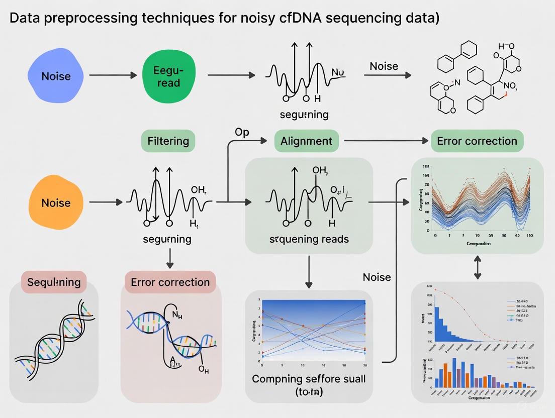
From Theory to Practice: A Toolkit of Preprocessing and Noise-Filtering Methods
Frequently Asked Questions (FAQs)
Q1: Why was a short, genuine genomic sequence mistakenly trimmed by Cutadapt as an adapter?
This occurs due to Cutadapt's default error-tolerant search algorithm. The tool can identify and remove sequences with even a small partial match (e.g., as low as 3 bp) to the provided adapter sequence if the number of errors (mismatches, insertions, deletions) falls within the allowed limit. The error allowance is calculated based on the full length of the adapter sequence, not the length of the matching segment. Therefore, a short genomic sequence with a few coincidental matches can be mistakenly identified for trimming [34].
- Solution: You can adjust the
--minimum-overlapparameter to require a longer minimum match between the read and the adapter sequence, making the search more stringent and reducing false positives [34].
Q2: My trimming report shows adapters were "trimmed," but all reads are still in the output file and seem unchanged. What does "trimmed" mean?
In this context, "trimmed" means that the adapter sequence and any subsequent bases were cut from the read, not that the entire read was removed or discarded. The shortened read is still written to the output file. If you need to filter out reads that became too short after trimming, you must use the -m or --minimum-length option to remove them [35].
Q3: After processing with fastp, my FastQC report shows new warnings for "Sequence Length Distribution" and "GC Content." Did fastp make my data worse?
Not necessarily. These new warnings are often expected and indicate that fastp has done its job correctly.
- Sequence Length Distribution: A warning here is normal after trimming because reads are shortened by different amounts, making their lengths variable rather than uniform [36].
- GC Content: Trimming can alter the overall composition of the read set, which may shift the GC content away from the theoretical distribution that FastQC uses. It is more important to compare the GC content to what is biologically expected for your organism [36]. As noted in one community discussion, the per-base sequence quality often visibly improves after fastp processing, which is a primary goal [36].
Q4: For a paired-end (PE) library, should I provide the reverse primer sequence for the R2 read as its reverse complement?
No, by default, you should provide all adapter sequences in the same 5' to 3' orientation as the reads. Cutadapt does not automatically consider the reverse complement of the adapters or the reads. If you are unsure, you may need to test both the original sequence and its reverse complement to see which one works [37].
Troubleshooting Common Issues
Cutadapt Trimming Specificity and Output
The table below summarizes common issues and solutions when using Cutadapt, based on real user experiences from support forums [34] [35] [37].
| Problem | Possible Cause | Diagnostic Steps | Solution & Recommended Parameters |
|---|---|---|---|
| Unexpected trimming of genuine genomic sequence [34]. | Overly liberal adapter matching with a short minimum overlap and default error rate. | Check the Cutadapt log file's "Overview of removed sequences" table. It shows the length and error count of all trimmed sequences. | Increase the stringency by setting a longer minimum overlap: --minimum-overlap 5 |
| No reads are removed after trimming; output file has the same number of reads as input [35]. | Misunderstanding of "trimming" vs. "filtering." Cutadapt trims (shortens) reads by default but does not remove them from the output. | Use the --length flag in grep to check the length of sequences in the input and output FASTQ files. You will notice the output sequences are shorter. |
Use the -m/--minimum-length parameter to discard reads that become too short after trimming: -m 20 |
| Incorrect primer/adapter not trimmed in single-end mode [37]. | The reverse primer might be provided in the wrong orientation. | Manually inspect a subset of raw reads to confirm the exact sequence and location of the adapter. | Provide the adapter sequence in the same 5' to 3' orientation as the read. Test with the reverse complement sequence if necessary. |
| Low trimming rate for a known adapter. | The adapter type (3' or 5') might be mis-specified. | Review the official Cutadapt guide to confirm the correct adapter type and option (-a for 3', -g for 5') [38]. |
Use -g ^ADAPTER for an anchored 5' adapter (must be at the very start of the read). Use -a ADAPTER$ for an anchored 3' adapter (must be at the very end) [38]. |
FastP Quality Control and Validation
The table below addresses common points of confusion when using fastp for quality control.
| Problem | Possible Cause | Diagnostic Steps | Solution & Recommended Parameters |
|---|---|---|---|
| How to run an analysis/preview mode to assess data quality without writing large output files? | fastp always requires output file parameters (-o, -O) but can be configured for minimal output. |
Omit the -o and -O parameters. Fastp will exit with an error, showing that outputs are mandatory. |
Use a two-step strategy. First, run fastp on a subset of data to generate the QC report and decide on parameters. A user's initial approach for BGI/MGI data was to first generate a report to diagnose quality before a full run [39]. |
| Interpreting FastQC warnings that appear only after running fastp [36]. | Expected consequences of trimming, not a degradation of data quality. | Compare the "Per base sequence quality" plot in FastQC before and after fastp. You will likely see quality improvements in the tails of the reads. | Trust the fastp report and the improved per-base quality. The length distribution warning is expected, and GC content can be checked against known biological expectations. |
| Need for comprehensive quality control in a single tool. | Using multiple, separate tools for QC and trimming can be inefficient. | Compare the fastp HTML report with a separate FastQC report. The fastp report consolidates both pre- and post-filtering statistics [40]. | Rely on the fastp HTML report, which provides all-in-one QC, including quality curves, base content, adapter content, and duplication rates, both before and after filtering [40]. |
Essential Workflows and Protocols
Standard Preprocessing Workflow for cfDNA Data
The following diagram illustrates a robust, generalized workflow for preprocessing cfDNA sequencing data, incorporating best practices from recent literature [41].
Detailed Protocol: A Two-Step Quality Control Strategy with FastP
This protocol is adapted from a user's approach for metagenomic data [39], which is highly relevant to the noisy data context of cfDNA research.
Preliminary Quality Assessment (Analysis-Only Mode):
- Objective: To diagnose the initial quality of the raw data and inform the parameters for the main filtering step.
- Command Example:
- Key Parameters:
--html/--json: Generate the quality control reports.--adapter_sequence/--adapter_sequence_r2: Manually specify known adapters for your library kit.--trim_poly_g: Especially important for data from NovaSeq/NextSeq platforms.
- Output Analysis: The HTML report will show adapter content, per-base quality, and poly-G tails, allowing you to decide if manual adapter specification or poly-G trimming is needed for the main run [39].
Comprehensive Filtering and Trimming:
- Objective: To execute the actual data cleaning with optimized parameters based on the preliminary report.
- Command Example:
- Key Parameters:
-l 50: Discard reads shorter than 50 bp after processing.-q 20: Set the qualified quality threshold to Q20.-e 30: Discard reads with an average quality below Q30.--correction: Enable base correction in overlapping regions for paired-end reads.
The Scientist's Toolkit: Research Reagent Solutions
For cfDNA studies, the choice of wet-lab reagents, particularly the library preparation kit, can introduce significant bias in downstream fragmentomic analysis. The following table lists common kits and their considerations, as evaluated in a recent study [41].
| Library Kit Name | Primary Application/Focus | Key Characteristics/Considerations |
|---|---|---|
| SureSelect XT HS2 (XTHS2) [41] | Targeted sequencing; sensitive mutation detection | Contains dual sample barcodes and dual molecular barcodes (UMIs), which help mitigate index hopping and improve mutation calling accuracy. |
| NEBNext Enzymatic Methyl-seq (EM_seq) [41] | Multi-omics (Methylation & Fragmentomics) | Allows for simultaneous analysis of genetic and epigenetic markers from the same library, which is valuable for multi-modal AI models in cancer detection. |
| Watchmaker DNA Library Prep Kit [41] | General cfDNA library prep | The study found it yielded a significantly higher fraction of mitochondrial DNA reads, which could be a confounder or a feature depending on the research question. |
| ThruPLEX Tag-Seq [41] | General cfDNA library prep | Known to produce a higher number of mismatches during alignment compared to other kits, which is an important factor for studies focused on single-nucleotide variations (SNVs). |
Frequently Asked Questions (FAQs)
Q1: What is the primary source of contamination that LBBC aims to correct? A1: LBBC primarily targets contamination from "low biomass" sources, where trace amounts of foreign DNA (e.g., from reagents, lab surfaces, or sample cross-talk) constitute a significant portion of the sequenced material in samples with very low native DNA content, such as plasma cfDNA.
Q2: How does LBBC differ from traditional background noise filters? A2: Traditional filters often rely on databases of known contaminants or simple abundance thresholds. LBBC is a data-driven method that does not require a priori knowledge. It identifies contaminants by analyzing two intrinsic properties of sequencing data: uneven coverage distribution across the genome (Coverage Uniformity) and systematic variation across experimental batches (Batch Variation).
Q3: My negative controls show minimal reads. Do I still need to apply LBBC? A3: Yes. Even with clean controls, low-level, batch-specific contamination can be present in your experimental samples and can bias downstream analyses, especially for rare variant detection in cfDNA. LBBC uses the controls to model this background, which may be below the threshold of casual observation but statistically significant across a batch.
Q4: What are the minimum sample and batch sizes required for a robust LBBC analysis? A4: While requirements can vary, a general guideline is:
- Minimum Samples per Batch: 8-10 samples (including controls).
- Minimum Batches: 3 or more distinct processing batches. Smaller sample or batch sizes reduce the statistical power to reliably distinguish batch-specific contaminants from true biological signal.
Q5: After applying LBBC, what is an acceptable post-correction contamination level? A5: The goal is to minimize contamination to a level where it does not interfere with your specific biological question. For cfDNA rare variant calling, a common benchmark is to reduce the contamination signal to below the expected variant allele frequency (e.g., <0.1% for ultra-deep sequencing).
Troubleshooting Guides
Problem: High False Positive Rate After LBBC
- Symptoms: True biological signals (e.g., known cancer mutations) are being filtered out alongside contaminants.
- Potential Causes & Solutions:
- Cause 1: Over-correction due to mis-specification of batch groups.
- Solution: Re-evaluate your batch definitions. Ensure batches are defined by distinct library prep kits, reagent lots, or processing dates, not arbitrary groupings.
- Cause 2: Genomic regions of genuine low coverage in your sample type are being mistaken for contaminant regions.
- Solution: Incorporate a sample-specific "coverage mask" based on a high-quality reference sample or a validated set of regions known to have low mappability in cfDNA.
- Cause 1: Over-correction due to mis-specification of batch groups.
Problem: Inconsistent LBBC Performance Across Batches
- Symptoms: Some batches show excellent contamination removal, while others show little to no effect.
- Potential Causes & Solutions:
- Cause 1: Extreme batch effect where one batch is an outlier, dominating the correction model.
- Solution: Implement a robust statistical method (e.g., median-based normalization instead of mean) that is less sensitive to outliers. Visually inspect PCA plots of coverage to identify and potentially exclude severe outlier batches.
- Cause 2: Insufficient sequencing depth in one or more batches.
- Solution: Ensure uniform and adequate sequencing depth (e.g., >100x for cfDNA) across all batches. Low-depth batches lack the statistical power for accurate coverage uniformity analysis.
- Cause 1: Extreme batch effect where one batch is an outlier, dominating the correction model.
Problem: LBBC Fails to Remove Known Contaminant Signal
- Symptoms: Reads from a known contaminant (e.g., E. coli) are still present post-correction.
- Potential Causes & Solutions:
- Cause 1: The contaminant is present uniformly across all batches and samples, making it indistinguishable from the true background via batch variation analysis.
- Solution: Combine LBBC with a database-driven approach. Use a "blacklist" of known contaminant genomes (e.g., phiX, common lab bacteria) to remove these reads as a primary filtering step before applying LBBC.
- Cause 1: The contaminant is present uniformly across all batches and samples, making it indistinguishable from the true background via batch variation analysis.
Experimental Protocols
Protocol 1: Generating Data for LBBC Analysis
- Sample Preparation:
- Extract cfDNA from plasma using a silica-membrane based kit.
- Quantify using a fluorescence-based assay (e.g., Qubit dsDNA HS Assay).
- Library Construction:
- Use a minimum of 8 patient cfDNA samples and 2 negative controls (e.g., water) per batch.
- Construct sequencing libraries using a targeted or whole-genome kit.
- Crucially, process at least 3 independent batches on different days using different reagent lots.
- Sequencing:
- Sequence on an Illumina platform to a minimum depth of 50x for WGS and 500x for targeted panels.
- Pool samples from multiple batches on the same sequencing lane to avoid confounding sequencing run with library prep batch.
Protocol 2: Computational Implementation of LBBC
- Data Preprocessing:
- Align FASTQ files to a reference genome (e.g., hg38) using BWA-MEM.
- Sort and index BAM files using SAMtools.
- Calculate read depth in non-overlapping genomic bins (e.g., 1kb bins) using
mosdepth.
- Coverage Uniformity & Batch Variation Modeling:
- Construct a coverage matrix (bins x samples).
- Perform Principal Component Analysis (PCA) on the normalized coverage matrix.
- Identify bins with strong loadings on batch-associated principal components (PCs). These represent regions with high batch variation.
- Contaminant Filtering:
- Remove or down-weight reads aligning to the identified contaminant bins from the final BAM files.
- Recalculate coverage metrics on the corrected BAM files.
Data Presentation
Table 1: Impact of LBBC on Simulated cfDNA Data with 0.5% Contamination
| Metric | Pre-LBBC | Post-LBBC | % Change |
|---|---|---|---|
| Mean Contamination Level | 0.50% | 0.07% | -86.0% |
| True Positive Rate (TPR) | 95.2% | 94.8% | -0.4% |
| False Discovery Rate (FDR) | 35.1% | 8.5% | -75.8% |
| Number of Significant Bins (FDR < 0.05) | 12,450 | 1,105 | -91.1% |
Table 2: Key Research Reagent Solutions for LBBC Experiments
| Item | Function in LBBC Context |
|---|---|
| cfDNA Extraction Kit | Isolate and purify low-concentration, fragmented cfDNA from plasma with minimal contamination. |
| Ultra-Pure Water | Serve as a negative control to detect contamination introduced during library preparation. |
| Targeted Sequencing Panel | Enrich for specific genomic regions of interest, allowing for deeper sequencing to better distinguish signal from background. |
| Unique Molecular Indices (UMIs) | Tag individual DNA molecules pre-amplification to correct for PCR duplicates and sequencing errors, improving variant calling accuracy post-LBBC. |
| Different Reagent Lots | Essential for creating the batch variation required to statistically identify lot-specific contaminants. |
Visualizations
LBBC Workflow
LBBC Core Concept
Frequently Asked Questions (FAQs)
Q1: What is DAGIP and what specific problem does it solve in cfDNA research? DAGIP is a novel data correction method that uses optimal transport theory and deep learning to correct for pre-analytical biases in cell-free DNA (cfDNA) sequencing data. It explicitly corrects for technical confounders introduced by variables such as library preparation protocols or sequencing platforms, which are major sources of non-biological variation that can obscure true biological signals. This allows for improved cancer detection, copy number alteration analysis, and fragmentomic analysis by alleviating sources of variation not of biological origin [42] [43].
Q2: What types of cfDNA data modalities can DAGIP correct? DAGIP is designed to correct multiple cfDNA data modalities, including:
- Genome-wide copy-number profiles (coverage profiles)
- Fragmentomics data (fragment size distributions)
- End motif frequencies
- Nucleosome positioning patterns The method operates in the original data space, making the corrections transparently interpretable, which is crucial for genetic research [42] [43].
Q3: How does DAGIP differ from traditional bias correction methods like GC-content correction? Unlike traditional methods that focus primarily on GC-content and mappability biases, DAGIP uses a sample-to-sample relationship approach guided by optimal transport theory. While methods like LOESS GC-content correction decorrelate per-bin GC-content from normalized read counts, DAGIP exploits information from the entire dataset to correct each individual sample, providing more comprehensive bias removal and better cancer signal isolation [42] [43].
Q4: What are the minimum data requirements to use DAGIP? DAGIP requires two groups of matched samples (preferably paired) sequenced under different protocols. The data should be structured as matrices where rows represent samples (e.g., coverage, methylation, or fragmentomic profiles) and columns represent features (e.g., genomic bins or DMRs). One group serves as the source domain (protocol 2) and the other as the target domain (protocol 1) for the correction [44].
Q5: Can DAGIP be used to integrate cohorts from different studies? Yes, a key advantage of DAGIP is its ability to integrate cohorts from different studies by explicitly correcting for technical biases introduced by different pre-analytical settings. This allows researchers to combine datasets produced by different sequencing pipelines or collected at different centers, effectively increasing the statistical power for downstream analyses [42].
Troubleshooting Guides
Issue 1: Installation and Dependency Problems
Problem: Errors during installation or when importing the DAGIP package. Solution:
- Ensure all Python dependencies are installed, including rpy2
- Install the required R packages: dplyr, GenomicRanges, and dryclean
- Install the DAGIP package using:
python setup.py install --user - Verify your environment can execute both Python and R code [44]
Issue 2: Data Formatting and Preparation Errors
Problem: The fit_transform() method fails with dimension or data type errors.
Solution:
- Ensure your input data (X and Y) are NumPy arrays with identical feature dimensions
- Confirm that rows represent profiles and columns represent features
- Verify that samples from protocols 1 and 2 are properly matched
- Check that no missing or infinite values are present in the arrays Example of correct implementation:
Issue 3: Poor Bias Correction Performance
Problem: The corrected data shows minimal improvement or unexpected artifacts. Solution:
- Verify that your source and target domain samples are well-matched biologically
- Ensure adequate sample size in both domains for reliable transport plan estimation
- Check that the neural network architecture and training parameters are appropriate for your data type
- Consider adjusting the optimal transport parameters based on your specific data characteristics
- Validate results using known biological controls if available [42]
Issue 4: Model Saving/Loading Failures
Problem: Errors when saving or loading trained models. Solution:
- Use the exact same package versions when saving and loading models
- Ensure sufficient write permissions in the save directory
- Verify the complete file path is specified when saving:
DAGIP Parameters and Performance Metrics
Table 1: Key Experimental Parameters for DAGIP Implementation
| Parameter Category | Specific Parameter | Recommended Setting | Function | ||
|---|---|---|---|---|---|
| Data Input | Feature Type | Coverage profiles, fragment sizes | Defines the input data modality | ||
| Sample Matching | Paired or biologically matched | Ensures valid domain translation | |||
| Matrix Orientation | Rows: samples, Columns: features | Proper data structure | |||
| Optimal Transport | Cost Function | Quadratic ( | xi-yj | ²) | Determines transport energy |
| Transport Plan | Sample-to-sample mapping | Guides correction direction | |||
| Neural Network | Architecture | Deep learning model | Estimates technical bias | ||
| Training | Paired samples | Learns bias correction function |
Table 2: Comparison of Bias Correction Methods in cfDNA Analysis
| Method | Approach | Data Modalities | Interpretability | Dependencies |
|---|---|---|---|---|
| DAGIP | Optimal transport + deep learning | Coverage, fragmentomics, end motifs | High (original space) | Python, R packages |
| GC-content LOESS | Local regression | Coverage profiles | Medium | None |
| BEADS | Read-level reweighting | Coverage profiles | Medium | None |
| dryclean | Robust PCA | Coverage profiles | Low | R |
| LIQUORICE | Fragment-level weighting | Coverage profiles | Medium | None |
Experimental Protocols
Protocol 1: Basic DAGIP Workflow for Coverage Profile Correction
Purpose: Correct technical biases in coverage profiles from different sequencing protocols.
Materials:
- NumPy arrays of coverage profiles (bins × samples)
- Matched samples from two different protocols
- DAGIP installation with dependencies
Procedure:
- Data Preparation: Format coverage data as NumPy matrices where rows are samples and columns are genomic bins
- Domain Specification: Assign protocol 2 data to X (source domain) and protocol 1 data to Y (target domain)
- Model Initialization: Create DomainAdapter instance
- Model Training and Transformation: Call
fit_transform()to correct source domain data - Model Saving: Save trained model for future use
- New Data Correction: Use
transform()method to correct new samples independently
Validation: Compare principal component analysis (PCA) plots before and after correction to confirm reduced technical variation while preserving biological signals [42] [44].
Protocol 2: Multi-modal cfDNA Analysis with DAGIP
Purpose: Correct technical biases across multiple cfDNA data types simultaneously.
Materials:
- Coverage profiles (read counts per genomic bin)
- Fragment size distributions
- End motif frequency profiles
- Nucleosome positioning data
Procedure:
- Feature Concatenation: Combine different data modalities into a unified feature matrix
- Normalization: Apply appropriate normalization to each data type
- DAGIP Application: Apply standard DAGIP workflow to the multi-modal matrix
- Post-processing: Separate corrected modalities for downstream analysis
- Validation: Assess preservation of known biological relationships across modalities
Note: This approach is particularly valuable for integrated analyses that leverage complementary information from different cfDNA features [42].
Workflow Visualization
Research Reagent Solutions
Table 3: Essential Materials for DAGIP Implementation
| Category | Item/Software | Function/Purpose | Implementation Notes |
|---|---|---|---|
| Computational Tools | DAGIP Python Package | Core bias correction algorithm | Install via: python setup.py install --user |
| R with dplyr, GenomicRanges | Data manipulation and genomic processing | Required dependency | |
| dryclean R package | Background correction reference | Used in comparative analyses | |
| NumPy | Numerical data processing | Handles matrix operations | |
| Data Types | Coverage Profiles | Read count per genomic bin | Primary input for CNA detection |
| Fragment Size Distributions | Fragment length frequencies | Fragmentomics analysis | |
| End Motif Frequencies | 4-nucleotide end patterns | Fragmentomics biomarker | |
| Methylation Profiles | DNA methylation patterns | Multi-modal integration | |
| Validation Methods | PCA Visualization | Technical variation assessment | Pre- vs. post-correction comparison |
| Biological Controls | Known positive/negative samples | Performance validation | |
| Domain Classifiers | Domain shift measurement | Quantify correction effectiveness |
Advanced Troubleshooting
Issue 5: Handling Large-Scale Genomic Data
Problem: Memory errors when processing whole-genome coverage data. Solution:
- Implement data chunking for large matrices
- Consider binning genomic regions to reduce dimensionality
- Use memory-mapped arrays for large datasets
- Ensure adequate RAM allocation for transport plan computation
Issue 6: Domain Shift Validation
Problem: Uncertainty about whether correction successfully reduced technical biases. Solution:
- Train domain classifiers (e.g., SVM) pre- and post-correction
- Use PCA to visualize separation between domains before and after correction
- Validate preservation of known biological signals using positive controls
- Assess improvement in downstream task performance (e.g., cancer detection accuracy) [42] [43]
Open Chromatin Regions (OCRs) are genomic regions associated with fundamental cellular physiological activities, and their accessibility significantly influences gene expression and function [45]. The accurate estimation of OCRs from cell-free DNA (cfDNA) sequencing data represents a crucial computational challenge in genomic and epigenetic studies. However, a persistent obstacle in this domain is the problem of noisy labels within the training data. Due to the dynamically variable nature of chromatin accessibility, obtaining training datasets with definitively pure OCRs or non-OCRs is particularly challenging [45]. These inaccurate labels, or false positives, can lead to overfitting and substantially degrade the performance of conventional machine learning models.
OCRFinder represents a novel computational solution to this problem. It is a learning-based OCR estimation approach specifically designed with a noise-tolerance architecture to mitigate the interference of noisy labels [45]. By integrating principles from ensemble learning and semi-supervised strategies, OCRFinder avoids the potential overfitting that plagues other methods when faced with imperfect training data. This framework is especially valuable for researchers and drug development professionals working with cfDNA sequencing data, where biological variability and technical artifacts frequently introduce label inaccuracies.
Table: Core Characteristics of the OCRFinder Framework
| Characteristic | Description |
|---|---|
| Primary Function | Estimation of Open Chromatin Regions (OCRs) from cfDNA-seq data |
| Core Innovation | Noise-tolerant machine learning design for handling inaccurate training labels |
| Technical Approach | Combination of ensemble learning and semi-supervised strategies |
| Input Data | cfDNA-seq data encoded as two-dimensional matrices and artificial features |
| Key Advantage | Maintains high accuracy and sensitivity despite noisy training data |
Technical Framework and Architecture
The OCRFinder framework operates through a structured, three-stage pipeline designed to progressively build model robustness against label noise. This systematic approach ensures that the model develops an initial discriminatory capability before tackling the more complex task of distinguishing clean from noisy samples.
Stage 1: Data Pre-processing
The first stage involves converting raw cfDNA sequencing data into a format suitable for deep learning. The cfDNA-seq data in FASTQ format is initially processed by tools like BWA and Samtools to obtain cfDNA-reads in BAM format [45]. OCRFinder then encodes these reads into a two-dimensional matrix T, where rows represent genomic coordinates and columns represent cfDNA-read lengths (Tij denotes the number of cfDNA-reads with genomic coordinate i and length j). The framework specifically considers cfDNA-reads with lengths between 50 bp and 250 bp [45]. Additionally, OCRFinder incorporates four artificially constructed features that help reflect gene expression: sequencing coverage, WPS score, and the density of the head and tail of cfDNA fragments. These are encoded in the same two-dimensional format as separate inputs to the model [45].
Stage 2: Model Pre-training
Before engaging in semi-supervised learning, the model undergoes a pre-training phase to establish an initial discriminative ability. This step is crucial because sample selection methods in subsequent stages rely on loss distributions. The underlying principle is that during iterative training, the model will typically learn to fit clean samples before noisy ones; consequently, clean samples tend to exhibit smaller losses than noisy samples early in training [45]. This pre-training phase must be carefully constrained to prevent the model from overfitting to the noisy labels present in the initial dataset, thereby preserving its capacity to differentiate between clean and noisy samples in the final stage.
Stage 3: Semi-Supervised Training with Sample Selection
The final stage implements a semi-supervised strategy and consists of three core components:
- Sample Selection: A reliable and adjustable criterion is designed to separate potentially clean samples from noisy ones based on their loss values.
- Ensemble Loss Calculation: An ensemble approach is used to compute a robust loss metric that mitigates the impact of individual model variances.
- Recirculation of Dirty Samples: The samples identified as having noisy labels are re-introduced into the training process in a controlled manner to maximize data utilization while balancing the training noise rate [45].
This sophisticated architecture allows OCRFinder to leverage the entire dataset effectively without being unduly influenced by the inaccuracies inherent in the labels.
Frequently Asked Questions (FAQs)
Framework Fundamentals
Q1: What exactly is a "noisy label" in the context of cfDNA sequencing, and why is it problematic?
A noisy label refers to an incorrect classification of a genomic region as either an Open Chromatin Region (OCR) or a closed region in the training data. This problem arises fundamentally due to the dynamic variability of chromatin accessibility, which can differ between individuals and even within the same individual over time [45]. Consequently, regions with active gene expression are sometimes mislabeled as silent, and vice-versa. These inaccuracies are highly problematic because standard machine learning models will diligently learn these errors, leading to severe overfitting and significantly reduced performance and generalizability of the model on new, real-world data.
Q2: How does OCRFinder's approach to handling noisy labels differ from traditional methods?
OCRFinder diverges from traditional noise-handling methods in several key aspects. Many existing methods rely on designing noise-resistant loss functions or implementing sample selection strategies that often depend on hard-to-obtain prior information about the data distribution [45]. In contrast, OCRFinder employs a more pragmatic combination of ensemble learning and semi-supervised strategies without requiring extensive a priori knowledge. It uses the model's own training dynamics—specifically, the observation that clean and noisy samples display different loss distributions during learning—to guide the separation and handling of samples. This data-driven approach makes it particularly suited for the complex, variable domain of cfDNA analysis.
Q3: What are the minimum computational resources required to implement the OCRFinder framework?
While the search results do not specify exact hardware requirements, the framework involves computationally intensive operations. These include deep neural networks for automated feature extraction, ensemble methods that may involve multiple models, and iterative semi-supervised training cycles. Implementation would typically require a high-performance computing environment with substantial CPU and RAM resources, as well as possibly GPUs to accelerate the training of deep learning models. The data pre-processing stage also requires standard bioinformatics tools like BWA and Samtools for sequence alignment [45].
Implementation and Troubleshooting
Q4: During pre-training, my model overfits rapidly to the noisy training data. What mitigation strategies can I employ?
To combat overfitting during the critical pre-training phase, consider the following strategies:
- Implement Early Stopping: Closely monitor the model's performance on a small, held-out validation set that is as clean as possible. Stop training as soon as performance on this set begins to degrade, even if training loss continues to decrease.
- Apply Strong Regularization: Utilize techniques such as dropout, L1/L2 weight regularization, and label smoothing to prevent the model from becoming overconfident on potentially erroneous labels.
- Use a Conservative Learning Rate: Opt for a lower learning rate and a simple optimizer (e.g., SGD without momentum) to allow for slower, more stable convergence rather than rapid fitting that may memorize noise.
- Constrain Model Capacity: While deep learning is powerful, an excessively complex model for the amount of reliable data can exacerbate overfitting. Start with a moderate architecture and increase capacity only if necessary.
Q5: The sample selection step in Stage 3 incorrectly flags too many clean samples as "noisy." How can I adjust the selection sensitivity?
The sample selection criterion in OCRFinder is designed to be adjustable. If the selection is too aggressive, you can:
- Relax the Division Criterion: Make the threshold for being considered a "clean" sample less strict. For example, if using a loss percentile threshold, increase the percentile value to allow more samples with moderately low loss to be included in the clean set.
- Incorporate Ensemble Agreement: Instead of relying on loss from a single model, use the agreement (e.g., consistent low loss) across multiple models in the ensemble to identify clean samples with higher confidence.
- Iterative Refinement: Perform the sample selection process iteratively. Use an initial, very conservative selection to train a slightly better model, then use that model to re-evaluate the remaining samples, potentially reclaiming some that were initially misclassified as noisy.
Q6: Can OCRFinder be applied to other data types beyond cfDNA-seq, such as ATAC-seq or DNase-seq data?
Yes, the foundational principles of OCRFinder are transferable. The article explicitly states that OCRFinder "also has an excellent performance in ATAC-seq or DNase-seq comparison experiments" [45]. The core innovation—using a noise-tolerant learning strategy to handle imperfect labels—is a general concept in machine learning. To adapt it, you would need to adjust the data pre-processing and feature encoding stages (Stage 1) to be appropriate for the specific data type (e.g., different encoding for ATAC-seq peaks), while the core noise-handling architecture of Stages 2 and 3 would remain largely applicable.
Troubleshooting Common Experimental Issues
Data Quality and Pre-processing Problems
Problem: High variance in model performance due to inconsistent cfDNA fragmentation patterns.
- Symptoms: Unstable loss curves during training, poor reproducibility on validation splits from the same sample cohort.
- Diagnosis: This often stems from pre-analytical variables in sample handling. The choice of blood collection tube, delay before centrifugation, DNA extraction platform, and library preparation kit can all introduce significant biases and variations in fragmentation profiles [41] [46]. For instance, different library kits exhibit variations in metrics like GC content, fraction of unmapped reads, and mitochondrial read counts [41].
- Solution: Implement stringent standardization of wet-lab protocols. For existing data, use bioinformatics tools designed to correct for these batch effects. Recently developed methods like DAGIP, which uses optimal transport theory, or standardized frameworks like the Trim Align Pipeline (TAP) and cfDNAPro R package, can help mitigate technical variations and align data distributions from different sources [42] [41].
Problem: Poor feature representation from cfDNA-seq data with low sequencing depth.
- Symptoms: Low accuracy even on training data, model fails to converge to a satisfactory loss level.
- Diagnosis: Artificially constructed features (e.g., coverage, WPS) are particularly susceptible to degradation at low sequencing depths [45]. The signal becomes too weak for the model to discern meaningful patterns.
- Solution:
- Increase Sequencing Depth: If feasible, sequence to a higher depth to obtain a denser signal.
- Leverage Automated Feature Extraction: Utilize OCRFinder's deep learning backbone to learn robust features directly from the encoded data, which can be more resilient than hand-crafted features in low-information scenarios [45].
- Data Augmentation: Carefully apply sensible in-silico augmentation techniques to the two-dimensional input matrices to artificially expand the training set.
Model Training and Convergence Issues
Problem: The ensemble model shows high disagreement on sample losses, making clean/noisy separation impossible.
- Symptoms: The loss distributions of clean and noisy samples overlap significantly, preventing the establishment of a clear selection threshold.
- Diagnosis: This can occur when the noise level in the dataset is very high (e.g., >50% of labels are incorrect) or when the models in the ensemble are too similar and fail to provide diverse opinions.
- Solution:
- Enforce Ensemble Diversity: Use different model architectures or bootstrap sampling of the training data for each model in the ensemble to ensure they learn different aspects of the data.
- Leverage Control Charts for Initial Filtering: As demonstrated in the related OCRClassifier framework, a robust Hotelling T² control chart can be applied first to identify a reliable subset of pure OCRs and non-OCRs [47]. This smaller, high-confidence set can then be used to bootstrap the training of the main OCRFinder model, providing a more stable start.
Problem: Final model performance is good on validation data but poor on independent test sets.
- Symptoms: High accuracy and sensitivity during cross-validation but a significant drop in performance when applied to data from a different study or sequencing center.
- Diagnosis: This is a classic sign of domain shift, where the test data comes from a different distribution (e.g., different library prep kit or sequencing platform) than the training data. Preanalytical variables are major confounders in cfDNA analysis [42].
- Solution:
- Incorporate Domain Adaptation: During pre-processing, apply domain adaptation techniques like optimal transport (e.g., DAGIP) to explicitly correct for technical biases between your training cohort and the target test cohort [42].
- Augment Training Data: If possible, incorporate a small amount of data from the target domain (even unlabeled) into your training process to allow the model to adapt to the new distribution.
The Scientist's Toolkit: Essential Research Reagents and Materials
Successful implementation of the OCRFinder framework relies on both robust computational methods and careful wet-lab experimentation. The following table details key reagents and tools referenced in the literature that are essential for generating high-quality, noise-reduced data for OCR estimation.
Table: Essential Research Reagent Solutions for cfDNA-based OCR Studies
| Reagent / Tool | Specific Example (from search results) | Primary Function in Workflow |
|---|---|---|
| Blood Collection Tubes (BCTs) with Stabilizers | Streck cfDNA BCT, Roche Cell-Free DNA Collection Tube [46] | Prevents white blood cell lysis during transport/storage, preserving native cfDNA profile and reducing background genomic DNA contamination. |
| cfDNA Extraction Kits | QIAamp Circulating Nucleic Acid Kit (Qiagen) [46] | Isulates and purifies short-fragment cfDNA from plasma with high efficiency and yield, crucial for downstream sequencing. |
| Library Preparation Kits | ThruPLEX Plasma-Seq, NEBNext Ultra II, Kapa HyperPrep [41] | Prepares cfDNA fragments for sequencing; kit choice significantly impacts GC bias, complexity, and final data quality. |
| Targeted Enrichment Probes | Integrated DNA Technologies (IDT) customized probes [4] | For hybrid capture-based sequencing, enabling focused sequencing on regions of interest (e.g., gene panels). |
| Reference Standard Genomic DNA | HD753 (Horizon Diagnostics) [4] | Provides a multiplexed reference standard with known mutations at defined allelic frequencies for assay validation and calibration. |
| Bioinformatics Pre-processing Tools | BWA, Samtools [45], Trim Align Pipeline (TAP) [41] | Performs sequence alignment, format conversion, and adapter trimming; TAP is specifically optimized for cfRNA data. |
| Bias Correction & DA Tools | DAGIP (Optimal Transport) [42] | Corrects for technical biases from different library prep or sequencing platforms, enabling robust data integration (Domain Adaptation). |
| Fragmentomic Feature Extractors | cfDNAPro R Package [41] | Provides standardized, cfDNA-optimized methods for calculating key fragmentation features like fragment length distributions and end motifs. |
Advanced Methodologies: Experimental Protocols
To ensure the reproducibility and robustness of your research, below are detailed protocols for key experimental and computational procedures referenced in the cited literature.
Protocol: Plasma cfDNA Extraction and Quality Control
This protocol is optimized to minimize pre-analytical noise, based on standardized methodologies [41] [46].
Blood Collection and Plasma Separation:
- Collect venous blood into specialized cell-free DNA BCTs (e.g., Streck tubes).
- Perform two-step centrifugation:
- First Spin: 1200–2000× g for 10 minutes at 4°C to separate plasma from blood cells. Carefully transfer the supernatant without disturbing the buffy coat.
- Second Spin: 12,000–16,000× g for 10 minutes at 4°C to remove any remaining cellular debris.
- Aliquot the purified plasma and store at -80°C to avoid freeze-thaw cycles.
cfDNA Extraction:
- Use the QIAamp Circulating Nucleic Acid Kit (or equivalent) according to the manufacturer's instructions. Automated platforms like QIAsymphony can enhance reproducibility.
- Elute the cfDNA in a low-EDTA buffer or nuclease-free water.
Quality Control (QC):
- Quantification and Size Profiling: Use a capillary electrophoresis system (e.g., Agilent Bioanalyzer with High Sensitivity DNA kit) to confirm a peak at ~167 bp and assess the degree of high molecular weight genomic DNA contamination. Avoid fluorometric methods alone for quantification.
- Assess Amplificability: Use a ddPCR or qPCR assay targeting both short and long amplicons to confirm the cfDNA is amplifiable and to detect significant gDNA contamination.
Protocol: Implementing the OCRFinder Framework
This computational protocol outlines the core steps for implementing the noise-tolerant learning framework [45].
Data Pre-processing and Feature Encoding:
- Alignment: Align paired-end FASTQ files to a reference genome (e.g., hg38) using BWA-MEM. Process the resulting files with Samtools to generate sorted BAM files.
- 2D Matrix Creation: For each genomic region of interest (e.g., a 5 Mb bin), create a two-dimensional matrix
Twhere the rows are genomic coordinates and the columns represent fragment lengths from 50 bp to 250 bp. The valueTijis the count of fragments of lengthjat positioni. - Artificial Feature Calculation: Compute the four auxiliary features (sequencing coverage, WPS, and head/tail density) for the same genomic regions and encode them in an identical 2D format.
Model Pre-training:
- Construct a Convolutional Neural Network (CNN) architecture capable of processing the 2D input.
- Train the model on the entire initial training set using a standard loss function (e.g., cross-entropy) but with early stopping based on a small, trusted validation set or by monitoring for a sharp increase in training accuracy (a potential sign of memorizing noise).
Semi-Supervised Training Cycle:
- Sample Selection: For each training epoch, compute the loss for each sample. Separate samples into a "clean" set (e.g., those with loss in the bottom 30th percentile) and a "noisy" set (the remainder).
- Ensemble Loss: Calculate the loss for the epoch using only the "clean" set, or use a weighted loss that gives higher weight to the clean samples.
- Recirculation: Periodically, or with a small weight, reintroduce samples from the "noisy" set into the training process to ensure the model does not forget them entirely and to potentially reclaim misclassified clean samples as the model improves.
Protocol: Cross-Validation Strategy for Noisy Data
Traditional random cross-validation can be unreliable with noisy labels. The following strategy, inspired by control chart methods, provides a more robust validation [47].
Initial Pure Set Creation:
- Use a robust statistical method (e.g., a Hotelling T² control chart) on simple, robust features (e.g., global coverage, WPS mean) to identify a high-confidence subset of genomic regions that are unequivocally OCRs or non-OCRs. This forms your "Pure Set".
Stratified Validation:
- When creating train/validation splits, ensure that the "Pure Set" is evenly distributed across all folds.
- Use the "Pure Set" within the validation fold as the primary metric for model selection and early stopping. This provides a more reliable signal of true performance than using the entire, noisy validation set.
Noise Level Estimation:
- After training, the proportion of samples consistently flagged as "noisy" by the sample selection step across multiple folds can serve as an estimate of the overall noise level in your dataset. This metric can be useful for reporting data quality.
Frequently Asked Questions (FAQs)
What are the primary fragmentomic features used for data quality assessment?
The primary fragmentomic features used to assess the quality of cfDNA sequencing data are the size profile and the end motif patterns.
- Size Profile: High-quality cfDNA from healthy individuals shows a prominent peak at approximately 166 base pairs (bp), corresponding to DNA wrapped around a single nucleosome, and exhibits a 10-bp periodicity for fragments shorter than 143 bp [48]. A deviation from this profile, such as an increased proportion of shorter fragments (<150 bp), can indicate the presence of tumor DNA or sample degradation [49] [50].
- End Motif Patterns: The sequences at the ends of cfDNA fragments are non-random. In healthy individuals, certain C-rich 4-mer motifs (e.g., CCCA) are more prevalent [48] [50]. A significant decrease in these preferred motifs and an overall increase in end motif diversity are associated with cancer-derived cfDNA and can serve as a quality metric for tumor enrichment [49] [41].
My cfDNA data shows a high duplication rate and low complexity. What could be the cause?
A high duplication rate often points to issues during the initial library preparation steps, typically related to insufficient input DNA or inefficient amplification [33].
- Root Cause: Using degraded cfDNA or an inaccurately quantified sample for library construction can lead to a low number of unique DNA molecules. Subsequent PCR amplification will then over-amplify the limited number of original molecules, creating duplicates [33].
- Solution:
- Re-quantify your cfDNA sample using a high-sensitivity fluorometric method (e.g., Qubit) instead of spectrophotometry, which can overestimate concentration by detecting contaminants [51].
- Verify sample integrity using a capillary electrophoresis system (e.g., Bioanalyzer) to ensure the cfDNA has the expected fragment size distribution and is not degraded [51] [50].
I suspect my sample is contaminated with genomic DNA. How would fragmentomics reveal this?
Fragmentomic analysis can readily identify high-molecular-weight genomic DNA (gDNA) contamination.
- Indicator: The classic cfDNA size profile shows a peak at ~166 bp. The presence of a significant amount of long fragments (>500 bp or a broad smear) in your size distribution plot is a clear sign of gDNA contamination [51].
- Impact: gDNA contamination can severely reduce the efficiency of sequence alignment and introduce biases that obscure true biological signals, such as tumor-derived fragmentomic patterns [51].
The end motif proportions in my data look abnormal. Is this a technical artifact or a biological signal?
Distinguishing between technical artifacts and biological signals requires careful analysis. Technical biases from library preparation kits and computational pipelines are known to affect end motif proportions [41].
- Action Plan:
- Review Your Bioinformatics Pipeline: Ensure you are using tools specifically designed for cfDNA fragmentomic analysis, which properly account for adapter sequences and alignment parameters [41].
- Compare to Healthy Controls: A significant deviation from the end motif patterns of healthy control samples processed with the same kit and pipeline is more likely to be a biological signal, such as a cancer-derived change [48] [49].
- Cross-validate with Other Metrics: Correlate the aberrant end motif patterns with other fragmentomic features. For example, if the sample also has an elevated proportion of short fragments and a high "N-index" (discordance with hematopoietic nucleosome positioning), it strengthens the evidence for a tumor-derived biological signal [48].
Troubleshooting Common Fragmentomic Data Issues
The following table outlines common problems, their diagnostic signals, and recommended corrective actions.
| Problem | Diagnostic Signals in Fragmentomic Data | Root Causes | Corrective & Preventive Actions |
|---|---|---|---|
| Low Library Complexity/High Duplication | - Abnormally flat or skewed fragment size profile [33].- High PCR duplicate rate in sequencing metrics. | - Degraded or insufficient input cfDNA [33].- Inaccurate quantification leading to suboptimal PCR cycles [33].- Contaminants inhibiting enzymes. | - Use fluorometric quantification (e.g., Qubit) and capillary electrophoresis for QC [51].- Re-purify sample to remove inhibitors [33].- Optimize the number of PCR cycles [33]. |
| gDNA Contamination | - Significant proportion of fragments >300bp in size distribution [51].- Loss of the characteristic ~166 bp nucleosomal peak. | - Improper blood sample processing or storage [52].- Inefficient cfDNA extraction. | - Process blood samples while fresh to prevent cell lysis [52].- Use extraction kits validated for cfDNA (e.g., QIAamp Circulating Nucleic Acid Kit) [50] [52].- Incorporate rigorous size selection during cleanup. |
| Biased End Motif Profiles | - Drastic deviation from expected C-rich motif frequencies (e.g., CCCA) in control samples [48] [41].- Inconsistent results across samples processed with different kits. | - Biases introduced by specific library preparation kits [41].- Suboptimal data processing with improper adapter trimming or alignment [41]. | - Standardize library prep protocol across sample batches [41].- Use standardized computational pipelines (e.g., TAP, cfDNAPro) designed for cfDNA [41]. |
| Weak Tumor Fragmentomic Signal | - ΔS150 and N-index values close to healthy control ranges, despite other evidence of cancer [48]. | - Low tumor fraction in plasma sample [48].- Dilution of signal by high background cfDNA. | - Computationally select fragments <150 bp to enrich for tumor-derived signals [48] [50].- Integrate multiple fragmentomic metrics (size, end motifs, coverage) with machine learning to boost signal [48] [49]. |
Quantitative Fragmentomic Reference Data
The following table summarizes key fragmentomic metrics and their values in healthy and cancerous states, providing a benchmark for data assessment.
| Metric | Description & Normal Range (Healthy Individuals) | Alteration in Cancer | Diagnostic Performance & Notes |
|---|---|---|---|
| Size Profile: Proportion of short fragments | - Peak at ~166 bp [48] [49].- Percentage of fragments ≤150 bp is relatively lower. | - Increase in proportion of fragments ≤150 bp [48] [49] [50]. | - ΔS150 (change in % of ≤150 bp fragments after end selection) showed significant differentiation in HCC [48]. |
| End Motif: CCCA Frequency | - CCCA is a preferred 4-mer end motif [48] [50]. | - Decrease in CCCA frequency [48] [49]. | - ΔMCCCA (change in CCCA frequency after end selection) is a potent biomarker [48]. Performance improved over measuring CCCA usage alone [48]. |
| N-index | - Low value, indicating cfDNA ends are concordant with hematopoietic nucleosome positioning [48]. | - Increase, indicating discordance due to presence of non-hematopoietic (tumor) cfDNA [48]. | - Achieved an AUC of 0.72-0.94 for detecting HCC across different stages [48]. |
| FrEIA Score | - A quantitative measure of a sample's distance from control 5' end trinucleotide composition [49]. | - Increase in score across multiple cancer types [49]. | - In a multi-cancer cohort, integrating this score with other features achieved 72% detection sensitivity at 95% specificity [49]. |
Experimental Workflow for Fragmentomic Quality Assessment
The diagram below illustrates a robust experimental and computational workflow for generating high-quality fragmentomic data.
Workflow for Fragmentomic Data Generation and QC
The Scientist's Toolkit: Essential Research Reagents & Materials
| Item | Function in Fragmentomic Analysis | Example Products & Kits |
|---|---|---|
| High-Sensitivity Fluorometer | Accurately quantifies low-concentration cfDNA, which is critical for successful library construction and avoiding bias [51]. | EzCube Fluorometer, Qubit dsDNA HS Assay Kit [51] [50]. |
| Capillary Electrophoresis System | Assesses cfDNA purity and fragment size distribution to check for gDNA contamination or degradation [51] [50]. | Agilent Bioanalyzer 2100. |
| cfDNA Extraction Kit | Isulates cfDNA from plasma with high efficiency and minimal gDNA contamination [50] [52]. | QIAamp Circulating Nucleic Acid Kit, QIAamp MinElute ccfDNA Midi Kit [50] [52]. |
| Library Prep Kit with UMI | Prepares sequencing libraries while incorporating Unique Molecular Identifiers (UMIs) to accurately track original molecules and reduce PCR duplicate bias [41]. | ThruPLEX Plasma-Seq, SureSelect XT HS2 [41]. |
| Standardized Bioinformatics Pipelines | Performs consistent adapter trimming, alignment, and feature extraction tailored to cfDNA's unique properties, minimizing technical variation [41]. | Trim Align Pipeline (TAP), cfDNAPro R package, FrEIA toolkit [49] [41]. |
Technical Support Center
Frequently Asked Questions (FAQs)
Q1: How does DNA sample quality and concentration impact nanopore sequencing success for cfDNA? Accurate DNA quantification is critical. Fluorometric methods (e.g., Qubit) are essential because they specifically measure dsDNA concentration, whereas photometric methods (e.g., Nanodrop) often overestimate concentration due to contaminants common in cfDNA samples. Insufficient concentration or quality is a primary cause of low sequencing coverage and assembly failure [53] [54]. For optimal results with cfDNA, ensure your sample meets the minimum volume and concentration requirements for your chosen library prep kit.
Q2: My consensus sequence has low-confidence bases. What are the common causes? Low-confidence calls often occur at specific challenging motifs. The most common are:
- Homopolymers: Stretches of identical bases (e.g., 'AAAAA'), where the basecaller may misestimate the length [55].
- Methylation Sites: Specific sequences like the Dam (
GATC) or Dcm (CCTGG,CCAGG) methylation sites in plasmids or native DNA [53] [55]. - Low-Complexity Regions: Repeats and reverse-complemented elements can also challenge basecalling algorithms [54]. These errors are often systematic and can be flagged by advanced bioinformatics pipelines.
Q3: What does a multi-peak read length histogram indicate about my sample? Multiple peaks in a read length histogram suggest a mixture of DNA molecules of different sizes. For cfDNA research, this could indicate:
- A mixture of plasmid constructs of different sizes if sequencing a library [53] [54].
- The presence of biological concatemers (e.g., dimeric or multimeric plasmid forms) [53].
- Significant host genomic DNA contamination alongside your target cfDNA [53]. A single, dominant peak is indicative of a clean, clonal population.
Q4: Can I use nanopore sequencing to detect methylation and other base modifications in cfDNA?
Yes. A key advantage of nanopore sequencing is its ability to detect base modifications like 5-methylcytosine (5mC) and 6-methyladenosine (6mA) from native DNA without special treatment. Tools like realfreq even enable real-time methylation calling during sequencing, which is highly relevant for epigenetic analysis of cfDNA [56]. This allows for simultaneous sequencing of the genome and its epigenome from a single sample.
Q5: How can I improve the accuracy of my nanopore-sequenced cfDNA genomes?
While nanopore raw read accuracy continually improves, achieving the highest consensus accuracy often involves polishing. For the most accurate results, a combination of long-read and short-read polishing is recommended. Long-read polishers like medaka correct errors using the original nanopore reads, while subsequent polishing with accurate short-read data using tools like NextPolish or Pilon can correct residual errors, particularly in homopolymers [57].
Troubleshooting Guides
Table 1: Common Nanopore Sequencing Issues and Solutions for cfDNA
| Issue | Possible Cause | Recommended Solution |
|---|---|---|
| Low or no coverage | Insufficient DNA concentration/quality; degraded DNA [53] [54]. | Use fluorometry (Qubit) for quantification; run gel electrophoresis to check for degradation; use Rolling Circle Amplification (RCA) for low-input circular DNA [54]. |
| No dominant peak in histogram | Sample is not a clonal population; high background contamination (e.g., host DNA) [53] [54]. | Re-prepare sample from a single colony; perform gel extraction or other size-selection to isolate the target cfDNA molecule [53]. |
| Multiple peaks in histogram | Plasmid mixture; biological concatemers; multiple plasmids of similar size [53] [54]. | Ensure clonal sample origin; use a recA- bacterial strain for propagation to prevent concatemer formation [53]. |
| High error rate in homopolymers | Systematic sequencing error in stretches of identical bases [55] [57]. | Use bioinformatic tools that are aware of this error mode; employ hybrid polishing with short reads for more accurate homopolymer lengths [57]. |
| Systematic errors at CCTGG/CCAGG | Dcm methylation site interfering with basecalling [53] [55]. | Use a methylation-aware basecaller or a bioinformatics pipeline that applies specialized correction algorithms for these known motifs [55]. |
Table 2: Essential Research Reagent Solutions for cfDNA Experiments
| Reagent / Material | Function in the Experiment |
|---|---|
| Ligation Sequencing Kit | Standard kit for routine sequencing where read length matches the input fragment length; ideal for characterizing cfDNA fragment size [58]. |
| Ultra-Long DNA Sequencing Kit | Specialized kit for generating reads >100 kb; not typically used for cfDNA but crucial for resolving complex genomic regions in reference genomes [58]. |
| Rapid Sequencing Kit | For quick sample-to-sequencer workflows; best for samples with input fragments >30 kb [58]. |
| Direct RNA Sequencing Kit | Prepares native RNA for sequencing; used for transcriptome analysis to discover full-length isoforms and fusion genes in cancer [58] [56]. |
| cDNA-PCR Sequencing Kit | Optimized for identifying and quantifying full-length isoforms from low input amounts; applicable for single-cell RNA-seq in cancer research [58] [56]. |
| Rolling Circle Amplification (RCA) | A pre-treatment method to selectively amplify circular DNA from very low input, such as a small volume of culture or low-concentration cfDNA samples [54] [56]. |
Experimental Protocols
Protocol 1: Nanopore Adaptive Sampling for Enriching Cancer Predisposition Genes
This protocol, adapted from Chevrier et al. (2025), uses adaptive sampling to target specific genomic regions from blood samples [56].
- Sample Preparation: Extract high-molecular-weight genomic DNA from blood samples of individuals with hereditary cancer predisposition.
- Library Preparation: Prepare the DNA library using the Ligation Sequencing Kit according to the standard protocol [58].
- Sequencing with Adaptive Sampling:
- Load the library onto a PromethION or GridION flow cell.
- Use the
ReadUntilAPI or integrated software to provide a BED file containing the coordinates of 152 known cancer predisposition genes. - During sequencing, the software will reject reads that do not map to the target regions in real-time, thereby enriching the sequencing output for genes of interest.
- Variant Calling and Analysis: Basecall the data and align to a human reference genome. Call single nucleotide variants (SNVs) and structural variants (SVs), noting that adaptive sampling enables detection even at low coverage [56].
Protocol 2: NanoRCS for Multimodal Cell-free Tumour DNA Profiling
This protocol, based on Chen et al. (2025), details a method for accurate, real-time profiling of cfDNA [56].
- cfDNA Extraction and Circularization: Isolate cell-free DNA from plasma. Convert the linear cfDNA molecules into circular form using a ligation reaction.
- Rolling Circle Amplification (RCA): Amplify the circularized cfDNA templates using phi29 polymerase. This step enhances signal and enables consensus sequencing.
- Nanopore Sequencing: Prepare the RCA product using a rapid or ligation sequencing kit and load onto a MinION or PromethION flow cell. Begin sequencing.
- Real-time, Multimodal Analysis:
- Variant Calling: Generate consensus sequences from the multi-pass RCA reads to achieve a very low error rate (~0.00072) [56].
- Copy Number Profiling: Analyze the coverage depth across the genome to identify copy number alterations.
- Fragmentomics: Determine the fragment size distribution of the original cfDNA molecules from the read length data.
- Tumor-specific SNVs can be detected within 20-110 minutes of sequencing start [56].
Workflow and Signaling Pathway Diagrams
Nanopore cfDNA Preprocessing Workflow
Methylation-Aware Polishing Logic
Optimizing Your cfDNA Pipeline: Troubleshooting Common Preprocessing Pitfalls
FAQ: Fundamental Concepts and Trade-offs
FAQ #1: What is the core trade-off when adjusting filtering parameters in cfDNA analysis, and how does it impact my results?
The core trade-off lies between sensitivity (the ability to detect true biological signals, like low-abundance cancer DNA) and specificity (the ability to correctly exclude noise, such as background contaminants). Overly stringent filtering may remove true positive signals along with noise, increasing false negatives. Conversely, overly relaxed filtering retains more noise, increasing false positives and potentially leading to misinterpretation of data.
This balance is critical in liquid biopsy applications, where the signal from circulating tumor DNA (ctDNA) can be exceptionally low amid a high background of normal cell-free DNA (cfDNA). The optimal parameter set is not universal; it depends on your specific experimental goal, whether it's early cancer detection (often requiring higher sensitivity) or cancer subtyping (which may prioritize specificity).
FAQ #2: Which fragmentomics metrics are most effective for cancer phenotyping in targeted panels, and how should I prioritize them?
Recent research on targeted exon panels, which are common in clinical settings, has systematically compared various fragmentomics metrics. The table below summarizes the performance of key metrics for predicting cancer types and subtypes, based on analyses of real patient cohorts [59].
Table: Performance Comparison of Key Fragmentomics Metrics on Targeted Panels
| Metric Category | Specific Metric | Average Performance (AUROC) | Best For |
|---|---|---|---|
| Normalized Depth | Depth across all exons [59] | 0.943 - 0.964 [59] | Overall best performance for cancer type prediction |
| Fragment Size Profile | Shannon Entropy (all exons) [59] | Varies by cohort [59] | Assessing diversity of fragment sizes |
| End Motif Analysis | End Motif Diversity Score (MDS) [59] | Up to 0.888 for SCLC [59] | Specific cancer types (e.g., Small Cell Lung Cancer) |
| Fragment Length | Proportion of small fragments (<150 bp) [59] | Varies by application [59] | Complementary signal |
The overarching finding is that normalized fragment read depth across all exons generally provides the strongest predictive power for differentiating cancer types. Interestingly, metrics calculated using all exons often perform as well as or better than those using only the first exon (E1), suggesting that downstream exons contain valuable additional information [59].
FAQ: Practical Implementation and Parameter Tuning
FAQ #3: What is a systematic method for establishing initial filtering thresholds for my cfDNA dataset?
A recommended and robust method involves visualizing the distribution of your data to identify natural "elbow" points or outliers, rather than relying on fixed thresholds from other datasets [60]. This is because optimal thresholds can vary significantly based on sample type, library preparation method, and sequencing technology.
- Step 1: Plot Distributions. Generate histograms, density plots, or violin plots for key metrics such as total read count per sample, fragment size distribution, and mapping rates.
- Step 2: Identify the "Elbow." Visually inspect the plots for points where the distribution shows a sharp change in frequency. This often indicates the transition between a technical artifact (e.g., empty droplets, adapter dimers) and your biological data of interest.
- Step 3: Use Statistical Guides (Optional). For a more automated approach, consider using Median Absolute Deviation (MAD). You can set a threshold, for example, at median + 3 MADs, to flag outliers programmatically [60].
- Step 4: Iterate and Validate. Start with a relaxed set of parameters, proceed with initial downstream analysis (e.g., clustering), and then revisit your thresholds. Biologically relevant cell populations or cfDNA profiles should form coherent groups; if not, adjust your filters and re-analyze [60].
FAQ #4: How can I tune parameters to improve the signal-to-noise ratio in microbial cfDNA analysis?
In microbial cfDNA (mDNA) analysis, a major challenge is distinguishing true pathogens from background contaminants, especially when using enrichment techniques. Fragment end motif analysis has emerged as a powerful tuning parameter for this purpose [61].
A highly specific strategy involves analyzing the ratio between the observed and expected (O/E) frequency of nucleotide motifs at fragment ends. Pathogen-derived DNA exhibits biased end motifs compared to contaminants. For example, in size-selected single-stranded DNA libraries:
- The GG dinucleotide is significantly enriched at the 3' end of pathogen-derived fragments compared to contaminants [61].
- Combining the O/E ratios for C and G nucleotides at the 3' end has been shown to achieve an AUROC of >0.98 for distinguishing common contaminants from culture-proven pathogens [61].
You can incorporate this by calculating these O/E ratios for your microbial reads and using them as a filtering criterion or a weighted feature in your classification model to significantly enhance the signal-to-noise ratio.
FAQ: Troubleshooting Common Scenarios
FAQ #5: My model performance is poor after filtering. What are the common pitfalls and how can I address them?
Poor performance often stems from inappropriate data preprocessing or mishandling of class imbalance, not the core model itself. Below is a troubleshooting guide for common issues.
Table: Troubleshooting Guide for Poor Model Performance
| Problem | Potential Cause | Solution |
|---|---|---|
| Low Sensitivity | Overly stringent filtering removed low-abundance true signals. | Relax thresholds (e.g., allow a lower read count). Use methods like Rolling Circle Amplification (RCA) to improve low-frequency variant detection [62]. |
| Low Specificity | Inadequate noise removal; class imbalance in the data. | Apply advanced class-balancing techniques (e.g., SMOTE, ADASYN) before model training [63]. Tighten thresholds based on end-motif analysis to remove contaminants [61]. |
| Inconsistent Results | Using fixed thresholds across different sample types or batches. | Determine and apply QC thresholds separately for different samples or batches if their QC covariate distributions differ [60]. |
| Black Box Model | Lack of interpretability hinders trust and debugging. | Integrate Explainable AI (XAI) tools like SHAP or LIME to understand which features (e.g., fragment depth, size) are driving predictions [63]. |
FAQ #6: Can I use smaller, commercially targeted panels for fragmentomics analysis, or is whole-genome sequencing required?
Yes, robust fragmentomics analysis can be performed using commercially available targeted panels (e.g., FoundationOne Liquid CDx, Guardant360 CDx), not just Whole-Genome Sequencing (WGS) [59]. Research has shown that normalizing depth metrics and utilizing all exons present on these smaller panels generally allow for excellent prediction of cancer phenotypes.
While there is a minimal decrease in performance compared to larger custom panels, the predictive power remains strong, making fragmentomics a viable add-on to existing clinical workflows that use these panels. The key is to use metrics like normalized depth that are effective even with limited genomic coverage [59].
The Scientist's Toolkit: Essential Research Reagents and Materials
Table: Key Reagents and Tools for cfDNA Fragmentomics Analysis
| Item | Function / Application | Example / Note |
|---|---|---|
| Streck Cell-Free DNA Blood Tubes | Stabilizes blood samples to prevent genomic DNA release and cfDNA degradation before processing [61]. | Critical for pre-analytical sample integrity. |
| ssDNA Library Prep Kit (e.g., SRSLY) | Enables capture of short, degraded DNA fragments often found in cfDNA; preserves both 5' and 3' end motifs [61]. | Superior to dsDNA kits for recovering diverse cfDNA fragments. |
| Size Selection System (e.g., Blue Pippin) | Physically enriches for cfDNA fragments of a specific size range (e.g., <110 bp) to deplete high-molecular-weight background DNA [61]. | Crucial for enriching microbial DNA or specific cfDNA populations. |
| Magnetic Beads (SPRI) | Clean up and size-select DNA fragments during library preparation. The bead-to-sample ratio is critical for efficient recovery of short cfDNA fragments [62]. | Optimize ratio (e.g., 1.8x) for best yield of short fragments [62]. |
| Targeted Sequencing Panels | Focuses sequencing power on genes of interest (e.g., cancer-related exons), enabling high-depth sequencing for variant and fragmentomics analysis [59]. | Examples: FoundationOne Liquid CDx (309 genes), Guardant360 CDx (55 genes) [59]. |
| Explainable AI (XAI) Tools (SHAP, LIME) | Provides interpretable insights into model predictions, identifying which fragmentomic features contributed most to a classification result [63]. | Fosters trust and provides biological insights. |
Experimental Workflow and Data Analysis Pathways
The following diagram illustrates a generalized, iterative workflow for tuning filtering parameters in cfDNA analysis, integrating the concepts and troubleshooting steps discussed in this guide.
Data Filtering and Tuning Workflow
The second diagram details the specific computational steps for extracting and analyzing key fragmentomics metrics from your aligned sequencing data, which feed into the tuning workflow above.
Fragmentomics Feature Extraction Pipeline
Frequently Asked Questions
How can data preprocessing tools inadvertently alter my mutation detection results? Different preprocessing algorithms handle adapter trimming, quality filtering, and base correction differently. These variations can lead to fluctuations in the calculated frequency of mutation detection. For instance, a base call that is trimmed by one tool might be retained by another, directly impacting whether a low-frequency mutation is called or missed [4].
Why did my HLA typing analysis fail after changing my preprocessing workflow? HLA typing is particularly sensitive to data quality and completeness. Some preprocessing tools can be overly aggressive, trimming sequences that are critical for accurately determining HLA alleles. This can lead to a complete failure of the typing analysis or produce erroneous results, as the necessary genetic information is removed before downstream analysis [4].
What are the main sources of technical bias in cfDNA sequencing data? Technical biases, also known as preanalytical variables, are major confounders in cfDNA analysis. Key sources include:
- Library Preparation Kits: Different polymerase enzymes have varying efficiencies in amplifying fragments with low vs. high GC-content [42] [7].
- DNA Extraction Platforms: Some platforms preferentially isolate short DNA fragments over long ones [42] [7].
- Sequencing Instruments: Different instruments (e.g., Illumina MiSeq vs. NextSeq) have been reported to produce different GC-content bias profiles [42] [7].
- Blood Collection and Plasma Separation Protocols: The choice of tube and centrifugation steps can affect cfDNA concentration and the level of background genomic DNA [42].
How can I correct for batch effects or protocol differences in my cfDNA dataset? Domain adaptation methods, such as those based on optimal transport theory, are designed for this purpose. These methods can explicitly correct for the effects of preanalytical variables, allowing for the integration of cohorts from different studies or processed with different wet-lab protocols. This improves the isolation of biological signals, such as cancer detection, from technical noise [42] [7].
Troubleshooting Guides
Problem: Inconsistent Low-Frequency Mutation Calls Across Replicates
Description When analyzing low-frequency mutations in ctDNA, results are inconsistent between technical replicates or samples processed with the same pipeline. The same sample, when preprocessed with different tools (e.g., Cutadapt, FastP, Trimmomatic), shows significant fluctuations in variant allele frequency.
Diagnosis This is a classic symptom of preprocessing-induced variability. The extremely low concentration of ctDNA (as low as 0.01%) means that the stochastic removal of even a few reads by a trimming algorithm can significantly impact the calculated frequency [4] [51]. Inconsistent adapter removal or quality trimming between replicates exacerbates this issue.
Solution
- Standardize and Validate: Choose one preprocessing tool and use it consistently across your entire study. Do not switch tools mid-analysis.
- Benchmark Tool Performance: If possible, use a reference standard genomic DNA (gDNA) with known mutation variants at defined allele frequencies (e.g., the HD753 standard used in [4]). Process this standard with different preprocessing tools and compare the observed allele frequencies to the known truth to select the most accurate tool for your specific data.
- Prioritize Comprehensive Tools: Consider using all-in-one preprocessing tools like FastP, which provides integrated quality control, adapter trimming, and error correction, potentially offering more consistent results [4].
- Implement Rigorous QC: Ensure accurate quantification of your cfDNA input using a fluorometer (e.g., EzCube) to prevent starting with insufficient or contaminated material, which can amplify downstream variability [51].
Problem: Failure or Inaccurate Results in HLA Typing
Description HLA typing analysis produces clearly erroneous results or fails entirely after data preprocessing. This may occur when reprocessing data with a new tool or when comparing data from different sequencing runs.
Diagnosis HLA typing requires specific, often conserved, sequence regions to be present and accurately sequenced. Overly stringent quality trimming can remove these critical sequences. Furthermore, different tools may trim the ends of reads to different degrees, which can be detrimental if the informative polymorphism is located near the end of a read [4].
Solution
- Audit Trimming Stringency: Re-run the preprocessing that led to the failure with less stringent trimming parameters, particularly for the minimum allowed read length.
- Tool Selection: Investigate whether your chosen preprocessing tool has specific modes or recommendations for amplicon-based sequencing or HLA typing applications, as these may preserve the necessary sequence context.
- Visualize Raw Data: Manually inspect the raw FASTQ files in a genomic viewer around the failed HLA loci to confirm the presence of the expected sequences before preprocessing. This will confirm if the data was truly lost during preprocessing versus being absent from the start.
- Use a Ground Truth Sample: Process a sample with known HLA type through your pipeline to identify at which step the error is introduced [4].
Problem: Technical Biases are Obscuring Biological Signals in Coverage Profiles
Description When performing copy number alteration (CNA) analysis on cfDNA whole-genome sequencing data, strong technical artifacts (e.g., correlated with GC-content) dominate the coverage profiles, making it difficult to distinguish true biological CNAs.
Diagnosis This is a common domain shift problem where technical effects from the wet-lab protocol (library kit, sequencer) create a stronger signal than the biological signal of interest, such as a focal copy number gain or loss [42] [7].
Solution
- Standard GC Correction: First, apply a standard GC-bias correction method (e.g., LOESS regression) to decorrelate the per-bin read counts from the GC-content percentage [7].
- Advanced Domain Adaptation: For more complex biases or when integrating datasets from different protocols, employ a domain adaptation method like DAGIP. This method uses optimal transport to map samples from a "source" domain (e.g., a new protocol) to a "target" domain (e.g., a reference protocol), effectively removing protocol-specific biases while preserving biological CNAs [42].
- Workflow Integration: The following diagram illustrates how a method like DAGIP integrates into a CNA analysis workflow to correct for technical biases.
Experimental Data and Protocols
Quantifying Preprocessing Impact on Mutation Calling
A 2020 study directly compared the impact of several preprocessing tools on mutation detection using a reference standard [4].
Key Experimental Protocol:
- Sample: Used HD753 reference genomic DNA (gDNA) containing 10 known mutation variants at defined allele frequencies (ranging from 2.5% to 18.2%) [4].
- Library Prep & Sequencing: Libraries were prepared using the NEBNext Ultra II DNA Library Prep Kit and sequenced on an Illumina NextSeq500 for 151bp paired-end reads [4].
- Data Preprocessing: The same raw sequencing data was processed using three different tools: Cutadapt, FastP, and Trimmomatic [4].
- Downstream Analysis: All resulting "clean" datasets were analyzed for mutation variations. The detected allele frequencies were then compared against the known values [4].
Results Summary: The study found that the choice of preprocessing software caused measurable fluctuations in the detected frequency of mutations. More critically, it was shown to directly lead to erroneous results in HLA typing [4].
Table: Comparison of Preprocessing Tools in Mutation Detection Study [4]
| Tool | Key Features | Impact on Mutation Frequency | Impact on HLA Typing |
|---|---|---|---|
| Cutadapt | Effective adapter removal, can trim low-quality ends | Fluctuations observed | Erroneous results reported |
| FastP | All-in-one; quality control, adapter trimming, read filtering, base correction | Fluctuations observed | Erroneous results reported |
| Trimmomatic | Pipeline-based architecture; variety of trimming and filtering steps | Fluctuations observed | Erroneous results reported |
The Scientist's Toolkit
Table: Essential Research Reagents and Materials for cfDNA Preprocessing Analysis
| Item | Function/Benefit | Example Use Case |
|---|---|---|
| Reference Standard gDNA | Contains known mutations at defined frequencies; essential for benchmarking preprocessing tool accuracy. | HD753 from Horizon Diagnostics [4] |
| Fluorometer (e.g., EzCube) | Provides highly sensitive and specific quantification of low-concentration cfDNA; critical for ensuring input quality. | Accurately quantifying cfDNA input for NGS library construction [51] |
| Micro-Volume Spectrophotometer (e.g., EzDrop) | Offers rapid assessment of sample purity (A260/280, A260/230) to detect contaminants like protein or solvent. | Initial quality check of extracted cfDNA samples [51] |
| Domain Adaptation Software (e.g., DAGIP) | Corrects for technical biases stemming from different library protocols or sequencing platforms, enabling data integration. | Improving cancer detection from coverage profiles by removing non-biological variation [42] |
For researchers in genomics and precision oncology, accurate cell-free DNA (cfDNA) analysis is paramount. cfDNA presents significant technical challenges: its concentration in plasma is typically very low (2-10 ng/mL), it is highly fragmented (~166 bp), and it exists against a background of potential genomic DNA contamination [51] [64]. These factors can introduce substantial "noise" in subsequent next-generation sequencing (NGS) data. Fluorometry has emerged as a foundational tool in the data preprocessing pipeline, providing the sensitive and specific quantification necessary to ensure that downstream sequencing results are reliable and reproducible. This guide details best practices for implementing fluorometry in your cfDNA QC workflow to mitigate pre-analytical variables and enhance data quality.
Technical FAQs: Addressing Common cfDNA Quantification Challenges
1. Why is fluorometry preferred over spectrophotometry for cfDNA quantification?
Spectrophotometry, while fast and simple, lacks the sensitivity and specificity for low-concentration cfDNA samples. It cannot reliably detect concentrations below 1 ng/μL and is susceptible to interference from contaminants like proteins, salts, and solvents, which can lead to inaccurate concentration readings [51]. Fluorometry uses fluorescent dyes that bind specifically to DNA (e.g., dsDNA), enabling accurate quantification of samples in the picogram-per-milliliter range and providing results unaffected by common contaminants [51]. This specificity is crucial for obtaining a true measure of the available DNA for NGS library construction.
2. My fluorometric quantification appears accurate, but my NGS library preparation failed. What could be wrong?
Even with accurate concentration, your cfDNA's purity or fragment size distribution may be unsuitable. Fluorometry quantifies total double-stranded DNA but does not assess fragment size or the presence of inhibitors that can derail enzymatic steps in library prep [51] [65]. A dual QC approach is essential:
- Use a spectrophotometer (like the EzDrop) to check purity via A260/280 and A260/230 ratios. Ideal ratios are ~1.8 and ~2.0, respectively [51] [65].
- Use capillary electrophoresis (e.g., Agilent TapeStation) to confirm the cfDNA fragment size distribution shows a peak at ~166 bp and lacks high-molecular-weight genomic DNA contamination [66] [64].
3. How do blood collection tubes and processing delays affect cfDNA yield and quality?
The choice of blood collection tube and time to plasma processing are critical pre-analytical variables. Research shows that cfDNA yield can be significantly impacted [66]:
- K2EDTA Tubes: Require rapid processing (ideally <60 minutes). Delays lead to leukocyte lysis and a massive increase in genomic DNA contamination, drastically altering the true cfDNA profile [66].
- Preservative Tubes (e.g., Streck, PAXgene): Allow plasma isolation to be delayed for days or up to a week by stabilizing nucleated blood cells. These tubes provide more consistent cfDNA yields over time and are superior for logistics involving sample transport [66]. Always note the tube type and processing timeline when interpreting fluorometry results, as these factors directly influence the sample's quality.
4. What is considered a sufficient amount of cfDNA input for NGS?
The required input depends on your application's sensitivity, but a general rule is that 1 ng of human cfDNA corresponds to approximately 300 haploid genome equivalents (GEs) [67]. For detecting low-frequency variants, you must ensure a sufficient number of mutant molecules are input. For example, with a 0.1% variant allele frequency, a 60 ng input provides only about 18 mutant GEs, making detection statistically challenging. Therefore, maximizing yield through optimized extraction and accurate quantification is key to assay success [67].
Essential Methodologies and Protocols
Standardized Protocol for cfDNA Quantification and Purity Assessment
This protocol outlines a dual-quality-control workflow to fully characterize cfDNA samples post-extraction.
Materials Required:
- Purified cfDNA sample
- Fluorometer (e.g., EzCube Fluorometer) and appropriate dsDNA-specific assay dye
- Spectrophotometer (e.g., EzDrop Spectrophotometer)
- Nuclease-free water or TE buffer
- Microcentrifuge tubes
Procedure:
- Spectrophotometric Purity Check:
- Dilute 1-2 μL of the cfDNA sample in the appropriate blank buffer.
- Load onto the spectrophotometer and measure absorbance at 230nm, 260nm, and 280nm.
- Record the concentration, A260/280, and A260/230 ratios.
Fluorometric Quantification:
- Prepare a working solution of the DNA-binding fluorescent dye according to the manufacturer's instructions.
- Prepare standards from a known DNA reference material across the expected concentration range.
- Mix a small volume of your cfDNA sample (e.g., 1-5 μL) with the dye working solution.
- Incubate the mixture as required (typically 2-5 minutes) protected from light.
- Load the mixture into the fluorometer and record the concentration.
Data Interpretation and Decision Making: The following workflow visualizes how to use the results from both instruments to determine sample suitability for NGS.
Table: Troubleshooting Common Fluorometry and QC Issues
| Problem | Potential Cause | Solution |
|---|---|---|
| Low Fluorometric Yield | Inefficient cfDNA extraction; sample degradation. | Optimize extraction protocol (e.g., switch to magnetic bead-based methods [64]); ensure fresh reagents are used. |
| High Fluorometric Yield butFailed Library Prep | gDNA contamination from white blood cell lysis. | Verify pre-analytical conditions: use preservative tubes or process K2EDTA tubes faster [66]; check fragment profile. |
| Poor A260/280 Ratio (<1.7) | Protein contamination. | Repeat purification step; ensure proper plasma separation during extraction [65]. |
| Poor A260/230 Ratio (<1.8) | Contamination from salts, solvents, or carryover from extraction kits. | Re-precipitate or re-purify the DNA; ensure complete removal of wash buffers during extraction [51]. |
| Inconsistent Replicate Readings | Improper pipetting; inadequate mixing with dye; dye degradation. | Use calibrated pipettes; vortex samples thoroughly after adding dye; prepare fresh dye working solution. |
Table: Key Research Reagent Solutions for cfDNA Analysis
| Item | Function in Workflow |
|---|---|
| Preservative Blood Collection Tubes (e.g., Streck, PAXgene) | Stabilizes blood cells during transport/storage, preventing lysis and genomic DNA contamination, which is a major source of noise [66]. |
| Magnetic Bead-based cfDNA Kits | Provides high-efficiency, automatable extraction of high-quality cfDNA, compatible with downstream NGS applications [64]. |
| Fluorometer with dsDNA Assay (e.g., EzCube) | Enables accurate and specific quantification of low-concentration cfDNA, which is critical for normalizing NGS input [51]. |
| Capillary Electrophoresis System (e.g., Agilent TapeStation) | Assesses cfDNA fragment size distribution and identifies gDNA contamination, a key quality metric [66] [64]. |
| Reference Standard Materials (e.g., Seraseq, nRichDx) | Contains known concentrations of fragmented DNA for spike-in experiments to validate extraction efficiency and quantification accuracy [64]. |
Advanced Topic: Connecting Robust QC to Data Preprocessing
In the context of data preprocessing for noisy cfDNA sequencing data, the steps of quantification and purity assessment are the first and most critical line of defense. Inaccurate quantification leads directly to suboptimal sequencing coverage, which exacerbates background noise and reduces the statistical power to detect low-frequency variants [67]. Furthermore, contaminants identified by poor purity ratios can inhibit enzymatic reactions, introduce biases, and lead to false-positive or false-negative variant calls. By standardizing fluorometry and complementary QC checks, you effectively "clean" the data at the pre-analytical stage, providing a high-fidelity input for the subsequent bioinformatic preprocessing steps, such as noise reduction algorithms and variant calling. A rigorous wet-lab QC protocol is the indispensable foundation for any successful dry-lab analysis.
FAQs: Understanding and Diagnosing Batch Effects
Q1: What are batch effects and why are they particularly problematic in multi-cohort cfDNA studies? Batch effects are technical variations introduced due to differences in labs, reagent batches, sequencing platforms, or data processing pipelines, rather than biological factors of interest [68] [69]. In multi-cohort cfDNA studies, these effects are especially problematic because they can confound the detection of true biological signals, such as low-frequency tumor-derived variants, leading to both false positives and false negatives [70] [2]. The challenge is magnified in confounded scenarios where biological groups are completely aligned with batch groups, making it difficult to distinguish technical artifacts from real biological differences [71] [72].
Q2: What are the main sources of batch effects in cfDNA sequencing workflows? Batch effects can originate at virtually every stage of a cfDNA study, as outlined in the table below.
Table: Common Sources of Batch Effects in cfDNA Studies
| Stage | Source of Variation | Impact |
|---|---|---|
| Sample Preparation & Storage | Different centrifugal forces, storage temperatures, freeze-thaw cycles [69] | Affects integrity of mRNA, proteins, and metabolites [69] |
| Wet-Lab Procedures | Different reagent lots, operators, DNA extraction kits, library prep protocols [73] [52] | Introduces technical biases in amplification and adapter ligation [73] |
| Sequencing | Different platforms (Illumina, Nanopore), flow cells, sequencing batches [70] [52] | Causes variations in error profiles, coverage, and read counts [70] |
| Data Analysis | Different alignment tools, bioinformatics pipelines, reference genomes [69] | Leads to inconsistencies in variant calling and quantification [2] |
Q3: How can I quickly diagnose if my dataset has significant batch effects? Principal Component Analysis (PCA) is a primary diagnostic tool. If samples cluster strongly by batch (e.g., sequencing run or lab) rather than by biological group in the first few principal components, it indicates substantial batch effects [68] [71]. Principal Variance Component Analysis (PVCA) can quantify the proportion of variance explained by batch factors versus biological factors [68]. A high signal-to-noise ratio (SNR) after integration also indicates successful separation of biological groups despite technical variation [68] [71].
Troubleshooting Guides: Solving Common Integration Challenges
Problem: Integrating Datasets with Missing Data
Challenge: Combining datasets where many features are missing completely in some batches but present in others.
Solution: Use the Batch-Effect Reduction Trees (BERT) algorithm, which is specifically designed for incomplete omic profiles [74].
- Methodology: BERT decomposes the integration task into a binary tree of batch-effect correction steps. For each pair of batches, it corrects features with sufficient data and propagates features unique to one batch without alteration. This approach retains significantly more data compared to methods like HarmonizR [74].
- Implementation:
- Input your data matrix and specify batch and covariate information.
- BERT uses established methods like ComBat or limma at each tree node for features with sufficient data.
- The algorithm outputs a fully integrated dataset, preserving unique features and correcting shared ones.
Table: BERT Performance on Simulated Incomplete Data (6,000 features, 20 batches)
| Method | Missing Value Ratio | Numeric Values Retained | Runtime Improvement vs. HarmonizR |
|---|---|---|---|
| BERT | Up to 50% | 100% (all values retained) | Up to 11x faster [74] |
| HarmonizR (Full Dissection) | Up to 50% | Up to 27% data loss | Baseline [74] |
| HarmonizR (Blocking of 4 batches) | Up to 50% | Up to 88% data loss | Slower than BERT [74] |
Problem: Correcting Batch Effects in Confounded Study Designs
Challenge: Biological groups of interest are completely confounded with batch groups (e.g., all controls in one batch and all cases in another). Most standard correction methods fail here as they may remove the biological signal along with the batch effect [71] [72].
Solution: Implement a reference-material-based ratio method (Ratio-G) [71] [72].
- Methodology: Concurrently profile one or more universal reference materials (e.g., Quartet project reference materials) alongside your study samples in every batch. Then, transform the absolute feature values of study samples into ratios relative to the corresponding feature values in the reference sample.
- Experimental Protocol:
- Select a Reference Material: Choose a well-characterized, stable reference material relevant to your study (e.g., commercial cfDNA reference standards or a pooled sample from your cohort).
- Concurrent Profiling: In every experimental batch, include the chosen reference material. The number of technical replicates for the reference should be determined by the desired precision [71].
- Data Generation: Process study samples and reference materials identically within the same batch.
- Ratio Calculation: For each feature (e.g., a protein or metabolite) in every study sample, calculate:
Ratio = Value_study_sample / Value_reference_material. This scales all data to a common baseline, effectively removing batch-specific technical variation [71] [72].
Problem: Choosing the Optimal Correction Level and Algorithm
Challenge: Determining whether to correct at the precursor, peptide, or protein level in MS-based proteomics, and which algorithm to use.
Solution: Evidence suggests that protein-level correction is generally the most robust strategy [68].
- Experimental Evidence: A comprehensive benchmark study leveraging the Quartet reference materials tested correction at precursor, peptide, and protein levels combined with three quantification methods (MaxLFQ, TopPep3, iBAQ) and seven batch-effect correction algorithms (ComBat, Median Centering, Ratio, RUV-III-C, Harmony, WaveICA2.0, NormAE). The key finding was that protein-level correction consistently provided superior robustness [68].
- Algorithm Recommendation: The Ratio-based method combined with MaxLFQ quantification demonstrated superior prediction performance in a large-scale case study involving 1,431 plasma samples from type 2 diabetes patients [68]. For other omics data, Harmony also performs well in both balanced and confounded scenarios [71].
Table: Benchmarking Batch-Effect Correction Algorithms (BECAs)
| Algorithm | Principle | Best-Suited Scenario | Considerations |
|---|---|---|---|
| Ratio-based | Scales values relative to a concurrently profiled reference material [71] [72] | Confounded designs, all omics types [71] [72] | Requires running reference samples in each batch [71] [72] |
| ComBat | Empirical Bayes to adjust for mean and variance shift across batches [75] [74] | Balanced designs, DNA methylation, proteomics [75] [74] | Assumes balanced design; may struggle with severe confounding [71] |
| Harmony | Iterative clustering based on PCA to remove batch effects [68] [71] | Single-cell RNA-seq, multi-omics data [68] [71] | Based on dimensionality reduction [68] |
| BERT | Tree-based integration using ComBat/limma for incomplete data [74] | Datasets with extensive missing values [74] | Handles arbitrarily missing data; efficient for large-scale studies [74] |
The Scientist's Toolkit: Essential Research Reagent Solutions
Table: Key Reagents and Materials for Robust Multi-Batch cfDNA Studies
| Item | Function | Example/Specification |
|---|---|---|
| Universal Reference Materials | Provides a stable baseline for ratio-based correction across batches [71] [72] | Quartet project multiomics reference materials (DNA, RNA, protein, metabolite) [71] [72] |
| Standardized cfDNA Extraction Kit | Minimizes pre-analytical variation during sample preparation [52] | QIAamp MinElute ccfDNA Midi Kit (validated for Nanopore sequencing) [52] |
| Native Barcoding Kit | Allows multiplexing of samples within a batch, reducing run-to-run variation [52] | Native Barcoding Kit 24 V14 (SQK-NBD114.24) [52] |
| DNA Repair Module | Repairs damaged cfDNA ends, ensuring consistent library prep efficiency [52] | NEBNext FFPE DNA Repair v2 Module [52] |
| Ligation Master Mix | Ensures high-efficiency adapter ligation for uniform library representation [52] | NEB Blunt/TA Ligase Master Mix [52] |
| Magnetic Beads | Used for consistent size selection and clean-up steps during library preparation [52] | Agencourt AMPure XP beads [52] |
Addressing GC-Content and Mappability Biases in Coverage-Based Analysis
Frequently Asked Questions (FAQs)
1. What are the main sources of GC-content bias in next-generation sequencing? GC-content bias primarily originates from laboratory procedures rather than the sequencing itself. The most significant sources are PCR amplification during library preparation and sequence-dependent fragmentation. During PCR, DNA fragments with extremely high or low GC content amplify less efficiently, leading to their under-representation in the final sequencing data. This results in a characteristic unimodal relationship between GC content and coverage, where both GC-rich and AT-rich fragments are underrepresented [76]. The specific library preparation kit and protocol used can dramatically affect both the direction and severity of this bias [77].
2. How does mappability bias affect my coverage analysis? Mappability bias arises from variations in sequence complexity across the genome. Regions with low complexity, such as repetitive elements, are less likely to yield reads that map uniquely to the reference genome. This leads to systematically lower coverage in these areas [78]. In the context of gene architecture, introns typically have significantly lower mappability (~88%) than exons (~94%) because they are denser in repetitive elements. This bias can be mistaken for genuine biological signals, potentially leading to incorrect conclusions in analyses like RNA polymerase II binding or chromatin accessibility [78].
3. Can I correct for these biases in a single sample without control data? Yes, specific computational methods have been developed for this purpose. For GC bias, the GuaCAMOLE algorithm can detect and correct bias using only the data from a single metagenomic sample by analyzing coverage patterns across different GC-content bins within assigned taxa [77]. Commercial platforms like Illumina's DRAGEN also include built-in GC bias correction modules that operate on target region counts [79]. However, the effectiveness of these methods depends on having sufficient genomic targets (e.g., >200,000 regions for WES) to robustly estimate the bias profile [79].
4. How do these biases impact the analysis of cell-free DNA (cfDNA) in cancer research? In liquid biopsy applications, both GC-content and mappability biases can obscure the detection of tumor-derived DNA, which is particularly problematic given the typically low tumor fraction in plasma samples. GC bias correction is crucial for accurate copy number aberration (CNA) detection from cfDNA [76] [79]. Furthermore, leveraging the distinct fragmentation patterns of ctDNA—which are influenced by chromatin structure—can provide complementary information to genetic analysis. This is especially valuable for pediatric cancers with low mutational burden, where filtering for shorter, tumor-derived fragments can enhance CNA detection and provide epigenetic insights [80] [81].
5. What are the best practices for diagnosing these biases in my dataset? A comprehensive diagnostic approach should include:
- GC Bias Analysis: Plot read coverage or fragment count against GC content percentage. Look for a non-random pattern, typically unimodal (peak around 50% GC) [76].
- Mappability Assessment: Generate genome-wide mappability maps using tools that simulate sequencing reads of your specific read length and determine unique mappability at each position [78].
- Protocol Documentation: Record the specific library preparation kit and PCR conditions, as the severity and direction of GC bias are highly protocol-dependent [77].
Troubleshooting Guides
Problem: Inaccurate Copy Number Variant (CNV) Calls Due to GC Bias
Symptoms:
- CNV calls correlate with genomic regions of specific GC content
- Poor concordance between technical replicates processed with different library kits
- Inconsistent detection of copy number changes in GC-extreme regions
Solutions:
- Wet-Lab Optimization
- Computational Correction
Table 1: Comparison of GC Bias Correction Tools
| Tool/Method | Applicability | Key Features | Requirements/Limitations |
|---|---|---|---|
| GuaCAMOLE | Metagenomic data | Alignment-free, uses k-mer based read assignment; works on single samples | Requires Kraken2 for taxonomic assignment [77] |
| DRAGEN GC Correction | WGS/WES data | Integrated in hardware-accelerated platform; smooths across GC bins | ≥200,000 target regions recommended for WES [79] |
| BEADS | DNA-seq, ChIP-seq | Single-base pair predictions; strand-specific correction | Reference genome required [76] |
Problem: Mappability Bias Skewing Coverage Comparisons Between Genomic Regions
Symptoms:
- Apparent depletion of coverage in repetitive regions
- Systematic differences in coverage between exons and introns
- Inability to distinguish true biological signals from technical artifacts
Solutions:
- Wet-Lab Considerations
- Use longer read lengths and paired-end sequencing to improve unique mappability [78]
- Consider sequencing depth requirements—higher depth helps but doesn't eliminate the bias
- Computational Approaches
- Generate and incorporate mappability tracks into your analysis pipeline [78]
- Filter out unmappable regions before comparative analyses
- Use mappability-aware normalization methods when comparing different genomic regions
Table 2: Mappability Bias Impact on Genomic Regions
| Genomic Region Type | Typical Mappability | Common Biases | Recommended Mitigation Strategies |
|---|---|---|---|
| Exons | ~94% | Minimal bias; high consistency | Standard normalization often sufficient [78] |
| Introns | ~88% | Significant under-representation | Mappability correction crucial [78] |
| Promoters | Variable | Dependent on local repeat content | Region-specific mappability assessment [78] |
| Repeat-rich regions | Very low | Extreme under-representation | Often excluded from analysis [78] |
Problem: Combined Biases in Cell-free DNA Analysis
Symptoms:
- Inability to detect tumor-derived CNAs in low tumor fraction samples
- Apparent "loss" of chromosomal segments with extreme GC content
- Poor sensitivity for detecting clinically relevant pathogens with atypical GC content
Solutions:
- Integrated Wet-Lab and Computational Approach
- Specialized Computational Pipelines
Experimental Protocols
Protocol 1: Implementing GuaCAMOLE for GC Bias Correction in Metagenomic Data
Principle: GuaCAMOLE (Guanosine Cytosine Aware Metagenomic Opulence Least Squares Estimation) detects and removes GC bias by comparing coverage patterns across different GC content bins within individual taxa in a single sample [77].
Procedure:
- Read Assignment: Process raw sequencing reads with Kraken2 to assign reads to taxonomic units [77]
- Probabilistic Redistribution: Use Bracken to probabilistically redistribute reads that cannot be unambiguously assigned [77]
- GC Bin Creation: Within each taxon, bin reads based on their GC content [77]
- Normalization: Normalize read counts in each taxon-GC-bin based on expected counts from genome lengths and genomic GC content distributions [77]
- Parameter Estimation: Compute bias-corrected abundance estimates and GC-dependent sequencing efficiencies from the normalized quotients [77]
Validation:
- Apply to mock communities with known compositions
- Compare corrected vs. uncorrected abundances for GC-extreme species (e.g., F. nucleatum at 28% GC) [77]
Protocol 2: Mappability Assessment and Correction Pipeline
Principle: Systematically identify genomic regions with reduced sequence complexity that prevent unique alignment of sequencing reads [78].
Procedure:
- Mappability Track Generation:
- Extract all possible k-mers (where k = read length) from the reference genome
- Map each k-mer back to the entire genome using your chosen aligner with identical parameters to your experimental data
- Label each genomic position as "mappable" only if the k-mer starting at that position maps uniquely to the genome [78]
- Mappability Integration in Analysis:
- Calculate mappability profiles for your specific read length (e.g., 32nt, 50nt, 75nt, 100nt)
- Incorporate mappability as a covariate in statistical models of coverage
- Filter out regions with mappability below a chosen threshold (e.g., <0.5) for comparative analyses [78]
Validation:
- Compare coverage distributions in regions with different mappability scores
- Assess whether apparent biological signals disappear after mappability correction [78]
The Scientist's Toolkit
Table 3: Essential Research Reagents and Computational Tools
| Resource | Type | Function | Application Context |
|---|---|---|---|
| Kraken2 & Bracken | Computational Tool | Taxonomic sequence classification system | Read assignment for GuaCAMOLE pipeline [77] |
| Illumina DRAGEN Platform | Hardware/Software | Accelerated secondary analysis with built-in bias correction | GC bias correction for WGS/WES data [79] |
| PCR-free Library Prep Kits | Wet-Lab Reagent | Minimize amplification-induced GC bias | Sensitive CNV detection, especially in GC-extreme regions [82] [76] |
| noisyR | Computational Tool | Comprehensive noise filtering for sequencing data | Technical noise reduction in various sequencing assays [84] |
| VarScan2 | Computational Tool | Variant detection in heterogeneous samples | Somatic mutation calling in tumor and cfDNA samples [83] |
| BEADS | Computational Tool | GC bias correction algorithm | Pre-processing for DNA-seq and ChIP-seq data [76] |
Workflow Diagrams
Diagram 1: Integrated GC Bias and Mappability Correction Workflow
Integrated Bias Correction Workflow
Diagram 2: GuaCAMOLE Algorithm Implementation
GuaCAMOLE GC Bias Correction Method
Benchmarking Success: Validation Frameworks and Comparative Analysis of Preprocessing Tools
FAQs: Core Concepts and Troubleshooting
FAQ 1: What constitutes an "orthogonal method" in the context of benchmarking a new NGS assay?
An orthogonal method is a fundamentally different, well-validated technique used to verify results from a new test. Its core principle is to minimize shared sources of error. When benchmarking a new cfDNA assay, you should use a method that relies on a different technological foundation (e.g., PCR-based detection vs. NGS) or a different sample type (e.g., tumor tissue vs. plasma). The goal is to establish a reliable "ground truth" against which the performance—including sensitivity, specificity, and limit of detection—of your new method can be measured. For infectious disease diagnostics, culture is often considered a gold standard orthogonal method for molecular tests like PCR. [85] [86]
FAQ 2: We are developing a targeted cfDNA sequencing panel. Our spike-in controls are performing well, but we are getting inconsistent results from patient samples. How can we use orthogonal methods to troubleshoot?
This discrepancy often points to issues with sample-specific inhibitors, DNA damage, or low tumor fraction that are not captured by idealized controls. To diagnose the problem, employ this orthogonal verification strategy:
- Split-Sample Analysis: Divide the patient's plasma sample and analyze one part with your cfDNA panel. From the other part, extract gDNA from circulating white blood cells (buffy coat) and perform droplet digital PCR (ddPCR) or a different validated PCR assay for a specific variant identified in the tumor tissue. [87]
- Interpretation: A positive ddPCR result for the variant from the buffy coat DNA indicates likely germline polymorphism or clonal hematopoiesis, not a somatic tumor variant, explaining the "inconsistency" in your cfDNA results. Conversely, if ddPCR confirms the variant in plasma but your panel does not, it suggests your panel's sensitivity in a complex background is lower than expected. If both methods agree on a negative call, it increases confidence that the variant is absent. [87]
FAQ 3: When using culture as an orthogonal method for a molecular diagnostic, what are its key limitations I must account for?
While culture is a powerful gold standard, its limitations can affect benchmarking:
- Viable but Non-Culturable (VBNC) State: Many microorganisms can enter a state where they are alive and metabolically active but cannot form colonies on a plate. Your molecular assay may detect these, leading to apparent "false positives" when compared to culture. [85]
- Time and Labor: Culture methods can take days to weeks, delaying the benchmarking process. [85]
- Sensitivity in Complex Matrices: The efficiency of pathogen recovery can be reduced in complex samples like sputum or stool due to competing flora or the sample matrix itself. Enrichment steps may be required to make culture a viable comparator. [85]
FAQ 4: How can I validate a metagenomic sequencing pipeline for pathogen detection when a universal gold standard is unavailable?
In the absence of a single perfect method, use a composite orthogonal standard. This combines results from multiple validated methods to create a more robust ground truth. [1] [86]
- For each sample, perform:
- Culture on appropriate media.
- Species-specific PCR assays for suspected pathogens.
- 16S rRNA gene sequencing.
- Define a true positive as a sample that tests positive by at least two of these orthogonal methods. This composite approach mitigates the individual weaknesses of each method and provides a higher-confidence dataset for benchmarking your metagenomic pipeline. [1]
FAQ 5: A collaborator used a different bioinformatic pipeline and found different variants in the same cfDNA dataset. How do we determine which pipeline is more accurate?
This is a classic case for orthogonal, in vitro validation.
- Identify Discrepant Variants: Compile a list of high-confidence discrepant variant calls (e.g., variants called by Pipeline A but not Pipeline B, and vice versa).
- Design Orthogonal Probes: For a subset of these variants, design targeted ddPCR or amplicon-based deep sequencing assays.
- Wet-Lab Validation: Re-test the original patient cfDNA sample using these orthogonal assays. The method that shows the highest concordance with the wet-lab results is demonstrably more accurate for your specific data type and use case. [5]
Experimental Protocols for Key Benchmarking Experiments
Protocol 1: Benchmarking a Real-Time PCR Assay Against Culture Methods
This protocol, adapted from a study on cosmetic quality control, outlines a direct comparison between a molecular method and the traditional culture gold standard. [85]
1. Sample Preparation and Inoculation:
- Select matrices representative of your final product (e.g., various cosmetic formulations, food products).
- Inoculate replicates with low levels (e.g., 3-5 CFU) of target microorganisms (E. coli, S. aureus, P. aeruginosa, C. albicans). Include uninoculated blanks.
- Dilute samples in an appropriate enrichment broth and incubate to allow microbial growth. [85]
2. Parallel Analysis with Orthogonal Methods:
- Culture Method (Gold Standard):
- After enrichment, spread samples onto selective agar plates as prescribed by relevant ISO standards (e.g., ISO 21150, ISO 22717, ISO 18416).
- Incubate plates at specified temperatures (e.g., 32.5°C) for 24-48 hours.
- Inspect for characteristic colony growth and confirm organism identity through standard microbiological techniques. [85]
- Real-Time PCR Method:
- From the same enrichment broth, extract genomic DNA using a commercial kit (e.g., PowerSoil Pro Kit) on an automated extractor (e.g., QIAcube Connect).
- Perform rt-PCR using validated, commercial kits for each pathogen. Include internal controls, no-template controls, and positive controls.
- Analyze samples in duplicate. A sample is considered positive if the Ct value is below the validated threshold. [85]
3. Data Analysis and Benchmarking:
- Compare the detection rates (number of positive replicates / total replicates) for each method and each matrix.
- Calculate the percent agreement, sensitivity, and specificity of the rt-PCR method using the culture method as the reference standard. [85]
Protocol 2: Orthogonal Verification of Variant Calls in cfDNA by ddPCR
This protocol is used to confirm low-frequency variants detected by NGS in cell-free DNA, a critical step for validating somatic mutations in liquid biopsy applications. [87]
1. NGS Variant Calling:
- Extract cfDNA from patient plasma.
- Prepare sequencing libraries and perform targeted NGS on your chosen platform.
- Perform bioinformatic analysis to identify somatic variants (SNVs, indels) and their variant allele frequencies (VAFs).
2. ddPCR Assay Design and Execution:
- Assay Design: For a selected list of variants (prioritize clinically relevant or technically challenging calls), design and order custom TaqMan ddPCR assays. Each assay requires two probes: one fluorescent dye for the mutant allele and another dye for the wild-type allele.
- Reaction Setup:
- Prepare a ddPCR reaction mix containing the remaining patient cfDNA extract, ddPCR Supermix, and the mutant/wild-type assay mix.
- Generate droplets using a droplet generator.
- PCR Amplification: Transfer the emulsified samples to a PCR plate and run the thermocycling protocol as per the assay's specifications.
- Droplet Reading and Analysis: Read the plate on a droplet reader. Use analysis software to quantify the number of mutant-positive, wild-type-positive, and negative droplets.
3. Orthogonal Confirmation Analysis:
- Calculate the VAF from ddPCR as: (Number of mutant droplets / (Number of mutant droplets + Number of wild-type droplets)) * 100.
- Compare the VAF from ddPCR with the VAF from NGS. High concordance (e.g., within 0.5-fold) validates the NGS call.
- A variant is considered orthogonally confirmed if the ddPCR VAF is significantly above the false-positive threshold established from negative control samples.
Table 1: Performance Comparison of Culture vs. Real-Time PCR for Pathogen Detection
Data adapted from a study inoculating 3-5 CFU of pathogens into cosmetic matrices (n=6 replicates per pathogen). The rt-PCR method demonstrated 100% detection rate across all replicates. [85]
| Pathogen | Culture Method Detection Rate | RT-PCR Method Detection Rate | Key Advantage of RT-PCR |
|---|---|---|---|
| Escherichia coli | 100% | 100% | Faster time-to-result; not reliant on colony morphology |
| Staphylococcus aureus | 100% | 100% | Detects viable but non-culturable (VBNC) cells |
| Pseudomonas aeruginosa | 100% | 100% | Superior performance in complex matrices |
| Candida albicans | 100% | 100% | Avoids issues with microbial competition on plates |
Summary of major sources of background noise identified in targeted deep sequencing data, which can inform the choice of orthogonal methods for validation. [88]
| Error Type | Substitution Class | Major Contributing Step in Workflow | Potential Mitigation Strategy |
|---|---|---|---|
| Oxidative Damage | C:G > A:T; C:G > G:C | Acoustic Shearing (DNA Fragmentation) | Use milder shearing conditions; employ antioxidant buffers |
| DNA Breakage | A > G; A > T | Acoustic Shearing (Fragment Ends) | Optimize shearing protocol; trim read ends |
| Hybridization Artifacts | C:G > A:T; C > T | Hybrid Capture Selection | Optimize bait design and hybridization conditions |
| Sequencing Run Errors | A > C; T > G | Sequencing Chemistry | Apply rigorous base quality filtering (e.g., Q30) |
Workflow and Pathway Diagrams
Orthogonal Method Benchmarking
Low Biomass cfDNA Analysis
The Scientist's Toolkit: Research Reagent Solutions
Table 3: Essential Reagents and Kits for Orthogonal Method Validation
| Item | Function in Benchmarking | Example Use Case |
|---|---|---|
| PowerSoil Pro Kit | DNA extraction from complex matrices. Standardized extraction is critical for reproducible PCR results. [85] | Isolating microbial DNA from cosmetic, food, or environmental samples for rt-PCR. |
| SureFast PLUS RT-PCR Kits | Commercial, pre-validated assays for pathogen detection. Reduces development time and ensures reliability as an orthogonal method. [85] | Detecting E. coli, S. aureus, or P. aeruginosa in a quality control setting. |
| TaqMan ddPCR Assays | Absolute quantification of DNA targets without a standard curve. Excellent for verifying low-frequency variants from NGS. [87] | Orthogonal confirmation of a somatic mutation (e.g., EGFR T790M) detected in patient cfDNA. |
| FDA-ARGOS Database | A public database of quality-controlled, regulatory-grade microbial reference genomes. Provides reliable sequences for assay design and benchmarking. [86] | Curating reference sequences for bioinformatics pipeline development or as a truth set for metagenomic studies. |
| Selective Agar Plates | Culture-based isolation and identification of specific microorganisms. The foundational orthogonal method for microbiology. [85] | ISO-standard methods for detecting specific pathogens in consumer products or clinical samples. |
In the field of medical diagnostics and biomedical research, particularly when working with challenging data like cell-free DNA (cfDNA) sequencing, accurately evaluating test performance is paramount. Three fundamental metrics form the cornerstone of diagnostic accuracy: sensitivity, specificity, and the Area Under the Receiver Operating Characteristic Curve (AUROC). These metrics provide researchers and clinicians with standardized measures to quantify how effectively a diagnostic test can distinguish between two conditions, such as the presence or absence of disease [89].
Understanding these metrics is especially crucial when developing assays for cfDNA analysis, where factors like low analyte concentration and high background noise can impact test performance [28] [90]. This guide provides practical troubleshooting advice and methodological frameworks for calculating and interpreting these essential metrics within your research workflow.
Defining the Metrics: FAQs for Researchers
What are sensitivity and specificity, and how do they differ?
Sensitivity (also called the True Positive Rate) measures a test's ability to correctly identify individuals who have the condition or disease. It is calculated as the proportion of actual positives that are correctly identified by the test [91] [89].
Sensitivity = True Positives (TP) / [True Positives (TP) + False Negatives (FN)]Specificity (also called the True Negative Rate) measures a test's ability to correctly identify individuals who do not have the condition. It is calculated as the proportion of actual negatives that are correctly identified [91] [89].
Specificity = True Negatives (TN) / [False Positives (FP) + True Negatives (TN)]
The key difference is that sensitivity focuses on the diseased population, while specificity focuses on the healthy population. A highly sensitive test is good at "ruling in" a disease if the test is positive, whereas a highly specific test is good at "ruling out" the disease if the test is negative.
What is the AUROC, and what does it tell me?
The AUROC (Area Under the Receiver Operating Characteristic Curve) is a single metric that summarizes the overall diagnostic ability of a test across all possible classification thresholds [91] [92].
- Interpretation: The AUROC represents the probability that a randomly selected positive example will have a higher predicted score or probability than a randomly selected negative example [92].
- Scale: AUROC values range from 0.0 to 1.0.
Why can't I rely on sensitivity and specificity alone?
While sensitivity and specificity are fundamental, they have limitations:
- Threshold Dependency: They are calculated at a single, often arbitrary, decision threshold and do not reflect the test's performance across other potential thresholds [93] [89].
- Prevalence Ignorance: They do not account for disease prevalence in the population, which can lead to misinterpretation of the practical value of a test in different clinical settings [93].
- Outcome Ambiguity: They do not incorporate the magnitude of patient benefits from true positives or the harms from false positives, which is critical for understanding the real-world impact of a diagnostic method [93].
The AUROC addresses the first limitation by providing a aggregate performance measure across all thresholds.
How do I calculate the AUROC for my model?
The AUROC is calculated by plotting the True Positive Rate (sensitivity) against the False Positive Rate (1 - specificity) at various threshold settings and then calculating the area under this curve [92]. In practice, this is typically done using statistical software.
Python Code Example:
Code adapted from source [92].
Troubleshooting Common Experimental Issues
Problem: My model has high sensitivity but low specificity (or vice versa).
This is a common trade-off in diagnostic test development. The balance between sensitivity and specificity is controlled by the classification threshold.
- Solution: Adjust the decision threshold. A lower threshold for declaring a test positive will generally increase sensitivity but decrease specificity. A higher threshold will do the opposite [91] [89].
- Actionable Step: Generate an ROC curve to visualize this trade-off across all thresholds. The optimal threshold is often chosen based on the clinical or research context—whether missing a positive case (false negative) or incorrectly labeling a negative case (false positive) is more consequential [89].
Problem: My AUROC is good, but the model doesn't seem useful in practice.
An AUROC summarizes overall ranking performance but may be "excessively optimistic" for imbalanced datasets where one class vastly outnumbers the other (e.g., healthy vs. diseased screening) [92].
- Solution: For imbalanced data, consider supplementing AUROC with the Area Under the Precision-Recall Curve (AUPRC). Precision-Recall curves are more informative when the positive class is rare because they focus on the performance of the positive class rather than being influenced by a large number of true negatives [92].
Problem: I am getting inconsistent performance metrics with my cfDNA assay.
cfDNA data, particularly in early-stage cancer detection, is often characterized by a low signal-to-noise ratio and low circulating tumor DNA (ctDNA) fraction, which can severely impact metric stability [28] [90].
- Solution 1: Optimize pre-analytical variables. The choice of blood collection tubes, centrifugation protocols, DNA extraction kits, and library preparation methods can introduce significant technical biases that confound biological signals [42]. Standardize these protocols across your experiments.
- Solution 2: Employ bias-correction methods. Computational techniques like DAGIP, which uses optimal transport theory, can correct for technical biases arising from different pre-analytical settings, leading to more robust performance metrics [42].
- Solution 3: Carefully control pre-amplification. When using pre-amplification to enhance sensitivity (e.g., TOP-PCR), be aware that it can introduce amplification errors and alter cfDNA size profiles. Establish stringent negative controls and mutation positivity thresholds to preserve specificity [90].
Workflow for Metric Calculation and Validation
The following diagram illustrates a standard workflow for calculating and validating these diagnostic metrics, integrating best practices for noisy data like cfDNA sequences.
Diagram 1: Diagnostic Metric Calculation Workflow
Quantitative Data Reference Tables
Table 1: Interpretation Guide for AUROC Values
| AUROC Value | Diagnostic Discrimination | Common Interpretation in Research |
|---|---|---|
| 0.90 - 1.00 | Excellent | Very good to excellent model for distinguishing groups. |
| 0.80 - 0.90 | Good | A model with good discriminatory ability. |
| 0.70 - 0.80 | Fair | Potentially useful discrimination, but may need improvement. |
| 0.60 - 0.70 | Poor | Discrimination is insufficient for most clinical applications. |
| 0.50 - 0.60 | Fail | No better than random chance. |
Data synthesized from [92] [94].
Table 2: Example from Meta-Analysis of AI in Emergency Medicine
This table shows real-world performance metrics from a meta-analysis, demonstrating how sensitivity and specificity can vary across different prediction tasks.
| Prediction Task | Pooled Sensitivity | Pooled Specificity | Pooled AUROC |
|---|---|---|---|
| Hospital Admission | 0.81 | 0.87 | 0.87 |
| Critical Care | 0.86 | 0.89 | 0.93 |
| Mortality | 0.85 | 0.94 | 0.93 |
Data directly sourced from [94].
Research Reagent Solutions for cfDNA Studies
The following table lists key reagents and their critical functions specifically for assays involving cell-free DNA, where accurate metric calculation is highly dependent on sample quality.
| Research Reagent / Tool | Primary Function | Considerations for Diagnostic Accuracy
| cfDNA Extraction Kits (e.g., QIAamp Circulating Nucleic Acid Kit) | Isolation of high-quality cfDNA from plasma or other biofluids. | Inefficient recovery can lower assay sensitivity. Inconsistent yields introduce variability, affecting reproducibility [95] [90]. |
| Streck Cell-Free DNA BCT Tubes | Stabilizes blood samples to prevent genomic DNA contamination and preserve cfDNA. | Reduces false positives caused by background wild-type DNA, thereby improving specificity [95]. |
| Library Preparation Kits (e.g., Illumina Nextera) | Prepares cfDNA for next-generation sequencing. | Different kits have varying efficiencies and GC-bias, which can skew coverage and impact sensitivity for certain genomic regions [42]. |
| Bias Correction Algorithms (e.g., DAGIP) | Computational correction of technical biases from pre-analytical variables. | Mitigates non-biological variance, enhancing the robustness of performance metrics like AUROC when integrating data from different protocols [42]. |
| Pre-amplification Kits (e.g., TOP-PCR) | Amplifies low-input cfDNA to increase material for analysis. | Can enhance sensitivity for low-frequency targets but requires rigorous optimization and controls to avoid introducing errors that hurt specificity [90]. |
Cell-free DNA (cfDNA) sequencing has emerged as a revolutionary technique in liquid biopsies, enabling non-invasive detection and monitoring of various pathophysiological conditions, including cancer, infectious diseases, and prenatal disorders [1] [28] [96]. However, the analysis of cfDNA faces significant challenges due to the intrinsically low biomass of microbial or tumor-derived DNA, making sequencing data susceptible to various noise sources that can compromise analytical specificity and clinical utility [1] [28]. Effective data preprocessing is therefore critical for distinguishing true biological signals from technical artifacts.
Bioinformatics tools for cfDNA data preprocessing employ diverse strategies to address specific noise types. These include filtering environmental contamination and alignment noise in metagenomic studies, correcting technical biases introduced during library preparation and sequencing, and managing noisy labels in chromatin accessibility studies [1] [42] [45]. This technical support guide provides a comparative analysis of four approaches—LBBC, DAGIP, OCRFinder, and Traditional Filters—to help researchers select and implement appropriate preprocessing strategies for their cfDNA research.
The following table summarizes the core characteristics, strengths, and limitations of each bioinformatics tool covered in this technical guide.
Table 1: Bioinformatics Tools for cfDNA Data Preprocessing: Overview and Comparison
| Tool Name | Primary Function | Designed Noise Type | Core Methodology | Advantages | Limitations |
|---|---|---|---|---|---|
| LBBC [1] | Background correction for metagenomic cfDNA sequencing | Environmental contamination; Alignment noise (digital crosstalk) | Filters based on coverage uniformity and batch variation in absolute microbial abundance | Dramatically reduces false positives (91.8% specificity demonstrated); Conserves true positives (100% sensitivity demonstrated) | Optimized for low-biomass settings; Requires batch-prepared samples |
| DAGIP [42] | Domain adaptation and technical bias correction | Preanalytical variables (library kits, sequencing platforms) | Optimal transport theory combined with deep learning | Enables cohort integration; Operates in original data space (interpretable corrections) | Requires data from multiple protocols/domains for correction model |
| OCRFinder [45] | Open Chromatin Region estimation from cfDNA-seq | Noisy labels in training data | Noise-tolerant deep learning with ensemble and semi-supervised strategies | Automates feature extraction; Robust to imperfect ground truth labels | Computationally intensive; Requires cfDNA-seq data specifically |
| Traditional Filters [97] | General-purpose low-signal filtering | Lowly expressed/abundant features | Data-driven thresholding (e.g., Jaccard similarity) | Simple implementation; Increases detection power for moderate/high signals | May remove low-abundance true positives; Not cfDNA-specific |
Troubleshooting Guides and FAQs
Tool Selection and Implementation
Q: My cfDNA metagenomic sequencing for UTI detection shows many atypical environmental bacteria. Which tool should I use? A: LBBC (Low Biomass Background Correction) is specifically designed for this scenario. It effectively removes environmental contaminants and alignment noise while preserving true pathogens [1].
- Implementation Protocol:
- Input Preparation: Sequence your cfDNA samples using a single-stranded DNA library preparation to enhance microbial cfDNA recovery.
- Alignment: Align sequences to microbial reference genomes using a tool like GRAMMy for abundance estimation [1].
- Parameter Optimization: Run LBBC with default parameters {ΔCVmax, σ2min} = {2.00, 3.16 pg²}, then adjust based on your positive controls.
- Validation: Compare filtered results with conventional culture or PCR results to verify sensitivity and specificity.
Q: I need to combine cfDNA sequencing data from multiple studies that used different library preparation kits. How can I handle the technical biases? A: DAGIP is explicitly designed for this domain adaptation task. It corrects for technical biases arising from different preanalytical variables, enabling robust data integration [42].
- Implementation Protocol:
- Data Organization: Structure your coverage or fragmentomic profiles from different protocols into distinct domain matrices.
- Model Training: Use DAGIP to compute pairwise distances between sample-derived profiles and solve the associated optimal transport problem.
- Bias Correction: Apply the trained model to correct samples from source domains toward your target domain.
- Downstream Analysis: Perform integrated analysis on the corrected dataset for cancer detection or copy number alteration analysis.
Q: I am estimating Open Chromatin Regions (OCRs) from cfDNA-seq data but am concerned about label inaccuracies from dynamic chromatin accessibility. What solution do you recommend? A: OCRFinder incorporates noise-tolerance specifically for this challenge, using ensemble learning and semi-supervised strategies to avoid overfitting to noisy labels [45].
- Implementation Protocol:
- Data Preprocessing: Convert your cfDNA-seq data (in BAM format) into two-dimensional matrices representing genomic coordinates and cfDNA read lengths.
- Feature Encoding: Encode additional features such as sequencing coverage, WPS score, and fragment end densities.
- Model Pre-training: Pre-train the model to establish initial discriminatory capability using a noise-resistant loss function.
- Semi-supervised Training: Implement the three-stage training process with sample selection and ensemble loss calculation to refine the model.
Performance Optimization and Validation
Q: After applying LBBC, I'm concerned about potentially removing true low-abundance pathogens. How can I validate my results? A: Incorporate positive controls and orthogonal validation:
- Spike-in Controls: Include known quantities of synthetic microbial DNA in your samples to establish a detection threshold.
- Clinical Correlation: Compare filtered results with clinical symptoms and other diagnostic tests (e.g., urine culture for UTI) [1].
- Parameter Sensitivity Analysis: Systematically vary ΔCVmax and σ2min parameters to assess their impact on reported taxa.
- Negative Controls: Include extraction and sequencing negative controls to identify persistent contaminants.
Q: The traditional filtering method I used appears to be removing potentially important signals. How can I set a more biologically informed threshold? A: Instead of using arbitrary thresholds, implement a data-driven approach:
- Jaccard Similarity Method: Calculate similarity indices among biological replicates to establish a threshold that maximizes filtering consistency [97].
- Replicate Concordance: Identify the threshold where most genes consistently show expression either above or below the cutoff across all replicates.
- Iterative Refinement: Start with conservative thresholds and gradually adjust based on positive control detection rates.
- Signal Preservation: Consider using maximum-based filters rather than mean-based filters to preserve features with condition-specific expression.
Experimental Protocols and Workflows
LBBC Workflow for Metagenomic cfDNA Analysis
The following diagram illustrates the complete LBBC workflow for filtering contamination and noise in metagenomic cfDNA sequencing data:
Figure 1: LBBC workflow for metagenomic cfDNA analysis showing key filtering steps.
Detailed Methodology:
- Library Preparation: Use single-stranded DNA library preparation, which improves recovery of microbial cfDNA relative to host cfDNA by up to 70-fold for plasma samples [1].
- Sequence Alignment: Align sequences to comprehensive microbial reference genomes using maximum likelihood estimation implemented in GRAMMy to handle closely related genomes [1].
- Coverage Uniformity Filter:
- Compute coefficient of variation (CV) in per-base genome coverage for all identified species
- Remove taxa where observed CV significantly differs from expected CV for a uniformly sequenced genome
- This eliminates inhomogeneous coverage indicative of digital crosstalk
- Batch Variation Filter:
- Estimate absolute abundance of microbial DNA using proportion of sequencing reads and measured biomass input
- Filter environmental contaminants by identifying species with consistent abundance across batch-prepared samples
- Negative Control Comparison: Compare microbial abundance in samples versus negative controls, setting a baseline threshold (typically 10-fold higher than negative control) [1].
DAGIP Bias Correction Methodology
Figure 2: DAGIP workflow for cross-protocol bias correction in cfDNA data.
Implementation Protocol:
- Data Structuring: Organize sample-derived profiles (coverage or fragment size profiles) from different wet-lab protocols into distinct domain matrices X and Y [42].
- Distance Calculation: Compute pairwise distances between sample-derived profiles to establish sample-to-sample relationships across protocols.
- Optimal Transport Solution: Solve the associated optimal transport problem to create a transport plan that defines sample similarities and guides correction direction.
- Model Training: Train a neural network model to explicitly estimate technical bias from sample profiles using the transport plan as guidance.
- Bias Correction: Apply the trained model to correct samples from source domains toward target domains, operating in the original data space for interpretability.
- Validation: Evaluate correction effectiveness through improved cancer detection accuracy and copy number alteration analysis across integrated cohorts.
Research Reagent Solutions and Materials
Table 2: Essential Research Reagents and Materials for cfDNA Preprocessing Experiments
| Reagent/Material | Function/Application | Specific Examples/Considerations |
|---|---|---|
| Single-stranded DNA Library Prep Kit | Enhances recovery of short, degraded microbial cfDNA | Critical for LBBC workflow; improves microbial cfDNA recovery by up to 70-fold compared to conventional kits [1] |
| Microbial Reference Genomes | Sequence alignment and abundance estimation | Use comprehensive databases; GRAMMy implementation recommended for handling closely related genomes [1] |
| Negative Control Templates | Identifies environmental contamination in reagents | Essential for both LBBC and traditional filters; helps establish baseline contamination thresholds [1] |
| Blood Collection Tubes with Stabilizers | Preserves cfDNA integrity and prevents background noise | Two-step centrifugation recommended over one-step to reduce genomic DNA contamination [42] |
| DNA Extraction Platforms | Isolates cfDNA with fragment size bias awareness | Maxwell and QIAsymphony platforms preferentially isolate short fragments over long ones [42] |
| Dual Indexing Adapters | Prevents barcode swapping in multiplexed sequencing | Particularly important for HiSeqX or 4000 platforms which have higher swapping rates [42] |
Effective preprocessing of cfDNA sequencing data is essential for extracting biologically meaningful signals from complex, noisy datasets. The choice among LBBC, DAGIP, OCRFinder, and traditional filters should be guided by the specific noise challenges in your experimental context—whether environmental contamination in metagenomics, technical batch effects in multi-study designs, or label inaccuracy in chromatin accessibility studies.
Future developments in cfDNA bioinformatics will likely focus on integrated approaches that combine elements from these specialized tools, creating comprehensive preprocessing pipelines that address multiple noise sources simultaneously. Additionally, as single-cell and spatial technologies converge with cfDNA analysis, new preprocessing challenges and solutions will emerge to handle increasing data complexity while preserving biological signals critical for clinical and research applications.
Cell-free DNA (cfDNA) analysis presents a transformative, non-invasive approach for diagnosing infections. Within the contexts of urinary tract infections (UTI) and intra-amniotic infection (IAI), cfDNA profiling moves beyond traditional, slower microbial cultures to offer rapid pathogen identification and insight into host inflammatory responses. This methodology is particularly valuable for addressing critical clinical challenges: in UTI, it can differentiate between uncomplicated and complicated infections or identify cases with symptoms but negative cultures; in IAI, it enables the early detection of microbial invasion and inflammation, which are major causes of spontaneous preterm birth (sPTB). The effective application of this technology, however, depends on robust data preprocessing techniques to overcome significant noise and bias in raw sequencing data. This case study explores specific experimental protocols, troubleshooting common issues, and detailing reagent solutions to support researchers in this complex field.
Experimental Protocols & Workflows
Metagenomic Next-Generation Sequencing (mNGS) for Intra-amniotic Infection
Principle: mNGS allows for the comprehensive, unbiased detection of microbial DNA in amniotic fluid, proving particularly useful for identifying fastidious or unculturable pathogens associated with IAI [98].
Detailed Protocol:
- Sample Collection: Under ultrasound guidance, perform amniocentesis using a puncture needle to penetrate the abdominal wall and myometrium into the amniotic cavity. Aspirate 20-30 mL of amniotic fluid (AF) [98].
- Sample Processing: Centrifuge the AF sample to remove cellular debris. Collect the supernatant for subsequent analysis [98].
- Nucleic Acid Extraction: Extract both DNA and RNA from the AF supernatant to ensure detection of all potential pathogens, including RNA viruses [98].
- Library Preparation: Convert the extracted nucleic acids into sequencing libraries. The choice of library preparation kit (e.g., Kapa HyperPrep) is critical, as it can introduce sequence-specific biases [99].
- Sequencing: Sequence the prepared libraries on a high-throughput platform (e.g., HiSeq 4000, NovaSeq) [99].
- Bioinformatic Analysis:
- Quality Control & Trimming: Use tools like FASTQC and Trimmomatic to assess read quality and remove low-quality sequences and adapters [100].
- Human Read Subtraction: Map sequencing reads to the human reference genome (GRCh38) using aligners like Bowtie2 and remove all human-aligned reads to enrich for microbial signals [100].
- Microbial Identification: Perform a BLAST search of the non-human reads against curated microbial databases, such as the Curated Phage Database (CPD) or human gut-specific viral databases (GPD, GVD), to identify pathogenic sequences [100].
- Criteria for Positivity: A sample is considered positive for a specific microbe if it has a minimum number of unique reads (e.g., ≥10) covering a significant portion of the genome (e.g., ≥500 bp) [100].
ELISA for Inflammation Biomarker Detection in Amniotic Fluid
Principle: Enzyme-Linked Immunosorbent Assay (ELISA) quantifies specific protein biomarkers of inflammation, such as Epithelial Neutrophil Activating Peptide-78 (ENA-78) and Matrix Metalloproteinase-8 (MMP-8), to diagnose microorganism-negative intra-amniotic inflammation (IAI) [98].
Detailed Protocol:
- Sample Preparation: Centrifuge amniotic fluid samples and store the supernatant at -80°C until analysis. Avoid repeated freeze-thaw cycles [98].
- Plate Coating: If performing a custom sandwich ELISA, coat the wells of a 96-well plate with a capture antibody specific to the target biomarker (e.g., ENA-78).
- Incubation with Samples and Standards: Add AF supernatants and a dilution series of known standard concentrations to the designated wells. Incubate to allow antigen-antibody binding.
- Washing: Wash the plate thoroughly with a buffer to remove unbound proteins.
- Detection Antibody Incubation: Add a biotinylated or enzyme-conjugated detection antibody that binds to a different epitope of the target biomarker.
- Signal Development: Add an enzyme substrate (e.g., TMB for HRP) to produce a colorimetric signal proportional to the amount of bound biomarker.
- Absorbance Measurement: Stop the reaction and measure the absorbance using a microplate reader.
- Data Interpretation: Generate a standard curve from the known standards and calculate the concentration of the biomarker in the AF samples. An ENA-78 level above a predetermined threshold indicates IAI [98].
Urinary cfDNA and Biomarker Analysis for UTI
Principle: Analyzing urine for cfDNA and associated biomarkers provides a non-invasive method for UTI diagnosis, classification, and differentiation from asymptomatic bacteriuria (ASB).
Detailed Protocol:
- Urine Collection: Collect mid-stream urine in a sterile container.
- cfDNA Isolation: Centrifuge urine to obtain a cell-free supernatant. Use commercial cfDNA extraction kits (e.g., from QIAGEN) to isolate nucleic acids. Note that some platforms may preferentially isolate shorter fragments [99].
- Downstream Analysis:
- mNGS: Follow a protocol similar to the AF mNGS workflow for pathogen identification.
- Biomarker Quantification: Use ELISA kits to measure levels of host response biomarkers, such as:
- cf-Nucleosome Analysis: Employ cell-free Chromatin Immunoprecipitation followed by sequencing (cfChIP-seq) to capture nucleosomes from urine and analyze histone modifications, which can reflect the tissue of origin and disease state, such as bladder cancer [102].
Troubleshooting Common Experimental Challenges
FAQ 1: Our mNGS results from amniotic fluid show a high background of human DNA, obscuring microbial signals. How can we improve pathogen detection sensitivity?
- Problem: Low microbial DNA signal-to-noise ratio due to high host DNA content.
- Solution:
- Optimize Bioinformatic Subtraction: Ensure you are using an up-to-date human reference genome (GRCh38) and a sensitive aligner (Bowtie2) for the removal of human reads. Follow this with a secondary, stringent BLAST search against the NCBI nuccore database to remove any residual human sequences [100].
- Consider Protocol Adjustments: While not always feasible for precious AF samples, some library prep kits include probes for depleting human ribosomal RNA and/or genomic DNA, which can be tested on sample replicates [99].
- Preventive Measure: The laboratory processing the plasma should employ a two-step centrifugation protocol during plasma separation to minimize contamination from lysed white blood cells [99].
FAQ 2: We observe inconsistent cfDNA fragment profiles between replicate urine samples processed in different batches. What could be causing this technical variation?
- Problem: Batch effects and technical noise in fragmentomic data.
- Solution:
- Standardize Pre-analytical Variables: Use the same blood collection tube type, minimize delay before centrifugation, and employ consistent DNA extraction platforms across all samples. For example, Maxwell and QIAsymphony platforms show different fragment length preferences [99].
- Apply Computational Bias Correction: Implement a data correction method like DAGIP, which uses optimal transport theory and deep learning to explicitly correct for the effects of pre-analytical variables (e.g., library prep kit, sequencer) [99]. This method operates in the original data space, preserving biological signals while removing technical noise.
- Preventive Measure: Process case and control samples in a randomized manner across batches to avoid confounding.
FAQ 3: How can we differentiate between true intra-amniotic inflammation and contamination introduced during amniocentesis?
- Problem: Distinguishing clinical IAI from procedural contamination.
- Solution:
- Utilize a Multi-Marker Approach: Rely on a combination of tests. The presence of a specific pathogen via mNGS, coupled with elevated levels of multiple inflammatory biomarkers (e.g., ENA-78, MMP-8, IL-6), strongly indicates true IAI [98].
- Set Rigorous Bioinformatic Thresholds: For mNGS, do not rely on single reads. Use stringent criteria, such as requiring a minimum of 10 unique reads covering at least 500 bp of a microbial genome, to filter out contaminants [100].
- Leverage Fetal-Specific Signals: The detection of bacteriophage DNA in cord blood at birth provides evidence of in utero exposure, helping to confirm a true fetal inflammatory response rather than contamination [100].
Research Reagent Solutions
The table below lists key reagents and their critical functions in cfDNA-based infection studies.
| Reagent / Kit | Function / Application | Technical Notes |
|---|---|---|
| Kapa HyperPrep Kit [99] | Library preparation for cfDNA sequencing. | Different polymerases in various kits can introduce GC-content bias; this kit is cited for use in cfDNA studies with minimal bias. |
| TruSeq Nano Kit [99] | Library preparation for cfDNA sequencing. | Another commonly used kit in cfDNA workflows; performance comparisons between kits are recommended. |
| ELISA Kits (e.g., for ENA-78, MMP-8) [98] | Quantification of specific protein biomarkers of inflammation in amniotic fluid or urine. | Critical for diagnosing microbe-negative intra-amniotic inflammation. ENA-78 showed 73.3% sensitivity and 100% specificity for IAI [98]. |
| Quant-It PicoGreen dsDNA Kit [98] | Quantification of cell-free DNA (cf-DNA) concentration. | Used to measure total cf-DNA, which can serve as a surrogate for neutrophil extracellular traps (NETs) in inflammation [98]. |
| Poly-L-lysine [98] | Coating for cell culture plates to promote neutrophil adhesion. | Essential for in vitro functional studies, such as inducing and visualizing NETosis in response to ENA-78 [98]. |
| Cell-Free DNA BCT Tubes [99] | Blood collection tubes for cfDNA stabilization. | Preserves cfDNA integrity and prevents dilution of the signal by genomic DNA from white blood cell lysis during transport and storage. |
Signaling Pathways and Experimental Workflows
ENA-78 Signaling in Neutrophil Activation
The following diagram illustrates the pathway by which the biomarker ENA-78 contributes to intra-amniotic inflammation by activating neutrophils and inducing the release of Neutrophil Extracellular Traps (NETs).
mNGS Workflow for Pathogen Detection
This diagram outlines the core steps in a metagenomic next-generation sequencing workflow for detecting pathogens in clinical samples like amniotic fluid or urine.
Table 1: Performance of Diagnostic Biomarkers for Intra-amniotic Infection/Inflammation
This table summarizes the performance metrics of various diagnostic methods and biomarkers for detecting IAI, as reported in the literature.
| Diagnostic Method / Biomarker | Target Condition | Sensitivity | Specificity | Key Findings / Cut-off | Citation |
|---|---|---|---|---|---|
| AF mNGS | MIAC | Not specified | Not specified | Higher diagnosis rate (17.5%) than culture (2.5%) | [98] |
| AF ENA-78 | IAI | 73.3% | 100% | Elevated in IAI; useful for predicting cerclage outcome | [98] |
| Serum Procalcitonin (PCT) | Acute Pyelonephritis in Children | 90.47% | 88.0% | Better than CRP for differentiating upper/lower UTI | [101] |
| Serum Procalcitonin (PCT) | Acute Pyelonephritis (Meta-analysis) | 86% | 76% | Cut-off ≥0.5 ng/mL | [101] |
| Serum IL-1β | Upper UTI in Children | 97% | 59% | Cut-off 6.9 pg/mL | [101] |
| Urine IL-6 | Differentiating ASB from UTI in elderly | 57% | 80% | Critical value of 25 pg/mL | [101] |
Table 2: Efficacy of Antibiotic Therapies in Intra-amniotic Infection
This table outlines the pregnancy outcomes associated with different treatment strategies for IAI in the context of preterm labor.
| Treatment Strategy | Patient Population | Effect on Gestational Period | Key Outcome | Citation |
|---|---|---|---|---|
| Appropriate Antibiotic Therapy (Macrolides for Ureaplasma/Mycoplasma; β-lactams for bacteria) | PTL with confirmed IAI | Prolonged by 4 weeks | Targeted therapy based on accurate infection identification is effective. | [103] |
| Inappropriate Antibiotic Therapy | PTL without IAI | Shortened | Highlights the need for accurate diagnosis to avoid unnecessary antibiotic use. | [103] |
| 17-alpha-hydroxyprogesterone caproate (17OHP-C) | PTL with mild intra-amniotic inflammation | Prolonged by 4 weeks | Effective only in cases of non-severe inflammation. | [103] |
| 17-alpha-hydroxyprogesterone caproate (17OHP-C) | PTL with severe intra-amniotic inflammation | No prolongation | Ineffective once severe inflammation is established. | [103] |
FAQs: Sequencing Panels in Clinical Practice
Q1: What are the key performance differences between targeted panels and Whole Genome Sequencing (WGS) in clinical diagnostics? A direct comparative study resequenced 20 patient samples using both WGS/Whole-Exome Sequencing (WES) and a targeted panel (TruSight Oncology 500). The analysis revealed that while panels identified most driver mutations, WGS/WES provided substantial additional clinical value [104].
- Therapy Recommendations: WGS/WES-based analysis produced a median of 3.5 therapy recommendations per patient, compared to 2.5 for the gene panel [104].
- Biomarker Coverage: Approximately one-third of therapy recommendations from WGS/WES relied on biomarkers not covered by the panel, including complex genomic features [104].
- Sensitivity and Specificity: One validation study of a clinical WGS procedure demonstrated excellent sensitivity, specificity, and accuracy when compared to orthogonal sequencing methods at commercial laboratories [105].
Q2: What types of biomarkers are missed by targeted panels? Targeted panels are highly effective for detecting simple variants in their covered genes but can miss complex or genome-wide biomarkers that are detectable by WGS. The following table summarizes the types of biomarkers identified in a WGS/WES study of 20 patients that are typically absent from panel sequencing [104].
Table 1: Biomarkers Identified by WGS/WES Beyond Typical Panel Scope
| Biomarker Category | Specific Types | Number Identified in Study | Clinical Utility |
|---|---|---|---|
| Composite Biomarkers | High Tumor Mutational Burden (TMB), Microsatellite Instability (MSI), Mutational Signatures, Homologous Recombination Deficiency (HRD) score | 33 | Informs immunotherapy response and PARP inhibitor eligibility [104]. |
| Somatic DNA Biomarkers | Structural Variants (SVs), Copy Number Variations (CNVs) - amplifications, deletions | 36 | Identifies oncogenic gene fusions and dosage-sensitive genes [104]. |
| RNA-based Biomarkers | Gene fusions, significantly increased or decreased mRNA expression | 65 | Confirms fusion events and identifies therapeutic targets like overexpressed receptors [104]. |
| Germline Biomarkers | Pathogenic germline single nucleotide variants (SNVs), deletions | 5 | Identifies hereditary cancer risk and guides therapy (e.g., PARP inhibitors for BRCA carriers) [104]. |
Q3: How does background noise differ between targeted capture and WGS, especially for cfDNA? Background noise presents unique challenges in each method, particularly for low-allelic fraction variant detection in cell-free DNA (cfDNA).
- Targeted Capture: Background noise arises from specific steps in the workflow. In one study, after filtering low-quality bases, errors were attributed to [88]:
- Acoustic Shearing: Caused C:G>A:T and C:G>G:C transversions due to guanine oxidation, and A>G/T substitutions at fragment ends.
- Hybrid Selection: Contributed to a portion of C:G>A:T and C>T errors.
- WGS (PCR-free): This method reduces allele capture bias and improves retention of complex genotypes in repetitive regions. It is less susceptible to errors associated with the hybrid capture step, which is a significant source of noise in targeted sequencing [105] [88].
- Noise Filtering for cfDNA: Metagenomic sequencing of cfDNA is susceptible to environmental contamination and alignment noise. The Low Biomass Background Correction (LBBC) tool was developed to filter this noise by analyzing the uniformity of microbial genome coverage and batch variation in abundance, dramatically reducing false positives while maintaining sensitivity [1].
Troubleshooting Guides
Problem: High Background Noise in Targeted Sequencing Data
Investigation Path:
Step-by-Step Protocols:
Protocol 1: Filtering Sequencing Run Errors
- Method: Analyze the Phred base quality score (Q-score) distribution of non-reference alleles.
- Procedure: Exclude bases with a quality score below a stringent threshold (e.g., Q30). One study found that after filtering bases with Q<30, the overall base quality distribution of background alleles was not significantly different from that of reference alleles, indicating most sequencing-run errors were removed [88].
- Expected Outcome: Removal of the majority of technical errors introduced during the sequencing cycle.
Protocol 2: Characterizing and Mitigating Sample Prep Errors
- Method: Analyze the pattern of substitution errors to pinpoint their source.
- Procedure:
- If an excess of C:G > A:T and C:G > G:C transversions is observed, investigate DNA fragmentation. Acoustic shearing has been identified as a source of these errors due to guanine oxidation [88].
- If A > G and A > T substitutions are localized to the end bases of sequenced fragments, this also suggests DNA breakage during shearing is a cause [88].
- Mitigation: Use milder acoustic shearing conditions or alternative fragmentation methods to minimize these artifacts [88].
Problem: Low Specificity in Metagenomic Analysis of cfDNA
Investigation Path:
Step-by-Step Protocol:
- Protocol: Low Biomass Background Correction (LBBC)
- Method: A bioinformatics workflow designed to filter two classes of noise: digital crosstalk (alignment errors) and physical contamination [1].
- Procedure:
- Filter Digital Crosstalk: Calculate the coefficient of variation (CV) in per-base coverage for all identified microbial genomes. Remove taxa where the observed CV significantly differs from the expected CV of a uniformly sequenced genome, as this indicates inhomogeneous, spurious alignment [1].
- Filter Batch Contamination: Perform batch variation analysis on the absolute abundance of microbial DNA (estimated using a tool like GRAMMy). Contaminants will show consistent biomass across samples prepared in the same batch [1].
- Final Filter: Remove species identified in negative controls (e.g., those with abundance less than 10-fold higher than in the control) [1].
- Expected Outcome: One study applying LBBC to urinary cfDNA data achieved a diagnostic sensitivity of 100% and specificity of 91.8%, a dramatic improvement over unfiltered data [1].
The Scientist's Toolkit
Table 2: Essential Reagents and Tools for Clinical Sequencing Validation
| Item | Function | Example from Literature |
|---|---|---|
| PCR-free Library Prep Kit | Reduces allele capture bias and improves variant detection in complex regions, ideal for WGS. | Illumina DNA PCR-Free Prep, Tagmentation kit [105]. |
| Targeted Hybrid-Capture Panel | For focused, cost-effective sequencing of known actionable genes; used as a comparator. | TruSight Oncology 500 (DNA) and TruSight Tumor 170 (RNA) panels [104]. |
| Orthogonal Validation Samples | Biobanked samples (blood, saliva) with prior commercial sequencing data to benchmark new assays. | 188 samples orthogonally sequenced at commercial labs for WGS LDP validation [105]. |
| Bioinformatics Noise Filter (LBBC) | Critical for cfDNA metagenomic studies to filter environmental and alignment noise in low-biomass samples. | Low Biomass Background Correction algorithm [1]. |
| Germline Reference DNA | Paired normal tissue (blood/saliva) DNA essential for distinguishing somatic from germline variants. | Used in both WGS and panel studies for accurate variant calling [105] [104]. |
Conclusion
Effective data preprocessing is not merely a preliminary step but a cornerstone of robust cfDNA analysis, directly determining the validity of downstream clinical interpretations. The journey from foundational understanding to methodological application and rigorous validation underscores that a multi-faceted approach is essential. This involves combining rigorous pre-analytical protocols, sophisticated bioinformatic tools like LBBC and DAGIP for bias correction, and noise-tolerant machine learning models. The future of cfDNA analysis in biomedical and clinical research hinges on the continued development and standardization of these preprocessing techniques, which will enable the reliable detection of ultra-low frequency variants, empower large-scale data integration, and ultimately fulfill the promise of liquid biopsy for early cancer detection, minimal residual disease monitoring, and comprehensive precision medicine.
References
- 1. Separating the signal from the noise in metagenomic cell ... [https://pmc.ncbi.nlm.nih.gov/articles/PMC7014780/]
- 2. Separating the signal from the noise in metagenomic cell-free ... [https://microbiomejournal.biomedcentral.com/articles/10.1186/s40168-020-0793-4]
- 3. cfDNA Sequencing: Technological Approaches and ... [https://pmc.ncbi.nlm.nih.gov/articles/PMC8234829/]
- 4. Assessing the Impact of Data Preprocessing on Analyzing ... [https://pmc.ncbi.nlm.nih.gov/articles/PMC7409520/]
- 5. Evaluating the Performance of NGS Pipelines on Noisy ... [https://blog.dnanexus.com/2018-01-16-evaluating-the-performance-of-ngs-pipelines-on-noisy-wgs-data]
- 6. DEEPGENTM—A Novel Variant Calling Assay for Low ... [https://pmc.ncbi.nlm.nih.gov/articles/PMC8065719/]
- 7. Alleviating cell-free DNA sequencing biases with optimal ... [https://elifesciences.org/reviewed-preprints/98116v1/pdf]
- 8. Guidelines for preventing and reporting contamination in ... [https://www.nature.com/articles/s41564-025-02035-2]
- 9. A metagenomic DNA sequencing assay that is robust ... [https://www.nature.com/articles/s41467-022-31654-0]
- 10. Contaminant DNA in bacterial sequencing experiments is a ... [https://pmc.ncbi.nlm.nih.gov/articles/PMC7053099/]
- 11. Avoiding Pre-analytical Errors in the Laboratory [https://www.blockscientific.com/avoiding-preanalytical-errors-laboratory?srsltid=AfmBOoqPTJku7qe2P58NRUzSrcQD5hUCsQIFzDLXu1hz209kw1GtoC4s]
- 12. Preanalytical Errors in Clinical Laboratory Testing at a ... [https://pmc.ncbi.nlm.nih.gov/articles/PMC10981510/]
- 13. The impact of preanalytical variables on the analysis of cell ... [https://www.frontiersin.org/journals/cell-and-developmental-biology/articles/10.3389/fcell.2024.1385041/full]
- 14. Guidelines for pre-analytical conditions for assessing the ... [https://clinicalepigeneticsjournal.biomedcentral.com/articles/10.1186/s13148-021-01182-7]
- 15. Adjustment of Cell-Type Composition Minimizes Systematic ... [https://journals.plos.org/plosone/article?id=10.1371/journal.pone.0147519]
- 16. Biases during DNA extraction of activated sludge samples ... [https://pmc.ncbi.nlm.nih.gov/articles/PMC3647099/]
- 17. Impact of DNA extraction method and targeted 16S-rRNA ... [https://www.nature.com/articles/s41598-018-34294-x]
- 18. Preanalytical variables that affect the outcome of cell-free ... [https://pubmed.ncbi.nlm.nih.gov/32393081/]
- 19. cfDNA UniFlow: a unified preprocessing pipeline for cell-free ... [https://pmc.ncbi.nlm.nih.gov/articles/PMC11659977/]
- 20. FAQs Next Generation Sequencing [https://eurofinsgenomics.eu/en/eurofins-genomics/product-faqs/next-generation-sequencing/]
- 21. Challenges in cfDNA Analysis [https://www.qiagen.com/us/knowledge-and-support/knowledge-hub/bench-guide/cell-free-dna-guide/introduction/challenges-in-cfdna-analysis]
- 22. Reagent and laboratory contamination can critically impact ... [https://pmc.ncbi.nlm.nih.gov/articles/PMC4228153/]
- 23. a source of biological noise in cell-free DNA analyses [https://pmc.ncbi.nlm.nih.gov/articles/PMC6442654/]
- 24. Tips on establishing a reliable cell free DNA (cfDNA) ... [https://www.revvity.com/blog/tips-establishing-reliable-cell-free-dna-cfdna-workflow-plasma-samples]
- 25. Preparation of DNA for Pronuclear Microinjection | [https://www.uwtransgenics.org/protocols/preparation-of-dna-for-pronuclear-microinjection/]
- 26. Ten common issues with reference sequence databases ... [https://www.frontiersin.org/journals/bioinformatics/articles/10.3389/fbinf.2024.1278228/full]
- 27. Troubleshooting Your First NGS Dataset [https://genomebeans.com/blog/troubleshooting-your-first-ngs-dataset-how-genomebeans-simplifies-it]
- 28. Challenges and proposed solutions to the adoption of cell ... [https://pmc.ncbi.nlm.nih.gov/articles/PMC12362566/]
- 29. Bioinformatics Pipeline Troubleshooting [https://www.meegle.com/en_us/topics/bioinformatics-pipeline/bioinformatics-pipeline-troubleshooting]
- 30. Garbage In, Garbage Out: Dealing with Data Errors ... [https://blog.omni-inc.com/blog/garbage-in-garbage-out-dealing-with-data-errors-bioinformatics]
- 31. Tips for improving genome annotation quality [https://www.maxapress.com/article/id/67e36fa6fa6c5866a7a3e810]
- 32. Building better genome annotations across the tree of life - PMC [https://pmc.ncbi.nlm.nih.gov/articles/PMC12047660/]
- 33. How to Troubleshoot Sequencing Preparation Errors (NGS ... [https://www.cd-genomics.com/resource-sequencing-preparation-troubleshooting.html]
- 34. Cutadapt adapter trimming with error behavior · Issue #619 [https://github.com/marcelm/cutadapt/issues/619]
- 35. Cutadapt not trimming adapters in PE mode [https://www.biostars.org/p/255845/]
- 36. If fastp output is not a good measure of FASTQ correctness ... [https://bioinformatics.stackexchange.com/questions/20303/if-fastp-output-is-not-a-good-measure-of-fastq-correctness-what-is]
- 37. problem with cutadapt trim-single - User Support [https://forum.qiime2.org/t/problem-with-cutadapt-trim-single/30675]
- 38. User guide — Cutadapt 5.2 documentation - Read the Docs [https://cutadapt.readthedocs.io/en/stable/guide.html]
- 39. Seeking advice on fastp command for BGI/MGI ... [https://github.com/OpenGene/fastp/issues/629]
- 40. OpenGene/fastp: An ultra-fast all-in-one FASTQ ... [https://github.com/OpenGene/fastp]
- 41. A standardized framework for robust fragmentomic feature ... [https://genomebiology.biomedcentral.com/articles/10.1186/s13059-025-03607-5]
- 42. DAGIP: alleviating cell-free DNA sequencing biases with ... [https://genomebiology.biomedcentral.com/articles/10.1186/s13059-025-03511-y]
- 43. Alleviating cell-free DNA sequencing biases with optimal ... [https://elifesciences.org/reviewed-preprints/98116]
- 44. JorisVermeeschLab/DAGIP: Bias correction tool for cell- ... [https://github.com/JorisVermeeschLab/DAGIP]
- 45. OCRFinder: a noise-tolerance machine learning method ... [https://pmc.ncbi.nlm.nih.gov/articles/PMC10267440/]
- 46. cfDNA Sequencing: Technological Approaches and ... [https://www.mdpi.com/1424-8247/14/6/596]
- 47. OCRClassifier: integrating statistical control chart into ... [https://www.frontiersin.org/journals/genetics/articles/10.3389/fgene.2024.1400228/full]
- 48. Cell-free DNA end characteristics enable accurate and ... [https://pmc.ncbi.nlm.nih.gov/articles/PMC11573786/]
- 49. Multi-modal cell-free DNA genomic and fragmentomic ... [https://pmc.ncbi.nlm.nih.gov/articles/PMC10829758/]
- 50. Characterization of fragment sizes, copy number ... [https://pmc.ncbi.nlm.nih.gov/articles/PMC8410516/]
- 51. Critical Considerations for cfDNA Analysis in NGS ... [https://www.blue-raybio.com/en/blog/Quantification-and-Quality-Control-Critical-Considerations-for-cfDNA-Analysis-in-NGS-Applications]
- 52. Human cfDNA multiplex sequencing from blood using SQK ... [https://nanoporetech.com/document/ligation-sequencing-v14-human-cfdna-multiplex-sqk-nbd114-24]
- 53. Troubleshooting & result interpretation [https://eurofinsgenomics.eu/en/custom-dna-sequencing/eurofins-services/whole-plasmid-sequencing/wps-data-interpretation/]
- 54. Nanopore Troubleshooting [https://sourcebioscience.com/genomics/plasmid-sequencing/nanopore-troubleshooting/]
- 55. Systematic sequencing errors and how to solve them [https://plasmidsaurus.com/blog/oxford-nanopore-specific-error-modes]
- 56. Science unlocked: publication picks from January 2025 [https://nanoporetech.com/blog/science-unlocked-publication-picks-from-january-2025]
- 57. Benchmarking short and long read polishing tools for ... [https://bmcgenomics.biomedcentral.com/articles/10.1186/s12864-024-10582-x]
- 58. Short, long, or ultra-long: which read length is right for you? [https://nanoporetech.com/blog/short-long-or-ultra-long-which-read-length-is-right-for-you]
- 59. Analysis of cfDNA fragmentomics metrics and commercial ... [https://www.nature.com/articles/s41467-025-64153-z]
- 60. FAQs And Tips To Avoid Pitfalls | ScRNA-Seq Analysis 101 [https://www.pythiabio.com/post/scrna-seq-analysis-faqs-and-tips-to-avoid-common-pitfalls-part-1-quality-control]
- 61. Fragment end motif analysis to distinguish pathogens from ... [https://www.medrxiv.org/content/10.1101/2025.11.06.25339688v1.full]
- 62. Advances of nanopore direct sequencing technology and ... [https://pmc.ncbi.nlm.nih.gov/articles/PMC12568403/]
- 63. XAI-HD: an explainable artificial intelligence framework for ... [https://link.springer.com/article/10.1007/s10462-025-11385-6]
- 64. Standardized Workflow and Analytical Validation of Cell ... [https://pmc.ncbi.nlm.nih.gov/articles/PMC12293894/]
- 65. Exploring Cell free DNA extraction and testing | IDT [https://www.idtdna.com/page/support-and-education/decoded-plus/cfdna-extraction-here-s-what-you-need-to-know/]
- 66. Evaluation of automatic cell free DNA extraction metrics ... [https://www.nature.com/articles/s41598-025-03508-4]
- 67. Real-World Technical Hurdles of ctDNA NGS Analysis [https://pmc.ncbi.nlm.nih.gov/articles/PMC12564474/]
- 68. Protein-level batch-effect correction enhances robustness ... [https://www.nature.com/articles/s41467-025-64718-y]
- 69. Assessing and mitigating batch effects in large-scale omics ... [https://pmc.ncbi.nlm.nih.gov/articles/PMC11447944/]
- 70. Nanopore-based consensus sequencing enables accurate multimodal... [https://pmc.ncbi.nlm.nih.gov/articles/PMC12047234/]
- 71. Correcting batch effects in large-scale multiomics studies ... [https://pmc.ncbi.nlm.nih.gov/articles/PMC10483871/]
- 72. Correcting batch effects in large-scale multiomics studies ... [https://genomebiology.biomedcentral.com/articles/10.1186/s13059-023-03047-z]
- 73. Batch Effect Correction [https://www.10xgenomics.com/analysis-guides/introduction-batch-effect-correction]
- 74. High performance data integration for large-scale analyses ... [https://www.nature.com/articles/s41467-025-62237-4]
- 75. iComBat: An incremental framework for batch effect ... [https://www.sciencedirect.com/science/article/pii/S2001037025003745]
- 76. Summarizing and correcting the GC content bias in high- ... [https://pmc.ncbi.nlm.nih.gov/articles/PMC3378858/]
- 77. Genomic GC bias correction improves species abundance ... [https://www.nature.com/articles/s41467-025-65530-4]
- 78. Detection and Removal of Biases in the Analysis of Next ... [https://journals.plos.org/plosone/article?id=10.1371/journal.pone.0016685]
- 79. GC Bias Correction [https://support-docs.illumina.com/SW/DRAGEN_v310/Content/SW/DRAGEN/BiasCorrections.htm]
- 80. Multimodal analysis of cell-free DNA whole-genome ... [https://www.nature.com/articles/s41467-021-23445-w]
- 81. Longitudinal cell-free DNA characterization by low- ... [https://www.sciencedirect.com/science/article/pii/S0167814024006340]
- 82. Identifying and Mitigating Bias in Next-Generation Sequencing ... [https://pmc.ncbi.nlm.nih.gov/articles/PMC4473780/]
- 83. Whole genome deep sequencing analysis of cell-free DNA in ... [https://bmccancer.biomedcentral.com/articles/10.1186/s12885-021-09160-1]
- 84. noisyR: enhancing biological signal in sequencing ... [https://pmc.ncbi.nlm.nih.gov/articles/PMC8373073/]
- 85. Development of real-time PCR methods for quality control ... [https://pmc.ncbi.nlm.nih.gov/articles/PMC12058689/]
- 86. FDA-ARGOS is a database with public quality-controlled ... [https://www.nature.com/articles/s41467-019-11306-6]
- 87. A clinician's handbook for using ctDNA throughout the patient ... [https://molecular-cancer.biomedcentral.com/articles/10.1186/s12943-022-01551-7]
- 88. Characterization of background noise in capture-based ... [https://genomebiology.biomedcentral.com/articles/10.1186/s13059-017-1275-2]
- 89. Receiver Operating Characteristic (ROC) Curve Analysis for ... [https://pmc.ncbi.nlm.nih.gov/articles/PMC3755824/]
- 90. Balancing Amplification Errors with Enhanced Sensitivity - PMC [https://pmc.ncbi.nlm.nih.gov/articles/PMC12191314/]
- 91. Classification Models | AUROC - Scaler Topics [https://www.scaler.com/topics/machine-learning/auroc/]
- 92. Measuring Performance : AUC ( AUROC ) – Glass Box Medicine [https://glassboxmedicine.com/2019/02/23/measuring-performance-auc-auroc/]
- 93. How to Determine If One Diagnostic , Such as an Artificial... Method [https://pmc.ncbi.nlm.nih.gov/articles/PMC10323419/]
- 94. A meta-analysis of the diagnostic test accuracy of artificial... [https://bmcmedinformdecismak.biomedcentral.com/articles/10.1186/s12911-025-03010-x]
- 95. Precision gynecologic oncology: circulating cell free DNA ... [https://www.nature.com/articles/s41598-022-23149-1]
- 96. Bioinformatics analysis methods for cell-free DNA [https://www.sciencedirect.com/science/article/abs/pii/S0010482522000750]
- 97. Data-based filtering for replicated high-throughput ... [https://pmc.ncbi.nlm.nih.gov/articles/PMC3740625/]
- 98. The clinic application of mNGS and ENA-78 assays to ... [https://www.frontiersin.org/journals/cellular-and-infection-microbiology/articles/10.3389/fcimb.2025.1510671/full]
- 99. DAGIP: alleviating cell-free DNA sequencing biases with ... [https://pmc.ncbi.nlm.nih.gov/articles/PMC11887355/]
- 100. Identification of bacteriophage DNA in human umbilical ... [https://pmc.ncbi.nlm.nih.gov/articles/PMC12288903/]
- 101. Urinary Tract Infections Detection with Molecular Biomarkers [https://www.mdpi.com/2218-273X/14/12/1540]
- 102. Urine cf-nucleosomes: A non-invasive window into human ... [https://www.sciencedirect.com/science/article/pii/S2666979X25002307]
- 103. Treatment strategies for intra-amniotic infection and/or ... [https://www.sciencedirect.com/science/article/pii/S2590161325000444]
- 104. Translational and clinical comparison of whole genome ... [https://www.nature.com/articles/s41698-024-00788-3]
- 105. Design and validation of a clinical whole genome ... [https://pmc.ncbi.nlm.nih.gov/articles/PMC12554564/]