Machine Learning Accelerated Pharmacophore Virtual Screening: Revolutionizing Early Drug Discovery
This article explores the transformative integration of machine learning (ML) with pharmacophore-based virtual screening (VS) to overcome critical bottlenecks in early drug discovery.
Machine Learning Accelerated Pharmacophore Virtual Screening: Revolutionizing Early Drug Discovery
Abstract
This article explores the transformative integration of machine learning (ML) with pharmacophore-based virtual screening (VS) to overcome critical bottlenecks in early drug discovery. Aimed at researchers, scientists, and drug development professionals, it provides a comprehensive examination of how ML models are being used to drastically accelerate screening speeds, improve hit identification from ultra-large chemical libraries, and enable scaffold hopping. The scope covers foundational concepts, modern methodological advances including deep learning and ensemble models, practical strategies for troubleshooting and optimizing predictive performance, and rigorous validation approaches comparing ML-powered workflows to traditional techniques. By synthesizing current research and real-world applications, this article serves as a guide for implementing these cutting-edge, data-driven approaches to make the drug discovery pipeline more efficient and cost-effective.
The Foundation: Understanding Pharmacophore Screening and Its AI-Driven Evolution
Technical Troubleshooting Guide: Common Pharmacophore Modeling Issues
Q1: My pharmacophore model retrieves too many false positives during virtual screening. How can I improve its specificity?
A: A high rate of false positives often indicates that the pharmacophore model lacks the steric and electronic constraints necessary to distinguish true actives from inactive compounds [1]. To address this:
- Add Exclusion Volumes: Incorporate exclusion volumes (XVols) into your model. These volumes represent regions in space occupied by the target protein itself, preventing the mapping of compounds that would cause steric clashes [1]. This mimics the shape of the binding pocket and is a critical step for structure-based models.
- Refine Feature Definitions: Review the chemical features in your hypothesis. Ensure that hydrogen bond donor and acceptor features are correctly defined as vectors, not just points, to enforce directional constraints [1]. You may also adjust the tolerance (radius) of each feature sphere to make it more or less restrictive.
- Validate with Inactive Compounds: Use a validation set containing known inactive molecules and decoys. A quality pharmacophore model should be able to exclude these inactive compounds while recovering known actives. Metrics like the Enrichment Factor (EF) and the Area Under the Curve (AUC) of the Receiver Operating Characteristic (ROC) plot can quantitatively assess this performance [1] [2].
Q2: My ligand-based pharmacophore model fails to identify active compounds with novel scaffolds. What is the likely cause?
A: This is typically a problem of over-fitting to the training set's specific chemical structures rather than the underlying functional features [1].
- Increase Training Set Diversity: The training set of active molecules should be structurally diverse. If all training molecules share a common core scaffold, the resulting model will be biased towards that specific chemistry and unable to perform "scaffold hopping" [1].
- Re-evaluate Feature Selection: The model may contain too many mandatory features. Try designating some features as "optional" or allowing the model to omit one feature during screening. This makes the model more flexible and able to recognize molecules that possess most, but not all, of the essential interaction points [1].
- Switch to a Structure-Based Approach: If possible, develop a structure-based pharmacophore model. This approach derives features directly from the target's binding site and is inherently scaffold-agnostic, making it highly effective for identifying novel chemotypes [3] [2].
Q3: How can I account for protein and ligand flexibility in my structure-based pharmacophore model?
A: Traditional models from a single static crystal structure may miss alternative interaction patterns. To incorporate flexibility:
- Use Molecular Dynamics (MD) Simulations: Run MD simulations of the protein-ligand complex. You can then generate a pharmacophore model from each snapshot of the trajectory. This produces an ensemble of models that capture the dynamic interaction landscape [4].
- Employ a Consensus or Hierarchical Approach: Instead of picking one "best" model, use multiple models from the MD simulation in a consensus screening method, such as the Common Hits Approach (CHA) [4]. Alternatively, represent all models in a single Hierarchical Graph Representation of Pharmacophore Models (HGPM), which provides an intuitive visualization of feature relationships and frequencies, allowing for informed model selection [4].
- Leverage Machine Learning: Recent methods use machine learning to learn the essential interaction patterns from docking scores or MD data, creating models that implicitly incorporate flexibility and can accelerate screening by several orders of magnitude [5].
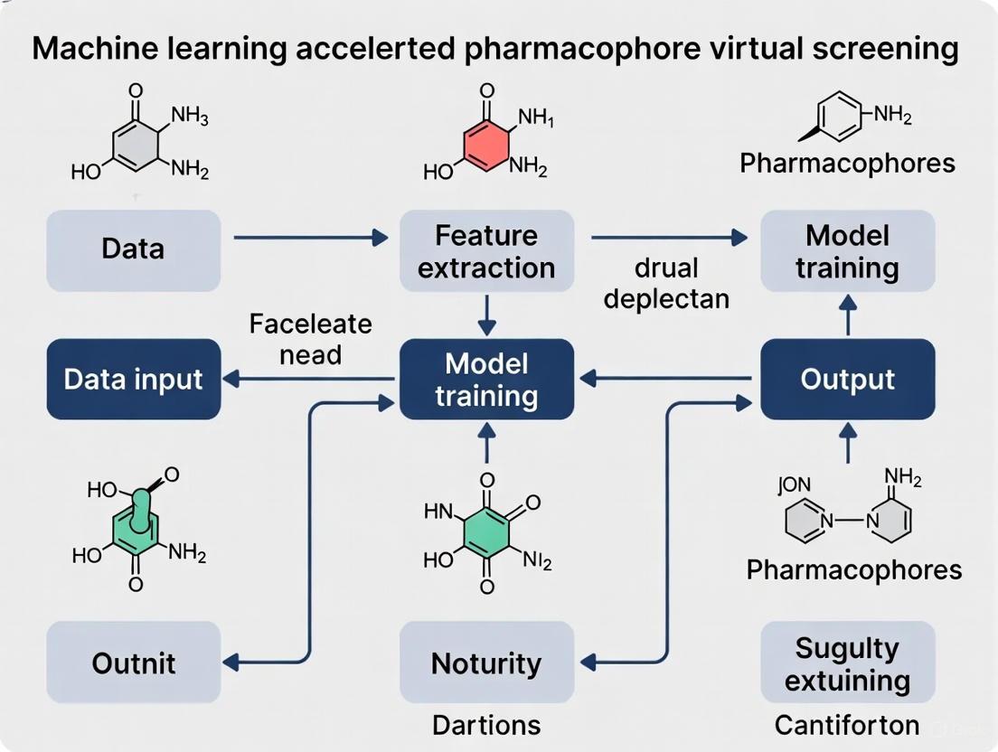
Machine Learning-Accelerated Pharmacophore Virtual Screening: Key Protocols
The integration of machine learning (ML) with pharmacophore modeling has created powerful new methodologies for ultra-rapid virtual screening. The core workflow and its acceleration via ML are summarized in the diagram below.
Protocol 1: Developing an ML Model to Predict Docking Scores
This protocol uses ML to bypass computationally expensive molecular docking, enabling the screening of ultra-large libraries [5].
- Data Preparation: Select a diverse set of ligands with known activity against your target (e.g., from ChEMBL). Generate their low-energy 3D conformations.
- Docking Score Calculation: Dock the prepared ligand set into your target's binding site using your preferred docking software (e.g., Smina) to obtain a docking score for each molecule. This score serves as the training label.
- Feature Extraction (Fingerprinting): Encode the structural and pharmacophoric characteristics of each ligand using molecular fingerprints. This can include:
- Classical Fingerprints: Extended Connectivity Fingerprints (ECFP).
- Pharmacophore Fingerprints: Ligand-based pharmacophore fingerprints that represent the spatial arrangement of chemical features [6].
- Model Training and Validation: Train an ensemble ML model (e.g., combining Random Forest and Support Vector Machines) to predict the docking score from the molecular fingerprints. Validate the model using a hold-out test set and scaffold-based splits to ensure its predictive power generalizes to new chemotypes [5].
- Virtual Screening: Apply the trained model to predict docking scores for millions of compounds in a database (e.g., ZINC). This ML-based screening can be ~1000 times faster than classical docking, allowing for the rapid prioritization of top-scoring compounds for synthetic acquisition and experimental testing [5].
Protocol 2: Pharmacophore-Guided Deep Learning for Molecular Generation
This protocol inverts the screening process by using a pharmacophore to generate novel, active molecules from scratch (de novo design) [7].
- Pharmacophore Hypothesis Input: Define a 3D pharmacophore hypothesis, either from a known active ligand or a protein structure. Represent this hypothesis as a complete graph where nodes are pharmacophore features and edges are inter-feature distances.
- Model Architecture: Employ a deep learning model such as PGMG (Pharmacophore-Guided Molecule Generation). This model typically uses:
- A Graph Neural Network (GNN) encoder to process the spatial distribution of pharmacophore features.
- A transformer decoder to generate SMILES strings of novel molecules.
- A latent variable to model the many-to-many relationship between pharmacophores and molecules, ensuring output diversity [7].
- Generation and Evaluation: The model generates molecules that match the input pharmacophore. Evaluate the generated molecules not only on their fit to the pharmacophore but also on key drug-like properties (validity, uniqueness, novelty) and predicted binding affinity through docking [7].
Performance Metrics & Benchmarking Data
The following tables summarize quantitative data relevant to evaluating and benchmarking pharmacophore and ML-based screening methods.
Table 1: Performance Comparison of Virtual Screening Methods
| Method | Key Metric | Reported Performance | Reference |
|---|---|---|---|
| Classical Pharmacophore VS | Hit Rate (vs. random) | 5-40% (vs. typically <1%) | [1] |
| ML-Accelerated Docking Score Prediction | Speed Increase vs. Classical Docking | ~1000x faster | [5] |
| Pharmacophore-Guided Deep Learning (PGMG) | Novelty / Uniqueness of Generated Molecules | High (>80% novelty achieved) | [7] |
| Structure-Based Pharmacophore Validation | AUC / Enrichment Factor (EF1%) | AUC: 0.98; EF1%: 10.0 | [2] |
Table 2: Essential Research Reagent Solutions for ML-Accelerated Pharmacophore Research
| Reagent / Resource | Function in Research | Example Sources |
|---|---|---|
| Compound Databases | Source of active/inactive ligands and decoys for model training and validation. | ChEMBL, ZINC, DrugBank, DUD-E [5] [1] |
| Protein Data Bank (PDB) | Source of 3D macromolecular structures for structure-based pharmacophore modeling. | RCSB PDB [4] [2] |
| Molecular Docking Software | Generates binding poses and scores for training ML models or validating hits. | Smina, AutoDock Vina [5] [2] |
| Pharmacophore Modeling Software | Creates 2D/3D pharmacophore hypotheses from structures or ligands. | LigandScout, Discovery Studio [4] [2] |
| MD Simulation Software | Samples protein-ligand dynamics to create ensembles of pharmacophore models. | AMBER, GROMACS [4] |
| Fingerprinting & ML Libraries | Generates molecular descriptors and builds predictive ML models. | RDKit, Scikit-learn [5] [6] |
FAQs on Core Concepts and Advanced Applications
Q1: What is the precise IUPAC definition of a pharmacophore?
A: According to IUPAC, a pharmacophore is "an ensemble of steric and electronic features that is necessary to ensure the optimal supramolecular interactions with a specific biological target structure and to trigger (or block) its biological response" [8] [9] [1]. It is crucial to understand that a pharmacophore is an abstract model of interactions, not a specific molecular scaffold or functional group.
Q2: What are the fundamental feature types used in building a 3D pharmacophore model?
A: The core features include [8] [1] [3]:
- Hydrogen Bond Acceptor (HBA)
- Hydrogen Bond Donor (HBD)
- Hydrophobic (H) / Aromatic (AR)
- Positive / Negative Ionizable (PI/NI) These features are represented as 3D objects (points, vectors, spheres) in space to define the necessary interactions for biological activity.
Q3: How does the structure-based pharmacophore approach differ from the ligand-based approach?
A:
- Structure-Based: Directly extracts pharmacophore features from a 3D protein-ligand complex (from PDB or docking). It benefits from knowing the exact interactions and allows the inclusion of exclusion volumes [3] [2].
- Ligand-Based: Derives common pharmacophore features by aligning the 3D structures of multiple known active molecules. It is used when the 3D structure of the target is unknown but a set of active ligands is available [1] [3]. The diagram below illustrates these two primary approaches and their integration with modern ML techniques.
Q4: What are the emerging applications of pharmacophore modeling beyond simple virtual screening?
A: The pharmacophore concept is now applied in advanced areas such as [9] [10]:
- ADMET and Toxicity Prediction: Modeling properties related to absorption, distribution, metabolism, excretion, and toxicity (e.g., blood-brain barrier permeation) [6].
- Off-Target and Side Effect Prediction: Identifying unintended interactions with other biological targets to anticipate potential adverse effects.
- Target Identification (Polypharmacology): For a compound with unknown mechanism, a pharmacophore model can be used to screen against a panel of targets to identify potential primary and secondary targets.
- De Novo Molecular Generation: As detailed in Protocol 2, pharmacophores guide AI to generate novel, active molecules from scratch [7].
Contrasting Structure-Based vs. Ligand-Based Pharmacophore Modeling Approaches
Core Concepts and Definitions
What is a pharmacophore? A pharmacophore is defined by the International Union of Pure and Applied Chemistry (IUPAC) as "the ensemble of steric and electronic features that is necessary to ensure the optimal supramolecular interactions with a specific biological target structure and to trigger (or to block) its biological response" [3] [11] [12]. It is an abstract model of the essential functional features a molecule must possess to bind to a specific target.
What are the key pharmacophoric features? The most common chemical features used in pharmacophore models are [3] [13]:
- Hydrogen Bond Acceptor (HBA)
- Hydrogen Bond Donor (HBD)
- Hydrophobic (H)
- Positively / Negatively Ionizable (PI / NI)
- Aromatic (AR)
- Exclusion Volumes (XVOL) - Represent regions sterically forbidden by the receptor.
Comparative Analysis: Structure-Based vs. Ligand-Based Approaches
Table 1: Core differences between structure-based and ligand-based pharmacophore modeling.
| Aspect | Structure-Based Pharmacophore | Ligand-Based Pharmacophore |
|---|---|---|
| Primary Data Input | 3D structure of the target protein or protein-ligand complex [3] | A set of known active ligands [14] |
| Key Prerequisite | Known 3D structure of the target (from PDB, homology modeling, or AlphaFold2) [3] | A collection of active compounds, ideally with diverse structures and known activities [11] |
| Fundamental Principle | Identifies key interaction points (features) directly from the binding site of the macromolecular target [3] [12] | Derives common chemical features from a set of superimposed active ligands [3] [14] |
| Typical Workflow | 1. Protein Preparation2. Binding Site Identification3. Interaction Points Mapping4. Feature Selection & Model Generation [3] | 1. Ligand Preparation & Conformational Analysis2. Molecular Alignment3. Common Feature Perception4. Hypothesis Generation [11] [14] |
| Ideal Use Case | Target with a known (or reliably modeled) 3D structure [3] | Target with unknown structure but multiple known active ligands [3] [15] |
| Advantages | Can identify novel interaction features not present in known ligands; does not require a set of pre-identified active compounds [3] | Does not require the 3D structure of the target; model is based on experimentally validated active compounds [15] |
| Challenges & Limitations | Quality of the model is highly dependent on the quality and resolution of the protein structure [3] | Handling ligand conformational flexibility and achieving a correct alignment are critical and non-trivial tasks [11] |
Workflow Visualization
Workflow for pharmacophore modeling.
Frequently Asked Questions (FAQs) & Troubleshooting
FAQ 1: When should I choose a structure-based approach over a ligand-based one? Answer: The choice is primarily dictated by data availability.
- Use Structure-Based if: The 3D structure of your target is available from the PDB, or can be reliably generated through homology modeling or tools like AlphaFold2 [3]. This approach is powerful for discovering novel scaffolds (scaffold hopping) as it is not biased by existing ligand structures.
- Use Ligand-Based if: The target's structure is unknown, but you have a set of known active ligands (a training set). This is common for many membrane proteins like GPCRs [11] [15]. The quality of the model increases with the number, diversity, and potency of the known actives.
FAQ 2: My ligand-based pharmacophore model fails to distinguish active compounds from inactives during validation. What could be wrong? Troubleshooting Guide:
- Issue: Poor training set. The set of active ligands used to build the model might be too structurally diverse (preventing identification of a common pattern) or too congeneric (leading to an overly specific model) [11].
- Solution: Re-curate your training set. Include ligands with a range of potencies and ensure they are known to act via the same mechanism. Using a set that includes confirmed inactive compounds can also help refine the model by defining exclusion volumes [16].
- Issue: Incorrect ligand alignment. The model is highly sensitive to the spatial alignment of the input ligands [14].
- Solution: If automatic alignment fails, consider using a known bioactive conformation (e.g., from a crystal structure) as a template for manual or constrained alignment [16].
- Issue: Suboptimal feature selection. The model may have too many or too few features.
- Solution: Manually adjust the feature constraints in your software (e.g., in Schrödinger's Phase, you can set the minimum and maximum number of features and specify required features) [16]. Validate multiple hypotheses against a test set with known actives and decoys.
FAQ 3: How can I improve the accuracy of my structure-based pharmacophore model? Troubleshooting Guide:
- Issue: Low-quality protein structure. The model is only as good as the input structure [3].
- Solution: Carefully prepare the protein structure. Add missing hydrogen atoms, assign correct protonation states to residues (especially in the binding site), and correct any structural anomalies. If using a crystal structure, check its resolution.
- Issue: Incorrect or incomplete binding site definition.
- Solution: Use multiple tools (e.g., GRID, SiteMap) to characterize the binding pocket and confirm its relevance through literature or mutational data [3]. If a co-crystallized ligand is present, it provides a crucial reference for defining key interactions.
- Issue: Model is too rigid. A single, static structure may not represent the dynamic nature of binding.
- Solution: Incorporate protein flexibility. Generate pharmacophore models from multiple molecular dynamics (MD) simulation snapshots to create a dynamic pharmacophore or an ensemble of models [13].
FAQ 4: How is Machine Learning (ML) integrated with pharmacophore modeling to accelerate virtual screening? Answer: ML enhances pharmacophore-based virtual screening in several key ways [17]:
- Feature Learning and Selection: ML algorithms, particularly deep learning, can automatically learn complex, non-intuitive pharmacophoric patterns and molecular descriptors from large datasets of active and inactive compounds, reducing reliance on manual feature engineering [17].
- Improved Performance: ML models can be trained to predict the pharmacophore-matching score or the biological activity of a compound directly from its structure (e.g., from SMILES strings or molecular graphs), leading to faster and more accurate screening [17].
- Handling Molecular Flexibility: Techniques like pharmacophore fingerprints, which encode the presence or absence of pharmacophore features, can be used as input for ML models to efficiently screen vast chemical libraries while implicitly accounting for conformational diversity [18] [13].
Essential Research Reagents & Computational Tools
Table 2: Key software and resources for pharmacophore modeling and virtual screening.
| Tool / Resource Name | Type / Category | Primary Function in Research |
|---|---|---|
| RCSB Protein Data Bank (PDB) | Data Repository | Source for experimental 3D structures of proteins and protein-ligand complexes, the essential input for structure-based modeling [3] |
| LigandScout | Software | Platform for both structure-based (from PDB complexes) and ligand-based pharmacophore modeling, visualization, and virtual screening [18] [12] |
| Schrödinger Phase | Software | Comprehensive tool for developing ligand-based pharmacophore hypotheses, creating screening databases, and performing virtual screening [16] |
| ELIXIR-A | Software (Open-Source) | Python-based tool for refining and comparing multiple pharmacophore models, useful for analyzing results from MD simulations or multiple ligands [18] |
| Pharmit | Online Platform | Interactive tool for pharmacophore-based virtual screening of large compound databases like ZINC and PubChem [18] |
| Directory of Useful Decoys (DUD-e) | Benchmark Dataset | A curated database containing active compounds and "decoys" (structurally similar but physiochemically distinct inactive molecules) for objective validation of virtual screening methods [18] [16] |
| RDKit | Open-Source Toolkit | A collection of cheminformatics and machine learning tools useful for ligand preparation, conformational analysis, and basic pharmacophore feature handling [12] |
Experimental Protocol: Developing a Ligand-Based Pharmacophore Model using Schrödinger Phase
This protocol outlines the key steps for generating a pharmacophore hypothesis from a set of active ligands, as detailed in the Schrödinger tutorial [16].
1. Project Setup and Ligand Preparation
- Create a new project in Maestro and set the working directory.
- Import your prepared 3D ligand structures into the project. Ensure ligands have been pre-processed (e.g., using LigPrep) with correct ionization states, tautomers, and stereochemistry.
2. Defining Actives and Inactives
- In the
Develop Pharmacophore Hypothesespanel, selectMultiple ligands (selected entries). - Click
Defineto specify the active and inactive ligands from your set. This requires a property column (e.g.,pIC50).- Example Thresholding: Set active ligands as those with
pIC50 >= 7.3(equivalent to IC50 ≤ 50 nM) and inactives as those withpIC50 <= 5.0(IC50 ≥ 10 µM) [16].
- Example Thresholding: Set active ligands as those with
3. Configuring Hypothesis Generation Settings
- In the
Featurestab ofHypothesis Settings:- Set the range for the
Number of features in the hypothesis(e.g., 5 to 6). - Optionally, specify a
Preferred minimum number of features. - You can set constraints for specific features (e.g., minimum 1 Donor (D) and 1 Negative (N)).
- Use
Feature presetslike "Make acceptor and negative equivalent" if chemically justified.
- Set the range for the
- In the
Excluded Volumestab:- Check
Create excluded volume shell. - Generate the shell from both
ActivesandInactivesto define regions sterically forbidden by the receptor, improving model selectivity [16].
- Check
4. Running the Job and Analyzing Results
- Provide a
Job name(e.g.,my_pharmacophore) and clickRun. - Upon completion, analyze the generated hypotheses in the
Project Table. Hypotheses are automatically named based on their features (e.g.,DHNRRR). - Critical Validation Steps:
- Visually inspect how well active ligands align with the hypothesis features and avoid the excluded volumes.
- Check how inactive ligands fail to match the hypothesis, either through poor feature alignment or steric clashes with excluded volumes.
- The best hypothesis is typically the one with the highest survival score that also convincingly separates known actives from inactives.
Frequently Asked Questions (FAQs)
FAQ 1: Why does my virtual screening workflow, which works well on small test sets, fail to scale effectively to ultra-large libraries?
The primary failure in scaling is the computational cost and time required by traditional methods. Classical molecular docking procedures become infeasible for screening billions of molecules [5]. Furthermore, the high steric tolerance and physical implausibility of poses generated by some deep learning methods can lead to false positives when applied to novel chemical spaces [19].
- Recommended Protocol for Scaling: Implement a hierarchical filtering approach. Begin with fast ligand-based methods like pharmacophore screening or 2D similarity searches if known active compounds exist [20]. Follow this with machine learning models trained to predict docking scores, which can accelerate screening by up to 1000 times compared to classical docking [5]. Reserve rigorous molecular docking for the final, greatly reduced subset of compounds.
FAQ 2: My QSAR model has high statistical accuracy, but its predictions for novel chemotypes are unreliable. What is the cause?
This is a classic problem of a model operating outside its Applicability Domain (AD). Traditional QSAR models suffer from the lack of a formal confidence score for each prediction, and their reliability is confined to the chemical space represented in their training data [21]. When presented with a novel scaffold (a new chemotype), the model's predictions cannot be trusted.
- Troubleshooting Guide:
- Assess Applicability Domain: Use tools to determine if your query molecules fall within the chemical space of the training set. A model cannot be expected to reliably extrapolate.
- Implement Conformal Prediction: Adopt newer QSAR approaches like Conformal Prediction (CP), which provides a confidence measure for each prediction and clearly identifies unreliable ones [21].
- Use Consensus Modeling: Combine predictions from multiple QSAR models built with different algorithms and descriptors. Consensus strategies have been proven to be more accurate and cover a wider area of chemical space than individual models on average [22].
FAQ 3: Why does a compound with an excellent docking score show no biological activity in the lab?
A high docking score does not equate to high binding affinity. Scoring functions are designed to identify plausible binding poses but are notoriously poor at predicting absolute binding affinity [23]. They often oversimplify critical physical phenomena.
- Key Limitations to Investigate:
- Inadequate Treatment of Solvation: Scoring functions struggle to model complex water networks and desolvation penalties accurately.
- Protein Rigidity: Most docking programs treat the protein as rigid, ignoring the induced fit and conformational changes that occur upon ligand binding [23].
- Ignoration of Entropy: The crucial role of conformational entropy and dynamics in binding is often not fully captured [23].
- Non-drug-like Properties: The compound might have poor pharmacokinetic properties, metabolic liability, or be synthetically inaccessible, issues docking does not assess [23].
FAQ 4: How can I improve the physical plausibility of the binding poses generated by deep learning docking tools?
While some DL docking methods, particularly generative diffusion models, achieve high pose accuracy, they can produce poses with steric clashes and incorrect bond geometries [19].
- Solution: Integrate post-docking refinement and validation. Use tools like the PoseBusters toolkit to systematically check generated poses for chemical and geometric consistency, including bond lengths, angles, and protein-ligand clashes [19]. Subsequently, refine top-scoring poses using more rigorous molecular dynamics (MD) simulations and rescore them with methods like MM-GBSA/MM-PBSA [23].
Troubleshooting Guides
Issue: Low Hit Rate and Poor Enrichment in Virtual Screening
Problem: Your VS campaign returns a high number of false positives, failing to enrich for truly active compounds.
| Potential Cause | Diagnostic Steps | Corrective Action |
|---|---|---|
| Inadequate Library Preparation | Check for correct protonation states, tautomers, and stereochemistry. Verify the generation of bioactive conformers. | Use software like LigPrep [20], OMEGA [20], or RDKit's MolVS [20] for standardized, high-quality 3D compound preparation. |
| Over-reliance on a Single Protein Conformation | Check if your protein structure has known flexible loops or multiple crystallographic structures with different binding site conformations. | Perform ensemble docking using multiple protein structures. Generate these from different crystal structures or by clustering frames from Molecular Dynamics (MD) simulations [23]. |
| Limitations of the Scoring Function | Test if your docking program can correctly re-dock and score known active ligands from co-crystal structures. | Use a consensus scoring approach. Combine results from multiple docking programs or different scoring functions to prioritize compounds identified by several methods [22]. |
| Ignoring Pharmacophore Constraints | Check if your top-scoring docking poses actually form key interactions known to be critical for activity (e.g., from SAR studies). | Develop a structure-based pharmacophore model from a protein-ligand complex and use it to filter docking results, ensuring poses match essential interaction features [3] [24]. |
Issue: QSAR Model with High Training Accuracy but Poor Predictive Performance
Problem: Your QSAR model performs well on its training and internal test sets but fails when applied to new external data.
| Potential Cause | Diagnostic Steps | Corrective Action |
|---|---|---|
| Overfitting | Check if the model performance on the training set is significantly higher than on a rigorous external test set. | Simplify the model by reducing the number of descriptors. Use validation techniques like scaffold splitting (splitting data based on Bemis-Murcko scaffolds) to ensure the model is tested on new chemotypes [5]. |
| Narrow Applicability Domain | Analyze the chemical space of your external dataset compared to the training set using PCA or similarity metrics. | Use conformal prediction frameworks to assign a confidence level to each prediction, allowing you to flag and disregard predictions for molecules outside the model's domain [21]. |
| Data Inconsistency and Bias | Evaluate the source and quality of your training data. Check for activity cliffs and significant imbalances between active and inactive compounds. | Curate a high-quality, diverse dataset. For imbalanced data, use techniques like class weighting during model training [21]. Apply consensus modeling to integrate predictions from multiple models, improving robustness [22]. |
Experimental Protocols & Data
Protocol 1: Building a Robust QSAR Model with an Defined Applicability Domain
This protocol is designed to create a generalizable QSAR model and quantify the confidence of its predictions.
- Data Curation and Preparation: Extract bioactivity data (e.g., IC₅₀, Ki) from curated databases like ChEMBL [21] [5]. Standardize structures, remove duplicates, and calculate molecular descriptors or fingerprints (e.g., Morgan fingerprints using RDKit) [21].
- Strategic Data Splitting: Instead of a simple random split, divide the dataset using Bemis-Murcko scaffolds. This ensures that the model is tested on structurally distinct scaffolds not seen during training, providing a more realistic assessment of its predictive power for novel chemotypes [5].
- Model Training and Validation: Train a machine learning model (e.g., Random Forest, Support Vector Machine) on the training set. Validate its performance on the scaffold-based test set.
- Define Applicability Domain with Conformal Prediction: Implement a conformal predictor using a calibration set. This will output predictions with a confidence measure (e.g., at 95% confidence), allowing you to identify and withhold predictions for molecules that are too dissimilar from the training data [21].
Protocol 2: Machine Learning-Accelerated Docking Score Prediction
This protocol bypasses slow molecular docking by using ML models to predict docking scores directly from 2D structures, enabling ultra-large library screening [5].
- Generate Training Data: Perform molecular docking with your chosen software (e.g., Smina) on a diverse but manageable set of ligands (e.g., 10,000-50,000 compounds) to obtain docking scores [5].
- Train an Ensemble ML Model: Use the docked ligands and their scores as training data. Represent each molecule with multiple types of molecular fingerprints and descriptors. Train an ensemble of machine learning models (e.g., using different algorithms or features) to predict the docking score. This ensemble reduces prediction errors [5].
- High-Throughput Screening: Apply the trained ensemble model to predict docking scores for millions or even billions of compounds in the virtual library. This process is orders of magnitude faster than classical docking.
- Validation and Docking: Select the top-ranked compounds from the ML screen and run a full molecular docking calculation for final verification and pose generation.
Troubleshooting Workflow for VS Bottlenecks
The Scientist's Toolkit: Research Reagent Solutions
| Item | Function & Application | Key Considerations |
|---|---|---|
| ZINC Database | A publicly available library of over 20 million commercially available compounds for virtual screening. Provides a vast chemical space for discovering high-quality hits [25]. | Compounds are purchasable but not available in-house. Vendor IDs are provided for purchasing hits [25]. |
| ChEMBL Database | A curated public database of bioactive molecules with drug-like properties. Used to extract bioactivity data (pChEMBL values) for training QSAR and machine learning models [21] [5]. | Data requires careful curation and standardization. Activity data can be sparse for certain targets [21]. |
| RDKit | An open-source cheminformatics toolkit. Used for calculating molecular descriptors, generating conformers (via its ETKDG method), and standardizing chemical structures [21] [20]. | The freely available DG algorithm is robust but may be outperformed by some commercial systematic conformer generators [20]. |
| PoseBusters | A validation toolkit to check the physical plausibility of AI-generated docking poses against chemical and geometric criteria (bond lengths, clashes, etc.) [19]. | Critical for identifying false positives from deep learning docking methods that may have good RMSD but invalid physics [19]. |
| OMEGA & ConfGen | Commercial, high-performance conformer generation software. Systematically sample conformational space to produce low-energy, biologically relevant 3D structures for docking and pharmacophore modeling [20]. | Outperforms simpler methods in benchmarking studies and is crucial for ensuring the bioactive conformation is represented [20]. |
| AutoDock Vina & Glide | Widely used molecular docking programs (Vina is open-source, Glide is commercial). Used for binding pose prediction and structure-based virtual screening [19]. | Traditional methods like Glide SP show high physical validity and robustness, especially on novel protein pockets [19]. |
ML-Accelerated Virtual Screening Protocol
Frequently Asked Questions & Troubleshooting
Q1: My ML model for predicting docking scores performs well on the training data but poorly on new, unseen chemotypes. How can I improve its generalization?
- Answer: This is a classic sign of overfitting, where the model learns the training data too well but fails to generalize. To address this:
- Implement Scaffold-Based Data Splitting: Instead of random splits, divide your dataset so that training and testing sets contain different molecular scaffolds (core structures). This ensures the model is tested on genuinely novel chemotypes and better simulates a real virtual screening scenario [5].
- Utilize Ensemble Models: Combine predictions from multiple models trained on different types of molecular fingerprints or descriptors (e.g., ECFP, molecular weight, topological descriptors). This approach reduces variance and overall prediction error, leading to more robust performance on diverse compounds [5].
Q2: Molecular docking is a bottleneck in my large-scale virtual screening workflow. Are there faster, structure-based alternatives?
- Answer: Yes, deep learning-based pharmacophore modeling offers a high-speed alternative.
- Solution: Tools like PharmacoNet provide fully automated, protein-based pharmacophore modeling. Instead of computationally intensive docking, these methods use a parameterized analytical scoring function to evaluate ligand potency based on pharmacophore feature alignment. This approach can screen hundreds of millions of compounds in a matter of hours on standard hardware, drastically accelerating the initial screening phase [26] [27].
- Troubleshooting Tip: If the hit rates from the initial pharmacophore screen are still too high for subsequent docking, you can apply a second filter using an ML model that predicts docking scores to further prioritize compounds [5].
Q3: How can I ensure the hits identified by my ML-driven virtual screening are not just artifacts but have real potential?
- Answer: Enhance the interpretability of your models and incorporate multiple validation strategies.
- Apply SHAP Analysis: Use SHapley Additive exPlanations (SHAP) analysis to interpret your ML model's predictions. This helps identify which specific molecular features or fingerprints are driving the activity prediction, adding a layer of plausibility and expert validation to the results [28].
- Conformational Ensembles: Train your ML models on docking results generated from multiple protein conformations, not just a single crystal structure. This accounts for protein flexibility and can improve the correlation between predicted and experimental activity [5].
- Experimental Triangulation: Always complement computational hits with in vitro testing. A successful workflow should synthesize and biologically evaluate top-ranked compounds to confirm inhibitory activity, as demonstrated in the MAO inhibitor study where 24 selected compounds were tested, leading to the discovery of active inhibitors [5].
Experimental Protocols for ML-Accelerated Pharmacophore Screening
The following protocol summarizes a methodology for machine learning-accelerated, pharmacophore-based virtual screening, as applied to Monoamine Oxidase (MAO) inhibitors [5].
1. Protein and Ligand Preparation
- Protein Structures: Obtain 3D coordinates of the target protein from the Protein Data Bank (PDB). For example, use PDB IDs 2Z5Y (for MAO-A) and 2V5Z (for MAO-B). Prepare the structure by removing native ligands and water molecules, and adding necessary hydrogen atoms [5].
- Ligand Library: Curate a library for screening, such as the ZINC database. Filter compounds based on drug-likeness rules (e.g., molecular weight < 700 Da) and remove highly flexible structures to simplify subsequent docking and modeling steps [5] [28].
2. Generating Training Data via Molecular Docking
- Docking Software: Perform molecular docking on a known set of active and inactive compounds using software like Smina to generate the ground-truth data [5].
- Output: The primary output is the docking score (DS) for each compound, which serves as the label for training the machine learning model.
3. Training the Machine Learning Model
- Feature Generation: Calculate multiple types of molecular representations for each compound, including:
- Molecular Fingerprints (e.g., ECFP4)
- Molecular Descriptors (e.g., molecular weight, logP, topological indices) [5]
- Model Training: Train an ensemble machine learning model (e.g., Random Forest) to predict the docking score based on the molecular features. The ensemble approach minimizes individual model errors [5] [28].
- Validation: Rigorously validate the model using a scaffold-based split to ensure its ability to generalize to new chemical classes [5].
4. Large-Scale Virtual Screening & Hit Identification
- Pharmacophore Constraint: Apply a pharmacophore model to filter the large screening library (e.g., ZINC), creating a constrained chemical space likely to bind the target [5].
- ML-Based Scoring: Use the trained ML model to rapidly predict docking scores for all compounds that pass the pharmacophore filter. This step is approximately 1000 times faster than running classical molecular docking on the entire library [5].
- Final Prioritization: Select the top-ranked compounds based on the predicted scores for synthesis and subsequent in vitro biological evaluation to confirm activity [5].
Research Reagent Solutions
The table below lists key computational tools and data resources essential for setting up an ML-accelerated virtual screening pipeline.
| Item Name | Function in the Experiment | Key Features / Notes |
|---|---|---|
| Smina [5] | Molecular docking software used to generate training data (docking scores) for the ML model. | Customizable scoring function; used for classic VS and creating labels for ML. |
| ZINC Database [5] | A publicly available library of commercially available compounds for virtual screening. | Source of millions to billions of purchasable molecules for screening. |
| Molecular Fingerprints (e.g., ECFP) [5] | 2D structural representations of molecules used as input features for the ML model. | Captures molecular patterns and features critical for activity prediction. |
| Pharmacophore Modeling Software | Defines essential steric and electronic features for molecular recognition, used as a constraint to filter libraries. | Can be traditional (e.g., in Schrodinger Maestro) or deep learning-based (e.g., PharmacoNet). |
| PharmacoNet [26] [27] | A deep learning framework for automated, structure-based pharmacophore modeling. | Enables ultra-fast screening; identifies key protein hotspots and pharmacophore features. |
| ChEMBL Database [5] | A manually curated database of bioactive molecules with drug-like properties. | Source of experimental bioactivity data (e.g., IC₅₀, Kᵢ) for known ligands. |
The table below consolidates key performance metrics from the reviewed studies, providing benchmarks for your own experiments.
| Metric | Reported Performance | Context / Model |
|---|---|---|
| Speed Gain | ~1000x faster than classical docking | ML-based docking score prediction vs. standard docking procedure [5]. |
| Screening Scale | 187 million compounds in < 21 hours | Performance of PharmacoNet on a single CPU for cannabinoid receptor inhibitors [26]. |
| Inhibition Activity | Up to 33% MAO-A inhibition | Best result from 24 synthesized and tested compounds identified via the ML/pharmacophore protocol [5]. |
| Model AUC | 0.99 | Interpretable Random Forest model for identifying GSK-3β inhibitors [28]. |
Workflow Diagram
The following diagram illustrates the integrated machine learning and pharmacophore screening workflow.
ML Model Development Process
This diagram details the core process of creating the machine learning model that predicts docking scores.
An FAQ for Machine Learning-Accelerated Pharmacophore Virtual Screening
This guide addresses frequently asked questions and common troubleshooting scenarios for researchers applying Core ML concepts to pharmacophore-based virtual screening in drug discovery.
Core Concepts and Experimental Design
FAQ: What are the fundamental machine learning paradigms used in cheminformatics, and how are they applied to pharmacophore virtual screening?
Machine learning in cheminformatics is broadly categorized into three types, each with distinct applications in virtual screening [29] [30].
- Supervised Learning: Models are trained on labeled datasets where the correct output (e.g., "active" or "inactive" against a target) is known for each input molecule. These are extensively used for classification tasks (predicting categorical labels like bioactivity) and regression tasks (predicting continuous values like binding affinity) in quantitative structure-activity relationship (QSAR) modeling and property prediction [29] [31] [30].
- Unsupervised Learning: Models find hidden patterns or intrinsic structures in unlabeled data. In virtual screening, this is crucial for clustering large chemical libraries to identify structural families or for dimensionality reduction to visualize and understand the chemical space of screened compounds [29] [30].
- Reinforcement Learning (RL): An agent learns optimal actions through trial-and-error interactions with an environment to maximize a cumulative reward. In drug discovery, RL is increasingly applied to de novo molecular design, where the agent learns to generate molecules with desired properties by sequentially building molecular structures [29] [32].
Table: Common Machine Learning Algorithms in Cheminformatics
| ML Paradigm | Algorithm Examples | Primary Use-Cases in Virtual Screening |
|---|---|---|
| Supervised Learning | Random Forest (RF), Support Vector Machine (SVM), k-Nearest Neighbors (kNN), Naïve Bayes (NB), Artificial Neural Networks (ANNs) [29] | Bioactivity classification, binding affinity prediction (regression), ADMET property forecasting [29] [31] |
| Unsupervised Learning | k-Means Clustering, Principal Component Analysis (PCA) [30] | Chemical library clustering, scaffold hopping, data exploration and visualization [29] |
| Reinforcement Learning | Deep Q-Networks (DQN), Policy Gradient Methods [29] | De novo drug design, optimizing multi-parameter objectives (potency, solubility, synthesizability) [29] [32] |
| Deep Learning (Subset of above) | Convolutional Neural Networks (CNNs), Recurrent Neural Networks (RNNs), Generative Adversarial Networks (GANs) [29] | Learning from complex data (e.g., molecular graphs, SMILES sequences), generating novel molecular structures [29] [33] |
FAQ: How do I choose the right machine learning algorithm for my virtual screening project?
Selecting an algorithm depends on your data and the specific question you are asking. The following workflow outlines a decision-making process for algorithm selection in a pharmacophore virtual screening context.
Troubleshooting Guide: My model's predictions are inaccurate and unreliable. What could be wrong?
Inaccurate models are often due to issues with input data or model configuration.
Problem: Poor Quality or Non-representative Training Data.
- Symptoms: Low accuracy on both training and test sets, model fails to generalize.
- Solution:
- Curate your data: For SMILES strings, use toolkits like RDKit to validate and standardize molecular structures. Remove duplicates and correct invalid representations [33].
- Address data bias: Ensure your negative dataset (inactives) is representative and not artificially easy to distinguish from actives.
- Apply feature scaling: Normalize or standardize numerical descriptors, especially for distance-based algorithms like SVM and kNN.
Problem: Data Leakage or Improper Validation.
- Symptoms: High accuracy during training/training but poor performance on truly external validation sets.
- Solution:
- Split data correctly: Strictly separate your data into training, validation, and test sets before any feature selection or model tuning [34].
- Use cross-validation: Apply k-fold cross-validation on the training set to get a more robust estimate of model performance and tune hyperparameters without touching the test set [34].
- Avoid target leakage: Ensure that no information from the validation/test set is used to inform the training process.
Problem: Suboptimal Algorithm or Hyperparameters.
- Symptoms: Model performance plateaus or is consistently worse than a simple baseline.
- Solution:
- Benchmark algorithms: Start by testing a few simple, interpretable models (e.g., Random Forest, SVM) as a baseline before moving to complex deep learning models [29].
- Perform hyperparameter tuning: Systematically search for the best hyperparameters (e.g., number of trees in RF, learning rate in neural networks) using methods like grid search or random search on the validation set.
Data Preparation and Feature Engineering
FAQ: How are molecular structures converted into a machine-readable format for model training?
Molecules are commonly represented as SMILES (Simplified Molecular Input Line Entry System) strings, a compact text-based notation that encodes the molecular structure [33]. Before training, these strings must be processed.
Table: Key Steps in SMILES Data Preprocessing
| Step | Description | Common Tools/Packages | Potential Issue if Skipped |
|---|---|---|---|
| Validation & Standardization | Check for and correct invalid SMILES; generate canonical forms to ensure one unique SMILES per molecule. | RDKit, OpenBabel | Model learns from incorrect or redundant structures, reducing generalizability. |
| Tokenization | Split the SMILES string into chemically meaningful units (e.g., atoms, bonds, branches). A regex-based tokenizer is preferred over character-level. | Custom regex functions, specialized cheminformatics libraries [33] | "Cl" (chlorine) is split into "C" and "l", confusing the model. |
| Embedding | Convert tokens into numerical vectors. This can be learned by the model or use pre-trained embeddings from models like ChemBERTa. | PyTorch (nn.Embedding), TensorFlow, Transformers libraries [33] |
Model treats each token as an independent symbol, missing chemical context. |
Troubleshooting Guide: My model fails to learn meaningful chemical patterns from SMILES data.
- Problem: Incorrect Tokenization of SMILES Strings.
- Symptoms: Model fails to converge, generates chemically invalid SMILES, or shows poor predictive performance.
- Solution: Implement a regular expression (regex)-based tokenizer that correctly handles multi-character atoms (e.g., "Cl", "Br") and complex bracketed expressions (e.g., "[Na+]") [33]. Do not use a naive character-level split.
Model Implementation and Workflow Integration
FAQ: What is a typical machine learning workflow for structure-based pharmacophore virtual screening?
A common strategy is to use a hybrid approach that combines structure-based methods with machine learning. The following workflow integrates SBVS with ML to improve screening efficiency.
Troubleshooting Guide: My virtual screening workflow is computationally too slow for large compound libraries.
- Problem: High Computational Cost of Structure-Based Screening.
- Symptoms: Docking millions of compounds takes days or weeks, creating a bottleneck.
- Solution:
- Implement a tiered screening approach: Use fast, ligand-based (LBVS) similarity searches (e.g., using Tanimoto coefficient on molecular fingerprints) or a simple ML classifier as a initial filter to reduce the library size before running more expensive SBVS [31].
- Optimize ML feature sets: Use dimensionality reduction (e.g., PCA) or feature selection algorithms (e.g., Recursive Feature Elimination) to reduce the number of molecular descriptors fed to the ML model, speeding up training and prediction [30] [32].
The Scientist's Toolkit: Essential Research Reagents and Software
Table: Key Resources for ML-Driven Pharmacophore Screening
| Tool/Resource | Type | Primary Function | Application in Workflow |
|---|---|---|---|
| RDKit | Cheminformatics Library | SMILES validation, descriptor calculation, fingerprint generation, molecular visualization. [33] | Data preprocessing, feature engineering, and result analysis. |
| Core ML Tools | Conversion Library | Converts models from PyTorch, TensorFlow into Core ML format for deployment on Apple devices. [35] [36] | Final model deployment and integration into mobile applications for on-device prediction. |
| Create ML | Model Training Tool (macOS) | Provides a no-code/low-code environment to train Core ML models on your Mac. [35] [36] | Rapid prototyping of ML models for tasks like image-based assay analysis or property prediction. |
| Schrödinger Suite | Commercial Software Platform | Physics-based molecular modeling, simulation, and high-throughput virtual screening. [37] | Structure-based pharmacophore generation, molecular docking (SBVS), and advanced simulation. |
| Exscientia AI Platform | AI-Driven Drug Discovery | Generative AI and automated design-make-test cycles for lead optimization. [37] | De novo molecular design and multi-parameter optimization of lead compounds. |
Deployment and Advanced Applications
FAQ: How can I deploy a trained cheminformatics model into a production environment for real-time screening?
Deployment strategies vary based on the target platform. For integrating models into applications on Apple devices (iOS, macOS, etc.), Core ML is the key framework [35] [36].
- Model Conversion: Trained models from popular frameworks like PyTorch and TensorFlow can be converted to the Core ML format (
.mlmodelfile) using the coremltools Python package [35] [36]. - On-Device Advantages: Core ML leverages the CPU, GPU, and Neural Engine on Apple hardware to run models on-device. This ensures data privacy, provides real-time inference without a network connection, and minimizes power consumption [35].
- Xcode Integration: The converted
.mlmodelfile can be directly integrated into your Xcode project, which automatically generates a ready-to-use Swift/Objective-C API for making predictions within your application [35].
Troubleshooting Guide: My deployed Core ML model performs differently than it did during training in Python.
- Problem: Discrepancy Between Training and Deployment Environments.
- Symptoms: Model accuracy drops or predictions are inconsistent after conversion to Core ML.
- Solution:
- Verify pre-processing parity: Ensure that the exact same data pre-processing steps (SMILES tokenization, feature scaling, etc.) are replicated identically in the deployment environment (e.g., your Swift app) as were used during model training in Python.
- Validate the converted model: Use Core ML Tools to validate the converted model on a sample dataset before deployment to check for conversion errors or precision losses [35] [36].
- Profile with Xcode: Use the Core ML performance reports and Instruments in Xcode to profile the model on the target device, checking for unexpected latency or hardware compatibility issues [35].
Methodologies in Action: AI Architectures and Workflows for Accelerated Screening
In machine learning-accelerated pharmacophore virtual screening, selecting and implementing the appropriate molecular representation is a foundational step that directly impacts the success of downstream tasks. This technical support center addresses common challenges researchers face when transitioning from traditional to modern AI-driven representation methods. The following guides and protocols are designed to help you navigate technical hurdles, optimize experimental setups, and validate your workflows within the context of advanced drug discovery research.
Frequently Asked Questions
FAQ 1: What are the primary considerations when choosing between SMILES strings and graph-based representations for a new virtual screening project?
The choice depends on your project's goals, data characteristics, and computational constraints. SMILES strings are text-based, human-readable, and work well with language models, but they can be syntactically invalid and struggle to explicitly capture complex topological information. Graph-based representations naturally model atoms (nodes) and bonds (edges), making them superior for tasks requiring an intrinsic understanding of molecular structure and topology, such as predicting complex bioactivity or generating novel scaffolds with specific stereochemistry. For a balanced approach, consider a hybrid model that uses both representations.
FAQ 2: Our model trained on ECFP fingerprints fails to generalize to novel scaffold classes. What could be the cause and potential solutions?
This is a classic problem of the "analogue bias" inherent in many fingerprint-based models. Extended-Connectivity Fingerprints (ECFPs) and other predefined descriptors may not capture the essential features responsible for bioactivity across structurally diverse compounds, leading to poor performance on out-of-distribution scaffolds.
- Solution 1: Employ Graph Neural Networks (GNNs). GNNs learn task-specific representations directly from the molecular graph, which can capture non-linear and complex structure-activity relationships that are opaque to fixed fingerprints [38].
- Solution 2: Utilize Contrastive Learning. Frameworks that use contrastive learning can learn embeddings by maximizing agreement between differently augmented views of the same molecule while pushing apart representations of different molecules. This forces the model to learn more robust, invariant features that are better for scaffold hopping [38].
- Solution 3: Implement a Hybrid Model. Combine ECFP features with learned representations from a GNN or other deep learning model to leverage both predefined chemical knowledge and data-driven insights.
FAQ 3: How can we effectively represent 3D molecular geometry and conformational information for pharmacophore-based models using standard 2D representations?
Standard 2D representations like SMILES or 2D graphs do not natively encode 3D conformation, which is critical for pharmacophore modeling where spatial relationships between features define biological activity.
- Approach 1: 3D Graph Representations. Enhance your graph representation by incorporating 3D spatial coordinates as node features. This allows GNNs to learn from both connectivity and geometric information.
- Approach 2: Geometric Deep Learning. Utilize specialized architectures like SE(3)-equivariant networks that are designed to be invariant to rotations and translations, making them inherently suited for 3D molecular data.
- Approach 3: Conformational Ensembles. For a simpler approach, generate multiple low-energy conformers for each molecule and represent the entire ensemble, either by pooling representations from each conformer or by using the conformer closest to the active pose for the target.
FAQ 4: What are the best practices for fine-tuning a pre-trained molecular transformer model (e.g., for a specific target family like GPCRs)?
Fine-tuning a pre-trained model on a smaller, target-specific dataset is an efficient way to achieve high performance.
- Model Selection: Choose a model pre-trained on a large, diverse chemical corpus (e.g., ChEMBL, ZINC).
- Data Preparation: Curate a high-quality, target-specific dataset. Ensure it is cleaned and standardized (e.g., canonical SMILES, removal of duplicates).
- Progressive Unfreezing: Do not unfreeze all layers at once. Start by fine-tuning only the last few layers, then progressively unfreeze earlier layers to avoid catastrophic forgetting.
- Task-Specific Head: Replace the pre-training head (e.g., masked language modeling) with a task-specific head suitable for your goal, such as a classification or regression layer.
- Learning Rate: Use a lower learning rate for the fine-tuning phase compared to the pre-training rate to make subtle adjustments to the weights.
Troubleshooting Guides
Issue 1: Handling Invalid or Unrealistic SMILES from Generative Models
Problem: A generative model (e.g., a VAE or Transformer) produces a high rate of invalid or chemically unrealistic SMILES strings, hindering the discovery of viable lead compounds.
Diagnosis Steps:
- Check Training Data Quality: Ensure the training data consists of valid, canonicalized SMILES.
- Quantify Invalidity Rate: Calculate the percentage of invalid SMILES generated during sampling.
- Analyze Chemical Plausibility: Use rule-based filters (e.g., for unwanted functional groups) or a separate model to predict synthetic accessibility (SAscore) to check for unrealistic molecules even among valid SMILES.
Resolution Protocol:
- Switch to SELFIES: Consider using SELFIES (SELF-referencing Embedded Strings) instead of SMILES as the molecular representation. SELFIES are grammatically correct by design, guaranteeing 100% validity [38].
- Reinforce Validity during Training: Implement a reinforcement learning (RL) framework where the model receives a positive reward for generating valid and novel molecules, in addition to the primary objective (e.g., high predicted activity).
- Apply Post-Hoc Filtering: Integrate a robust post-processing pipeline that automatically discards invalid SMILES and filters chemically undesirable molecules using tools like RDKit.
Issue 2: Poor Model Performance on Imbalanced High-Throughput Screening (HTS) Data
Problem: A model trained on HTS data, where active compounds are rare, fails to identify true actives because it is biased toward the majority class (inactives).
Diagnosis Steps:
- Calculate Class Balance: Determine the ratio of active to inactive compounds in your dataset.
- Analyze Performance Metrics: Rely on metrics beyond accuracy, such as Precision-Recall (PR) curves, Area Under the PR Curve (AUPRC), and F1-score, which are more informative for imbalanced data.
- Review Data Splitting: Check if your training/test split preserves the class distribution (stratified splitting).
Resolution Protocol:
- Advanced Sampling Techniques:
- Apply SMOTE: Use Synthetic Minority Over-sampling Technique (SMOTE) to generate synthetic examples of the active class in the descriptor or embedding space.
- Use Weighted Random Sampling: During training, weight the sampling probability so that the model sees more examples from the under-represented active class.
- Algorithmic Cost-Sensitivity:
- Implement Focal Loss: Use Focal Loss, a modified cross-entropy loss that down-weights the loss assigned to well-classified examples, forcing the model to focus on hard, misclassified negatives (inactives mistaken for actives).
- Adjust Class Weights: Most ML libraries allow you to automatically adjust class weights inversely proportional to class frequencies in the loss function.
- Leverage Transfer Learning:
- Start with a model pre-trained on a large, balanced chemical dataset.
- Fine-tune the final layers on your specific, imbalanced HTS dataset. This provides a strong foundational understanding of chemistry before learning the specific task.
Issue 3: Inefficient Training or Inference with Graph Neural Networks on Large Virtual Compound Libraries
Problem: Training a GNN on a library of millions of compounds is prohibitively slow, or generating predictions (inference) for a virtual screen takes too long.
Diagnosis Steps:
- Profile Code: Use profiling tools to identify bottlenecks (e.g., data loading, graph convolution operations).
- Monitor Hardware Utilization: Check GPU/CPU and memory usage to see if resources are saturated.
- Check Batch Size: Determine if the batch size is too small (inefficient) or too large (causes memory overflow).
Resolution Protocol:
- Optimize Data Loading:
- Use a
DataLoaderthat supports parallel data loading. - Precompute and cache graph structures for the entire dataset to avoid on-the-fly processing during training.
- Use a
- Employ Graph Sampling:
- Use neighbor sampling methods (e.g., GraphSAGE) instead of full-graph training. This samples a node's local neighborhood for each batch, drastically reducing memory footprint and computation.
- Implement mini-batching of graphs for link prediction or graph classification tasks.
- Utilize Mixed-Precision Training:
- Use 16-bit floating-point precision (FP16) alongside 32-bit precision (FP32) to speed up computations and reduce memory usage on supported GPUs.
- Model Simplification:
- Reduce the number of GNN layers. Very deep GNNs can suffer from over-smoothing and are computationally expensive.
- Consider using simpler, faster convolution operations (e.g., SAGEConv instead of GINConv) for large-scale screening.
Experimental Protocols & Data
Protocol 1: Benchmarking Molecular Representations for Scaffold Hopping
Objective: To systematically evaluate the performance of different molecular representations in identifying structurally diverse compounds (different scaffolds) with similar biological activity.
Materials:
- Dataset: A curated bioactivity dataset (e.g., from ChEMBL) for a well-defined protein target, with known active compounds clustered by Bemis-Murcko scaffolds.
- Software: RDKit, DeepChem, PyTorch Geometric, or equivalent libraries.
Methodology:
- Data Preparation & Splitting:
- Cluster molecules by their Bemis-Murcko scaffolds.
- Perform a scaffold split, where all molecules from entire scaffolds are held out for the test set. This rigorously tests the model's ability to generalize to novel chemotypes.
- Model Training & Representation:
- Train identical machine learning models (e.g., a Random Forest or a simple GNN) using different molecular representations on the training set.
- Representations to Test:
- Fingerprint-based: ECFP6, MACCS Keys.
- Descriptor-based: A set of physicochemical descriptors (e.g., molecular weight, logP, TPSA).
- Graph-based: A Graph Isomorphism Network (GIN) operating on 2D graphs.
- Language model-based: Features extracted from a pre-trained molecular Transformer model.
- Evaluation:
- Predict activity on the held-out test set containing novel scaffolds.
- Evaluate using metrics focused on early retrieval: Hit Rate (top 1%), Enrichment Factor (EF) at 1%, and Area Under the Accumulative Recall Curve.
Anticipated Results: Graph-based and language model-based representations are expected to significantly outperform traditional fingerprints and descriptors on the scaffold-split test set, demonstrating their superior ability to capture the essential features of bioactivity beyond simple structural similarity [38].
Protocol 2: Integrating Multimodal Representations for ADMET Prediction
Objective: To improve the predictive accuracy of ADMET (Absorption, Distribution, Metabolism, Excretion, Toxicity) properties by combining multiple molecular representations.
Methodology:
- Feature Extraction:
- Extract features for each molecule in the dataset using several representation methods concurrently.
- Modality 1 (Graph): A GNN to extract topological features.
- Modality 2 (SMILES): A Transformer encoder to extract sequential features.
- Modality 3 (3D): Geometric features from a low-energy conformer (e.g., 3D coordinates fed to a different network or used to compute 3D descriptors).
- Multimodal Fusion:
- Employ a fusion strategy to combine the extracted feature vectors. Simple early fusion (concatenation) can be used, or more complex late fusion (e.g., using an attention mechanism to weight the importance of each modality) can be implemented.
- Prediction:
- Feed the fused, multimodal representation into a final prediction head (a fully connected network) for the specific ADMET endpoint (e.g., hERG inhibition, microsomal stability).
Validation: Compare the performance of the multimodal model against unimodal baselines (using only graph, SMILES, or 3D features) via cross-validation on standard ADMET benchmarks like those in the MoleculeNet dataset. The multimodal approach is designed to provide a more holistic view of the molecule, leading to more robust and accurate predictions [38].
Table 1: Comparison of Key Molecular Representation Methods
| Representation Type | Example Methods | Data Structure | Key Advantages | Common Use Cases |
|---|---|---|---|---|
| String-Based | SMILES, SELFIES [38] | Sequential Text | Simple, compact, works with NLP models [38] | Molecular generation, language model pre-training |
| Fingerprint-Based | ECFP, MACCS Keys [38] | Fixed-length Bit Vector | Fast, interpretable, good for similarity search [38] | QSAR, high-throughput virtual screening, clustering |
| Graph-Based | GIN, MPNN [38] | Graph (Nodes/Edges) | Naturally encodes structure, powerful for property prediction [38] | Predicting complex bioactivity, scaffold hopping, lead optimization |
| 3D & Geometric | SchNet, SE(3)-Transformer | 3D Coordinates / Point Cloud | Captures spatial and conformational data | Pharmacophore screening, protein-ligand interaction prediction |
Table 2: Performance Benchmark of Representations on a Public Activity Dataset (e.g., HIV)
| Representation | Model | AUC-ROC (Random Split) | AUC-ROC (Scaffold Split) | Inference Speed (molecules/sec) |
|---|---|---|---|---|
| ECFP6 | Random Forest | 0.81 | 0.65 | > 100,000 |
| Molecular Graph | GIN | 0.85 | 0.78 | ~ 10,000 |
| SMILES String | Transformer | 0.83 | 0.75 | ~ 5,000 |
| Multimodal (Graph+SMILES) | Fused GIN-Transformer | 0.87 | 0.80 | ~ 3,000 |
The Scientist's Toolkit: Research Reagent Solutions
Table 3: Essential Software and Libraries for Molecular Representation Research
| Item | Function | Resource Link |
|---|---|---|
| RDKit | Open-source cheminformatics toolkit for working with molecules (generating SMILES, fingerprints, graphs, descriptors). | https://www.rdkit.org |
| DeepChem | Deep learning library specifically for drug discovery, offering implementations of various molecular models and datasets. | https://deepchem.io |
| PyTorch Geometric | Library for deep learning on graphs, with extensive GNN architectures and easy-to-use molecular data loaders. | https://pytorch-geometric.readthedocs.io |
| Hugging Face Mol-Community | Platform hosting pre-trained molecular transformer models (e.g., ChemBERTa) for transfer learning. | https://huggingface.co/models?library=transformers&search=mol |
| Open Babel | A chemical toolbox for interconverting file formats and handling molecular data. | http://openbabel.org/wiki/Main_Page |
Workflow Diagrams
Molecular Representation and Model Evaluation Workflow
Multimodal Molecular Representation Fusion
What is the fundamental principle behind using Machine Learning to predict docking scores?
The core principle involves training machine learning (ML) models to learn the relationship between a compound's chemical structure and its docking score, bypassing the need for computationally intensive molecular docking simulations. These models use molecular fingerprints or descriptors as input to directly predict the binding energy score that traditional docking software would calculate. This approach can accelerate virtual screening by up to 1000 times compared to classical docking-based methods, enabling the rapid evaluation of extremely large chemical databases [5].
How does this ML approach differ from traditional molecular docking and QSAR models?
This ML methodology represents a hybrid approach that addresses key limitations of both traditional docking and Quantitative Structure-Activity Relationship (QSAR) models.
- Versus Traditional Docking: Molecular docking is computationally expensive, requiring time-consuming pose generation and scoring for each compound, making it infeasible for screening billions of molecules [5]. The ML approach predicts scores directly from 2D structures, dramatically increasing speed.
- Versus QSAR Models: Traditional QSAR models predict biological activity (e.g., IC₅₀) based on ligand structure but are highly dependent on the quality and quantity of experimental training data. They can be unreliable for novel chemotypes not represented in the training set [5]. In contrast, the described ML method learns from docking results, which are more readily generated in silico, and does not rely on scarce or incoherent experimental activity data [5].
The following table summarizes the key differences:
Table 1: Comparison of Virtual Screening Approaches
| Feature | Traditional Docking | Classical QSAR Models | ML-based Docking Score Prediction |
|---|---|---|---|
| Basis of Prediction | Physical simulation of binding | Ligand structure -> Experimental activity | Ligand structure -> Docking score |
| Computational Speed | Slow (hours/days for large libraries) | Fast | Very Fast (~1000x faster than docking) [5] |
| Data Dependency | Requires protein structure | Limited by available bioactivity data | Limited by docking data (can be generated) |
| Handling Novel Chemotypes | Good (structure-based) | Poor | Good (trained on diverse docking data) |
Experimental Protocol & Workflow
This section outlines a detailed, step-by-step protocol for implementing an ML-based docking score prediction pipeline, as demonstrated in the development of monoamine oxidase (MAO) inhibitors [5].
Step 1: Data Set Curation and Preparation
- Source Bioactivity Data: Download known ligands for your target protein from public databases like ChEMBL. For MAO inhibitors, researchers retrieved 2,850 MAO-A and 3,496 MAO-B records with Ki and IC₅₀ values [5].
- Filter Compounds: Remove compounds with high molecular weight (e.g., >700 Da) and highly flexible structures to reduce docking complexity and errors [5].
- Generate Docking Scores:
- Prepare the protein structure (e.g., from PDB IDs 2Z5Y for MAO-A, 2V5Z for MAO-B) by removing native ligands and water molecules [5].
- Use docking software (e.g., Smina) to calculate a docking score for every compound in your curated dataset [5]. This score will be the target variable for the ML model.
- Transform Activity Data (Optional): For models predicting bioactivity directly, convert IC₅₀ values to pIC₅₀ (-log₁₀IC₅₀) to normalize the data distribution [5].
Step 2: Data Splitting Strategy
To rigorously evaluate the model's performance and generalizability, employ a careful data splitting strategy:
- Random Split: A simple random split (e.g., 70/15/15 for train/validation/test) repeated multiple times to account for data variability [5].
- Scaffold-Based Split: Split data based on compound Bemis-Murcko scaffolds to ensure the model is tested on entirely new chemotypes, providing a more realistic assessment of its screening capability [5].
- Kolmogorov-Smirnov (KS) Guided Split: Sample multiple splits and select those where the distribution of activity labels (e.g., pIC₅₀) across training, validation, and test sets is most similar, as measured by the lowest D statistic in a two-sample KS test [5].
Step 3: Feature Engineering and Molecular Representation
Generate numerical representations (features) for each compound that the ML model can learn from. Using an ensemble of different representations can reduce prediction errors [5].
- Molecular Fingerprints: Binary vectors that represent the presence or absence of specific substructures or patterns in the molecule.
- Molecular Descriptors: Numerical values that capture physicochemical properties (e.g., molecular weight, logP, polar surface area).
Step 4: Machine Learning Model Training and Validation
- Model Selection: Train an ensemble model or use algorithms like Random Forest (RF), which is well-suited for this task and has been successfully applied in scoring functions like RF-Score [39].
- Training: Use the training set (molecular features as input, docking scores as output) to fit the model.
- Validation & Hyperparameter Tuning: Use the validation set to optimize model parameters and prevent overfitting.
- Performance Evaluation: Evaluate the final model on the held-out test set. Common metrics include Pearson's correlation coefficient (R) and Root Mean Square Error (RMSE) between predicted and actual docking scores.
The entire workflow for creating and applying the model is summarized in the diagram below.
The Scientist's Toolkit: Research Reagent Solutions
Table 2: Essential Resources for ML-based Docking Score Prediction
| Resource Name | Type | Primary Function | Reference/URL |
|---|---|---|---|
| ChEMBL | Database | Source of bioactive molecules with reported binding affinities (Ki, IC₅₀) for model training. | https://www.ebi.ac.uk/chembl/ [5] |
| PDBbind | Database | Comprehensive collection of protein-ligand complexes with binding data for benchmarking. | http://www.pdbbind.org.cn/ [39] |
| ZINC | Database | Large, commercially available database of compounds for virtual screening. | https://zinc.docking.org/ [5] |
| TCMBank / HERB | Database | Libraries of natural products for screening, as used in identifying GSK-3β inhibitors [28]. | N/A |
| Smina | Software | Molecular docking software used to generate the target docking scores for training. | https://sourceforge.net/projects/smina/ [5] |
| RF-Score | Software/Algorithm | A Random Forest-based scoring function demonstrating the application of ML to binding affinity prediction. | N/A [39] |
| KarmaDock | Software/Algorithm | A deep learning-based molecular docking platform used in integrated screening frameworks [28]. | N/A |
| ICM | Software | Commercial molecular modeling software suite with docking and scripting capabilities. | https://www.molsoft.com/ [40] |
Troubleshooting FAQs
Q1: My ML model shows excellent performance on the test set but fails to identify active compounds in real-world screening. What could be wrong?
A: This is a classic sign of the model failing to generalize, often due to an improper data splitting strategy.
- Solution: Avoid simple random splits. Use a scaffold-based splitting method, which ensures that the test set contains molecular scaffolds not seen during training. This more rigorously tests the model's ability to predict for truly novel chemotypes and provides a better estimate of real-world screening performance [5].
- Proactive Measure: Enhance the diversity and size of your training data. The performance of ML scoring functions like RF-Score has been shown to improve dramatically with increased training set size [39].
Q2: The predicted docking scores from my model do not correlate well with experimental bioactivity data. How can I improve this?
A: This discrepancy can arise because the model is trained to predict a docking score, not experimental activity. The docking score itself is a theoretical approximation with its own inaccuracies.
- Solution: Ensure the docking protocol is optimized and validated. Re-dock a known native ligand and check if the software can reproduce the crystallized pose and provide a reasonable score. A good ICM docking score, for example, is generally considered to be below -32, but this is system-dependent [40].
- Alternative Approach: If sufficient experimental data is available, consider training a model to predict bioactivity (e.g., pIC₅₀) directly, or use an ensemble that combines both docking score prediction and activity prediction.
Q3: During the docking phase to generate training data, my ligands are sampling outside the defined binding pocket. Why?
A: This is a common issue in computational docking with several potential causes [40] [41]:
- Incorrect Box Placement: The grid box defining the docking search space may be misplaced.
- Fix: Visually inspect the box placement in a molecular viewer after receptor setup. Ensure it is centered on the binding site of interest. Tools like ICM's "PocketFinder" can help identify binding pockets automatically [40].
- Probe Misplacement: The initial probe/ligand position might have been accidentally moved outside the box during setup.
- Fix: During receptor setup, do not move the probe unless necessary, and always verify its final position [40].
- Software Setting: Some docking interfaces have an option to "Use Current Ligand Position."
- Fix: If this option is checked for a ligand outside the pocket, uncheck it [40].
Q4: How can I add interpretability to my "black-box" ML model to understand which molecular features drive the prediction?
A: Model interpretability is crucial for building trust and generating hypotheses for chemists.
- Solution: Use interpretable ML models like Random Forest and employ post-hoc explanation tools. For instance, in a study identifying GSK-3β inhibitors, SHAP (SHapley Additive exPlanations) analysis was used on a Random Forest model to uncover the key fingerprint features that were most important for predicting activity [28]. This provides insight into the chemical substructures the model associates with high predicted docking scores or activity.
Q5: What are the best practices for preparing protein and ligand structures for the initial docking calculations?
A: Proper preparation is critical for generating reliable docking scores for training [41]:
- Protein Preparation:
- Carefully remove unwanted chains, heteroatoms (HETATMS), and water molecules from the original PDB file, but do not remove CONECT records.
- If using a predicted protein structure, ensure it is complete and of high quality.
- Ligand Preparation:
- Use reliable tools like PyMOL or Open Babel for file format conversion (e.g., SDF, MOL to PDBQT). These tools generally preserve ligand structure integrity.
- For energy minimization of ligands, Avogadro is a recommended tool [41].
- Pay attention to the assignment of correct protonation states and charges at the desired pH.
Frequently Asked Questions (FAQs)
Q1: What are the core strengths of CNNs, RNNs, and Transformers for representing molecular data?
A1: Each architecture processes fundamentally different molecular representations, making them suitable for distinct tasks.
- CNNs (Convolutional Neural Networks): Excel at processing local, grid-like data. They use convolutional layers with kernels to detect local patterns and pooling layers for dimensionality reduction [42] [43]. In molecular property prediction (MPP), they are effectively applied to 1D data such as SMILES strings or biological sequences, where they learn to identify important local substructures or sequence motifs [42] [44].
- RNNs (Recurrent Neural Networks): Are designed for sequential data. They process inputs step-by-step (e.g., one character of a SMILES string at a time), maintaining a "memory" of previous elements through hidden states. This makes them historically well-suited for tasks like generating SMILES strings [45] [44]. However, they can struggle with long-range dependencies in sequences.
- Transformers: Revolutionized the handling of sequential data with the self-attention mechanism. This mechanism allows the model to weigh the importance of all elements in a sequence (e.g., all atoms or tokens in a SMILES string) simultaneously, regardless of their distance from each other. This is crucial for capturing long-range interactions in a molecule or protein sequence [45] [46] [44]. Their ability to process and relate all parts of a sequence in parallel makes them highly powerful and efficient for a wide range of MPP tasks.
Q2: For a researcher focused on pharmacophore-based virtual screening, which architecture is most suitable?
A2: While all architectures can be applied, Transformers and specialized Graph Neural Networks currently offer the most direct path for integrating pharmacophore information. Traditional CNNs and RNNs operate on SMILES or other 1D representations, which do not explicitly encode 3D pharmacophoric features like hydrogen bond donors/acceptors or aromatic rings.
Advanced models like the Pharmacophoric-constrained Heterogeneous Graph Transformer (PharmHGT) are specifically designed for this context. PharmHGT represents a molecule as a heterogeneous graph where nodes can be both atoms and larger molecular fragments (functional groups) obtained through methods like BRICS. It then uses a transformer architecture to learn from this multi-scale representation, effectively capturing the vital chemical information that defines a pharmacophore [47]. This allows the model to learn from the functional substructures and their spatial relationships directly, leading to superior performance in property prediction [47].
Q3: What are the most common reasons for poor model generalization in molecular property prediction?
A3: Poor generalization, where a model performs well on its training data but fails on new compounds, typically stems from:
- Data Bias and Scarcity: Models trained on small, non-diverse datasets (e.g., the PDBbind database has limited ligand diversity) tend to "memorize" structural biases instead of learning generalizable patterns of protein-ligand interaction [48] [49]. This is a significant challenge for docking-free deep learning methods.
- Incorrect Data Splitting: If the training and test sets contain molecules with the same or very similar chemical scaffolds (Bemis-Murcko scaffolds), the model may appear accurate but fail on novel chemotypes. To truly test generalization, data should be split so that scaffolds in the test set are not present in the training set [5].
- Overfitting: A model with too many parameters (e.g., a very deep network) trained on a small dataset can overfit the training data, including its noise. Techniques like data augmentation, transfer learning, and regularization are essential to mitigate this [42].
Table 1: Common Generalization Issues and Mitigation Strategies
| Issue | Description | Mitigation Strategy |
|---|---|---|
| Data Scarcity | Limited and non-diverse training data. [48] | Use data augmentation, transfer learning from larger datasets, or employ models with simpler, parameterized analytical functions. [48] [42] |
| Scaffold Bias | Test compounds have different core structures from training compounds. [5] | Implement rigorous scaffold-based data splitting during model evaluation. [5] |
| Overfitting | Model learns noise and details from the training set that negatively impact performance on new data. [42] | Apply regularization techniques (e.g., dropout, weight decay), use early stopping, and simplify the model architecture. |
Troubleshooting Common Experimental Issues
Problem 1: Long Training Times and Computational Bottlenecks
- Cause: Training deep learning models, especially Transformers on large molecular datasets, is computationally intensive and can take days or weeks.
- Solution:
- Utilize Pre-trained Models: Start with models that have been pre-trained on massive unlabeled molecular datasets (e.g., GROVER). Fine-tuning a pre-trained model for your specific task is significantly faster and requires less data than training from scratch [47].
- Simplify the Model: For pharmacophore-based screening, consider ultra-fast models like PharmacoNet, which uses a deep learning-guided pharmacophore model and a parameterized analytical scoring function. This approach can achieve speedups of 3000x or more compared to traditional docking while maintaining reasonable accuracy [48].
- Hardware Acceleration: Use GPUs or TPUs for model training, as they are optimized for the parallel computations required by deep learning.
Problem 2: Model Fails to Predict Accurate Binding Affinities for Novel Targets
- Cause: The model has learned biases in the training data and cannot extrapolate to new protein targets or unrelated chemical spaces.
- Solution:
- Leverage Protein Structure Information: Move beyond ligand-based models to structure-based approaches. PharmacoNet, for instance, uses only protein structure to automatically identify interaction hotspots and construct a pharmacophore model, boosting generalization to unseen targets [48].
- Focus on Pharmacophore Abstraction: As performed in PharmHGT, abstracting the problem from the atomistic level to the pharmacophore level can reduce overfitting and improve generalization by focusing on essential non-covalent interactions [48] [47].
- Benchmark on Rigorous Datasets: Evaluate your model on unbiased benchmarks like LIT-PCBA, which is derived from PubChem bioassays and removes structural biases, providing a more realistic assessment of screening power [48].
Table 2: Troubleshooting Experimental Protocols
| Problem Area | Diagnostic Check | Corrective Action Protocol |
|---|---|---|
| Data Quality & Splitting | Check for scaffold overlap between training and test sets using the Bemis-Murcko method. [5] | Re-split the data using a scaffold-based splitting function to ensure no core scaffolds are shared between training and test sets. |
| Model Generalization | Evaluate the model on an external, unbiased benchmark dataset like LIT-PCBA. [48] | If performance drops, incorporate more diverse training data or switch to a structure-based or pharmacophore-based model that relies less on ligand structural bias. [48] |
| Training Performance | Monitor validation loss vs. training loss for signs of overfitting (diverging curves). | Introduce or increase dropout layers, apply L2 regularization, and employ early stopping based on validation loss. |
Experimental Protocol: ML-Accelerated Pharmacophore Virtual Screening
This protocol outlines the methodology for a machine learning-accelerated, pharmacophore-constrained virtual screening pipeline, as exemplified by recent studies [48] [5] [47].
Objective: To rapidly screen ultra-large chemical libraries (millions to billions of compounds) for potential active molecules against a specific protein target by combining pharmacophore constraints with a machine learning scoring function.
Workflow Overview:
Step-by-Step Methodology:
Pharmacophore Modeling:
- Input: 3D structure of the target protein (from PDB or predicted by AlphaFold2).
- Process:
- Structure-Based: Use a tool like PharmacoNet to perform fully automated, deep learning-based pharmacophore modeling [48]. The model identifies key protein functional groups ("hotspots") and determines optimal locations for corresponding ligand pharmacophore points (e.g., hydrogen bond donors, acceptors, hydrophobic regions).
- Output: A set of spatial constraints that define the essential features a ligand must possess to bind to the target.
Library Preparation:
- Source: Download or access a large-scale chemical library (e.g., ZINC, Enamine REAL).
- Preprocessing: Filter compounds based on drug-like properties (e.g., molecular weight, log P). Generate multiple low-energy 3D conformers for each molecule.
Pharmacophore-Constrained Screening:
- Process: Screen the entire prepped library against the pharmacophore model generated in Step 1. This step rapidly filters out molecules that do not fit the basic spatial and chemical constraints, drastically reducing the number of candidates for more detailed scoring.
- Output: A subset of molecules that match the pharmacophore model.
Machine Learning-Based Scoring:
- Objective: Rank the pharmacophore-matched molecules by predicted binding affinity or activity.
- Model: Use a pre-trained or custom-built ML model to predict the docking score or binding affinity.
- Example: An ensemble model trained on docking scores (e.g., from Smina) can predict binding energy 1000x faster than actual docking [5].
- Advanced Model: Use a model like PharmHGT that directly consumes a graph-based molecular representation which includes pharmacophoric information (atoms and functional group fragments) to predict properties [47].
- Output: A ranked list of top-scoring compounds.
Hit Identification and Experimental Validation:
- Process: Select the top-ranked compounds (e.g., top 24-100) from the ML scoring step.
- Validation: Synthesize or procure these compounds and evaluate their biological activity (e.g., IC50, % inhibition) through in vitro assays [5].
Research Reagent Solutions
Table 3: Essential Tools and Datasets for ML-Driven Pharmacophore Screening
| Resource Name | Type | Function in Research | Relevant Architecture |
|---|---|---|---|
| ZINC Database [5] | Chemical Library | A freely available database of commercially available compounds for virtual screening. | All |
| ChEMBL Database [5] | Bioactivity Data | A large-scale repository of bioactive molecules with drug-like properties and assay data, used for model training. | All |
| PDBbind Database [48] | Protein-Ligand Structure Data | Provides a curated collection of protein-ligand complex structures and their binding affinities for structure-based model training. | CNN, Transformer |
| LIT-PCBA [48] | Benchmark Dataset | An unbiased virtual screening benchmark constructed from PubChem bioassays, used for rigorous model evaluation. | All |
| BRICS [47] | Fragmentation Algorithm | A retrosynthesis-compatible method to break molecules into meaningful fragments containing functional groups for building heterogeneous molecular graphs. | Transformer (PharmHGT) |
| PharmacoNet [48] | Deep Learning Software | Provides fully automated, protein-based pharmacophore modeling and ultra-fast scoring for virtual screening. | CNN / Other DL |
| PharmHGT [47] | Deep Learning Model | A heterogeneous graph transformer for molecular property prediction that incorporates pharmacophore information from fragments and reactions. | Transformer |
In the field of machine learning-accelerated pharmacophore virtual screening, the predictability of biological activity is paramount. Single-algorithm models often struggle with generalization, suffering from high variance or bias. Ensemble models address this by combining the predictions of multiple base algorithms, thereby enhancing robustness and reducing prediction error. This technical guide explores the implementation of ensemble models within pharmacophore-based drug discovery, providing troubleshooting and methodological support for researchers.
Core Concepts & FAQs
FAQ 1: What is an ensemble model in the context of virtual screening?
An ensemble model is a machine learning technique that combines predictions from multiple individual models (often called "base learners" or "weak learners") to produce a single, superior prediction. In virtual screening, this typically involves using different molecular fingerprinting methods or algorithmic approaches to create a committee of models whose consensus prediction is more accurate and stable than any single model alone [5] [50].
FAQ 2: Why does combining multiple algorithms reduce prediction error?
Ensemble methods reduce error through two primary mechanisms:
- Bias Reduction: By combining models with different inductive biases (e.g., a tree-based model and a neural network), the ensemble can capture more complex patterns in the data that a single model might miss.
- Variance Reduction: Averaging the predictions from multiple models, each trained on different data subsets or with different parameters, smooths out the idiosyncrasies (variance) of individual models, leading to more stable predictions on new data. This is analogous to reducing noise in an experimental measurement through repeated trials.
FAQ 3: What are the common ensemble strategies used in pharmacophore screening?
The most prevalent strategies are:
- Voting and Stacking: Different pharmacophore models are generated, and their predictions are combined through a voting system or used as features for a second-level "meta-learner" (stacking) to make the final prediction [51].
- Cluster-then-Predict: An unsupervised learning step (like K-means clustering) first groups similar data points or models. A classifier (like logistic regression) is then trained to predict the performance category of new models based on their features, effectively selecting the best-performing ensemble members [52].
- Fingerprint/Descriptor Fusion: An ensemble is built by training models on multiple, distinct molecular representations—such as Avalon, MACCS keys, and Pharmacophore fingerprints—and aggregating their outputs [5] [50].
Experimental Protocols & Data Presentation
Protocol: Implementing a Multi-Fingerprint Ensemble
This protocol is adapted from methodologies that successfully identified Monoamine Oxidase (MAO) inhibitors and anti-leishmanial compounds [5] [50].
Step 1: Data Preparation and Feature Encoding
- Obtain a dataset of compounds with known activities (e.g., IC₅₀, Ki) from sources like ChEMBL or PubChem [5] [50].
- Encode each molecule using several distinct molecular fingerprinting or descriptor methods. For example:
- Avalon Fingerprints: Encode general hashed molecular substructures.
- MACCS Keys: Encode the presence or absence of specific, pre-defined substructural patterns.
- Pharmacophore Fingerprints: Encode the spatial arrangement of key chemical features [50].
- Split the dataset into training, validation, and test sets. Use scaffold-based splitting to ensure the model generalizes to new chemotypes, not just structurally similar molecules [5].
Step 2: Base Model Training
- Train multiple base machine learning algorithms (e.g., Random Forest, Gradient Boosting, Multilayer Perceptron) on each of the encoded fingerprint sets. This creates a diverse pool of models [50].
Step 3: Ensemble Construction
- Averaging/Voting: For regression tasks (e.g., predicting docking scores), average the numerical predictions from all base models. For classification tasks (e.g., active/inactive), use a majority vote [5].
- Stacking: Use the predictions from all base models as input features to a final "meta-model" (e.g., a logistic regression) that learns the optimal way to combine them [51].
Step 4: Validation and Performance Assessment
- Validate the ensemble model on the held-out test set. Compare its performance against any single base model using metrics like AUC-ROC, enrichment factor (EF), and goodness-of-hit (GH) score [51] [52].
Performance Metrics and Benchmarking
The table below summarizes quantitative results from published studies employing ensemble methods, providing a benchmark for expected performance.
Table 1: Benchmarking Ensemble Model Performance in Virtual Screening
| Study / Application | Ensemble Method | Key Performance Metrics | Reported Performance |
|---|---|---|---|
| MAO Inhibitor Screening [5] | Ensemble of multiple fingerprint-based models predicting docking scores | Prediction speed vs. classical docking | 1000x faster than classical docking-based screening |
| Anti-leishmanial Compound Screening [50] | Ensemble of Random Forest, MLP, XGBoost on multiple fingerprints | Accuracy, AUC-ROC | Accuracy: 83.65%, AUC: 0.8367 |
| Apelin Agonist Screening [51] | Ensemble (voting/stacking) of pharmacophore models | AUC, Enrichment Factor (EF1%), Güner-Henry (GH) Score | AUC: 0.994, EF1%: 50.07, GH: 0.956 |
| GPCR-targeted Screening [52] | Cluster-then-predict (K-means + Logistic Regression) | Positive Predictive Value (PPV) for selecting high-enrichment models | PPV: 0.88 (experimental structures), 0.76 (modeled structures) |
Troubleshooting Common Experimental Issues
Problem: The ensemble model is overfitting, performing well on training data but poorly on the test set.
- Potential Cause 1: The base models are too complex and/or the training dataset is too small.
- Solution: Increase regularization parameters in your base models (e.g., increase
max_depthrestriction in Random Forest, add dropout in neural networks). Apply techniques like bagging (Bootstrap Aggregating) to introduce more diversity and reduce variance [51].
- Solution: Increase regularization parameters in your base models (e.g., increase
- Potential Cause 2: Data leakage between training and validation sets, often due to inappropriate splitting.
- Solution: Implement scaffold-based data splitting to ensure that molecules with similar core structures are grouped together in the same split. This tests the model's ability to generalize to truly novel chemotypes [5].
Problem: The ensemble shows no significant improvement over the best single model.
- Potential Cause: Lack of diversity among the base models. If all models make the same errors, combining them will not help.
- Solution: Increase model diversity. Use fundamentally different algorithms (e.g., tree-based vs. linear models vs. neural networks). Use different types of molecular representations as input (e.g., ECFP fingerprints combined with pharmacophore fingerprints) [5] [50]. Alternatively, use the same algorithm but train each instance on a different subset of the data or features.
Problem: High computational cost and slow prediction times.
- Potential Cause: The ensemble contains too many complex models, creating a computational bottleneck.
- Solution: Perform feature selection prior to model training to reduce dimensionality. Use a simpler meta-learner in stacking (e.g., linear models instead of another complex model). Prune the ensemble by selecting only the most accurate and diverse models for inclusion, rather than using all generated models [51] [52].
The Scientist's Toolkit: Essential Research Reagents
The table below lists key computational "reagents" and their functions for building ensemble models in virtual screening.
Table 2: Key Research Reagents for Ensemble Model Implementation
| Research Reagent / Tool | Function / Description | Application in Ensemble Workflow |
|---|---|---|
| Molecular Fingerprints (e.g., Avalon, MACCS, ECFP) [5] [50] | Binary vectors representing molecular structure and features. | Provide diverse input representations to create varied base models. |
| Butina Clustering Algorithm [51] | An unsupervised clustering method to group molecules by structural similarity (using Tanimoto coefficient on fingerprints). | Used in data preparation to create structurally diverse training sets or to group pharmacophore models. |
| Random Forest Classifier/Regressor [50] [53] | An ensemble method itself, combining multiple decision trees. | Often used as a robust and high-performing base learner within a larger ensemble framework. |
| K-means Clustering [52] | Partitions data into 'k' distinct clusters based on feature similarity. | Used in the "cluster-then-predict" workflow to group generated pharmacophore models before performance prediction. |
| Logistic Regression [52] | A linear model for classification. | Frequently used as a simple, effective, and interpretable meta-learner in stacking ensembles. |
| ZINC Database [5] [54] | A public database of commercially available compounds for virtual screening. | The primary source of chemical compounds to be screened using the trained ensemble model. |
Workflow Visualization: Ensemble Model Generation
The following diagram illustrates a generalized workflow for creating and applying an ensemble model in pharmacophore-based virtual screening, integrating concepts from the cited research.
Troubleshooting Guide & FAQs
This technical support center is designed for researchers conducting machine learning (ML)-accelerated pharmacophore virtual screening for Monoamine Oxidase (MAO) inhibitors. The guidance is framed within the context of a broader thesis on optimizing these computational workflows for drug discovery.
Frequently Asked Questions
Q1: Our ML model performs well on training data but fails to generalize to new chemical scaffolds during virtual screening. What could be the cause and solution?
A: This is a classic sign of overfitting, where the model learns the noise in the training data rather than the underlying structure-activity relationship.
- Cause: The model has likely memorized specific chemotypes in the training set and lacks the ability to extrapolate to novel structures.
- Solution:
- Implement Scaffold-Based Splitting: During data preprocessing, split your dataset so that the same Bemis-Murcko scaffolds do not appear in both the training and test sets. This forces the model to learn more generalizable features [5].
- Use an Ensemble Model: Combine predictions from multiple models trained on different types of molecular fingerprints or descriptors (e.g., ECFP, MACCS, topological torsions). This reduces prediction errors and improves robustness [5] [55].
- Employ Transfer Learning: If data is scarce for your specific target, pre-train a model on a larger, related biochemical dataset before fine-tuning it on your MAO-specific data [56].
Q2: We are encountering performance bottlenecks when trying to screen ultra-large chemical libraries. How can we accelerate the process?
A: The primary advantage of ML in this context is a dramatic increase in screening speed over traditional methods.
- Direct Speed Comparison: The referenced methodology demonstrated a 1000-fold acceleration in binding energy predictions compared to classical molecular docking-based screening [5] [55].
- Optimization Strategy:
- Leverage High-Performance Computing Cloud Platforms: Utilize scalable resources from AWS, Google Cloud, or Azure to run multiple predictions in parallel [56].
- Optimize Feature Calculation: Precompute and store molecular fingerprints and descriptors for your entire screening library to avoid redundant calculations during model inference.
Q3: Our molecular docking results are inconsistent with experimental bioactivity data. How can we make our ML predictions more reliable?
A: The discrepancy often lies in the quality of the data used to train the ML model.
- Root Cause: Traditional QSAR models are limited by "insufficient and incoherent experimental activity data" sourced from various assays and laboratories [5].
- Proposed Methodology: Instead of relying solely on experimental IC₅₀ or Kᵢ values, train your ML model to predict molecular docking scores. This creates a more consistent and unified activity metric, allowing you to choose the docking software that best aligns with your experimental goals [5]. The model learns the scoring function's behavior, enabling fast and accurate affinity predictions.
Q4: What are the best practices for data splitting in machine learning experiments for virtual screening?
A: Choosing the right data-splitting strategy is critical for evaluating your model's real-world predictive power.
The following table summarizes two key strategies used in the case study:
| Strategy | Description | Purpose | Outcome |
|---|---|---|---|
| Random Split | Dataset is randomly divided into training (70%), validation (15%), and test (15%) subsets [5]. | Provides a baseline performance measure under ideal conditions. | Reports mean scores and standard deviations across multiple splits. |
| Scaffold-Based Split | Division ensures that compounds sharing Bemis-Murcko scaffolds are confined to a single subset (training, validation, or test) [5]. | Tests the model's ability to generalize to entirely new chemotypes, which is crucial for novel drug discovery. | Generally yields lower but more realistic performance scores that better reflect screening capability. |
Experimental Protocols & Workflows
Machine Learning-Accelerated Virtual Screening Workflow
This diagram outlines the integrated computational pipeline for discovering MAO inhibitors, from data preparation to final candidate selection.
Detailed Pharmacophore-Based Screening Protocol
This protocol details the steps for building a pharmacophore model and using it for constrained virtual screening, as applied to MAO-B inhibitors [57].
Objective: To identify novel alkaloids and flavonoids with potential MAO-B inhibitory activity.
Methodology:
Ligand Set Curation:
- Identify potential active molecules from scientific literature and public databases like PubChem.
- Optimize the 2D structures using computational chemistry software (e.g., HyperChem) with semi-empirical methods (e.g., RM1) to clean geometry and minimize energy.
Pharmacophore Model Generation:
- Input the optimized structures into a specialized webserver like PharmaGist.
- The server aligns the structures and identifies common 3D chemical features critical for binding (e.g., aromatic rings, hydrogen bond donors/acceptors, hydrophobic regions).
- Assign feature weights in the scoring function to prioritize important interactions (e.g., Aromatic=3.0, Hydrophobic=3.0, H-Bond=1.5) [57].
Virtual Screening with Pharmacophore Constraints:
- Use the generated 3D pharmacophore model to screen a large compound library (e.g., ZINC) using a platform like ZINCPharmer.
- Key Parameters: Set RMSD tolerance (e.g., 1.5 Å), molecular weight filter (e.g., < 400 g/mol for CNS drugs), and limit the number of hits per molecule/conformer [57].
- This step rapidly filters millions of compounds to a manageable number of top-fitting candidates.
Post-Screening Analysis:
- Subject the top hits to molecular docking to evaluate binding poses and interaction energies with the MAO-B binding site.
- Further analyze promising candidates for pharmacokinetic and toxicological properties.
The logical flow of this protocol is as follows:
The Scientist's Toolkit: Research Reagent Solutions
The following table catalogs essential computational tools and resources used in the featured ML-accelerated virtual screening experiments.
| Item | Function / Application | Example Use in MAO Inhibitor Screening |
|---|---|---|
| Smina Docking Software [5] | Structure-based molecular docking for calculating binding affinity (docking scores). | Used to generate the training data for the ML model by docking known MAO ligands from ChEMBL. |
| Molecular Fingerprints & Descriptors [5] [55] | Numerical representations of chemical structure used as input for ML models. | An ensemble of different fingerprint types (e.g., ECFP) was used to build a robust predictive model for docking scores. |
| ZINC Database [5] [57] | A public database of commercially available compounds for virtual screening. | Screened over 1 billion compounds; top hits were synthesized and biologically evaluated. |
| PharmaGist [57] | A webserver for pharmacophore model generation from a set of aligned active molecules. | Used to create a 3D pharmacophore model for MAO-B inhibitors based on aligned alkaloids and flavonoids. |
| ZINCPharmer [57] | An online platform for pharmacophore-based screening of the ZINC database. | Used to rapidly search for molecules matching the MAO-B pharmacophore model, constraining the chemical search space. |
| ChEMBL Database [5] | A manually curated database of bioactive molecules with drug-like properties. | Source for experimental bioactivity data (IC₅₀, Kᵢ) for MAO-A and MAO-B ligands. |
| Protein Data Bank (PDB) [5] | Repository for 3D structural data of proteins and nucleic acids. | Source for obtaining the crystal structures of MAO-A (e.g., PDB: 2Z5Y) and MAO-B (e.g., PDB: 2V5Z) for docking studies. |
Integrating Pharmacophore Constraints with ML Predictions for Focused Library Screening
Conceptual Foundations & FAQs
What is the core principle behind integrating pharmacophore constraints with machine learning for library screening?
This integration creates a powerful hybrid approach that combines the biochemical insight of pharmacophore models with the pattern recognition power of machine learning (ML). A pharmacophore is defined as "the ensemble of steric and electronic features that is necessary to ensure the optimal supramolecular interactions with a specific biological target structure and to trigger (or to block) its biological response" [3]. When used to guide ML models, these constraints ensure that generated molecules are not just statistically likely but also biophysically plausible, leading to more effective focused libraries [7].
What are the most common failure points when combining these methodologies?
Based on recent implementations, researchers frequently encounter these specific technical challenges:
- Data Imbalance and Model Bias: When training ML classifiers for virtual screening, the number of inactive compounds vastly exceeds known actives. This severe imbalance can cause models to over-learn the majority "non-hit" class, resulting in poor recall for the precious active compounds you aim to find [58] [59]. One study reported an initial recall of only 0.11 for the hit class before addressing this imbalance [59].
- Incorrect Binding Site Characterization: For structure-based pharmacophores, the quality of the input protein structure directly dictates the quality of the resulting pharmacophore model. Errors in protonation states, missing residues, or incorrect binding site detection will propagate through the entire workflow, yielding non-functional constraints [3].
- Pharmacophore Feature Misalignment: The spatial and electronic features (e.g., hydrogen bond donors, hydrophobic areas) defined in the pharmacophore must be accurately translated into a machine-readable format for the ML model. Misrepresentation of these features, such as using inappropriate distance metrics, leads to the generation of molecules that match the hypothesis in silico but fail in biological assays [7].
Troubleshooting Common Experimental Issues
Problem: ML Model Has High Precision but Poor Recall for Active Compounds
This is a classic symptom of a severely imbalanced dataset.
Diagnosis Steps:
- Calculate the ratio of active to inactive compounds in your training set.
- Evaluate model performance on a held-out test set using a confusion matrix, paying special attention to the number of false negatives.
Solutions:
- Apply Strategic Down-Sampling: Remove a portion of the majority class (non-hits) from the training data to create a more balanced dataset. Empirical results show that adjusting the hit-to-non-hit ratio from 1:19 to approximately 1:1 can dramatically improve recall from 0.11 to 0.86 for the hit class, albeit with a trade-off in overall accuracy [59].
- Utilize Ensemble Models with Strict Voting: Combine multiple ML algorithms (e.g., Random Forest, XGBoost, Neural Networks) into an ensemble. Require that a molecule must be predicted as "active" by all models in the ensemble to be selected. This consensus approach has been shown to reduce the false positive rate to 0% on a challenging external dataset [58].
- Leverage the Conformal Prediction Framework: This advanced statistical framework allows you to control the error rate of predictions. It is particularly effective for imbalanced datasets as it provides class-specific confidence levels, helping to identify the top-scoring compounds with guaranteed reliability [60].
Problem: Generated Molecules Match the Pharmacophore but Have Poor Docking Scores or Synthetic Complexity
Diagnosis Steps:
- Verify the chemical validity and synthetic accessibility of generated molecules using tools like RDKit [7].
- Check if the pharmacophore model includes exclusion volumes to represent forbidden areas in the binding pocket [3].
Solutions:
- Incorporate Multiple Constraints in Generation: Use a model like Pharmacophore-Guided Molecule Generation (PGMG), which introduces latent variables to manage the many-to-many mapping between pharmacophores and molecules. This improves the diversity and validity of generated compounds, with one model achieving a 93.8% validity score and a 6.3% improvement in the ratio of novel, available molecules [7].
- Implement a Multi-Stage Screening Funnel: Do not rely on a single method. Adopt a sequential workflow where a massive library is first filtered by a fast ML classifier, then by pharmacophore mapping, and finally by rigorous molecular docking for a greatly reduced subset [60].
Problem: Workflow is Computationally Prohibitive for Ultra-Large Libraries (Billions of Molecules)
Diagnosis Steps:
- Profile your code to identify bottlenecks, typically in the molecular docking or feature calculation steps.
- Assess the size of your initial compound library and the computational cost of your docking protocol.
Solutions:
- Adopt an ML-Guided Docking Workflow: Instead of docking all billions of compounds, train a machine learning classifier (e.g., CatBoost with Morgan fingerprints) on docking scores from a smaller, representative subset (e.g., 1 million compounds). Use this model to predict the top-scoring compounds in the ultra-large library, then only perform explicit docking on this much smaller, pre-filtered set. This strategy has been demonstrated to reduce the computational cost of structure-based virtual screening by more than 1,000-fold [60].
Detailed Experimental Protocols
Protocol 1: Creating an Ensemble ML Model for a Focused Library
This protocol is adapted from a successful study that generated a CDK8-focused library, reducing a parent library of 1.6 million molecules by over 99.9% [58].
1. Feature Engineering and Data Preparation:
- Input Data: Collect known active molecules and, if available, generate a target-specific fragment library using a substructure miner (e.g., MoSS) [58].
- Molecular Representation: Encode molecules using molecular fingerprints (e.g., ECFP4). Additionally, create features based on matches to the target-specific fragment library.
- Labeling: Label compounds as "active" or "inactive" based on experimental data (e.g., enzymatic assays, SPR binding data) [59].
2. Model Training and Ensemble Construction:
- Algorithm Selection: Train multiple diverse classification algorithms, such as Logistic Regression (LR), K-Nearest Neighbors (KNN), Random Forest (RF), XGBoost, and Multilayer Perceptron (MLP) [58].
- Imbalance Mitigation: Address class imbalance via down-sampling of the majority class during training.
- Ensemble Rule: Optimize the ensemble to require a molecule be predicted as "active" by all (or a strict majority) of the constituent models to be included in the final focused library.
3. Validation:
- Test the ensemble model on an external dataset like the Available Chemical Directory (ACD) to ensure a low false positive rate [58].
The following workflow diagram illustrates the key stages of this protocol:
Workflow for Ensemble ML Model Development
Protocol 2: Machine Learning-Accelerated Docking of Ultra-Large Libraries
This protocol leverages the Conformal Prediction (CP) framework to enable the screening of billion-member libraries [60].
1. Initial Docking and Training Set Creation:
- Sample and Dock: Randomly sample a subset (e.g., 1 million compounds) from an ultra-large library (e.g., Enamine REAL space).
- Prepare Training Data: Perform molecular docking for this subset against your target. Label the top 1% of scorers as the "active" class and the remainder as "inactive" [60].
2. Classifier Training and Conformal Prediction:
- Train Model: Train a classifier (CatBoost with Morgan2 fingerprints is recommended for speed and accuracy) on the labeled subset.
- Apply CP Framework: Use the Mondrian Conformal Prediction framework to predict the class of the remaining compounds in the multi-billion-member library. Select a significance level (ε) that controls the error rate and defines the size of the "virtual active" set for docking.
3. Final Docking and Validation:
- Dock Filtered Library: Perform molecular docking only on the much smaller "virtual active" set identified by the CP model.
- Experimental Testing: Select top-ranking compounds from this final docked set for experimental validation (e.g., enzymatic assays).
The following workflow diagram illustrates this high-efficiency screening protocol:
Workflow for ML-Accelerated Ultra-Large Library Screening
Performance Metrics & Validation Data
Successful implementation of these protocols should yield results comparable to those reported in recent literature. The following tables summarize key quantitative benchmarks.
Table 1: Performance Metrics of Integrated ML-Pharmacophore Models
| Model / Study | Library Size Reduction | Hit Rate Enrichment | Key Metric (Value) | Validation Outcome |
|---|---|---|---|---|
| Ensemble ML (CDK8) [58] | 1.6M to 1,672 (99.9%) | N/A | False Positive Rate: 0% (6-vote) | 6 novel CDK8 inhibitors confirmed, one with IC₅₀ ≤100 nM |
| ML + Down-sampling (CAG DNA) [59] | N/A | 5.2% to 20.6% | Recall (Hit Class): 0.86 | Identified novel binders for CAG repeat DNA |
| PGMG (Generated Molecules) [7] | N/A | N/A | Validity: 93.8%, Novelty: 82.5% | Generated molecules with strong predicted docking affinities |
Table 2: Efficiency of ML-Accelerated Docking Workflows
| Workflow / Step | Library Size | Computational Efficiency | Key Outcome |
|---|---|---|---|
| Standard Docking [60] | ~11 Million | Baseline | Top 1% of compounds identified via full docking |
| ML-Guided (CatBoost + CP) [60] | 3.5 Billion | >1,000-fold cost reduction | Identified ligands for GPCRs (A2AR, D2R) by docking only ~10% of the library |
| Performance Metric | Sensitivity | Precision | Significance Level (ε) |
| CatBoost+CP (A2AR) [60] | 0.87 | High | 0.12 |
| CatBoost+CP (D2R) [60] | 0.88 | High | 0.08 |
The Scientist's Toolkit: Essential Research Reagents & Software
| Item Name | Category | Function / Application | Example Tools / Sources |
|---|---|---|---|
| Pharmacophore Modeling | Software | Creates 2D/3D pharmacophore hypotheses from structure or ligands. | Phase (Schrödinger) [61], LigandScout [62], MOE [62] |
| Machine Learning | Library / Algorithm | Trains classification models for virtual screening. | Scikit-learn (LR, RF) [58] [59], XGBoost [58] [59], CatBoost [60] |
| Molecular Descriptors | Software | Generates numerical representations of chemical structures. | RDKit [7] [60], Dragon [59] |
| Molecular Docking | Software | Performs structure-based virtual screening and scoring. | Glide [61], FlexX [62] |
| Compound Libraries | Database | Source of screening compounds. | Enamine REAL [60], ZINC [60], Commercial vendors (Mcule, MolPort) [61] |
| Fragment Library | Resource | Provides target-specific chemical features for ML model training. | Generated via substructure mining (e.g., MoSS) [58] |
Overcoming Challenges: Data, Generalization, and Model Optimization Strategies
Troubleshooting Guides
Guide 1: Addressing Low Hit Rates in Virtual Screening
Problem: Machine learning (ML)-accelerated virtual screening fails to identify biologically active compounds, yielding a low hit rate in subsequent experimental validation.
Solution:
- Re-analyze Actives/Decoys Distribution: Ensure your dataset has a balanced representation of physicochemical properties between active compounds and decoys. Employ a 2D Principal Component Analysis (2D PCA) to visually inspect the spatial relationship and overlap between actives and decoys in chemical space [63].
- Mitigate Analogue Bias: A high number of active compounds from the same chemotype can artificially inflate model accuracy. Prioritize structural diversity by analyzing fragment fingerprints to ensure your training set encompasses a wide range of scaffolds [63].
- Incorpose Exclusion Volumes: In your pharmacophore model, add exclusion volumes (XVOL) to represent the 3D shape of the binding pocket. This prevents the model from selecting compounds that fit the pharmacophore but are sterically hindered from binding [3].
Guide 2: Handling Poor Model Generalization
Problem: The trained ML model performs well on its training data but fails to accurately predict the activity of novel chemotypes or data from external sources.
Solution:
- Implement Advanced Data-Splitting: Instead of random splits, divide your dataset based on compound Bemis-Murcko scaffolds. Minimize scaffold overlap between training, validation, and testing subsets to rigorously evaluate the model's ability to generalize to new chemical structures [5].
- Apply a Consensus Holistic Approach: Do not rely on a single ML model or screening method. Combine scores from multiple methods—such as QSAR, pharmacophore, molecular docking, and 2D shape similarity—into a single consensus score. This approach has been shown to outperform individual methods and prioritize compounds with higher experimental activity [63].
- Continuous Iteration with Wet-Lab Data: Establish a robust, iterative cycle between computational (dry-lab) and experimental (wet-lab) teams. Use early experimental results to identify model weaknesses and refine the training data, even if the model is initially imperfect. This active learning approach is powerful even with limited data [64].
Guide 3: Managing Computational Bottlenecks in Large-Scale Screening
Problem: Screening ultra-large chemical libraries (e.g., billions of compounds) using classical methods like molecular docking is computationally infeasible.
Solution:
- Use ML to Predict Docking Scores: Train lightweight ML models on the results of a representative docking study. These models can learn to predict docking scores directly from 2D molecular structures, achieving a speedup of 1000 times or more compared to classical docking-based screening, without the need for explicit 3D pose prediction [5].
- Employ a Data Curation Pipeline: Utilize specialized "curator" models to filter large screening libraries before the main processing stage. For example, in code reasoning tasks, a combination of semantic and execution filters reduced a corpus to a concise, high-signal 38% of its original size, dramatically lowering computational costs while maintaining model performance [65].
Frequently Asked Questions (FAQs)
FAQ 1: What are the concrete steps for curating data for a pharmacophore-based ML project?
A robust data curation workflow involves a series of interconnected steps [66]:
- Data Collection: Gather relevant data from diverse sources like public databases (ChEMBL, PubChem, PDB) and proprietary collections [5] [66] [63].
- Data Cleaning: Handle missing values, eliminate duplicates, correct inconsistencies, and remove salts or small fragments [5] [66].
- Data Annotation & Transformation: Label data with experimental values (e.g., pIC50) and transform it into suitable formats for ML (e.g., calculating molecular fingerprints and descriptors) [5] [66].
- Data Integration & Validation: Combine data from multiple sources consistently. Critically, perform bias assessment by comparing physicochemical property distributions between active and decoy compounds to ensure the dataset is representative and unbiased [63].
- Data Maintenance: Continuously update and refine the dataset with new experimental results and information [66].
FAQ 2: Our dataset is limited. How can we possibly train a good model?
A limited dataset makes data quality paramount. Focus on these strategies:
- Leverage Transfer Learning: Start with a foundation model pre-trained on broad biological and chemical data (e.g., AMPLIFY, ESM). Then, fine-tune this model on your smaller, high-quality proprietary dataset. This approach injects prior knowledge and can yield high performance without needing billions of new data points [64].
- Prioritize Data Quality over Quantity: As demonstrated by Amgen's AMPLIFY model, high-quality, relevant data can surpass the utility of massive, less-curated datasets. Strategic focus on generating a smaller set of highly reliable data can be more effective [64].
FAQ 3: What is the difference between data cleaning and data curation?
These terms are related but distinct [66]:
- Data Cleaning is a subset of data curation. It is a targeted process focused on correcting technical errors within a dataset, such as handling missing values, removing duplicates, and correcting inconsistencies.
- Data Curation is an end-to-end, iterative process. It encompasses data cleaning but also includes the broader activities of collection, annotation, integration, validation, and maintenance. It requires deep domain expertise to ensure the final dataset is not just clean, but also relevant, representative, and valuable for the specific ML task [67].
FAQ 4: How do we balance the need for diverse data with the risk of introducing bias?
All datasets contain some bias; the goal is to understand and manage it [67].
- Proactive Measures: Collect data from a wide range of sources and develop transparent, objective annotation processes involving diverse annotators.
- Bias Detection: Utilize tools and techniques to detect and measure bias. Regularly audit both data and models, checking for representation across different demographics or chemical spaces.
- Rigorous Validation: Use external validation datasets that were not seen during training and employ scaffold-based splitting to ensure your model's performance is not inflated by analogue bias [63].
Quantitative Performance Data
The impact of rigorous data curation and consensus methods on model performance and efficiency is significant, as demonstrated by published studies.
Table 1: Performance Gains from Advanced Data Curation
| Curation Method / Metric | Reported Outcome | Context / Domain |
|---|---|---|
| Model-based Data Curation [65] | ~2x speedup in training; matched/exceeded performance using <50% of data | Math Reasoning (NuminaMath dataset) |
| Ensemble Curator Filtering [65] | Reduced corpus to 38% of original size while preserving high-signal data | Code Reasoning (NVIDIA OCR corpus) |
| ML-based Docking Score Prediction [5] | 1000x faster than classical docking | Virtual Screening (MAO inhibitors) |
Table 2: Efficacy of Consensus Virtual Screening
| Screening Method | Performance (AUC) | Target Protein |
|---|---|---|
| Consensus Holistic Scoring [63] | 0.90 | PPARG |
| Consensus Holistic Scoring [63] | 0.84 | DPP4 |
| Individual Methods (e.g., Docking, QSAR alone) | Lower than consensus | Multiple targets |
Experimental Protocols
Protocol 1: Building an ML Model to Accelerate Docking-Based Screening
This methodology avoids time-consuming molecular docking by training a model to predict docking scores from chemical structure [5].
- Activity Dataset Preparation: Download ligands and activity data (e.g., IC50, Ki) from databases like ChEMBL. Retain only compounds with reliable activity values and filter by molecular weight (e.g., <700 Da).
- Molecular Docking: For the curated set of compounds, run a standard molecular docking procedure (e.g., using Smina) against the prepared protein structure to obtain a docking score for each molecule.
- Descriptor Calculation: Compute a diverse array of molecular fingerprints and descriptors (e.g., ECFP, Atom-pairs, Avalon, topological descriptors) for all compounds using toolkits like RDKit.
- Model Training & Validation: Using the docking scores as labels and the molecular descriptors as features, train an ensemble of machine learning models. Employ a scaffold-based data-splitting strategy to validate the model's ability to generalize to new chemotypes.
Protocol 2: Implementing a Consensus Holistic Virtual Screening Workflow
This protocol integrates multiple screening methods to improve hit rates and scaffold diversity [63].
- Dataset Curation & Bias Assessment: Obtain active compounds and decoys from PubChem and DUD-E. Rigorously assess and mitigate bias by comparing the distributions of 17+ physicochemical properties between actives and decoys and by analyzing fragment fingerprints for analogue bias.
- Multi-Method Scoring: Score the entire dataset using four distinct methods:
- QSAR model predicting pIC50.
- Pharmacophore model fit.
- Molecular Docking score.
- 2D Shape Similarity to a known active.
- Consensus Model Training & Weighting: Train ML models on the scores from step 2. Rank the performance of these models using a novel metric (e.g., "w_new") that combines multiple coefficients of determination and error metrics.
- Consensus Scoring & Enrichment: Calculate a final consensus score for each compound as a weighted average of the Z-scores from the four methods, using the model-derived weights. Perform an enrichment study to compare the consensus method's performance against each individual method.
Experimental Workflow Visualization
Pharmacophore ML Screening Workflow
Data Curation & Bias Mitigation Process
The Scientist's Toolkit: Research Reagent Solutions
Table 3: Essential Resources for ML-Accelerated Pharmacophore Screening
| Resource / Reagent | Function / Purpose | Example Sources |
|---|---|---|
| Public Bioactivity Databases | Source of chemical structures and experimental activity data for model training. | ChEMBL [5], PubChem BioAssay [63] |
| Protein Structure Database | Source of 3D macromolecular structures for structure-based pharmacophore modeling and docking. | Protein Data Bank (PDB) [5] [3] |
| Directory of Useful Decoys | Source of pharmaceutically relevant decoy molecules to test model specificity and for virtual screening. | DUD-E [63] |
| Cheminformatics Toolkit | Open-source software for calculating molecular descriptors, fingerprints, and handling chemical data. | RDKit [63] |
| Molecular Docking Software | Tool for predicting binding poses and scores of protein-ligand complexes; used to generate labels for ML models. | Smina [5], AutoDock, Vina [63] |
| Pharmacophore Modeling Software | Software to build and run structure-based and ligand-based pharmacophore queries. | Commercial and open-source platforms [3] |
Frequently Asked Questions (FAQs)
FAQ 1: Why is random splitting of datasets considered insufficient for benchmarking virtual screening models? Random splitting randomly divides a dataset into training and test sets. This often results in molecules that are structurally very similar appearing in both sets. A model can then appear to perform well simply by "memorizing" these structural similarities, rather than by learning generalizable structure-activity relationships. This creates an overly optimistic performance estimate that does not reflect the real-world virtual screening scenario, where models are applied to libraries containing predominantly novel, structurally distinct chemotypes [68].
FAQ 2: What is the core principle behind scaffold-based data splitting? Scaffold splitting, also known as Bemis-Murcko scaffold splitting, groups molecules based on their core molecular framework or scaffold. During data splitting, all molecules that share a common scaffold are assigned to the same set (either training or test). This ensures that the model is tested on entirely new core structures that it did not encounter during training, providing a more realistic and challenging assessment of its ability to generalize to new chemotypes [68].
FAQ 3: Recent studies suggest that scaffold splits can still lead to overoptimistic performance. Why is that? Emerging research indicates that scaffold splits can still overestimate model performance. The reason is that molecules with different core scaffolds can still be structurally similar to each other in their overall properties or functional groups. This means that a training set molecule from one scaffold can be highly similar to a test set molecule from a different scaffold, providing the model with an "unfair" advantage. More rigorous splitting methods, such as those based on UMAP clustering, have been shown to provide a more challenging and realistic benchmark by creating a greater degree of structural distinction between training and test molecules [68].
FAQ 4: How can Active Learning be used as an adaptive strategy for data selection? Active Learning can be employed as an advanced, adaptive subsampling strategy. Instead of using a static, pre-defined split, an initial model is trained on a very small, randomly selected subset of the data. This model is then used to predict the remaining data (the "pool set"), and the molecules for which the model is most uncertain are selectively added to the training set. This iterative process builds a highly informative training set and has been shown to improve model performance significantly, even beyond training on the full dataset, while also being robust to noisy data [69].
FAQ 5: What are some best practices for reporting model performance to ensure reliability? To ensure reliable and realistic performance reporting:
- Use Multiple Splits: Always benchmark your models using multiple data splitting methods (e.g., random, scaffold, and more advanced clustering-based splits) to understand the sensitivity of your model's performance to the splitting strategy.
- Go Beyond Accuracy: For imbalanced datasets common in drug discovery (e.g., high-throughput screens with very few active compounds), use metrics like Matthews Correlation Coefficient (MCC) or F1-score, which provide a more realistic picture of performance than accuracy alone [69].
- Disclose the Split Method: Clearly state the data splitting methodology used in any publication or report to provide context for the performance metrics [68] [70].
Troubleshooting Guides
Issue 1: Poor Model Performance on External Test Sets or Newly Synthesized Compounds
Problem: Your model shows excellent performance during cross-validation on your dataset but performs poorly when predicting on an external test set or newly proposed compounds from medicinal chemists.
Diagnosis: This is a classic sign of overfitting, likely caused by the model learning dataset-specific biases or memorizing local chemical patterns rather than generalizable rules. The data splitting strategy during training and validation was not rigorous enough to expose this weakness.
Solution: Implement a More Rigorous Data Splitting Protocol.
- Step 1: Move beyond simple random splits. Apply scaffold-based splitting to ensure your model is validated on entirely new core structures.
- Step 2: For critical benchmarking, consider using even more stringent splitting methods like Butina clustering or UMAP clustering, which can create a greater molecular dissimilarity between training and test sets [68].
- Step 3: If your dataset is highly imbalanced (e.g., many more inactive compounds than active ones), combine scaffold splitting with techniques to address class imbalance, such as active learning-based subsampling, which has been shown to improve performance on minority classes [69].
Experimental Workflow for Robust Model Validation: The following diagram illustrates a recommended workflow for training and validating models to ensure generalization.
Issue 2: Handling Highly Imbalanced Screening Datasets
Problem: Your virtual screening dataset has a very low hit rate (e.g., 0.1% active compounds). A trained model achieves 99% accuracy by simply predicting "inactive" for every compound, making it useless for identifying new hits.
Diagnosis: Standard machine learning algorithms are biased towards the majority class ("inactive") in imbalanced datasets. The model has not learned the characteristics of the "active" class.
Solution: Employ Advanced Subsampling or Active Learning.
- Step 1: Avoid Naive Subsampling. Randomly removing inactive compounds can lead to a loss of crucial information.
- Step 2: Implement Active Learning-based Subsampling. This adaptive strategy starts with a small, balanced training set and iteratively adds the most informative compounds from the pool set, focusing the model's learning on the critical decision boundaries [69].
- Step 3: Benchmark Performance. Compare the active learning approach against other subsampling strategies. Studies have shown that active subsampling can lead to performance increases of over 100% compared to training on the full imbalanced dataset [69].
Protocol: Active Learning Subsampling for Imbalanced Data
- Initialization: Split your data into an active learning set (80%) and a final validation set (20%) using a scaffold split. From the active learning set, randomly select one active and one inactive compound to form the initial training set ( T_i ). The remainder is the pool set ( U ) [69].
- Iterative Loop: For a predetermined number of iterations
N: a. Train a model (e.g., Random Forest) on the current training set ( Ti ). b. Use this model to predict the classes of all molecules in the pool set ( U ). c. Calculate the predictive uncertainty for each molecule in ( U ) (e.g., using the variance of predictions from all trees in the Random Forest). d. Select the molecule ( dk ) from ( U ) with the highest predictive uncertainty. e. Remove ( dk ) from ( U ) and add it to ( Ti ). - Termination and Evaluation: The process terminates after
Niterations. The performance of the model is evaluated on the held-out validation set to determine the final performance metrics [69].
Table 1: Comparison of Data Splitting Strategies on Model Performance
This table summarizes a comparative study on the impact of different data splitting methods on the performance of AI models, as evaluated on NCI-60 datasets. The performance drop with UMAP splits highlights their rigor [68].
| Splitting Method | Core Principle | Reported Performance | Advantages | Limitations |
|---|---|---|---|---|
| Random Split | Divides data randomly into training/test sets. | Overestimates performance (Optimistic) | Simple and fast to implement. | Fails to assess generalization to new chemotypes. |
| Scaffold Split | Groups molecules by core Bemis-Murcko scaffold. | Overestimates performance (Less Optimistic) | More realistic; tests generalization to new scaffolds. | Can overestimate performance if scaffolds are similar [68]. |
| Butina Clustering | Uses molecular similarity to create clusters. | More realistic than scaffold splits. | Creates more distinct train/test sets. | Computationally more intensive. |
| UMAP Clustering | Uses a non-linear dimensionality reduction to cluster. | Lowest performance (Most Realistic) | Provides the most challenging and realistic benchmark [68]. | Complex to implement and tune. |
Table 2: Impact of Active Learning Subsampling on Model Performance
This table compares the performance of a Random Forest model trained with active learning subsampling against training on the full dataset and random selection across four benchmark molecular property prediction tasks (BBBP, BACE, ClinTox, HIV). Data is synthesized from a study that reported percentage performance changes [69].
| Dataset | Performance with Full Dataset (Baseline) | Performance with Random Selection | Performance with Active Subsampling | Relative Improvement vs. Full Dataset |
|---|---|---|---|---|
| BBBP | Baseline Metric Value | Slightly below/above baseline | Significantly higher than baseline | Increase of up to 139% [69] |
| BACE | Baseline Metric Value | Slightly below/above baseline | Significantly higher than baseline | Significant increase |
| ClinTox | Baseline Metric Value | Slightly below/above baseline | Significantly higher than baseline | Significant increase |
| HIV | Baseline Metric Value | Slightly below/above baseline | Significantly higher than baseline | Significant increase |
The Scientist's Toolkit
Table 3: Essential Research Reagents and Computational Tools
| Item / Software | Function / Description | Application in Scaffold-Split Research |
|---|---|---|
| RDKit | An open-source cheminformatics toolkit. | Used to compute molecular descriptors, generate Morgan fingerprints, and perform Bemis-Murcko scaffold decomposition [69]. |
| DeepChem | An open-source platform for deep learning in drug discovery. | Provides built-in functions for performing scaffold splits on molecular datasets, facilitating streamlined benchmarking [69]. |
| Scikit-learn | A core library for machine learning in Python. | Used to implement and train models like Random Forests and to build custom active learning pipelines [69]. |
| UMAP | A non-linear dimensionality reduction technique. | Used to create clusters of molecules based on structural similarity for constructing rigorous train/test splits [68] [70]. |
| ChemProp | A deep learning library for molecular property prediction. | A state-of-the-art Graph Neural Network (GNN) method often used for benchmarking, whose performance is also evaluated using different data splits [70]. |
FAQs: Core Concepts for Researchers
FAQ 1: Why is interpretability a critical issue in machine learning for drug discovery, particularly in virtual screening?
In high-stakes domains like medicine and drug discovery, a model's failure can have severe implications for patient health and research validity. Interpretability is essential for building trust with scientists, ensuring regulatory acceptance, and verifying that a model's reasoning aligns with established medical knowledge. An opaque "black box" model might achieve high accuracy but could be relying on spurious correlations or biases in the training data, leading to failures when applied to new chemical spaces or patient populations. Interpretable models allow researchers to understand why a particular compound was predicted to be active, enabling better decision-making and faster lead optimization [71] [72].
FAQ 2: What is the fundamental difference between an interpretable model and a black-box model with post-hoc explanations?
The key difference lies in whether transparency is built into the model's architecture or applied after the fact.
- Interpretable (or Explainable) Models: These models are designed to be transparent by their very structure. Their decision-making process is inherently understandable. Examples include simple linear models, decision trees, or specifically designed models like RG-MPNN that integrate pharmacophore concepts. The explanations are exact and derived directly from the model's mechanics [71] [73].
- Black-Box Models with Post-Hoc Explanations: These are complex models (e.g., deep neural networks) whose internal workings are difficult to understand. Tools like SHAP or LIME are used after a prediction is made to provide an approximate explanation. While useful, these explanations are estimations and can sometimes be inconsistent or misleading, making them less reliable for critical applications [71].
FAQ 3: Our team has developed a high-performing ML model for virtual screening. How can we assess its trustworthiness before deployment?
Assessing model trustworthiness requires a multi-faceted approach beyond just predictive accuracy:
- Robustness and Reproducibility: Ensure your model's results can be reproduced. This involves using standard reporting guidelines, partitioning data correctly to avoid data leakage, and reporting the variance of performance metrics. Pre-registering studies and statistical plans can also help uphold methodological standards [72].
- Fairness and Bias Mitigation: Actively check for and address potential biases in your data collection, model development, and application. This may involve requiring diverse training datasets, establishing fairness metrics, and conducting regular audits to ensure equitable performance across different populations or chemical classes [74].
- Uncertainty Quantification: Models should ideally provide confidence estimates for their predictions. Techniques like Bayesian modeling or ensemble methods can help convey how certain a model is about a particular prediction, which is crucial for prioritizing compounds for synthesis and testing [71].
Troubleshooting Guides: Common Experimental Issues
Problem 1: Poor Model Generalization to Novel Chemotypes
- Symptoms: High accuracy on the training and validation sets (compounds with similar scaffolds) but a significant performance drop when predicting activity for compounds with new or different molecular frameworks.
- Investigation & Diagnosis:
- Check Your Data Splitting Strategy: A random split of the dataset can lead to artificially high performance because structurally similar compounds end up in both training and test sets. This fails to test the model's ability to generalize.
- Diagnostic Tool: Implement a scaffold-based split, where the data is divided such that compounds sharing a Bemis-Murcko scaffold are grouped together, and the overlap of scaffolds between training and testing subsets is minimized [5].
- Interpretation: If performance drops with a scaffold split, your model is likely memorizing local chemical patterns rather than learning the underlying pharmacophoric rules for binding.
- Solutions:
- Data-Level Fix: Incorporate a more diverse set of chemical scaffolds into your training data if possible.
- Model-Level Fix: Integrate pharmacophore-based features directly into the model architecture. For example, use a model like RG-MPNN, which performs hierarchical learning on both atom-level graphs and pharmacophore-based reduced graphs. This forces the model to learn abstract features that are more transferable across different scaffolds [73].
- Protocol: Retrain the model using the scaffold-based split from the outset to ensure it is evaluated fairly on its ability to generalize.
Problem 2: The Virtual Screening Model is a "Black Box" and Lacks Actionable Insights
- Symptoms: The model successfully prioritizes compounds with predicted high activity, but the team cannot understand the structural or pharmacophoric reasons for these predictions, hindering lead optimization.
- Investigation & Diagnosis:
- Identify the Need: Determine what kind of insight is needed. Is it the key molecular features driving activity? Or the specific protein-ligand interactions the model deems important?
- Diagnostic Tool: Analyze the model's interpretability output. For a pharmacophore-integrated model, this could be the importance weights assigned to different pharmacophore nodes. For a kernel-based model, it could be the weights on different feature kernels [71] [73].
- Solutions:
- Adopt Intrinsically Interpretable Models: Transition to models that provide built-in transparency.
- Sparse Models: Use L1-regularized (Lasso) models that drive many feature coefficients to zero, effectively performing feature selection and highlighting the most important molecular descriptors [71].
- Kernel Methods with Multiple Kernel Learning (MKL): MKL models use separate kernels for different data types or feature subsets (e.g., hydrogen-bond donors, hydrophobic features). The learned weights on these kernels indicate which pharmacophoric feature groups are most predictive for the target [71].
- Pharmacophore-Integrated GNNs: Implement an RG-MPNN framework. The model's architecture allows for direct analysis of the importance of various pharmacophore nodes in the reduced graph, providing a chemically meaningful explanation for predictions [73].
- Adopt Intrinsically Interpretable Models: Transition to models that provide built-in transparency.
Experimental Protocols for Key Interpretable ML Techniques
Protocol 1: Implementing a Scaffold-Based Data Split for Rigorous Validation
This protocol is essential for diagnosing and preventing poor generalization to novel chemical structures [5].
- Input: A dataset of compounds with associated activity values (e.g., IC₅₀, Ki).
- Generate Molecular Scaffolds: For each compound in the dataset, compute its Bemis-Murcko scaffold. This scaffold represents the core molecular framework by removing side chains and retaining ring systems and linkers [5].
- Group by Scaffold: Group all compounds that share an identical Bemis-Murcko scaffold.
- Split the Data: Assign entire scaffold groups to the training, validation, and testing subsets. Common proportions are 70/15/15. The goal is to minimize or eliminate scaffold overlap between these subsets.
- Verify the Split: Check that the distributions of activity values (e.g., pIC₅₀) are similar across the training, validation, and test sets using a statistical test like Kolmogorov-Smirnov to avoid introducing bias.
Protocol 2: Integrating Pharmacophore Features into a Graph Neural Network (RG-MPNN)
This protocol details the methodology for creating a more interpretable and powerful GNN by leveraging pharmacophore knowledge [73].
- Input: Molecular structures as graphs
G = (V, E), whereVare atoms (nodes) andEare bonds (edges). - Atom-Level Message Passing: The first phase is a standard Message-Passing Neural Network (MPNN). The model learns representations by passing messages (node and bond features) between connected atoms over several steps.
- Pharmacophore-Based Graph Reduction (Pooling): This is the key hierarchical step.
- Apply predefined pharmacophore rules to collapse atom groups into pharmacophore nodes (e.g., Hydrogen Bond Donor, Acceptor, Hydrophobic, Aromatic).
- The original molecular graph is transformed into a Reduced Graph (RG), where nodes represent pharmacophore features and edges represent their topological connections.
- RG-Level Message Passing: A second MPNN phase is performed on this new Reduced Graph. This allows the model to learn from the abstract pharmacophore features and their spatial relationships.
- Readout and Prediction: The final representations from the RG-level are pooled to form a single molecular representation, which is used for the activity prediction.
- Interpretation: Analyze the learned representations and attention mechanisms (if applicable) at the RG level to identify which pharmacophore features were most critical for the model's predictions.
Performance and Methodology Comparison Tables
Table 1: Comparison of ML Model Interpretability Approaches in Drug Discovery
| Approach | Core Methodology | Interpretability Strength | Best Use-Case in Virtual Screening | Key Considerations |
|---|---|---|---|---|
| Sparsity Constraints (e.g., Lasso) | Uses L1 regularization to drive feature coefficients to zero. | High; provides a clear, short list of the most important molecular descriptors. | Initial feature selection; identifying key physicochemical properties for activity. | Assumes linear relationships; may miss complex, non-linear feature interactions. |
| Multiple Kernel Learning (MKL) | Learns an optimal combination of kernels, each representing a different feature type (e.g., pharmacophore). | High; reveals which data modalities or feature groups (e.g., hydrophobicity) are most predictive. | Multi-target activity modeling; understanding which pharmacophore features drive selectivity. | Requires careful kernel design; can be computationally intensive for very large datasets. |
| Pharmacophore-Integrated GNN (e.g., RG-MPNN) | Hierarchical GNN that pools atoms into pharmacophore nodes for a second learning phase. | High; provides insight into important pharmacophores and their topological relationships. | Scaffold hopping; lead optimization by highlighting crucial functional groups. | More complex architecture; depends on the quality of the pharmacophore reduction rules. |
| Post-Hoc Explanations (e.g., SHAP) | Approximates the contribution of each feature to a single prediction from any model. | Medium; provides local explanations for specific predictions but can be approximate. | Diagnosing predictions from a pre-existing, complex model without retraining. | Explanations are model-agnostic approximations; can be inconsistent. |
Table 2: Key Research Reagents and Computational Tools for Interpretable ML
| Research Reagent / Tool | Type | Function in Interpretable ML | Example in Context |
|---|---|---|---|
| Bemis-Murcko Scaffolds | Computational Descriptor | Enables rigorous, scaffold-based data splitting to test model generalization to new chemotypes. | Used to create training/test sets with no scaffold overlap, preventing over-optimistic performance estimates [5]. |
| Reduced Graphs (RGs) | Molecular Representation | Simplifies a molecular structure into a graph of pharmacophore nodes, abstracting away specific atoms. | Serves as the input for the RG-level message passing in the RG-MPNN model, injecting prior chemical knowledge [73]. |
| Molecular Fingerprints & Descriptors | Feature Vector | Encodes molecular structure into a numerical format. Used as input for models like MKL and Lasso. | An ensemble of different fingerprints (ECFP, physicochemical) can be used as separate kernels in an MKL model to identify important feature types [5]. |
| SiteFinder / Pharmer | Pharmacophore Generation Software | Identifies potential interaction features in a protein binding site or from a reference ligand. | Can be used to generate structure-based pharmacophore constraints for virtual screening or to validate model-prioritized features [75] [76]. |
Workflow and Model Architecture Diagrams
RG-MPNN Hierarchical Architecture
Model Interpretation Strategy Workflow
Hyperparameter Tuning and Feature Selection for Enhanced Model Performance
FAQs and Troubleshooting Guides
Hyperparameter Tuning
1. What is hyperparameter tuning and why is it critical in pharmacophore-based virtual screening?
Hyperparameter tuning is the process of selecting the optimal values for a machine learning model's hyperparameters, which are configuration variables set before the training process begins. They control aspects of the learning process itself [77] [78]. In the context of machine learning-accelerated pharmacophore virtual screening, effective tuning is paramount. It helps the model learn better patterns from complex chemical data, avoid overfitting on limited bioactivity datasets, and achieve higher accuracy in predicting docking scores or biological activity for new compounds. This directly leads to more reliable identification of promising drug candidates [5]. A well-tuned model can significantly outperform an untuned one, making the difference between successful and failed screening campaigns.
Table 1: Key Hyperparameters in Virtual Screening Models
| Hyperparameter | Description | Impact on Model Performance |
|---|---|---|
| Learning Rate | Controls how much the model updates its weights after each step [79]. | Too high causes divergence; too low makes training slow [79]. |
| Number of Estimators (e.g., in Random Forest) | The number of trees in the ensemble [80]. | Too few can lead to underfitting; too many may overfit and increase compute time [77]. |
| Max Depth (in Decision Trees) | The maximum depth of a tree [77]. | Controls model complexity; deeper trees can overfit, shallower ones can underfit [77]. |
| Dropout Rate | Fraction of neurons randomly disabled during training [79]. | Prevents overfitting; too high drops useful information, too low may not prevent overfitting [79]. |
| Batch Size | Number of training samples processed before updating model weights [79]. | Larger batches train faster but may generalize poorly; smaller ones can help escape local minima [79]. |
2. My virtual screening model is overfitting to the training data. Which hyperparameters should I adjust first?
Overfitting is a common challenge when working with the often limited and noisy bioactivity data from sources like ChEMBL. To address this, prioritize tuning the following hyperparameters [77] [79]:
- Increase Regularization Strength (L1/L2): This adds a penalty to the loss function for large weights, forcing the model to become simpler and less sensitive to noise in the training data.
- Increase Dropout Rate: Randomly disabling more neurons during training prevents the model from becoming overly reliant on any single node, enhancing generalization.
- Reduce Model Complexity: Decrease hyperparameters like
max_depthin tree-based models or the number of layers/units in neural networks. A simpler model is less capable of memorizing the training data. - Adjust Learning Rate Schedule: Using a scheduler to decay the learning rate over time can help the model converge to a broader, more generalizable minimum in the loss landscape.
3. What is the most efficient hyperparameter tuning method for large chemical libraries like ZINC?
For large libraries containing billions of molecules, exhaustive methods like Grid Search become computationally infeasible [5]. The following table compares suitable optimization techniques for this high-throughput context.
Table 2: Hyperparameter Optimization Methods for Large-Scale Screening
| Method | Core Principle | Advantages for Virtual Screening |
|---|---|---|
| Bayesian Optimization | Builds a probabilistic model of the objective function to predict promising hyperparameters [77] [78]. | Highly sample-efficient; ideal when model evaluation (training) is expensive [5] [79]. |
| Random Search | Randomly samples combinations of hyperparameters from defined distributions [77] [78]. | Explores hyperparameter space more broadly than grid search; often finds good settings faster [78] [79]. |
| Automated ML (AutoML) | Uses high-level tools or platforms to automate the tuning process [80]. | Reduces manual effort; accessible to non-experts; leverages cloud computing resources [80]. |
Experimental Protocol: Implementing Bayesian Optimization for a Scoring Function Predictor
- Define the Objective Function: This is the function to be minimized (e.g., negative predictive accuracy) or maximized (e.g., R² score between predicted and actual docking scores). The function will take a set of hyperparameters as input, train your model, and return its performance on a validation set [78].
- Specify the Search Space: Define the hyperparameters and their value ranges to explore. For a Random Forest model predicting Smina docking scores [5], this could include:
n_estimators: Integer range (e.g., 50 to 500)max_depth: Integer range (e.g., 5 to 50) or Nonemin_samples_split: Integer range (e.g., 2 to 10)max_features: Categorical choices (e.g., 'sqrt', 'log2')
- Choose a Surrogate Model: Select a probabilistic model, such as a Gaussian Process or Tree-structured Parzen Estimator (TPE), to model the objective function [77].
- Select an Acquisition Function: Choose a function (e.g., Expected Improvement) to determine the next hyperparameter combination to evaluate by balancing exploration (trying uncertain areas) and exploitation (focusing on promising areas) [77] [79].
- Iterate and Converge: Run the optimization for a set number of iterations or until performance plateaus. In each iteration, the surrogate model is updated with the new result, and the acquisition function suggests the next best set of hyperparameters to try [79].
Diagram: Bayesian Optimization Workflow for Hyperparameter Tuning. This diagram illustrates the iterative process of using a surrogate model to efficiently find optimal hyperparameters.
Feature Selection
4. What are the main types of feature selection methods, and how do I choose one for high-dimensional chemical descriptor data?
Feature selection methods are broadly categorized into three groups, each with its own strengths and trade-offs [81]. The choice depends on your dataset size, computational resources, and model interpretability needs.
Table 3: Comparison of Feature Selection Techniques
| Method Type | Core Principle | Pros & Cons | Suitability for Chemical Data |
|---|---|---|---|
| Filter Methods | Selects features based on statistical measures (e.g., correlation) with the target, independent of a model [81]. | Pro: Fast, model-agnostic, good for very high-dimensional data [82] [81].Con: Ignores feature interactions and the model itself [81]. | High; excellent for initial, rapid dimensionality reduction of thousands of molecular descriptors [82]. |
| Wrapper Methods | Uses the performance of a specific model (e.g., SVM) to evaluate and select feature subsets [81]. | Pro: Model-specific, can find high-performing subsets [81].Con: Computationally very expensive, high risk of overfitting [81]. | Medium; use for smaller datasets or a final tuning step when computational budget allows. |
| Embedded Methods | Performs feature selection as an inherent part of the model training process [81]. | Pro: Efficient, balances performance and computation [81].Con: Tied to specific algorithms, can be less interpretable [81]. | High; algorithms like Random Forest or LASSO provide built-in feature importance, offering a good compromise. |
5. The features selected from my dataset are unstable—they change drastically with small changes in the data. How can I improve stability?
Instability in feature selection is a known issue, especially with complex and high-dimensional biological data [83]. It reduces the credibility of your findings. To improve stability, employ Ensemble Feature Selection [83]. This method aggregates the results from multiple feature selectors or multiple data samples to produce a more robust and stable feature subset.
Experimental Protocol: Implementing Ensemble Feature Selection for Stable Feature Subsets
- Generate Diverse Data Subsets: Instead of simple random sampling, create data segments based on different characteristics. For time-series bioactivity data, this could involve splitting data collected in different periods or under different experimental conditions to ensure diversity [83].
- Apply Multiple Feature Selectors: Run a diverse set of feature selection algorithms (e.g., a filter method, a wrapper method, and an embedded method) on each generated data subset [83].
- Evaluate Selector Performance: Assign a performance weight to each feature selector based on criteria like the prediction accuracy of a model built using its selected features and its stability across different data subsets [83].
- Aggregate Results with Expert Weighting: Combine the selected feature subsets from the high-performing selectors. Incorporate domain knowledge (e.g., known important pharmacophoric features like hydrogen bond donors/acceptors, aromatic rings) to weight and filter the final aggregated list, ensuring selected features are both stable and chemically meaningful [3] [83].
Diagram: Ensemble Workflow for Stable Feature Selection. This process combines multiple selectors and data views to produce a reliable feature set.
6. How can deep learning and graph-based methods advance feature selection in pharmacophore research?
Traditional feature selection methods may struggle to capture the complex, non-linear relationships between molecular features. Deep learning-based feature selection methods, particularly those using graph representations, offer a powerful alternative [82]. In this approach, the initial feature space is represented as a graph, where each node is a feature (e.g., a molecular descriptor). A deep learning model then uses a similarity measure to group these feature-nodes into communities or clusters [82]. Finally, the most influential feature from each cluster (using metrics like node centrality) is selected. This method automatically determines the number of clusters and can capture intricate patterns and dependencies that traditional methods overlook, potentially leading to more informative feature subsets for activity prediction [82].
The Scientist's Toolkit: Research Reagent Solutions
Table 4: Essential Computational Tools for ML-Accelerated Virtual Screening
| Tool / Resource | Type | Function in Research |
|---|---|---|
| Smina | Docking Software | Used to generate the docking scores that machine learning models are trained to predict, providing a fast alternative to exhaustive docking [5]. |
| ZINC Database | Compound Library | A large, publicly available database of purchasable compounds used for virtual screening to identify potential lead molecules [5]. |
| ChEMBL Database | Bioactivity Database | A curated database of bioactive molecules with drug-like properties, providing essential experimental data (e.g., IC₅₀, Ki) for training predictive models [5]. |
| Scikit-learn | Machine Learning Library | A Python library providing implementations of many machine learning algorithms, hyperparameter tuning methods (GridSearchCV, RandomizedSearchCV), and feature selection techniques [77]. |
| Optuna | Hyperparameter Optimization Framework | A library specifically designed for efficient and automated hyperparameter optimization, using algorithms like Bayesian optimization [80]. |
| Protein Data Bank (PDB) | Structural Database | The primary source for 3D structures of biological macromolecules (e.g., MAO-A, MAO-B), which are essential for structure-based pharmacophore modeling and molecular docking [5] [3]. |
The emergence of ultra-large chemical libraries, containing billions of readily available compounds, represents a transformative opportunity in early drug discovery. These libraries provide unprecedented access to diverse chemical space, increasing the probability of identifying novel lead compounds for therapeutic targets. However, this opportunity comes with significant computational challenges, as traditional virtual screening methods like molecular docking are computationally infeasible for libraries of this scale. This technical support center article, framed within the context of machine learning-accelerated pharmacophore virtual screening research, provides troubleshooting guidance and detailed methodologies to help researchers navigate these complex computational landscapes effectively.
# FAQs: Navigating Computational Challenges
Q1: What are the main computational bottlenecks when screening ultra-large libraries, and how can they be overcome?
The primary bottlenecks include the tremendous computational time and resources required for structure-based screening methods like molecular docking. Traditional molecular docking procedures become infeasible when applied to billions of compounds due to costly computations needed to discover optimal binding poses [5]. Machine learning (ML) approaches can accelerate virtual screening by 1000 times compared to classical docking-based screening [5]. For instance, ML models can be trained to predict docking scores directly from 2D molecular structures, bypassing the need for explicit pose prediction and scoring calculations [60].
Q2: How can we ensure our machine learning models generalize well to novel chemotypes?
Generalization to new chemotypes remains challenging for traditional QSAR models. To address this, implement scaffold-based data splitting strategies during model training. Divide datasets into training, validation, and testing subsets based on compound Bemis-Murcko scaffolds, ensuring minimal overlap of scaffolds between subsets [5]. This approach tests the model's ability to generalize to genuinely new chemotypes rather than just similar compounds. Additionally, using ensemble models that combine multiple types of molecular fingerprints and descriptors can further reduce prediction errors and improve generalization [5].
Q3: What strategies are most effective for exploring combinatorial make-on-demand libraries?
Combinatorial make-on-demand libraries constructed from lists of substrates and chemical reactions present unique opportunities. Evolutionary algorithms like REvoLd can efficiently search these combinatorial spaces without enumerating all molecules [84]. These algorithms exploit the combinatorial nature of these libraries, using selection, crossover, and mutation operations to navigate the chemical space with full ligand and receptor flexibility. This approach can improve hit rates by factors between 869 and 1622 compared to random selections while maintaining synthetic accessibility [84].
Q4: How do we balance speed and accuracy in ultra-large library screening?
Achieving this balance requires hybrid approaches that combine machine learning pre-screening with subsequent structure-based validation. The conformal prediction (CP) framework is particularly valuable here, as it allows users to control the error rate of predictions [60]. For example, applying CP with CatBoost classifiers trained on Morgan2 fingerprints can reduce the library requiring explicit docking from 234 million to approximately 20 million compounds while maintaining sensitivity values of 0.87-0.88 [60]. This approach enables the identification of nearly 90% of virtual actives by docking only about 10% of the library.
# Troubleshooting Guides
Problem: High Computational Time in Initial Screening Phase
Symptoms: Screening a billion-compound library using conventional docking would take months or years with available computational resources.
Solution: Implement a machine learning-guided docking screen with the following protocol:
- Training Set Creation: Randomly sample 1 million compounds from the target library and dock them against your protein target [60].
- Model Training: Train an ensemble of machine learning classifiers (e.g., CatBoost with Morgan2 fingerprints) to predict docking scores based on the molecular structures [60].
- Conformal Prediction: Apply the Mondrian conformal prediction framework to identify compounds likely to be top-scoring binders in the full library [60].
- Focused Docking: Perform explicit docking only on the predicted active set (typically 5-15% of the full library) [60].
Validation: Check that the percentage of incorrectly classified compounds does not exceed your selected significance level (typically 8-12%) [60].
Problem: Poor Enrichment of Active Compounds
Symptoms: High throughput of screening but low hit rates in subsequent experimental validation.
Solution: Incorporate pharmacophore constraints and ensemble docking:
- Pharmacophore Constraints: Generate pharmacophore models from known active compounds or protein active site features before screening [5].
- Ensemble Docking: Use multiple protein conformations rather than a single static structure for docking [5].
- ML Ensemble: Combine multiple types of molecular fingerprints and descriptors to construct an ensemble model that reduces prediction errors [5].
Validation: Perform retrospective screening on datasets with known actives and decoys to calculate enrichment factors before proceeding to prospective screening.
Problem: Handling Combinatorial Libraries Without Full Enumeration
Symptoms: Inability to screen ultra-large make-on-demand libraries due to storage and computational limitations of fully enumerated collections.
Solution: Implement an evolutionary algorithm approach:
- Initial Population: Start with 200 randomly created ligands from the combinatorial space [84].
- Generational Evolution: Run for 30 generations with a population size of 50 individuals advancing between generations [84].
- Genetic Operations: Apply crossover between fit molecules and mutation steps that switch fragments to low-similarity alternatives [84].
- Multiple Runs: Conduct multiple independent runs (typically 20) to explore different regions of the chemical space [84].
Validation: Confirm that the algorithm continues to discover new scaffolds across multiple independent runs rather than converging prematurely.
# Performance Comparison of Computational Strategies
Table 1: Comparison of Strategies for Screening Ultra-Large Chemical Libraries
| Strategy | Throughput | Key Advantages | Limitations | Best Suited For |
|---|---|---|---|---|
| ML-Guided Docking [60] | ~1000x acceleration over docking | High sensitivity (87-88%), controlled error rates | Requires initial docking training set | Targets with known structures and diverse chemotypes |
| Evolutionary Algorithms [84] | 49,000-76,000 compounds screened vs. billions | No full enumeration needed, maintains synthetic accessibility | May miss some optimal compounds | Combinatorial make-on-demand libraries |
| Affinity Selection-MS [85] | Up to 10^8 diversity in single pass | Direct experimental readout, identifies binders not just dockers | Limited to lower affinity binders, complex instrumentation | Protein-protein interaction targets |
| Deep Learning [5] | Extremely fast prediction once trained | Can screen over 1 billion compounds rapidly | Black box predictions, data hungry | Targets with abundant structural data |
# Experimental Protocols
Protocol 1: Machine Learning-Accelerated Pharmacophore Screening
This protocol adapts the methodology from Cieślak et al. for machine learning-accelerated pharmacophore-based virtual screening [5]:
Materials:
- Target protein structure (PDB format)
- Chemical library (e.g., ZINC, Enamine REAL)
- Docking software (Smina recommended)
- Machine learning environment (Python with scikit-learn, CatBoost)
Procedure:
Prepare Protein Structure:
- Obtain crystal structure from PDB (e.g., 2Z5Y for MAO-A)
- Remove ligands and water molecules
- Prepare active site for docking
Generate Training Data:
- Select diverse 1 million compounds from target library
- Perform molecular docking with Smina
- Record docking scores for each compound
Train Machine Learning Model:
- Calculate molecular fingerprints (Morgan, ECFP4) and descriptors
- Split data using scaffold-based partitioning (70/15/15)
- Train CatBoost classifier to predict docking scores from fingerprints
- Validate using 5-fold cross-validation
Screen Ultra-Large Library:
- Compute fingerprints for all compounds in target library
- Apply trained model to predict docking scores
- Apply pharmacophore constraints to filter predictions
- Select top-ranked compounds for experimental validation
Experimental Validation:
- Synthesize or procure top-ranked compounds
- Evaluate biological activity (e.g., IC50 determination)
- Iterate model with experimental results
Protocol 2: Evolutionary Algorithm Screening of Make-on-Demand Libraries
This protocol implements the REvoLd approach for screening combinatorial libraries without full enumeration [84]:
Materials:
- List of available substrates and reaction schemes
- Rosetta software suite with REvoLd module
- Target protein structure
Procedure:
Define Chemical Space:
- Specify available building blocks and compatible reactions
- Define reaction rules for combinatorial library
Set Evolutionary Parameters:
- Population size: 200 initial ligands
- Generations: 30
- Selection: Top 50 individuals advance
- Genetic operations: Crossover and mutation with low-similarity fragment switching
Run Evolutionary Optimization:
- Initialize with random population
- Score each ligand using RosettaLigand flexible docking
- Apply selection based on docking scores
- Perform crossover between high-scoring ligands
- Implement mutation operations
- Advance to next generation
Diversity Enhancement:
- Implement secondary crossover excluding fittest molecules
- Run multiple independent runs (minimum 20)
- Combine results from all runs
Hit Validation:
- Select diverse high-scoring compounds from final populations
- Place synthesis orders for make-on-demand compounds
- Experimental activity testing
# Workflow Visualization
Workflow for Screening Ultra-Large Libraries
# Research Reagent Solutions
Table 2: Essential Computational Tools and Resources
| Resource Type | Specific Tools | Primary Function | Application Context |
|---|---|---|---|
| Chemical Libraries | ZINC, Enamine REAL | Source of compounds | Provides access to billions of commercially available compounds [5] [60] |
| Docking Software | Smina, RosettaLigand | Structure-based screening | Predicts protein-ligand interactions and binding affinities [5] [84] |
| Machine Learning | CatBoost, Deep Neural Networks | Predictive modeling | Accelerates screening by predicting docking scores [60] |
| Molecular Descriptors | Morgan Fingerprints, CDDD | Compound representation | Encodes molecular structures for machine learning [60] |
| Validation Resources | ChEMBL, PDB | Experimental reference | Provides bioactivity data and protein structures for benchmarking [5] |
Validation and Benchmarking: Measuring Performance and Impact
This technical support document provides benchmarking protocols and troubleshooting guidance for researchers implementing machine learning (ML) to accelerate pharmacophore-based virtual screening. The core promise of this approach is a dramatic reduction in computational time while maintaining high accuracy in identifying potential drug candidates. This guide quantifies these speed improvements and offers solutions to common experimental challenges.
Key Performance Metrics from recent studies demonstrate the significant acceleration achievable:
Table 1: Documented Speed Accelerations in Virtual Screening
| ML Method / Platform | Reported Speedup vs. Classical Docking | Baseline for Comparison | Key Citation |
|---|---|---|---|
| Ensemble ML Model (for MAO inhibitors) | ~1000-fold faster | Smina Docking Software | [5] |
| PharmacoNet (Deep Learning Framework) | ~3000-4000-fold faster (3956x on core set) | AutoDock Vina | [48] |
| PharmacoNet (vs. high-accuracy docking) | ~30,000-fold faster (34,117x on core set) | GLIDE SP | [48] |
The following workflow diagram illustrates the general process for achieving this acceleration, integrating both traditional and ML-accelerated paths:
Detailed Experimental Protocols for Benchmarking
To ensure the reproducibility of speed benchmarks, follow these detailed experimental protocols.
Protocol: Establishing a 1000x Speedup Baseline (MAO Inhibitor Study)
This protocol recreates the core methodology that demonstrated a 1000-fold acceleration [5].
Objective: To train an ensemble ML model that predicts molecular docking scores, bypassing the need for explicit docking calculations.
Required Reagents & Data:
- Ligand Dataset: Obtain known active and inactive compounds for your target from a database like ChEMBL (e.g., 2,850 MAO-A and 3,496 MAO-B records) [5].
- Docking Software: Smina (or your preferred docking software to generate training labels) [5].
- Molecular Descriptors: Calculate multiple types of molecular fingerprints and descriptors for all compounds (e.g., using RDKit or similar tools) [5].
- ML Framework: A standard machine learning library (e.g., Scikit-learn) for building ensemble models.
Step-by-Step Procedure:
- Data Preparation & Docking Score Generation:
- Prepare the 3D structure of your target protein (e.g., PDB IDs: 2Z5Y for MAO-A, 2V5Z for MAO-B). Remove native ligands and water molecules [5].
- For every compound in your ligand dataset, run molecular docking with Smina to generate a "ground truth" docking score. This is computationally expensive but is a one-time cost for creating the training set.
- Feature Generation:
- For the same set of compounds, compute various 2D molecular descriptors and fingerprints. This creates the feature set (X) for the ML model.
- Model Training & Validation:
- Split the dataset: 70% for training, 15% for validation, and 15% for testing. It is critical to perform scaffold-based splitting to ensure the model is evaluated on novel chemotypes, providing a realistic measure of its screening power [5].
- Train an ensemble ML model (e.g., a random forest or gradient boosting model) to predict the docking score (from Step 1) based on the molecular features (from Step 2).
- Benchmarking & Speed Measurement:
- Speed Test: Select a hold-out set of 10,000 compounds. Time how long it takes to generate predictions for all 10,000 using the trained ML model.
- Baseline Test: Time how long it takes to run classical molecular docking for the same 10,000 compounds.
- Calculate Acceleration: Divide the docking time by the ML prediction time. The cited study achieved a 1000-fold speedup at this stage [5].
Protocol: Implementing an Ultra-Fast Screening Pipeline with PharmacoNet
This protocol is based on the PharmacoNet framework, which demonstrated speedups of 3,000x and more [48].
Objective: To use a deep learning-guided pharmacophore model for ultra-large-scale virtual screening on a standard CPU.
Required Reagents & Data:
- Protein Structure: A 3D structure of the target protein (from PDB or predicted by AlphaFold2).
- Screening Library: A large chemical library (e.g., ZINC, Enamine REAL) in a standard format like SDF or SMILES.
- PharmacoNet/OpenPharmaco Software: The dedicated software for deep learning-based pharmacophore modeling and screening [48].
Step-by-Step Procedure:
- Pharmacophore Model Creation:
- Input the prepared protein structure into PharmacoNet.
- The deep learning model performs instance segmentation to automatically identify protein "hotspots" (key interaction sites) [48].
- The model then determines the optimal locations for corresponding pharmacophore points (e.g., hydrogen bond donors, acceptors, hydrophobic features).
- Ligand Evaluation via Coarse-Grained Matching:
- The system evaluates library ligands using a parameterized analytical scoring function. Instead of calculating millions of atom-pairwise interactions (as in docking), it checks for compatibility with the pharmacophore model at a coarse-grained, non-covalent interaction level [48].
- Benchmarking & Speed Measurement:
- Throughput Test: Measure the number of compounds screened per second on a single CPU core. PharmacoNet reported processing millions of compounds per day on a single machine, a task that would take years with classical docking [48].
- Comparative Benchmark: Run a smaller library (e.g., 100,000 compounds) through both PharmacoNet and a traditional docking tool like AutoDock Vina. Calculate the speedup factor. The referenced study achieved a ~4000-fold speedup over Vina [48].
The Scientist's Toolkit: Essential Research Reagents
Table 2: Key Software and Data Resources for ML-Accelerated Screening
| Reagent / Resource | Type | Function & Explanation | Key Citation |
|---|---|---|---|
| Smina | Software | A fork of AutoDock Vina optimized for scoring and customizability; used to generate docking scores for training ML models. | [5] |
| ZINC Database | Data | A public repository of commercially available compounds, commonly used as a source for virtual screening libraries. | [5] |
| ChEMBL Database | Data | A manually curated database of bioactive molecules with drug-like properties; provides bioactivity data for training models. | [5] |
| Molecular Fingerprints/Descriptors | Data/Chemoinformatic Tool | Numerical representations of molecular structure that serve as input features for machine learning models. | [5] |
| PharmacoNet / OpenPharmaco | Software | A deep learning framework for fully automated, protein-based pharmacophore modeling and ultra-fast virtual screening. | [48] |
| Protein Data Bank (PDB) | Data | The single worldwide repository for 3D structural data of proteins and nucleic acids, essential for structure-based methods. | [5] [3] |
Frequently Asked Questions (FAQs) & Troubleshooting
Q1: The ML model's predictions are fast but inaccurate when tested on new compound scaffolds. How can I improve its generalization?
- Problem: The model is overfitting to the chemotypes present in its training data.
- Solution: Implement a rigorous scaffold-based data splitting strategy during model training and validation [5]. Ensure that the training and test sets contain distinct molecular scaffolds. This forces the model to learn generalizable rules about molecular interactions rather than memorizing specific structures.
- Prevention: Use a large and structurally diverse set of active compounds for training. Techniques like ensemble modeling, which combines predictions from multiple models trained on different descriptors, can also reduce errors and improve generalization [5].
Q2: My ML-accelerated screening is missing known active compounds (high false-negative rate). What could be going wrong?
- Problem: The pharmacophore model or the ML training set may be too restrictive, missing valid chemotypes.
- Solution:
- For Pharmacophore Models: Re-evaluate the pharmacophore hypothesis. Consider if it contains unnecessary exclusion volumes or is missing key alternative interaction features. Using an ensemble of pharmacophore models derived from multiple protein-ligand complexes can improve coverage [3].
- For ML Models: Audit your training data. If it lacks certain types of active compounds, the model cannot learn to recognize them. Augment the training set with more diverse actives.
- Verification: Always validate your accelerated pipeline by ensuring it can successfully recall a set of known active compounds not used in training.
Q3: How can I trust the predictions of a "black box" ML model for a critical drug discovery project?
- Problem: Lack of model interpretability and uncertainty quantification.
- Solution:
- Interpretability: Employ model explanation techniques (e.g., SHAP, LIME) to determine which molecular features contributed most to a prediction. This can provide insights similar to analyzing interaction diagrams from docking [86].
- Uncertainty Quantification: Use methods that provide confidence estimates for predictions. If a model is uncertain about a compound, it can be flagged for further inspection or classical docking.
- Validation: The ultimate validation is experimental. The MAO inhibitor study synthesized 24 ML-prioritized compounds and found several active inhibitors, demonstrating the real-world predictive power of the approach [5].
Q4: The classical docking step for generating training data is itself a major bottleneck. How can this be optimized?
- Problem: Creating a large labeled dataset for ML is slow.
- Solution:
- Use faster docking software (e.g., Smina, AutoDock Vina) in a high-throughput computing environment.
- Focus docking on a diverse but representative subset of the chemical space you wish to screen. A well-chosen subset of 10,000-50,000 compounds can be sufficient to train a robust model.
- Consider using publicly available bioactivity data (e.g., from ChEMBL) as a starting point, though it may require careful curation for consistency [5] [86].
Frequently Asked Questions
Q1: My machine learning (ML) model shows a high correlation with docking scores, but it fails to find true active compounds in virtual screening. What is going wrong? A high correlation (e.g., high Pearson R-value) across an entire test set does not guarantee that the model can correctly identify the extreme, top-scoring compounds you are seeking. A study screening over 6 billion molecules found that models could achieve high overall correlations yet still fail to enrich for the top 0.01% of docking-ranked molecules or experimentally confirmed binders [87]. This occurs because overall correlation metrics can be dominated by the majority of mediocre-scoring compounds and may not reflect performance on the critical "active" subset.
Q2: What is the most effective way to split my data to get a realistic assessment of my model's predictive power? To test your model's ability to generalize to truly novel chemical structures, you should split data based on compound scaffolds rather than randomly. One effective method involves using the Bemis-Murcko scaffolds to divide the dataset, ensuring that the training, validation, and testing subsets have minimal scaffold overlap [88]. This approach more accurately simulates a real virtual screening scenario where you are searching for new chemotypes, and typically results in lower but more realistic performance scores compared to a random split.
Q3: How much training data is sufficient for creating a robust ML model for docking score prediction? Performance generally improves with more data. Benchmarking studies have systematically explored training set sizes, showing that model sensitivity, precision, and significance values improve as the size increases from 25,000 to 1 million compounds [60]. For many targets, performance metrics tend to stabilize at a training size of around 1 million molecules, which can be established as a standard for robust model development.
Q4: Why is my model's performance poor even when using a sophisticated deep learning algorithm? The problem may not be your model but your data. A systematic assessment revealed that superior predictive performance (e.g., 99% accuracy) can be achieved with conventional ML algorithms like Support Vector Machines (SVM) when using the right data and molecular representation [89]. Deficiencies often stem from poor data quality, erroneous use of decoys as inactives (which can inflate false positive rates), or suboptimal molecular representations, rather than the complexity of the AI algorithm itself.
Q5: Can I use ML to screen a billion-compound library without docking every single molecule? Yes. A proven strategy is to use an ML classifier as a rapid filter. In this workflow, a model is trained to predict docking scores based on a subset of the library (e.g., 1 million compounds). This model then screens the vast multi-billion-scale library to select a much smaller subset of promising compounds (the "virtual active" set) for actual molecular docking [60]. This hybrid approach has been shown to reduce the computational cost of structure-based virtual screening by more than 1,000-fold while still identifying the majority of true top-scoring compounds.
Troubleshooting Guides
Poor Correlation Between ML Predictions and Docking Scores
| Problem | Potential Causes | Solutions |
|---|---|---|
| Low Overall Correlation | Inadequate training data size or quality [89]. | Increase the diversity and size of the training set, aiming for ~1 million compounds if possible [60]. |
| Suboptimal molecular fingerprint or descriptor choice [89]. | Test multiple fingerprint types (e.g., ECFP, Morgan) and consider merging different representations for a more complete molecular description [89]. | |
| Good Overall Correlation, Poor Top-Scorer Identification | Improper data sampling strategy during training [87]. | Use stratified sampling: oversample from the top 1% of scoring molecules (e.g., 80% from top 1%, 20% from the rest) to teach the model to recognize key features of actives [87]. |
| Use of an evaluation metric that doesn't reflect the screening goal. | Prioritize metrics like logAUC that measure the enrichment of the top 0.01% of molecules over overall Pearson correlation [87]. |
Model Fails to Generalize to New Scaffolds
| Problem | Potential Causes | Solutions |
|---|---|---|
| High Performance on Random Test Split, Low Performance on Scaffold Split | The model has memorized specific structural features rather than learning generalizable binding rules [88]. | Implement a scaffold-based data splitting strategy during model validation to ensure you are testing on genuinely novel chemotypes [88]. |
| The training data lacks sufficient chemical diversity. | Curate a training set that encompasses a wide range of chemical scaffolds to help the model learn fundamental interactions rather than specific sub-structures. |
Experimental Protocols for Validation
Protocol 1: Establishing a Robust Validation Framework
This protocol outlines how to set up an experiment to reliably assess the predictive power of an ML model for docking score prediction.
- Data Collection and Curation: Assemble a large and diverse set of compounds with their corresponding docking scores from a high-throughput docking campaign [88] [87].
- Define the "Active" Class: Determine an energy threshold to classify compounds as "virtual actives," typically based on the top-scoring 1% of the docked library [60].
- Strategic Data Splitting:
- Create a random split (e.g., 70/15/15 for train/validation/test) to estimate baseline performance.
- Create a scaffold-based split using Bemis-Murcko scaffolds to assess the model's ability to generalize to new chemical series [88].
- Model Training with Appropriate Sampling: When training the model, especially on imbalanced datasets, employ a stratified sampling strategy to ensure the model is exposed to a sufficient number of top-scoring compounds [87].
- Comprehensive Performance Evaluation: Calculate the following metrics on both the random and scaffold test sets:
- Overall Pearson Correlation: Measures the linear relationship across all data points.
- Sensitivity/Recall for the Active Class: The fraction of true top-scorers correctly identified by the model.
- logAUC: Quantifies the fraction of the true top 0.01% of molecules found as a function of the screened library fraction, providing a more relevant measure of early enrichment [87].
Protocol 2: Implementing a Hybrid ML-Docking Screening Workflow
This protocol describes a method for using ML to enable ultra-large virtual screens, validated to achieve over 1,000-fold acceleration [60].
- Initial Docking and Training Set Creation: Perform molecular docking on a representative, randomly selected subset of the vast library (e.g., 1 million compounds) [60].
- Classifier Training: Train a machine learning classifier (e.g., CatBoost with Morgan fingerprints is recommended for its speed and accuracy) to distinguish between "virtual active" and "inactive" compounds based on the docking results from Step 1 [60].
- Conformal Prediction for Library Screening: Use the conformal prediction (CP) framework with the trained model to screen the entire multi-billion-compound library. The CP framework allows you to select a significance level (ε) that controls the error rate of the predictions. It will output a much smaller "virtual active" set [60].
- Final Docking and Validation: Perform explicit molecular docking only on the compounds in the predicted "virtual active" set. Experimentally test the top-ranking compounds from this final list to validate the workflow [60].
Research Reagent Solutions
Table: Essential computational tools and their functions in ML-guided virtual screening.
| Item Name | Function/Brief Explanation |
|---|---|
| Smina | A fork of AutoDock Vina optimized for scoring and docking, often used to generate docking scores for training ML models [88]. |
| RDKit | An open-source cheminformatics toolkit used for generating molecular fingerprints (e.g., Morgan fingerprints), standardizing molecules, and conformer generation [20] [60]. |
| ZINC/Enamine REAL | Publicly accessible databases containing millions to billions of commercially available compounds, used as source libraries for virtual screening [88] [60]. |
| CatBoost | A high-performance, open-source gradient boosting library on decision trees, frequently identified as a top-performing algorithm for classifying docking scores [60]. |
| ChEMBL | A manually curated database of bioactive molecules with drug-like properties, providing experimental bioactivity data for model building and validation [88]. |
| DOCK | Molecular docking software used in large-scale docking campaigns; its results are part of a large-scale benchmarking database [87]. |
| Chemprop | A message passing neural network for molecular property prediction, used in proof-of-concept studies for predicting docking scores [87]. |
| Conformer Generation (e.g., OMEGA, ConfGen, RDKit ETKDG) | Software tools used to generate realistic 3D conformations of 2D molecular structures, which is a critical step before docking or 3D pharmacophore modeling [20]. |
Workflow Visualization
The following diagram illustrates the core hybrid workflow that combines machine learning and molecular docking for efficient virtual screening, as described in the troubleshooting guides and protocols.
Data Splitting Strategy for Generalization
A critical step in validating your model is to use a data splitting strategy that tests its ability to predict activity for new types of molecules, not just those similar to its training data.
FAQs: Core Concepts for Experimental Validation
What is the primary goal of experimental validation in an ML-driven discovery pipeline? The primary goal is to provide experimental confirmation of the binding affinity and biological activity predicted by machine learning models for candidate compounds. This step is crucial to transition from in silico predictions to real-world therapeutic candidates, as ML models can propose numerous hits, but only laboratory experiments can confirm their true efficacy and potential for further development [90] [91].
Why might an ML-identified hit show no activity in a subsequent biological assay? This is a common challenge and can occur for several reasons [91]:
- Insufficient Model Training: The ML model may have been trained on limited, noisy, or biased data, reducing its predictive accuracy for novel chemotypes.
- Incorrect Assay Conditions: The biochemical assay (e.g., an ELISA) may contain interfering substances in the sample buffer, such as detergents, which can affect protein-ligand interactions or the assay's detection method [92] [93].
- Compound Stability: The hit compound may be unstable in the assay buffer or degrade before the readout, leading to a false negative.
- Off-Target Effects: The compound's activity might be mediated through a different, unpredicted biological target.
How can I troubleshoot high background noise or nonspecific binding in my validation assays? High background is frequently encountered and can be addressed by [92]:
- Optimizing Blocking Buffers: Use efficient blocking buffers (e.g., ChonBlock) to prevent nonspecific reactions and reduce background signal.
- Including Proper Controls: Always run nonspecific binding (NSB) controls, which include all reagents except the primary sample, to assess the contribution of the detection system to the background.
- Validating Antibodies: Ensure the specificity of detection antibodies, especially for novel targets or non-model species, by using two different assay methods if possible.
- Sample Preparation: Dilute the sample or dialyze it to reduce the concentration of interfering substances.
Our ML model has high accuracy, but the confirmed hit rate from experiments is low. What could be wrong? A discrepancy between model accuracy and experimental hit rate often points to a data mismatch [90] [91]. The chemical space or the protein-ligand interaction data used to train the ML model may not be fully representative of the actual experimental conditions used for validation. This can be mitigated by ensuring the training data's biological context and chemical diversity align closely with your validation screen. Additionally, applying more stringent structural filtration and physicochemical property filters (e.g., for solubility, molecular weight) during the virtual screening phase can improve the quality of candidates selected for testing [91].
Troubleshooting Guides
Guide 1: Troubleshooting Low or No Absorbance/Signal in Assays
A weak or absent signal can prevent the detection of true positives.
| Problem & Symptom | Possible Cause | Recommended Solution |
|---|---|---|
| Low signal in all samples and standards [93] | Old or improperly stored detection reagent. | Prepare fresh reagents and ensure proper storage (e.g., Bradford reagent at 4°C). |
| Assay reagent is too cold. | Bring all reagents to room temperature before use. | |
| Incorrect measurement wavelength. | Verify the correct wavelength for your assay (e.g., 595 nm for Bradford assay). | |
| Signal only absent in test samples [93] | Protein concentration is below the assay's detection limit. | Concentrate your sample or use a more sensitive assay (e.g., switch from Bradford to BCA). |
| The protein is too small (e.g., < 3-5 kDa). | The Bradford assay dye may not bind effectively; use an alternative method. | |
| High signal in negative controls [92] | Nonspecific binding of detection antibodies. | Optimize blocking conditions and antibody concentrations. Include and review NSB control values. |
| Contaminated reagents or poorly washed plates. | Use fresh, clean reagents and ensure thorough washing steps. |
Guide 2: Troubleshooting Inconsistent Results Between Replicates
High variability undermines confidence in validation data.
| Problem & Symptom | Possible Cause | Recommended Solution |
|---|---|---|
| High well-to-well variability [92] | Inconsistent pipetting technique. | Calibrate pipettes, use reverse pipetting for viscous liquids, and change tips between samples. |
| Uneven temperature distribution during incubation. | Avoid stacking plates in the incubator and use plate sealers to prevent evaporation. | |
| Inconsistent results between assay runs [92] [93] | Different lots of reagents from suppliers. | Use the same lot numbers for critical reagents throughout a project. |
| Improper preparation of standard curves. | Freshly prepare standard dilutions precisely for each assay run. | |
| Substrate incubation in varying light conditions. | Incubate substrate in the dark to prevent inaccurate readings. |
Experimental Protocols for Key Validation Assays
Protocol 1: Direct Binding Validation using Surface Plasmon Resonance (SPR)
This protocol is based on the methodology used to validate novel binders for the WDR91 protein, as described in the open ML framework study [90].
1. Principle: SPR measures biomolecular interactions in real-time by detecting changes in the refractive index on a sensor chip surface when a ligand binds to an immobilized target.
2. Key Research Reagent Solutions:
| Reagent / Material | Function in the Experiment |
|---|---|
| CM5 Sensor Chip | A carboxymethylated dextran matrix for covalent immobilization of the target protein. |
| Amine Coupling Kit | Contains N-hydroxysuccinimide (NHS) and N-ethyl-N'-(3-dimethylaminopropyl)carbodiimide (EDC) to activate the chip surface for protein binding. |
| HBS-EP Buffer | Running buffer; provides a consistent pH and ionic strength, and contains a surfactant to minimize nonspecific binding. |
| Serial Dilutions of ML-Nominated Compounds | Analytes used to test for binding against the immobilized target. |
3. Step-by-Step Methodology:
- Chip Preparation: The CM5 sensor chip is docked into the SPR instrument.
- Target Immobilization:
- The chip surface is activated with a mixture of NHS and EDC.
- The target protein (e.g., WDR91 WD domain, 20-50 µg/mL in sodium acetate buffer pH 5.0) is injected over the flow cell, leading to covalent immobilization.
- Remaining activated groups are blocked with ethanolamine.
- A reference flow cell is prepared similarly but without protein to account for bulk shift and nonspecific binding.
- Binding Experiment:
- HBS-EP buffer is flowed continuously.
- ML-nominated compounds are serially diluted in running buffer and injected over both the target and reference flow cells at a constant flow rate.
- The association of the compound is monitored.
- Buffer is then flowed to monitor the dissociation of the bound compound.
- Data Analysis:
- The reference cell sensorgram is subtracted from the target cell sensorgram to yield a specific binding curve.
- Equilibrium dissociation constants (KD) are calculated by fitting the binding curves to a 1:1 Langmuir binding model or other appropriate models.
This process successfully confirmed seven novel binders for WDR91 with KD values ranging from 2.7 to 21 µM [90].
Protocol 2: Functional Enzyme Inhibition Assay
This protocol is adapted from the work on monoamine oxidase (MAO) inhibitors, where 24 ML-prioritized compounds were synthesized and tested for their biological activity [5].
1. Principle: This assay measures the ability of a compound to inhibit the catalytic activity of an enzyme (e.g., MAO-A or MAO-B) by monitoring the change in absorbance or fluorescence resulting from the enzyme's reaction with a substrate.
2. Key Research Reagent Solutions:
| Reagent / Material | Function in the Experiment |
|---|---|
| Recombinant Enzyme | The purified target enzyme (e.g., hMAO-A or hMAO-B). |
| Enzyme-Specific Substrate | A compound the enzyme converts into a detectable product (e.g., kynuramine for MAO). |
| Positive Control Inhibitor | A known, potent inhibitor of the enzyme (e.g., harmine for MAO-A) to validate the assay's performance. |
| Detection Reagent | A reagent that reacts with the enzyme's product to generate a colorimetric or fluorescent signal. |
3. Step-by-Step Methodology:
- Reaction Setup:
- In a microplate, prepare a reaction mixture containing a fixed concentration of the enzyme in an appropriate buffer (e.g., Potassium Phosphate Buffer, pH 7.4).
- Pre-incubate the enzyme with various concentrations of the ML-identified test compounds or a positive control inhibitor for 15-30 minutes.
- Reaction Initiation:
- Start the enzymatic reaction by adding the substrate (e.g., kynuramine for MAO assays) at its predetermined Km concentration.
- Incubation and Detection:
- Allow the reaction to proceed for a linear period of time at 37°C.
- Stop the reaction by adding a detection reagent (e.g., NaOH for the MAO-kynuramine reaction, which yields a chromophore).
- Data Analysis:
- Measure the absorbance or fluorescence of the product (e.g., at 316 nm for the MAO product).
- Calculate the percentage of enzyme inhibition for each compound concentration compared to a no-inhibitor control.
- Determine the half-maximal inhibitory concentration (IC50) by fitting the dose-response data to a nonlinear regression model.
In the MAO study, this approach discovered weak inhibitors of MAO-A, with some showing a percentage efficiency index close to a known drug at the lowest tested concentration [5].
Workflow and Decision Pathways
Experimental Validation Workflow
Assay Troubleshooting Decision Tree
This technical support center addresses key questions for researchers conducting comparative analyses of Machine Learning (ML) and Traditional Virtual Screening (VS) methods. The content is framed within a thesis investigating ML-accelerated pharmacophore virtual screening, providing troubleshooting and methodological guidance.
- Virtual Screening (VS) is a computational technique for evaluating large chemical libraries to identify potential bioactive molecules. It serves as a digital simulation of high-throughput screening (HTS), using mathematical models and algorithms to predict ligand-target interactions [94].
- Traditional VS Methods encompass well-established computational strategies that often rely on predefined rules and physical simulations. The two primary categories are:
- Structure-Based VS (SBVS): Requires the 3D structure of the target protein and typically uses molecular docking to predict ligand binding poses and affinity [95] [94].
- Ligand-Based VS (LBVS): Used when the target structure is unknown; it leverages known active compounds through pharmacophore modeling, similarity searching, and Quantitative Structure-Activity Relationship (QSAR) models [1] [94].
- Machine Learning (ML) in VS refers to the application of data-driven algorithms that learn to predict activity from existing bioactivity data. ML models can approximate docking scores, create target-specific scoring functions, or directly predict bioactivity from molecular structures, dramatically accelerating the screening process [5] [96].
Performance Metrics FAQ: Hit Rate and Scaffold Diversity
Q1: What quantitative performance gains can I expect from ML-based VS compared to traditional methods?
The primary advantages of ML-based VS are its superior speed and its ability to achieve higher hit rates with greater scaffold diversity. The table below summarizes a core quantitative comparison.
Table 1: Comparative Performance of Traditional vs. ML-Based Virtual Screening
| Feature | Traditional VS | ML-Based VS | Key Evidence from Literature |
|---|---|---|---|
| Typical Hit Rates | Often below 1% in HTS; ~5-40% in prospective pharmacophore VS [1]. | Can achieve hit rates of 55% or higher in prospective studies [97] [5]. | A study screening a 140M compound library for CB2 antagonists reported a 55% experimentally validated hit rate using ML-enhanced docking [97]. |
| Scaffold Diversity | Can be limited by the reliance on predefined chemical rules and similarity metrics. | Enhanced ability for "scaffold hopping", identifying novel core structures with similar activity [98] [38]. | AI-driven molecular representation methods facilitate exploration of broader chemical spaces, moving beyond predefined rules to discover new scaffolds [38]. |
| Screening Speed | Docking billions of compounds is computationally infeasible [5]. | 1000 times faster than classical docking-based screening by predicting binding energies without docking [5]. | An ML-based methodology for MAO inhibitors enabled rapid binding energy predictions, vastly outpacing molecular docking [5]. |
| Key Limitation | High false positive rates (e.g., median of 83% in docking), rigid treatment of proteins [94]. | Performance depends on quality and size of training data; risk of poor generalizability if not properly validated [95] [99]. | ML model accuracy is critically dependent on the strategy for selecting decoys (inactive compounds) for training [99]. |
Q2: How do ML methods enhance "scaffold hopping" compared to ligand-based pharmacophore models?
Scaffold hopping is the discovery of new core structures (scaffolds) that retain the biological activity of a known lead compound [38]. While traditional ligand-based pharmacophore models can perform scaffold hopping by identifying molecules that share a common 3D arrangement of functional features, their scope is often limited by the chemical diversity of the input active molecules and the predefined nature of the features [1] [38].
ML methods, particularly modern deep learning approaches, transform this process. They use advanced molecular representations (e.g., graph neural networks, transformer-based models) that learn continuous, high-dimensional embeddings of molecules. These embeddings capture subtle, non-linear relationships between structure and function that are not explicitly defined by rules. This allows ML models to identify functionally similar compounds that are structurally diverse and would be missed by traditional similarity searches, thereby significantly enhancing scaffold hopping [98] [38].
Experimental Protocol Troubleshooting
Q3: What is a robust experimental workflow for a comparative VS study?
A robust workflow integrates both traditional and ML approaches, using rigorous validation to ensure generalizability. The following diagram outlines a recommended protocol.
Workflow for Comparative VS Study
Troubleshooting Common Issues in the Workflow:
Problem: Low Hit Rate in Prospective Validation
- Check 1 (Data Quality): Ensure your active compound dataset for training contains only molecules with experimentally confirmed, direct target interactions (e.g., from receptor binding assays). Avoid cell-based assay data for model training, as off-target effects or poor pharmacokinetics can mislead the model [1].
- Check 2 (Model Overfitting): Validate your ML model using a scaffold-split or temporal-split rather than a random split. This tests the model's ability to generalize to truly novel chemotypes, which is the goal of a prospective screen [95] [5]. If performance drops significantly with a scaffold split, your model is likely overfitted.
Problem: Poor Scaffold Diversity in Final Hit List
- Solution 1 (Chemical Space Clustering): After the VS step, cluster the top-ranking compounds based on their molecular scaffolds (e.g., using Bemis-Murcko scaffolds) before selecting compounds for experimental testing. This ensures you are picking representatives from diverse chemical classes [97].
- Solution 2 (AI-Driven Generation): Consider using generative AI models (e.g., VAEs, GANs) for de novo molecular generation. These models can be conditioned on desired activity profiles to create novel scaffolds absent from existing screening libraries, directly addressing scaffold hopping [38].
Q4: How should I select decoys for training and evaluating my ML model?
Decoy selection is critical for building a robust ML model with high screening power [99]. The goal is to choose molecules that are physically similar to actives but are presumed inactive.
Recommended Workflows for Decoy Selection:
- Random Selection from Large Databases (ZNC): Select decoys randomly from large databases like ZINC15, matched to actives on key physicochemical properties (e.g., molecular weight, logP). This is a viable and efficient default strategy [99].
- Use of Dark Chemical Matter (DCM): Leverage compounds from HTS campaigns that have never shown activity in any assay (recurrent non-binders). These provide a source of real-world, high-quality negative examples [99].
- Data Augmentation from Docking (DIV): Use diverse, low-scoring binding conformations of known active molecules as decoys. This approach is computationally intensive but can help the model learn to discriminate based on binding pose rather than just ligand structure [99].
Troubleshooting Decoy Bias:
- Problem: Your model has high accuracy on the test set but performs poorly on external validation.
- Check: Analyze the physicochemical property distribution (e.g., using Principal Component Analysis - PCA) of your actives versus decoys. Significant biases can lead to models that discriminate based on these simple properties rather than true biological activity [100]. Tools like the Directory of Useful Decoys: Enhanced (DUD-E) are designed to mitigate this bias [1] [100].
The Scientist's Toolkit: Essential Research Reagents & Materials
Table 2: Key Software, Databases, and Tools for Comparative VS
| Item Name | Type | Primary Function in Research | Key Consideration |
|---|---|---|---|
| ZINC/Enamine REAL | Compound Library | Source of purchasable and on-demand synthesizable compounds for screening [97] [5]. | Ultra-large libraries (billions of compounds) enable exploration of vast chemical space but require efficient VS methods [97]. |
| ChEMBL | Bioactivity Database | Public repository of bioactive molecules with drug-like properties, used to curate datasets of active compounds for training ML models [1] [5]. | Data must be carefully filtered for direct target interactions and appropriate activity cut-offs to ensure quality [1]. |
| DUD-E | Decoy Dataset | Provides optimized decoy molecules for specific targets, helping to benchmark and train VS methods by reducing bias [1] [100]. | A standard resource for creating balanced datasets for model training and evaluation. |
| AutoDock Vina/Smina | Docking Software | Widely used tools for Structure-Based VS that predict ligand binding poses and scores [95] [5]. | Smina is a variant optimized for scoring; often used to generate data for ML models [5]. |
| RDKit | Cheminformatics Toolkit | Open-source software for calculating molecular descriptors, fingerprints, and handling chemical data [100]. | Essential for featurizing molecules (e.g., ECFP fingerprints) as input for ML models [38] [100]. |
| PADIF | ML Scoring Function | A target-specific scoring function based on protein-ligand interaction fingerprints, trained with ML to improve screening power [99]. | Performance is superior to classical scoring functions like ChemPLP and less dependent on the specific decoy set used [99]. |
Advanced Strategy: Consensus and Holistic Screening
Q5: How can I further improve the robustness of my VS hits beyond a single method?
Adopting a consensus or holistic screening approach that combines multiple VS methods can significantly enhance results and reduce the false positive rate associated with any single method [100].
- Strategy: Instead of relying solely on docking scores or a single ML model, combine scores from multiple sources. These can include:
- SBVS: Docking scores from different programs (Vina, Smina) or scoring functions.
- LBVS: Pharmacophore fit scores, 2D shape similarity scores.
- ML: Predictions from QSAR models or other ML classifiers.
- Implementation: The scores from these different methods are normalized (e.g., into Z-scores) and then combined into a single consensus score. This can be a simple average (mean/median) or a weighted average where better-performing methods contribute more [100]. Machine learning models can also be trained to act as the consensus method, learning which individual technique is most reliable for a given target or chemical series.
- Benefit: This approach has been shown to outperform individual screening methods in specific targets, achieving higher AUC values and better prioritizing compounds with higher experimental potency (pIC50) [100].
This technical support center provides troubleshooting guides and FAQs for researchers employing machine learning-accelerated pharmacophore virtual screening in drug discovery. This methodology integrates traditional computational drug design with modern artificial intelligence to significantly enhance the efficiency and success rate of identifying novel bioactive molecules across a wide range of therapeutic areas. The following sections detail documented success stories, complete with experimental protocols and solutions to common challenges, to support your research efforts.
FAQs & Troubleshooting Guides
FAQ 1: How can I screen ultra-large chemical libraries when molecular docking is too computationally expensive?
Answer: Implement a machine learning (ML) model trained on docking results to predict binding energies without performing explicit docking for every compound.
- Documented Success: Researchers developed an ensemble ML model that used molecular fingerprints and descriptors to predict Smina docking scores for Monoamine Oxidase (MAO) inhibitors. This approach accelerated the binding energy prediction phase by 1000 times compared to classical docking-based screening. The model was used to prioritize compounds from the ZINC database, leading to the synthesis of 24 compounds, several of which showed weak inhibitory activity against MAO-A [5].
- Experimental Protocol:
- Data Preparation: Curate a dataset of known active and inactive compounds for your target. Generate their 3D structures and calculate a set of molecular fingerprints (e.g., ECFP, MACCS) and descriptors (e.g., Mordred, RDKit descriptors).
- Docking & Labeling: Perform molecular docking (e.g., using Smina, AutoDock Vina) for all compounds in this initial dataset to obtain a docking score for each.
- Model Training: Train a supervised ML regression model (e.g., Support Vector Machine, Random Forest) using the molecular fingerprints/descriptors as input features and the docking scores as the target label. Use a portion of the data for validation and testing.
- Virtual Screening: Use the trained model to predict the docking scores for millions of compounds in your ultra-large library, allowing for rapid prioritization of top candidates for subsequent experimental validation [5] [101].
Troubleshooting:
- Problem: Poor correlation between ML-predicted scores and subsequent docking scores.
- Solution: Ensure your initial training set is representative of the chemical space you plan to screen. Consider using an ensemble of different fingerprints and ML algorithms to reduce prediction errors [5].
FAQ 2: What is the best strategy when there are very few known active compounds for my target?
Answer: Utilize a structure-based pharmacophore generation approach, which requires only the 3D structure of the target protein and does not rely on a large set of known ligands.
- Documented Success: For the discovery of novel Acetylcholinesterase (AChE) inhibitors, the
dyphAIworkflow was employed. This involved generating multiple complex-based pharmacophore models from molecular dynamics (MD) simulation snapshots of the protein target. These models were combined into a pharmacophore ensemble, which captured key dynamic interactions. This ensemble was used to screen the ZINC database, identifying 18 novel molecules. Experimental testing confirmed that two of these (P-1894047 and P-2652815) exhibited IC₅₀ values lower than or equal to the control drug galantamine [102]. - Experimental Protocol:
- Protein Preparation: Obtain a high-resolution crystal structure of your target (e.g., from the PDB). Prepare it by adding hydrogen atoms, assigning correct protonation states, and removing unwanted water molecules.
- Binding Site Analysis: Define the binding site of interest, either from a co-crystallized ligand or using binding site detection software (e.g., GRID).
- Pharmacophore Generation: Use software (e.g., Pharmit, ConPhar) to detect key interaction features (hydrogen bond donors/acceptors, hydrophobic areas, charged regions) within the binding site. This creates a 3D pharmacophore hypothesis.
- Virtual Screening: Use this hypothesis as a query to screen chemical databases. Retain compounds that match the spatial arrangement of the defined features for further analysis [13] [3] [103].
Troubleshooting:
- Problem: The structure-based pharmacophore model retrieves too many hits with false positives.
- Solution: Incorporate exclusion volumes to represent steric constraints of the binding pocket. Furthermore, use MD simulations to generate multiple protein conformations and create a consensus pharmacophore model that captures essential, persistent interactions, improving model robustness [103] [102].
Answer: Combine ligand-based and structure-based approaches into a multi-layer virtual screening workflow that also includes ADMET (Absorption, Distribution, Metabolism, Excretion, and Toxicity) prediction.
- Documented Success: In the search for novel HPPD (4-Hydroxyphenylpyruvate dioxygenase) inhibitors, a multi-layer workflow was designed. It simultaneously used:
- A ligand-based pharmacophore model built from known active inhibitors.
- A receptor-based pharmacophore model derived from the HPPD crystal structure.
- Molecular docking to refine the hit list.
- ADMET predictions to filter for drug-like properties. This integrated approach identified compounds C-139 and C-5222, which displayed excellent inhibitory effects on plant and human HPPD, respectively (IC₅₀ = 0.742 µM and 6 nM) [104].
- Experimental Protocol:
- Parallel Screening: Run separate virtual screenings using your ligand-based and structure-based pharmacophore models.
- Intersection Analysis: Take the intersection of the hits from both screens to obtain a high-confidence set of compounds that satisfy both the ligand and receptor constraints.
- Docking & ADMET Filtering: Subject these high-confidence hits to molecular docking to study binding modes and predict affinity. Finally, filter the top docking hits using ADMET prediction tools to select candidates with favorable pharmacokinetic profiles for experimental testing [104].
Troubleshooting:
- Problem: The ligand-based and structure-based models yield no common hits.
- Solution: This may indicate that the ligand-based model is biased toward a specific scaffold. Relax the stringency of the pharmacophore queries (e.g., by turning some features from "required" to "optional") or revisit the composition of your training set for the ligand-based model [104].
The table below summarizes the performance metrics from several documented success stories of ML-accelerated pharmacophore virtual screening.
Table 1: Documented Performance Metrics Across Diverse Therapeutic Targets
| Therapeutic Target | Screening Library | Key Methodology | Experimental Outcome | Reference |
|---|---|---|---|---|
| Monoamine Oxidase (MAO) | ZINC Database | ML-based docking score prediction (ensemble model) | 1000x faster screening; 24 compounds synthesized; weak MAO-A inhibitors identified | [5] |
| ULK1 Kinase | 13 million compounds | Machine Learning (Naive Bayes with ECFP6 fingerprint) vs. Deep Learning | 3 novel inhibitors with µM IC₅₀ identified; ML outperformed DL with limited data | [105] |
| Acetylcholinesterase (AChE) | ZINC Database | Dynamic pharmacophore ensemble (dyphAI) with ML |
18 novel molecules identified; 2 hits with IC₅₀ ≤ control drug galantamine | [102] |
| Poly ADP-ribose polymerase 1 (PARP1) | Docked compound library | PARP1-specific SVM regressor with PLEC fingerprints | High enrichment (NEF1% = 0.588) on challenging test set; outperformed classical scoring functions | [101] |
| HPPD | ~110,000 compounds | Multi-layer workflow: Ligand + Receptor Pharmacophore, Docking | C-139 (IC₅₀ = 0.742 µM vs. AtHPPD) and C-5222 (IC₅₀ = 6 nM vs. hHPPD) discovered | [104] |
Essential Research Reagent Solutions
The following table lists key software, data resources, and descriptors commonly used in successful ML-accelerated pharmacophore screening campaigns.
Table 2: Key Research Reagents and Computational Tools
| Item Name | Type | Function in Workflow | Example Use Case |
|---|---|---|---|
| ZINC Database | Compound Library | Source of commercially available compounds for virtual screening. | Screening for MAO and AChE inhibitors [5] [102]. |
| ChEMBL / BindingDB | Bioactivity Database | Source of known active and inactive compounds for model training. | Curating datasets for ULK1 and PARP1 ML models [105] [101]. |
| RDKit | Cheminformatics Toolkit | Calculates molecular descriptors (RDKit, Mordred) and fingerprints (ECFP, MACCS). | Used universally for molecular representation in ML model training [105] [101]. |
| Smina | Docking Software | Performs molecular docking to generate binding poses and scores for training data. | Used to generate labels for the ML model in MAO inhibitor discovery [5]. |
| Pharmit / ConPhar | Pharmacophore Tool | Used for structure-based pharmacophore generation and screening. | Generating consensus pharmacophore models for SARS-CoV-2 Mpro [103]. |
| ECFP6 Fingerprint | Molecular Representation | A circular topological fingerprint that encodes molecular structure for ML models. | The best-performing fingerprint in the ULK1 inhibitor screening model [105]. |
| PLEC Fingerprint | Molecular Representation | A protein-ligand interaction fingerprint that captures key interaction features. | The best-performing descriptor for the PARP1-specific SVM model [101]. |
Workflow Visualization
The following diagram illustrates a consolidated, high-level workflow integrating the key steps from the documented success stories.
Integrated ML-Pharmacophore Virtual Screening Workflow
Structure-Based Strategy for Data-Scarce Targets
Frequently Asked Questions (FAQs)
FAQ 1: Why does my ML model perform well on internal tests but fails to identify valid hits for a novel protein target?
This is a classic sign of the generalization gap. The model may have learned "shortcuts" from its training data, such as specific structural motifs in the compounds it was trained on, rather than the underlying principles of molecular binding. When it encounters a new protein family or a different chemical space, these shortcuts fail.
- Troubleshooting Guide:
- Challenge: Model fails on novel protein families or chemical scaffolds.
- Root Cause: Over-reliance on dataset-specific structural features instead of generalizable binding principles.
- Solution: Implement a targeted model architecture that focuses on the physicochemical interaction space between atom pairs, rather than the entire raw chemical structure. This forces the model to learn the transferable rules of binding [106].
- Validation Protocol: Adopt a more rigorous evaluation benchmark. During testing, simulate real-world conditions by leaving out entire protein superfamilies and all their associated chemical data from the training set. This provides a realistic assessment of the model's ability to generalize to truly novel targets [106].
FAQ 2: How can I trust a "black-box" ML model's virtual screening results for critical lead optimization decisions?
The opacity of complex models like deep neural networks is a major barrier to trust and adoption. The solution is to integrate Explainable AI (XAI) methods to make the model's reasoning more transparent.
- Troubleshooting Guide:
- Challenge: Lack of interpretability and trust in ML model predictions.
- Root Cause: Inherent "black-box" nature of complex AI/ML models.
- Solution: Apply XAI techniques such as SHAP (SHapley Additive exPlanations) or LIME (Local Interpretable Model-agnostic Explanations). These tools can identify which molecular features or substructures are the primary drivers of the model's activity prediction, thereby clarifying its decision-making process [28] [107].
- Validation Protocol: Use XAI to validate that the model is prioritizing pharmacophoric features and interactions known to be critical for binding from medicinal chemistry knowledge. This bridges the gap between computational predictions and expert understanding [107].
FAQ 3: My model identifies compounds with high predicted affinity, but they have poor drug-likeness or high toxicity. What went wrong?
The virtual screening pipeline may be overly focused on a single endpoint (like binding affinity) and lacks integrated filters for ADMET (Absorption, Distribution, Metabolism, Excretion, and Toxicity) properties.
- Troubleshooting Guide:
- Challenge: Identification of compounds with unfavorable pharmacokinetic or safety profiles.
- Root Cause: Narrow model focus on efficacy, neglecting essential drug-like properties.
- Solution: Incorporate ADMET prediction models into the early screening workflow. Use ML models trained on large, curated datasets to predict properties like metabolic stability, hERG channel inhibition (cardiotoxicity), and bioavailability [108] [109].
- Validation Protocol: Implement a multi-stage screening framework where compounds must first pass drug-likeness and ADMET filters (e.g., quantitative estimate of drug-likeness - QED) before undergoing more computationally intensive affinity prediction [28] [110].
FAQ 4: Why is scaffold hopping, a key goal of pharmacophore modeling, still challenging for ML models?
While AI-driven models excel at exploring chemical space, they can be biased towards the scaffolds present in their training data. Effective scaffold hopping requires models that capture the essential functional features of a pharmacophore beyond simple structural similarity.
- Troubleshooting Guide:
- Challenge: Difficulty in discovering novel molecular scaffolds that retain biological activity.
- Root Cause: Traditional molecular representations may not adequately capture the functional groups and spatial relationships critical for binding.
- Solution: Utilize advanced, AI-driven molecular representation methods. Graph Neural Networks (GNNs) natively model a molecule as a graph of atoms and bonds, which can better learn the key substructures responsible for activity, facilitating the identification of structurally distinct but functionally equivalent scaffolds [38].
- Validation Protocol: Benchmark the model's ability to retrieve known active compounds with diverse scaffolds from a database, using the original active molecule as a query.
FAQ 5: For multi-target drug design, how can I balance activity against multiple targets while maintaining drug-likeness?
Designing a single ligand that effectively modulates multiple targets is a complex multi-objective optimization problem. A key challenge is the "balancing act" between affinity for different targets and ensuring the compound retains good pharmacokinetic properties.
- Troubleshooting Guide:
- Challenge: Designing a single ligand with balanced affinity for multiple targets and favorable ADMET properties.
- Root Cause: Complexity of optimizing for multiple, sometimes conflicting, objectives simultaneously.
- Solution: Leverage in silico methods like inverse docking, pharmacophore modeling, and machine learning to predict a compound's polypharmacology profile early in the design process [111].
- Validation Protocol: Use hybrid pharmacophore models that integrate information from the structures of multiple targets to identify common binding features. Continuously evaluate and optimize the candidate molecules using multi-parameter optimization scores that weigh both potency against all targets and drug-likeness predictors [111].
Key Limitations at a Glance
The table below summarizes the core limitations discussed, their implications for ML-accelerated pharmacophore screening, and the proposed solutions.
Table 1: Summary of Key Limitations and Mitigation Strategies in ML-Accelerated Pharmacophore Screening
| Limitation | Impact on Research | Recommended Solution |
|---|---|---|
| Generalization Gap [106] | Poor performance on novel targets or chemical scaffolds, limiting real-world utility. | Use task-specific model architectures focused on molecular interactions; implement rigorous, leave-out-family validation. |
| Lack of Interpretability [107] | Low trust in model predictions, hindering adoption for critical decision-making. | Integrate Explainable AI (XAI) techniques like SHAP and LIME to reveal decision drivers. |
| Inadequate ADMET Profiling [108] | Late-stage failure due to toxicity or poor pharmacokinetics, wasting resources. | Incorporate ADMET prediction models and drug-likeness filters (e.g., QED) early in the screening pipeline. |
| Bias-Variance Trade-off [112] | Model is either too simple (underfitting, high bias) or too complex (overfitting, high variance). | Find the optimal trade-off to minimize total error; regular retraining can help adapt to new data. |
| Data Quality & Coverage [108] [38] | Models are only as good as their training data; gaps in data lead to blind spots in prediction. | Use curated, high-quality datasets and combine global data with local project data for fine-tuning. |
Experimental Protocols for Model Validation and Improvement
Protocol 1: Rigorous Generalizability Testing
Objective: To evaluate a model's performance in a realistic scenario, simulating the discovery of a novel protein family.
Methodology:
- Dataset Curation: Assemble a large and diverse dataset of protein-ligand complexes.
- Data Splitting: Partition the data using a "leave-out-family" split. This involves selecting entire protein superfamilies and removing all compounds associated with them from the training set.
- Model Training: Train the ML model exclusively on the training set.
- Model Evaluation: Test the model's predictive power on the held-out protein superfamilies. This measures its ability to generalize to truly novel targets, rather than just performing well on variations of proteins it has already seen [106].
Protocol 2: Building a Trustworthy ML Pipeline with XAI
Objective: To enhance the interpretability and trustworthiness of ML model predictions in virtual screening.
Methodology:
- Model Training: Train a predictive model (e.g., a random forest or graph neural network) for a specific target.
- Explanation Generation: For a given prediction, apply an XAI tool like SHAP.
- Feature Attribution: SHAP calculates the contribution of each input feature (e.g., the presence of a specific molecular fingerprint or a graph node) to the final prediction score.
- Visualization & Validation: Map the top-contributing features back to the molecular structure. This allows researchers to see if the model is correctly identifying key pharmacophoric elements, such as hydrogen bond donors/acceptors or hydrophobic regions, thereby validating its reasoning [28] [107].
Protocol 3: Frequent Model Retraining for Lead Optimization
Objective: To keep ML models accurate and relevant as a drug discovery program evolves and explores new chemical space.
Methodology:
- Initial Model: Start with a model trained on a combination of global public/proprietary data and any available local project data.
- Weekly Cycle: As new experimental ADME or activity data is generated each week, add this data to the training set.
- Retraining: Retrain the model on the updated dataset weekly. This allows the model to rapidly learn the local structure-activity relationship (SAR) and adjust to "activity cliffs" – small structural changes that lead to large changes in activity [109].
- Impact Assessment: A retrospective analysis can show that models retrained weekly maintain higher prediction accuracy (e.g., higher Spearman R correlation) for the most recent data compared to static models [109].
Workflow Diagram: Integrated ML Virtual Screening with Explainability
The following diagram illustrates a robust workflow for ML-accelerated pharmacophore virtual screening that incorporates strategies to address key limitations like generalizability and interpretability.
Integrated ML Screening Workflow
The Scientist's Toolkit: Essential Research Reagents & Solutions
This table details key computational "reagents" and resources essential for implementing and troubleshooting ML-based pharmacophore screening.
Table 2: Key Research Reagents and Computational Tools for ML-Accelerated Screening
| Tool / Resource | Function | Relevance to Limitations |
|---|---|---|
| SHAP/LIME (XAI Tools) [107] | Provides post-hoc interpretations of ML model predictions, highlighting influential molecular features. | Addresses the "black-box" problem by making model decisions transparent and interpretable. |
| Graph Neural Networks (GNNs) [38] | A deep learning architecture that represents molecules as graphs (atoms=nodes, bonds=edges) for property prediction. | Excellent for molecular representation and capturing features important for scaffold hopping. |
| Quantitative Estimate of Drug-likeness (QED) [110] | A quantitative score that measures a compound's overall drug-likeness based on key physicochemical properties. | Used as an early filter to prioritize compounds with favorable ADMET potential and reduce late-stage attrition. |
| Structured Toxicity Databases [108] | Curated databases (e.g., for hepatotoxicity, cardiotoxicity) used to train predictive models. | Provides the high-quality data needed to build reliable computational toxicology models. |
| Interaction-Specific Model Architectures [106] | ML models constrained to learn only from protein-ligand interaction features, not full structures. | Designed specifically to improve model generalizability to novel targets by focusing on physicochemical principles. |
Conclusion
The integration of machine learning with pharmacophore-based virtual screening represents a profound advancement in computational drug discovery. By synthesizing the key takeaways, it is evident that ML-driven workflows deliver unprecedented speed, successfully screening billion-member libraries in feasible timeframes, while also improving the identification of novel, potent, and diverse chemical scaffolds through advanced molecular representations. These approaches directly address the high costs and lengthy timelines that have long plagued traditional drug development. Looking forward, the field will be shaped by the development of more interpretable AI models, the integration of multimodal data, the creation of even larger and more diverse training datasets, and the wider application of federated learning to leverage data across institutions securely. As these technologies mature, ML-accelerated pharmacophore screening is poised to become an indispensable, standard tool, dramatically increasing the efficiency and success rate of bringing new therapeutics to patients.
References
- 1. Concepts and Applications Exemplified on Hydroxysteroid ... [https://pmc.ncbi.nlm.nih.gov/articles/PMC6332202/]
- 2. Structure based pharmacophore modeling, virtual ... [https://www.nature.com/articles/s41598-021-83626-x]
- 3. Drug Design by Pharmacophore and Virtual Screening ... [https://pmc.ncbi.nlm.nih.gov/articles/PMC9145410/]
- 4. Hierarchical Graph Representation of Pharmacophore ... [https://pmc.ncbi.nlm.nih.gov/articles/PMC7793842/]
- 5. Machine learning accelerates pharmacophore-based ... [https://www.nature.com/articles/s41598-024-58122-7]
- 6. Pharmacophore Modeling Using Machine Learning for ... [https://pmc.ncbi.nlm.nih.gov/articles/PMC9602466/]
- 7. A pharmacophore-guided deep learning approach for ... [https://www.nature.com/articles/s41467-023-41454-9]
- 8. Pharmacophore [https://en.wikipedia.org/wiki/Pharmacophore]
- 9. Pharmacophore - an overview [https://www.sciencedirect.com/topics/biochemistry-genetics-and-molecular-biology/pharmacophore]
- 10. Pharmacophore modeling in drug design [https://pubmed.ncbi.nlm.nih.gov/40175047/]
- 11. Pharmacophore modeling and applications in drug discovery [https://www.sciencedirect.com/science/article/abs/pii/S135964461000111X]
- 12. T009 · Ligand - based — TeachOpenCADD... pharmacophores [https://projects.volkamerlab.org/teachopencadd/talktorials/T009_compound_ensemble_pharmacophores.html]
- 13. Pharmacophore modeling in drug design [https://www.sciencedirect.com/science/article/abs/pii/S1054358925000109]
- 14. Ligand-based Pharmacophore Modeling [https://www.creative-biolabs.com/ligand-based-pharmacophore-modeling.html]
- 15. What is pharmacophore modeling and its applications? [https://synapse.patsnap.com/article/what-is-pharmacophore-modeling-and-its-applications]
- 16. Schrödinger Notes—Ligand-based Pharmacophore Modeling [https://blog.jiangshen.org/2024/schrodinger-notes-ligand-based-pharmacophore-modeling/]
- 17. Transforming modern drug discovery with machine learning [https://www.sciencedirect.com/science/article/abs/pii/S0065774325000016]
- 18. ELIXIR-A: An Interactive Visualization Tool for Multi-Target ... [https://pmc.ncbi.nlm.nih.gov/articles/PMC9025992/]
- 19. Decoding the limits of deep learning in molecular docking for ... [https://pmc.ncbi.nlm.nih.gov/articles/PMC12401186/]
- 20. The Light and Dark Sides of Virtual Screening [https://pmc.ncbi.nlm.nih.gov/articles/PMC6470506/]
- 21. Large scale comparison of QSAR and conformal prediction ... [https://jcheminf.biomedcentral.com/articles/10.1186/s13321-018-0325-4]
- 22. Consensus versus Individual QSARs in Classification [https://pmc.ncbi.nlm.nih.gov/articles/PMC7997107/]
- 23. The Truth About Docking: Uses and Limitations [https://averysader.com/the-truth-about-docking-uses-and-limitations/]
- 24. Schrodinger-Notes—Target-based Pharmacophore Modeling [https://blog.jiangshen.org/2024/schrodinger-notes-target-based-pharmacophore-modeling/]
- 25. Drug Discovery Institute Resources and FAQs [https://icahn.mssm.edu/research/ddi/capabilities/drug-discovery/resources]
- 26. deep learning-guided pharmacophore modeling for ultra-large ... [https://pubs.rsc.org/en/content/articlelanding/2024/sc/d4sc04854g]
- 27. PharmacoNet: Accelerating Large-Scale Virtual Screening ... [https://arxiv.org/abs/2310.00681]
- 28. Integrated machine learning and deep learning-based ... [https://pubmed.ncbi.nlm.nih.gov/40668407/]
- 29. The role of artificial intelligence in drug screening ... [https://pmc.ncbi.nlm.nih.gov/articles/PMC11637864/]
- 30. Machine Learning Tutorial [https://www.geeksforgeeks.org/machine-learning/machine-learning/]
- 31. Virtual Screening Algorithms in Drug Discovery: A Review ... [https://www.mdpi.com/2813-2998/2/2/17]
- 32. Advances in Artificial Intelligence (AI)-assisted approaches ... [https://www.sciencedirect.com/science/article/pii/S2949747723000398]
- 33. How Machine Learning Models Learn Chemistry from ... [https://neovarsity.org/blogs/how-ml-models-learn-chemistry-from-smiles]
- 34. MLU-Explain [https://mlu-explain.github.io/]
- 35. Core ML - Machine Learning [https://developer.apple.com/machine-learning/core-ml/]
- 36. mikeroyal/CoreML-Guide: Core ML Guide [https://github.com/mikeroyal/CoreML-Guide]
- 37. Leading AI-Driven Drug Discovery Platforms [https://www.sciencedirect.com/science/article/abs/pii/S0031699725075118]
- 38. Recent advances in molecular representation methods and ... [https://www.nature.com/articles/s44386-025-00017-2]
- 39. A machine learning approach to predicting protein-ligand ... [https://pmc.ncbi.nlm.nih.gov/articles/PMC3524828/]
- 40. ICM User's Guide: FAQ-Docking [https://www.molsoft.com/icmpro/faq-docking.html]
- 41. Common mistakes made during computational docking. [https://bioinformaticsreview.com/20230707/common-mistakes-made-during-computational-docking/]
- 42. Convolutional neural networks (CNNs): concepts and ... [https://pmc.ncbi.nlm.nih.gov/articles/PMC8342355/]
- 43. Convolutional Neural Networks: A Comprehensive Guide [https://medium.com/thedeephub/convolutional-neural-networks-a-comprehensive-guide-5cc0b5eae175]
- 44. Advancements in Molecular Property Prediction: A Survey ... [https://arxiv.org/html/2408.09461v1]
- 45. Transformers for small molecule property prediction [https://medium.com/@falk_hoffmann/transformers-for-small-molecule-property-prediction-236076e2f8f1]
- 46. A review of transformer models in drug discovery and beyond [https://www.sciencedirect.com/science/article/pii/S2095177924001783]
- 47. Pharmacophoric-constrained heterogeneous graph ... [https://www.nature.com/articles/s42004-023-00857-x]
- 48. PharmacoNet: deep learning-guided pharmacophore ... [https://pmc.ncbi.nlm.nih.gov/articles/PMC11575537/]
- 49. Decoding the limits of deep learning in molecular docking for ... [https://pubs.rsc.org/en/content/articlelanding/2025/sc/d5sc05395a]
- 50. Ensemble techniques for predictive modeling of ... [https://pmc.ncbi.nlm.nih.gov/articles/PMC12522471/]
- 51. Integration of the Butina algorithm and ensemble learning ... [https://www.frontiersin.org/journals/chemistry/articles/10.3389/fchem.2024.1382319/full]
- 52. Structure-based pharmacophore modeling 2. Developing a ... [https://www.sciencedirect.com/science/article/abs/pii/S1093326323000864]
- 53. An Ensemble Learning-Based Method for Inferring Drug ... [https://pmc.ncbi.nlm.nih.gov/articles/PMC8093043/]
- 54. Ligand-based Pharmacophore Modeling, Virtual Screening ... [https://www.sciencedirect.com/science/article/pii/S2001037018302617]
- 55. Machine learning accelerates pharmacophore-based ... [https://pubmed.ncbi.nlm.nih.gov/38589405/]
- 56. Drug Discovery Algorithms [https://www.meegle.com/en_us/topics/algorithm/drug-discovery-algorithms]
- 57. Pharmacophore-Based Virtual Screening of Alkaloids and ... [https://www.mdpi.com/2076-3417/15/18/10162]
- 58. An ensemble machine learning model generates a focused ... [https://pmc.ncbi.nlm.nih.gov/articles/PMC11081523/]
- 59. A machine learning approach toward generating the focused ... [https://pubs.rsc.org/en/content/articlehtml/2024/dd/d3dd00160a]
- 60. Rapid traversal of vast chemical space using machine ... [https://www.nature.com/articles/s43588-025-00777-x]
- 61. Phase - Schrödinger [https://www.schrodinger.com/platform/products/phase/]
- 62. A Comprehensive Guide to the Top 8 Pharmacophore ... [https://parssilico.com/blogs/79-top-8-pharmacophore-modeling-software]
- 63. Consensus holistic virtual screening for drug discovery [https://pmc.ncbi.nlm.nih.gov/articles/PMC11134635/]
- 64. From Data to Drugs: The Role of Artificial Intelligence in Drug ... [https://wyss.harvard.edu/news/from-data-to-drugs-the-role-of-artificial-intelligence-in-drug-discovery/]
- 65. Data Curation: The secret sauce for enterprise AI excellence [https://blog.collinear.ai/p/post-training-data-curation]
- 66. Data Curation in Machine Learning: Ultimate Guide 2024 [https://atlan.com/data-curation-in-machine-learning/]
- 67. Data Curation: The Beast Behind Every AI Model [https://medium.com/@jasoncorso/data-curation-the-beast-behind-every-ai-model-d136eac4da6b]
- 68. Scaffold Splits Overestimate Virtual Screening Performance [https://arxiv.org/abs/2406.00873]
- 69. Improving molecular machine learning through adaptive ... [https://pubs.rsc.org/en/content/articlehtml/2023/dd/d3dd00037k]
- 70. Advanced machine learning for innovative drug discovery [https://pmc.ncbi.nlm.nih.gov/articles/PMC12333061/]
- 71. Machine Learning for Medicine Must Be Interpretable ... [https://arxiv.org/html/2508.16097v1]
- 72. A critical moment in machine learning in medicine [https://pmc.ncbi.nlm.nih.gov/articles/PMC10791964/]
- 73. Integrating concept of pharmacophore with graph neural ... [https://jcheminf.biomedcentral.com/articles/10.1186/s13321-022-00634-3]
- 74. Applying Machine Learning Techniques to Predict Drug ... [https://pmc.ncbi.nlm.nih.gov/articles/PMC12166244/]
- 75. Linking machine learning and biophysical structural ... [https://pmc.ncbi.nlm.nih.gov/articles/PMC11798802/]
- 76. PharmacoForge: pharmacophore generation with diffusion ... [https://www.frontiersin.org/journals/bioinformatics/articles/10.3389/fbinf.2025.1628800/full]
- 77. Hyperparameter Tuning [https://www.geeksforgeeks.org/machine-learning/hyperparameter-tuning/]
- 78. Hyperparameter optimization [https://en.wikipedia.org/wiki/Hyperparameter_optimization]
- 79. The Ultimate Guide to Deep Learning Hyperparameter ... [https://www.blog.trainindata.com/the-ultimate-guide-to-deep-learning-hyperparameter-tuning/]
- 80. Hyperparameter Tuning for AI Model Success [https://www.teamitserve.com/hyperparameter-tuning-how-to-boost-ai-model-performance-in-2025/]
- 81. Feature Selection Techniques in Machine Learning [https://www.geeksforgeeks.org/machine-learning/feature-selection-techniques-in-machine-learning/]
- 82. A new feature selection method using deep learning and ... [https://www.sciencedirect.com/science/article/abs/pii/S0950705125013772]
- 83. Ensemble learning-based stability improvement method for ... [https://www.sciencedirect.com/science/article/pii/S0278612524000487]
- 84. Ultra-large library screening with an evolutionary algorithm ... [https://pmc.ncbi.nlm.nih.gov/articles/PMC12594993/]
- 85. Ultra-large chemical libraries for the discovery of high- ... [https://www.nature.com/articles/s41467-020-16920-3]
- 86. Questions you should ask AI-based drug discovery ... [https://medium.com/data-science/questions-you-should-ask-ai-based-drug-discovery-companies-78c5a011c915]
- 87. A Database for Large-Scale Docking and Experimental ... [https://pmc.ncbi.nlm.nih.gov/articles/PMC12226175/]
- 88. accelerates Machine -based learning ... pharmacophore virtual [https://www.nature.com/articles/s41598-024-58122-7?error=cookies_not_supported&code=54e3e1dc-dacf-4234-9d76-72f14f3b7c48]
- 89. Establishing the foundations for a data-centric AI approach ... [https://elifesciences.org/reviewed-preprints/97821v1]
- 90. Enabling Open Machine Learning of Deoxyribonucleic Acid ... [https://pmc.ncbi.nlm.nih.gov/articles/PMC12557371/]
- 91. Editorial: Approaches to improve the performance of virtual ... [https://www.frontiersin.org/journals/pharmacology/articles/10.3389/fphar.2025.1658699/abstract]
- 92. Troubleshooting common problems during ELISA ... [https://www.mybiosource.com/learn/troubleshooting-common-problems-during-elisa-experiments/]
- 93. Bradford Assay Troubleshooting Guide Common Errors ... [https://go.zageno.com/blog/bradford-assay-troubleshooting]
- 94. How useful is virtual screening in drug discovery? [https://synapse.patsnap.com/article/how-useful-is-virtual-screening-in-drug-discovery]
- 95. Deep learning and virtual drug screening - PMC [https://pmc.ncbi.nlm.nih.gov/articles/PMC6563286/]
- 96. Artificial intelligence: Machine learning approach for ... [https://www.sciencedirect.com/science/article/abs/pii/S0166354223002188]
- 97. Virtual Screening of a Chemically Diverse “Superscaffold ... [https://pmc.ncbi.nlm.nih.gov/articles/PMC12034570/]
- 98. Convergence of Computer-Aided Drug Discovery and ... [https://www.sciencedirect.com/science/article/pii/S2773216925000388]
- 99. Efficient decoy selection to improve virtual screening using ... [https://jcheminf.biomedcentral.com/articles/10.1186/s13321-025-01107-z]
- 100. Consensus holistic virtual screening for drug discovery [https://jcheminf.biomedcentral.com/articles/10.1186/s13321-024-00855-8]
- 101. Comprehensive machine learning boosts structure-based ... [https://jcheminf.biomedcentral.com/articles/10.1186/s13321-024-00832-1]
- 102. dyphAI dynamic pharmacophore modeling with AI: a tool ... [https://www.frontiersin.org/journals/chemistry/articles/10.3389/fchem.2025.1479763/full]
- 103. Pharmacophore Modeling for Targets with Extensive ... [https://www.jove.com/t/68933/pharmacophore-modeling-for-targets-with-extensive-ligand-libraries]
- 104. Virtual screening based on pharmacophore model for ... [https://www.sciencedirect.com/science/article/abs/pii/S0048357522000761]
- 105. Discovery of novel ULK1 inhibitors through machine ... [https://pmc.ncbi.nlm.nih.gov/articles/PMC11485869/]
- 106. Vanderbilt scientist tackles key roadblock for AI in drug discovery [https://medschool.vanderbilt.edu/basic-sciences/2025/10/16/vanderbilt-scientist-tackles-key-roadblock-for-ai-in-drug-discovery/]
- 107. Explainable Artificial Intelligence: A Perspective on Drug ... [https://pmc.ncbi.nlm.nih.gov/articles/PMC12472777/]
- 108. Computational toxicology in drug discovery: applications of ... [https://pmc.ncbi.nlm.nih.gov/articles/PMC12499773/]
- 109. Machine Learning ADME Models in Practice [https://pmc.ncbi.nlm.nih.gov/articles/PMC11318014/]
- 110. Current Trends and Challenges in Drug-Likeness Prediction [https://pmc.ncbi.nlm.nih.gov/articles/PMC10880170/]
- 111. What are the challenges with multi-targeted drug design for ... [https://pubmed.ncbi.nlm.nih.gov/35549603/]
- 112. Bias-Variance Trade Off - Machine Learning [https://www.geeksforgeeks.org/machine-learning/ml-bias-variance-trade-off/]