Longitudinal ctDNA Monitoring in Lung Cancer: From MRD Detection to Personalized Treatment Strategies
This article comprehensively reviews the transformative role of longitudinal circulating tumor DNA (ctDNA) monitoring in the management of lung cancer.
Longitudinal ctDNA Monitoring in Lung Cancer: From MRD Detection to Personalized Treatment Strategies
Abstract
This article comprehensively reviews the transformative role of longitudinal circulating tumor DNA (ctDNA) monitoring in the management of lung cancer. It explores the foundational principle of ctDNA as a dynamic biomarker for minimal residual disease (MRD) and early relapse detection, detailing the latest methodological advances in tumor-informed and tissue-agnostic assays. The scope includes troubleshooting for technical and biological challenges, alongside rigorous validation of ctDNA's prognostic and predictive utility across NSCLC and SCLC. By synthesizing evidence from recent clinical trials and real-world studies, this resource provides researchers and drug development professionals with a critical overview of how ctDNA integration is refining risk stratification, guiding adjuvant therapy decisions, and accelerating novel endpoint development in oncology.
The Basis of ctDNA Dynamics: Prognostic Power and Clinical Applications in Lung Cancer
Circulating tumor DNA (ctDNA) refers to the fraction of cell-free DNA (cfDNA) in the bloodstream that originates from tumor cells, carrying tumor-specific genetic and epigenetic alterations [1]. As a minimally invasive "liquid biopsy," ctDNA analysis provides real-time insights into tumor genetics, enabling molecular profiling, therapy selection, and disease monitoring [2] [3]. This application note delineates the biological foundations of ctDNA, its shedding mechanisms, and relationship with tumor burden, contextualized within longitudinal monitoring for lung cancer research. We further provide structured experimental data, detailed protocols, and visual workflows to support researchers and drug development professionals in implementing robust ctDNA analyses.
Biological Foundations of ctDNA
Origins and Molecular Characteristics
CtDNA is released into the circulation through passive and active mechanisms, primarily from apoptotic and necrotic tumor cells, though secretory processes also contribute [1]. These fragments are typically short, often below 100 base pairs, and circulate in plasma as part of nucleosome complexes or within extracellular vesicles such as exosomes [1]. The key distinction from total cfDNA lies in its tumor-specific markers, including point mutations, copy number variations, insertions/deletions, and methylation patterns, which are absent in DNA from healthy cells [1].
Table 1: Fundamental Characteristics of ctDNA vs. cfDNA
| Characteristic | Cell-Free DNA (cfDNA) | Circulating Tumor DNA (ctDNA) |
|---|---|---|
| General Description | All DNA fragments in circulation | DNA fragments derived from tumor cells |
| Sources | Healthy cells, inflammatory cells, necrotic cells | Tumor cells, cells in the tumor microenvironment |
| Presence in Population | Healthy individuals and patients | Cancer patients |
| Specificity | Non-specific; reflects general cellular turnover | Highly specific; carries tumor-related mutations |
| Typical Fragment Size | 100 bp to 21 kbp | Less than 100 bp |
| Approx. Plasma Concentration in Cancer Patients | 10 - 1000 ng/mL | 0.01 - 100 ng/mL |
| Proportion of Total cfDNA | 100% | Typically <1% to 10% (can be higher in advanced disease) |
ctDNA Shedding Mechanisms and Variability
The release of ctDNA into the bloodstream is a complex process influenced by tumor biology and microenvironmental factors. The tumor microenvironment, comprising immune cells, stromal cells, and the vascular network, plays a critical role. Tumor-associated macrophages (TAMs) can promote the epithelial-mesenchymal transition (EMT), a process that enhances cell detachment and intravasation [4]. Furthermore, exosomes can carry EMT-promoting factors like TGF-β, regulating key genes that facilitate CTC migration and metastasis [4].
Vascular permeability is another critical factor. Tumor-derived exosomes rich in miR-27b-3p can disrupt endothelial cell tight junctions by inhibiting VE-cadherin and p120-catenin, increasing vascular leakage and enabling ctDNA entry into the circulation [4]. Similarly, ADAM17-positive exosomes shear VE-cadherin, further compromising endothelial barrier integrity [4].
A critical challenge in ctDNA analysis is inter-patient shedding variability. In stage IV EGFR-mutated non-small cell lung cancer (NSCLC), only about 65% of patients had detectable mutant EGFR (mEGFR) in baseline plasma samples and were classified as "shedders" [2]. This variability means that a negative ctDNA result does not always rule out the presence of disease, potentially leading to false negatives if the tumor does not shed sufficient DNA into the bloodstream [2] [1].
Table 2: Clinical and Tumor Characteristics Associated with ctDNA Shedding in NSCLC Based on a study of 40 stage IV mEGFR-NSCLC patients [2]
| Characteristic | Association with Shedding Status | P-value |
|---|---|---|
| ECOG Performance Status | Higher ECOG PS (worse performance status) associated with shedding | 0.04 |
| Primary Tumor Localization | Bilateral localization associated with shedding | 0.04 |
| Disease Spread | Presence of intrathoracic/extrathoracic disease associated with shedding | 0.05 |
| Progression-Free Survival (PFS) | Shedders had significantly shorter PFS compared to non-shedders | 0.03 |
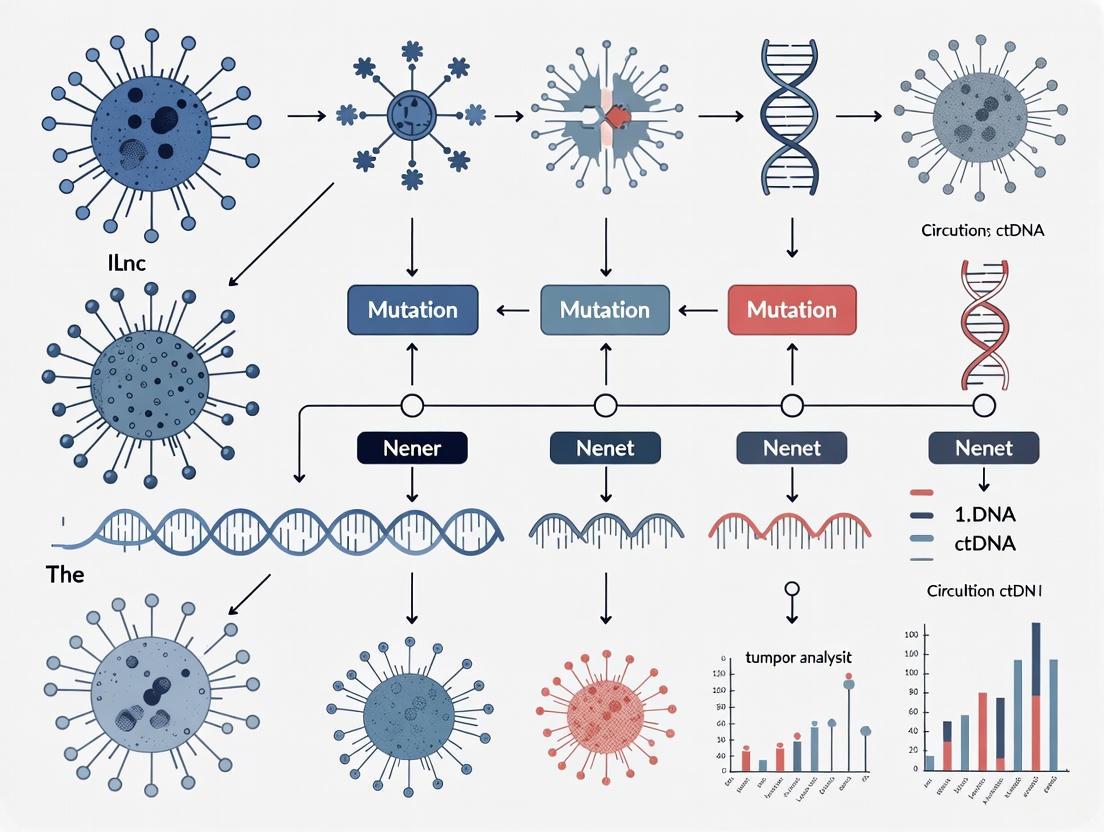
The following diagram illustrates the multi-step process of ctDNA shedding and release into the circulation.
Correlation Between ctDNA and Tumor Burden
The Relationship and Its Modulating Factors
While ctDNA levels generally correlate with tumor burden, the relationship is complex and influenced by factors beyond mere tumor volume. A study on metastatic melanoma found a modest positive correlation between ctDNA concentration and total tumor burden (TTB) across all disease states (R² = 0.49) [5]. However, this correlation strengthened markedly under conditions of progressive disease (R² = 0.91) [5]. This suggests that dynamic tumor proliferation and cell death, which are heightened during progression, are key drivers of ctDNA release.
The underlying principle is that ctDNA concentration in plasma represents a steady state maintained by a balance between the release of DNA from tumor cells and its rapid elimination from the bloodstream, with a half-life of approximately 35 minutes [6]. To maintain a detectable concentration, a continuous "infusion" of ctDNA from the tumor is required, making ctDNA levels a function of both tumor burden and the cellular turnover rate [6]. Consequently, a highly aggressive tumor with a high proliferation and death rate may yield higher ctDNA levels than a larger, more indolent lesion.
Table 3: Correlation of ctDNA with Tumor Burden and Clinical State Synthesized data from metastatic melanoma and lung cancer studies [2] [7] [5]
| Parameter | Correlation / Finding | Clinical Context / Implication |
|---|---|---|
| Overall Tumor Burden | Modest correlation (R² ≈ 0.49) | Relationship is not linear; influenced by tumor type and disease activity. |
| During Progressive Disease | Strong correlation (R² = 0.91) | High cellular turnover during progression increases ctDNA shedding. |
| Anatomic Distribution | Gradient: Primary Tumor > Pulmonary Vein > Peripheral Vein | Confirmed in lung cancer; indicates "spill-over" from tumor site [7]. |
| ctDNA Detection vs. Radiographic Disease | 81% detection in patients with radiographic tumor burden | ctDNA is a specific but not perfectly sensitive biomarker [5]. |
Clinical Utility in Monitoring Therapy Response
Longitudinal ctDNA monitoring can provide an early and dynamic readout of therapeutic efficacy. In a study of 204 patients with advanced solid tumors, increasing ctDNA levels during therapy (a positive "delta" or "slope") were strongly associated with radiographic progression and shorter time to treatment failure [3]. Notably, rising ctDNA predicted clinical or radiologic progression in 73% of patients with a median lead time of 23 days [3].
In the context of targeted therapy, the clearance of ctDNA is a significant positive indicator. For EGFR-mutated NSCLC patients treated with TKIs, those who cleared mEGFR from plasma at the first reassessment exhibited better progression-free survival compared to those who did not [2]. This "ctDNA clearance" can serve as an early molecular response marker, potentially preceding radiographic changes.
Experimental Protocols for ctDNA Analysis
Pre-Analytical Phase: Blood Collection and Plasma Separation
Critical Step: Standardized procedures are essential to prevent contamination with genomic DNA from lysed blood cells.
- Blood Collection: Draw a minimum of 10 mL of whole blood into EDTA or specialized cell-stabilization blood collection tubes [2] [3].
- Initial Centrifugation: Process samples within 2 hours of collection. Centrifuge at 1900 × g for 10 minutes at room temperature to separate plasma from cellular components [2].
- Secondary Centrifugation: Transfer the supernatant (plasma) to a new tube without disturbing the buffy coat. Centrifuge again at 1900 × g for 15 minutes to remove any remaining cells and debris [2] [3].
- Storage: Aliquot the cleared plasma and store at -80°C until DNA extraction.
Analytical Phase: cfDNA Extraction and Mutational Analysis
cfDNA Extraction
Extract cfDNA from 3-4 mL of plasma using commercially available kits, such as the QIAamp Circulating Nucleic Acid Kit, following the manufacturer's instructions [2] [3]. Quantify the extracted cfDNA using a fluorescence-based assay (e.g., Quant-iT PicoGreen dsDNA Assay) for high sensitivity [3].
Mutational Analysis
Two primary methods are used for ctDNA analysis:
- Digital PCR (dPCR) / Droplet Digital PCR (ddPCR): This method is highly sensitive and suitable for tracking known mutations.
- Principle: The PCR reaction is partitioned into thousands of individual droplets or wells. This allows for absolute quantification of mutant allele copies without the need for a standard curve [1].
- Sensitivity: Can detect mutant allele frequencies (MAF) as low as 0.001% [1] [8]. It is ideal for longitudinal monitoring of a predefined genetic alteration (e.g., EGFR T790M) [2] [3].
- Next-Generation Sequencing (NGS): This method allows for broader profiling.
- Tumor-Informed NGS: A personalized panel is designed based on the mutational profile of the patient's tumor tissue, achieving ultra-high sensitivity (LoD 95%: 0.001%) [8].
- Fixed-Panel NGS: Uses a predetermined gene panel to identify a range of alterations without prior tumor sequencing, useful for comprehensive profiling at progression [2].
The following workflow diagram outlines the key steps from sample collection to data analysis.
The Scientist's Toolkit: Essential Reagents and Materials
Table 4: Key Research Reagent Solutions for ctDNA Analysis
| Item | Function / Application | Example Product / Note |
|---|---|---|
| Cell-Free DNA Blood Collection Tubes | Stabilize nucleated blood cells to prevent genomic DNA contamination and enable longer sample transport times. | Streck Cell-Free DNA BCT, PAXgene Blood cDNA Tube |
| cfDNA Extraction Kit | Isolate high-purity, short-fragment cfDNA from plasma samples. | QIAamp Circulating Nucleic Acid Kit [2] [3] |
| Fluorescent DNA Quantification Kit | Accurately quantify low-concentration cfDNA samples. | Quant-iT PicoGreen dsDNA Assay Kit [3] |
| dPCR/ddPCR Systems | Absolute quantification and ultra-sensitive detection of known low-frequency mutations. | QIAcuity One dPCR System [2], Qx200 Droplet Digital PCR System [3] |
| NGS Library Prep Kit | Prepare sequencing libraries from low-input cfDNA for targeted or whole-genome sequencing. | AVENIO ctDNA Expanded Kit [2] |
| Tumor-Informed NGS Assay | Ultra-sensitive patient-specific ctDNA detection for minimal residual disease (MRD) and monitoring. | Commercial or custom assays (LoD 95%: 0.001%) [8] |
The Critical Role of ctDNA in Detecting Minimal Residual Disease (MRD)
In the field of lung cancer research, particularly for non-small cell lung cancer (NSCLC), the detection of minimal residual disease (MRD) represents a critical challenge in therapeutic management. MRD refers to the presence of residual tumor cells following curative-intent treatment that remains undetectable by conventional imaging techniques [9]. These occult cells are the hypothesized source of subsequent disease recurrence, which occurs in 30%-55% of early-stage NSCLC patients after radical resection [10]. Circulating tumor DNA (ctDNA), consisting of fragmented DNA released by tumor cells into the bloodstream, has emerged as a powerful biomarker for detecting MRD [9]. ctDNA fragments typically range from 130-150 base pairs in length and have a relatively short half-life of 16 minutes to 2.5 hours, enabling real-time monitoring of disease burden [9]. The integration of longitudinal ctDNA monitoring into lung cancer research protocols provides unprecedented opportunities to understand tumor evolution, identify patients at highest recurrence risk, and guide personalized adjuvant therapy decisions.
Performance Characteristics of ctDNA-Based MRD Detection
Diagnostic Accuracy of Detection Strategies
The clinical validity of ctDNA-based MRD detection is well established, with studies demonstrating that postoperative ctDNA positivity is significantly associated with increased recurrence risk and shorter survival outcomes [11]. A recent comprehensive meta-analysis of 30 studies involving 3,287 postoperative NSCLC patients revealed compelling evidence for the diagnostic performance of ctDNA-based MRD testing [10].
Table 1: Diagnostic Performance of ctDNA MRD Detection Strategies in NSCLC
| Detection Strategy | Sensitivity | Specificity | AUC | Optimal Use Case |
|---|---|---|---|---|
| Landmark Analysis | ||||
| Tumor-informed | 42% | 97% | 0.81 | Early postoperative risk stratification |
| Tumor-agnostic | 44% | 93% | 0.70 | Situations without tumor tissue availability |
| Longitudinal Monitoring | ||||
| Tumor-informed | 76% | 96% | 0.86 | Dynamic recurrence risk assessment |
| Tumor-agnostic | 79% | 88% | 0.91 | Long-term surveillance |
Temporal Patterns of MRD Detection
The timing of blood collection for MRD assessment is a critical factor influencing detection sensitivity and prognostic value. Research indicates that ctDNA detection can identify recurrent disease 70-151 days earlier than conventional radiographic imaging [9]. The optimal sampling schedule appears to be influenced by the treatment modality received:
- Surgically-treated patients: Landmark testing at 3-4 weeks postoperatively followed by longitudinal monitoring every 3-6 months for 2-3 years [9]
- Radiation/chemoradiation-treated patients: Later landmark testing at 4.5-7.5 months post-treatment may be more predictive of outcomes than earlier timepoints [11]
Methodological Approaches for ctDNA-Based MRD Detection
Experimental Workflow for MRD Detection
The standard workflow for ctDNA-based MRD detection involves multiple critical steps from sample collection to data analysis. The following diagram illustrates the two primary approaches and their respective workflows:
Research Reagent Solutions for ctDNA MRD Detection
Table 2: Essential Research Reagents and Materials for ctDNA MRD Detection
| Reagent/Material | Function | Examples/Specifications |
|---|---|---|
| Blood Collection Tubes | Stabilize cfDNA for up to 24-48 hours | EDTA tubes, Streck Cell-Free DNA BCT, PAXgene Blood cDNA Tubes |
| cfDNA Extraction Kits | Isolate cell-free DNA from plasma | QIAamp Circulating Nucleic Acid Kit, MagMAX Cell-Free DNA Isolation Kit |
| Library Preparation Kits | Prepare sequencing libraries from low-input cfDNA | KAPA HyperPrep Kit, Illumina DNA Prep with Enrichment |
| Hybrid Capture Reagents | Target cancer-associated genomic regions | IDT xGen Lockdown Probes, Twist Human Core Exome plus Comprehensive Exome Panel |
| Sequencing Platforms | High-throughput DNA sequencing | Illumina NovaSeq 6000, Illumina NextSeq 550 |
| ctDNA Reference Standards | Assay validation and quality control | Seraseq ctDNA Mutation Mix, Horizon Multiplex I cfDNA Reference Standard |
Protocol: Tumor-Informed MRD Detection Using CAPP-Seq
Sample Collection and Processing
Materials:
- EDTA blood collection tubes or specialized cfDNA stabilization tubes
- Refrigerated centrifuge capable of 2000×g
- Plasma separation accessories
- cfDNA extraction kit
- Fragment Analyzer or Bioanalyzer
Procedure:
- Collect 10-20 mL of peripheral blood into EDTA tubes
- Invert tubes gently 8-10 times for proper mixing
- Process within 2 hours of collection for optimal results
- Centrifuge at 2000×g for 10 minutes at 4°C to separate plasma
- Transfer supernatant to fresh tube without disturbing buffy coat
- Perform second centrifugation at 16,000×g for 10 minutes to remove residual cells
- Aliquot plasma into cryovials and store at -80°C if not processing immediately
- Extract cfDNA using validated commercial kits according to manufacturer's protocol
- Quantify cfDNA yield using fluorometric methods (Qubit)
- Assess fragment size distribution using Fragment Analyzer
Tumor Sequencing and Personalized Panel Design
Materials:
- Tumor tissue (FFPE or fresh frozen)
- DNA extraction kit for tissue
- Quality control instruments
Procedure:
- Extract genomic DNA from tumor tissue and matched normal (buffy coat)
- Perform quality control to ensure DNA integrity (DV200 > 30% for FFPE)
- Sequence using whole exome sequencing or large targeted panels (500+ genes)
- Identify somatic mutations using bioinformatics pipelines (SAGE, MuTect2)
- Select 16-50 clonal mutations for personalized panel based on:
- High variant allele frequency in tumor
- Clonality (truncal mutations preferred)
- Representation across tumor subclones
- Analytical performance in plasma
- Design custom hybridization probes for selected mutations
Library Preparation and Targeted Sequencing
Materials:
- Library preparation kit compatible with low DNA input
- Custom hybridization probes
- Sequencing platform with sufficient capacity
Procedure:
- Prepare sequencing libraries from 10-30 ng of cfDNA
- Incorporate unique molecular identifiers (UMIs) to distinguish true mutations from PCR errors
- Perform hybrid capture with custom probes
- Sequence to ultra-high depth (>100,000× coverage)
- Include negative controls (healthy donor plasma) and positive controls (diluted tumor DNA) in each run
Bioinformatic Analysis and MRD Calling
Materials:
- High-performance computing cluster
- Bioinformatic pipelines for ctDNA analysis
Procedure:
- Demultiplex sequencing data and assign reads to samples
- Process UMIs to create consensus reads and eliminate PCR duplicates
- Align reads to reference genome (hg38)
- Call variants using UMI-aware variant callers
- Apply error suppression algorithms to reduce technical noise
- Determine MRD positivity using statistical thresholds (typically VAF > 0.01% with p-value < 0.01)
Protocol: Tumor-Agnostic MRD Detection Using Fixed Panels
Sample Processing and Library Preparation
Materials:
- Fixed gene panel targeting cancer-associated mutations
- Library preparation reagents
Procedure:
- Process plasma samples as described in Section 4.1
- Prepare sequencing libraries from 20-50 ng of cfDNA
- Use fixed panels covering 50-700+ cancer-associated genes
- Include genomic regions for:
- Driver mutations common in NSCLC (EGFR, KRAS, TP53, etc.)
- Copy number alterations
- Epigenetic modifications when possible
- Sequence to moderate depth (10,000-30,000×)
Variant Calling and Interpretation
Procedure:
- Align sequences to reference genome
- Call variants using sensitive detection algorithms
- Filter out common polymorphisms using population databases
- Apply clonal hematopoiesis filters to exclude age-related mutations
- Use fragmentomic analysis to distinguish tumor-derived fragments
- Determine MRD positivity based on presence of tumor-derived mutations above background noise
Advanced Applications: Quantitative ctDNA Dynamics for Response Monitoring
MinerVa-Delta Algorithm for Molecular Response Assessment
For advanced disease settings where complete ctDNA clearance may not occur, quantitative assessment of ctDNA dynamics provides valuable insights into treatment response. The MinerVa-Delta algorithm was developed specifically to address this need by calculating weighted mutation changes in samples with multiple tracked variants [12].
Table 3: MinerVa-Delta Algorithm Implementation for Response Assessment
| Parameter | Specification | Clinical/Rearch Utility |
|---|---|---|
| Input Data | Multiple tracked variants from pretreatment and posttreatment plasma | Captures tumor heterogeneity and evolution |
| Calculation | Weighted mutation changes accounting for VAF uncertainty | More reliable than simple VAF ratios |
| Threshold | <30% decrease defines molecular response | Identifies patients with favorable outcomes |
| Validation | Tested in advanced LUSC cohorts receiving immunochemotherapy | Proven prognostic value in aggressive disease |
| Advantage | Identifies responders among radiologic stable disease patients | Enhances traditional imaging assessment |
Procedure for MinerVa-Delta Calculation:
- Identify variants de novo in pretreatment plasma using 769-gene NGS panel
- Track these variants in posttreatment plasma after 2 cycles of treatment
- Calculate weighted ratio change for each variant considering:
- Depth of sequencing
- Variance of VAF measurements
- Statistical confidence intervals
- Compute composite MinerVa-Delta score across all variants
- Classify as molecular responder (MinerVa-Delta <30%) or non-responder (≥30%)
Technical Considerations and Limitations
Despite the promising clinical applications of ctDNA for MRD detection, several technical challenges remain. The inherently low abundance of ctDNA in early-stage disease or following treatment represents a fundamental limitation, with ctDNA often comprising <0.1% of total cell-free DNA in these settings [13]. Factors influencing ctDNA levels include tumor burden, metastatic volume, tumor location, and biological features affecting DNA release and clearance [13]. Preanalytical variables such as blood collection methods, processing time, and sample storage conditions can significantly impact assay performance [14]. Additionally, clonal hematopoiesis represents a important confounding factor that must be addressed through careful bioinformatic filtering or paired normal sequencing [10]. Ongoing efforts to improve sensitivity include integration of multi-modal approaches combining genomic, fragmentomic, and epigenetic features to enhance detection capabilities [14].
The integration of ctDNA-based MRD detection into lung cancer research represents a paradigm shift in how residual disease is quantified and monitored. Both tumor-informed and tumor-agnostic approaches offer complementary strengths, with the former providing higher specificity for early postoperative assessment and the latter offering practical advantages for long-term monitoring [10]. The development of quantitative dynamic monitoring approaches like MinerVa-Delta further extends the utility of ctDNA to response assessment in advanced disease settings [12]. As standardization improves and larger clinical validation studies are completed, ctDNA-based MRD detection is poised to become an essential component of lung cancer research and clinical management, enabling more personalized treatment approaches and ultimately improving patient outcomes.
Longitudinal Monitoring for Early Relapse Detection and Lead Time Over Imaging
In the management of lung cancer, the early detection of residual disease following curative-intent treatment is a critical challenge. Current standard surveillance relies on radiological imaging, which can only identify macroscopic disease recurrence, often at a point when therapeutic options may be limited. Longitudinal monitoring of circulating tumor DNA (ctDNA), a component of cell-free DNA (cfDNA) shed by tumors into the bloodstream, has emerged as a powerful tool for identifying minimal residual disease (MRD). This Application Note details the protocols and data supporting the use of longitudinal ctDNA monitoring for the early detection of relapse and the significant lead time it provides over conventional imaging in a lung cancer research context. The data presented herein underpins a broader thesis that ctDNA dynamics can serve as a real-time, sensitive, and specific biomarker to guide personalized adjuvant therapy and improve patient outcomes.
Key Quantitative Findings from Recent Studies
Research consistently demonstrates that the presence of ctDNA after definitive treatment is a potent predictor of future clinical recurrence. The quantitative findings below summarize the performance of ctDNA monitoring across multiple studies involving patients with non-small cell lung cancer (NSCLC) and small cell lung cancer (SCLC).
Table 1: Summary of Key Studies on ctDNA for Relapse Detection in Lung Cancer
| Study (Citation) | Cohort & Design | Pre-Treatment ctDNA Detection Rate | Longitudinal Sensitivity for Progression | Longitudinal Specificity for Progression | Median Lead Time Over Imaging |
|---|---|---|---|---|---|
| Natera (Frontiers, 2023) [15] | 17 pts, unresectable Stage I-III NSCLC; Definitive radiotherapy ± chemo | 82% (14/17) | 100% (9/9) | 100% (8/8) | 5.4 months |
| LUCID (Annals of Oncology, 2022) [16] | 88 pts, Stage I-IIIB NSCLC; Curative-intent surgery or chemoradiotherapy | 51% (Stage I: 24%, II: 77%, III: 87%) | 64.3% (18/28) for primary tumour recurrence | >98.5% | 212.5 days (~7 months) |
| IASLC WCLC (2025) [17] | 177 pts, Limited-Stage SCLC; Chemoradiotherapy ± consolidation immunotherapy | Not Specified | ctDNA-positive patients post-induction had significantly better PFS and OS with ICIs | ctDNA-negative patients showed no added benefit from ICIs | Not Specified |
Table 2: Prognostic Value of Post-Treatment ctDNA Detection
| Study | Landmark Timepoint | Hazard Ratio (HR) for Recurrence/Progression | Hazard Ratio (HR) for Overall Survival | Statistical Significance |
|---|---|---|---|---|
| LUCID Study [16] | 2 weeks to 4 months after treatment | HR: 14.8 | HR: 5.48 | P < 0.00001 (RFS); P < 0.0003 (OS) |
| Natera Study [15] | First timepoint after radiotherapy | HR: 24.2 (Single timepoint); HR: 13.4 (Multivariate analysis) | Not Specified | P = 0.004; P = 0.02 |
Experimental Protocols for ctDNA Analysis
Protocol A: Tumor-Informed Personalized ctDNA Assay
This protocol, as utilized in the Signatera (Natera) and LUCID studies, leverages whole exome sequencing (WES) of tumor tissue to create a patient-specific assay for unparalleled sensitivity and specificity in longitudinal monitoring [16] [15].
Sample Collection and Processing:
- Tissue: Obtain formalin-fixed, paraffin-embedded (FFPE) tumor tissue from a primary tumor biopsy or surgical resection.
- Blood: Collect peripheral blood in EDTA or Streck tubes. Process within 1-2 hours of collection with a double centrifugation protocol (e.g., 1600 g for 10 min, then 20,000 g for 10 min) to isolate plasma. Aliquot and store at -80°C.
Tissue Whole Exome Sequencing (WES) and Assay Design:
- Extract DNA from FFPE tissue using a dedicated kit (e.g., QIAamp DNA FFPE Tissue Kit) with DNA repair steps.
- Perform WES on tumor DNA and matched germline DNA (from buffy coat) to identify somatic mutations.
- Select up to 48 clonal, tumor-specific somatic variants unique to the patient to design a personalized, multiplex PCR assay (e.g., RaDaR assay).
Plasma Analysis via Personalized Assay:
- Extract cfDNA from plasma samples using a commercial kit (e.g., QIAsymphony DSP Circulating DNA kit).
- Analyze serial plasma samples (collected pre-treatment, during treatment, and post-treatment during surveillance) using the patient-specific assay.
- Utilize ultra-sensitive sequencing or digital PCR technology to detect and quantify the presence of these variants in the cfDNA, with a reported sensitivity to detect variant allele fractions as low as 0.003% [16].
Protocol B: Multiplex Methylation Analysis via REM-DREAMing
For cases where tumor tissue is unavailable, or to leverage epigenetic alterations, this protocol assesses DNA methylation heterogeneity using a multiplex digital PCR approach [18].
Sample Collection and Bisulfite Conversion:
- Collect and process plasma as described in Protocol A.
- Extract cfDNA and subject it to bisulfite conversion. This process deaminates unmethylated cytosines to uracils, while methylated cytosines remain unchanged, translating methylation status into sequence differences.
Multiplex Digital High-Resolution Melt (dHRM):
- Design "methylation-agnostic" TaqMan probes for a panel of target biomarkers (e.g., a 5-gene panel for NSCLC). These probes incorporate degenerate bases to hybridize to the target locus regardless of its methylation pattern.
- Label identical probes for each locus with two different fluorophores (e.g., Cy5 and HEX) at a predefined stoichiometric ratio, creating a unique ratiometric fluorescence signature for each locus in the panel.
- Perform PCR amplification in a digital microfluidic device (e.g., containing 10,400 nanowells) in the presence of the probe mix and a DNA binding dye (e.g., EvaGreen).
- Post-PCR, perform a high-resolution melt analysis on each individual partition. The melt temperature (Tm) of an amplicon is proportional to its methylation density.
Data Analysis:
- Use two-channel fluorescence to identify the target locus in each partition based on its unique ratiometric signature.
- Analyze the melt curve from each positive partition to determine the methylation density of the original template molecule on a copy-by-copy basis.
- Assess intermolecular methylation density distributions across the biomarker panel to classify samples as positive or negative for tumor-derived ctDNA.
Workflow Visualization
The following diagram illustrates the logical sequence and decision points for implementing longitudinal ctDNA monitoring in a clinical research setting for lung cancer.
Diagram 1: Longitudinal ctDNA Monitoring Workflow. The diagram outlines the key decision points in a post-treatment monitoring protocol, highlighting how ctDNA status at a landmark timepoint and during surveillance can stratify patient risk.
The Scientist's Toolkit: Essential Research Reagents & Platforms
Table 3: Key Research Reagent Solutions for ctDNA-Based MRD Detection
| Item / Technology | Function / Application | Specific Examples / Notes |
|---|---|---|
| Tumor-Informed MRD Assays | Detects patient-specific mutations in plasma for ultra-sensitive MRD assessment. | Signatera (Natera) [15], RaDaR assay (Inivata) [16]. Requires matched tumor-normal sequencing. |
| Methylation-Specific Assays | Detects cancer-specific epigenetic alterations; useful when tumor tissue is unavailable. | REM-DREAMing platform for multiplex methylation heterogeneity analysis [18]. |
| Next-Generation Sequencing (NGS) | Enables comprehensive mutation profiling for personalized assay design and variant discovery. | Whole exome sequencing (WES) for identifying clonal somatic mutations [17] [16]. |
| Digital PCR (dPCR) Platforms | Provides absolute quantification of nucleic acids without a standard curve; high sensitivity for rare targets. | Crystal Digital PCR (3-color multiplexing) [19]; other platforms for target-specific assays. |
| Cell-free DNA Extraction Kits | Isolates high-quality, high-integrity cfDNA from blood plasma samples. | QIAsymphony DSP Circulating DNA kit (Qiagen), QIAamp DNA FFPE Tissue Kit for tumor DNA [16]. |
| Bisulfite Conversion Kits | Chemically modifies DNA to differentiate methylated from unmethylated cytosines for methylation assays. | Essential for protocols like REM-DREAMing and other bisulfite sequencing-based methods [18]. |
ctDNA as a Dynamic Biomarker for Predicting Patient Outcomes (PFS/OS)
Circulating tumor DNA (ctDNA) has emerged as a pivotal biomarker in oncology, offering a non-invasive method for monitoring tumor dynamics and predicting patient outcomes. As fragments of DNA shed by tumor cells into the bloodstream, ctDNA carries tumor-specific genetic alterations that provide real-time insights into disease burden and therapeutic response [20]. In lung cancer research, particularly non-small cell lung cancer (NSCLC), longitudinal ctDNA monitoring has demonstrated significant utility for predicting progression-free survival (PFS) and overall survival (OS) with greater sensitivity than traditional imaging methods [21] [22]. The short half-life of ctDNA (approximately 16 minutes to several hours) enables rapid assessment of treatment response and disease evolution, making it an ideal dynamic biomarker for clinical decision-making in both early-stage and advanced disease settings [20].
Quantitative Evidence: ctDNA Dynamics and Survival Outcomes
Extensive clinical research has established robust correlations between ctDNA dynamics and survival outcomes across various treatment modalities. The tables below summarize key quantitative evidence from recent studies.
Table 1: ctDNA Dynamics and Survival Outcomes in Advanced Solid Tumors
| Cancer Type | ctDNA Metric | Threshold | Overall Survival (Months) | Hazard Ratio (HR) | Reference |
|---|---|---|---|---|---|
| Advanced Solid Tumors | maxVAF | >4% | 5.9 vs 12.1* | 2.17 [1.76-2.70] (p<0.001) | [23] |
| Advanced LUSC | MinerVa-Delta | ≥30% (Non-responder) | Significantly reduced | 0.24 (OS, p<0.001) | [22] |
| Advanced LUSC | MinerVa-Delta | ≥30% (Non-responder) | - | 0.19 (PFS, p<0.001) | [22] |
| NSCLC (Early-stage) | ctDNA Status | Postoperative Detection | Highly prognostic | - | [21] |
Compared with patients with maxVAF ≤4%; *Exact months not specified in source; reported as statistically significant improvement
Table 2: Technical Approaches for ctDNA-Based Monitoring
| Methodology | Genomic Coverage | Key Features | Reported Clinical Utility |
|---|---|---|---|
| MinerVa-Delta | 769-gene panel | Weighted mutation accounting for VAF variance | Identified molecular responders in LUSC despite radiographic stable disease [22] |
| Tumor-informed whole-genome | 1,800 variants | Ultrasensitive detection (<80 parts per million) | Prognostic stratification pre-/post-operation; identified intermediate-risk group [21] |
| FoundationOne Liquid CDx | 309 genes | FDA-approved comprehensive genomic profiling | Independent prognostic value in advanced solid tumors [23] |
Protocols for Longitudinal ctDNA Monitoring
Blood Collection and Processing Protocol
Principle: Optimal sample collection and processing are critical for maintaining ctDNA integrity and ensuring accurate analysis.
Materials:
- Cell-free DNA blood collection tubes (e.g., Streck, PAXgene)
- Refrigerated centrifuge capable of 1,600-3,000 × g
- DNA extraction kits optimized for low-input cell-free DNA
- -80°C freezer for plasma storage
Procedure:
- Blood Collection: Draw 10-20 mL peripheral blood into cell-free DNA collection tubes
- Transport: Store tubes at room temperature and process within 6 hours of collection
- Plasma Separation:
- Centrifuge at 1,600-3,000 × g for 10 minutes at 4°C
- Transfer supernatant to fresh tube without disturbing buffy coat
- Perform second centrifugation at 16,000 × g for 10 minutes at 4°C
- Plasma Storage: Aliquot cleared plasma and store at -80°C until DNA extraction
- DNA Extraction: Use silica membrane-based columns or magnetic beads to isolate cell-free DNA
- Quality Control: Quantify DNA using fluorometric methods and assess fragment size distribution
MinerVa-Delta Algorithm for Molecular Response Assessment
Principle: The MinerVa-Delta algorithm quantifies ctDNA dynamics by calculating weighted mutation changes that account for sequencing depth and variance at each variant position [22] [24].
Materials:
- Pretreatment plasma sample
- Posttreatment plasma sample (after 2 cycles of therapy)
- DNA library preparation kit
- Hybridization capture-based next-generation sequencing panel (769 genes)
- High-throughput sequencer
Procedure:
- Baseline Variant Identification:
- Extract ctDNA from pretreatment plasma
- Perform whole-genome sequencing or targeted NGS
- Identify somatic variants de novo using variant calling algorithm
Posttreatment Tracking:
- Design patient-specific variant panel based on baseline findings
- Sequence posttreatment sample targeting identified variants
- Calculate deduplicated read depth for each variant
MinerVa-Delta Calculation:
- For each variant, compute ratio change in VAF between timepoints
- Assign weight to each variant based on depth and VAF variance
- Calculate weighted sum of ratio changes across all tracked variants
- Apply 30% threshold to classify molecular responders (<30%) versus non-responders (≥30%)
Clinical Correlation:
- Correlate MinerVa-Delta status with radiographic assessment
- Evaluate PFS and OS based on molecular response classification
Diagram 1: MinerVa-Delta Molecular Response Assessment Workflow
Ultrasensitive Tumor-Informed ctDNA Monitoring
Principle: This approach leverages whole-genome sequencing of tumor tissue to create patient-specific mutation panels for highly sensitive ctDNA detection in plasma, enabling minimal residual disease (MRD) assessment [21].
Materials:
- Tumor tissue sample (fresh frozen or FFPE)
- Matched normal sample (blood or saliva)
- Whole-genome sequencing kit
- Unique molecular identifiers (UMIs)
- Multiplex PCR reagents
Procedure:
- Tumor Sequencing:
- Extract DNA from tumor and matched normal samples
- Perform whole-genome sequencing at high coverage (≥80x)
- Identify somatic variants (1,800 variants recommended)
Personalized Panel Design:
- Select clonal and subclonal variants across genome
- Design patient-specific primers for targeted amplification
Plasma Analysis:
- Extract ctDNA from serial plasma samples
- Prepare sequencing libraries with UMIs
- Amplify using patient-specific panel
- Sequence at high depth (≥50,000x)
Variant Calling:
- Apply duplex sequencing for error correction
- Detect ctDNA down to 80 parts per million
- Monitor kinetic changes across timepoints
The Scientist's Toolkit: Essential Research Reagents
Table 3: Key Research Reagents for ctDNA Analysis
| Reagent/Kit | Function | Application Note |
|---|---|---|
| Cell-free DNA Blood Collection Tubes | Stabilize nucleated blood cells during transport | Prevents genomic DNA contamination and preserves ctDNA profile for up to 7 days at room temperature |
| Magnetic Bead-based cfDNA Extraction Kits | Isolate cell-free DNA from plasma | Optimized for short fragment recovery (90-150 bp) characteristic of ctDNA |
| Unique Molecular Identifiers (UMIs) | Tag individual DNA molecules pre-amplification | Enables consensus sequencing to eliminate PCR errors and sequencing artifacts |
| Hybridization Capture Panels | Enrich cancer-related genomic regions | FoundationOne Liquid CDx covers 309 genes; custom panels enable tumor-informed approaches |
| High-Sensitivity DNA Quantitation Kits | Accurately measure low-concentration cfDNA | Fluorometric methods superior to spectrophotometry for fragmented DNA samples |
| Multiplex PCR Master Mixes | Amplify multiple targets simultaneously | Enables efficient amplification of patient-specific variant panels from limited ctDNA input |
Analytical Framework and Data Interpretation
Kinetic Monitoring and Clinical Decision Points
Longitudinal ctDNA monitoring provides multiple decision points throughout the patient journey. The dynamic nature of ctDNA enables real-time assessment of treatment efficacy, often weeks to months before radiographic changes become apparent [20] [21].
Diagram 2: ctDNA Kinetic Monitoring in Early-Stage Lung Cancer
Integration with Radiographic Assessment
ctDNA dynamics provide complementary information to traditional imaging, particularly in clinically challenging scenarios:
- Radiographic Stable Disease: MinerVa-Delta classification identified molecular responders with significantly improved PFS (HR=0.19) and OS (HR=0.24) within this heterogeneous group [22]
- Pseudoprogression: ctDNA kinetics can distinguish true progression from inflammatory responses in patients receiving immunotherapy
- Minimal Residual Disease: Ultrasensitive ctDNA detection identifies MRD undetectable by imaging, enabling earlier intervention [21]
For patients with advanced disease, the combination of ctDNA dynamics and radiographic assessment creates a more comprehensive response evaluation framework. The 4% maxVAF threshold provides prognostic stratification independent of traditional factors, while molecular response algorithms like MinerVa-Delta offer quantitative metrics for treatment continuation decisions [23] [22].
Longitudinal ctDNA monitoring represents a transformative approach for predicting PFS and OS in lung cancer patients. The methodologies outlined in this document provide researchers with standardized protocols for implementing ctDNA-based dynamic biomarkers in both clinical trials and translational research. As ctDNA analysis continues to evolve toward greater sensitivity and standardization, its integration into routine oncology practice will enable more precise therapeutic guidance and improved patient outcomes.
Circulating tumor DNA (ctDNA) analysis has emerged as a transformative approach in lung cancer management, providing a non-invasive method for assessing tumor dynamics. This application note details the foundational evidence and methodologies establishing the prognostic value of longitudinal ctDNA monitoring in both non-small cell lung cancer (NSCLC) and small cell lung cancer (SCLC). For researchers and drug development professionals, this document synthesizes key quantitative findings from pivotal studies and provides detailed experimental protocols for implementing these assays in research settings.
The evidence presented herein supports the integration of ctDNA monitoring across the lung cancer continuum, from detecting minimal residual disease (MRD) after curative-intent therapy to guiding treatment in metastatic settings. By capturing real-time tumor dynamics, ctDNA analysis enables risk stratification, early response assessment, and intervention before clinical or radiographic progression becomes evident.
Foundational Evidence: Prognostic Value of ctDNA in Lung Cancer
Quantitative Synthesis of Key Studies
Table 1: Prognostic Value of ctDNA Across Lung Cancer Types and Disease Stages
| Cancer Type & Study | Patient Population | Key ctDNA Metric | Prognostic Impact (Hazard Ratio, HR) | Clinical Implications |
|---|---|---|---|---|
| Early-Stage NSCLC (IPD Meta-Analysis) [25] | 1,686 operable (I-III) patients | Positive ctDNA post-operation | DFS HR: 3.96 (2.19-7.16) | Identifies patients at high risk of recurrence who may benefit from adjuvant therapy. |
| Early-Stage NSCLC (TRACERx) [26] [21] | 431 patients | Ultrasensitive detection (<80 ppm) | Highly prognostic for relapse | Defines an intermediate-risk group; ctDNA clearance during adjuvant therapy predicts improved outcomes. |
| Metastatic NSCLC (IMpower150) [27] | 466 patients from Phase 3 trial | Machine learning model of ctDNA dynamics | OS HR: 3.2-3.3 for high vs. low-risk | Enables early risk stratification within weeks of treatment, outperforming early radiographic imaging. |
| EGFR-mutant NSCLC [28] | 72 patients on osimertinib | ctDNA clearance at 6-week follow-up | PFS (P=0.022); OS (P=0.009) | Clearance correlates with superior survival; molecular progression detected 2.5 months before radiological progression. |
| Limited-Stage SCLC [17] [29] | 177 patients post-chemoradiotherapy | ctDNA-positive post-induction | OS HR: 0.41 with ICI benefit | Identifies patients most likely to benefit from consolidation immunotherapy. |
| Limited-Stage SCLC [30] | 23 patients post-definitive therapy | ctDNA ever detected post-treatment | PFS (P<0.001); OS (P=0.081) | Predicts disease relapse and death; never detected ctDNA associates with prolonged PFS (>48 months). |
Key Interpretations of the Evidence
The consolidated data from these foundational studies demonstrate consistent and powerful prognostic value of ctDNA across lung cancer subtypes. In operable NSCLC, the detection of ctDNA post-surgery (MRD) is a robust biomarker for recurrence risk, far exceeding the predictive power of conventional staging alone [25]. The ultrasensitive methodologies now enable risk stratification at parts-per-million sensitivity, identifying distinct intermediate-risk groups that require refined clinical management strategies [26].
In the advanced setting, longitudinal ctDNA dynamics provide an early indicator of treatment efficacy and survival outcomes. The IMpower150 analysis highlights that machine learning models integrating multiple ctDNA metrics can risk-stratify patients as early as the first treatment cycles, with high-risk patients showing median overall survival of less than 9 months compared to over 28 months for low-risk patients [27]. Similarly, in oncogene-addicted NSCLC, ctDNA clearance during targeted therapy serves as an early marker of therapeutic response [28].
For SCLC, a cancer type with limited biomarkers, ctDNA monitoring shows particular promise in guiding immunotherapy use. The 2025 WCLC study demonstrates that ctDNA status after induction chemotherapy can personalize consolidation immunotherapy, maximizing benefit while sparing unlikely responders from unnecessary treatment [17] [29].
Experimental Protocols for Longitudinal ctDNA Monitoring
Core Workflow for Tumor-Informed ctDNA Analysis
The following diagram illustrates the comprehensive workflow for tumor-informed ctDNA analysis, as used in foundational studies like TRACERx [26] and IMpower150 [27]:
Figure 1: Workflow for Tumor-Informed Longitudinal ctDNA Analysis
Detailed Methodological Components
Sample Collection and Processing
Plasma Collection Protocol:
- Blood Draw: Collect 10-20 mL of peripheral blood into cell-stabilizing tubes (e.g., Streck Cell-Free DNA BCT) [30].
- Processing: Centrifuge at 1200-1600 × g for 10-20 minutes within 4-6 hours of collection to separate plasma from cellular components [28] [30].
- Secondary Centrifugation: Perform a second centrifugation at 16,000 × g for 10 minutes to remove residual cells [30].
- Storage: Aliquot plasma and store at -80°C until DNA extraction.
Cell-Free DNA Extraction:
- Utilize commercial kits specifically designed for low-concentration cell-free DNA (e.g., QIAamp Circulating Nucleic Acid Kit) [28] [30].
- Input: 4-5 mL plasma typically yields 10-50 ng cell-free DNA [28].
- Quantify yield using fluorescence-based methods (e.g., Qubit fluorometer) [30].
Sequencing Approaches
Table 2: Comparison of ctDNA Sequencing Methodologies
| Methodology | Target Approach | Genomic Coverage | Sequencing Depth | Key Applications | Representative Studies |
|---|---|---|---|---|---|
| Tumor-Informed | Patient-specific variants | ~20-200 variants per patient | 50,000-100,000× | MRD detection, ultra-sensitive monitoring | TRACERx [26], IMpower150 [27] |
| Tumor-Agnostic | Fixed gene panel | 139-168 genes | 10,000-30,000× | Treatment monitoring, resistance detection | NSCLC TKI Study [28], SCLC Study [17] |
| Whole Genome | Genome-wide | ~80,000 variants | 0.1-1× | Comprehensive profiling, structural variants | Research applications |
Tumor-Informed Sequencing (e.g., TRACERx Protocol):
- Tumor Sequencing: Perform whole-genome or whole-exome sequencing of tumor tissue and matched germline DNA to identify patient-specific somatic variants [26].
- Panel Design: Create a custom panel targeting ~20-200 patient-specific variants for ultra-sensitive tracking.
- Plasma Analysis: Use hybrid capture and unique molecular identifiers (UMIs) for error-suppressed sequencing at ultra-deep coverage (≥50,000×) [26] [27].
Tumor-Agnostic Panel Sequencing:
- Fixed Panels: Utilize commercially available or custom panels targeting frequently mutated genes in lung cancer (e.g., 139-168 gene panels) [28] [17] [29].
- Sequencing Depth: Typically 10,000-30,000× coverage to detect variants at 0.1% variant allele frequency (VAF) [28] [27].
- Bioinformatic Processing: Implement duplex sequencing with UMI-based error correction to distinguish true somatic variants from sequencing artifacts [27].
Bioinformatic Analysis
Variant Calling Pipeline:
- Alignment: Map sequencing reads to reference genome (GRCh37/38) using optimized aligners (e.g., BWA-MEM).
- UMI Processing: Group read families by unique molecular identifiers to generate consensus sequences and reduce sequencing errors [27].
- Variant Calling: Apply statistical models to distinguish somatic variants from background noise, with filtering for clonal hematopoiesis (CHIP) using matched PBMC sequencing when available [27].
- ctDNA Quantification: Calculate mean tumor molecule concentration or aggregate VAF across tracked variants.
Kinetic Modeling:
- Implement machine learning approaches to integrate multiple ctDNA metrics (baseline level, early kinetics, clearance) for survival prediction [27].
- Define molecular response (clearance) and molecular progression (emergence of new mutations or increasing VAF) [28].
The Scientist's Toolkit: Essential Research Reagents & Platforms
Table 3: Key Research Reagents and Platforms for ctDNA Analysis
| Category | Specific Product/Platform | Research Application | Key Features |
|---|---|---|---|
| Blood Collection Tubes | Streck Cell-Free DNA BCT tubes | Cell-free DNA stabilization | Preserves blood sample integrity for up to 7 days at room temperature [30] |
| Nucleic Acid Extraction | QIAamp Circulating Nucleic Acid Kit (Qiagen) | Cell-free DNA isolation from plasma | Optimized for low-abundance cell-free DNA; typical input 4-5 mL plasma [28] [30] |
| Library Prep | KAPA HyperPrep Kit (Roche) | NGS library construction | Compatible with low-input cell-free DNA; incorporates UMIs |
| Hybrid Capture | IDT xGen Lockdown Probes | Target enrichment | Customizable panels for tumor-informed or fixed-panel approaches [27] |
| Sequencing Platforms | Illumina NextSeq 500/550 | Target sequencing | Mid-output flow cells ideal for targeted panels at high depth [28] |
| ctDNA Analysis Software | FoundationOne Liquid CDx | Comprehensive ctDNA analysis | 394-gene panel; FDA-approved; includes CHIP filtering [27] |
| Lung Cancer Panels | OncoScreen (168 genes) | Tumor-agnostic profiling | Covers key lung cancer drivers and resistance mechanisms [28] |
The foundational studies summarized in this application note provide compelling evidence for the prognostic utility of longitudinal ctDNA monitoring across the spectrum of lung cancer. The methodologies detailed herein enable researchers to implement these approaches in both basic and translational research settings. As the field advances, standardization of protocols and analytical frameworks will be crucial for broader adoption in clinical trial design and ultimately in routine practice. The integration of ctDNA monitoring represents a paradigm shift toward more dynamic, personalized cancer management with the potential to significantly improve patient outcomes through earlier intervention and more precise treatment selection.
Advanced ctDNA Assay Technologies and Their Clinical Implementation
Circulating tumor DNA (ctDNA) analysis has emerged as a cornerstone of liquid biopsy, enabling non-invasive tumor genotyping and longitudinal monitoring of treatment response in lung cancer research [20]. The detection of ctDNA, which often constitutes less than 0.1% of total cell-free DNA, requires ultrasensitive methods capable of identifying tumor-specific genetic alterations against a background of wild-type DNA [31]. The two principal technological approaches for ctDNA detection are PCR-based methods—including droplet digital PCR (ddPCR) and BEAMing—and next-generation sequencing (NGS)-based platforms. This application note provides a comparative analysis of these platforms, detailing their operational principles, performance characteristics, and practical implementation for longitudinal ctDNA monitoring in lung cancer studies.
Principle of Operation
PCR-based platforms utilize a targeted approach for absolute quantification of specific DNA sequences. ddPCR partitions samples into thousands of nanodroplets, enabling end-point amplification and binary counting of mutant and wild-type DNA molecules without the need for standard curves [32]. BEAMing (beads, emulsion, amplification, and magnetics) similarly employs emulsion PCR to amplify mutant DNA fragments bound to magnetic beads, which are then detected and enumerated via flow cytometry [33] [20].
NGS-based platforms employ a broader sequencing approach to simultaneously interrogate multiple genomic regions. Targeted NGS panels focus on cancer hotspot regions (e.g., 50-500 genes), while whole-exome/genome sequencing provides comprehensive genomic coverage [32] [20]. Unique molecular identifiers (UMIs) are incorporated to distinguish true low-frequency variants from PCR and sequencing artifacts, with advanced error-correction methods such as SaferSeqS and CODEC significantly enhancing detection accuracy [20].
Comparative Performance Metrics
Table 1: Analytical Performance Comparison of ctDNA Detection Platforms
| Parameter | ddPCR | BEAMing | Targeted NGS | Whole-Genome NGS |
|---|---|---|---|---|
| Sensitivity (VAF) | 0.01%-0.1% [32] | 0.01%-0.1% [20] | 0.02%-0.1% [32] [20] | 0.02%-0.05% (tumor-informed) [21] |
| Multiplexing Capacity | 1-5 targets per reaction [34] [35] | Moderate | High (50-500 genes) [32] | Very High (entire genome) |
| Sample Throughput | Medium | Low-Medium | High | Medium |
| Turnaround Time | 1-2 days | 3-5 days | 5-10 days | 10-15 days |
| DNA Input Requirement | 5-20 ng [31] | 10-30 ng | 20-100 ng [31] | 50-200 ng |
| Cost per Sample | Low ($50-150) [32] | Medium ($200-400) | High ($500-1000) | Very High ($1500-3000) |
| Applications | Treatment monitoring, MRD detection [32] [21] | Mutation quantification | Genomic profiling, MRD detection [21] | Comprehensive genomic analysis |
Clinical Application in Lung Cancer
Table 2: Platform Selection Guide for Lung Cancer Applications
| Research Application | Recommended Platform | Key Considerations | Reported Performance in Lung Cancer |
|---|---|---|---|
| MRD Detection | Tumor-informed ddPCR or NGS | Sensitivity requirements, cost constraints | Ultrasensitive NGS detects ctDNA below 80 parts per million; prognostic for recurrence [21] |
| Treatment Response Monitoring | ddPCR or targeted NGS | Turnaround time, quantitative accuracy | Methylation-specific ddPCR multiplex shows 70.2-83.0% sensitivity in metastatic disease [35] |
| Comprehensive Genomic Profiling | Targeted NGS panels | Breadth of genomic coverage, ability to detect novel alterations | Identifies actionable mutations in KRAS, EGFR, TP53 for targeted therapy selection [36] [20] |
| Therapy Resistance Mechanism Elucidation | NGS with error correction | Ability to detect emerging resistant subclones | Captures heterogeneous resistance mutations across metastatic sites [20] |
Experimental Protocols
Pre-analytical Sample Processing
Blood Collection and Plasma Separation:
- Collect 20-30 mL whole blood using butterfly needles into Streck Cell-Free DNA BCT tubes or K2EDTA tubes [31].
- Process within 2-6 hours for EDTA tubes or within 7 days for stabilized BCT tubes at room temperature [31].
- Centrifuge at 2,000 × g for 10 minutes to separate plasma, followed by a second centrifugation at 10,000 × g for 10 minutes to remove residual cells [35].
- Aliquot and store plasma at -80°C until cfDNA extraction.
cfDNA Extraction:
- Extract cfDNA from 4-8 mL plasma using the QIAsymphony DSP Circulating DNA Kit or similar silica-membrane based methods [35].
- Elute in 20-60 μL of low-EDTA TE buffer or manufacturer's elution buffer.
- Quantify cfDNA using fluorometric methods (Qubit dsDNA HS Assay); expected yield ranges from 5-50 ng/mL plasma depending on tumor burden [31].
- Assess fragment size distribution using Bioanalyzer or TapeStation; expected peak at ~167 bp [20].
Platform-Specific Detection Protocols
ddPCR Assay Protocol
Mutation-Specific ddPCR:
- Design assays targeting known lung cancer driver mutations (e.g., EGFR T790M, KRAS G12C) using Bio-Ral ddPCR mutation assays or custom-designed probes [32].
- Prepare 20-40 μL reaction mixture containing:
- 5-20 ng cfDNA template
- 1× ddPCR Supermix for Probes
- 900 nM primers
- 250 nM FAM and HEX-labeled probes
- Generate droplets using Automated Droplet Generator or manual oil-emulsion methods.
- Perform PCR amplification with the following cycling conditions:
- 95°C for 10 minutes (enzyme activation)
- 40 cycles of: 94°C for 30 seconds, 55-60°C for 60 seconds
- 98°C for 10 minutes (enzyme deactivation)
- 4°C hold
- Read plates using QX200 Droplet Reader and analyze with QuantaSoft software.
- Calculate variant allele frequency (VAF) as (mutant droplets/total droplets) × 100 [32].
Methylation-Specific ddPCR Multiplex for Lung Cancer:
- Bisulfite convert 20-40 ng cfDNA using EZ DNA Methylation-Lightning Kit [35].
- Design primers and probes targeting lung cancer-specific methylated regions (e.g., HOXA9) [35].
- Prepare multiplex reaction with 5 methylation markers to increase sensitivity (38.7-83.0% across disease stages) [35].
- Include quality controls: exogenous spike-in DNA for extraction efficiency, immunoglobulin gene assay for lymphocyte contamination [35].
NGS-Based ctDNA Profiling Protocol
Library Preparation for Targeted NGS:
- Use 20-100 ng cfDNA for library construction [31].
- Repair DNA ends and ligate with adapters containing unique molecular identifiers (UMIs).
- Amplify libraries with 8-12 PCR cycles using panels targeting lung cancer genes (e.g., Ion AmpliSeq Cancer Hotspot Panel v2 covering 50 genes) [32].
- Purify libraries with AMPure XP beads and quantify by qPCR.
Sequencing and Data Analysis:
- Sequence on Illumina or Ion Torrent platforms to achieve minimum 5,000× coverage for ctDNA detection [32].
- Process raw data through bioinformatic pipeline:
- Demultiplex samples and trim adapters
- Group reads by UMIs to generate consensus sequences
- Align to reference genome (hg38)
- Call variants using specialized ctDNA callers (e.g., MuTect, VarScan2)
- Apply duplex sequencing error correction when possible [20]
- For tumor-informed MRD assays, sequence tumor tissue to identify clonal mutations, then design patient-specific panels tracking 100-1,800 variants [21].
The Scientist's Toolkit: Essential Research Reagents and Materials
Table 3: Key Research Reagent Solutions for ctDNA Analysis
| Reagent/Material | Function | Example Products | Application Notes |
|---|---|---|---|
| Cell-Free DNA Blood Collection Tubes | Preserves blood cell integrity, prevents genomic DNA contamination | Streck Cell-Free DNA BCT, PAXgene Blood ccfDNA | Enables room temperature storage for up to 7 days; critical for multi-center trials [31] |
| cfDNA Extraction Kits | Isolation of high-quality cfDNA from plasma | QIAsymphony DSP Circulating DNA Kit, QIAamp Circulating Nucleic Acid Kit | Optimized for low-abundance cfDNA; typical yields of 5-50 ng/mL plasma [31] [35] |
| ddPCR Supermix | Digital PCR reaction setup for absolute quantification | ddPCR Supermix for Probes, ddPCR Mutation Detection Assays | Enables detection down to 0.01% VAF; no standard curve required [32] |
| Targeted NGS Panels | Capture and sequencing of cancer-relevant genes | Ion AmpliSeq Cancer Hotspot Panel v2, Illumina TruSight Oncology | Covers 50+ oncogenes/tumor suppressors; identifies >90% of mutations in lung cancer [32] |
| Bisulfite Conversion Kits | DNA modification for methylation analysis | EZ DNA Methylation-Lightning Kit | Converts unmethylated cytosines to uracils; preserves methylated cytosines [35] |
| Unique Molecular Identifiers (UMIs) | Error correction for NGS sequencing | IDT Duplex UMIs, Twist UMI Adapters | Reduces sequencing errors; essential for low-frequency variant detection [20] |
The selection between PCR-based and NGS-based platforms for longitudinal ctDNA monitoring in lung cancer research involves careful consideration of analytical sensitivity, multiplexing capability, and practical constraints. ddPCR offers exceptional sensitivity and quantitative precision for tracking known mutations during treatment response monitoring, while NGS provides comprehensive genomic profiling capabilities essential for discovering resistance mechanisms and tumor evolution. The emerging application of methylation-based ddPCR assays further expands the toolkit for lung cancer detection, particularly for cases without known driver mutations. As ctDNA technologies continue to evolve, integration of these complementary approaches will provide unprecedented insights into lung cancer dynamics and treatment responses, ultimately advancing personalized oncology research.
Circulating tumor DNA (ctDNA) analysis enables minimally invasive, longitudinal monitoring of tumor dynamics in lung cancer, offering a real-time snapshot of the tumor's genetic landscape [37] [20]. A critical decision in designing a monitoring study is the choice between two primary assay strategies: tumor-informed and tissue-agnostic (also referred to as tumor-naïve) approaches [10]. Tumor-informed assays require prior sequencing of a tumor tissue sample to create a patient-specific panel, while tissue-agnostic assays use a fixed, predetermined panel of cancer-associated genes and do not require baseline tumor tissue [10] [11]. This document outlines the application, performance, and protocols for both strategies to guide the development of personalized monitoring panels within lung cancer research.
Comparative Performance of Assay Strategies
The choice between tumor-informed and tissue-agnostic assays involves a trade-off between sensitivity, specificity, and practical logistics. The table below summarizes key performance characteristics, with data derived from a recent meta-analysis of minimal residual disease (MRD) detection in non-small cell lung cancer (NSCLC) [10].
Table 1: Performance comparison of tumor-informed and tissue-agnostic assays for MRD detection in early-stage NSCLC [10]
| Performance Metric | Landmark Analysis (Single Post-Op Time Point) | Longitudinal Monitoring (Multiple Time Points) | ||
|---|---|---|---|---|
| Tumor-Informed | Tissue-Agnostic | Tumor-Informed | Tissue-Agnostic | |
| Pooled Sensitivity | 0.42 | 0.44 | 0.76 | 0.79 |
| Pooled Specificity | 0.97 | 0.93 | 0.96 | 0.88 |
| Area Under Curve (AUC) | 0.81 | 0.70 | 0.86 | 0.91 |
This data indicates that tumor-informed assays generally provide higher specificity, making them excellent for confirming the presence of disease and minimizing false positives [10]. Tissue-agnostic assays can offer strong performance, particularly in longitudinal settings, and their logistical simplicity facilitates broader clinical application [10] [11].
Experimental Protocols for ctDNA MRD Detection
The following protocols detail the core workflows for implementing both assay strategies in a longitudinal monitoring study.
Protocol for Tumor-Informed ctDNA Assay
This protocol is designed for ultra-sensitive detection of MRD by targeting a set of patient-specific mutations [26].
3.1.1. Step 1: Tumor and Matched Normal Sequencing
- Input Materials: Formalin-fixed, paraffin-embedded (FFPE) tumor tissue block and patient-matched peripheral blood mononuclear cells (PBMCs) or buccal swab as a source of germline DNA.
- Procedure: Isolate DNA from both samples. Perform whole-exome sequencing (WES) or comprehensive genomic profiling (CGP) on the tumor DNA. Sequence the germline DNA to filter out germline variants and polymorphisms.
- Output: A list of somatic mutations (e.g., SNVs, indels) unique to the patient's tumor.
3.1.2. Step 2: Personalized Panel Design
- Input Materials: The list of somatic mutations from Step 1.
- Procedure: Select 16-50 high-confidence, clonal somatic mutations. Design a custom capture panel or set of PCR primers targeting these specific mutations. This panel is unique to the patient.
- Output: A patient-specific sequencing panel for ctDNA tracking.
3.1.3. Step 3: Plasma Collection and Processing
- Input Materials: Patient peripheral blood collected in cell-stabilizing tubes (e.g., Streck Cell-Free DNA BCT).
- Procedure:
- Collect two 10 mL blood draws at each longitudinal time point (e.g., pre-treatment, post-surgery, during adjuvant therapy, during surveillance) [26].
- Process plasma within 6 hours of draw by double centrifugation (e.g., 1600 × g for 10 min, then 16,000 × g for 10 min) to isolate platelet-poor plasma.
- Extract cell-free DNA (cfDNA) from 4-8 mL of plasma using a commercial kit (e.g., QIAamp Circulating Nucleic Acid Kit).
- Output: Purified cfDNA.
3.1.4. Step 4: Library Preparation and Ultra-Deep Sequencing
- Input Materials: Purified cfDNA.
- Procedure: Prepare sequencing libraries from the cfDNA. Use the custom-designed panel from Step 2 for hybrid capture-based enrichment. Sequence to an ultra-high depth (often >100,000x coverage) to detect variants at very low allele frequencies (e.g., < 0.01%).
- Output: High-depth sequencing data for the patient-specific mutations.
3.1.5. Step 5: Bioinformatic Analysis and MRD Calling
- Input Materials: Sequencing data from Step 4.
- Procedure: Use unique molecular identifiers (UMIs) for error correction. Apply a duplex sequencing method (e.g., SaferSeqS) to generate consensus reads and eliminate sequencing artifacts [20]. The sample is called MRD-positive if two or more tumor-informed mutations are detected with high confidence.
- Output: Qualitative (detected/not detected) and/or quantitative (ctDNA tumor fraction) MRD status.
The following workflow diagram illustrates the tumor-informed assay process:
Figure 1: Tumor-Informed Assay Workflow. This multi-step process involves creating a patient-specific panel from tumor tissue, which is then used to analyze plasma cfDNA with high sensitivity. UMI: Unique Molecular Identifier.
Protocol for Tissue-Agnostic ctDNA Assay
This protocol uses a fixed gene panel, streamlining the process for monitoring without the need for prior tumor tissue [38] [11].
3.2.1. Step 1: Plasma Collection and Processing
- This step is identical to Section 3.1.3. Collect blood in stabilized tubes, process to platelet-poor plasma, and extract cfDNA.
3.2.2. Step 2: Library Preparation and Targeted Sequencing
- Input Materials: Purified cfDNA.
- Procedure: Prepare sequencing libraries from the cfDNA. Use a fixed, commercially available hybrid-capture panel (e.g., FoundationOne Monitor, CAPP-seq) targeting a predefined set of genes commonly mutated in lung cancer (e.g., 70-100 genes) [38] [11]. Sequence to a moderate depth (e.g., 10,000x coverage).
- Output: Sequencing data for the fixed gene panel.
3.2.3. Step 3: Bioinformatic Analysis and Tumor Fraction Quantification
- Input Materials: Sequencing data from Step 2.
- Procedure: Align sequences and call variants. Use computational methods to differentiate tumor-derived variants from noise and clonal hematopoiesis, often leveraging fragmentomics (ctDNA size patterns) [20] [38]. The key output is the ctDNA Tumor Fraction (TF), which quantifies the proportion of ctDNA in total cfDNA.
- Output: Quantitative ctDNA TF and a list of detected somatic alterations.
The following workflow diagram illustrates the tissue-agnostic assay process:
Figure 2: Tissue-Agnostic Assay Workflow. This streamlined process uses a fixed gene panel to analyze plasma cfDNA, with bioinformatic analysis focused on quantifying the ctDNA tumor fraction. CH: Clonal Hematopoiesis.
The Scientist's Toolkit: Essential Research Reagents
The table below lists key materials and their functions for establishing a ctDNA monitoring protocol.
Table 2: Key research reagents and materials for ctDNA-based monitoring [37] [20] [38]
| Item | Function/Application |
|---|---|
| Cell-Free DNA BCT Tubes (e.g., Streck) | Preserves blood sample integrity by stabilizing nucleated cells and preventing genomic DNA contamination during transport and storage. |
| cfDNA Extraction Kits (e.g., QIAamp Circulating Nucleic Acid Kit) | Isolate and purify short-fragment cfDNA from plasma samples with high efficiency and low contamination. |
| Hybrid-Capture-Based NGS Panels | Target enrichment for sequencing. Either fixed panels (e.g., FoundationOne Monitor) for tissue-agnostic approaches or custom panels for tumor-informed approaches. |
| Unique Molecular Identifiers (UMIs) | Short DNA barcodes ligated to each original DNA fragment before PCR amplification, enabling bioinformatic error correction and accurate variant calling. |
| Matched Normal Sample (PBMCs or saliva) | Critical for distinguishing somatic tumor mutations from germline variants and polymorphisms in tumor-informed assays. |
Application in Longitudinal Lung Cancer Studies
Integrating these assays into a longitudinal framework is key for advanced research applications. Key time points for plasma collection include: pre-treatment (baseline), post-curative intent therapy (e.g., 2-4 weeks after surgery or radiotherapy), during adjuvant therapy, and every 3-6 months during surveillance [11] [26]. A study using a tissue-agnostic CAPP-seq approach found that the optimal timing for MRD detection depends on treatment type; for patients receiving radiotherapy, later time points (4.5-7.5 months post-treatment) were more prognostic than earlier ones [11]. Research shows that ctDNA dynamics, such as clearance during adjuvant therapy, are highly predictive of patient outcomes and can identify an intermediate-risk group that may benefit most from treatment escalation [26]. Furthermore, undetectable ctDNA or a ≥90% reduction in tumor fraction during treatment is strongly associated with significantly longer progression-free and overall survival in advanced NSCLC and SCLC [38].
Defining Key Timepoints for Landmark and Longitudinal Monitoring in Treatment Pathways
Within the broader thesis on longitudinal circulating tumor DNA (ctDNA) monitoring in lung cancer research, defining optimal assessment timepoints is paramount for translating this biomarker into regulatory-grade endpoints. Circulating tumor DNA, with its short half-life, enables real-time assessment of tumor dynamics and therapeutic response, offering a significant advantage over traditional imaging-based endpoints [39]. The ctDNA for Monitoring Treatment Response (ctMoniTR) project, a collaborative initiative aggregating patient-level data from randomized clinical trials, has identified that ctDNA reductions at both early and later timepoints are significantly associated with improved overall survival (OS) in advanced non-small cell lung cancer (aNSCLC) [40]. This application note synthesizes current evidence and provides detailed protocols for implementing landmark and longitudinal ctDNA monitoring in drug development workflows, with a specific focus on timing considerations that affect patient outcomes and trial integrity [41].
Defining Critical Timepoints for ctDNA Assessment
Landmark versus Longitudinal Monitoring Strategies
ctDNA monitoring strategies are broadly categorized into two approaches: landmark detection at single, fixed timepoints and longitudinal monitoring through serial assessments. Landmark detection involves a single postoperative or on-treatment assessment within a defined window, providing a snapshot of molecular response [10]. In contrast, longitudinal monitoring refers to multiple timepoint assessments during follow-up, allowing dynamic observation of minimal residual disease (MRD) status over time [10]. Research indicates these approaches offer complementary strengths, with longitudinal monitoring generally providing enhanced sensitivity for recurrence detection [10].
Quantitative Evidence for Timepoint Selection
Table 1: Optimal Timepoints for ctDNA Monitoring in Treatment Pathways
| Treatment Setting | Recommended Timepoints | Key Associations & Performance Metrics | Evidence Source |
|---|---|---|---|
| Advanced NSCLC (on-treatment monitoring) | T1 (Early): Up to 7 weeks post-treatment initiationT2 (Late): 7-13 weeks post-treatment initiation | • MRD at both T1 & T2 significantly associated with improved OS across all thresholds (≥50% decrease, ≥90% decrease, 100% clearance)• T2 showed marginally stronger OS association than T1• Patients with MRD at both T1 & T2 had strongest OS associations | ctMoniTR Project [40] |
| Early-Stage NSCLC (Post-operative MRD) | Landmark: Within 3 months after surgery (Day 10 to Day 120)Longitudinal: Every 3 months after therapy completion | • Landmark: Tumor-informed assays demonstrated higher specificity (0.97 vs. 0.93) and AUC (0.81 vs. 0.70) than tumor-agnostic• Longitudinal: Tumor-agnostic methods exhibited modestly higher sensitivity (0.79 vs. 0.76) and AUC (0.91 vs. 0.86) | Meta-analysis of 30 studies [10]; Clinical protocol [41] |
| Neoadjuvant Setting | • Baseline (before chemotherapy)• Cycle 2 Day 1• Cycle 4 Day 1 | • Clearance by Cycle 2 Day 1 associated with significantly better outcomes• Cycle 4 Day 1 correlates strongly with pathologic complete response | Clinical protocol [41] |
| Limited-Stage SCLC | • Post-induction chemotherapy (t1)• Post-radiotherapy (t2) | • ctDNA at post-induction (t1) more predictive of treatment response than post-radiotherapy (t2)• Maintaining ctDNA negativity during immunotherapy associated with better prognosis | IASLC 2025 WCLC [17] |
Molecular Response Definitions and Thresholds
The ctMoniTR project established three predefined molecular response (MR) thresholds based on percent change in ctDNA levels from baseline, each demonstrating significant association with overall survival [40]:
- ≥50% decrease: Provides early signal of treatment activity
- ≥90% decrease: Indicates substantial molecular response
- 100% decrease (clearance): Represents complete elimination of detectable ctDNA
These thresholds should be applied at both T1 and T2 timepoints for comprehensive assessment, with the understanding that ctDNA dynamics may differ between treatment modalities (e.g., immunotherapy versus chemotherapy) [40].
Experimental Protocols and Workflows
Sample Collection and Processing Protocol
Table 2: Research Reagent Solutions for ctDNA Analysis
| Item | Function | Specification Notes |
|---|---|---|
| Blood Collection Tubes | Stabilizes cell-free DNA for plasma separation | Use Streck or EDTA tubes per manufacturer guidelines |
| Plasma Isolation Kits | Separates plasma from cellular components | Double centrifugation recommended (e.g., 1600× g, 10 min; then 16,000× g, 10 min) |
| Cell-Free DNA Extraction Kits | Isolves ctDNA from plasma | Silica membrane or magnetic bead-based methods; elution in low-EDTA TE buffer |
| Target Enrichment Panels | Captures genomic regions of interest | Custom fixed panels (∼330 kb, 311 genes) or tumor-informed personalized panels |
| Hybridization Capture Reagents | Enriches for target sequences | Include blocker oligonucleotides to reduce non-specific binding |
| Library Preparation Kits | Prepares sequencing libraries | Incorporate unique molecular identifiers (UMIs) for error correction |
| Matched Normal DNA | Distinguishes somatic from germline/CHIP variants | PBMCs at high sequencing coverage (∼5,400×) |
Detailed Experimental Workflow:
Sample Acquisition: Collect 10-20mL whole blood in cell-stabilizing collection tubes. Process within 2-6 hours of collection [27].
Plasma Separation: Perform sequential centrifugation: first at 1600× g for 10 minutes at 4°C to separate plasma from blood cells, followed by a second centrifugation at 16,000× g for 10 minutes to remove remaining cellular debris [27].
Cell-Free DNA Extraction: Extract cfDNA from 2-5mL plasma using commercially available kits, quantifying yield by fluorometry. Expected yields range from 1-100ng total cfDNA, with tumor-derived fraction varying by disease burden [27].
Library Preparation and Sequencing: Convert 10-50ng cfDNA into sequencing libraries incorporating unique molecular identifiers (UMIs) for error correction. For tumor-informed approaches, design personalized panels targeting 16-50 patient-specific mutations identified through tumor tissue sequencing [10].
Sequencing and Data Analysis: Sequence to high depth (typically 10,000-100,000×) using Illumina or similar platforms. Process data through bioinformatics pipelines that apply UMI-based error correction and remove technical artifacts [27].
Figure 1: Experimental Workflow for ctDNA Analysis
Analytical Considerations and Quality Control
Variant Calling and Validation: Implement duplex sequencing with molecular barcodes to achieve sensitivities down to 0.01% variant allele frequency (VAF). Establish limit of detection (LOD) through spike-in experiments with synthetic DNA standards [27].
CHIP Mitigation: Address clonal hematopoiesis of indeterminate potential (CHIP) by sequencing matched peripheral blood mononuclear cells (PBMCs) or using computational removal of common CHIP-associated genes (TET2, DNMT3A, ASXL1, etc.) [27]. In the IMpower150 study, PBMC correction resulted in 45 patients (10.3%) switching from ctDNA-positive to ctDNA-negative status due to germline/CHIP variants [27].
Sample Timing and Handling: Adhere strictly to defined timepoint windows. For post-surgical MRD detection, the optimal window is 2-12 weeks after resection, with later timepoints (7-13 weeks) potentially providing stronger prognostic value in advanced disease [40] [41].
Advanced Analytical Approaches
Machine Learning for Survival Prediction
Advanced computational approaches can integrate multiple ctDNA metrics to improve risk stratification. The methodology employed in the IMpower150 analysis provides a framework for predictive modeling [27]:
Feature Engineering: Calculate multiple ctDNA metrics including:
- Maximum VAF at each timepoint
- Percentage change from baseline
- Slope of ctDNA change across timepoints
- Molecular response status using 50%, 90%, and 100% thresholds
Model Training: Apply machine learning algorithms (e.g., random survival forests, Cox proportional hazards with regularization) to jointly model multiple ctDNA features and their association with survival outcomes.
Risk Stratification: Develop models that can identify high-risk patients even within radiologic response categories (stable disease or partial response). In IMpower150, the ctDNA model effectively stratified patients with stable disease (HR=3.2) and partial response (HR=3.3) into distinct prognostic groups [27].
Figure 2: ctDNA Data Analysis and Risk Stratification Pathway
Clinical Validation and Implementation
Association with Survival Endpoints: Validate ctDNA metrics against overall survival (OS) using multivariable Cox proportional hazards models, adjusting for established prognostic factors (e.g., performance status, tumor burden, line of therapy) [40]. In the ctMoniTR analysis, ctDNA reductions maintained significant OS associations across all MR thresholds after adjustment for confounding variables [40].
Lead Time Advantage: Longitudinal monitoring provides early indication of treatment response. One study reported that increasing ctDNA quantity predicted clinical and/or radiologic progressive disease in 73% of patients with a median lead time of 23 days [39]. In colorectal cancer, ctDNA monitoring provided a median lead time of 8.0 months for recurrence detection [42].
Application in Clinical Trial Design
Endpoint Selection and Trial Acceleration
ctDNA monitoring presents opportunities for innovative clinical trial designs:
Early Go/No-Go Decisions: Implement ctDNA response at T1 (up to 7 weeks) as an early indicator of drug activity for internal decision-making.
Enrichment Strategies: Use baseline ctDNA levels or early molecular response to enrich trials with patients more likely to experience clinical events.
Adaptive Designs: Incorporate ctDNA dynamics to adapt treatment assignments or sample size calculations.
Simulations based on the IMpower150 dataset suggest that early ctDNA testing outperforms early radiographic imaging for predicting trial outcomes [27]. This supports the use of ctDNA as an intermediate endpoint that could potentially accelerate oncology drug development.
Regulatory Considerations
For regulatory-grade endpoints, the ctMoniTR project recommends [40]:
- Standardized timing: Consistent application of T1 (up to 7 weeks) and T2 (7-13 weeks) windows across trials
- Multiple MR thresholds: Reporting of ≥50%, ≥90%, and 100% reduction thresholds
- Assay validation: Demonstration of analytical validity with LOD between 0.1% to 0.5% VAF
- Prospective validation: Continued collection of trial-level evidence for meta-analyses
The strategic implementation of landmark and longitudinal ctDNA monitoring at defined critical timepoints provides a powerful framework for assessing treatment response in lung cancer clinical trials. The standardized timepoints and molecular response thresholds detailed in this application note enable robust risk stratification and early detection of treatment failure, with potential to significantly accelerate drug development timelines. As the field evolves, prospective validation of these monitoring strategies across diverse patient populations and treatment modalities will be essential for establishing ctDNA as a regulatory-grade endpoint.
Circulating tumor DNA (ctDNA) analysis has emerged as a transformative approach in oncology, enabling non-invasive detection of tumor-specific genetic alterations through liquid biopsy. In lung cancer management, longitudinal ctDNA monitoring provides critical insights into tumor dynamics, offering unprecedented opportunities for guiding adjuvant therapy and monitoring treatment response. The short half-life of ctDNA (approximately 8-147 minutes) allows for real-time assessment of tumor burden and therapeutic efficacy, addressing significant limitations of conventional imaging-based approaches [3]. Within the broader context of longitudinal ctDNA monitoring research in lung cancer, this application note outlines specific clinical scenarios, provides validated protocols, and demonstrates how ctDNA dynamics can inform clinical decision-making for researchers and drug development professionals.
The integration of ctDNA monitoring into lung cancer management represents a paradigm shift from reactive to proactive care. Traditional response assessment using RECIST 1.1 criteria typically occurs 6-10 weeks after treatment initiation, potentially exposing patients to ineffective therapies and associated toxicities [3]. ctDNA monitoring enables earlier response assessment, identification of resistance mechanisms, and detection of minimal residual disease (MRD) – a precursor to clinical recurrence. For drug development, ctDNA serves as a pharmacodynamic biomarker that can accelerate therapeutic evaluation and support go/no-go decisions in clinical trials.
Current Evidence and Clinical Validation
Key Studies Supporting ctDNA Utility in Lung Cancer
Table 1: Evidence Summary for ctDNA Monitoring in Lung Cancer Clinical Scenarios
| Clinical Scenario | Key Findings | Study Details | Implications |
|---|---|---|---|
| Early Response Assessment | Increasing ctDNA quantity predicted radiologic progression in 73% of patients with median lead time of 23 days [3]. | 204 patients, 260 systemic therapies; ddPCR monitoring at baseline, day 21, and restaging. | Enables early intervention and therapy modification before clinical deterioration. |
| Predicting Treatment Benefit | ctDNA clearance during neoadjuvant therapy associated with improved recurrence-free interval (HR: 2.89) [8]. | 119 patients with early breast cancer; tumor-informed assay. | Identifies patients benefiting from treatment continuation; supports adaptive therapy trials. |
| Post-Treatment MRD Detection | Postoperative ctDNA detection demonstrated 100% PPV for recurrence with median lead time of 374 days [8]. | Real-world cohort receiving neoadjuvant therapy; high-sensitivity tumor-informed assay. | Enables identification of candidates for adjuvant therapy and second-line trial recruitment. |
| Combined Modality Assessment | Radiomics + ctDNA status predicted complete pathological response (AUC 0.84) in resectable NSCLC [43]. | Exploratory analysis of AEGEAN trial (n=111). | Supports multi-modal assessment strategies for enhanced prediction accuracy. |
Integration with Emerging Biomarker Technologies
Beyond standalone utility, ctDNA monitoring demonstrates enhanced prognostic capability when integrated with complementary technologies. Artificial intelligence (AI)-driven analysis of CT imaging has emerged as a powerful adjunct to liquid biopsy. In the AEGEAN trial, changes in radiomic features from screening to surgery predicted complete pathological response with an AUC of 0.82, which improved to 0.84 when combined with ctDNA status [43]. Similarly, AI-derived early response assessment in the CROWN trial successfully stratified ALK-positive patients with baseline brain metastases into risk groups with significantly different median progression-free survival (33.3 months versus 7.8 months) [43].
Novel immune biomarkers are also advancing the personalization of immunotherapy. Recent research presented at ESMO 2025 demonstrated that thymic health, assessed through AI analysis of routine chest CT scans, correlates with immunotherapy outcomes. Patients with higher thymic health showed a 35% lower risk of cancer progression and 44% lower risk of death when treated with immune checkpoint inhibitors for NSCLC [44]. This approach highlights the growing importance of host factors in predicting treatment response alongside tumor-derived biomarkers like ctDNA.
Experimental Protocols and Methodologies
Longitudinal ctDNA Monitoring Protocol
Protocol Title: Longitudinal ctDNA Monitoring for Assessment of Treatment Response in Advanced Lung Cancer
Objective: To quantitatively monitor ctDNA dynamics during systemic therapy for prediction of treatment response and progression.
Materials and Reagents:
- EDTA blood collection tubes
- Centrifuge with refrigeration capability
- QIAamp Circulating Nucleic Acid Kit (QIAGEN) or equivalent
- Quant-iT PicoGreen dsDNA Assay Kit (Thermo Fisher)
- Droplet Digital PCR System (Bio-Rad) or next-generation sequencing platform
- Custom ddPCR probes for patient-specific mutations
Sample Collection Workflow:
- Baseline Collection: Draw 10-20 mL blood prior to treatment initiation (Cycle 1, Day 1)
- Early Monitoring Point: Collect 10-20 mL blood at Day 21 (± 2 days) of Cycle 1
- Restaging Timepoint: Collect 10-20 mL blood at first radiologic restaging (6-10 weeks)
- Additional Monitoring: Serial collections every 2-3 cycles or as clinically indicated
Sample Processing Protocol:
- Plasma Separation: Centrifuge blood at 1600×g for 10 minutes at 4°C within 2 hours of collection
- Secondary Centrifugation: Transfer supernatant to new tube; centrifuge at 16,000×g for 10 minutes
- Plasma Storage: Aliquot plasma and store at -80°C until DNA extraction
- cfDNA Extraction: Use QIAamp Circulating Nucleic Acid Kit per manufacturer's instructions
- DNA Quantification: Measure cfDNA concentration using Quant-iT PicoGreen dsDNA Assay
ctDNA Analysis:
- Mutation Identification: Determine tumor-specific mutations via prior tissue testing or NGS
- Assay Design: Design dual-labeled (FAM/HEX) fluorescent probes for ddPCR
- Droplet Generation and PCR: Prepare reaction mixture with ddPCR Supermix; run 40 cycles of PCR
- Droplet Reading and Analysis: Use QX200 Droplet Reader; analyze with QuantaSoft software
- Variant Allele Frequency Calculation: Calculate aggregate VAF for all detected mutations
Figure 1: ctDNA Analysis Workflow from Sample Collection to Data Interpretation
Tumor-Informed ctDNA Assay Protocol
Protocol Title: High-Sensitivity Tumor-Informed ctDNA Assay for Minimal Residual Disease Detection
Objective: To detect minimal residual disease following curative-intent therapy using a tumor-informed, high-sensitivity ctDNA assay.
Materials and Reagents:
- Fresh frozen or FFPE tumor tissue specimen
- MACRO or ALLPREP DNA/RNA extraction kit
- Whole exome sequencing kit (Illumina TruSeq or equivalent)
- Custom panel design software
- Unique molecular identifiers (UMIs)
- Hybridization capture reagents
- High-sensitivity DNA library preparation kit
Methodology:
- Tumor Sequencing and Variant Identification:
- Extract genomic DNA from tumor tissue and matched normal (buffy coat)
- Perform whole exome sequencing at median coverage >250x
- Identify somatic mutations using variant calling pipeline
- Select 16-48 variants for personalized panel (excluding germline and clonal hematopoiesis)
Personalized Panel Design:
- Design probes for patient-specific variants
- Include UMIs for error correction
- Finalize panel with median of 48 variants (range: 22-53)
ctDNA Analysis:
- Extract cfDNA from plasma
- Prepare sequencing libraries with UMIs
- Hybridize with custom probes
- Sequence to high coverage (>50,000x)
- Analyze using duplex sequencing methods
Result Interpretation:
- Apply threshold for positive ctDNA detection (typically >2 tumor-derived fragments)
- Calculate estimated variant allele frequency (eVAF)
- Monitor dynamics across timepoints
Table 2: Research Reagent Solutions for ctDNA Analysis
| Category | Specific Product/Technology | Application/Function | Key Features |
|---|---|---|---|
| Sample Collection | EDTA Blood Collection Tubes | Plasma separation for ctDNA analysis | Prevents coagulation and preserves cfDNA integrity |
| Nucleic Acid Extraction | QIAamp Circulating Nucleic Acid Kit (QIAGEN) | Isolation of cell-free DNA from plasma | Optimized for low-abundance cfDNA recovery |
| DNA Quantification | Quant-iT PicoGreen dsDNA Assay (Thermo Fisher) | Accurate quantification of double-stranded DNA | Sensitive detection for low-concentration samples |
| Targeted Analysis | Droplet Digital PCR (Bio-Rad) | Absolute quantification of mutant alleles | High sensitivity (0.001%-0.01% VAF) without NGS |
| Comprehensive Profiling | Tumor-Informed Assays (e.g., Signatera) | MRD detection and monitoring | Personalized tracking based on tumor mutational profile |
| UMI Adapters | Unique Molecular Identifiers | Error correction in NGS workflows | Distinguishes true mutations from PCR errors |
Clinical Application Scenarios
Scenario 1: Monitoring Response to Targeted Therapy in EGFR-Mutant NSCLC
Clinical Context: A patient with newly diagnosed EGFR-mutant advanced NSCLC initiating osimertinib therapy.
Monitoring Protocol:
- Baseline ctDNA measurement prior to first dose
- Day 21 ctDNA assessment for early response prediction
- Serial monitoring every 4-8 weeks concurrently with imaging
- Comprehensive genomic profiling upon ctDNA progression to identify resistance mechanisms
Interpretation Framework:
- Rapid Clearance: >50% decrease in VAF at Day 21 predicts radiographic response and prolonged PFS
- Stable Detection: Persistent ctDNA suggests primary resistance and need for alternative therapy
- Re-emergence: Rising VAF indicates acquired resistance, with median lead time of 23 days before radiographic progression [3]
Clinical Action: Upon ctDNA progression, consider:
- Repeat tissue or liquid biopsy for resistance mechanism identification
- Therapy modification based on resistance pattern (e.g., adding chemotherapy for MET amplification)
- Clinical trial enrollment for novel resistance-targeting agents
The phase 3 COMPEL study supports continuing osimertinib while adding chemotherapy upon progression, demonstrating superior progression-free survival (8.4 months versus 4.4 months) and reduced incidence of new brain metastases [45].
Scenario 2: Minimal Residual Disease Detection After Curative-Intent Therapy
Clinical Context: A patient with stage II-III NSCLC following complete surgical resection.
Monitoring Protocol:
- Pre-operative baseline ctDNA sample
- Post-operative sample at 4 weeks to confirm clearance
- Serial monitoring every 3-6 months for 2 years, then every 6-12 months for years 3-5
- Additional sampling if clinical suspicion of recurrence
Interpretation Framework:
- Post-operative Clearance: ctDNA negative status predicts low recurrence risk
- Persistent Detection: Post-operative ctDNA positivity indicates residual disease and high recurrence risk
- Molecular Relapse: Conversion from negative to positive precedes radiographic recurrence by median 374 days [8]
Clinical Action: For MRD-positive patients:
- Consider adjuvant therapy escalation (e.g., immunotherapy if not previously administered)
- Enhanced imaging surveillance
- Enrollment in clinical trials evaluating novel adjuvant strategies
The high positive predictive value (100%) of post-operative ctDNA detection for future recurrence supports intervention in MRD-positive patients [8].
Figure 2: Clinical Decision Pathway for MRD Detection in Early-Stage NSCLC
Scenario 3: Early Response Assessment in Immunotherapy-Treated NSCLC
Clinical Context: A patient with advanced NSCLC without actionable mutations initiating immune checkpoint inhibitor therapy.
Monitoring Protocol:
- Baseline ctDNA measurement
- Day 21 assessment for early dynamics
- Cycle 3, Day 1 evaluation coinciding with first imaging
- Continued monitoring every 2-3 cycles
Interpretation Framework:
- Molecular Response: >50% decrease in VAF predicts clinical benefit
- Pseudoprogression: Radiographic ambiguity with declining ctDNA supports therapy continuation
- Hyperprogression: Rapid ctDNA increase suggests accelerated disease and need for immediate intervention
Clinical Action:
- Molecular responders: Continue immunotherapy
- Molecular non-responders: Consider combination therapy or switch to chemotherapy
- Hyperprogressors: Rapid transition to alternative regimens
Complementary biomarkers such as thymic health assessment via AI analysis of CT scans may further stratify immunotherapy candidates. Research demonstrates patients with higher thymic health have 35% lower risk of progression and 44% lower risk of death with immunotherapy [44].
Data Analysis and Interpretation Guidelines
Quantitative Framework for ctDNA Dynamics
Variant Allele Frequency (VAF) Calculation:
- Calculate aggregate VAF for all detected mutations
- Report as mean VAF across all variants or sum of mutant molecules per mL plasma
- Establish patient-specific baseline for dynamics interpretation
Response Categories:
- Molecular Response: >50% decrease in VAF from baseline
- Molecular Stability: VAF change between -50% and +50%
- Molecular Progression: >50% increase in VAF from nadir
Statistical Considerations:
- Account for tumor shedding heterogeneity (high vs. low shedders)
- Establish institution-specific limits of detection for assay validation
- Implement significance thresholds for change detection (typically 2-fold)
Integration with Other Biomarker Modalities
Radiomic Integration:
- Combine ctDNA dynamics with AI-derived imaging features
- In AEGEAN trial, radiomics + ctDNA predicted pCR with AUC 0.84 versus 0.82 for radiomics alone [43]
Immune Biomarker Correlation:
- Assess thymic health via AI analysis of routine CT scans
- Evaluate PD-L1 expression and tumor mutational burden
- Incorporate T-cell receptor sequencing when available
Clinical Composite Score: Develop integrated response assessment incorporating:
- ctDNA dynamics (weight: 40%)
- Radiographic features (weight: 30%)
- Symptom assessment (weight: 20%)
- Laboratory parameters (weight: 10%)
Longitudinal ctDNA monitoring represents a transformative approach for guiding adjuvant therapy and monitoring treatment response in lung cancer. The protocols and application scenarios outlined provide researchers and drug development professionals with a framework for implementing these approaches in clinical trials and translational research. The ability to detect minimal residual disease, assess early treatment response, and identify resistance mechanisms with lead time before radiographic progression positions ctDNA as a cornerstone of precision oncology.
Future developments will likely focus on standardizing assays across platforms, validating interventional trials based on ctDNA dynamics, and further integrating liquid biopsy with complementary modalities like AI-enhanced imaging and immune profiling. As the field advances, ctDNA monitoring is poised to transition from research tool to clinical standard, fundamentally reshaping lung cancer management through truly personalized, dynamic treatment approaches.
Integrating ctDNA with Radiographic Assessments (RECIST) for Comprehensive Disease Monitoring
The comprehensive monitoring of disease burden in oncology is pivoting towards a multi-modal paradigm that integrates traditional radiographic assessments with advanced molecular tools. The Response Evaluation Criteria in Solid Tumours (RECIST) has long served as the standard for evaluating treatment response via serial imaging, tracking macroscopic changes in tumor volume [46]. Concurrently, analysis of circulating tumor DNA (ctDNA)—a subset of cell-free DNA shed by tumor cells into the bloodstream—has emerged as a powerful, non-invasive tool for the real-time assessment of tumor dynamics and molecular response [47] [48]. This protocol details the methodology for synergistically combining longitudinal ctDNA monitoring with RECIST to achieve a more sensitive and dynamic system for disease monitoring, with a specific focus on non-small cell lung cancer (NSCLC) within a broader thesis on longitudinal ctDNA research.
Background and Rationale
Radiographic imaging, while foundational, has inherent limitations. It assesses treatment response based on dynamic changes in gross macroscopic tumour volume in pre-selected target lesions, which may fail to detect smaller, global changes in tumour burden or early evidence of subclinical progression [48]. Furthermore, RECIST assessments occur at discrete, often widely spaced, time points.
In contrast, ctDNA levels broadly correlate with tumor burden and proliferation status, offering a real-time, molecular snapshot of disease activity [47] [48]. Key advantages of integrating ctDNA include:
- Early Response Indication: Changes in ctDNA levels can signal a molecular response to treatment weeks before such changes become apparent on imaging [47]. For instance, a decline in ctDNA levels predicted radiographic response more accurately than follow-up imaging in patients with NSCLC [47].
- Detection of Minimal Residual Disease (MRD): Ultrasensitive ctDNA assays can detect molecular relapse months before clinical or radiographic evidence of recurrence, allowing for earlier therapeutic intervention [47] [21].
- Insight into Resistance: The emergence of new mutations in ctDNA can reveal resistance mechanisms long before clinical progression is evident [47] [46].
The synergy of these two modalities—molecular and anatomical—provides a more complete and time-sensitive picture of tumor behavior, enabling more informed clinical decision-making in both standard care and drug development [48] [49].
Quantitative Evidence for Integration
Empirical data from multiple studies and aggregate analyses robustly support the association between ctDNA dynamics and clinical outcomes, forming the evidence base for this integrated protocol.
Table 1: Key Evidence Linking ctDNA Dynamics to Clinical Outcomes in Advanced NSCLC
| Study / Analysis | Treatment Context | Key ctDNA Metric | Clinical Correlation |
|---|---|---|---|
| ctMoniTR (Step 2) [49] | Anti-PD(L)1 and/or Chemotherapy | Reduction in ctDNA levels at 0-7 weeks and 8-13 weeks | Associated with improved overall survival |
| ctMoniTR (Step 2) [49] | TKI Therapy | Clearance of ctDNA on treatment | Associated with improved overall survival and progression-free survival |
| Personalis Inc. Study [21] | Early-Stage NSCLC (Post-op) | Ultrasensitive detection (<80 parts per million) | Highly prognostic for recurrence; identified intermediate-risk group |
| IMpower150 Model [50] | Chemoimmunotherapy | Longitudinal ctDNA dynamics in first 21 weeks | Predictive of overall survival beyond 21 weeks |
| Sanz-Garcia et al. [48] | Phase I Trials | Changes in Tumor Fraction (TF) | Indicated early molecular response before imaging |
Table 2: Comparison of Monitoring Modalities
| Parameter | Radiographic (RECIST) | ctDNA Monitoring | Integrated Advantage |
|---|---|---|---|
| Basis of Measurement | Macroscopic tumor dimensions/volume [48] | Molecular tumor burden [47] [48] | Anatomical + Molecular correlation |
| Sampling Frequency | Discrete intervals (e.g., 6-12 weeks) [46] | Frequent, real-time (e.g., weekly) [47] | High-resolution kinetic profiling |
| Turnaround Time | Days to weeks for readout | Hours to days with rapid assays [47] | Near real-time response assessment |
| Sensitivity for MRD | Limited | High (detects parts per million) [47] [21] | Earlier detection of recurrence |
| Insight into Biology | None | Can reveal resistance mutations [47] [46] | Guides subsequent therapy choices |
Integrated Monitoring Protocol
This protocol outlines a standardized workflow for the simultaneous collection and interpretation of radiographic and ctDNA data in patients with advanced NSCLC.
The following diagram illustrates the integrated monitoring pathway, from initial testing to clinical decision-making.
Pre-Analytical and Baseline Phase
- Baseline Radiographic Scan: A CT or PET/CT scan must be performed within 28 days prior to treatment initiation to establish baseline tumor burden and identify target lesions according to RECIST 1.1 [46].
- Tumor Genotyping:
- Tissue-Based: Sequence formalin-fixed paraffin-embedded (FFPE) tumor tissue using a validated method (e.g., Denaturing Capillary Electrophoresis (DCE), large-panel NGS) to identify one or more tumor-specific somatic mutations (single nucleotide variants, indels, or structural variants) [46]. This serves as the "tumor-informed" anchor for subsequent ctDNA assays.
- Liquid Biopsy-Based: If tissue is unavailable, a baseline plasma sample can be subjected to NGS-based liquid biopsy for mutation discovery, though this is costlier for longitudinal monitoring [46].
- Baseline Blood Draw: Collect a pre-treatment blood sample (see Section 4.4 for blood collection details) to determine the baseline level of ctDNA, often reported as variant allele frequency (VAF) or tumor fraction (TF) [48].
On-Treatment Monitoring Schedule
A staggered schedule for sample and data collection optimizes the complementary nature of both tools.
Table 3: Integrated Monitoring Schedule for First-Line Therapy
| Timepoint | Radiographic (RECIST) | ctDNA Analysis | Primary Purpose |
|---|---|---|---|
| Baseline | Required | Required (Baseline VAF/TF) | Benchmark for all future assessments |
| Early (e.g., Week 3) | Not performed | Blood draw & analysis | Early molecular response signal |
| First Response (e.g., Week 6-9) | Required | Blood draw & analysis (paired) | Correlate molecular & anatomic response |
| Subsequent Cycles | Per standard of care (e.g., q9w) | With each cycle or q3w | Longitudinal kinetic monitoring |
| Suspected Progression | Triggered as clinically indicated | Immediate blood draw | Discern pseudoprogression; identify resistance |
Detailed Experimental Methodologies
A. Blood Collection and Plasma Processing for ctDNA Analysis
Objective: To ensure standardized pre-analytical handling for high-quality ctDNA recovery. Reagents & Materials:
- Blood collection tubes (K₂EDTA or Streck Cell-Free DNA BCT tubes)
- Refrigerated centrifuge
- Phosphate-buffered saline (PBS)
- QIAamp Circulating Nucleic Acid Kit (or equivalent)
Protocol:
- Blood Draw: Collect 10-20 mL of peripheral venous blood into approved tubes.
- Plasma Separation: Process within 2 hours of collection. Centrifuge at 800-1600 × g for 10 minutes at 4°C to separate plasma from cellular components.
- Plasma Clarification: Transfer the supernatant (plasma) to a new tube without disturbing the buffy coat. Perform a second centrifugation at 16,000 × g for 10 minutes at 4°C to remove any residual cells.
- Storage: Aliquot clarified plasma and store at -80°C until DNA extraction.
- cfDNA Extraction: Use the QIAamp Circulating Nucleic Acid Kit according to the manufacturer's instructions to isolate cell-free DNA (cfDNA) from 1-5 mL of plasma. Elute in a low-EDTA TE buffer or nuclease-free water.
- Quality Control: Quantify the extracted cfDNA using a fluorometer (e.g., Qubit dsDNA HS Assay). Fragment analysis (e.g., Bioanalyzer) is recommended to confirm a peak at ~160-170 bp.
B. Tumor-Informed ctDNA Analysis via Denaturing Capillary Electrophoresis (DCE)
Objective: A cost-effective method for longitudinal monitoring of a known tumor-specific mutation [46]. Reagents & Materials:
- PCR reagents (primers, dNTPs, HotStart Taq Polymerase)
- ABI 3500 Genetic Analyzer (or equivalent capillary electrophoresis system)
- GenomeLab Separation Gel (or equivalent)
Protocol:
- Assay Design: Design PCR primers to amplify a 150-250 bp region encompassing the mutation identified in the tumor tissue.
- PCR Amplification: Amplify the target region from the extracted cfDNA using a standard PCR protocol. Include a no-template control (NTC) and a wild-type control.
- Heteroduplex Formation: Denature the PCR products at 95°C for 5 minutes, then reanneal by gradual cooling to 45°C over 45 minutes. This step forms heteroduplexes between wild-type and mutant DNA strands.
- DCE Analysis: Inject the heteroduplexed products into the capillary electrophoresis system under denaturing conditions. The presence of a mutant allele creates a heteroduplex with a distinct electrophoretic mobility shift, allowing for detection and relative quantification.
- Data Interpretation: The mutant allele fraction is calculated based on the peak areas of the heteroduplex and homoduplex peaks. A mutation is considered detectable if the signal exceeds the baseline noise of the NTC.
C. Ultrasensitive Next-Generation Sequencing (NGS) for MRD
Objective: To achieve the highest sensitivity (<0.01% VAF) for minimal residual disease detection. Reagents & Materials:
- Hybrid-capture or multiplex PCR-based NGS library preparation kit
- Unique Molecular Identifier (UMI) adapters
- Target enrichment panels (e.g., for structural variants)
- High-output sequencing flow cell (e.g., Illumina)
Protocol:
- Library Preparation: Construct sequencing libraries from cfDNA using UMI adapters to tag individual DNA molecules, enabling bioinformatic error correction.
- Target Enrichment: Enrich for tumor-specific targets using a personalized panel based on the baseline tumor sequencing (e.g., for structural variants) [47] or a fixed cancer gene panel.
- Sequencing: Perform ultra-deep sequencing (>50,000x coverage) on an NGS platform.
- Bioinformatic Analysis: Use a pipeline that:
Data Interpretation and Integration
The final and most critical step is the synergistic interpretation of results from both modalities.
The Research Process diagram below maps the logical pathway from data collection to clinical insight.
Response Scenarios:
- Molecular Response Precedes Radiographic Response: A significant decrease (>50%) or clearance of ctDNA at an early timepoint (e.g., Week 3) is strongly associated with subsequent radiographic response (PR or SD) and improved overall survival [47] [49]. This confirms the biological activity of the treatment.
- Molecular Progression Precedes Radiographic Progression: A sustained rise in ctDNA levels or the de novo appearance of a resistance mutation (e.g., EGFR T790M) indicates molecular progression, often occurring weeks to months before radiographic progression is confirmed [47] [46]. This provides a window for early intervention or therapy switch.
- Discordant Results (e.g., Radiographic PD with undetectable ctDNA): This may indicate a non-shedding tumor or a specific metastatic site with low ctDNA release. In this scenario, the radiographic assessment takes precedence. The reverse scenario (elevated ctDNA with stable imaging) should trigger heightened surveillance and consideration of a treatment change based on molecular evidence.
The Scientist's Toolkit: Essential Research Reagents
Table 4: Key Reagents and Materials for Integrated Monitoring Studies
| Item | Specification / Example | Primary Function in Protocol |
|---|---|---|
| Cell-Free DNA Blood Collection Tubes | Streck Cell-Free DNA BCT | Stabilizes nucleated blood cells for up to 14 days, preventing genomic DNA contamination and preserving ctDNA integrity. |
| Nucleic Acid Extraction Kit | QIAamp Circulating Nucleic Acid Kit | Iserts high-purity, high-yield cell-free DNA from plasma samples for downstream molecular analysis. |
| PCR Reagents | HotStart Taq Polymerase, dNTPs, Primer Pairs | Amplifies specific genomic regions of interest for mutation detection via DCE or dPCR. |
| Digital PCR System | Bio-Rad QX200 ddPCR System | Provides absolute quantification of mutant allele fraction without the need for a standard curve; offers high sensitivity. |
| NGS Library Prep Kit with UMIs | QIAseq Ultra Panels | Prepares sequencing libraries and tags each original DNA molecule with a Unique Molecular Identifier (UMI) for error correction. |
| Hybrid-Capture Probes | Personalized structural variant panels [47] | Enriches for patient-specific genomic rearrangements in NGS libraries, enabling ultra-sensitive MRD detection. |
| Capillary Electrophoresis System | ABI 3500 Genetic Analyzer | Separates heteroduplexed PCR products by size and sequence under denaturing conditions for DCE mutation detection. |
| Bioinformatic Pipeline | AI-based error suppression software [47] | Analyzes NGS data, corrects errors using UMIs, and calls low-frequency variants with high confidence. |
The integration of longitudinal ctDNA monitoring with standard RECIST-based radiographic assessments represents a transformative approach to comprehensive disease monitoring in lung cancer. This protocol provides a detailed framework for implementing this dual-modality strategy, enabling researchers and clinicians to capture a more dynamic, sensitive, and biologically informed picture of tumor response and evolution. The adoption of this integrated model is poised to accelerate drug development, refine personalization of therapy, and ultimately improve patient outcomes.
Addressing Technical and Biological Challenges in ctDNA Analysis
Overcoming Low ctDNA Shedding in Early-Stage and Low-Burden Disease
The analysis of circulating tumor DNA (ctDNA) has emerged as a transformative tool in oncology, enabling non-invasive detection of molecular residual disease (MRD), monitoring of treatment response, and assessment of tumor heterogeneity. However, a significant challenge persists in the context of early-stage cancers and low-burden disease: low ctDNA shedding. In early-stage lung cancer, ctDNA can constitute as little as 0.01% of the total cell-free DNA (cfDNA), presenting substantial analytical hurdles for reliable detection [51] [20]. Overcoming this limitation is critical for expanding the clinical utility of liquid biopsy into early cancer detection and minimal residual disease monitoring, ultimately improving patient outcomes through earlier intervention.
This document outlines advanced strategies and detailed protocols to enhance ctDNA detection sensitivity, specifically framed within longitudinal monitoring studies in lung cancer research. The approaches described herein leverage multi-analyte detection, innovative sequencing technologies, and integrated bioinformatic analyses to address the fundamental technical challenges of low tumor DNA fraction in plasma.
Technological Strategies to Enhance Detection Sensitivity
Multi-Modal and Multi-Feature Analysis
Relying on a single class of genomic alterations is insufficient for reliable detection of low-shedding tumors. Combining multiple analytical approaches significantly improves detection rates by providing orthogonal lines of evidence for tumor-derived DNA.
Table 1: Multi-Analyte Approaches for Enhanced ctDNA Detection
| Analytical Approach | Target Features | Advantages in Low-Shedding Context | Technical Considerations |
|---|---|---|---|
| Whole-Genome Methylation Profiling | Cancer-specific hyper/hypomethylation patterns | High tissue-of-origin specificity; early carcinogenic changes | Requires reference methylation atlas; computational complexity |
| Somatic Copy Number Alteration (CNA) Analysis | Genome-wide amplifications and deletions | Broad genomic coverage not limited to point mutations | Requires sufficient sequencing depth; confounded by germline CNVs |
| Fragmentomics | ctDNA size distribution, end motifs, nucleosomal positioning | Exploits physiological differences in DNA release and processing | Needs paired-end sequencing; specialized bioinformatic pipelines |
| Somatic Mutation Tracking | Single nucleotide variants, small indels | High specificity with tumor-informed approaches | Limited by tumor heterogeneity; requires deep sequencing |
The synergistic application of these methods is particularly powerful. For instance, while somatic mutations provide high specificity when detected, methylation patterns offer an additional layer of cancer signals that can be identified even when mutant allele fractions fall below detection limits [51]. Fragmentomics leverages the finding that ctDNA fragments typically exhibit different size distributions and end motifs compared to non-tumor-derived cfDNA, providing a detection method that does not rely on identifying genetic sequence alterations [20].
Ultrasensitive Sequencing Methodologies
Conventional next-generation sequencing (NGS) approaches are limited by PCR amplification errors and base substitution artifacts that obscure true low-frequency variants. Advanced error-suppression techniques are essential for distinguishing true ctDNA fragments from technical noise.
Personalized, Tumor-Informed Assays: These assays begin with whole-exome or whole-genome sequencing of tumor tissue to identify patient-specific somatic variants (typically 16-48 mutations). This personalized mutation panel is then used to create a highly sensitive and specific assay for tracking ctDNA in plasma. The multi-mutation approach significantly enhances detection probability compared to single-mutation assays [52] [53].
Unique Molecular Identifiers (UMIs) and Duplex Sequencing: UMIs are short random nucleotide sequences added to each DNA fragment prior to PCR amplification. This allows bioinformatic distinction between true molecules and PCR amplification errors. More advanced techniques like Duplex Sequencing tag and sequence both strands of DNA molecules, requiring mutations to be present on both strands for validation, reducing error rates to less than one per 10⁷ nucleotides [20].
The analytical sensitivity achieved through these methods is demonstrated in recent studies where ctDNA detection below 80 parts per million (0.008%) was shown to be highly prognostic in non-small cell lung cancer (NSCLC) patients, enabling improved risk stratification [21].
Experimental Protocols for Sensitive ctDNA Detection
Protocol: Tumor-Informed Personalized ctDNA Detection
This protocol outlines the complete workflow for implementing a personalized, tumor-informed ctDNA assay for longitudinal monitoring in lung cancer studies, adapted from methodologies used in the TRACERx study and commercial assays such as Signatera [21] [53].
Workflow Description: Tumor-Informed Personalized ctDNA Detection
Step 1: Sample Collection and Processing
- Tumor Tissue: Collect formalin-fixed paraffin-embedded (FFPE) tumor blocks or fresh frozen tissue with minimum tumor content of 20%. For FFPE samples, ensure a total tissue thickness of ≥60 μm across sections.
- Matched Normal Sample: Collect peripheral blood mononuclear cells (PBMCs) via blood draw in EDTA or Streck tubes, or buccal swab for germline DNA isolation.
- Plasma for ctDNA: Collect longitudinal blood samples in cell-stabilization tubes (e.g., Streck, EDTA). Process within 2-6 hours of collection with double centrifugation (1,600-3,000 × g for 10-20 minutes) to isolate platelet-poor plasma. Store at -80°C [52] [53].
Step 2: Tumor Whole Exome/Genome Sequencing and Variant Selection
- Extract DNA from tumor and normal samples using validated kits (e.g., QIAamp DNA Investigator Kit for FFPE, QIAamp DNA Blood Mini Kit for blood).
- Prepare sequencing libraries using kits such as KAPA HyperPlus with unique dual indices.
- Perform whole-exome sequencing (minimum 100x coverage) or whole-genome sequencing.
- Bioinformatic Analysis:
Step 3: Personalized Panel Design and Validation
- Design target-specific primers for selected variants.
- Include additional primers for 21 common single nucleotide polymorphisms (SNPs) for quality control.
- Validate panel performance using synthetic controls and pre-characterized samples [52].
Step 4: Plasma ctDNA Analysis
- Extract cfDNA from 2-10 mL plasma using specialized kits (e.g., QIAamp Circulating Nucleic Acid Kit).
- Prepare sequencing libraries incorporating unique molecular identifiers (UMIs) before amplification.
- Enrich for target regions using the personalized panel.
- Sequence on high-throughput platforms (e.g., Illumina NovaSeq) with sufficient depth (typically 50,000-100,000x raw coverage) [52] [53].
Step 5: Bioinformatic Processing and MRD Calling
- Process raw sequencing data: demultiplex, align to reference genome, group reads by UMI families.
- Apply error-suppression algorithms to eliminate PCR and sequencing errors.
- Use statistical models to determine sample-level ctDNA presence based on cumulative evidence from all tracked variants.
- Report results as mean tumor molecules per milliliter (MTM/mL) and variant allele frequency (VAF) [52].
Protocol: Methylation-Based ctDNA Detection Without Matched Tumor
For cases where tumor tissue is unavailable, methylation-based approaches provide an alternative sensitive method for ctDNA detection.
Workflow Description: Methylation-Based ctDNA Detection
Step 1: Plasma Processing and Bisulfite Conversion
- Extract cfDNA from plasma as described in Section 3.1.
- Treat 5-30 ng cfDNA with bisulfite using commercial kits (e.g., EZ DNA Methylation Kit) to convert unmethylated cytosines to uracils while preserving methylated cytosines.
Step 2: Library Preparation and Sequencing
- Prepare sequencing libraries from bisulfite-converted DNA using methylation-compatible kits.
- Perform either:
- Targeted methylation sequencing using panels covering 10,000-100,000 informative CpG sites
- Whole-genome bisulfite sequencing for comprehensive coverage (higher cost)
- Sequence to appropriate depth (30-50x for WGBS, higher for targeted) [51].
Step 3: Bioinformatic Analysis
- Align bisulfite-treated sequences using specialized aligners (e.g., Bismark, BS-Seeker).
- Extract methylation levels (β-values) for all CpG sites.
- Compare methylation patterns to reference databases of cancer and normal tissue methylation atlas.
- Use machine learning classifiers to identify cancer-derived methylation patterns and determine tissue of origin [51].
Comparative Performance of Advanced ctDNA Assays
Table 2: Analytical Performance of ctDNA Detection Methods in Low-Shedding Context
| Assay Type | Detection Sensitivity | Lead Time to Clinical Recurrence | Sample Requirements | Optimal Use Case |
|---|---|---|---|---|
| Tumor-informed dPCR (1-2 mutations) | ~0.1% VAF | 3.9 months [52] | Tumor tissue, 2 mL plasma | Tracking known mutations; limited variant number |
| Tumor-informed Personalized NGS (16-48 mutations) | ~0.01% VAF | 6.1 months [52] | Tumor tissue, 2-10 mL plasma | MRD detection; longitudinal monitoring |
| Methylation-Based Profiling | ~0.01% (varies by panel size) | Not fully established | 4-10 mL plasma (no tumor needed) | Tissue-agnostic screening; early detection |
| Whole Genome CNA + Fragmentomics | ~0.05% (combined approach) | Emerging data | 3-6 mL plasma (no tumor needed) | Comprehensive analysis; low-input applications |
The Scientist's Toolkit: Essential Research Reagents and Materials
Table 3: Key Research Reagents for Sensitive ctDNA Detection
| Reagent/Material | Function | Example Products/Alternatives |
|---|---|---|
| Cell-Stabilization Blood Collection Tubes | Preserve blood cell integrity and prevent genomic DNA contamination during transport and storage | Streck Cell-Free DNA BCT, PAXgene Blood cDNA Tube, EDTA tubes (with rapid processing) |
| cfDNA Extraction Kits | Isolve ctDNA from plasma with high recovery efficiency and minimal fragmentation | QIAamp Circulating Nucleic Acid Kit, MagMAX Cell-Free DNA Isolation Kit, Circulating Nucleic Acid Extraction Kit |
| Library Preparation Kits | Prepare sequencing libraries from low-input cfDNA with UMIs | KAPA HyperPrep Kit, NEBNext Ultra II DNA Library Prep Kit, Swift Accel Amplification Kit |
| Hybridization Capture Reagents | Enrich target regions for tumor-informed or methylation panels | IDT xGen Lockdown Probes, Twist Human Methylation Panels, Agilent SureSelectXT |
| UMI Adapters | Tag individual DNA molecules to enable error correction | IDT Unique Dual Indexes, Twist UMI Adapters |
| Bisulfite Conversion Kits | Convert unmethylated cytosines to uracils for methylation analysis | EZ DNA Methylation Kit, MethylCode Bisulfite Conversion Kit |
| Positive Control Materials | Validate assay performance and sensitivity | Seraseq ctDNA Reference Materials, Horizon HDx ctDNA Standards |
Overcoming the challenge of low ctDNA shedding in early-stage and low-burden lung cancer requires a multi-faceted approach that combines technological innovation in sequencing, multi-analyte detection, and careful experimental execution. The protocols and methodologies outlined herein provide a roadmap for researchers to achieve the requisite sensitivity for meaningful longitudinal monitoring in minimal residual disease settings. As these technologies continue to evolve, their integration into clinical trial designs and ultimately routine practice will enhance our ability to detect lung cancer recurrence earlier and guide more personalized treatment interventions.
Clonal hematopoiesis of indeterminate potential (CHIP) is an age-related phenomenon characterized by the acquisition of somatic mutations in hematopoietic stem cells, leading to their clonal expansion in the blood, without the presence of overt hematological malignancy [54] [55]. CHIP mutations occur in genes recurrently mutated in myeloid malignancies, most frequently in DNMT3A, TET2, and ASXL1 (collectively known as DTA genes), which account for approximately two-thirds of all recurrent mutations [54]. The prevalence of CHIP increases substantially with age, affecting approximately 5% of the general adult population (40-70 years old) and rising to 10-20% in individuals over 70 years [54].
In liquid biopsy applications, CHIP presents a significant challenge as a source of biological noise. The majority of cell-free DNA (cfDNA) in plasma originates from hematopoietic cells [55]. Consequently, CHIP-derived mutations are released into the bloodstream and can be detected in cfDNA, creating false positive signals that can be misinterpreted as tumor-derived circulating tumor DNA (ctDNA) [55] [56]. This is particularly problematic in lung cancer research, where distinguishing true tumor-derived variants from CHIP-derived mutations is critical for accurate disease monitoring, minimal residual disease detection, and treatment response assessment.
CHIP Mutational Landscape and Detection Challenges
Genetic Drivers and Lineage Penetrance
The mutational landscape of CHIP is dominated by genes involved in epigenetic regulation, splicing, and DNA damage response. Beyond the common DTA mutations, other frequently mutated genes include JAK2, TP53, PPM1D, SF3B1, and SRSF2 [54] [55]. CHIP was initially considered a predominantly myeloid phenomenon, with studies showing higher variant allele frequencies (VAFs) in monocytes, granulocytes, and NK-cells compared to B- or T-cells [54]. This lineage-specific penetrance has implications for the representation of CHIP mutations in cfDNA.
The Clonal Hematopoiesis Risk Score (CHRS) is used to stratify progression risk, incorporating factors such as age, blood laboratory values, and the specific type and number of gene mutations [57]. Mutations in splicing factor genes (SF3B1, SRSF2), TP53, IDH1/2, and RUNX1 carry the highest risk of progression to hematological neoplasms [54] [57].
Technical Considerations for Detection
CHIP mutations are defined by having a VAF of ≥2% (or ≥4% for X-linked genes in males) in blood or bone marrow cells [54]. The detection of CHIP in cfDNA presents unique technical challenges. Studies have shown variable concordance between CHIP measurements in cfDNA versus paired blood cell-derived DNA [56]. While excellent concordance is observed in patients with hematologic malignancies, particularly those with large CH clones, healthy aging individuals show poorer concordance between cfDNA and paired blood samples [56].
Table 1: Common CHIP Mutations and Their Clinical Associations
| Gene | Frequency in CHIP | Associated Cancer Risk | Non-Malignant Disease Associations |
|---|---|---|---|
| DNMT3A | ~30-40% | Lower risk (especially single mutations) | Moderate cardiovascular risk |
| TET2 | ~15-20% | Intermediate risk | High cardiovascular risk, strong association with TI-CH in lung cancer |
| ASXL1 | ~10-15% | Intermediate risk | Moderate cardiovascular risk |
| JAK2 | ~3-5% | Intermediate risk | High thrombotic risk, cardiovascular disease |
| TP53 | ~2-4% | High risk | - |
| SRSF2, SF3B1 | ~2-4% each | High risk (especially for MDS) | - |
| PPM1D | ~3-6% | Associated with therapy-related CH | - |
Impact on Liquid Biopsy Specificity in Lung Cancer
CHIP as a Confounding Factor in ctDNA Analysis
In lung cancer liquid biopsy applications, CHIP mutations can significantly impact test specificity by mimicking tumor-derived mutations. This biological noise is particularly challenging when CHIP mutations occur in genes commonly mutated in solid tumors, such as TP53, KRAS, PIK3CA, and others [55] [58]. The risk of misclassification is heightened in cases with low tumor burden, where ctDNA fractions are minimal, and CHIP-derived mutations may constitute a relatively larger proportion of the variant pool.
Recent research has revealed that CHIP-derived cells can directly infiltrate lung tumors, a phenomenon termed tumor-infiltrating clonal hematopoiesis (TI-CH) [59]. Approximately one in eight lung cancer patients have TI-CH, and these patients demonstrate significantly worse outcomes, including reduced treatment response and more aggressive disease [59]. Mutations in TET2 strongly predict the likelihood of TI-CH, with experimental models showing that TET2-mutated myeloid cells promote faster tumor growth [59].
Quantitative Impact on Assay Performance
The interference of CHIP with ctDNA detection can be quantified by several parameters:
- False Positive Rates: CHIP can increase false positive rates in liquid biopsy assays, particularly when using fixed gene panels that include common CHIP genes.
- Variant Allele Frequency: CHIP mutations typically demonstrate VAFs below 10-15% in cfDNA, which overlaps with the VAF range often observed in early-stage cancer ctDNA.
- Clonal Complexity: Patients may harbor multiple CHIP mutations simultaneously, with approximately 10% of CHIP carriers having more than one mutation [54].
Table 2: Distinguishing Features of CHIP vs. Tumor-derived Mutations in cfDNA
| Feature | CHIP-derived Mutations | Tumor-derived Mutations |
|---|---|---|
| Typical VAF Range | Often 0.5%-10% | Can range from <0.1% to >50% |
| Genes Commonly Affected | DNMT3A, TET2, ASXL1, JAK2, PPM1D | Lung cancer drivers: EGFR, KRAS, TP53, etc. |
| Mutation Persistence | Stable over time without cancer progression | May increase with disease progression or decrease with response |
| VAF in Matched White Blood Cells | Present at similar or higher VAF | Absent or at very low VAF |
| Fragmentomics Profile | Follows non-tumor fragmentation patterns | May show tumor-specific fragmentation features |
Experimental Protocols for CHIP Management
Paired White Blood Cell Sequencing Protocol
Purpose: To definitively identify CHIP mutations by comparing cfDNA variants with matched white blood cell (WBC) DNA.
Materials:
- 10-20 mL whole blood collected in EDTA or Streck tubes
- Plasma separation equipment (centrifuge)
- DNA extraction kits for both cfDNA and WBC DNA
- Targeted sequencing panel covering CHIP genes
- High-sensitivity library preparation reagents
Procedure:
- Sample Collection and Processing: Centrifuge blood at 800-1600 × g for 10 minutes within 2 hours of collection. Transfer plasma to a new tube and centrifuge at 16,000 × g for 10 minutes to remove residual cells [55].
- cfDNA Extraction: Extract cfDNA from 2-5 mL plasma using commercial cfDNA extraction kits. Elute in 20-50 µL buffer.
- WBC DNA Extraction: Isolate WBCs from the initial blood pellet and extract genomic DNA using standard methods.
- Library Preparation and Sequencing: Prepare sequencing libraries for both cfDNA and WBC DNA using identical protocols. Utilize molecular barcoding to reduce sequencing errors [55].
- Variant Calling and Filtering: Call variants in both cfDNA and WBC samples. Filter out any variants present in WBC DNA at similar or higher VAF than in cfDNA as CHIP-derived [58].
Validation: Include control samples with known CHIP mutations to verify detection sensitivity.
Computational Subtraction Approach
Purpose: To identify CHIP mutations when paired WBC sequencing is not available.
Materials:
- Population-level CHIP databases (e.g., from gnomAD, COSMIC)
- Bioinformatics pipeline for variant annotation
- Machine learning classifiers for CHIP prediction
Procedure:
- Variant Annotation: Annotate all cfDNA variants with population frequency data from CHIP databases.
- Gene-based Filtering: Flag variants in known CHIP genes (DNMT3A, TET2, ASXL1, etc.) as potential CHIP [58].
- VAF-based Filtering: Identify variants with VAFs in the typical CHIP range (1-15%).
- Contextual Analysis: Evaluate variant patterns – multiple mutations in CHIP genes with similar VAFs suggest CHIP.
- Predictive Modeling: Apply trained classifiers to distinguish CHIP from tumor variants based on sequence context, VAF, and genomic position.
Limitations: This approach has lower specificity than paired WBC sequencing and may misclassify true tumor mutations in CHIP genes.
Fragmentomics-Based Discrimination
Purpose: To leverage DNA fragmentation patterns to distinguish CHIP-derived from tumor-derived cfDNA.
Materials:
- High-depth sequencing data (>1000x) from cfDNA
- Bioinformatics tools for fragmentation analysis
- Reference fragmentation profiles from healthy individuals and cancer patients
Procedure:
- Deep Sequencing: Sequence cfDNA libraries to high depth to obtain sufficient coverage for fragmentation analysis.
- Fragment Size Analysis: Calculate fragment size distributions for wild-type and mutant molecules.
- End Motif Analysis: Analyze sequence preferences at fragment ends for different variant types.
- Nucleosome Positioning: Infer nucleosome positioning patterns associated with mutant fragments.
- Machine Learning Classification: Train classifiers to distinguish CHIP-derived fragments based on fragmentation features.
Advantages: This method can be applied without paired WBC sequencing and provides orthogonal validation.
Research Reagent Solutions
Table 3: Essential Research Reagents for CHIP Management in Liquid Biopsy
| Reagent/Category | Specific Examples | Function/Application |
|---|---|---|
| Blood Collection Tubes | EDTA tubes, Streck Cell-Free DNA BCT | Stabilize blood cells to prevent genomic DNA contamination of plasma |
| cfDNA Extraction Kits | QIAamp Circulating Nucleic Acid Kit, MagMAX Cell-Free DNA Isolation Kit | Isolate high-quality cfDNA with minimal contamination |
| WBC DNA Extraction Kits | DNeasy Blood & Tissue Kit, PureLink Genomic DNA Mini Kit | Extract genomic DNA from white blood cells for paired sequencing |
| Targeted Sequencing Panels | MSK-IMPACT, Illumina TSO500, Custom CHIP panels | Enrich for genes of interest including CHIP drivers and cancer genes |
| Library Preparation | Illumina DNA Prep, KAPA HyperPrep, NEBNext Ultra II DNA | Prepare sequencing libraries with molecular barcodes for error suppression |
| Bioinformatics Tools | GATK, VarScan2, custom CHIP filtration scripts | Analyze sequencing data, call variants, and filter CHIP mutations |
Workflow Visualization
Diagram 1: Comprehensive workflow for CHIP management in lung cancer liquid biopsy studies. The workflow integrates wet-lab and computational approaches to identify and filter CHIP-derived mutations.
Effective management of CHIP-derived biological noise is essential for maintaining the specificity and clinical utility of liquid biopsy in lung cancer research. The integration of paired white blood cell sequencing, computational filtering approaches, and emerging fragmentomics methods provides a multi-layered defense against CHIP interference. As lung cancer liquid biopsy applications advance toward earlier detection and minimal residual disease monitoring, robust CHIP mitigation strategies will become increasingly critical for accurate biomarker development and clinical translation. Future directions should focus on standardized CHIP reporting, validation of fragmentomics approaches, and the development of integrated bioinformatics solutions that can reliably distinguish tumor-derived from CHIP-derived variants across diverse patient populations.
Within the framework of longitudinal circulating tumor DNA (ctDNA) monitoring in lung cancer research, the translation of these liquid biopsy approaches from research settings to clinical practice and drug development is hampered by significant standardization challenges. The pre-analytical phase, encompassing all procedures from sample collection to analyte isolation, and the analytical phase, concerning the choice of detection assay, introduce substantial variability that can confound the interpretation of ctDNA dynamics. This Application Note details the critical variables identified in recent studies and provides structured protocols to guide the implementation of robust, reproducible longitudinal ctDNA monitoring for non-small cell lung cancer (NSCLC) research and clinical trials.
Pre-analytical Variables: A Critical Source of Variability
The pre-analytical phase is a major source of variability in ctDNA analysis. Recognizing and controlling these factors is essential for generating reliable and comparable data across different study sites and time points.
Table 1: Key Pre-analytical Variables and Recommended Protocols for ctDNA Analysis in Lung Cancer Research
| Pre-analytical Variable | Impact on ctDNA Analysis | Recommended Protocol | Supporting Evidence |
|---|---|---|---|
| Blood Collection Tube | Affects cfDNA yield and quality; influences ex-vivo release of genomic DNA from blood cells [60]. | Use dedicated cell-free DNA blood collection tubes (e.g., Streck, PAXgene) that stabilize nucleated blood cells. | Systematic review identifying tube type as a significant pre-analytical factor [60]. |
| Sample Processing Delay | Delay can lead to cell lysis, increasing background wild-type cfDNA and diluting the ctDNA fraction [60] [37]. | Process plasma within 2-4 hours of draw if using EDTA tubes. With stabilizing tubes, follow manufacturer's guidelines (e.g., 3-7 days for Streck tubes). | Noted as a critical factor affecting the degree of ex-vivo DNA release [60]. |
| Centrifugation Protocol | Incomplete removal of cells and platelets leads to contamination of plasma with genomic DNA [37]. | Perform a double centrifugation protocol: 1) 800-1600 RCF for 10 min to isolate plasma; 2) 16,000 RCF for 10 min to remove residual cells/platelets. | Plasma is preferred over serum due to lower contamination risk from clotting [37]. |
| Plasma vs. Serum | Serum is contaminated with genomic DNA released from leukocytes during clotting, diluting ctDNA [37]. | Use plasma as the standard sample matrix for ctDNA isolation. | Plasma recommended due to lower risk of contamination by genomic DNA [37]. |
| cfDNA Isolation Method | The choice of kit influences the relative abundance and quality of isolated ctDNA [60]. | Use silica-membrane or magnetic bead-based commercial cfDNA isolation kits. Validate the kit for yield and fragment size representation. | Method of cfDNA isolation impacts relative ctDNA abundance and subsequent assay performance [60]. |
| Sample Storage | Improper storage can lead to DNA degradation, impacting assay sensitivity [37]. | Store isolated cfDNA at a minimum of -80°C. Avoid multiple freeze-thaw cycles. | Long-term storage of centrifuged samples should be at least -80°C [37]. |
Quantifying Background ctDNA Variability
A key challenge in longitudinal monitoring is distinguishing true biological change from background "noise." A recent study systematically quantified this intrinsic variability in paired pretreatment plasma samples from 360 patients with advanced EGFR-mutant NSCLC [61].
Table 2: Observed Background ctDNA Variability in Paired Pretreatment Samples from Advanced NSCLC Patients [61]
| Magnitude of Change | Prevalence in FLAURA Trial (1st-line, n=132) | Prevalence in AURA3 Trial (2nd-line, n=228) | Potential for Misinterpretation |
|---|---|---|---|
| ≥20% Reduction | 23.5% (31/132) | 18.9% (43/228) | Could be mistaken for an early molecular response. |
| ≥50% Reduction | 9.1% (12/132) | 10.1% (23/228) | Aligns with some molecular response (MR) thresholds. |
| 100% Reduction (Clearance) | 0% (0/132) | 2.2% (5/228) | Could be misinterpreted as complete MR without treatment. |
This study concluded that evaluating on-treatment changes must account for this background variability, and baseline samples should be obtained as close as possible to treatment initiation to minimize its impact [61]. Larger changes were associated with low variant allele frequency (VAF) and low cfDNA input, highlighting the need for sensitive and robust assays.
Assay Selection and Harmonization for Longitudinal Monitoring
The choice of analytical platform profoundly impacts the sensitivity, specificity, and overall utility of ctDNA for longitudinal monitoring. Assays vary widely in their technological approach, sensitivity, and the type of molecular features they Interrogate.
Table 3: Common Analytical Methods for ctDNA Detection in Lung Cancer
| Method Category | Specific Techniques | Key Advantages | Key Limitations | Common Applications in Lung Cancer |
|---|---|---|---|---|
| PCR-based | Droplet Digital PCR (ddPCR), BEAMing | High sensitivity for known mutations; precise quantification; relatively low cost and fast turnaround [60] [37]. | Limited multiplexing capability; requires prior knowledge of target mutations [60]. | Tracking known driver mutations (e.g., EGFR T790M) for therapy monitoring [37] [61]. |
| Sequencing-based | Next-Generation Sequencing (NGS) | High multiplexing; untargeted discovery of novel variants; enables analysis of mutations, copy number alterations, and fusions [60] [37]. | Higher cost; complex bioinformatic analysis; longer turnaround time [60]. | Comprehensive genomic profiling, tumor mutation burden (TMB) assessment, MRD detection [27] [62]. |
| Tumor-informed NGS | CAPP-Seq, NeXT Personal | Ultra-high sensitivity (down to 1-3 ppm); high specificity due to patient-specific mutation panel [63]. | Requires tumor tissue for sequencing; longer lead time for panel design; higher cost [63]. | Molecular residual disease (MRD) detection, ultra-early response assessment, high-resolution risk stratification [21] [26] [63]. |
| Tumor-agnostic NGS | Methylation-based (e.g., Galleri), Fragmentomics | Does not require tumor tissue; can provide tissue-of-origin information [60]. | Generally lower sensitivity than tumor-informed approaches for early-stage disease [60]. | Multi-cancer early detection (MCED), pan-cancer screening [60]. |
The Impact of Assay Sensitivity on Clinical Stratification
The choice of assay sensitivity directly impacts the ability to risk-stratify patients, particularly in early-stage disease. Research using the NeXT Personal platform, an ultrasensitive tumor-informed assay, demonstrated that increasing the limit of detection (LOD) from 80 parts per million (ppm) to ~1.3 ppm dramatically improved preoperative ctDNA detection in lung adenocarcinoma (LUAD) [63].
- Stage I Detection: The detection rate in pathological stage I LUAD increased from 14% with the older assay to 57% with NeXT Personal [63].
- Prognostic Power: Patients with ctDNA levels below 80 ppm, who would have been classified as "negative" by less sensitive assays, showed significantly worse overall survival (HR=12.33) and relapse-free survival (HR=18.07) compared to truly ctDNA-negative patients [63].
This underscores that assay harmonization must account for sensitivity, as results from different platforms are not directly interchangeable.
Experimental Protocols for Key Applications
Protocol: Longitudinal ctDNA Monitoring for Therapy Response in Metastatic NSCLC
This protocol is adapted from the methodology used in the IMpower150 trial, which developed a machine learning model integrating multiple ctDNA metrics to predict survival [27].
Application: Predicting overall survival (OS) and stratifying risk in patients with metastatic NSCLC receiving systemic therapy. Sample Collection Time Points:
- Baseline (C1D1): Pre-treatment.
- Early On-Treatment (C2D1): ~3 weeks after initiation.
- Early On-Treatment (C3D1): ~6 weeks after initiation.
Methodology:
- Baseline Profiling: Perform comprehensive NGS (e.g., 300+ gene panel) on baseline plasma to identify tumor-derived somatic alterations. Correct for CHIP by sequencing matched PBMCs or using a bioinformatic filter [27].
- Longitudinal Tracking: For subsequent time points, use a custom, high-depth sequencing panel targeting the patient-specific mutations identified at baseline.
- ctDNA Quantification: Calculate the variant allele frequency (VAF) for each tracked mutation. The model in IMpower150 used metrics like maximum VAF allele and mean VAF of all tracked mutations [27].
- Data Integration: A machine learning model (e.g., random survival forest) can integrate ctDNA metrics (e.g., clearance, rate of change) with clinical variables to stratify patients into high- and low-risk groups. In IMpower150, this model identified high-risk patients with stable disease (HR for OS=3.2) and partial response (HR for OS=3.3) [27].
Protocol: Ultrasensitive Detection of Molecular Residual Disease (MRD) in Early-Stage NSCLC
This protocol is based on the TRACERx study, which utilized the NeXT Personal platform for high-resolution risk prediction [21] [26] [63].
Application: Detecting MRD after curative-intent surgery to predict relapse and guide adjuvant therapy decisions. Sample Collection Time Points:
- Pre-operative: For tumor tissue and baseline plasma.
- Post-operative (MRD window): 2-4 weeks after surgery.
- During adjuvant therapy: To monitor ctDNA clearance.
- Long-term follow-up: Every 3-6 months for surveillance.
Methodology:
- Tumor and Normal Whole Genome Sequencing: Sequence tumor tissue and matched germline DNA (from PBMCs or buffy coat) to a high depth (e.g., 80-100x). Identify ~1,800 patient-specific somatic single nucleotide variants (SNVs) across the entire genome, prioritizing those with high signal-to-noise ratio [63].
- Personalized Panel Design: Design a bespoke hybridization capture panel targeting the selected SNVs.
- Plasma Analysis and Ultrasensitive Sequencing: Isolate cfDNA from post-operative and follow-up plasma. Sequence using the personalized panel at an ultra-high depth (e.g., >100,000x). Use molecular barcoding and consensus calling to suppress sequencing errors [63].
- ctDNA Calling and Kinetics: Aggregate the signal from all tracked variants. ctDNA detection below 80 ppm post-operatively is highly prognostic. Patients who "clear" ctDNA during adjuvant therapy have improved outcomes. Specific patterns of post-operative ctDNA kinetics can predict the timing and anatomical pattern of relapse [21] [26].
The Scientist's Toolkit: Essential Research Reagent Solutions
Table 4: Key Reagents and Materials for ctDNA Research in Lung Cancer
| Item | Function/Application | Example Products / Notes |
|---|---|---|
| cfDNA Stabilizing Blood Tubes | Prevents cell lysis and preserves in vivo cfDNA profile during storage and transport. | Streck Cell-Free DNA BCT, PAXgene Blood ccfDNA Tube |
| cfDNA Extraction Kits | Isolation of high-quality, short-fragment cfDNA from plasma. | QIAamp Circulating Nucleic Acid Kit, MagMAX Cell-Free DNA Isolation Kit |
| NGS Library Prep Kits | Preparation of cfDNA libraries for sequencing, often optimized for low-input, fragmented DNA. | KAPA HyperPrep Kit, Illumina DNA Prep with Enrichment |
| Targeted Hybridization Panels | Enrichment of cancer-associated genes or patient-specific mutations for deep sequencing. | FoundationOne Liquid CDx, Guardant360, Custom Panels (NeXT Personal) |
| ddPCR Supermixes | Absolute quantification of known hotspot mutations with high sensitivity. | Bio-Rad ddPCR Supermix for Probes, QIAcuity Digital PCR Master Mix |
| UMI Adapters | Incorporation of Unique Molecular Identifiers (UMIs) to correct for PCR and sequencing errors. | TruSeq Unique Dual Indexes, IDT xGen UDI adapters |
| Matched Normal DNA | Essential for distinguishing somatic tumor mutations from germline variants and CHIP. | Isolated from PBMCs or Buffy Coat |
Workflow and Relationship Diagrams
The following diagram illustrates the critical decision points and their impacts in the standardized ctDNA analysis workflow for lung cancer research.
Diagram Title: Standardization Hurdles in ctDNA Analysis Workflow
This diagram maps the critical steps in the ctDNA analysis workflow (yellow, green, blue) against the potential consequences of standardization failures (red). Adherence to standardized protocols at each pre-analytical and analytical step is essential to avoid the introduction of artifacts that compromise data integrity and clinical interpretation.
Optimizing Sensitivity and Specificity through Molecular Barcoding and Error Correction
Circulating tumor DNA (ctDNA) analysis has emerged as a transformative tool in oncology, enabling non-invasive assessment of tumor dynamics and treatment response. In lung cancer research, longitudinal ctDNA monitoring presents particular promise for tracking disease evolution, detecting minimal residual disease (MRD), and guiding therapeutic decisions [64]. The core challenge, however, lies in reliably detecting the vanishingly low concentrations of ctDNA present in patient plasma, especially in early-stage disease or during treatment response monitoring [31].
Molecular barcoding and advanced error correction techniques represent breakthrough methodologies that significantly enhance the sensitivity and specificity of ctDNA detection. These approaches are critical for distinguishing true tumor-derived mutations from artifacts introduced during sample preparation and sequencing [65]. In the context of a broader thesis on longitudinal ctDNA monitoring in lung cancer, optimizing these technical parameters is fundamental to accurate risk stratification and real-time assessment of therapeutic efficacy [27] [21].
Technical Foundations: Molecular Barcoding and Error Correction Strategies
The Need for Enhanced Detection Sensitivity
The pre-analytical and analytical phases of ctDNA testing present multiple challenges. ctDNA typically constitutes only 0.025–2.5% of total circulating cell-free DNA (ccfDNA), with concentrations often falling below 1-100 copies per milliliter of plasma [31]. This low abundance is further complicated by the natural decay of ctDNA, which has a half-life between 16 minutes and several hours [64], and the introduction of errors during PCR amplification and sequencing [65].
Traditional next-generation sequencing (NGS) methods encounter limitations in detecting low-frequency variants due to their error rates, which typically range from 0.1% to 1% [64]. This is particularly problematic in lung cancer applications where detecting molecular residual disease or early treatment response requires identifying mutant allele frequencies below this threshold [21].
Core Principles of Molecular Barcoding
Molecular barcoding, also known as unique molecular identifier (UMI) tagging, involves labeling individual DNA molecules with unique nucleotide sequences before PCR amplification [65]. This process enables bioinformatic discrimination between true mutations and PCR/sequencing errors by tracking the original DNA molecules through the amplification process.
The fundamental principle relies on the fact that true mutations will appear in multiple PCR duplicates derived from the same original molecule, while sequencing errors will appear randomly and inconsistently [64]. Advanced implementations of this technology have evolved to address specific limitations:
- Single-Strand Barcoding: Early approaches tagged single DNA strands, providing partial error correction but remaining vulnerable to errors occurring in the original strand or early amplification cycles.
- Duplex Sequencing: This gold-standard method tags and sequences both strands of a DNA duplex independently, requiring that true mutations be confirmed on both complementary strands [64]. This approach reduces error rates by up to 10,000-fold compared to conventional NGS but requires substantial sequencing depth.
- Emerging Methods: Newer techniques including SaferSeqS, NanoSeq, and Singleton Correction have been developed to improve upon duplex sequencing efficiency [64]. Most recently, Concatenating Original Duplex for Error Correction (CODEC) achieves 1000-fold higher accuracy than conventional NGS while using up to 100-fold fewer reads than duplex sequencing [64].
Table 1: Evolution of Error Correction Methods in ctDNA Analysis
| Method | Principle | Error Reduction | Key Advantage | Limitation |
|---|---|---|---|---|
| Standard UMI | Single-strand barcoding | ~10-100 fold | Simple implementation | Limited error correction |
| Duplex Sequencing | Independent sequencing of both strands | Up to 10,000 fold | Gold standard accuracy | Inefficient; high read requirements |
| SaferSeqS | Enhanced duplex consensus | >10,000 fold | Improved efficiency over duplex | Complex workflow |
| CODEC | Concatenates both strands in single read | 1000-fold over NGS | High accuracy with fewer reads | New technology; limited validation |
Computational Error Correction Approaches
Beyond molecular barcoding, bioinformatic methods further enhance specificity by filtering variants based on fragmentomics patterns, clonal hematopoiesis of indeterminate potential (CHIP) signatures, and population-level error databases. In lung cancer studies, correction for CHIP variants is particularly crucial, as hematopoietic mutations can be misclassified as tumor-derived [27]. The IMpower150 study demonstrated that failure to account for CHIP variants using matched peripheral blood mononuclear cells (PBMCs) can lead to false positive calls in 64% of patients [27].
Application Notes: Implementing Optimized ctDNA Detection in Lung Cancer Research
Pre-Analytical Considerations for Optimal Sensitivity
The reliability of ctDNA analysis begins with appropriate sample collection and processing. Standardized protocols are essential for maintaining DNA integrity and minimizing background noise:
- Blood Collection: Utilize butterfly needles with appropriate gauge to prevent hemolysis and avoid prolonged tourniquet use. Collect a minimum of 2×10 mL of blood per single-analyte liquid biopsy [31].
- Sample Stabilization: For ctDNA-specific analysis, use blood collection tubes containing cell-stabilizing preservatives (e.g., cfDNA BCT by Streck, PAXgene Blood ccfDNA by Qiagen) that maintain sample integrity for up to 7 days at room temperature [31]. Conventional EDTA tubes require processing within 2-6 hours at 4°C.
- Plasma Processing: Implement double-centrifugation protocols to efficiently separate plasma from cellular components. The first centrifugation at 1600×g for 10 minutes separates plasma from cells, followed by a second centrifugation at 16,000×g for 10 minutes to remove remaining cellular debris [31].
- Timing Considerations: Account for circadian influences on ctDNA release, with evidence suggesting higher concentrations at night. Avoid immediate post-surgical collection due to trauma-induced ccfDNA increases that can persist for weeks [31].
Integrated Workflow for Ultrasensitive ctDNA Detection
The following diagram illustrates the complete optimized workflow for ctDNA analysis incorporating molecular barcoding and error correction:
Optimized ctDNA Analysis Workflow diagram illustrates the integrated process from sample collection to clinical interpretation, highlighting critical stages for sensitivity optimization.
Key Protocol: Duplex Sequencing with Molecular Barcodes
The following protocol details the optimized wet-lab procedures for implementing duplex sequencing in longitudinal lung cancer monitoring studies:
Step 1: DNA Input Qualification and Fragmentation
- Quantity extracted cfDNA using fluorometric methods (e.g., Qubit dsDNA HS Assay).
- Assess fragment size distribution via Bioanalyzer or TapeStation; expect peak at ~160-200 bp.
- Require minimum input of 10-30 ng cfDNA for optimal library preparation.
Step 2: UMI Ligation and Library Preparation
- Dilute extracted cfDNA to appropriate concentration for the sequencing platform.
- Prepare duplex adapter mix containing unique molecular identifiers with the following composition:
- 2.5 µL 10× T4 DNA Ligase Buffer
- 1.0 µL Duplex Adapters (15 µM)
- 1.0 µL T4 DNA Ligase (5 U/µL)
- 20.5 µL diluted cfDNA
- Incubate ligation reaction at 25°C for 30 minutes, followed by 5 minutes at 95°C to inactivate ligase.
- Clean up ligation product using solid-phase reversible immobilization (SPRI) beads with a 1.8× ratio.
Step 3: Target Enrichment and Amplification
- For tumor-informed approaches: Use personalized panels targeting patient-specific variants identified through whole-exome sequencing of tumor tissue. The TRACERx study successfully employed panels targeting 1,800 variants across 2,994 plasma samples [21].
- For tumor-agnostic approaches: Utilize commercially available lung cancer panels (e.g., FoundationOne Liquid CDx, Guardant360) covering key driver genes including EGFR, KRAS, TP53, and ALK.
- Perform PCR amplification with 10-12 cycles to minimize amplification bias.
- Pool enriched libraries and quantify using qPCR before sequencing.
Step 4: High-Depth Sequencing
- Sequence on appropriate Illumina platform (NovaSeq recommended for depth requirements).
- Target minimum sequencing depth of 10,000× for ctDNA detection at 0.01% variant allele frequency.
- Include sequencing of matched PBMCs to enable CHIP variant filtering.
Performance Metrics and Validation in Lung Cancer Studies
Quantitative Assessment of Methodological Improvements
Implementation of molecular barcoding with advanced error correction has demonstrated significant improvements in ctDNA detection capabilities. The following table summarizes key performance metrics from recent lung cancer studies:
Table 2: Performance Metrics of Advanced ctDNA Detection Methods in Lung Cancer Studies
| Method/Study | Limit of Detection (VAF) | Sensitivity | Specificity | Clinical Application |
|---|---|---|---|---|
| Standard NGS (IMpower150) [27] | 0.1% | 84% (baseline) | >99% | Treatment monitoring in metastatic NSCLC |
| CAPP-Seq [64] | 0.02% | 93% | >99% | MRD detection in early-stage NSCLC |
| Duplex Sequencing (TRACERx) [21] | 0.001% | >95% | >99.9% | Ultrasensitive MRD detection |
| TEC-Seq [64] | 0.03% | 91% | >99% | Multi-cancer early detection |
| CODEC [64] | 0.0001%* | >98%* | >99.99%* | Emerging technology |
*Theoretical performance based on initial publications
Clinical Validation in Lung Cancer Cohorts
The clinical utility of optimized ctDNA detection has been demonstrated across multiple lung cancer studies:
- The IMpower150 trial, analyzing 466 metastatic NSCLC patients across 5 time points, established that ctDNA dynamics during treatment provided superior risk stratification compared to radiographic imaging [27]. The machine learning model incorporating multiple ctDNA metrics successfully identified high-risk patients with significantly shorter overall survival (HR = 3.2 for stable disease, HR = 3.3 for partial response).
- The TRACERx study implemented ultrasensitive tumor-informed ctDNA detection in 431 NSCLC patients, demonstrating that postoperative ctDNA detection below 80 parts per million was highly prognostic for recurrence [21]. Combinatorial analysis of pre- and postoperative ctDNA status identified intermediate-risk groups, refining disease stratification.
- Research by Black et al. showed that ctDNA kinetics during adjuvant therapy had clinical utility, with patients who cleared ctDNA experiencing improved outcomes [21]. Characterization of postoperative ctDNA kinetics provided insights into the timing, risk, and anatomical patterns of relapse.
The Scientist's Toolkit: Essential Reagents and Platforms
Successful implementation of optimized ctDNA detection requires appropriate selection of reagents and platforms. The following table details key solutions for lung cancer-focused research:
Table 3: Essential Research Reagent Solutions for Optimized ctDNA Detection
| Category | Product/Platform | Key Features | Application in Lung Cancer |
|---|---|---|---|
| Blood Collection Tubes | cfDNA BCT (Streck) | Cell stabilization for 7 days at RT | Preserves ctDNA integrity during transport |
| Extraction Kits | QIAamp Circulating Nucleic Acid Kit (Qiagen) | High recovery of short fragments | Optimal for fragmented ctDNA |
| Library Prep | AVENIO ctDNA Library Prep Kit (Roche) | Integrated UMI tagging | Targeted sequencing of lung cancer genes |
| UMI Adapters | IDT Duplex Sequencing Adapters | Dual-strand barcoding | Ultrasensitive error correction |
| Target Enrichment | FoundationOne Liquid CDx | 311-gene panel with CHIP correction | Comprehensive lung cancer profiling |
| Sequencing Platforms | Illumina NovaSeq | Ultra-high throughput | Deep sequencing for MRD detection |
| Bioinformatic Tools | UMI-based error correction pipelines | Consensus sequence generation | Distinguishing true variants from artifacts |
The integration of molecular barcoding and advanced error correction techniques has fundamentally enhanced the sensitivity and specificity of ctDNA analysis in lung cancer research. These methodological improvements enable reliable detection of mutant allele frequencies as low as 0.001%, opening new possibilities for MRD detection, therapy monitoring, and early intervention [21].
For longitudinal lung cancer studies, these technical advances provide unprecedented opportunities to track tumor evolution in real time, identify emerging resistance mechanisms, and guide adaptive therapeutic strategies. The ongoing challenge remains standardization and validation of these methods across diverse patient populations and cancer stages.
Future developments will likely focus on streamlining the complex workflows, reducing costs, and integrating multi-omic approaches that combine mutation analysis with fragmentomics and methylation patterns. As these technologies mature, they hold immense promise for transforming lung cancer management through precision monitoring and personalized treatment adaptation.
Strategies for Tumor Heterogeneity and Overcoming False-Negative Results
Tumor heterogeneity presents a significant challenge in oncology, complicating diagnosis, treatment selection, and monitoring of therapeutic response. In lung cancer, this heterogeneity manifests at multiple levels, including intertumor (variations between different tumors), intratumor (variations within a single tumor), and interpatient diversity [66]. This variability, driven by genetic, epigenetic, and microenvironmental factors, can lead to false-negative results in circulating tumor DNA (ctDNA) analysis, as subclonal populations may shed insufficient DNA for detection. Within the context of longitudinal ctDNA monitoring in lung cancer research, developing robust strategies to overcome these limitations is paramount for accurate disease assessment and guiding personalized treatment strategies.
Understanding Tumor Heterogeneity in Lung Cancer
Mechanisms and Manifestations
Tumor heterogeneity in lung cancer arises from diverse biological mechanisms that contribute to spatial and temporal variations in tumor composition. Understanding these mechanisms is crucial for developing effective ctDNA monitoring strategies.
- Genetic Sources: Chromosomal instability (CIN) is a hallmark, leading to gains and losses of large chromosome regions. In non-small cell lung cancer (NSCLC), elevated copy-number heterogeneity is associated with an increased risk of recurrence or death [66]. Mutant allele specific imbalance (MASI), frequently observed in oncogenes like EGFR, can promote heterogeneity and impact therapeutic responses [66].
- Epigenetic and Non-Genetic Sources: Heterogeneity is not solely genetic. Epigenetic mechanisms, including DNA methylation and chromatin remodeling, induce variability in gene expression [66]. Furthermore, cancer stem cells (CSCs) and the immune contexture of the tumor microenvironment are crucial non-genetic sources. The lung tumor microenvironment, comprising diverse immune and stromal cells, creates variable selective pressures that sustain different clones and can affect drug resistance [66] [67].
- Intertumoral Heterogeneity: Significant molecular differences exist between lung cancer histotypes. Lung adenocarcinomas (LUAD) and squamous cell carcinomas (LUSC) demonstrate distinct copy number alteration profiles and transcriptional programs [67]. Single-cell RNA sequencing studies reveal that LUSC often exhibits higher inter- and intratumor heterogeneity compared to LUAD [67].
- Heterogeneity in Small Cell Lung Cancer (SCLC): SCLC is characterized by profound non-genetic heterogeneity and cell state plasticity. Tumors are categorized into subtypes based on the expression of key transcription factors (ASCL1, NEUROD1, POU2F3), and an "inflamed" subtype (SCLC-I) has been proposed. Critically, multiple cancer cell states can coexist within a single tumor, and subtype switching may occur after therapy, contributing to rapid therapeutic resistance [68].
Table 1: Sources of Tumor Heterogeneity in Lung Cancer
| Source Type | Specific Mechanism | Impact on Tumor Heterogeneity |
|---|---|---|
| Genetic | Chromosomal Instability (CIN) | Causes gains/losses of chromosome regions; associated with poor prognosis [66]. |
| Mutant Allele Specific Imbalance (MASI) | Amplifies mutant alleles (e.g., EGFR); may influence therapy response [66]. | |
| Epigenetic | DNA Methylation / Chromatin Remodeling | Alters gene expression patterns without changing DNA sequence, contributing to phenotypic diversity [66]. |
| Non-Genetic | Cancer Stem Cells (CSCs) | Drive tumor formation, progression, and drug resistance through self-renewal and differentiation hierarchies [66]. |
| Tumor Microenvironment | Immune and stromal cells create selective pressures that sustain specific clones and influence therapy resistance [66] [67]. |
Impact on ctDNA Analysis and False-Negative Results
Tumor heterogeneity directly challenges the sensitivity and accuracy of ctDNA analysis. False-negative results can occur when the genetic alterations targeted by a ctDNA assay are not present in all tumor subclones, or when certain tumor regions or metastases shed DNA inefficiently. Spatial heterogeneity means a tissue biopsy may not capture the complete genomic landscape of the entire tumor mass, leading to a tumor-informed assay that misses key alterations from unsampled regions [69] [66]. Temporal heterogeneity and clonal evolution under treatment pressure can render previously identified mutations obsolete for monitoring, allowing resistant subclones to expand undetected [20] [68]. Furthermore, generally low ctDNA abundance in early-stage disease or low-shedding tumors remains a fundamental technical hurdle, as tumor-derived DNA can constitute less than 0.1% of total cell-free DNA [20] [47].
Strategic Approaches to Overcome Heterogeneity and False Negatives
To mitigate the impact of heterogeneity and reduce false-negative rates, a multi-faceted approach leveraging advanced technologies and methodologies is required.
Assay Design and Selection
Choosing the appropriate analytical approach is the first critical step.
- Tumor-Informed vs. Tumor-Agnostic Assays: Tumor-informed assays (e.g., FoundationOne Tracker) sequence the tumor tissue first to identify patient-specific alterations, then monitor these in plasma. This approach offers high specificity and sensitivity for minimal residual disease (MRD) detection but requires available tumor tissue and is vulnerable to spatial heterogeneity [69]. Tumor-agnostic assays (e.g., Guardant360 CDx, FoundationOne Liquid CDx) profile plasma without prior tissue knowledge using large gene panels. They better capture overall tumor heterogeneity and are ideal for profiling in advanced disease when tissue is unavailable, though they may have lower sensitivity for MRD [69] [20].
- Beyond Single Nucleotide Variants: Moving beyond single mutations to target structural variants (SVs) is a powerful strategy. SVs (translocations, insertions, deletions) are highly tumor-specific and can be detected with parts-per-million sensitivity, effectively acting as a unique fingerprint for a patient's cancer [47]. Another emerging approach is the detection of phased variants (multiple single-nucleotide variants on the same DNA fragment), which further improves specificity and sensitivity [47].
- Multi-Modal Analysis: Integrating multiple analytic dimensions dramatically increases robustness. Combining mutation analysis with methylation profiling provides an orthogonal layer of tumor-specific information. Aberrant DNA methylation is an early event in carcinogenesis, and methylation panels can detect and quantify tumor development with high accuracy [69] [47]. Additionally, leveraging fragmentomics—analyzing the size and end-motif patterns of ctDNA fragments—can differentiate tumor-derived DNA from normal cell-free DNA, as ctDNA tends to be shorter in length [20] [47]. Enriching for these short fragments during library preparation can increase the fractional abundance of ctDNA and enhance the detection of low-frequency variants [47].
Technological Innovations for Ultrasensitive Detection
Technological advances are pushing the limits of detection to attomolar levels.
- Advanced Sequencing and Error Suppression: Next-generation sequencing (NGS) methods are essential. Techniques employing unique molecular identifiers (UMIs) tag DNA fragments before amplification to filter out PCR and sequencing errors [20]. Ultra-sensitive methods like Duplex Sequencing (sequencing both strands of DNA) and its successors (e.g., SaferSeqS, CODEC) achieve exceptional accuracy by requiring mutations to be present on both strands, reducing error rates by up to 1000-fold compared to conventional NGS [20].
- Novel Biosensing Platforms: Nanomaterial-based electrochemical sensors use conductive properties to transduce DNA-binding events into electrical signals. Platforms utilizing magnetic nanoparticles conjugated with DNA probes can capture and enrich target ctDNA, achieving attomolar limits of detection within minutes, which is promising for point-of-care applications [47].
Table 2: Ultrasensitive ctDNA Detection Technologies
| Technology | Core Principle | Advantage | Reported Sensitivity |
|---|---|---|---|
| PhasED-Seq [47] | Targets multiple phased single-nucleotide variants on a single DNA fragment. | Dramatically improved specificity over single-mutation assays. | Enables detection at very low variant allele frequencies (<0.0001%). |
| Structural Variant (SV) Assays [47] | Detects tumor-specific chromosomal rearrangements. | Avoids errors from PCR/sequencing artifacts; highly tumor-specific. | Parts-per-million sensitivity; detected ctDNA in 96% of early-stage breast cancer patients [47]. |
| Magnetic Nano-Electrode Systems [47] | Combines PCR with magnetic nanoparticles for electrochemical detection. | Rapid, highly sensitive, and adaptable to point-of-care devices. | Attomolar (3 aM) detection within 7 minutes of PCR [47]. |
| CODEC [20] | Reads both strands of a DNA duplex with single NGS read pairs for error correction. | 1000-fold higher accuracy than NGS; uses fewer reads than duplex sequencing. | Enables reliable detection of ultra-rare variants. |
Experimental Protocols for Longitudinal Monitoring
A robust protocol for longitudinal ctDNA monitoring in lung cancer research must account for heterogeneity and maximize sensitivity.
Pre-Analytical Workflow
- Blood Collection and Plasma Separation: Collect blood into cell-stabilizing tubes (e.g., Streck Cell-Free DNA BCT). Process within 6 hours to prevent lysis of white blood cells and contamination of plasma with genomic DNA. Centrifuge twice to obtain pure plasma: first at 1600 × g to separate plasma from cells, then at 16,000 × g to remove residual cells and debris.
- Cell-Free DNA Extraction and Quality Control: Extract cfDNA from plasma using silica-membrane or bead-based kits optimized for low concentrations. Prefer manual extraction over automated systems for higher yield. Quantify cfDNA using fluorometry (e.g., Qubit dsDNA HS Assay). Confirm fragment size distribution (peak ~167 bp) using a bioanalyzer or tape station.
Analytical Protocol: A Multi-Modal, Tumor-Informed Approach
This protocol combines a tumor-informed structural variant panel with methylation-sensitive sequencing to counter heterogeneity.
Step 1: Tumor Whole Genome Sequencing (WGS)
- Objective: To identify patient-specific structural variants (SVs) and copy number alterations for creating a personalized liquid biopsy panel.
- Procedure: Isolate genomic DNA from FFPE tumor tissue and matched normal (e.g., buffy coat). Prepare WGS libraries. Sequence to a minimum coverage of 60x. Use bioinformatic tools (e.g., Manta, Delly) to call SVs (translocations, inversions, large indels). Select 10-20 high-confidence, clonal SVs for panel design.
Step 2: Design and Synthesis of a Personalized SV Capture Panel
- Objective: To create a targeted assay for highly specific tracking of tumor-derived DNA.
- Procedure: Design hybrid-capture probes targeting the breakpoint regions of the selected SVs. Synthesize the panel. This personalized panel will be used for all subsequent longitudinal plasma samples.
Step 3: Multi-Modal Library Preparation from Plasma cfDNA
- Objective: To prepare sequencing libraries that enable simultaneous detection of SVs and methylation patterns from a single, low-input cfDNA sample.
- Procedure: Use a dual-purpose library prep kit (e.g., one compatible with both hybrid capture and bisulfite sequencing). The protocol should include:
- End-repair and A-tailing of cfDNA fragments.
- Ligation of Adapters with Unique Molecular Identifiers (UMIs) to enable error correction.
- Bisulfite Conversion of an aliquot of the library to analyze methylation patterns.
- Size Selection to enrich for cfDNA fragments in the 90-150 bp range, increasing the relative tumor fraction [47].
- Hybrid Capture of the main library using the personalized SV panel.
Step 4: High-Depth Sequencing and Bioinformatic Analysis
- Objective: To generate and analyze data for SV quantification, methylation classification, and fragmentomics.
- Procedure: Sequence the captured library and the bisulfite-converted library on a high-throughput sequencer. Achieve a minimum deduplicated coverage of 50,000x for the SV panel.
- Bioinformatic Pipeline:
- UMI Processing: Group reads by UMI families to generate consensus sequences and eliminate PCR/sequencing errors.
- SV Quantification: Count reads spanning each personalized SV breakpoint. Calculate the variant allele frequency for each.
- Methylation Analysis: Map bisulfite-converted reads to the reference genome and call methylation status at CpG sites. Use a pre-trained classifier to assign a "tumor-derived" score based on the methylation profile [69] [47].
- Fragmentomics Analysis: Compute the size distribution of sequencing reads and analyze end-motif patterns. Apply a machine learning model to identify fragmentomic features characteristic of lung cancer-derived ctDNA [20].
- Integrated Result: Generate a final molecular response value by combining the VAF from SVs, the quantitative methylation score, and the fragmentomic score. This multi-parameter approach mitigates the risk of a false negative from any single analyte.
Diagram 1: Experimental workflow for multi-modal ctDNA analysis. The process integrates pre-analytical sample processing, analytical steps incorporating multiple technological strategies, and a consolidated bioinformatic pipeline to generate a final, robust result.
The Scientist's Toolkit: Key Research Reagents and Materials
Successful implementation of the described protocols relies on specific, high-quality reagents and platforms.
Table 3: Essential Research Reagents and Materials
| Item | Function/Application | Example Kits/Platforms |
|---|---|---|
| Cell-Free DNA Collection Tubes | Preserves blood sample integrity by preventing white blood cell lysis during transport and storage, protecting plasma from genomic DNA contamination. | Streck Cell-Free DNA BCT, Roche Cell-Free DNA Collection Tubes |
| cfDNA Extraction Kit | Isolves and purifies low-abundance cfDNA from plasma samples with high efficiency and minimal contamination. | QIAamp Circulating Nucleic Acid Kit, MagMAX Cell-Free DNA Isolation Kit |
| Ultra-Sensitive Library Prep Kit | Prepares sequencing libraries from low-input cfDNA, often incorporating UMI adapters for error correction. | KAPA HyperPrep Kit, NEBNext Ultra II DNA Library Prep Kit |
| Bisulfite Conversion Kit | Chemically converts unmethylated cytosines to uracils, allowing for subsequent methylation profiling via sequencing. | EZ DNA Methylation-Lightning Kit, Premium Bisulfite Kit |
| Hybrid Capture Probes (Custom) | Synthesized oligonucleotides designed to target and enrich for patient-specific structural variants or gene panels of interest. | IDT xGen Lockdown Probes, Twist Custom Panels |
| UMI Adapters | Molecular barcodes ligated to individual DNA fragments pre-amplification, enabling bioinformatic error correction and accurate variant calling. | Integrated DNA Technologies (IDT) UMI Adapters |
| High-Sensitivity DNA Assay | Accurately quantifies minute amounts of double-stranded DNA, essential for quality control of extracted cfDNA and final libraries. | Agilent High Sensitivity DNA Kit (Bioanalyzer), Qubit dsDNA HS Assay |
Overcoming the challenges posed by tumor heterogeneity and false-negative results in ctDNA analysis requires a concerted shift from single-analyte to multi-modal strategies. By integrating tumor-informed structural variant detection, methylation profiling, and fragmentomics within ultra-sensitive technological frameworks, researchers can achieve a more comprehensive and accurate view of tumor dynamics. The experimental protocols and tools outlined herein provide a foundation for robust longitudinal monitoring in lung cancer research, paving the way for more reliable biomarkers for early detection, MRD assessment, and therapeutic guidance in the era of precision oncology.
Clinical Validation, Comparative Effectiveness, and Real-World Utility
Table of Contents
- Introduction
- TRACERx Study: Deciphering Cancer Evolution
- ADRIATIC Study: Establishing a New Standard of Care
- Integrated ctDNA Biomarkers for Prognostication
- The Scientist's Toolkit: Essential Research Reagents
- Comparative Analysis of Trial Designs and Outcomes
- Conclusion and Future Directions
The landscape of lung cancer research has been fundamentally transformed by two landmark studies: TRACERx and ADRIATIC. These trials represent complementary approaches to addressing critical challenges in lung cancer management. TRACERx provides unprecedented insights into cancer evolution and metastatic dissemination through sophisticated circulating tumor DNA (ctDNA) analysis, while ADRIATIC establishes a new therapeutic standard for limited-stage small-cell lung cancer (LS-SCLC) through immunotherapy consolidation. Together, these studies form a cohesive narrative on the potential of longitudinal ctDNA monitoring to guide personalized treatment strategies across the lung cancer spectrum. This integration of sophisticated biomarker science with practice-changing clinical trials represents the forefront of oncology research, offering a roadmap for advancing drug development through biologically-informed trial designs.
TRACERx Study: Deciphering Cancer Evolution
Study Design and Objectives
The TRACERx (Tracking Cancer Evolution through Therapy) study is a prospective cohort study designed to decipher the evolutionary trajectories of non-small cell lung cancer (NSCLC). The study enrolled 197 patients with early-stage NSCLC who underwent curative-intent surgical resection, with longitudinal plasma collection for ctDNA analysis over a median follow-up of 4.6 years in event-free patients [70]. The core objective was to characterize intratumor heterogeneity and track the evolutionary dynamics of lung cancers through comprehensive genomic analysis, with particular focus on the predictive value of ctDNA for minimal residual disease (MRD) detection and recurrence monitoring.
The study implemented a sophisticated ctDNA detection approach using patient-specific multiplex PCR (AMP) panels targeting a median of 200 tumor-specific mutations identified through multi-region exome sequencing of surgical specimens [70]. This extensive profiling enabled unparalleled sensitivity in detecting molecular residual disease at variant allele frequencies as low as 0.003%, providing a window into the earliest stages of metastatic dissemination.
Key Methodological Protocols
Patient-Specific Multiplex PCR Assay
The TRACERx study established a robust protocol for ultrasensitive ctDNA detection:
- Tumor Tissue Processing: Multi-region DNA extraction from formalin-fixed paraffin-embedded (FFPE) tumor samples using QIAamp DNA FFPE Tissue Kit (Qiagen)
- Whole Exome Sequencing: Identification of clonal and subclonal mutations through multi-region tumor sequencing
- Panel Design: Development of patient-specific panels targeting a median of 200 mutations (126 clonal, 64 subclonal) representing the phylogenetic architecture of individual tumors
- Plasma Processing: Collection of 10mL peripheral blood in EDTA tubes, centrifugation at 1,800 × g for 10 minutes within 2 hours of collection, followed by plasma separation and cfDNA extraction using QIAamp DNA extraction kit (Qiagen)
- Library Preparation: Using KAPA Hyper Prep kit with end repair and adapter ligation steps, followed by hybridization capture with customized probes
- Sequencing: High-depth sequencing on Illumina HiSeq4000 platform with a target depth of 30,000× for plasma samples
- Bioinformatic Analysis: Implementation of the ECLIPSE algorithm for subclonal architecture tracking at low ctDNA levels, with variant calling using SCALPEL for SNVs and GATK for indels [71] [70]
ctDNA Detection and MRD Algorithm
The molecular residual disease detection algorithm incorporated several critical components:
- Error Rate Estimation: Evaluation of background non-variant sequencing positions to establish library-specific error rates
- Statistical Significance Threshold: Implementation of a P value threshold of 0.01 for MRD detection, optimized through analysis of a 10-patient pilot cohort
- Analytical Validation: 659 spike-in samples analyzed at DNA inputs of 2ng to 80ng and variant DNA levels of 0.003% to 0.1%
- Orthogonal Validation: Confirmation of preoperative ctDNA positive calls using digital droplet PCR [70]
Key Findings and Clinical Implications
The TRACERx study yielded several practice-informing insights into NSCLC biology and ctDNA dynamics:
Table: Key Findings from the TRACERx Study
| Finding Category | Specific Result | Clinical Implication |
|---|---|---|
| Preoperative ctDNA Detection | 39/93 (42%) LUAD patients ctDNA+; 78/85 (92%) non-LUAD patients ctDNA+ | Distinguishes biologically indolent vs aggressive adenocarcinoma |
| Postoperative MRD Detection | 25% of patients ctDNA+ within 120 days post-surgery; detected in 49% of all future relapses | Identifies patients at highest recurrence risk for adjuvant therapy |
| Lead Time to Recurrence | Median 6-11 months earlier than radiological detection | Window for early intervention prior to clinical recurrence |
| Preoperative ctDNA Negative LUAD | 100% 5-year OS; 94% RFS | Potential for de-escalation strategies in low-risk patients |
| Polyclonal Dissemination | Associated with poor clinical outcome | Identifies particularly aggressive disease phenotypes |
The study demonstrated that preoperative ctDNA detection in lung adenocarcinoma (LUAD) was strongly prognostic, with ctDNA-negative patients exhibiting 100% 5-year overall survival and 94% relapse-free survival [72]. Conversely, patients with preoperative ctDNA detection had significantly worse outcomes, with only 24% 2-year overall survival in the high ctDNA group compared to 90% in ctDNA-negative patients [70]. Postoperative ctDNA surveillance identified molecular relapse with a median lead time of 6-11 months before radiological confirmation, creating a potential window for therapeutic intervention [72].
The development of the ECLIPSE bioinformatic tool enabled non-invasive tracking of subclonal architecture, revealing that patients with polyclonal metastatic dissemination had particularly poor outcomes [71] [70]. Furthermore, analysis of preoperative plasma samples demonstrated that subclones which eventually seeded future metastases were significantly more expanded compared to non-metastatic subclones, providing insights into the fundamental process of metastatic dissemination [70].
TRACERx ctDNA Analysis Workflow: The workflow illustrates the comprehensive process from sample collection through clinical application, highlighting the integration of tissue and plasma analysis.
ADRIATIC Study: Establishing a New Standard of Care
Study Design and Rationale
The ADRIATIC study is a phase III, randomized, double-blind, placebo-controlled, multicenter global trial that addresses a significant unmet need in limited-stage small-cell lung cancer (LS-SCLC). Despite standard curative-intent platinum-based chemoradiotherapy (cCRT), most LS-SCLC patients experience disease relapse, highlighting the need for more effective consolidation strategies [73]. Building on the success of durvalumab in stage III NSCLC and extensive-stage SCLC, ADRIATIC was designed to evaluate the efficacy of immune checkpoint inhibition as consolidation therapy following cCRT in LS-SCLC.
The study randomized approximately 600 patients with LS-SCLC who had not progressed after 4 cycles of cCRT in a 1:1:1 ratio to three treatment arms:
- Arm 1: Durvalumab (1500 mg) plus placebo every 4 weeks (q4w) for 4 cycles, followed by durvalumab 1500 mg q4w
- Arm 2: Durvalumab (1500 mg) plus tremelimumab (75 mg) q4w for 4 cycles, followed by durvalumab 1500 mg q4w
- Arm 3: Dual placebo q4w for 4 cycles, followed by single placebo q4w [73]
Randomization was stratified by disease stage and receipt of prophylactic cranial irradiation, with treatment initiation within 1-42 days of completing cCRT. The primary endpoints were progression-free survival (PFS) and overall survival (OS), with secondary endpoints including OS and PFS rates, objective response rate, and safety and tolerability [73].
Key Methodological Protocols
Patient Population and Treatment Protocol
The ADRIATIC study established clear inclusion criteria and treatment parameters:
- Patient Population: Histologically or cytologically documented LS-SCLC, WHO/ECOG performance status 0 or 1, no progression after 4 cycles of cCRT
- Radiotherapy Parameters: Standard total dose of either 60-66 Gy in 30 fractions over 6 weeks for standard regimen or 45 Gy in 15 fractions over 3 weeks for hyper-fractionated bid schedules
- Immunotherapy Administration: Durvalumab 1500 mg with or without tremelimumab 75 mg every 4 weeks for 4 cycles, followed by durvalumab monotherapy until completion of 12 months of treatment, disease progression, or unacceptable toxicity
- Assessment Schedule: Radiographic tumor assessments at baseline, every 8 weeks for the first 12 months, and every 12 weeks thereafter until objective progression
- Statistical Considerations: The study was powered to detect a hazard ratio of 0.70 for both PFS and OS with approximately 80% power at a two-sided alpha of 0.05 [73]
Key Findings and Clinical Implications
The ADRIATIC study demonstrated significant clinical benefits for consolidation immunotherapy in LS-SCLC:
Table: Key Outcomes from the ADRIATIC Study
| Endpoint | Durvalumab Group | Placebo Group | Hazard Ratio |
|---|---|---|---|
| Overall Survival (Median) | 55.9 months | 33.4 months | 0.73 |
| Progression-free Survival (Median) | 16.6 months | 9.2 months | 0.76 |
| 2-Year Overall Survival | Not reported | Not reported | Significant improvement |
| Safety Profile | Manageable toxicity | Lower toxicity | Consistent with known IO safety |
The study met both primary endpoints, demonstrating statistically significant and clinically meaningful improvements in both overall survival and progression-free survival with durvalumab consolidation compared to placebo [74]. The hazard ratios of 0.73 for OS and 0.76 for PFS represent a substantial reduction in the risk of death or disease progression, establishing a new standard of care for patients with LS-SCLC who do not progress after cCRT.
The ADRIATIC study represents a paradigm shift in the management of LS-SCLC, marking the first time a significant survival benefit has been demonstrated with immune checkpoint inhibition in this setting. The results support the integration of durvalumab consolidation into standard treatment protocols for LS-SCLC, analogous to the PACIFIC regimen's impact on stage III NSCLC management.
ADRIATIC Study Treatment Timeline: The diagram outlines the treatment sequence from chemoradiation through consolidation immunotherapy and follow-up, highlighting the randomization scheme.
Integrated ctDNA Biomarkers for Prognostication
Prognostic Algorithm Development
Recent research has focused on developing integrated ctDNA-based algorithms to stratify progression risk and predict survival benefit from consolidation immunotherapy in LS-SCLC. A comprehensive study analyzing 203 LS-SCLC patients with baseline tumor tissue and 86 patients receiving post-dCRT consolidation immunotherapy developed a Bayesian inference prognostic algorithm that combines multiple parameters [74].
The algorithm incorporates:
- Post-induction chemotherapy (post-ICT) ctDNA detection status
- ctDNA detection during thoracic radiotherapy (TRT)
- Receipt of prophylactic cranial irradiation (PCI)
- Post-ICT tumor shrinkage assessment
This integrated approach demonstrated significant predictive accuracy for 3-year progression with a time-dependent area under the curve (AUC) of 0.796 in the training cohort (n=49) and 0.745 in the test cohort (n=32) [74]. The algorithm effectively stratified patients into distinct prognostic subgroups with significantly different progression-free survival (PFS in training cohort: p=0.008; test cohort: p=0.098).
Biomarker-Guided Patient Selection
A critical finding from this research was the differential benefit from consolidation immunotherapy based on ctDNA risk classification. Patients identified as high-risk by the ctDNA-based algorithm demonstrated significantly improved PFS with consolidation immunotherapy (p=0.004), with increasing benefit observed at higher risk thresholds [74]. This suggests that ctDNA monitoring during dCRT could serve as a valuable non-invasive approach for identifying patients most likely to benefit from consolidation immunotherapy, potentially optimizing resource utilization and maximizing therapeutic efficacy.
The study also identified tissue-based prognostic biomarkers, noting that PTEN mutations were associated with antigen processing and presentation pathway enrichment (p.adjust=0.008) and better progression-free survival (p=0.047) and overall survival (p=0.040) [74]. These findings highlight the potential for integrating tissue and liquid biopsy biomarkers to refine prognostic stratification in LS-SCLC.
The Scientist's Toolkit: Essential Research Reagents
Table: Essential Research Reagents for ctDNA Analysis and Clinical Trial Implementation
| Reagent/Category | Specific Product | Application Function |
|---|---|---|
| DNA Extraction Kits | QIAamp DNA FFPE Tissue Kit; QIAamp DNA Blood Kit | High-quality DNA extraction from tumor tissue and plasma |
| Library Preparation | KAPA Hyper Prep Kit | Construction of sequencing libraries from low-input cfDNA |
| Target Enrichment | Customized Pulmocan probes; Patient-specific AMP panels | Hybridization capture of tumor-specific mutations |
| Sequencing Platforms | Illumina HiSeq4000 NGS platform | High-depth sequencing for variant detection |
| Bioinformatic Tools | ECLIPSE algorithm; GATK; SCALPEL | Variant calling and phylogenetic tracking |
| Validation Technologies | Digital Droplet PCR | Orthogonal validation of ctDNA detection |
| Immunotherapy Agents | Durvalumab (anti-PD-L1); Tremelimumab (anti-CTLA-4) | Immune checkpoint inhibition for consolidation therapy |
The successful implementation of complex studies like TRACERx and ADRIATIC relies on carefully validated research reagents and platforms. The TRACERx study utilized sophisticated patient-specific multiplex PCR panels targeting a median of 200 mutations identified through multi-region exome sequencing [70]. This approach required optimized DNA extraction methods, with the QIAamp DNA FFPE Tissue Kit employed for tumor tissue and the QIAamp DNA extraction kit for plasma cfDNA [74] [70].
For library preparation, the KAPA Hyper Prep kit provided robust performance for the low-input cfDNA samples typical of MRD detection scenarios [74]. The customized Pulmocan targeted capture panel enabled comprehensive mutation profiling, while the Illumina HiSeq4000 platform delivered the high sequencing depth (30,000× for plasma samples) necessary for detecting variants at very low allele frequencies [74].
The bioinformatic pipeline incorporated established tools like GATK and SCALPEL for variant calling, complemented by custom algorithms like ECLIPSE specifically developed for subclonal architecture tracking at low ctDNA levels [71] [70]. Digital droplet PCR served as an essential orthogonal validation method to confirm ctDNA detection calls [70].
For the ADRIATIC study, the immunotherapy agents durvalumab (anti-PD-L1 antibody) and tremelimumab (anti-CTLA-4 antibody) represented the critical therapeutic interventions being evaluated [73]. The successful implementation of this global phase III trial required standardized administration protocols and rigorous safety monitoring, establishing a new treatment paradigm for LS-SCLC.
Comparative Analysis of Trial Designs and Outcomes
Table: Comparative Analysis of TRACERx and ADRIATIC Studies
| Parameter | TRACERx Study | ADRIATIC Study |
|---|---|---|
| Study Design | Prospective observational cohort | Phase III randomized controlled trial |
| Patient Population | 197 early-stage NSCLC patients | ~600 limited-stage SCLC patients |
| Primary Focus | Cancer evolution and ctDNA monitoring | Immunotherapy consolidation after chemoradiation |
| ctDNA Methodology | Patient-specific AMP panels tracking 200 mutations | Not specified in available results |
| Key Intervention | Observation only | Durvalumab ± tremelimumab vs. placebo |
| Primary Endpoints | ctDNA detection correlation with recurrence | Overall survival and progression-free survival |
| Major Finding | Pre-op ctDNA- LUAD: 100% 5-year OS | Durvalumab: 55.9 mo OS vs 33.4 mo placebo |
| Clinical Impact | Risk stratification and recurrence monitoring | New standard of care for LS-SCLC |
The TRACERx and ADRIATIC studies, while differing in design and objectives, share complementary strengths in advancing lung cancer care. TRACERx provides deep biological insights into cancer evolution and metastatic dissemination, establishing ctDNA as a powerful biomarker for risk stratification and recurrence monitoring [71] [72] [70]. ADRIATIC translates immunological principles into practice-changing therapy, demonstrating that consolidation immunotherapy significantly improves survival outcomes in LS-SCLC [73] [74].
The integration of these research paradigms represents the future of oncology drug development. TRACERx-like biomarker studies can identify patient subsets most likely to benefit from specific interventions, while ADRIATIC-like therapeutic trials establish new treatment standards. The recent development of ctDNA-based prognostic algorithms for LS-SCLC exemplifies this integration, using ctDNA dynamics during chemoradiation to identify patients who derive the greatest benefit from consolidation immunotherapy [74].
The TRACERx and ADRIATIC studies collectively represent significant advancements in lung cancer research and clinical care. TRACERx has established a new paradigm for cancer evolution tracking and minimal residual disease detection through sophisticated ctDNA analysis, while ADRIATIC has demonstrated the life-extending potential of immunotherapy consolidation in limited-stage small-cell lung cancer.
Future research directions should focus on the integration of comprehensive ctDNA monitoring into therapeutic trial designs, enabling biologically informed patient selection and response assessment. The development of standardized, commercially available assays for MRD detection, such as the NeXT Personal platform which demonstrated 100% sensitivity in pre-surgical non-LUAD samples and 81% sensitivity in LUAD in TRACERx analyses, will be crucial for widespread clinical implementation [72].
Additionally, further investigation is needed to determine optimal therapeutic approaches for ctDNA-positive patients following definitive therapy, including the evaluation of novel agents and treatment strategies specifically targeting minimal residual disease. As these technologies evolve, the integration of longitudinal ctDNA monitoring into standard oncology practice promises to revolutionize personalized cancer care, enabling earlier intervention, more precise response assessment, and ultimately improved patient outcomes across the lung cancer spectrum.
Circulating tumor DNA (ctDNA) analysis has emerged as a powerful, non-invasive tool for longitudinal monitoring in lung cancer. However, its translation into clinical practice necessitates rigorous validation of real-world performance across diverse patient populations and settings. This application note synthesizes evidence from major studies, including TRACERx and IMpower150, to detail the prognostic value of ctDNA, provide standardized protocols for its implementation, and validate its utility in predicting survival and treatment response in non-small cell lung cancer (NSCLC). Framed within a broader thesis on longitudinal ctDNA monitoring, this document provides researchers and drug development professionals with structured data, experimental workflows, and critical reagents required to deploy these biomarkers effectively in both research and clinical trial contexts.
Longitudinal ctDNA monitoring provides high-resolution risk stratification and early response assessment, as demonstrated by several pivotal studies in lung cancer. The tables below summarize key quantitative findings.
Table 1: Clinical Validation of ctDNA for Prognostication in Lung Cancer Studies
| Study (Population) | Sample Size | Key ctDNA Metric | Clinical Utility / Prognostic Value |
|---|---|---|---|
| TRACERx (Early-Stage NSCLC) [21] | 431 patients2,994 plasma samples | Ultrasensitive detection (<80 PPM) pre-/post-surgery | Identified an intermediate-risk group; highly prognostic for recurrence. Combinatorial pre-/post-op analysis improved stratification. |
| IMpower150 (Metastatic NSCLC) [75] | 466 patients1,954 samples total | Machine learning model of longitudinal kinetics (Baseline, C2D1, C3D1) | Risk stratification within radiological response groups (Stable Disease & Partial Response). HR for OS in High vs Low-Int risk: 3.2 & 3.3. |
| MD Anderson (Advanced Solid Tumors) [3] | 204 patients260 therapies | ctDNA detection rate & kinetics (Delta/Slope) | ctDNA detection associated with shorter TTF. Increasing ctDNA predicted PD in 73% of patients (median lead time: 23 days). |
Table 2: Analytical and Performance Characteristics of ctDNA Assays
| Parameter | TRACERx (Tumor-Informed) [21] | IMpower150 (Custom Panel) [75] | Tumor-Uninformed Approach [76] |
|---|---|---|---|
| Technology | Whole-genome, tumor-informed | Hybridization capture (311 genes) | Fixed panel without prior tumor sequencing |
| Sensitivity | <80 parts per million (PPM) | ~0.1% VAF | Varies by panel; generally lower sensitivity |
| Specificity | High (leverages patient-specific mutations) | High (with PBMC correction for CHIP) | High, but potential for false positives |
| Turnaround Time | Not Specified | Not Specified | 7-14 days |
| Key Application | Molecular residual disease, relapse timing | Early therapy response, survival prediction | Broad screening, rapid results |
Experimental Protocols for Longitudinal ctDNA Analysis
Protocol 1: Tumor-Informed Longitudinal Monitoring (TRACERx Study)
This protocol is designed for high-sensitivity detection of minimal residual disease (MRD) and relapse monitoring in early-stage lung cancer [21].
- Tumor Whole Genome Sequencing: Isolate and sequence DNA from resected tumor tissue and matched germline DNA (from PBMCs) to identify patient-specific somatic mutations.
- Panel Design: Create a patient-specific ctDNA assay targeting a median of 1,800 somatic variants.
- Plasma Collection and Processing:
- Collection: Collect peripheral blood in EDTA or Streck tubes at predefined timepoints (e.g., pre-surgery, post-surgery, and during follow-up).
- Plasma Isolation: Perform two-step centrifugation (e.g., 1,600 × g for 10 min, then 16,000 × g for 10 min) to separate plasma from cellular components.
- cfDNA Extraction: Extract cfDNA from 4-6 mL of plasma using a commercial kit (e.g., QIAamp Circulating Nucleic Acid Kit). Quantify using a fluorescence-based assay (e.g., Quant-iT PicoGreen).
- Library Preparation and Sequencing: Prepare sequencing libraries from the extracted cfDNA. Use a custom hybridization capture panel to enrich for the patient-specific mutations. Sequence to a high depth (typically >50,000x coverage).
- Bioinformatic Analysis: Process sequencing data using a dedicated ctDNA pipeline. Apply error-correction methods (e.g., molecular barcoding) to suppress sequencing artifacts and distinguish true tumor-derived variants from noise.
Protocol 2: Longitudinal Kinetics for Treatment Response (IMpower150 Study)
This protocol uses a fixed gene panel to monitor ctDNA dynamics during systemic therapy in advanced NSCLC, enabling early prediction of treatment efficacy [75].
- Baseline Plasma Profiling:
- Sample: Collect baseline plasma before treatment initiation.
- Assay: Analyze using a comprehensive ctDNA panel (e.g., FoundationOne Liquid CDx) covering 394 genes to identify baseline tumor-derived mutations.
- PBMC Collection for CHIP Correction: Collect PBMCs at baseline. Sequence to high coverage to identify mutations associated with clonal hematopoiesis (CHIP). Subtract these variants from all subsequent plasma analyses to ensure specificity.
- On-Treatment Monitoring:
- Time Points: Collect serial blood samples at critical early timepoints (e.g., Cycle 2 Day 1 [C2D1] and Cycle 3 Day 1 [C3D1]).
- Targeted Sequencing: Analyze on-treatment plasma using a custom, focused panel targeting mutations identified at baseline. This allows for deeper, more cost-effective sequencing.
- Variant Calling: Use proprietary software to estimate the variant allele frequency (VAF) for each tracked mutation.
- Data Integration and Modeling: Integrate multiple ctDNA metrics (e.g., clearance, VAF dynamics) from the serial time points. Apply a machine learning model to classify patients into risk groups (high vs. low-intermediate) for overall survival.
Visualization of Workflows and Relationships
ctDNA Analysis Workflow for Treatment Response
Clinical Utility of Longitudinal ctDNA Monitoring
The Scientist's Toolkit: Research Reagent Solutions
Table 3: Essential Materials and Reagents for Longitudinal ctDNA Studies
| Item / Reagent | Function / Application | Exemplars / Notes |
|---|---|---|
| Blood Collection Tubes | Stabilizes nucleated cells and preserves cfDNA profile for plasma isolation. | EDTA tubes (requires rapid processing); Cell-free DNA BCT Streck tubes (allows longer transport). |
| cfDNA Extraction Kit | Isolation of high-quality, pure cfDNA from plasma samples for downstream sequencing. | QIAamp Circulating Nucleic Acid Kit (QIAGEN). |
| DNA Quantitation Assay | Accurate quantification of low-concentration cfDNA samples prior to library prep. | Fluorescence-based assays (e.g., Quant-iT PicoGreen dsDNA Assay Kit). |
| Hybridization Capture Panels | Target enrichment for sequencing; can be fixed (off-the-shelf) or custom (patient-specific). | FoundationOne Liquid CDx (broad panel); Custom panels (e.g., 311-gene panel in IMPower150 [75]). |
| Molecular Barcoding Kits | Unique molecular identifiers (UMIs) attached to DNA fragments pre-PCR to correct for sequencing errors and PCR duplicates. | Essential for achieving high sensitivity (<0.1% VAF) in tumor-informed assays [21]. |
| PBMC Isolation Kits | Separation of peripheral blood mononuclear cells for matched germline DNA sequencing and CHIP variant filtering. | Density gradient centrifugation kits (e.g., Ficoll-Paque). Critical for specificity [75]. |
| Digital PCR Systems | Absolute quantification of specific mutant alleles without the need for NGS; useful for validating specific variants. | Droplet Digital PCR (ddPCR) (e.g., Bio-Rad Qx200 system) [3]. |
The clinical management of lung cancer has evolved beyond the broad histological classification of non-small cell lung cancer (NSCLC) versus small cell lung cancer (SCLC) toward a personalized medicine paradigm. Liquid biopsies, particularly the analysis of circulating tumor DNA (ctDNA), have emerged as powerful, minimally invasive tools for cancer detection, monitoring, and therapeutic stratification [77] [78]. This application note delineates the comparative utility of liquid biopsy biomarkers in NSCLC and SCLC, contextualized within a framework of longitudinal ctDNA monitoring. We detail specific applications, validated biomarkers, and standardized protocols to guide researchers and drug development professionals in leveraging these tools for advanced lung cancer research.
Subtype-Specific Clinical Applications and Biomarkers
The applications and relevant biomarkers for liquid biopsy diverge significantly between NSCLC and SCLC, reflecting their distinct molecular landscapes and clinical needs.
Table 1: Comparative Utility of Liquid Biopsy in NSCLC and SCLC
| Feature | Non-Small Cell Lung Cancer (NSCLC) | Small Cell Lung Cancer (SCLC) |
|---|---|---|
| Primary Liquid Biopsy Application | Detection of actionable mutations for targeted therapy; monitoring of Minimal Residual Disease (MRD) and therapy resistance [77] [21] [79] | Predicting response to chemo-immunotherapy; understanding tumor evolution and metastatic mechanisms [80] [81] |
| Key Circulating Biomarkers | ctDNA (EGFR, KRAS, BRAF mutations) [77] [79]; ctRNA (microRNAs) [77]; Circulating Tumor Cells (CTCs) [78] | Plasma proteins (VASN, PARD3, PTGES3) [81]; CTCs [80] |
| Representative Actionable Targets | EGFR, ALK, KRASG12C, BRAFV600E [79] [82] | Limited targetable drivers; focus on predictive biomarkers for immunotherapy [80] [81] |
| Prognostic/Monitoring Utility | Ultrasensitive ctDNA detection pre-/post-operation is highly prognostic for recurrence; ctDNA kinetics predict adjuvant therapy benefit [21] [83] | Plasma proteomic models stratify patients into high- and low-risk groups for progression-free survival on immunotherapy [81] |
Experimental Protocols for Key Applications
Protocol 1: Whole-Genome Tumor-Informed ctDNA Analysis for MRD in NSCLC
This protocol, derived from the TRACERx study, is designed for ultrasensitive longitudinal monitoring of ctDNA in early-stage NSCLC patients [21] [83].
- Sample Collection & Processing: Collect whole blood (e.g., 10-20 mL) in Cell-Free DNA BCT tubes (Streck). Process within 48 hours with a double centrifugation protocol: first at 1,600 × g for 10 minutes, then the plasma supernatant at 16,000 × g for 10 minutes. Store plasma at -80°C [79] [83].
- cfDNA Extraction: Extract circulating cell-free DNA (cfDNA) from 2 mL of plasma using the QiaAMP Circulating Nucleic Acid Kit (Qiagen). Elute in 47 μL of AVE buffer. Quantify using the Qubit dsDNA HS Assay (Thermo Fisher Scientific) and the LiquidIQ Panel (Agena Bioscience) for quality assessment [79].
- Tumor Whole-Genome Sequencing (WGS): For the "tumor-informed" approach, subject matched tumor and normal DNA to WGS to identify up to 1,800 somatic single nucleotide variants (SNVs) specific to the patient's tumor [21].
- Custom ctDNA Assay Design: Design a patient-specific multiplex PCR assay targeting the identified panel of somatic variants.
- ctDNA Detection & Sequencing: Amplify and sequence the target regions from plasma-derived cfDNA using high-throughput sequencing. Bioinformatic analysis detects the presence of tumor-derived variants amidst the background of wild-type cfDNA.
- Data Analysis: Quantify ctDNA levels in parts per million (PPM). Monitor kinetics over time; "clearing" of ctDNA during adjuvant therapy is associated with improved outcomes. The presence of ctDNA post-operatively, even at levels below 80 PPM, indicates Minimal Residual Disease (MRD) and high recurrence risk [21] [83].
Protocol 2: Plasma Proteomic Profiling for Immunotherapy Response in SCLC
This protocol outlines the process for developing a predictive protein signature for response to anti-PD-L1 plus chemotherapy in SCLC patients [81].
- Sample Collection: Collect longitudinal plasma samples from SCLC patients before and during treatment with chemo-immunotherapy.
- Protein Quantification: Perform quantitative proteomic profiling of plasma samples using mass spectrometry.
- Machine Learning Model Development:
- In a discovery cohort, identify differentially expressed proteins between responders and non-responders.
- Use a least absolute shrinkage and selection operator (LASSO) regression model to select the most predictive proteins and build a prognostic signature (e.g., the VPP model incorporating VASN, PARD3, and PTGES3) [81].
- Model Validation: Validate the model's performance in an independent validation cohort, assessing the Area Under the Curve (AUC) for response prediction and stratifying patients into low- and high-risk groups for progression-free survival analysis [81].
- Assay Translation: Confirm the clinical utility of the model in an external cohort using a standardized method like enzyme-linked immunosorbent assay (ELISA) for the target proteins [81].
Visualizing Workflows and Signaling Pathways
NSCLC Liquid Biopsy Workflow for MRD Detection
SCLC Signaling Pathways in Therapy Response
The Scientist's Toolkit: Research Reagent Solutions
Table 2: Essential Reagents and Kits for Lung Cancer Liquid Biopsy Research
| Product Name | Function/Application | Specific Use Case |
|---|---|---|
| Cell-Free DNA BCT Tubes (Streck) | Stabilizes nucleated blood cells to prevent genomic DNA contamination and preserve cfDNA profile during storage and transport [79]. | Standardized pre-analytical blood collection for both NSCLC ctDNA and SCLC proteomic studies. |
| QiaAMP Circulating Nucleic Acid Kit (Qiagen) | Efficient extraction of high-quality, low-abundance cfDNA from plasma samples [79]. | cfDNA isolation for downstream mutation detection and sequencing in NSCLC. |
| UltraSEEK Lung Panel v2 (Agena Bioscience) | Mid-sized targeted panel for detection of 78 SNVs/indels in genes like BRAF, EGFR, and KRAS [79]. | Sensitive, cost-effective detection of actionable mutations in NSCLC ctDNA. |
| LiquidIQ Panel (Agena Bioscience) | Quantitative and qualitative control of extracted cfDNA, assessing fragment size and concentration [79]. | Quality control of input material for ctDNA assays to ensure assay reliability. |
| Mass Spectrometry Platforms | High-throughput quantification of protein abundance in complex biological samples like plasma [81]. | Discovery and validation of predictive protein biomarkers in SCLC. |
Within the broader thesis on longitudinal circulating tumor DNA (ctDNA) monitoring in lung cancer research, the precise identification of patients who will benefit from consolidation immunotherapy represents a critical advancement in precision oncology. For patients with limited-stage small cell lung cancer (LS-SCLC), the standard of care has evolved to include consolidation immune checkpoint inhibitors (ICIs) following definitive chemoradiotherapy (dCRT), as demonstrated by the significant survival benefits reported in the ADRIATIC study [84]. However, not all patients derive equal benefit from this intensified treatment approach, creating an urgent need for predictive biomarkers to guide therapeutic personalization [84].
Liquid biopsy, particularly the analysis of ctDNA, has emerged as a powerful non-invasive tool for monitoring tumor dynamics in real-time. ctDNA comprises fragmented DNA shed by tumor cells into the bloodstream and other bodily fluids, carrying tumor-specific genetic alterations that reflect the current tumor burden and molecular heterogeneity [85] [86]. The clinical utility of serial ctDNA monitoring lies in its ability to provide dynamic insights into treatment response and resistance mechanisms, often before radiographic evidence becomes apparent [85] [87]. This application note details the development, validation, and implementation of a ctDNA-based prognostic algorithm for identifying LS-SCLC patients most likely to benefit from consolidation immunotherapy, thereby enabling more precise treatment allocation and improved clinical outcomes.
Predictive Algorithm Development and Validation
Core Components of the ctDNA-Based Predictive Model
The development of a Bayesian inference prognostic algorithm for LS-SCLC integrates multiple clinical and molecular parameters obtained through longitudinal liquid biopsy monitoring. This sophisticated model demonstrated accurate prediction of 3-year progression risk with a time-dependent area under the curve (AUC) of 0.796 in the training cohort and 0.745 in the validation cohort [84]. The algorithm strategically combines the following critical elements:
- Post-Induction Chemotherapy ctDNA Status: ctDNA measurement after initiation of induction chemotherapy (post-ICT) serves as a primary predictor of treatment response and subsequent outcomes [17] [84].
- Thoracic Radiotherapy ctDNA Monitoring: Continued ctDNA assessment during subsequent thoracic radiotherapy (TRT) provides complementary data on tumor response to multimodal therapy [84].
- Prophylactic Cranial Irradiation: Inclusion of PCI receipt status as a clinical covariate significantly enhances the prognostic accuracy of the model [84].
- Radiographic Tumor Shrinkage: Post-ICT tumor shrinkage measurement provides structural correlation with molecular findings [84].
This integrated approach effectively stratifies patients into distinct risk subgroups with significantly different progression-free survival (PFS) outcomes, enabling identification of candidates most likely to benefit from consolidation immunotherapy [84].
Clinical Validation and Predictive Performance
Recent clinical studies have validated the utility of ctDNA-based stratification for guiding consolidation immunotherapy decisions in LS-SCLC. A 2025 study presented at the International Association for the Study of Lung Cancer World Conference demonstrated that ctDNA monitoring could effectively personalize immunotherapy use in LS-SCLC [17]. The key validation findings include:
Table 1: Clinical Validation of ctDNA-Based Immunotherapy Guidance in LS-SCLC
| Study Parameter | Findings | Clinical Implications |
|---|---|---|
| Consolidation ICI Benefit | Significant overall survival improvement with ICI vs CCRT alone (HR: 0.41; p = 0.031) [17] | Establishes baseline efficacy of consolidation immunotherapy |
| ctDNA-Positive Patients | Significantly better PFS and OS with ICI compared to CCRT alone [17] | Identifies patient subgroup deriving substantial benefit |
| ctDNA-Negative Patients | No significant added benefit from ICI observed [17] | Prevents overtreatment in patients unlikely to benefit |
| Temporal Predictive Value | ctDNA at post-induction more predictive than post-radiotherapy [17] | Guides optimal timing for treatment decisions |
The posterior Bayesian algorithm analysis further established ctDNA-based risk classification as an independent predictor of PFS (p < 0.001), with significantly improved PFS under consolidation immunotherapy exclusively observed in patients predicted as high-risk (p = 0.004) [84]. This predictive capacity showed a direct relationship with risk thresholds, with increasing benefit observed at higher risk thresholds [84].
Experimental Protocols and Methodologies
Sample Collection and Processing Protocol
Proper specimen collection and processing are fundamental to obtaining reliable ctDNA results. The following standardized protocol ensures sample integrity throughout the pre-analytical phase:
- Blood Collection: Collect peripheral blood (10 mL recommended) in ethylenediaminetetraacetic acid (EDTA) tubes or dedicated cell-free DNA blood collection tubes [84].
- Sample Processing: Centrifuge samples at 1800 × g for 10 minutes at room temperature within 2 hours of collection to separate plasma from cellular components [84].
- Plasma Isolation: Transfer supernatant plasma to a fresh tube without disturbing the buffy coat, followed by a second centrifugation step at 16,000 × g for 10 minutes to remove residual cells [84].
- Storage Conditions: Store plasma at -80°C until DNA extraction to prevent nucleic acid degradation [84].
- cfDNA Extraction: Extract cell-free DNA from approximately 2 mL plasma using the QIAamp DNA extraction kit (Qiagen) or equivalent system [84].
- Quality Control: Verify cfDNA concentration and purity using Nanodrop2000 (Thermo Fisher Scientific) and quantify using dsDNA HS Assay Kit on Qubit 3.0 (Life Technologies) [84].
Next-Generation Sequencing Workflow
The following detailed protocol outlines the ctDNA sequencing process using targeted next-generation sequencing approaches:
- Library Preparation: Perform library construction using KAPA Hyper Prep kit (KAPA Biosystems) involving end repair and A-tailing, followed by adapter ligation using Agencourt AMPure XP beads (Beckman Coulter) for cleanup [84].
- Library Amplification: Amplify sequencing libraries by polymerase chain reaction (8-12 cycles typically), followed by purification to remove amplification reagents [84].
- Quality Assessment: Check library size distribution and quality using the 2100 Bioanalyzer (Agilent Technologies) to ensure appropriate fragment distribution [84].
- Target Enrichment: Perform hybridization capture using customized lung cancer-specific panels (e.g., Pulmocan 139-gene lung cancer panel) [17] [84].
- Sequencing: Sequence enriched libraries on Illumina platforms (e.g., Hiseq4000) at ultra-deep coverage of at least 30,000× read depth to detect low-frequency variants [17] [84].
Bioinformatic Analysis Pipeline
The computational analysis of sequencing data requires a robust bioinformatic pipeline to accurately identify tumor-derived mutations:
- Sequence Quality Control: Process FASTQ files using Trimmomatic to remove low-quality or N bases (quality reading < 20) [84].
- Sequence Alignment: Align high-quality reads to the reference genome (GRCh37-hg19) using Burrows-Wheeler Aligner with BWA-MEM algorithm (BWA-mem, v0.7.12) [84].
- Duplicate Removal: Remove PCR duplicates to minimize amplification bias in variant calling [84].
- Variant Identification: Call single nucleotide variants (SNVs) with SCALPEL and insertions/deletions (indels) with the Genome Analysis Toolkit (GATK) [84].
- Germline Filtering: Filter out germline mutations using matched leukocyte samples as normal controls [84].
- Variant Annotation: Annotate remaining variants and filter against population databases (1000 Genomes Project, Exome Aggregation Consortium, gnomAD) to remove common polymorphisms [84].
- Variant Prioritization: For liquid biopsies, retain variants with variant allele frequency ≥ 0.02% and ≥ 2 supporting reads [84].
The following workflow diagram illustrates the complete ctDNA analysis process from sample collection to clinical interpretation:
Research Reagent Solutions
The following essential materials and reagents are critical for successful implementation of ctDNA analysis for immunotherapy prediction:
Table 2: Essential Research Reagents for ctDNA-Based Immunotherapy Prediction
| Reagent/Kit | Manufacturer | Primary Function | Application Notes |
|---|---|---|---|
| QIAamp DNA FFPE Tissue Kit | Qiagen | Genomic DNA extraction from tumor tissue | Required for tumor-informed analysis approaches [84] |
| QIAamp DNA Extraction Kit | Qiagen | Plasma cell-free DNA extraction | Processes approximately 2 mL plasma per extraction [84] |
| KAPA Hyper Prep Kit | KAPA Biosystems | NGS library preparation | Includes end repair, A-tailing, and ligation modules [84] |
| Agencourt AMPure XP Beads | Beckman Coulter | Nucleic acid purification | Used for size selection and cleanup during library prep [84] |
| Pulmocan Hybridization Panel | Nanjing Geneseeq | Target enrichment | 139-gene lung cancer panel for comprehensive profiling [17] [84] |
| Custom Lung Cancer Panel | Various | Target enrichment | Typically includes TP53, RB1, and other SCLC-relevant genes [84] |
Clinical Applications and Implementation
Integration into Treatment Decision Pathways
The ctDNA-based predictive algorithm provides a structured approach to personalizing consolidation immunotherapy in LS-SCLC. Implementation in clinical practice follows a defined pathway with critical decision points:
- Baseline Assessment: Establish pre-treatment ctDNA profile and tumor mutation spectrum when possible [84].
- Post-Induction Chemotherapy Evaluation: Assess ctDNA status after initiation of induction chemotherapy as the primary predictive timepoint [17] [84].
- Risk Stratification: Apply Bayesian algorithm integrating ctDNA status, PCI receipt, and radiographic response to classify patients into risk categories [84].
- Treatment Allocation: Recommend consolidation immunotherapy for ctDNA-positive/high-risk patients, while considering observation or alternative approaches for ctDNA-negative/low-risk patients [17] [84].
- Longitudinal Monitoring: Continue ctDNA surveillance during consolidation immunotherapy to monitor response and early detection of resistance [17].
The following decision pathway illustrates the clinical implementation of ctDNA monitoring for consolidation immunotherapy guidance:
Analytical Considerations and Limitations
While ctDNA monitoring provides a powerful approach for predicting immunotherapy benefit, several analytical and clinical considerations must be addressed for optimal implementation:
- Tumor Shedding Heterogeneity: Variations in ctDNA release between patients and tumor subtypes may affect detection sensitivity, particularly in early-stage disease or tumors with low shedding characteristics [85] [87].
- Technical Sensitivity Requirements: Detection methods must achieve high sensitivity (variant allele frequency ≥ 0.02%) to identify minimal residual disease following chemoradiotherapy [84].
- Temporal Dynamics: The optimal timing for ctDNA assessment appears to be after induction chemotherapy rather than post-radiotherapy, emphasizing the importance of strategic sampling schedules [17].
- Integration with Other Biomarkers: Combining ctDNA with other potential biomarkers, such as inflammatory indexes (neutrophil-to-lymphocyte ratio, prognostic nutritional index) may enhance predictive accuracy [88].
- Standardization Needs: Pre-analytical variables, assay standardization, and bioinformatic pipelines require harmonization across laboratories to ensure reproducible results [87] [89].
Longitudinal ctDNA monitoring represents a transformative approach for identifying LS-SCLC patients who will benefit from consolidation immunotherapy, aligning with the broader thesis of dynamic biomarker assessment in lung cancer research. The development and validation of integrated prognostic algorithms that combine serial ctDNA measurements with clinical parameters enable sophisticated risk stratification and treatment personalization. The experimental protocols outlined provide a framework for implementing this approach in both research and clinical settings, with standardized methodologies for sample processing, sequencing, and bioinformatic analysis. As validation of these approaches continues across larger prospective cohorts, ctDNA-based treatment guidance promises to optimize immunotherapy utilization, improve patient outcomes, and advance precision oncology in thoracic malignancies.
Cost-Effectiveness and Impact on Clinical Decision-Making and Guideline Adoption
Application Note: Assessing Cost-Effectiveness in Lung Cancer Management
The integration of longitudinal circulating tumor DNA (ctDNA) monitoring into the management of lung cancer represents a paradigm shift towards more personalized and cost-effective healthcare. This application note synthesizes recent clinical evidence and technical protocols to outline the value proposition of ctDNA analysis in non-small cell lung cancer (NSCLC), with a focus on its impact on clinical decision-making and emerging guideline recommendations.
Table 1: Clinical Evidence for ctDNA Monitoring in Lung Cancer
| Study / Trial (Citation) | Key Finding Related to Cost-Effectiveness and Clinical Impact | Clinical Context |
|---|---|---|
| TRACERx (NeXT Personal) [26] [63] | Ultrasensitive detection (1-3 ppm) identified an intermediate-risk group; post-operative ctDNA kinetics predicted relapse timing and pattern, enabling refined stratification. | Early-Stage NSCLC (LUAD) |
| IMpower150 [27] | Machine learning model using longitudinal ctDNA dynamics enabled risk stratification; simulations showed ctDNA outperformed early radiographic imaging for predicting trial outcomes. | Metastatic NSCLC |
| ctMoniTR Project [90] | Pooled analysis showed ctDNA clearance within 10 weeks on TKIs correlated with better overall survival; supports ctDNA as an early endpoint for accelerated drug development. | Advanced NSCLC (aNSCLC) |
| European Liquid Biopsy Society Workshop [91] | Established expert consensus for standardized ctDNA test reporting to ensure optimal communication between labs and clinicians, facilitating broader adoption. | Cross-Cancer Context |
The economic rationale for ctDNA monitoring is underpinned by its ability to act as a highly accurate predictive biomarker. In the early-stage setting, the ultrasensitive NeXT Personal platform demonstrated that preoperative ctDNA detection, even at levels below 80 parts per million (ppm), was highly prognostic for reduced overall survival in lung adenocarcinoma [63]. This precise risk stratification allows for a more rational allocation of adjuvant therapy. Patients with a negative ctDNA status post-surgery, who have a very low risk of recurrence, can potentially be spared the cost and toxicity of unnecessary chemotherapy. Conversely, for those with positive ctDNA, indicating minimal residual disease (MRD), intervention can be initiated earlier and more confidently [20] [26].
In advanced disease, longitudinal ctDNA monitoring provides a dynamic and rapid assessment of treatment efficacy. Data from the phase 3 IMpower150 trial showed that a machine learning model incorporating multiple ctDNA metrics could stratify patients with stable or responding disease into distinct risk groups with significantly different median overall survival (e.g., 7.1 versus 22.3 months for high- versus low-intermediate risk in stable disease) [27]. This enables clinicians to identify non-responders early, avoiding the continued cost and side effects of ineffective therapies and allowing for a quicker switch to alternative treatments.
Experimental Protocols for Longitudinal ctDNA Monitoring
Protocol: Tumor-Informed Ultrasensitive ctDNA Detection (NeXT Personal)
Principle: This protocol utilizes whole-genome sequencing (WGS) of tumor tissue to design a patient-specific assay for tracking hundreds to thousands of somatic variants in plasma, achieving detection limits as low as 1-3 ppm [63].
Workflow:
Materials and Reagents:
- QIAamp DNA FFPE Tissue Kit (QIAGEN) [92]: For high-quality DNA extraction from formalin-fixed, paraffin-embedded (FFPE) tumor tissue.
- ACD or EDTA blood collection tubes [92]: For stabilization of blood samples before plasma separation.
- NeXT Personal Platform Reagents [63]: Includes reagents for WGS library preparation, bespoke panel hybridization capture, and sequencing.
- High-Sensitivity DNA Assay Kits (e.g., Qubit dsDNA HS Assay Kit) [92]: For accurate quantification of low-input cfDNA.
Procedure:
- Tissue and Blood Collection: Obtain FFPE tumor tissue blocks with high tumor cell content (>50% for resection, >20% for biopsy) and a matched normal sample (e.g., peripheral blood mononuclear cells, PBMCs). Collect longitudinal blood samples (e.g., 10 mL in EDTA tubes) at predefined time points (e.g., pre-surgery, post-surgery, during adjuvant therapy, during surveillance) [92] [63].
- DNA Extraction and Quality Control: Extract DNA from tumor and normal samples. Assess DNA quality and quantity using spectrophotometry (e.g., Nanodrop) and fluorometry (e.g., Qubit) [92].
- Whole-Genome Sequencing: Perform WGS on tumor and normal DNA to identify all somatic mutations.
- Bioinformatic Panel Design: Using a proprietary algorithm, select approximately 1,800 somatic variants per patient based on a high signal-to-noise ratio, prioritizing for the ctDNA assay [63].
- Plasma Processing and cfDNA Extraction: Isolate plasma from blood samples via centrifugation. Extract cfDNA from plasma using a commercial cfDNA extraction kit.
- Library Preparation and Target Enrichment: Prepare sequencing libraries from patient cfDNA. Use the patient-specific bespoke panel to perform hybridization-based enrichment of the target variants.
- Ultra-Deep Sequencing: Sequence the enriched libraries to a very high depth to detect extremely low-frequency variants.
- Data Analysis and Error Suppression: Analyze sequencing data using a pipeline that employs unique molecular identifiers (UMIs) and molecular consensus techniques (e.g., Concatenating Original Duplex for Error Correction - CODEC) to suppress sequencing errors and distinguish true tumor-derived variants from noise and clonal hematopoiesis (CHIP) [20] [63]. The result is a highly sensitive and specific quantification of ctDNA levels in parts per million (ppm).
Protocol: Longitudinal ctDNA Dynamics for Survival Prediction
Principle: This protocol involves tracking ctDNA levels at multiple time points during systemic therapy and using a machine learning model to integrate these dynamics for early prediction of overall survival [27].
Workflow:
Materials and Reagents:
- FoundationOne Liquid CDx or similar NGS panel [27]: A commercially available, CLIA-validated panel for robust ctDNA variant detection across a broad gene set.
- PBMC Isolation Kits: For obtaining matched normal DNA to filter out germline and CHIP variants, which is critical for accurate variant calling in longitudinal studies [27].
- Bioinformatic Pipeline with CHIP Correction: Software capable of processing raw sequencing data, applying UMI-based error correction, and subtracting variants found in the matched normal sample.
Procedure:
- Baseline Sample Collection and Analysis: Collect a pre-treatment plasma sample and PBMCs. Analyze the plasma using a targeted NGS panel (e.g., 311+ genes) to identify tumor-derived somatic mutations and establish a baseline variant allele frequency (VAF) [27].
- Longitudinal On-Treatment Sampling: Collect subsequent plasma samples at critical early treatment time points, such as Cycle 2 Day 1 (C2D1) and Cycle 3 Day 1 (C3D1).
- Targeted Sequencing and Quantification: Sequence the longitudinal samples using a customized, focused panel to track the mutations identified at baseline. Quantify the VAF for each mutation.
- CHIP and Germline Variant Filtering: Use the PBMC sequencing data to remove any variants detected in the matched normal, ensuring that the analyzed ctDNA signal is purely tumor-derived [27].
- Feature Calculation and Model Application: Calculate dynamic ctDNA metrics from the longitudinal data, such as:
- ctDNA Clearance: Whether ctDNA becomes undetectable.
- Maximum VAF Drop: The largest relative decrease in VAF from baseline.
- Molecular Response: A predefined percent reduction in ctDNA level. Input these features into a pre-validated machine learning model to generate a risk score that stratifies patients into high-, intermediate-, and low-risk groups for overall survival [27].
The Scientist's Toolkit: Research Reagent Solutions
Table 2: Essential Reagents and Kits for ctDNA Research
| Item | Function in Protocol | Key Characteristics |
|---|---|---|
| QIAamp DNA FFPE Tissue Kit [92] | DNA extraction from archived tumor tissue. | Optimized for fragmented, cross-linked DNA from FFPE samples. |
| ACD/EDTA Blood Collection Tubes [92] | Plasma sample preservation. | Prevents cell lysis and preserves cfDNA integrity before processing. |
| Roche AVENIO ctDNA Kits [92] | Targeted NGS for ctDNA mutation profiling. | Pre-designed panels (e.g., 77 genes) for tumor-agnostic or -informed approaches. |
| Signatera Assay [93] | Tumor-informed MRD detection. | Custom-built, patient-specific assay used in large clinical trials like CIRCULATE. |
| Digital Droplet PCR (ddPCR) Reagents [90] | Absolute quantification of specific mutations. | High sensitivity, fast turnaround, cost-effective for monitoring known variants. |
| PBMC Isolation Kit [27] | Source of matched normal DNA. | Critical for filtering germline and clonal hematopoiesis (CHIP) variants. |
| Qubit dsDNA HS Assay Kit [92] | Accurate quantification of low-concentration cfDNA. | Essential for standardizing input material for library preparation. |
Impact on Clinical Decision-Making and Guideline Adoption
The evidence generated by the described protocols is fundamentally reshaping clinical pathways and guidelines. The National Comprehensive Cancer Network (NCCN), American Society of Clinical Oncology (ASCO), and European Society for Medical Oncology (ESMO) already recommend ctDNA testing for specific diagnostic and treatment decisions in certain cancer types, with lung cancer being a primary focus [90].
The key impacts are:
- Adjuvant Therapy De-escalation: In resected early-stage NSCLC, a negative ctDNA test post-surgery identifies patients with a very low risk of recurrence, creating an evidence-based pathway to potentially omit adjuvant chemotherapy and its associated costs and toxicities [26] [63].
- Early Switch of Therapy: In metastatic NSCLC, a lack of molecular response (insufficient drop in ctDNA) or rising ctDNA levels on treatment can signal primary or acquired resistance long before radiographic progression, allowing clinicians to change treatment strategies earlier [27].
- Accelerated Drug Development: The FDA has recognized ctDNA's potential as an early surrogate endpoint "reasonably likely to predict clinical benefit" [90]. Using ctDNA dynamics as an endpoint in clinical trials can significantly shorten trial readout times, accelerating the approval of new drugs and reducing the overall cost of drug development [27]. Initiatives like the ctMoniTR project are critical for standardizing these endpoints [90].
In conclusion, longitudinal ctDNA monitoring presents a compelling cost-effectiveness profile by enabling precise, risk-adapted therapy. This minimizes ineffective treatment-related costs and improves patient outcomes. Its integration into clinical guidelines and routine care is supported by robust and standardized experimental protocols that ensure reliability and actionability.
Conclusion
Longitudinal ctDNA monitoring represents a paradigm shift in lung cancer management, transitioning from a research tool to a clinically actionable biomarker with proven prognostic and predictive power. The synthesis of evidence confirms its critical role in MRD detection, offering a lead time of months over standard imaging and enabling unprecedented risk stratification. While methodological challenges around sensitivity and standardization persist, ongoing technological refinements in tumor-informed sequencing and error correction are steadily overcoming these hurdles. The future of ctDNA lies in its integration as a standard endpoint in clinical trials, potentially accelerating drug development, and its implementation in guiding personalized adjuvant and consolidation therapies. For researchers and drug developers, the priority is now on prospective validation of ctDNA-guided intervention strategies, establishing standardized clinical-grade assays, and exploring its synergy with other liquid biopsy analytes to build a comprehensive, real-time monitoring system for precision oncology.
References
- 1. Revolution of Circulating Tumor DNA [https://pmc.ncbi.nlm.nih.gov/articles/PMC12192262/]
- 2. Clinical characteristics of EGFR-ctDNA shedders in ... [https://pmc.ncbi.nlm.nih.gov/articles/PMC11832947/]
- 3. Longitudinal Monitoring of Circulating Tumor DNA to ... [https://pmc.ncbi.nlm.nih.gov/articles/PMC9307306/]
- 4. Circulating tumor cells: mechanisms and clinical significance ... [https://molecular-cancer.biomedcentral.com/articles/10.1186/s12943-025-02465-w]
- 5. Corresponding ctDNA and tumor burden dynamics in ... [https://www.sciencedirect.com/science/article/pii/S1936523324000093]
- 6. The Correlation between Plasma Circulating Tumor DNA and ... [https://pmc.ncbi.nlm.nih.gov/articles/PMC11524323/]
- 7. Distribution of circulating tumor DNA in lung cancer [https://pmc.ncbi.nlm.nih.gov/articles/PMC5601731/]
- 8. Longitudinal evaluation of circulating tumor DNA in ... [https://www.nature.com/articles/s41467-025-56658-4]
- 9. Clinical application of minimal residual disease detection by ... [https://pmc.ncbi.nlm.nih.gov/articles/PMC12000943/]
- 10. Predictive Effectiveness of Circulating Tumor DNA in ... [https://pmc.ncbi.nlm.nih.gov/articles/PMC12513050/]
- 11. ctDNA can detect minimal residual disease in curative ... [https://www.sciencedirect.com/science/article/pii/S0169500225004209]
- 12. Quantitative and Dynamic ctDNA as a Biomarker of ... [https://www.sciencedirect.com/science/article/pii/S1556086425007518]
- 13. The changing face of circulating tumor DNA (ctDNA) profiling [https://pmc.ncbi.nlm.nih.gov/articles/PMC11006350/]
- 14. A Review of Circulating Tumor DNA (ctDNA) and the Liquid ... [https://pmc.ncbi.nlm.nih.gov/articles/PMC12032849/]
- 15. New MRD Publication in Unresectable Stage I-III Lung ... [https://www.natera.com/company/news/new-mrd-publication-in-unresectable-stage-i-iii-lung-cancer-demonstrates-signateras-ability-to-risk-stratify-and-detect-progression-early/]
- 16. Residual ctDNA after treatment predicts early relapse in ... [https://pmc.ncbi.nlm.nih.gov/articles/PMC9067454/]
- 17. Circulating Tumor DNA May Guide Immunotherapy Use in ... [https://www.iaslc.org/iaslc-news/press-release/circulating-tumor-dna-may-guide-immunotherapy-use-limited-stage-sclc-new]
- 18. Multiplex digital profiling of DNA methylation heterogeneity for ... [https://pmc.ncbi.nlm.nih.gov/articles/PMC11584010/]
- 19. Three-color crystal digital PCR [https://www.sciencedirect.com/science/article/pii/S2214753516300328]
- 20. Circulating tumor DNA to monitor treatment response in solid tumors and advance precision oncology | npj Precision Oncology [https://www.nature.com/articles/s41698-025-00876-y]
- 21. Longitudinal ultrasensitive ctDNA monitoring for high- ... [https://pubmed.ncbi.nlm.nih.gov/41205598/]
- 22. Quantitative and Dynamic ctDNA as a Biomarker of Response and Survival in Patients With Advanced Lung Squamous Cell Carcinoma Receiving Immunochemotherapy or Chemotherapy Alone - PubMed [https://pubmed.ncbi.nlm.nih.gov/40473106/]
- 23. Circulating Tumor DNA as a Prognostic Biomarker for ... [https://pmc.ncbi.nlm.nih.gov/articles/PMC12416488/]
- 24. Quantitative and Dynamic ctDNA as a Biomarker of Response and Survival in Patients With Advanced Lung Squamous Cell Carcinoma Receiving Immunochemotherapy or Chemotherapy Alone - ScienceDirect [https://www.sciencedirect.com/science/article/abs/pii/S1556086425007518]
- 25. Prognostic value of circulating tumor DNA in operable non ... [https://bmcmedicine.biomedcentral.com/articles/10.1186/s12916-023-03181-2]
- 26. Longitudinal ultrasensitive ctDNA monitoring for high- ... [https://www.sciencedirect.com/science/article/pii/S0092867425011924]
- 27. A longitudinal circulating tumor DNA-based model ... [https://www.nature.com/articles/s41591-023-02226-6]
- 28. The prognostic value of longitudinal circulating tumor DNA ... [https://pmc.ncbi.nlm.nih.gov/articles/PMC7867761/]
- 29. WCLC 2025: Circulating tumour DNA may guide ... [https://ecancer.org/en/news/27015-wclc-2025-circulating-tumour-dna-may-guide-immunotherapy-use-in-limited-stage-sclc-new-study-shows]
- 30. Blood-Based Surveillance Monitoring of Circulating Tumor ... [https://pmc.ncbi.nlm.nih.gov/articles/PMC8474488/]
- 31. Improvement of the sensitivity of circulating tumor DNA- ... [https://www.explorationpub.com/Journals/etat/Article/1002333]
- 32. Performance Comparison of Droplet Digital PCR and Next ... [https://pmc.ncbi.nlm.nih.gov/articles/PMC12062871/]
- 33. Clinical applications of cell-free DNA-based liquid biopsy ... [https://www.sciencedirect.com/science/article/pii/S1936523325002505]
- 34. Detection of circulating tumor DNA in colorectal cancer ... [https://pubmed.ncbi.nlm.nih.gov/41239188/]
- 35. Development and validation of a methylation-specific ... [https://www.nature.com/articles/s41598-025-15228-w]
- 36. Clinical Utility of Monitoring Circulating Tumor DNA Using ... [https://pubmed.ncbi.nlm.nih.gov/40528408/]
- 37. Clinical Utility of ctDNA Analysis in Lung Cancer—A Review [https://pmc.ncbi.nlm.nih.gov/articles/PMC12189613/]
- 38. Real-World Validity of Tissue-Agnostic Circulating Tumor DNA ... [https://pmc.ncbi.nlm.nih.gov/articles/PMC12311516/]
- 39. Longitudinal Monitoring of Circulating Tumor DNA to Predict ... [https://pubmed.ncbi.nlm.nih.gov/35834760/]
- 40. Molecular response cutoffs and ctDNA collection ... [https://pmc.ncbi.nlm.nih.gov/articles/PMC12458711/]
- 41. Timing of MRD Testing in Lung Cancer Affects Patient ... [https://www.targetedonc.com/view/timing-of-mrd-testing-in-lung-cancer-affects-patient-outcomes]
- 42. Clinical trial and real-world evidence of circulating tumor ... [https://www.sciencedirect.com/science/article/pii/S2949820124000547]
- 43. New AI-based biomarkers offer insight into treatment ... [https://dailyreporter.esmo.org/esmo-congress-2025/ai-digital-oncology/new-artificial-intelligence-based-biomarkers-offer-early-insight-into-treatment-response]
- 44. ESMO 2025: Thymic health linked to immunotherapy ... [https://ecancer.org/en/news/27140-esmo-2025-thymic-health-linked-to-immunotherapy-response]
- 45. New Insights in Treating Metastatic Lung Cancer ... [https://www.lungevity.org/blogs/new-insights-in-treating-metastatic-lung-cancer-from-wclc-2025]
- 46. Detection and Quantification of ctDNA for Longitudinal ... [https://www.por-journal.com/journals/pathology-and-oncology-research/articles/10.3389/pore.2022.1610308/full]
- 47. Monitoring and Assessment of Circulating Tumor DNA in ... [https://pmc.ncbi.nlm.nih.gov/articles/PMC12550271/]
- 48. A perspective review on the systematic implementation of ... [https://pmc.ncbi.nlm.nih.gov/articles/PMC11871688/]
- 49. ctMoniTR Project [https://friendsofcancerresearch.org/ctdna/]
- 50. Treatment-Agnostic Joint Modeling of Longitudinal ... [https://www.sciencedirect.com/science/article/pii/S0928098725002738]
- 51. Advances and challenges in circulating tumor DNA-based ... [https://pmc.ncbi.nlm.nih.gov/articles/PMC12605629/]
- 52. Comparison of Circulating Tumor DNA Assays for Molecular ... [https://pmc.ncbi.nlm.nih.gov/articles/PMC10870111/]
- 53. Personalized Circulating Tumor DNA Assay to Assess ... [https://www.mdpi.com/2072-6694/17/23/3804]
- 54. Clonal Hematopoiesis: Impact on Health and Disease - PMC [https://pmc.ncbi.nlm.nih.gov/articles/PMC12167641/]
- 55. Clonal Hematopoiesis in Liquid Biopsy: From Biological ... [https://pmc.ncbi.nlm.nih.gov/articles/PMC7463455/]
- 56. Expanding approaches to detect clonal hematopoiesis - PMC [https://pmc.ncbi.nlm.nih.gov/articles/PMC9335110/]
- 57. 5 things to know about clonal hematopoiesis [https://www.mdanderson.org/cancerwise/5-things-to-know-about-clonal-hematopoiesis.h00-159780390.html]
- 58. Clonal Hematopoiesis Mutations in Patients with Lung ... [https://pmc.ncbi.nlm.nih.gov/articles/PMC8815061/]
- 59. Why Clonal Hematopoiesis, a Blood Condition Related to ... [https://www.mskcc.org/news/why-clonal-hematopoiesis-blood-condition-related-to-aging-is-linked-to-worse-outcomes-for-cancer-patients]
- 60. Circulating tumour DNA analysis for early detection of lung ... [https://pmc.ncbi.nlm.nih.gov/articles/PMC11304429/]
- 61. Variability of Circulating Tumor DNA Levels in Plasma ... [https://pmc.ncbi.nlm.nih.gov/articles/PMC12591551/]
- 62. Implementation of Circulating Tumor DNA (ctDNA) Testing ... [https://www.sciencedirect.com/science/article/pii/S2950195425000359]
- 63. Ultrasensitive ctDNA detection for preoperative disease ... [https://www.nature.com/articles/s41591-024-03216-y]
- 64. Circulating tumor DNA to monitor treatment response in solid ... [https://pmc.ncbi.nlm.nih.gov/articles/PMC11930993/]
- 65. Strategies for improving detection of circulating tumor DNA ... [https://www.sciencedirect.com/science/article/pii/S0305737223000889]
- 66. Molecular heterogeneity in lung cancer: from mechanisms ... [https://pmc.ncbi.nlm.nih.gov/articles/PMC6643125/]
- 67. Single-cell profiling of tumor heterogeneity and the ... [https://www.nature.com/articles/s41467-021-22801-0]
- 68. Tumor Heterogeneity and Plasticity in Small Cell Lung Cancer [https://pmc.ncbi.nlm.nih.gov/articles/PMC12519990/]
- 69. Clinical applications of circulating tumor DNA profiling in ... [https://pmc.ncbi.nlm.nih.gov/articles/PMC11567053/]
- 70. Tracking early lung cancer metastatic dissemination in ... [https://pmc.ncbi.nlm.nih.gov/articles/PMC7614605/]
- 71. Tracking early lung cancer metastatic dissemination ... - PubMed [https://pubmed.ncbi.nlm.nih.gov/37055640/]
- 72. Analysis from TRACERx Study Reveals More Sensitive ... [https://investors.personalis.com/news-releases/news-release-details/analysis-tracerx-study-reveals-more-sensitive-and-earlier-ctdna]
- 73. The ADRIATIC Study [https://pubmed.ncbi.nlm.nih.gov/31948903/]
- 74. Integrated circulating tumor DNA-based prognostic ... [https://pmc.ncbi.nlm.nih.gov/articles/PMC12595657/]
- 75. A longitudinal circulating tumor DNA-based model... | Nature Medicine [https://www.nature.com/articles/s41591-023-02226-6?error=cookies_not_supported&code=853bcccb-a4e5-4818-a72a-11ed4b0ae59a]
- 76. Circulating Tumor DNA–Guided Risk Stratification in Colorectal Cancer ... [https://ascopost.com/issues/june-10-2025/circulating-tumor-dna-guided-risk-stratification-in-colorectal-cancer-evolving-evidence-and-future-utility/]
- 77. Clinical applications of circulating biomarkers in non-small cell ... [https://pmc.ncbi.nlm.nih.gov/articles/PMC11375801/]
- 78. Circulating tumor cells versus circulating tumor DNA in ... [https://pmc.ncbi.nlm.nih.gov/articles/PMC5099512/]
- 79. Detection of actionable mutations in circulating tumor DNA ... [https://www.nature.com/articles/s43856-025-00921-8]
- 80. Small cell lung cancer: emerging subtypes, signaling ... [https://ehoonline.biomedcentral.com/articles/10.1186/s40164-024-00548-w]
- 81. Plasma proteomics profiling identifies predictive ... [https://www.sciencedirect.com/science/article/pii/S0169500225007032]
- 82. Diagnostic, Predictive, and Prognostic Biomarkers in Non ... [https://pmc.ncbi.nlm.nih.gov/articles/PMC8624402/]
- 83. Longitudinal ultrasensitive ctDNA monitoring for high- ... [https://www.crick.ac.uk/research/publications/longitudinal-ultrasensitive-ctdna-monitoring-for-high-resolution-lung-cancer-risk-prediction]
- 84. Integrated circulating tumor DNA-based prognostic algorithm ... [https://biosignaling.biomedcentral.com/articles/10.1186/s12964-025-02462-y]
- 85. Circulating Tumor DNA as a Prognostic and Predictive ... [https://pmc.ncbi.nlm.nih.gov/articles/PMC12564818/]
- 86. Circulating Tumor DNA: One of Our Newest Tools for ... [https://www.nrgoncology.org/Home/News/Post/circulating-tumor-dna-one-of-our-newest-tools-for-managing-solid-tumors/]
- 87. Clinical Management of Circulating Tumor DNA in Breast ... [https://pmc.ncbi.nlm.nih.gov/articles/PMC12479222/]
- 88. Development and validation of a serum inflammatory ... [https://pmc.ncbi.nlm.nih.gov/articles/PMC12432609/]
- 89. Cell-free DNA Testing (Liquid Biopsy) for the Management of ... [https://guidelines.carelonmedicalbenefitsmanagement.com/cell-free-dna-testing-liquid-biopsy-for-the-management-of-cancer-2024-11-17-updated-2025-07-01/]
- 90. Drug Discovery News — ctDNA monitoring is providing a ... [https://friendsofcancerresearch.org/news/drug-discovery-news-ctdna-monitoring-is-providing-a-smarter-way-to-track-and-treat-cancer/]
- 91. Reporting of molecular test results from cell-free DNA ... [https://www.sciencedirect.com/science/article/pii/S2352396425000805]
- 92. A comparative study on ctDNA and tumor DNA mutations in ... [https://pmc.ncbi.nlm.nih.gov/articles/PMC10691057/]
- 93. CIRCULATE-NORTH AMERICA Colon Adjuvant ... [https://www.nrgoncology.org/patients/patient-study-webpages/nrg-gi008-circulate-north-america-colon-adjuvant-chemotherapy-based-on-evaluation-of-residual-disease/]