Cross-Validation Strategies for Genomic Cancer Classifiers: A Guide for Robust Model Development
This article provides a comprehensive guide to cross-validation (CV) strategies for developing and validating machine learning models in genomic cancer classification.
Cross-Validation Strategies for Genomic Cancer Classifiers: A Guide for Robust Model Development
Abstract
This article provides a comprehensive guide to cross-validation (CV) strategies for developing and validating machine learning models in genomic cancer classification. Tailored for researchers and drug development professionals, it covers the foundational principles of CV, including its critical role in preventing overoptimistic performance estimates in high-dimensional genomic data. The content explores methodological applications of various CV techniques, from k-fold to nested designs, specifically within cancer genomics contexts. It addresses common pitfalls and optimization strategies for handling dataset shift and class imbalance, and concludes with frameworks for rigorous model validation and comparative analysis to ensure clinical translatability, ultimately supporting the development of reliable diagnostic and prognostic tools in precision oncology.
Why Cross-Validation is Non-Negotiable in Genomic Cancer Classification
The Problem of Overfitting in High-Dimensional Genomic Data
In the field of genomic cancer research, high-dimensional data presents both unprecedented opportunities and significant analytical challenges. Advances in high-throughput technologies like RNA sequencing (RNA-seq) now enable researchers to generate massive biological datasets containing tens of thousands of gene expression features [1]. While these datasets offer unprecedented opportunities for cancer subtype classification and biomarker discovery, their high dimensionality, redundancy, and the presence of irrelevant features pose significant challenges for computational analysis and predictive modeling [1]. The fundamental problem lies in the "p >> n" scenario, where the number of features (genes) vastly exceeds the number of samples (patients), creating conditions where models can easily memorize noise rather than learning biologically meaningful signals [2].
This overfitting problem is particularly acute in cancer genomics, where sample sizes are often limited due to the difficulty and cost of collecting clinical specimens, yet each sample may contain expression data for over 20,000 genes [3]. The consequences of overfitting are severe: models that appear highly accurate during training may fail completely when applied to new patient data, potentially leading to incorrect biological conclusions and flawed clinical predictions. Thus, developing robust strategies to mitigate overfitting is not merely a statistical concern but an essential prerequisite for reliable genomic cancer classification.
Comparative Analysis of Anti-Overfitting Strategies
Internal Validation Methods
Internal validation strategies are crucial for obtaining realistic performance estimates and mitigating optimism bias in high-dimensional genomic models. A recent simulation study specifically addressed this challenge by comparing various internal validation methods for Cox penalized regression models in transcriptomic data from head and neck tumors [4]. The researchers simulated datasets with clinical variables and 15,000 transcripts across various sample sizes (50-1000 patients) with 100 replicates each, then evaluated multiple validation approaches.
Table 1: Performance Comparison of Internal Validation Methods for Genomic Data
| Validation Method | Stability with Small Samples (n=50-100) | Performance with Larger Samples (n=500-1000) | Risk of Optimism Bias | Recommended Use Cases |
|---|---|---|---|---|
| Train-Test Split (70/30) | Unstable performance | Moderate stability | High | Preliminary exploration only |
| Conventional Bootstrap | Overly optimistic | Still optimistic | Very high | Not recommended |
| 0.632+ Bootstrap | Overly pessimistic | Becomes more accurate | Low (but pessimistic) | Specialized applications |
| k-Fold Cross-Validation | Moderate stability | High stability and reliability | Low | Recommended standard |
| Nested Cross-Validation | Moderate stability (varies with regularization) | High stability (with careful tuning) | Very low | Recommended for final models |
The findings demonstrated that train-test validation showed unstable performance, while conventional bootstrap was over-optimistic [4]. The 0.632+ bootstrap method, though less optimistic, was found to be overly pessimistic, particularly with small samples (n = 50 to n = 100) [4]. Both k-fold cross-validation and nested cross-validation showed improved performance with larger sample sizes, with k-fold cross-validation demonstrating greater stability across simulations [4]. Based on these comprehensive simulations, k-fold cross-validation and nested cross-validation are recommended for internal validation of high-dimensional time-to-event models in genomics [4].
Feature Selection Techniques
Feature selection represents another powerful strategy for combating overfitting by reducing dimensionality before model training. By selecting only the most informative genes, researchers can eliminate noise and redundancy while improving model interpretability [1].
Nature-Inspired Feature Selection Algorithms: The Dung Beetle Optimizer (DBO) is a recent nature-inspired metaheuristic algorithm that has shown promise for feature selection in high-dimensional gene expression datasets [1]. DBO simulates dung beetles' foraging, rolling, obstacle avoidance, stealing, and breeding behaviors to effectively identify informative and non-redundant subsets of genes [1]. When integrated with Support Vector Machines (SVM) for classification, this DBO-SVM framework achieved 97.4-98.0% accuracy on binary cancer datasets and 84-88% accuracy on multiclass datasets, demonstrating how feature selection can enhance performance while reducing computational cost [1].
Regularization-Based Feature Selection: Penalized regression methods like Lasso (Least Absolute Shrinkage and Selection Operator) and Ridge Regression provide embedded feature selection capabilities [3]. Lasso incorporates L1 regularization that drives some coefficients exactly to zero, effectively performing automatic feature selection, while Ridge Regression uses L2 regularization to shrink coefficients without eliminating them entirely [3]. These methods are particularly valuable for RNA-seq data characterized by high dimensionality, gene-gene correlations, and significant noise [3].
Table 2: Comparison of Feature Selection Methods for Genomic Data
| Method | Mechanism | Key Advantages | Performance on Cancer Data | Implementation Considerations |
|---|---|---|---|---|
| Dung Beetle Optimizer (DBO) | Nature-inspired metaheuristic search | Balances exploration and exploitation; avoids local optima | 97.4-98.0% accuracy (binary), 84-88% (multiclass) [1] | Requires parameter tuning; computationally intensive |
| Lasso (L1) Regression | Shrinks coefficients to zero via L1 penalty | Automatic feature selection; produces sparse models | Identifies compact gene subsets with high discriminative power [3] | Sensitive to correlated features; may select arbitrarily from correlated groups |
| Ridge (L2) Regression | Shrinks coefficients without eliminating via L2 penalty | Handles multicollinearity well; more stable than Lasso | Provides stable feature weighting but doesn't reduce dimensionality [3] | All features remain in model; less interpretable for high-dimensional data |
| Random Forest | Feature importance scoring | Robust to noise; handles non-linear relationships | Effective for identifying biomarker candidates [3] | Computationally intensive for very high dimensions; importance measures can be biased |
Data Balancing and Augmentation
Cancer datasets frequently exhibit class imbalance, where certain cancer subtypes are significantly underrepresented [2]. This imbalance can further exacerbate overfitting, as models may become biased toward majority classes. The synthetic minority oversampling technique (SMOTE) algorithm has been successfully applied to address this challenge by artificially synthesizing new samples for minority classes [2]. The basic SMOTE approach analyzes minority class samples and generates synthetic examples along line segments connecting each minority class sample to its k-nearest neighbors [2]. When combined with deep learning architectures, this approach has demonstrated improved classification performance for imbalanced cancer subtype datasets [2].
Experimental Protocols for Robust Genomic Classification
Protocol 1: DBO-SVM Framework for Cancer Classification
The Dung Beetle Optimizer with Support Vector Machines represents a sophisticated wrapper approach to feature selection and classification [1]:
Step 1: Problem Formulation - For a dataset with D features, feature selection is formulated as finding a subset S ⊆ {1,...,D} that minimizes classification error while keeping |S| small. Each candidate solution (dung beetle) is represented by a binary vector x = (x₁, x₂, ..., xD) where xj = 1 indicates feature j is selected [1].
Step 2: Fitness Evaluation - The quality of each candidate solution is evaluated using a fitness function that combines classification error and subset size: Fitness(x) = α·C(x) + (1-α)·|x|/D, where C(x) denotes the classification error on a validation set, |x| is the number of selected features, and α ∈ [0.7,0.95] balances accuracy versus compactness [1].
Step 3: DBO Optimization - The population of candidate solutions evolves through simulated foraging, rolling, breeding, and stealing behaviors, which balance exploration (global search) and exploitation (local refinement) [1].
Step 4: Classification - The optimal feature subset identified by DBO is used to train an SVM classifier with Radial Basis Function (RBF) kernels, which provide robust decision boundaries even in high-dimensional spaces [1].
Validation: The entire process should be embedded within a nested cross-validation framework to ensure reliable performance estimates [4].
Protocol 2: Deep Learning with Data Balancing
For deep learning approaches applied to genomic cancer classification, a specific protocol addresses both dimensionality and class imbalance:
Step 1: Data Balancing - Apply SMOTE to equalize cancer subtype class distributions. For each sample xi in minority classes, calculate Euclidean distance to all samples in the minority class set to find k-nearest neighbors, then construct synthetic samples using: xnew = xi + (xn - xi) × rand(0,1), where xn is a randomly selected nearest neighbor [2].
Step 2: Feature Normalization - Standardize gene expression data using Z-score normalization: X' = (x - u)/σ, where u is the feature mean and σ is the standard deviation, ensuring all features have zero mean and unit variance [2].
Step 3: Deep Learning Architecture - Implement a hybrid neural network such as DCGN that combines convolutional neural networks (CNN) for local feature extraction with bidirectional gated recurrent units (BiGRU) for capturing long-range dependencies in genomic data [2].
Step 4: Regularized Training - Incorporate dropout layers and L2 weight regularization during training to prevent overfitting, with careful monitoring of validation performance for early stopping [2].
Validation: Use stratified k-fold cross-validation to maintain class proportions across splits and obtain reliable performance estimates [4].
The Scientist's Toolkit: Essential Research Reagents
Implementing robust genomic cancer classifiers requires both computational tools and carefully curated data resources. The following table outlines key solutions available to researchers:
Table 3: Research Reagent Solutions for Genomic Cancer Classification
| Resource Name | Type | Primary Function | Key Features | Access Information |
|---|---|---|---|---|
| genomic-benchmarks | Python Package | Standardized datasets for genomic sequence classification | Curated regulatory elements; interface for PyTorch/TensorFlow [5] | https://github.com/ML-Bioinfo-CEITEC/genomic_benchmarks |
| TraitGym | Benchmark Dataset | Causal variant prediction for Mendelian and complex traits | 113 Mendelian and 83 complex traits with carefully constructed controls [6] | https://huggingface.co/datasets/songlab/TraitGym |
| DNALONGBENCH | Benchmark Suite | Long-range DNA dependency tasks | Five genomics tasks considering dependencies up to 1 million base pairs [7] | Available via research publication [7] |
| TCGA RNA-seq Data | Genomic Data | Cancer gene expression analysis | 801 samples across 5 cancer types; 20,531 genes [3] | UCI Machine Learning Repository |
| SCANDARE Cohort | Clinical Genomic Data | Head and neck cancer prognosis | 76 patients with clinical variables and transcriptomic data [4] | NCT03017573 |
The problem of overfitting in high-dimensional genomic data remains a significant challenge in cancer research, but methodological advances in validation strategies, feature selection, and data balancing provide powerful countermeasures. The experimental evidence consistently demonstrates that approaches combining rigorous internal validation like k-fold cross-validation [4] with sophisticated feature selection [1] and appropriate data preprocessing [2] yield more reliable and generalizable cancer classifiers.
As the field progresses, standardized benchmark datasets [6] [5] and comprehensive validation protocols will be essential for comparing methods and ensuring reproducible research. By adopting these robust strategies, researchers can develop genomic cancer classifiers that not only achieve high accuracy on training data but, more importantly, maintain their predictive power when applied to new patient populations, ultimately accelerating progress toward precision oncology.
Defining Generalization Performance for Clinical Trust
In translational oncology, the transition of machine learning models from research tools to clinical assets hinges on their generalization performance—the ability to maintain diagnostic accuracy across diverse patient populations, sequencing platforms, and healthcare institutions. This capability forms the cornerstone of clinical trust, particularly for genomic cancer classifiers that must operate reliably in the complex, heterogeneous landscape of human cancers. Within cross-validation strategies for genomic cancer classifier research, generalization performance transcends conventional performance metrics to encompass model robustness, institutional transferability, and demographic stability.
The clinical imperative for generalization is most acute in cancers of unknown primary (CUP), where accurate tissue-of-origin identification directly determines therapeutic pathways and significantly impacts patient survival outcomes. Current molecular classifiers face substantial challenges in achieving true generalization due to technical variability in genomic sequencing platforms, institutional biases in training datasets, and the inherent biological heterogeneity of malignancies across patient populations. This comparative analysis examines the generalization performance of three prominent genomic cancer classifiers—OncoChat, GraphVar, and CancerDet-Net—through the lens of their architectural innovations, validation methodologies, and clinical applicability.
Comparative Performance Analysis of Genomic Classifiers
Table 1: Generalization Performance Metrics Across Cancer Classifiers
| Classifier | Architecture | Cancer Types | Sample Size | Accuracy | F1-Score | Validation Framework | Clinical Validation |
|---|---|---|---|---|---|---|---|
| OncoChat | Large Language Model (Genomic alterations) | 69 | 158,836 tumors | 0.774 | 0.756 | Multi-institutional (AACR GENIE) | 26 confirmed CUP cases (22 correct) |
| GraphVar | Multi-representation Deep Learning (Variant maps + numeric features) | 33 | 10,112 patients | 0.998 | 0.998 | TCGA holdout validation | Pathway enrichment analysis |
| CancerDet-Net | Vision Transformer + CNN (Histopathology images) | 9 | 7,078 images | 0.985 | N/R | Cross-dataset (LC25000, ISIC 2019, BreakHis) | Web and mobile deployment |
Performance metrics compiled from respective validation studies [8] [9] [10]
The generalization performance of OncoChat is particularly notable for its validation across 19 institutions within the AACR GENIE consortium, demonstrating consistent performance with a precision-recall area under the curve (PRAUC) of 0.810 (95% CI, 0.803-0.816) across diverse sequencing panels and demographic groups [8]. This institutional robustness suggests a lower likelihood of performance degradation when deployed across heterogeneous clinical settings—a critical consideration for clinical trust.
GraphVar exemplifies exceptional classification performance on TCGA data, achieving remarkable accuracy (99.82%) across 33 cancer types through its innovative multi-representation learning framework that integrates both image-based variant maps and numeric genomic features [10]. However, its generalization to non-TCGA datasets remains to be established, highlighting the fundamental tension between single-source optimization and multi-institutional applicability.
CancerDet-Net addresses generalization through a different modality, employing cross-scale feature fusion to maintain performance across diverse histopathology imaging platforms and staining protocols [9]. Its reported 98.51% accuracy across four major cancer types using vision transformers with local-window sparse self-attention demonstrates the potential of computer vision approaches for multi-cancer classification, though its applicability to genomic data is limited.
Experimental Protocols and Methodological Frameworks
OncoChat: Multi-Institutional Validation Protocol
The experimental protocol for OncoChat's validation exemplifies contemporary best practices for establishing generalization performance in genomic classifiers:
Data Curation and Partitioning
- Data Source: 163,585 targeted panel sequencing samples from AACR Project GENIE spanning 19 institutions [8]
- Cohort Composition: 158,836 cancers with known primary (CKP) across 69 tumor types + 4,749 CUP cases
- Preprocessing: Genomic alterations (SNVs, CNVs, SVs) formatted into instruction-tuning compatible dialogues for LLM integration
- Dataset Partitioning: Random split of CKP dataset into training/testing sets with rigorous separation to prevent data leakage
- External Validation: Three independent CUP datasets (n=26, n=719, n=158) with subsequent type confirmation and survival outcomes
Model Architecture and Training
- Foundation: Large language model architecture adapted for genomic alteration sequences
- Input Representation: Diverse genomic alterations encoded as structured textual dialogues
- Integration: Combined SNVs, copy number variations, and structural variants in a flexible representation schema
- Comparative Baseline: Performance benchmarked against OncoNPC and GDD-ENS using identical test sets
This multi-institutional framework with independent CUP validation provides compelling evidence for real-world generalization, particularly the survival outcome correlations in larger CUP cohorts, which substantiate clinical relevance beyond mere classification accuracy [8].
GraphVar: Multi-Representation Learning Framework
GraphVar's methodology introduces a novel approach to feature representation that enhances model performance:
Data Preparation and Transformation
- Data Source: 10,112 patient samples from TCGA across 33 cancer types [10]
- Variant Map Construction: Somatic variants encoded into N×N matrices with pixel intensities representing variant categories (SNPs=blue, insertions=green, deletions=red)
- Numeric Feature Extraction: 36-dimensional feature matrix derived from allele frequencies and somatic variant spectra
- Data Partitioning: 70% training, 10% validation, 20% testing with patient-level separation and stratified sampling
Dual-Stream Architecture
- Image Processing Branch: ResNet-18 backbone for spatial feature extraction from variant maps
- Numeric Processing Branch: Transformer encoder for contextual pattern recognition in feature matrices
- Feature Fusion: Concatenated representations processed through fully connected classification head
- Implementation: Python/PyTorch framework with scikit-learn for metric computation
The multi-representation approach demonstrates how integrating complementary data modalities can enhance feature richness and potentially improve generalization, though the exclusive reliance on TCGA data limits cross-institutional validation [10].
Cross-Validation Strategies for Generalization Assessment
Each classifier employed distinct cross-validation strategies reflective of their clinical aspirations:
OncoChat: Institutional hold-out validation assessing performance consistency across MSK, DFCI, and DUKE cancer centers, with specific evaluation of metastatic vs. primary tumor classification performance [8]
GraphVar: Standardized TCGA hold-out validation with stratified sampling to maintain class balance, complemented by Grad-CAM interpretability analysis and KEGG pathway enrichment for biological validation [10]
CancerDet-Net: Cross-dataset validation using LC25000, ISIC 2019, and BreakHis datasets individually and in combined multi-cancer configurations to assess domain adaptation capabilities [9]
These methodological approaches highlight the evolving understanding of generalization in genomic cancer classification, where traditional train-test splits are increasingly supplemented with institutional, demographic, and technological variability assessments.
Visualization of Experimental Workflows
OncoChat Validation Framework
OncoChat employs a comprehensive multi-stage validation framework emphasizing real-world CUP cases.
GraphVar Multi-Representation Architecture
GraphVar's dual-stream architecture processes complementary genomic representations for enhanced feature learning.
The Scientist's Toolkit: Essential Research Reagent Solutions
Table 2: Key Experimental Resources for Genomic Classifier Development
| Resource Category | Specific Tools/Platforms | Function in Research | Exemplary Implementation |
|---|---|---|---|
| Genomic Datasets | AACR GENIE, TCGA, LC25000, ISIC 2019, BreakHis | Provide standardized, annotated multi-cancer genomic and histopathology data for training and validation | OncoChat: 158,836 GENIE tumors [8]; GraphVar: 10,112 TCGA samples [10] |
| Sequencing Platforms | Targeted panels (MSK-IMPACT), NGS, WGS, WES | Generate genomic alteration profiles (SNVs, CNVs, SVs) from tumor samples | OncoChat: Targeted cancer gene panels [8]; Market shift from Sanger to NGS [11] |
| Machine Learning Frameworks | PyTorch, TensorFlow, scikit-learn | Provide algorithms, neural architectures, and training utilities for model development | GraphVar: PyTorch implementation [10]; General ML tools [12] [13] [14] |
| Interpretability Tools | Grad-CAM, LIME, pathway enrichment analysis | Enable model transparency and biological validation of predictions | GraphVar: Grad-CAM + KEGG pathways [10]; CancerDet-Net: LIME + Grad-CAM [9] |
| Clinical Validation Resources | CUP cohorts with confirmed primaries, survival outcomes, treatment response | Establish clinical relevance and prognostic value of classifier predictions | OncoChat: 26 CUP cases with subsequent confirmation [8] |
The evolving landscape of genomic cancer diagnostics reflects increasing integration of automated platforms like the Idylla system, which enables rapid biomarker assessment with turnaround times under 3 hours, and liquid biopsy technologies that facilitate non-invasive monitoring through ctDNA analysis [11]. These technological advances expand the potential application domains for genomic classifiers while introducing additional dimensions of generalization across specimen types and temporal sampling.
The comparative analysis of OncoChat, GraphVar, and CancerDet-Net reveals that generalization performance in genomic cancer classifiers is multidimensional, encompassing technical robustness across sequencing platforms, institutional stability across healthcare systems, and biological relevance across cancer subtypes. While each approach demonstrates distinctive strengths—OncoChat in real-world CUP validation, GraphVar in multi-representation feature learning, and CancerDet-Net in histopathology cross-dataset adaptation—their collective progress underscores several fundamental principles for building clinical trust.
First, scale and diversity of training data correlate strongly with generalization capability, as evidenced by OncoChat's performance across 19 institutions. Second, architectural innovations that capture complementary representations of genomic information, such as GraphVar's dual-stream approach, can enhance classification accuracy. Third, rigorous clinical validation with prospective cohorts and outcome correlations remains indispensable for establishing true clinical utility beyond technical performance metrics.
For researchers and drug development professionals, these findings emphasize that generalization performance must be designed into genomic classifiers from their inception, through multi-institutional data collection, comprehensive cross-validation strategies that extend beyond random splits to include institutional and demographic hold-outs, and purposeful clinical validation frameworks. As the field advances toward increasingly sophisticated multi-modal approaches integrating genomic, histopathological, and clinical data, the definition of generalization performance will continue to evolve, but its central role in building clinical trust will remain paramount for translating computational innovations into improved cancer patient outcomes.
In the field of genomic cancer research, the development of robust classifiers is fundamentally constrained by the high-dimensional nature of omics data, where the number of features (e.g., genes) vastly exceeds the number of biological samples [15] [16]. This reality makes the choice of data partitioning strategy and the management of the bias-variance tradeoff not merely theoretical considerations but critical determinants of a model's clinical utility. Bias-variance tradeoff describes the inverse relationship between a model's simplicity and its stability when faced with new data [17] [18]. Proper data partitioning through validation strategies is the primary methodological tool for navigating this tradeoff, providing realistic estimates of how a classifier will perform on independent datasets [15] [19].
The central thesis of this guide is that while simple hold-out validation is sufficient for low-dimensional data, the complexity and scale of genomic data necessitate more sophisticated strategies like k-fold and nested cross-validation to produce reliable, clinically actionable models. This article objectively compares these partitioning methods, providing experimental data from genomic studies to guide researchers and drug development professionals in selecting the optimal validation framework for their cancer classifiers.
Theoretical Foundations: The Bias-Variance Tradeoff
Decomposing Prediction Error
In machine learning, the error a model makes on unseen data can be systematically broken down into three components: bias, variance, and irreducible error. This decomposition is formalized for a squared error loss function as follows [17]:
E[(y - ŷ)²] = (Bias[ŷ])² + Var[ŷ] + σ²
- Bias is the error stemming from overly simplistic assumptions made by a model. A high-bias model fails to capture complex patterns in the data, leading to underfitting. This is characterized by consistently poor performance on both training and test data [17] [18] [20].
- Variance is the error due to a model's excessive sensitivity to small fluctuations in the training set. A high-variance model learns the training data too closely, including its noise, leading to overfitting. Such a model typically shows a large performance gap between high training accuracy and low testing accuracy [17] [18].
- Irreducible Error is the inherent noise in the data itself, which cannot be reduced by any model [17].
The tradeoff arises because reducing bias (by increasing model complexity) typically increases variance, and reducing variance (by simplifying the model) typically increases bias [17] [20]. The goal is to find a balance where the total of these two errors is minimized.
Impact of Model Complexity
The relationship between model complexity and the bias-variance tradeoff is fundamental. The following conceptual diagram illustrates how bias, variance, and total error change as a model grows more complex, highlighting the optimal zone for model performance.
- Underfitting Region (High Bias, Low Variance): This occurs with overly simple models, such as linear regression applied to a complex, non-linear genomic phenomenon. These models make strong assumptions, cannot capture important patterns, and perform poorly on both training and test data [18] [21].
- Overfitting Region (Low Bias, High Variance): This occurs with overly complex models, such as deep decision trees or high-degree polynomials trained on limited genomic samples. They model the noise in the training data and fail to generalize to new data, showing high training accuracy but low test accuracy [18] [20].
- Optimal Region: The point of minimum total error represents the best balance, where the model is complex enough to capture the true underlying biological signals but simple enough to remain stable across different datasets [18] [21].
Data Partitioning Strategies for Validation
Data partitioning strategies are practical implementations of the bias-variance tradeoff principle, designed to estimate a model's true performance on unseen data.
Common Validation Methods
The table below summarizes the core data partitioning methods used in model validation.
| Method | Core Principle | Key Characteristics | Typical Use Case |
|---|---|---|---|
| Hold-Out (Train-Test Split) | Data is randomly partitioned into a single training set and a single test set [19]. | Simple and fast; performance can be highly variable and dependent on a single, arbitrary data split [16] [19]. | Initial model prototyping with large datasets. |
| K-Fold Cross-Validation | Data is divided into K equal-sized folds. The model is trained K times, each time using K-1 folds for training and the remaining fold for testing [19]. | Reduces the variance of the performance estimate compared to hold-out; makes efficient use of all data [16] [19]. | Standard for model selection and evaluation with moderate-sized datasets. |
| Leave-One-Out Cross-Validation (LOOCV) | A special case of K-Fold where K equals the number of samples. Each sample is used once as a single-item test set [22]. | Nearly unbiased estimate; computationally expensive and can have high variance in its estimate [15] [22]. | Very small datasets where maximizing training data is critical. |
| Nested Cross-Validation | Uses two layers of CV: an outer loop for performance estimation and an inner loop exclusively for model/hyperparameter tuning [15] [19]. | Provides an almost unbiased estimate of true error; computationally very intensive [15] [16]. | Final evaluation of a modeling process that involves tuning, especially with small, high-dimensional data. |
| Bootstrap Validation | Creates multiple training sets by sampling from the original data with replacement; the out-of-bag samples are used for testing [16]. | Useful for estimating statistics like model parameter confidence; the simple bootstrap can be optimistic [16]. | Methods like Random Forest, and for estimating sampling distributions. |
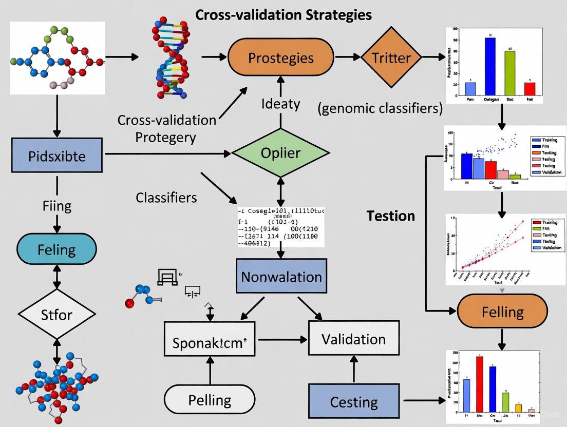
Workflow for Model Development and Validation
A robust machine learning pipeline involves sequential steps that must be properly integrated with the chosen validation strategy. The following diagram outlines a generalized workflow for developing a genomic classifier, highlighting where different data partitioning strategies are applied.
Comparative Analysis of Partitioning Strategies in Genomic Studies
Quantitative Performance Comparison
Empirical evidence from healthcare and genomic simulation studies demonstrates the relative performance of different validation strategies. The table below summarizes findings from key studies, highlighting the impact of each method on performance estimation.
| Source | Experimental Context | Validation Methods Compared | Key Finding on Performance Estimation |
|---|---|---|---|
| Varma et al. (2006) [15] | "Null" and "non-null" datasets using Shrunken Centroids and SVM classifiers. | Standard CV with tuning, Nested CV, and evaluation on an independent test set. | Standard CV with parameter tuning outside the loop gave substantially biased (optimistic) error estimates. Nested CV gave an estimate very close to the independent test set error. |
| Lemoine et al. (2025) [16] | Simulation of high-dimensional transcriptomic data (15,000 genes) with time-to-event outcomes. Sample sizes from 50 to 1000. | Train-test, Bootstrap, 0.632+ Bootstrap, 5-Fold CV, Nested CV (5x5). | Train-test was unstable. Bootstrap was over-optimistic. 0.632+ Bootstrap was overly pessimistic for small n. K-fold CV and Nested CV were recommended for stability and reliability. |
| Wilimitis & Walsh (2023) [19] | Tutorial using MIMIC-III clinical data for classification and regression tasks. | Hold-out validation vs. various Cross-Validation methods. | Nested cross-validation reduces optimistic bias but comes with additional computational challenges. Cross-validation is generally favored over hold-out for smaller healthcare datasets. |
Case Study: Bias in Cross-Validation for Classifier Tuning
A critical study by Varma et al. [15] illustrates a common pitfall in validation. The researchers created "null" datasets where no real difference existed between sample classes. They then used CV to find classifier parameters that minimized the CV error. This process alone produced deceptively low error estimates (<30% on 38% of "null" datasets for SVM), even though the classifier's performance on a true independent test set was no better than chance. This demonstrates that using the same data for both tuning and performance estimation introduces significant optimism bias. The nested CV procedure, where tuning is performed inside each fold of the outer validation loop, successfully corrected this bias.
The Scientist's Toolkit: Research Reagent Solutions
Building and validating a genomic cancer classifier requires a suite of computational and data resources. The following table details key components of the research pipeline.
| Item | Function in Genomic Classifier Research |
|---|---|
| High-Dimensional Omics Data | The foundational input for model training. Public repositories like The Cancer Genome Atlas (TCGA) provide large-scale, well-annotated genomic (e.g., RNA-seq), epigenomic, and clinical data [3] [22]. |
| Programming Environment (Python/R) | Provides the ecosystem for data manipulation, analysis, and modeling. Key libraries (e.g., scikit-learn in Python, caret in R) implement cross-validation, machine learning algorithms, and performance metrics [3] [19]. |
| Feature Selection Algorithms | Critical for reducing data dimensionality and mitigating overfitting. Methods like Lasso (L1 regularization) and Ridge (L2 regularization) regression are commonly used to identify a subset of predictive genes from thousands of candidates [16] [3]. |
| High-Performance Computing (HPC) | Essential for computationally intensive tasks like nested cross-validation on large genomic datasets or training complex ensemble models, significantly reducing computation time [22] [21]. |
| Stratified Cross-Validation | A specific technique that preserves the percentage of samples for each class (e.g., cancer type) in every fold. This is crucial for handling class imbalance often found in biomedical datasets and for obtaining realistic performance estimates [19] [23]. |
The selection of a data partitioning strategy is a direct application of the bias-variance tradeoff principle. For genomic cancer classification, where high-dimensional data and small sample sizes are the norm, simple hold-out validation is often inadequate and can be misleading.
Evidence from multiple studies consistently shows that k-fold cross-validation offers a stable and reliable balance between bias and variance for general model evaluation [16]. When the modeling process involves parameter tuning or feature selection, nested cross-validation is the gold standard for obtaining an almost unbiased estimate of the true error, preventing optimistic bias from creeping into performance reports [15] [19]. By rigorously applying these advanced partitioning strategies, researchers and drug developers can build more generalizable and trustworthy genomic classifiers, ultimately accelerating the path to clinical impact.
In the pursuit of precision oncology, genomic classifiers have emerged as powerful tools for cancer diagnosis, prognosis, and treatment selection. These molecular classifiers, developed from high-throughput genomic, transcriptomic, and proteomic data, promise to tailor cancer care to the unique biological characteristics of each patient's tumor [24]. However, the development of classifiers from high-dimensional data presents a complex analytical challenge fraught with potential methodological pitfalls that may result in spuriously high performance estimates [25]. The stakes for proper validation are exceptionally high in this domain, as erroneous classifiers can lead to misdiagnosis, inappropriate treatment selections, and ultimately, patient harm.
Cross-validation (CV) has become a cornerstone methodology for assessing the performance and generalizability of genomic classifiers, particularly when limited samples are available. This technique provides a framework for estimating how well a classifier will perform on unseen data, simulating its behavior in real-world clinical settings. Yet, not all cross-validation approaches are created equal, and inappropriate application can generate misleadingly optimistic performance estimates [25] [26]. This guide examines current cross-validation strategies, compares their methodological rigor, and provides experimental protocols to ensure reliable assessment of genomic classifiers in oncology applications.
The Validation Gap: Empirical Evidence of Performance Inflation
Substantial empirical evidence demonstrates that common cross-validation practices can significantly overestimate the true performance of genomic classifiers. A comprehensive assessment of molecular classifier validation practices revealed that most studies employ cross-validation methods likely to overestimate performance, with marked discrepancies between internal validation and independent external validation results [25].
Table 1: Performance Discrepancy Between Cross-Validation and Independent Validation
| Performance Metric | Cross-Validation Median | Independent Validation Median | Relative Diagnostic Odds Ratio |
|---|---|---|---|
| Sensitivity | 94% | 88% | 3.26 (95% CI 2.04-5.21) |
| Specificity | 98% | 81% |
This validation gap stems from multiple methodological challenges. Simple resubstitution analysis of training sets is well-known to produce biased performance estimates, but even more sophisticated internal validation methods like k-fold cross-validation and leave-one-out cross-validation can yield inflated accuracy when inappropriately applied [25]. Specific sources of bias include population selection bias, incomplete cross-validation, optimization bias, reporting bias, and parameter selection bias [25].
The computational intensity of proper validation presents another challenge, particularly for complex classifiers. Standard implementations of leave-one-out cross-validation require training a model m times for m instances, while leave-pair-out methods require O(m²) training rounds [27]. These computational demands can become prohibitive with larger datasets, creating pressure to adopt less rigorous but more computationally efficient validation approaches.
Cross-Validation Techniques: A Comparative Analysis
Standard Cross-Validation Approaches
Random Cross-Validation (RCV) represents the most common approach, where samples are randomly partitioned into k folds. While theoretically sound, RCV can produce over-optimistic performance estimates when test samples are highly similar to training samples, as often occurs with biological replicates in genomic datasets [26]. This approach assumes that randomly selected test sets well-represent unseen data, an assumption that may not hold when samples come from different experimental conditions or biological contexts [26].
Leave-One-Out Cross-Validation (LOO) provides an almost unbiased estimate of performance but suffers from high variance, particularly with small sample sizes [27]. For AUC estimation, LOO can demonstrate substantial negative bias in small-sample settings [27].
Leave-Pair-Out Cross-Validation (LPO) has been proposed specifically for AUC estimation, as it is almost unbiased and maintains deviation variance as low as the best alternative approaches [27]. In this method, all possible pairs of positive and negative instances are left out for testing, making it computationally intensive but statistically favorable for AUC-based evaluations.
Advanced Approaches for Genomic Data
Clustering-Based Cross-Validation (CCV) addresses a critical flaw in RCV by first clustering experimental conditions and including entire clusters of similar conditions as one CV fold [26]. This approach tests a method's ability to predict gene expression in entirely new regulatory contexts rather than similar conditions, providing a more realistic estimate of generalizability.
Simulated Annealing Cross-Validation (SACV) represents a more controlled approach that constructs partitions spanning a spectrum of distinctness scores [26]. This enables researchers to evaluate classifier performance across varying degrees of training-test similarity, offering insights into how methods will perform when applied to datasets with different relationships to the training data.
Table 2: Comparison of Cross-Validation Techniques for Genomic Classifiers
| Technique | Key Principles | Strengths | Limitations | Best Use Cases |
|---|---|---|---|---|
| Random CV (RCV) | Random partitioning into k folds | Simple implementation; Widely understood | May overestimate performance; Sensitive to sample similarity | Initial model assessment; Large, diverse datasets |
| Leave-One-Out CV | Each sample alone as test set | Low bias; Uses maximum training data | High variance; Computationally intensive | Very small datasets; Nearly unbiased estimation needed |
| Leave-Pair-Out CV | All positive-negative pairs left out | Excellent for AUC estimation; Low bias | Extremely computationally intensive (O(m²)) | Small datasets where AUC is primary metric |
| Clustering-Based CV | Entire clusters as folds | Tests generalizability across contexts; More realistic performance estimates | Dependent on clustering algorithm and parameters | Assessing biological generalizability; Context-shift evaluation |
| Simulated Annealing CV | Partitions with controlled distinctness | Enables performance spectrum analysis; Controlled distinctness | Complex implementation; Computationally intensive | Comprehensive method comparison; Distinctness-impact analysis |
Experimental Protocols for Robust Validation
Protocol 1: Distinctness-Based Cross-Validation
The distinctness of test sets from training sets significantly impacts performance estimation [26]. This protocol provides a methodological framework for assessing this relationship:
Compute Distinctness Score: For each potential test experimental condition, calculate its distinctness from a given set of training conditions using only predictor variables (e.g., transcription factor expression values), independent of the target gene expression values.
Construct Partitions: Use simulated annealing to generate multiple partitions with gradually increasing distinctness scores, creating a spectrum from highly similar to highly distinct test-training set pairs.
Evaluate Performance: Train and test classifiers across these partitions, measuring performance metrics (sensitivity, specificity, AUC) at each distinctness level.
Analyze Trends: Plot performance against distinctness scores to evaluate how classifier accuracy degrades as test sets become increasingly distinct from training data.
This approach enables comparison of classifiers not merely based on average performance, but on their robustness to increasing dissimilarity between training and application contexts [26].
Protocol 2: Cross-Condition Validation for GRN Inference
For gene regulatory network (GRN) inference, standard CV may not adequately assess generalizability across biological conditions:
Cluster Conditions: Perform clustering on experimental conditions based on TF expression profiles to identify groups of similar regulatory contexts.
Form Folds: Assign entire clusters to cross-validation folds rather than individual samples.
Train and Test: Iteratively leave out each cluster-fold, train GRN inference methods on remaining data, and test prediction accuracy on the held-out cluster.
Compare to RCV: Execute standard random CV on the same dataset for comparative analysis.
Studies implementing this approach have demonstrated that RCV typically produces more optimistic performance estimates than CCV, with the discrepancy revealing the degree to which performance depends on similarity between training and test conditions [26].
Visualization of Cross-Validation Strategies
Figure 1: Cross-Validation Workflow Comparison. This diagram illustrates the key differences between standard Random Cross-Validation (RCV) and Clustering-Based Cross-Validation (CCV) approaches, highlighting how CCV tests generalizability across distinct experimental contexts.
The Researcher's Toolkit: Essential Solutions for Validation
Table 3: Research Reagent Solutions for Cross-Validation Experiments
| Solution Category | Specific Tools/Frameworks | Function in Validation | Key Considerations |
|---|---|---|---|
| Statistical Computing | R, Python (scikit-learn) | Provides base CV implementations | Customization needed for genomic specificities |
| Machine Learning Frameworks | TensorFlow, PyTorch | Enable custom classifier development | Computational efficiency for large-scale CV |
| Specialized CV Algorithms | Leave-Pair-Out, SACV | Address specific biases in performance estimation | Implementation complexity; Computational demands |
| Clustering Methods | k-means, hierarchical clustering | Enables CCV implementation | Sensitivity to parameters; Distance metrics |
| Distinctness Scoring | Custom implementations | Quantifies test-training dissimilarity | Must use only predictor variables, not outcomes |
| Performance Metrics | AUC, sensitivity, specificity | Standardized performance assessment | AUC particularly important for class imbalance |
The development of genomic classifiers for cancer diagnostics carries tremendous responsibility, as these tools directly impact patient care decisions. The evidence clearly demonstrates that standard cross-validation approaches often yield optimistic performance estimates that do not translate to independent validation [25]. This validation gap represents a significant concern for clinical translation, potentially leading to the implementation of classifiers that underperform in real-world settings.
Moving forward, the field requires a shift toward more rigorous validation practices that explicitly account for the distinctness between training and test conditions. Clustering-based cross-validation and distinctness-controlled approaches like SACV provide promising frameworks for more realistic performance estimation [26]. Additionally, researchers should prioritize external validation in independent datasets whenever possible, as this remains the gold standard for establishing generalizability [25].
The computational burden of rigorous validation remains a challenge, particularly for complex classifiers and large genomic datasets. However, the stakes are too high to accept methodological shortcuts that compromise the reliability of performance estimates. By adopting more stringent cross-validation practices and transparently reporting validation methodologies, the research community can enhance the development of genomic classifiers that truly deliver on the promise of precision oncology.
A Practical Toolkit of Cross-Validation Methods for Cancer Genomics
In genomic cancer classifier research, where models are built on high-dimensional molecular data to predict phenotypes like cancer subtypes or survival outcomes, robust model evaluation is paramount. Cross-validation provides an essential framework for assessing how well a predictive model will generalize to independent datasets, thereby flagging problems like overfitting to the limited samples typically available in biomedical studies [28]. Among various techniques, K-Fold Cross-Validation has emerged as a widely adopted standard, striking a practical balance between computational feasibility and reliable performance estimation [29]. For researchers and drug development professionals, understanding the parameters and alternatives to K-Fold is crucial for developing classifiers that can reliably inform biological hypothesis generation and potential clinical applications [30] [31]. This guide provides an objective comparison of K-Fold's performance against other cross-validation strategies, with a specific focus on evidence from genomic and cancer classification studies.
Understanding the K-Fold Cross-Validation Algorithm
K-Fold Cross-Validation is a resampling procedure used to evaluate machine learning models on a limited data sample. The method's core premise involves dividing the entire dataset into 'K' equally sized folds or segments. For each unique group, the algorithm treats it as a test set while using the remaining groups as a training set. This process repeats 'K' times, with each fold used exactly once as the testing set. The 'K' results are then averaged to produce a single estimation of model performance [32] [33].
The following diagram illustrates the workflow and data flow in a standard 5-fold cross-validation process:
The brilliance of K-Fold Cross-Validation lies in its ability to mitigate the bias associated with random shuffling of data into training and test sets. It ensures that every observation from the original dataset has the opportunity to appear in both training and test sets, which is crucial for models sensitive to specific data partitions [33]. This is particularly important in genomic studies where sample sizes may be limited, and each data point represents valuable biological information.
Comparative Analysis of Cross-Validation Techniques
Performance Comparison Across Methods
Different cross-validation techniques offer varying trade-offs between bias, variance, and computational requirements. The table below summarizes a comparative analysis of three common methods based on experimental data from model evaluation studies:
Table 1: Comparative Performance of Cross-Validation Techniques on Balanced and Imbalanced Datasets
| Cross-Validation Method | Best Performing Model (Imbalanced Data) | Sensitivity | Balanced Accuracy | Best Performing Model (Balanced Data) | Sensitivity | Balanced Accuracy | Computational Time (Seconds) |
|---|---|---|---|---|---|---|---|
| K-Fold Cross-Validation | Random Forest | 0.784 | 0.884 | Support Vector Machine | 0.878 | 0.892 | 21.480 (SVM) |
| Repeated K-Fold | Support Vector Machine | 0.541 | 0.764 | Support Vector Machine | 0.886 | 0.894 | ~1986.570 (RF) |
| Leave-One-Out (LOOCV) | Random Forest/Bagging | 0.787/0.784 | 0.883/0.881 | Support Vector Machine | 0.893 | 0.891 | High (Model Dependent) |
Data adapted from comparative analysis by Lumumba et al. (2024) [29]
Key Trade-Offs and Characteristics
Each cross-validation method carries distinct advantages and limitations that researchers must consider within their specific genomic context:
K-Fold Cross-Validation (typically with K=5 or K=10) generally offers a balanced compromise between computational efficiency and reliable performance estimation. It demonstrates strong performance across various models while maintaining reasonable computation times, making it suitable for medium to large genomic datasets [29] [34].
Leave-One-Out Cross-Validation (LOOCV), an exhaustive method where the number of folds equals the number of instances, provides nearly unbiased error estimation but suffers from higher variance and computational cost, particularly with large datasets. In biomedical contexts with small sample sizes, LOOCV is sometimes preferred as it maximizes the training data in each iteration [31] [28].
Repeated K-Fold Cross-Validation enhances reliability by averaging results from multiple K-fold runs with different random partitions, effectively reducing variance. However, this comes at a significant computational cost, as evidenced by processing times nearly 100 times longer than standard K-fold in some experimental comparisons [29].
Stratified K-Fold Cross-Validation preserves the class distribution in each fold, making it particularly valuable for imbalanced genomic datasets, such as those comparing cancer subtypes with unequal representation [23] [34].
Experimental Protocols in Genomic Studies
Case Study: Genomic Prediction Models in Plant Science (with Implications for Cancer Research)
Frontiers in Plant Science published a comprehensive methodological comparison of genomic prediction models using K-fold cross-validation, with protocols directly transferable to genomic cancer classifier development [30]. The experimental methodology proceeded as follows:
Dataset Preparation: Public datasets from wheat, rice, and maize were utilized, comprising 599 wheat lines with 1,279 DArT markers, 1,946 rice lines from the 3,000 Rice Genomes Project, and maize lines from the "282" Association Panel. These genomic datasets mirror the high-dimensional characteristics of cancer genomic data.
Model Selection: The study evaluated a variety of statistical models from the "Bayesian alphabet" (e.g., BayesA, BayesB, BayesC) and genomic relationship matrix models (e.g., G-BLUP, EG-BLUP), representing common approaches in genomic prediction.
Cross-Validation Protocol: The researchers implemented paired K-fold cross-validation to compare model performances. The key innovation was the use of statistical tests based on equivalence margins borrowed from clinical research to identify differences in model performance with practical relevance.
Hyperparameter Tuning: The study assessed how hyperparameters (parameters not directly estimated from data) affect predictive accuracy across models, using cross-validation to guide selection.
Performance Assessment: Predictive accuracy was evaluated through the cross-validation process, with emphasis on identifying statistically significant differences between models that would impact genetic gain - analogous to clinical utility in cancer diagnostics.
This experimental design highlights how K-fold cross-validation enables robust model comparison in high-dimensional biological data contexts, providing a template for cancer genomic classifier development.
Case Study: Bivariate Monotonic Classifiers for Biomarker Discovery
A 2025 study in BMC Bioinformatics addresses genome-scale discovery of bivariate monotonic classifiers (BMCs), with direct implications for cancer biomarker identification [31]. The research team developed the fastBMC algorithm to efficiently identify pairs of features with high predictive performance, using leave-one-out cross-validation as an integral component of their methodology:
Classifier Design: BMCs are based on pairs of input features (e.g., gene pairs) that capture nonlinear patterns while maintaining interpretability - a crucial consideration for biological hypothesis generation.
Validation Approach: The original naïveBMC algorithm used leave-one-out cross-validation to estimate classifier performance, requiring this computation for each possible pair of features. With high-dimensional genomic data, this becomes computationally prohibitive.
Computational Optimization: The fastBMC algorithm introduced a mathematical bound for the LOOCV performance estimate, dramatically speeding up computation by a factor of at least 15 while maintaining optimality.
Biological Validation: The approach was applied to a glioblastoma survival predictor, identifying a biomarker pair (SDC4/NDUFA4L2) that demonstrates the method's utility for generating testable biological hypotheses with potential therapeutic implications.
This case study illustrates how specialized cross-validation approaches can enable biomarker discovery in cancer genomics while balancing computational constraints with statistical rigor.
Table 2: Essential Computational Tools for Cross-Validation in Genomic Research
| Tool/Resource | Function | Implementation Example |
|---|---|---|
| scikit-learn Cross-Validation Module | Provides comprehensive cross-validation functionality | from sklearn.model_selection import KFold, cross_val_score |
| Stratified K-Fold | Preserves class distribution in imbalanced datasets | StratifiedKFold(n_splits=5) |
| Repeated K-Fold | Reduces variance through multiple iterations | RepeatedStratifiedKFold(n_splits=5, n_repeats=10) |
| Bivariate Monotonic Classifier (BMC) | Identifies interpretable feature pairs for biomarker discovery | Python implementation available at github.com/oceanefrqt/fastBMC [31] |
| Pipeline Construction | Ensures proper data preprocessing without data leakage | make_pipeline(StandardScaler(), SVM(C=1)) |
| Multiple Metric Evaluation | Enables comprehensive model assessment | cross_validate(..., scoring=['precision_macro', 'recall_macro']) |
Parameter Optimization in K-Fold Cross-Validation
Selecting the Optimal K Value
The choice of K in K-fold cross-validation represents a critical decision point that balances statistical properties with computational practicality:
K=5 or K=10: These values have been empirically shown to yield test error rate estimates that suffer neither from excessively high bias nor from very high variance, making them recommended defaults for many applications [32] [29].
Lower K values (2-3): May lead to higher variance in performance estimates because the training data size is substantially reduced in each iteration.
Higher K values (approaching n): Increase the training data size in each fold, potentially reducing variance but increasing computational burden and potentially introducing higher bias [33].
Stratified Variants: For classification problems with imbalanced classes, such as rare cancer subtypes, stratified K-fold ensures each fold preserves the percentage of samples for each class, providing more reliable performance estimates [23] [35].
The following decision diagram guides researchers in selecting appropriate cross-validation parameters based on their dataset characteristics and research goals:
Integration with Hyperparameter Tuning
In genomic cancer classifier development, K-fold cross-validation is frequently integrated with hyperparameter optimization through techniques such as grid search or random search. The proper implementation requires nesting the cross-validation procedures:
- Inner Loop: Used for hyperparameter tuning and model selection
- Outer Loop: Used for performance assessment of the final selected model
This nested approach prevents optimistic bias in performance estimates that occurs when the same cross-validation split is used for both parameter tuning and final evaluation [35]. For example, when optimizing the C parameter in Support Vector Machines or the number of trees in Random Forests, the inner cross-validation loop systematically evaluates different parameter combinations across the training folds, while the outer loop provides an unbiased estimate of how well the selected model will generalize.
K-Fold Cross-Validation remains the go-to standard for model evaluation in genomic cancer classifier development due to its optimal balance between statistical reliability and computational efficiency. As evidenced by comparative studies, K=5 or K=10 generally provide the most practical defaults, though researchers working with specialized classifiers or particularly challenging data structures may benefit from variations like stratified or repeated K-fold. The experimental protocols and toolkit presented here offer researchers a foundation for implementing these methods in their genomic studies, with appropriate attention to the unique characteristics of high-dimensional biomedical data. As cross-validation methodologies continue to evolve, including recent developments like irredundant K-fold cross-validation [36], the fundamental importance of robust validation practices in translating genomic discoveries to clinical applications remains undiminished.
In the field of genomic cancer classification, the problem of class imbalance presents a fundamental challenge that can severely compromise the validity of machine learning models. Cancer datasets frequently exhibit significant skewness, where the number of samples from one class (e.g., healthy patients or a common cancer subtype) vastly outnumbers others (e.g., rare cancer subtypes or metastatic cases) [37]. This imbalance is particularly pronounced in genomic studies characterized by high-dimensional feature spaces and limited sample sizes [37]. Traditional cross-validation approaches, which randomly split data into training and testing sets, risk creating folds that poorly represent the minority class, leading to overly optimistic performance estimates and models that fail to generalize to real-world clinical scenarios [38] [39].
Stratified K-Fold Cross-Validation has emerged as a critical methodological solution to this problem. It is a variation of standard K-Fold cross-validation that ensures each fold preserves the same percentage of samples for each class as the complete dataset [40]. This preservation of class distribution is not merely a technical refinement but a statistical necessity for generating reliable performance estimates in genomic cancer research, where accurately identifying minority classes (such as rare malignancies) can be of paramount clinical importance. This guide provides a comprehensive comparison of Stratified K-Fold against alternative validation strategies, supported by experimental data from cancer classification studies.
Experimental Comparisons of Cross-Validation Strategies
Performance Comparison on Imbalanced Biomedical Datasets
The table below summarizes findings from a large-scale study comparing Stratified Cross-Validation (SCV) and Distribution Optimally Balanced SCV (DOB-SCV) across 420 datasets, involving several sampling methods and classifiers including Decision Trees, k-NN, SVM, and Multi-layer Perceptron [38].
| Validation Method | Key Principle | Reported Advantage | Classifier Context |
|---|---|---|---|
| Stratified K-Fold (SCV) | Ensures each fold has same class proportion as full dataset [40] [38]. | Provides a more reliable estimate of model performance on imbalanced data; avoids folds with missing classes [38] [39]. | Foundation for robust evaluation; often combined with sampling techniques [38]. |
| DOB-SCV | Places nearest neighbors of the same class into different folds to better maintain original distribution [38]. | Can provide slightly higher F1 and AUC values when combined with sampling [38]. | Performance gain is often smaller than the impact of selecting the right sampler-classifier pair [38]. |
The core finding was that while DOB-SCV can sometimes offer marginal improvements, the choice between SCV and DOB-SCV is generally less critical than the selection of an effective sampler-classifier combination [38]. This underscores that Stratified K-Fold provides a sufficiently robust foundation for model evaluation, upon which other techniques for handling imbalance can be built.
Efficacy of Ensemble Classifiers with Stratified K-Fold
Stratified K-Fold is frequently used to validate powerful ensemble classifiers in cancer diagnostics. The following table synthesizes results from multiple studies on breast cancer classification that utilized Stratified K-Fold validation, demonstrating state-of-the-art performance [23] [41].
| Study Focus | Classifier/Method | Key Performance Metric(s) | Stratified Validation Role |
|---|---|---|---|
| Breast Cancer Classification [23] | Majority-Voting Ensemble (LR, SVM, CART) | 99.3% Accuracy [23] | Ensured reliable performance estimate on imbalanced Wisconsin Diagnostic Breast Cancer dataset. |
| Breast Cancer Classification [41] | Ensembles (AdaBoost, GBM, RGF) | 99.5% Accuracy [41] | Used alongside Stratified Shuffle Split to validate performance and ensure class representation. |
| Multi-Cancer Prediction [42] | Stacking Ensemble (12 base learners) | 99.28% Accuracy, 97.56% Recall, 99.55% Precision (average across 3 cancers) [42] | Critical for fair evaluation across lung, breast, and cervical cancer datasets with different imbalance levels. |
These results highlight a consistent trend: combining Stratified K-Fold validation with ensemble methods produces exceptionally high and, more importantly, reliable performance metrics, making them a gold standard for imbalanced cancer classification tasks.
Methodologies and Protocols
Standardized Workflow for Genomic Classifier Validation
The following diagram illustrates a recommended experimental workflow that integrates Stratified K-Fold at its core, ensuring that class imbalance is addressed at both the data and validation levels.
This workflow emphasizes two critical best practices. First, resampling techniques like SMOTE or KDE must be applied exclusively to the training folds after the split to prevent data leakage from the test set, which would invalidate the performance estimate [37] [43]. Second, the final model performance is derived from the aggregated results across all test folds, providing a robust measure of how the model will generalize to new, unseen data [39] [44].
The Scientist's Toolkit: Essential Research Reagents and Solutions
The table below catalogues key computational "reagents" and their functions essential for implementing Stratified K-Fold validation in genomic cancer studies.
| Research Reagent / Solution | Function / Purpose | Example / Notes |
|---|---|---|
| StratifiedKFold (scikit-learn) | Core cross-validator; splits data into K folds while preserving class distribution [40]. | from sklearn.model_selection import StratifiedKFold Essential for initial, reliable data splitting [39]. |
| Resampling Algorithms (e.g., SMOTE, KDE) | Balances class distribution within the training set by generating synthetic minority samples [37] [43]. | SMOTE: Generates samples via interpolation [43]. KDE: Resamples from estimated probability density; can outperform SMOTE on genomic data [37]. |
| High-Performance Ensemble Classifiers | Combines multiple models to improve predictive accuracy and robustness [23] [42]. | XGBoost, Random Forest, and Majority-Voting ensembles have shown >99% accuracy in stratified validation [23] [42]. |
| Imbalance-Robust Metrics | Provides a truthful evaluation of model performance on imbalanced data beyond simple accuracy [37] [43]. | AUC, F1-Score, Matthews Correlation Coefficient (MCC), Precision-Recall AUC [42] [43]. |
The consistent theme across comparative studies is that Stratified K-Fold Cross-Validation is a non-negotiable starting point for reliable model evaluation on imbalanced cancer datasets. While alternative methods like DOB-SCV can offer minor enhancements, the primary gain in performance and robustness comes from coupling Stratified K-Fold with appropriate ensemble classifiers and data-level resampling techniques like SMOTE or KDE [23] [38].
For researchers and clinicians developing genomic cancer classifiers, the evidence strongly supports a standardized protocol: using Stratified K-Fold as the validation backbone to ensure fair class representation, then systematically exploring combinations of modern resampling methods and powerful ensemble models like XGBoost or stacking ensembles to achieve state-of-the-art performance. This rigorous approach ensures that predictive models are not only accurate in a technical sense but also generalizable and trustworthy in high-stakes clinical environments.
In genomic cancer research, accurately estimating a classifier's real-world performance is paramount for clinical translation. Cross-validation (CV) serves as the standard for assessing model generalization, yet common practices introduce a subtle but critical flaw: optimistic bias caused by data leakage during hyperparameter tuning [45]. When the same data informs both parameter tuning and performance estimation, the test set is no longer "statistically pure," leading to inflated performance metrics and models that fail in production [45]. This problem is particularly acute in genomic studies, where datasets are often characterized by high dimensionality (thousands of genes) and small sample sizes, amplifying the risk of overfitting.
Nested cross-validation (NCV) provides a robust solution to this problem. It is a disciplined validation strategy that strictly separates the model selection process from the model assessment process [46]. By employing two layers of data folding, NCV delivers a realistic and unbiased estimate of how a model, with its tuned hyperparameters, will perform on unseen data. For researchers developing genomic cancer classifiers, adopting NCV is not merely a technical refinement but a foundational practice for building trustworthy and reliable predictive models.
Understanding Nested Cross-Validation: Architecture and Workflow
The Core Principle: Separation of Model Selection and Evaluation
The fundamental strength of nested cross-validation lies in its clear separation of duties [46]. It consists of two distinct loops, an outer loop for performance estimation and an inner loop for model and hyperparameter selection, which operate independently to prevent information leakage.
- Outer Loop (Performance Estimation): The dataset is split into ( K ) folds. Sequentially, each fold serves as a test set, while the remaining ( K-1 ) folds constitute the development set. The key is that this test set is never used for any decision-making during the model building process for that split [47].
- Inner Loop (Model Tuning): For each development set from the outer loop, a second, independent cross-validation process is performed. This inner loop is used to train models with different hyperparameters and select the best-performing set. The outer loop's test set is completely untouched during this phase [46] [45].
This hierarchical structure ensures that the final performance score reported from the outer loop is a true estimate of generalization error, as it is derived from data that played no role in selecting the model's configuration [48].
Workflow Diagram of Nested Cross-Validation
The following diagram illustrates the two-layer structure of the nested cross-validation process.
Comparative Analysis: Nested vs. Non-Nested Cross-Validation
Quantitative Performance Comparison
Empirical studies across various domains, including healthcare and genomics, consistently demonstrate that non-nested cross-validation produces optimistically biased performance estimates. The following table summarizes key findings from the literature.
Table 1: Empirical Comparison of Nested and Non-Nested Cross-Validation Performance
| Study / Domain | Metric | Non-Nested CV Performance | Nested CV Performance | Bias Reduction |
|---|---|---|---|---|
| Tougui et al. (2021) [46] | AUROC | Higher estimate | Realistic estimate | 1% to 2% |
| Area Under Precision-Recall (AUPR) | Higher estimate | Realistic estimate | 5% to 9% | |
| Wilimitis et al. (2023) [49] | Generalization Error | Over-optimistic, biased | Lower, more realistic | Significant |
| Ghasemzadeh et al. (2024) [46] | Statistical Power & Confidence | Lower | Up to 4x higher confidence | Notable |
| Usher Syndrome miRNA Study [50] | Classification Accuracy | Prone to overfitting | 97.7% (validated) | Critical for robustness |
Procedural and Conceptual Differences
The quantitative differences stem from fundamental methodological flaws in the non-nested approach.
Table 2: Conceptual and Practical Differences Between Validation Methods
| Aspect | Non-Nested Cross-Validation | Nested Cross-Validation |
|---|---|---|
| Core Procedure | Single data split for both tuning and evaluation. | Two separate, layered loops for tuning and evaluation. |
| Information Leakage | High risk; test data influences hyperparameter choice. | Prevented by design; outer test set is completely hidden from tuning. |
| Performance Estimate | Optimistically biased, unreliable for generalization. | Nearly unbiased, realistic estimate of true performance [46]. |
| Computational Cost | Lower. | Significantly higher (e.g., K x L models for K outer and L inner folds). |
| Model Selection | Vulnerable to selection bias, overfits the test set. | Robust model selection; identifies models that generalize better. |
| Suitability for Small Datasets | Poor, high variance and bias. | Recommended, makes efficient and rigorous use of limited data [50]. |
Implementing Nested Cross-Validation in Genomic Cancer Research
Experimental Protocol for Genomic Classifiers
Implementing NCV for a genomic cancer classifier involves a sequence of critical steps to ensure biological relevance and statistical rigor.
Dataset Preparation and Partitioning:
- Subject-Wise Splitting: Given the correlated nature of genomic measurements from the same patient, splits must be performed subject-wise (or patient-wise) rather than record-wise. This ensures all samples from a single patient are contained within either the training or test set of a given fold, preventing inflated performance due to patient re-identification [49].
- Stratification: For classification tasks, it is crucial to use stratified k-fold in both the inner and outer loops. This preserves the percentage of samples for each class (e.g., cancer vs. normal) across all folds, which is especially important for imbalanced genomic datasets [49].
Inner Loop Workflow (Hyperparameter Tuning):
- The development set from the outer loop is used for an inner ( L )-fold cross-validation.
- A predefined hyperparameter search space (e.g., using GridSearchCV or RandomizedSearchCV) is explored. For a Random Forest classifier, this might include
max_depth,n_estimators, andmax_features. - A model is trained for each hyperparameter combination on the inner training folds and evaluated on the inner validation folds.
- The set of hyperparameters that yields the best average performance across the inner folds is selected.
Outer Loop Workflow (Performance Evaluation):
- Using the optimal hyperparameters found in the inner loop, a final model is trained on the entire development set.
- This model is then evaluated on the held-out outer test fold, which has not been used in any way during the tuning process. A performance metric (e.g., AUC, Accuracy) is recorded.
- This process repeats for each of the ( K ) outer folds.
Final Model and Reporting:
- The final output of NCV is not a single model, but a distribution of ( K ) performance scores. The mean and standard deviation of these scores provide a robust estimate of the model's generalization capability and its uncertainty [48].
- To deploy a model, one can refit it on the entire dataset using the hyperparameters that performed best on average during the NCV process.
Data Partitioning Strategy Diagram
The following diagram details the data partitioning strategy for a single outer fold, highlighting the strict separation of training, validation, and test data.
The Scientist's Toolkit: Essential Research Reagents and Computational Tools
Successfully implementing nested cross-validation in genomic research requires a combination of computational tools and rigorous statistical practices.
Table 3: Essential Tools and Practices for Rigorous Genomic Classifier Validation
| Tool / Practice | Category | Function in Nested CV | Example Technologies |
|---|---|---|---|
| Stratified K-Fold | Data Partitioning | Ensures class ratios are preserved in all training/test splits, critical for imbalanced cancer datasets. | StratifiedKFold (scikit-learn) |
| Group K-Fold | Data Partitioning | Enforces subject-wise splitting by grouping all samples from the same patient to prevent data leakage. | GroupKFold (scikit-learn) |
| Hyperparameter Optimizer | Model Tuning | Automates the search for optimal model parameters within the inner loop. | GridSearchCV, RandomizedSearchCV (scikit-learn), Optuna |
| High-Performance Computing (HPC) | Infrastructure | Manages the high computational cost of NCV through parallelization across multiple CPUs/GPUs. | SLURM, Multi-GPU frameworks, Cloud computing [48] |
| Nested CV Code Framework | Software | Provides a reusable, scalable structure for implementing the complex nested validation process. | NACHOS framework [48], custom scripts in Python/R |
| Reproducibility Practices | Methodology | Ensures results are trustworthy and verifiable. | Setting random seeds, version control (Git), containerization (Docker) |
Nested cross-validation represents a paradigm shift from a model-centric to a reliability-centric approach in genomic cancer classifier development. While computationally demanding, its rigorous separation of model tuning and evaluation is the most effective method to quantify and reduce optimistic bias, providing a trustworthy estimate of how a model will perform in a real-world clinical setting [48]. For research aimed at informing drug development or clinical decision-making, where the cost of failure is high, adopting nested cross-validation is not just a best practice—it is an ethical imperative to ensure that reported performance metrics reflect true predictive power.
In the field of genomic cancer research, selecting the proper validation strategy is not merely a technical formality—it is a fundamental determinant of a classifier's real-world utility. The choice between hold-out validation and more computationally intensive methods like k-fold cross-validation carries significant implications for the reliability, generalizability, and ultimate clinical applicability of predictive models. This guide provides an objective comparison of these strategies, focusing on their application in genomic cancer classifier development, to equip researchers with evidence-based criteria for selection.
Understanding the Validation Methods
What is Hold-Out Validation?
Hold-out validation, also known as the train-test split method, involves partitioning a dataset into two distinct subsets: one for training the model and another for testing its performance [34] [51]. This approach typically allocates 70-80% of the data for training and reserves the remaining 20-30% for testing [52]. The primary advantage of this method is its computational efficiency, as models are trained and evaluated only once [34].
What is Cross-Validation?
Cross-validation, particularly k-fold cross-validation, represents a more robust approach to model evaluation. This technique divides the dataset into k equal-sized folds (commonly k=5 or k=10) [34] [35]. The model is trained k times, each time using k-1 folds for training and the remaining fold for testing, with results averaged across all iterations [34] [35]. This process ensures that every data point contributes to both training and testing, providing a more comprehensive assessment of model performance [34].
Figure 1: Workflow comparison between hold-out validation and k-fold cross-validation
Comparative Analysis: Key Differences at a Glance
Table 1: Direct comparison between hold-out validation and k-fold cross-validation
| Feature | Hold-Out Validation | K-Fold Cross-Validation |
|---|---|---|
| Data Split | Single split into training and test sets [34] | Multiple splits into k folds; each fold serves as test set once [34] |
| Training & Testing | Model trained once, tested once [34] | Model trained and tested k times [34] |
| Bias & Variance | Higher bias if split isn't representative; results can vary significantly [34] | Lower bias; more reliable performance estimate [34] |
| Computational Time | Faster; single training cycle [34] [51] | Slower; requires k training cycles [34] [51] |
| Data Utilization | Only partial data used for training; may miss patterns [34] | All data points used for both training and testing [34] |
| Best Use Cases | Very large datasets, quick evaluation, initial modeling [34] [51] | Small to medium datasets where accurate estimation is crucial [34] |
When to Use Hold-Out Validation: Research Contexts and Applications
With Very Large Datasets
When working with extensive genomic datasets containing thousands of samples, the computational efficiency of hold-out validation becomes advantageous [34] [51]. The single training-testing cycle significantly reduces processing time while still providing reasonable performance estimates.
During Initial Model Development
In the exploratory phases of research, hold-out serves as a rapid assessment tool for comparing multiple algorithms or establishing baseline performance before committing to more resource-intensive validation [52].
When Implementing Strict Data Segregation
For research requiring absolute separation between training and testing data—particularly in clinical validation contexts—hold-out validation enables clear demarcation [53]. This approach prevents any potential data leakage that might occur during complex cross-validation procedures.
For Independent External Validation
Hold-out validation is particularly valuable when simulating real-world scenarios where a model trained on one dataset must generalize to entirely separate data [25] [54]. This approach more accurately reflects clinical deployment conditions where models encounter truly unseen data.
Inherent Risks and Limitations of Hold-Out Validation
High Variance in Performance Estimates
A single train-test split provides limited information about model stability [34]. The performance metric obtained is highly dependent on the specific random partition of the data, potentially leading to misleading conclusions if the split is unrepresentative [34] [53].
Potential for Optimistic Bias
When the test set is used repeatedly for model selection or hyperparameter tuning, knowledge of the test set can "leak" into the model, creating over-optimistic performance estimates [35]. This risk necessitates three-way splits (training, validation, and test sets) for proper model selection [52].
Reduced Statistical Power in Small Datasets
In genomic studies with limited samples, reserving a portion for testing alone may substantially reduce the training data available, potentially leading to underfitting and poor model performance [53]. For small sample sizes, cross-validation provides more reliable performance estimates [53].
Evidence from Genomic Cancer Research: A Critical Comparison
Performance Discrepancies in Molecular Classifiers
Empirical assessments of molecular classifier validation reveal significant performance gaps between internal validation methods and independent testing. A comprehensive review of 35 studies comparing cross-validation versus external validation demonstrated that cross-validation practices often overestimate classifier performance [25].
Table 2: Performance comparison between cross-validation and independent hold-out validation in molecular classifier studies
| Validation Method | Reported Sensitivity (%) | Reported Specificity (%) | Relative Diagnostic Odds Ratio |
|---|---|---|---|
| Internal Cross-Validation | 94% | 98% | Baseline |
| Independent Hold-Out Validation | 88% | 81% | 3.26 (95% CI: 2.04-5.21) |
Data adapted from an empirical assessment of 35 studies on molecular classifier validation [25]
The relative diagnostic odds ratio of 3.26 indicates significantly worse performance in independent validation compared to cross-validation, highlighting the potential optimism bias in internal validation approaches [25].
Case Study: Cancer Transcriptomics Model Selection
Research on cancer transcriptomic predictive models directly tested the assumption that smaller, simpler gene signatures generalize better across datasets [55]. The study compared model selection based solely on cross-validation performance versus combining cross-validation with regularization strength.
Findings revealed that more regularized (simpler) signatures did not demonstrate superior generalization across datasets (from cell lines to human tumors and vice versa) or biological contexts (holding out entire cancer types from pan-cancer data) [55]. This result held for both linear models (LASSO logistic regression) and non-linear ones (neural networks) [55].
The authors concluded that when the goal is producing generalizable predictive models, researchers should select models performing best on held-out data or in cross-validation rather than preferring smaller or more regularized models [55].
GWAS Research on Prostate Cancer Toxicity
A study on genome-wide association data for predicting prostate cancer radiation therapy toxicity employed both cross-validation and hold-out validation [54]. Researchers used a cohort of 324 patients, with two-thirds for training and one-third for hold-out validation [54].
The preconditioned random forest regression method achieved an area under the curve (AUC) of 0.70 (95% CI: 0.54-0.86) for the weak stream endpoint on hold-out data, significantly outperforming competing methods [54]. This example demonstrates appropriate use of hold-out validation for final model assessment after hyperparameter tuning via cross-validation.
Best Practices for Implementation in Genomic Studies
Strategic Dataset Partitioning
For genomic data with inherent structures (e.g., patient cohorts, tissue sources, or batch effects), implement stratified splitting to maintain consistent distribution of key characteristics across training and test sets [34]. This approach is particularly crucial for imbalanced datasets where class proportions must be preserved [34].
Three-Way Data Splitting
For comprehensive model development, implement separate training, validation, and test sets [52]. Use the training set for model fitting, the validation set for hyperparameter tuning and model selection, and the test set exclusively for final performance assessment [52].
Implementing Nested Cross-Validation
For optimal model selection with limited data, employ nested cross-validation: an outer loop for performance estimation and an inner loop for model selection [56]. This approach provides nearly unbiased performance estimates while maximizing data utilization.
Table 3: Key computational tools and resources for validation studies in genomic cancer research
| Tool/Resource | Function | Application Context |
|---|---|---|
| Scikit-learn traintestsplit | Random partitioning of datasets into training/test subsets [35] | Initial model assessment; baseline establishment |
| Scikit-learn crossvalscore | Automated k-fold cross-validation with performance metrics [35] | Robust performance estimation; model comparison |
| StratifiedKFold | Cross-validation with preserved class distribution [34] | Imbalanced genomic datasets; rare cancer subtype classification |
| Pipeline Class | Chains transformers and estimators; prevents data leakage [35] | Preprocessing integration; feature selection validation |
| RandomState Parameter | Controls randomness for reproducible splits [35] | Result reproducibility; method comparison studies |
Decision Framework: Selecting the Appropriate Validation Strategy
Figure 2: Decision framework for selecting between hold-out and cross-validation strategies
Hold-out validation remains a valuable tool in the genomic researcher's arsenal, particularly for large-scale datasets, initial model screening, and simulating true external validation scenarios. However, its limitations—including potential high variance and optimistic bias—necessitate careful consideration of research context and goals. In genomic cancer classification, where model generalizability directly impacts clinical translation, combining hold-out validation with cross-validation approaches provides the most rigorous assessment framework. By implementing context-appropriate validation strategies and transparently reporting validation methodologies, researchers can advance the development of robust, clinically relevant cancer classifiers.
In genomic cancer research, the integrity of a classifier's performance hinges on the validation strategy employed. A fundamental aspect of this process is how data is partitioned into training and testing sets. Subject-wise and record-wise splitting represent two divergent philosophies, with the choice between them having profound implications for the realism and clinical applicability of a model's reported performance. This guide objectively compares these approaches within the context of developing genomic cancer classifiers, providing a framework for robust validation.
The Core Concepts: Why Splitting Strategy Matters
At its heart, the distinction is about what constitutes an independent sample.
Record-wise splitting randomly divides individual data points (e.g., genomic measurements from a single CpG site, a gene expression value) into training and test sets, without regard for which patient they came from. This can lead to a phenomenon known as data leakage, where measurements from the same patient appear in both the training and test sets. The model may then learn to recognize a patient's specific biological "signature" rather than generalizable disease patterns, resulting in optimistically biased performance estimates that fail to translate to new patient cohorts [57].
Subject-wise splitting ensures that all data pertaining to a single patient are kept together in either the training or test set. This mirrors the real-world clinical scenario where a classifier is applied to a new, previously unseen patient. It provides a more honest and realistic estimate of a model's performance and is the recommended standard for developing robust, clinically relevant genomic classifiers [57].
The following diagram illustrates the logical relationship between the splitting method and the risk of data leakage, which is critical for assessing a model's real-world applicability.
Quantitative Comparison of Splitting Strategies
The theoretical risks of record-wise splitting manifest in tangible, often dramatic, differences in model evaluation metrics. The table below summarizes the core distinctions.
Table 1: A direct comparison of subject-wise and record-wise splitting methodologies.
| Aspect | Subject-Wise Splitting | Record-Wise Splitting |
|---|---|---|
| Core Principle | All records from a single biological subject (patient) are kept in the same set (training or test). | Individual records are randomly assigned to training or test sets, independent of subject origin. |
| Handling of Repeated Measures | Correctly groups repeated samples or multiple genomic features from the same patient. | Splits repeated samples/features from one patient across training and test sets. |
| Risk of Data Leakage | Minimal. Prevents the model from learning patient-specific noise. | High. Inflates performance by allowing the model to "memorize" patient-specific signatures. |
| Estimated Performance | Realistic/Pessimistic. Better reflects performance on genuinely new patients. | Overly Optimistic. Often leads to poor generalizability in clinical practice. |
| Recommended Use Case | Clinical application development, robust model validation. | Generally avoided in patient-centric genomic studies. |
Experimental Evidence from Genomic Studies
Case Study: DNA Methylation-Based Cancer Classification
DNA methylation analysis, commonly performed using platforms like the Illumina Infinium MethylationEPIC (850K) chip, provides a clear example of this principle [58] [59]. A typical dataset comprises hundreds of thousands of methylation β-values (ranging from 0, unmethylated, to 1, fully methylated) for each patient sample [57].
Experimental Protocol:
- Dataset: A public dataset (e.g., from GEO, such as GSE68777) is loaded, containing methylation β-values and patient phenotype data (e.g., cancer vs. normal) [60].
- Classifier Training: A machine learning classifier (e.g., a linear model or random forest) is trained to distinguish cancer types based on methylation patterns.
- Validation Scenarios:
- Scenario A (Record-Wise): The entire data matrix (all CpG sites from all patients) is randomly split, with 70% of all measurements used for training and 30% for testing.
- Scenario B (Subject-Wise): The patient list is randomly split, with 70% of all patients used for training and 30% for testing.
- Performance Evaluation: Model accuracy, precision, and recall are calculated on the test set for both scenarios.
Anticipated Outcome: Studies consistently show that Scenario A (Record-Wise) will yield an inflated accuracy, as the model is tested on CpG sites from patients it was already trained on. Scenario B (Subject-Wise) will report a lower but more trustworthy accuracy, indicative of how the model would perform on data from a new hospital or study cohort [57].
Supporting Workflow in Methylation Analysis
The standard bioinformatics workflow for analyzing methylation array data, as implemented in R packages like minfi or ChAMP, inherently operates on a per-sample basis, making subject-wise splitting the logical choice [60] [59]. The workflow below outlines the key steps from data loading to validation, highlighting where subject-wise splitting is critical.
Building and validating a genomic classifier requires a suite of bioinformatics tools and data resources. The following table details key solutions, with a focus on their role in facilitating proper subject-wise analysis.
Table 2: Key research reagent solutions and software for genomic classifier development.
| Research Reagent / Solution | Function & Relevance to Splitting Strategy |
|---|---|
| Illumina MethylationEPIC (850K) BeadChip | The platform for generating DNA methylation data. Provides ~850,000 CpG site measurements per patient sample, forming the high-dimensional data for classification [58] [59]. |
| R Statistical Software & Bioconductor | The primary computational environment for analysis. Essential for implementing subject-wise splitting procedures [60]. |
minfi / ChAMP R Packages |
Comprehensive pipelines for methylation data import, normalization, and differential analysis. They process data by sample, naturally aligning with subject-wise workflows [60] [59]. |
GEOquery R Package |
Facilitates the download of public datasets from the Gene Expression Omnibus (GEO). Allows researchers to access large patient cohorts with clinical annotations necessary for validation [60] [57]. |
SeSAMe R Package |
Provides a updated pipeline for methylation data preprocessing, including quality control and inference of sample metadata (e.g., cell type composition), which can be critical confounders to account for during subject-wise validation [59]. |
For researchers developing genomic cancer classifiers, the choice of data splitting strategy is not merely a technical detail but a foundational decision that impacts the clinical validity of their work. Subject-wise splitting is the unequivocal standard for producing realistic performance estimates and building models that can genuinely inform drug development and patient care. While record-wise splitting might offer comforting but misleading metrics during development, subject-wise validation provides the rigorous testing necessary to advance the field of precision oncology.
Overcoming Critical Pitfalls in Genomic CV: From Data Scarcity to Batch Effects
The Peril of 'Tuning to the Test Set' and How to Avoid It
In the high-stakes field of genomic cancer research, where classifiers guide diagnostic and treatment decisions, the integrity of model validation is paramount. A critical yet often overlooked threat to this integrity is the practice of 'tuning to the test set'—using the test set to guide model development decisions, particularly hyperparameter tuning. This creates a form of information leakage where the model indirectly learns from data that should remain completely unseen, resulting in performance estimates that are overly optimistic and do not reflect true generalization to new patient data [61].
This article examines the perils of test set contamination through the lens of genomic cancer classification, objectively compares robust validation methodologies, and provides a practical toolkit for researchers to implement scientifically sound cross-validation strategies. The consequences of these pitfalls are not merely statistical—they can directly impact clinical translation and patient outcomes.
The Pitfall: How Tuning to the Test Set Compromises Research
The Underlying Mechanisms of Failure
Tuning hyperparameters directly on the test set undermines model validity through several interconnected mechanisms:
- Information Leakage: When the test set influences model development, information about the test set 'leaks' into the training process. The model is no longer evaluated on truly independent data, making the test set an ineffective proxy for real-world performance [61].
- Selection Bias: Hyperparameters become optimized for the specific sample characteristics of a single test set rather than the underlying disease biology. This bias is particularly dangerous in genomics, where dataset sizes are often limited, and samples may not fully represent population diversity [61] [3].
- Overfitting: The model may learn patterns that are idiosyncratic to the test set but do not generalize to new data from different institutions, sequencing platforms, or patient demographics [61].
Evidence from Cancer Genomics Research
A 2025 study on machine learning approaches to identify significant genes for cancer classification highlights the standard practice of keeping the test set completely separate. The researchers used a 70/30 train-test split followed by 5-fold cross-validation exclusively on the training partition to tune their eight different classifiers, including Support Vector Machines and Random Forests. This rigorous separation allowed them to report a likely realistic classification accuracy of 99.87% for their best-performing model under cross-validation, providing confidence in its generalizability [3].
Comparative Analysis of Validation Methodologies
To objectively evaluate validation strategies, we compare three fundamental approaches used in genomic classifier development.
Table 1: Comparison of Model Validation Strategies
| Validation Method | Key Principle | Procedure | Advantages | Limitations | Reported Performance in Genomic Studies |
|---|---|---|---|---|---|
| Simple Hold-Out | Single split into training, validation, and test sets. | Data divided once; validation set used for tuning, test set used for final evaluation only. | Simple, computationally efficient. | High variance based on a single data split; inefficient use of limited genomic data. | Commonly used with 70/30 or 80/20 splits [3]. |
| K-Fold Cross-Validation | Repeated splits to use all data for both training and validation. | Data partitioned into K folds; model trained K times, each time using a different fold as validation. | Reduces overfitting; more reliable estimate of performance; efficient data use. | Computationally intensive; requires careful implementation to avoid data leakage. | Achieved 99.60% Top-1 accuracy in a 10-fold cross-validation study for cotton leaf disease classification, demonstrating robustness [62]. |
| Nested Cross-Validation | Two layers of cross-validation for unbiased tuning and evaluation. | Outer loop for performance estimation, inner loop for hyperparameter tuning. | Provides nearly unbiased performance estimates; gold standard for small genomic datasets. | Very computationally expensive; complex implementation. | Considered a rigorous standard for high-dimensional data like genomics, though not always feasible for large deep-learning models. |
The following workflow diagram illustrates the proper implementation of K-Fold Cross-Validation, a robust strategy that mitigates the risk of tuning to the test set.
Implementing Robust Experimental Protocols
Detailed Methodology for K-Fold Cross-Validation
Based on best practices from recent literature, here is a detailed protocol for implementing k-fold cross-validation in genomic classifier development:
- Data Preparation: Partition the entire dataset into
kmutually exclusive folds of approximately equal size. In genomic studies, ensure stratification by class labels (e.g., cancer type) to maintain similar class distribution across folds [62]. - Iteration Cycle: For each iteration
i(from 1 tok):- Designate fold
ias the validation set. - Combine the remaining
k-1folds to form the training set. - Train the model (e.g., SVM, Random Forest) on the training set.
- Tune hyperparameters using only this training split, potentially via an additional inner validation loop.
- Validate the tuned model on the held-out fold
iand record performance metrics (accuracy, precision, recall, F1-score).
- Designate fold
- Performance Aggregation: Calculate the average and standard deviation of all recorded metrics from the
kiterations. This provides a robust estimate of model generalizability [62]. - Final Model Training: After the cross-validation cycle, train a final model using the entire dataset for deployment. This model benefits from all available data while its expected performance has been reliably estimated through the cross-validation process.
Researcher's Toolkit: Essential Research Reagent Solutions
Table 2: Key Computational Tools for Robust Validation in Genomic Research
| Tool / Reagent | Category | Primary Function in Validation | Application Example |
|---|---|---|---|
| Python Scikit-Learn | Software Library | Provides implementations of cross_val_score, GridSearchCV, and train/test splitters. |
Implementing 5-fold cross-validation for a Random Forest classifier on RNA-seq data [3]. |
| TCGA RNA-Seq Dataset | Genomic Data | A comprehensive, publicly available benchmark dataset for training and validating cancer classifiers. | Sourcing gene expression data for multiple cancer types to build a pancancer classifier [3]. |
| Lasso / Ridge Regression | Feature Selection Method | Regularized algorithms that perform embedded feature selection to handle high-dimensional genomic data. | Identifying the most significant genes from thousands of features to reduce overfitting [3]. |
| Hyperparameter Optimization Frameworks (e.g., Optuna, Ray Tune) | Software Library | Automates the search for optimal hyperparameters within a defined space, separate from the test set. | Efficiently tuning the learning rate and number of estimators for a gradient boosting model. |
Pathway to Robust Model Validation
The logical sequence of steps below, from problem identification to solution, ensures a rigorous approach to model validation that avoids the pitfall of tuning to the test set.
The peril of 'tuning to the test set' is a fundamental threat to the validity of genomic cancer classifiers. It produces models that appear highly accurate during development but fail when applied to new clinical data. By adopting rigorous cross-validation strategies like k-fold cross-validation, researchers can obtain honest performance estimates and build more reliable classifiers.
The core best practices for avoiding this pitfall are:
- Strict Separation: Treat the test set as a sacred, unseen dataset until the very final evaluation.
- Systematic Tuning: Use only the training data (via hold-out validation or cross-validation) for all model development decisions, including hyperparameter tuning and feature selection [61] [63].
- Robust Evaluation: Prioritize k-fold cross-validation, especially for smaller genomic datasets, to maximize data use and obtain stable performance estimates [62].
Building classifiers with these disciplined validation practices is not just a technical exercise—it is a scientific and ethical imperative for translating genomic research into meaningful clinical applications.
Addressing Data Scarcity and High Dimensionality with Resampling
In genomic cancer research, the pursuit of reliable classifiers is consistently challenged by two major obstacles: data scarcity, often exemplified by a small number of patient samples, and high dimensionality, characterized by a vast number of genomic features. These challenges are frequently compounded by class imbalance, where clinically critical cases, such as specific cancer subtypes, are underrepresented [64] [37]. This combination can severely bias machine learning models, reducing their sensitivity to the minority class of interest and threatening the clinical validity of findings.
Resampling techniques offer a potential solution by rebalancing class distributions in training data. This guide provides an objective comparison of current resampling strategies, evaluates their performance in high-dimensional genomic settings, and integrates them with robust cross-validation protocols to guide researchers and drug development professionals.
Performance Comparison of Resampling Strategies
The effectiveness of resampling strategies is highly context-dependent, varying with dataset characteristics, the classifier used, and the performance metrics prioritized. The table below summarizes key findings from recent empirical evaluations.
Table 1: Comparative Performance of Resampling Strategies
| Strategy | Key Findings | Optimal Use Cases | Supporting Evidence |
|---|---|---|---|
| Random Oversampling (ROS) | Improves sensitivity & F1 at 0.5 threshold; same effect achievable via threshold tuning with strong classifiers [65]. | • "Weak" learners (e.g., Decision Trees, SVM) • Models without probabilistic output [65]. | Empirical study on 58 datasets [66]. |
| SMOTE & Variants | Can improve performance for weak learners; no consistent superiority over ROS. Risks overfitting and amplifying noise [65] [37]. | • Addressing moderate imbalance • Weak learners where ROS helps [65]. | Systematic comparison across multiple datasets [65]. |
| KDE Oversampling | Outperforms SMOTE in high-dimensional genomic data; improves AUC in tree-based models by estimating global distribution [37]. | • High-dimensional, small-sample genomic data • Tree-based models (Random Forests) [37]. | Evaluation on 15 genomic datasets with Naïve Bayes, Decision Trees, Random Forests [37]. |
| Random Undersampling (RUS) | Can improve model performance in some datasets, but benefits are inconsistent. Simpler and faster than complex cleaning methods [65]. | • Large-scale datasets where computation time is a concern • Initial benchmarking [65]. | Comparison of undersampling methods across public datasets [65]. |
| Cost-Sensitive Learning | Often outperforms data-level resampling, especially at high imbalance ratios; underreported in medical AI [64] [65]. | • Strong classifiers (e.g., XGBoost) with class weight parameters • High imbalance ratios (IR < 10%) [64] [65]. | Systematic review and empirical evaluation [64] [66]. |
| Specialized Ensembles (e.g., EasyEnsemble, Balanced RF) | Can outperform standard ensembles like AdaBoost; Balanced RF and EasyEnsemble are computationally efficient and promising [65]. | • Scenarios where ensemble methods are preferred • Handling complex imbalance structures [65]. | Testing on multiple public datasets [65]. |
Key Insights from Comparative Studies
A large-scale empirical evaluation of 20 algorithms across 58 imbalanced datasets found that the effectiveness of each strategy varies significantly depending on the evaluation metric used [66]. This underscores the importance of selecting metrics aligned with clinical objectives, such as sensitivity or F1-score, rather than relying solely on accuracy.
Furthermore, the emergence of strong classifiers like XGBoost and CatBoost has changed the conversation. Evidence suggests that with these algorithms, tuning the decision threshold often provides similar benefits to resampling, simplifying the modeling pipeline [65]. However, for weaker learners or in the presence of severe data-level complexities, resampling remains a crucial tool.
Experimental Protocols and Validation
The reliability of any genomic classifier, including those trained on resampled data, hinges on a rigorous internal validation strategy that accounts for optimism bias.
Internal Validation for High-Dimensional Data
A simulation study focusing on high-dimensional transcriptomic data for prognosis offers clear recommendations [4]:
- Unstable Methods: Train-test splits showed unstable performance, while conventional bootstrap was over-optimistic.
- Recommended Methods: K-fold cross-validation and nested cross-validation are recommended for internal validation of models in high-dimensional settings, as they demonstrate greater stability and reliability, particularly with sufficient sample sizes [4].
Table 2: Internal Validation Strategies for High-Dimensional Genomic Models
| Validation Method | Performance in High-Dimensional Settings | Recommendation |
|---|---|---|
| Train-Test Split | Unstable and sensitive to specific data partition [4]. | Not recommended for small-sample genomic studies. |
| Bootstrap | Conventional bootstrap is over-optimistic; the 0.632+ bootstrap can be overly pessimistic with small samples [4]. | Use with caution and awareness of its biases. |
| K-Fold Cross-Validation | Provides stable and reliable performance with larger sample sizes [4]. | Recommended for internal validation. |
| Nested Cross-Validation | Provides robust performance but can fluctuate with the regularization method [4]. | Recommended for both model selection and performance estimation. |
Protocol: KDE Oversampling for Genomic Data
The following protocol is adapted from a 2025 study that successfully applied Kernel Density Estimation (KDE) oversampling to 15 real-world genomic datasets [37].
1. Problem Formulation: * Objective: Improve classifier performance for a minority class (e.g., a rare cancer subtype) in a high-dimensional genomic dataset (e.g., gene expression data with 15,000+ features and <100 samples). * Evaluation Metrics: Primary: AUC of the IMCP curve. Secondary: F1-score, G-mean. Avoid accuracy [37].
2. Data Preparation and Partitioning: * Preprocessing: Perform standard normalization of genomic features. * Validation Structure: Implement a nested cross-validation framework [4]. * Outer Loop: 5-fold CV for performance estimation. * Inner Loop: 5-fold CV within each training fold for model selection and hyperparameter tuning.
3. Resampling Process (Applied Only to Training Fold):
* Technique: Apply KDE Oversampling to the minority class within the training data of each inner fold.
* KDE Workflow:
* Input: Minority class instances from the training fold.
* Distribution Estimation: Use a Gaussian kernel to estimate the global probability density function of the minority class. The bandwidth parameter h is determined by optimizing the Mean Integrated Square Error (MISE) [37].
* Synthetic Generation: Generate new synthetic minority class samples by drawing from the estimated probability distribution. This creates a more balanced training set without replicating noise.
4. Model Training and Evaluation: * Classifier Training: Train classifiers (e.g., Naïve Bayes, Decision Trees, Random Forests) on the KDE-resampled training data. * Performance Assessment: Evaluate the trained model on the pristine, non-resampled test fold from the outer loop. This provides an unbiased estimate of generalization performance.
The Scientist's Toolkit
Table 3: Essential Research Reagents and Computational Tools
| Tool / Solution | Function in Resampling Workflow |
|---|---|
| Imbalanced-Learn (Python) | Open-source library providing a comprehensive suite of resampling techniques (ROS, SMOTE, KDE, undersampling) and specialized ensembles (EasyEnsemble) [65]. |
| Scikit-Learn (Python) | Provides base classifiers, cost-sensitive learning via class_weight parameter, and essential modules for cross-validation and metrics [65] [66]. |
| XGBoost / CatBoost | "Strong" classifier implementations that are often robust to class imbalance and can be used with cost-sensitive learning or as a benchmark against resampling methods [65]. |
| R/Bioconductor | Ecosystem for genomic data analysis, offering packages for high-dimensional data handling, penalized regression, and survival analysis integrated with resampling. |
| Structured Clinical Codes (ICD, LOINC) | Standardized vocabularies within Electronic Medical Records (EMRs) that enable the extraction of well-defined patient cohorts for building clinical genomic datasets [67]. |
Integrated Workflow for Genomic Data
The following diagram illustrates the integration of resampling within a robust validation workflow for high-dimensional genomic data, designed to prevent over-optimism and data leakage.
Resampling within Nested Cross-Validation
No single resampling strategy dominates across all genomic classification tasks. The choice depends on a triad of factors: data characteristics, model selection, and clinical objectives.
For researchers building genomic cancer classifiers, the following evidence-based pathway is recommended:
- Establish a Strong Baseline: Begin with a strong classifier like XGBoost and optimize the decision threshold, before applying any resampling [65].
- Prioritize Cost-Sensitive Learning: If the classifier supports it, use cost-sensitive learning as it often outperforms data-level interventions [64] [65].
- Select a Simple Resampler for Weak Learners: If using weaker learners or if cost-sensitive learning is not viable, start with simple methods like Random Oversampling or Random Undersampling [65].
- Consider Advanced Methods for Complex Genomics: For high-dimensional genomic data with small sample sizes, KDE-based oversampling presents a statistically grounded and effective alternative [37].
- Never Compromise on Validation: Always embed resampling within a rigorous nested cross-validation framework to obtain trustworthy performance estimates and ensure that the promise of resampling translates into genuine clinical utility [4].
Managing Dataset Shift and Batch Effects from Multi-Site Genomic Data
The integration of multi-site genomic data is a cornerstone of modern precision oncology, enabling researchers to assemble cohorts with sufficient statistical power for robust analysis. However, this integration is frequently complicated by technical variations and unwanted biases introduced when data are generated across different laboratories, using different protocols, or from different biological systems. These confounding factors, collectively known as batch effects, can obscure true biological signals and compromise the validity of downstream analyses [68] [69]. The challenge is particularly acute in cancer research, where molecular data may originate from diverse platforms including RNA sequencing, DNA methylation arrays, and emerging technologies like optical genome mapping [70] [71].
The clinical implications of improperly handled batch effects are significant. In the context of genomic cancer classifiers, batch effects can lead to inaccurate molecular subtyping, biased biomarker discovery, and ultimately, reduced generalizability of predictive models. Therefore, implementing effective batch effect correction strategies is not merely a technical preprocessing step but a critical component in the development of reliable, clinically applicable genomic tools [72] [73]. This guide provides a comparative analysis of current methodologies, their experimental protocols, and performance in addressing these challenges, with a specific focus on cross-validation strategies for genomic cancer classifier research.
Comparison of Batch Effect Correction Methods
Various computational approaches have been developed to address batch effects in genomic data, each with distinct theoretical foundations, advantages, and limitations. The following table summarizes key methods used in the field.
Table 1: Comparison of Batch Effect Correction Methods for Genomic Data
| Method | Underlying Algorithm | Best-Suited Data Types | Key Strengths | Key Limitations |
|---|---|---|---|---|
| sysVI [68] | Conditional Variational Autoencoder (cVAE) with VampPrior & cycle-consistency | Single-cell RNA-seq (scRNA-seq), data with substantial technical/biological confounders (e.g., cross-species, different protocols) | Maintains biological signal while integrating datasets with strong batch effects; suitable for complex atlas-level integration. | Can mix embeddings of unrelated cell types if batch correction strength is too high. |
| BERT [69] | Batch-Effect Reduction Trees (Leverages ComBat/limma) | Incomplete omic profiles (Transcriptomics, Proteomics, Metabolomics), large-scale datasets | High performance; handles data incompleteness; considers covariates; minimal data loss. | Requires appropriate pre-processing to remove singular numerical values per batch. |
| ComBat-met [71] | Beta Regression | DNA Methylation data (β-values) | Accounts for bounded, proportion-based nature of methylation data; superior statistical power for differential methylation analysis. | Specifically designed for methylation data, not directly applicable to other data types like RNA-seq. |
| HarmonizR [69] | Matrix Dissection (ComBat/limma) | Omic data with missing values | Imputation-free; allows integration of arbitrarily incomplete datasets. | High data loss with increasing missing values; slower runtime compared to BERT. |
| Adversarial Learning (e.g., GLUE) [68] | cVAE with Adversarial Module | Multiple omic data types | Effective batch correction in standard scenarios. | Prone to removing biological signal and mixing unrelated cell types in datasets with unbalanced cell type proportions. |
Experimental Protocols and Validation
Robust validation is critical for assessing the performance of any batch correction method. The following sections detail experimental protocols and key metrics used in benchmark studies.
Performance Metrics for Batch Correction
Researchers typically evaluate methods using metrics that quantify both the removal of technical batch effects and the preservation of biological variance.
- Batch Mixing (iLISI): The graph integration Local Inverse Simpson's Index (iLISI) evaluates batch composition in the local neighborhoods of individual cells. Higher iLISI scores indicate better mixing of cells from different batches, signifying successful technical correction [68].
- Biological Preservation (NMI): Normalized Mutual Information (NMI) is used to assess how well cell-type level biological information is preserved after integration. It compares clusters from the integrated data to ground-truth annotations [68].
- Average Silhouette Width (ASW): This metric measures both intra-cluster and inter-cluster distances. It can be calculated with respect to biological conditions (ASW label) to measure biological preservation, or with respect to batch of origin (ASW batch) to measure residual batch effects. Scores range from -1 to 1, with higher absolute values indicating better separation [69].
Case Study: sysVI for Complex Integrations
Objective: To integrate single-cell RNA-seq datasets from substantially different biological systems (e.g., cross-species, organoid-tissue, single-cell/single-nuclei protocols) while preserving nuanced biological signals [68].
Protocol:
- Data Collection: Assemble datasets from different systems (e.g., human and mouse pancreatic islets, retinal organoids and primary tissue).
- Baseline Confirmation: Calculate per-cell type distances between samples to confirm that distances between systems are significantly larger than within systems.
- Model Training:
- Train a conditional Variational Autoencoder (cVAE) using a VampPrior (a multimodal prior for the latent space) to better capture the data distribution.
- Apply cycle-consistency constraints to ensure that translating a data point from one system to another and back preserves its original biological state.
- Evaluation: Compare sysVI against baseline cVAE and adversarial methods (e.g., GLUE) using iLISI and NMI metrics. Visualize latent embeddings to check for both batch mixing and biological separation of cell types.
Finding: sysVI (the VAMP + CYC model) successfully integrates datasets with substantial batch effects while maintaining higher biological preservation compared to methods that rely solely on KL divergence regularization or adversarial learning [68].
Case Study: BERT for Large-Scale, Incomplete Data
Objective: To efficiently integrate large-scale omic datasets (up to 5000 batches) afflicted with missing values, a common scenario in real-world meta-analyses [69].
Protocol:
- Data Input: Input a data matrix with numerous features across multiple batches, where many values are missing.
- Tree Construction: Decompose the integration task into a binary tree. At each level, pairs of batches are selected for correction.
- Pairwise Correction:
- For features with sufficient data in both batches, apply established methods like ComBat or limma.
- For features with data in only one of the two batches, propagate the values without change.
- Parallelization: Process independent sub-trees simultaneously to drastically improve runtime.
- Evaluation: Compare against HarmonizR (the only other method for arbitrarily incomplete data) in terms of retained numeric values, runtime, and ASW scores on both simulated and experimental data.
Finding: BERT retains up to five orders of magnitude more numeric values and achieves up to 11× runtime improvement compared to HarmonizR, while providing comparable or better integration quality [69].
Table 2: Quantitative Performance Comparison of BERT vs. HarmonizR
| Metric | BERT | HarmonizR (Full Dissection) | HarmonizR (Blocking of 4 Batches) |
|---|---|---|---|
| Data Retention (with 50% missing values) | Retains all numeric values | Up to 27% data loss | Up to 88% data loss |
| Runtime | Faster for all missing value scenarios | Slower | Slowest |
| ASW Score on Complete Data | Equivalent to HarmonizR | Reference | Reference |
Case Study: ComBat-met for DNA Methylation Data
Objective: To correct batch effects in DNA methylation data (β-values), which are bounded between 0 and 1 and often exhibit skewness and over-dispersion, making standard correction methods suboptimal [71].
Protocol:
- Model Fitting: For each methylation site (feature), fit a beta regression model to the data. The model accounts for batch-associated effects and biological conditions (covariates).
- Parameter Estimation: Calculate the parameters for a theoretical, batch-free distribution.
- Quantile Mapping: Adjust the data by mapping the quantile of each original data point on its estimated batch-specific distribution to the corresponding quantile on the batch-free distribution.
- Validation via Simulation:
- Simulate 1000 methylation features with known differential methylation status (100 truly differential) and known batch effects.
- Apply ComBat-met and other methods (e.g., M-value ComBat, SVA, RUVm).
- Perform differential methylation analysis and compute True Positive Rates (TPR) and False Positive Rates (FPR) over 1000 simulation repeats.
Finding: ComBat-met followed by differential methylation analysis shows superior statistical power (higher TPR) without compromising false positive rates compared to methods that rely on logit-transforming β-values to M-values [71].
Workflow Visualization
The following diagram illustrates the generalized workflow for managing batch effects in multi-site genomic studies, from experimental design to validated integration.
The Scientist's Toolkit
Successful management of batch effects requires both computational tools and well-characterized biological materials. The table below lists key reagents and resources used in the featured studies.
Table 3: Essential Research Reagents and Resources for Multi-Site Genomic Studies
| Resource / Reagent | Function in Experimental Context | Example Source / Implementation |
|---|---|---|
| Reference Cell Lines or Control Samples | Used to estimate and adjust for batch effects across sites, especially when covariate levels are unknown for some samples. | BERT allows users to designate specific samples as references for batch effect estimation [69]. |
| Covariate Metadata | Critical biological conditions (e.g., sex, disease status) preserved during correction via design matrices in ComBat, limma, and BERT. | Must be meticulously collected for all samples [69]. |
| Validated Genomic Panels | Standardized sets of genomic targets for consistent profiling and cross-site comparison, ensuring technical reproducibility. | A cross-validated NGS panel for lymphoid cancer prognostication [72]. |
| High-Quality Clinical Samples with SOC Data | Well-annotated samples with Standard of Care (SOC) results serve as a gold standard for validation. | 200 prenatal samples with SOC cytogenomic results for OGM validation [70]. |
| Standardized Bioinformatics Pipelines | Containerized or scripted workflows (e.g., in R, Python) to ensure consistent data processing and analysis across sites. | BERT is implemented as a user-friendly R library available on Bioconductor [69]. |
In genomic cancer classifier research, the standard practice of randomly partitioning data into training and test sets rests on a critical assumption: that randomly selected test samples adequately represent the unseen data the model will encounter. However, this assumption often fails in genomics, where samples may originate from fundamentally different experimental conditions, tissue types, or regulatory contexts. Random cross-validation (RCV) can produce over-optimistic estimates of model generalizability when test samples are highly similar to training data, creating a false impression of predictive performance that may not translate to clinically relevant scenarios where models encounter truly novel sample types [26].
The core challenge lies in ensuring that test sets are sufficiently 'distinct' from training data to provide meaningful evaluation of a model's ability to generalize. This distinctness requirement is particularly crucial in cancer genomics, where classifiers must perform reliably across diverse cancer subtypes, experimental batches, and patient populations. Traditional random partitioning often inadvertently creates test sets containing biological replicates or highly similar experimental conditions to those in the training set, allowing models to achieve high accuracy through pattern recognition rather than true biological insight [26].
Beyond Random Splits: Advanced Partitioning Strategies
Clustering-Based Cross-Validation (CCV)
Clustering-based cross-validation addresses the limitations of RCV by strategically partitioning data to ensure test sets contain samples that are fundamentally distinct from training data. In CCV, experimental conditions are first clustered based on their characteristics (e.g., gene expression patterns), and entire clusters are assigned to CV folds [26]. This approach tests a model's ability to predict gene expression in entirely new regulatory contexts rather than similar conditions seen during training.
Experimental Protocol:
- Perform clustering on all samples using relevant features (e.g., transcription factor expression values)
- Assign entire clusters to different cross-validation folds
- Iteratively train models on K-1 folds and test on the held-out cluster
- Compare performance against RCV to assess generalizability gap
Quantifying Distinctness: The Simulated Annealing Approach (SACV)
To systematically evaluate how distinctness affects model performance, researchers have developed a simulated annealing approach (SACV) that generates partitions with controlled levels of distinctness [26]. This method introduces a quantitative 'distinctness score' that measures how different a test experimental condition is from training conditions based solely on predictor variables (e.g., TF expression values), independent of the target gene's expression levels.
Distinctness Score Calculation: The distinctness of a test sample is computed by comparing its predictor variable profile to all samples in the training set, typically using distance metrics in the feature space. This generates a continuum of train-test partitions with gradually increasing distinctness, allowing researchers to evaluate model performance across a spectrum of generalization challenges.
Experimental Comparison of Partitioning Strategies
Case Study: Cancer Type Classification from RNA-seq Data
A 2025 study on cancer classification from RNA-seq data provides compelling experimental evidence for the importance of appropriate data partitioning strategies [3]. The research utilized the Gene Expression Cancer RNA-Seq dataset from UCI, containing 801 cancer tissue samples across five cancer types (BRCA, KIRC, COAD, LUAD, PRAD) with expression data for 20,531 genes.
Table 1: Performance of ML Classifiers with 70/30 Split Validation
| Classifier | Accuracy | Validation Method |
|---|---|---|
| Support Vector Machine | 99.87% | 5-fold Cross-validation |
| Random Forest | 96.18% | 70/30 Train-Test Split |
| Decision Tree | 93.16% | 70/30 Train-Test Split |
| K-Nearest Neighbors | 90.14% | 70/30 Train-Test Split |
| Naïve Bayes | 87.62% | 70/30 Train-Test Split |
Despite these impressive results with standard validation, the study acknowledged critical challenges specific to genomic data: high dimensionality (20,531 genes vs. 801 samples), significant gene-gene correlations, potential noise, and class imbalance across cancer types [3]. These factors necessitate specialized approaches to data partitioning to avoid over-optimistic performance estimates.
Comparative Performance: RCV vs. CCV
Research comparing random CV with clustering-based CV reveals significant differences in perceived model performance. In one analysis using LARS (Least Angle Regression) for gene expression prediction, RCV created partitions where test folds were "relatively easily predictable" due to similarity to training data [26]. In contrast, CCV provided more realistic performance estimates by ensuring test sets contained fundamentally distinct regulatory contexts.
Table 2: Impact of Data Sampling Techniques on Accuracy Estimation
| Sampling Technique | Estimated Accuracy | Required Train-Test Runs | Variance Characteristics |
|---|---|---|---|
| Leave-One-Out | Highest (0.81-0.79) | N/A | Low variance but optimistic |
| 95%-5% CV | Most optimistic | >5000 | High variance, reduces with many runs |
| 75%-25% CV | Moderate | >1000 | Moderate variance |
| 50%-50% CV | Most conservative | >500 | Lower variance |
| Bootstrap | Similar to 50%-50% | >1000 | Comparable to cross-validation |
The table illustrates how different sampling techniques produce varying accuracy estimates, with methods using larger training portions (like 95%-5% CV) typically generating more optimistic assessments [74]. The number of train-test experiments required to achieve stable estimates also varies substantially between approaches.
Implementation Framework for Genomic Applications
Workflow for Strategic Data Partitioning
The following diagram illustrates a comprehensive workflow for implementing non-standard partitioning strategies in genomic cancer classifier development:
The Scientist's Toolkit: Essential Research Reagents and Solutions
Table 3: Key Research Reagent Solutions for Genomic Data Partitioning
| Research Reagent | Function | Example Application |
|---|---|---|
| RNA-seq Data | Comprehensive gene expression profiling | Input data for cancer classifier development [3] |
| Lasso Regression | Feature selection with built-in regularization | Identifies statistically significant genes from high-dimensional data [3] |
| Ridge Regression | Addresses multicollinearity in genetic markers | Handles gene-gene correlations in genomic datasets [3] |
| TCGA PANCAN Dataset | Standardized cancer genomics resource | Benchmarking partitioning strategies across cancer types [3] |
| Silhouette Index | Intrinsic cluster validation | Evaluates cluster quality without ground truth [75] |
| Adjusted Rand Index | Extrinsic cluster validation | Compares calculated clusters to known subtypes [75] |
| Distinctness Score | Quantifies test-training dissimilarity | Measures partition quality independent of model [26] |
The strategic partitioning of data into distinct training and test sets represents a critical methodological consideration in genomic cancer classifier research. While traditional random splitting provides a quick baseline assessment, it often fails to adequately test model generalizability to truly novel data. Clustering-based approaches and distinctness-controlled partitioning offer more rigorous evaluation frameworks that better simulate real-world deployment scenarios where models encounter fundamentally different sample types.
The experimental evidence demonstrates that partitioning strategy significantly impacts performance assessment, with RCV often producing optimistic estimates compared to more structured approaches. By implementing the advanced partitioning strategies outlined in this guide—particularly CCV and distinctness-controlled SACV—researchers can develop more robust, generalizable cancer classifiers that maintain performance across diverse patient populations and experimental conditions.
As genomic datasets continue growing in size and complexity, strategic data partitioning will remain essential for translating computational models into clinically relevant tools. The frameworks presented here provide a foundation for developing evaluation protocols that truly test a model's biological insights rather than its ability to recognize similar patterns.
Implementing Cluster-Based and Simulated Annealing CV for Robust Assessment
In genomic cancer classification, the reliability of a predictive model is only as strong as the validation strategy behind it. Standard random cross-validation (RCV) can produce over-optimistic performance estimates, a critical flaw when model predictions may influence clinical decisions. This occurs because RCV can inadvertently place highly similar biological samples in both training and test sets, allowing models to "cheat" by memorizing data patterns rather than learning generalizable genetic relationships [76]. To address this, advanced strategies like cluster-based cross-validation (CCV) and simulated annealing cross-validation (SACV) have been developed. These methods rigorously control the data splitting process to provide a more realistic assessment of how a classifier will perform on truly unseen genomic data. This guide provides a comparative analysis of these advanced methods, detailing their protocols, performance, and optimal applications within genomic cancer research.
Core Principles of Advanced CV Strategies
- Cluster-Based Cross-Validation (CCV): This method first clusters samples based on their feature-space similarities, such as gene expression profiles. Instead of splitting data randomly, entire clusters are assigned to different folds [76]. This ensures that samples within a single fold are more similar to each other than to samples in other folds, and, crucially, that no highly similar samples are present in both the training and test sets. This forces the model to generalize to new genetic contexts rather than capitalize on minor variations of seen samples.
- Simulated Annealing Cross-Validation (SACV): Inspired by a thermodynamic process, SACV is a global optimization technique used to construct custom train-test splits [76]. It intelligently searches the space of possible data partitions to create splits with a predefined "distinctness" score—a measure of dissimilarity between the training and test sets. This allows researchers to systematically evaluate a model's performance across a spectrum of generalizability challenges, from easy (low distinctness) to difficult (high distinctness).
Comparative Strengths and Applications
The table below summarizes the key characteristics of these methods against traditional RCV.
Table 1: Comparison of Cross-Validation Strategies for Genomic Data
| Feature | Random CV (RCV) | Cluster-Based CV (CCV) | Simulated Annealing CV (SACV) |
|---|---|---|---|
| Core Principle | Random partitioning of samples [76] | Partitioning based on pre-defined sample clusters [76] | Optimized search for partitions with desired distinctness [76] |
| Primary Goal | Estimate performance on data from the same distribution | Estimate performance on data from new clusters/contexts [76] | Profile performance across a spectrum of train-test dissimilarities [76] |
| Bias in Estimation | Often over-optimistic for genomic data [76] | More conservative and realistic [76] | Tunable, provides a performance-distinctness curve |
| Handling Data Structure | Ignores underlying sample relationships | Explicitly uses feature-space to define similarity [76] [77] | Uses a distinctness score to quantify similarity [76] |
| Computational Cost | Low | Moderate (depends on clustering algorithm) | High (due to iterative optimization process) |
| Ideal Use Case | Initial model benchmarking | Robust evaluation for clinical translation; balanced datasets [77] | Method comparison; understanding model failure modes |
Experimental Protocols for Genomic Cancer Data
Implementing these CV strategies requires a structured workflow. The following diagram and detailed protocols outline the key steps for applying CCV and SACV to a cancer gene expression dataset.
Protocol 1: Cluster-Based Cross-Validation
This protocol is recommended for achieving a robust performance estimate, particularly on balanced genomic datasets [77].
Data Preprocessing and Feature Selection:
- Input: Raw gene expression matrix (samples × genes).
- Normalization: Apply standard scaling (e.g., Z-score normalization) to make features comparable.
- Dimensionality Reduction: Use feature selection techniques like Lasso (L1 regularization) to identify a subset of statistically significant genes. Lasso is particularly effective for high-dimensional genomic data as it drives less important feature coefficients to zero, aiding interpretability [3].
Clustering Samples:
- Algorithm Selection: Apply a clustering algorithm to the preprocessed data. While various algorithms can be used, Mini Batch K-Means has shown strong performance in this context, especially when combined with class stratification for balanced datasets [77].
- Stratification: For balanced datasets, incorporate class labels (e.g., cancer type) during the cluster assignment to ensure each fold maintains a representative class distribution [77].
Fold Formation and Model Validation:
- Partitioning: Assign entire clusters to different cross-validation folds. For 5-fold CV, the clusters are distributed into 5 groups.
- Iterative Training/Testing: For each fold, train a classifier (e.g., Support Vector Machine, Random Forest) on the data from the other four groups of clusters and validate it on the held-out group.
- Performance Metrics: Calculate accuracy, precision, recall, F1-score, and ROC-AUC for each fold. The final performance is the average across all folds [3].
Protocol 2: Simulated Annealing for CV
This protocol is ideal for a more exploratory analysis, profiling how model performance degrades as the test set becomes increasingly distinct from the training data [76].
Define a Distinctness Metric:
- Objective: Create a quantitative score that measures the dissimilarity between a potential test set and a training set. This score should be based solely on the predictor variables (e.g., TF expression values) without using the target gene's expression levels [76].
Configure the Simulated Annealing Optimizer:
- Objective Function: The optimizer's goal is to find data partitions (splits into training and test sets) that have a specific, pre-defined distinctness score.
- Hyperparameters: Set an initial "temperature," a cooling schedule, and a number of iterations. The algorithm will probabilistically accept worse solutions early on to escape local minima, gradually becoming more greedy as the "temperature" cools [76] [78].
Generate Partitions and Evaluate Models:
- Partition Generation: Run the simulated annealing algorithm to produce a series of train-test partitions across a desired range of distinctness scores.
- Model Profiling: Train and validate your classifier on each of these generated partitions. This allows you to plot a curve of model performance (e.g., prediction accuracy) versus distinctness score, providing a comprehensive view of model generalizability [76].
Performance Analysis in Cancer Genomics
Quantitative Results from Benchmarking Studies
Experimental comparisons on real genomic datasets reveal clear performance differences between CV methods.
Table 2: Experimental Performance Comparison of CV Methods on Genomic Data
| Study Context | Random CV (RCV) Performance | Cluster-Based CV (CCV) Performance | Simulated Annealing CV (SACV) Insight |
|---|---|---|---|
| Gene Expression Prediction [76] | Over-optimistic estimates of generalizability | Provided more realistic and conservative performance estimates | Enabled performance comparison across a spectrum of distinctness, revealing method strengths |
| Cancer Type Classification (Balanced Data) [77] | N/A | Mini Batch K-Means + Stratification: Outperformed others in bias and variance | N/A |
| Cancer Type Classification (Imbalanced Data) [77] | N/A | Traditional Stratified CV: Lower bias, variance, and cost; recommended safe choice | N/A |
| DNA-Based Cancer Prediction [79] | 5-fold CV used for final model assessment (Accuracy up to 100% for some types) | N/A | N/A |
Key Findings and Recommendations
- RCV's Over-optimism is Confirmed: Studies consistently show that RCV can significantly overestimate a model's ability to generalize to new data, as it fails to account for the complex correlations and structure within genomic datasets [76].
- CCV for Robust Validation: CCV is a powerful and relatively straightforward replacement for RCV when the goal is a realistic performance estimate. Its effectiveness can be enhanced by using advanced clustering like Mini Batch K-Means with class stratification on balanced datasets [77].
- Know Your Data's Balance: On imbalanced datasets, a study found that traditional stratified cross-validation can be a safer and more effective choice than cluster-based methods, achieving lower bias and variance [77].
- SACV for In-Depth Analysis: SACV's primary strength is not in producing a single performance number, but in allowing researchers to understand and compare how different models behave as the generalization challenge intensifies. This is invaluable for method development and for stress-testing classifiers intended for clinical use [76].
The Scientist's Toolkit: Essential Research Reagents
Table 3: Key Computational Tools for Advanced Cross-Validation
| Tool / Reagent | Type | Function in Workflow |
|---|---|---|
| Lasso (L1) Regression [3] | Statistical / Embedded Method | Performs feature selection by shrinking less relevant gene coefficients to zero, reducing dimensionality and noise. |
| Ridge Regression [3] | Statistical / Embedded Method | Addresses multicollinearity among genetic markers via L2 regularization, improving model stability. |
| Mini Batch K-Means [77] | Clustering Algorithm | Efficiently clusters large-scale genomic data for CCV, enabling robust data splits. |
| Simulated Annealing Optimizer [76] [78] | Optimization Algorithm | Navigates the complex space of data partitions to create splits with specific distinctness properties for SACV. |
| SHAP (SHapley Additive exPlanations) [79] | Explainable AI (XAI) Algorithm | Interprets model predictions post-validation, identifying dominant genes and providing biological insight. |
| Stratified Sampling [77] | Sampling Technique | Maintains original class distribution in CV folds, crucial for validating models on imbalanced genomic data. |
Benchmarking and Validating Model Performance for Clinical Readiness
Selecting appropriate performance metrics is a critical step in the development and validation of genomic cancer classifiers. While accuracy provides an intuitive initial assessment, its limitations in imbalanced genomic datasets can lead to overly optimistic and misleading performance evaluations. This guide provides a comparative analysis of evaluation metrics—including AUC-ROC, AUC-PR, precision, and recall—within the context of cross-validation strategies for genomic cancer classification. We present experimental data from cancer classification studies, detail essential methodologies, and provide a structured framework for researchers to select metrics that accurately reflect classifier performance in imbalanced genomic contexts, thereby supporting robust model selection and translational potential in oncology.
In genomic cancer classification, where datasets are frequently characterized by imbalanced class distributions across different cancer types, the choice of evaluation metric directly impacts the assessment of a classifier's clinical utility. Models optimized for accuracy alone may fail to detect rare but critical cancer subtypes, potentially overlooking biologically significant patterns. The integration of robust cross-validation strategies is essential to ensure that performance metrics provide reliable estimates of generalization ability, guarding against overfitting given the high-dimensional nature of genomic data. This guide moves beyond traditional accuracy measurements to explore metric suites that offer more nuanced insights into classifier performance, particularly for the positive class (e.g., a specific cancer type) which is often the primary focus in diagnostic and prognostic applications.
Key Performance Metrics: A Comparative Framework
Different evaluation metrics illuminate distinct aspects of classifier performance. Understanding their calculations, interpretations, and optimal use cases is fundamental for objective model comparison.
Table 1: Core Classification Metrics and Their Formulae
| Metric | Formula | Interpretation |
|---|---|---|
| Accuracy | $(TP + TN) / (TP + TN + FP + FN)$ | Overall correctness across both classes [80] [81]. |
| Precision | $TP / (TP + FP)$ | Proportion of positive predictions that are correct [80] [82]. |
| Recall (Sensitivity/TPR) | $TP / (TP + FN)$ | Proportion of actual positives that are correctly identified [80] [81]. |
| F1-Score | $2 * (Precision * Recall) / (Precision + Recall)$ | Harmonic mean of precision and recall [83] [81]. |
| ROC-AUC | Area under ROC curve | Model's ability to separate classes across all thresholds; threshold-independent [84] [85]. |
| PR-AUC | Area under Precision-Recall curve | Model's performance focused on the positive class across all thresholds [86] [87]. |
Threshold-Dependent vs. Threshold-Independent Metrics
- Threshold-Dependent Metrics (Accuracy, Precision, Recall, F1-Score): These are calculated after converting predicted probabilities into class labels based on a specific threshold (typically 0.5). They provide a snapshot of performance at a single operating point but can be highly sensitive to the chosen threshold [80] [85].
- Threshold-Independent Metrics (AUC-ROC, AUC-PR): These evaluate model performance across all possible classification thresholds, providing a more comprehensive view of the model's ranking and discrimination capabilities without committing to a single operating point [84] [85].
The ROC Curve and AUC-ROC
The Receiver Operating Characteristic (ROC) curve is a graphical plot that illustrates the diagnostic ability of a binary classifier by plotting the True Positive Rate (Recall) against the False Positive Rate (FPR) at various threshold settings [84] [81]. The Area Under the ROC Curve (AUC-ROC) is a single scalar value that summarizes this curve.
- Interpretation: The AUC-ROC represents the probability that the model will rank a randomly chosen positive instance higher than a randomly chosen negative instance [85]. A perfect model has an AUC of 1.0, while a random classifier has an AUC of 0.5 [84].
- Best For: Balanced datasets or when the cost of false positives and false negatives is similar [84] [87]. It gives an overall picture of classification performance across both classes.
The Precision-Recall Curve and AUC-PR
The Precision-Recall (PR) curve plots Precision against Recall at various threshold settings [86] [84]. The Area Under the PR Curve (AUC-PR), also known as Average Precision, summarizes this curve.
- Interpretation: Unlike ROC-AUC, the baseline for a random classifier in a PR curve is equal to the proportion of positive examples in the dataset. In a highly imbalanced dataset (e.g., 1% positives), the random baseline AUC-PR is 0.01 [84].
- Best For: Imbalanced datasets where the positive class is the primary focus, and when false positives and false negatives have different costs [86] [87]. It provides a more informative view of performance on the minority class.
Experimental Data and Comparative Performance in Genomic Studies
Empirical evidence from cancer genomics research demonstrates how metric selection can dramatically alter performance interpretation.
Table 2: Metric Performance Across Cancer Classification Studies
| Study / Dataset | Class Imbalance | Accuracy | ROC-AUC | PR-AUC | Key Finding |
|---|---|---|---|---|---|
| Credit Card Fraud [86] | High (<1% positive) | - | 0.957 | 0.708 | ROC-AUC was deceptively high, while PR-AUC revealed challenges in identifying the rare class. |
| Pima Indians Diabetes [86] | Mild (35% positive) | - | ~0.838 | ~0.733 | PR-AUC was moderately lower than ROC-AUC, a common pattern with imbalance. |
| Wisconsin Breast Cancer [86] | Mild (37% positive) | - | ~0.998 | ~0.999 | Both metrics were high, indicating robust performance despite mild imbalance. |
| GraphVar (Multi-Cancer) [88] | 33 cancer types | 99.82% | - | - | High reported accuracy and F1-score, but full ROC/PR analysis is crucial for multi-class, imbalanced scenarios. |
| DNA Sequencing (5 cancers) [79] | 5 cancer types | Up to 100% | 0.99 | - | Demonstrated high performance on a balanced multi-class problem using a blended ensemble model. |
Analysis of Experimental Results
The data in Table 2 underscores a critical pattern: as class imbalance intensifies, the disparity between ROC-AUC and PR-AUC widens. The credit card fraud example is a canonical case where a high ROC-AUC (0.957) could be misinterpreted as excellent performance, while the substantially lower PR-AUC (0.708) provides a more realistic assessment of the model's ability to correctly identify the rare, positive class [86]. This is because ROC-AUC incorporates true negatives (the overwhelming majority in imbalanced sets) into the FPR calculation, making the score appear robust even if the model fails on the positive class. In contrast, PR-AUC focuses solely on the model's performance concerning the positive class (precision and recall), making it more sensitive to the challenges posed by imbalance [86] [87].
Essential Workflow for Metric Selection in Genomic Classifiers
Selecting the right metric requires a systematic approach that considers dataset characteristics and the research or clinical objective. The following workflow provides a logical decision framework.
Diagram 1: A workflow for selecting performance metrics, emphasizing the use of PR-AUC for imbalanced datasets where the positive class is critical.
Application to Genomic Cancer Classification
In the context of genomic cancer classifiers, this workflow typically leads to prioritizing PR-AUC and F1-score. For instance:
- Cancer Screening or Detecting Rare Subtypes: The goal is to miss as few positive cases as possible. Here, Recall is paramount, even at the cost of more false positives. The PR curve helps visualize the trade-offs at different recall levels [80] [87].
- Confirmatory Diagnostics or Guiding Targeted Therapy: A false positive could lead to unnecessary invasive procedures or incorrect treatment. Here, Precision is critically important. The PR curve shows how precision drops as the model attempts to capture more true positives (higher recall) [87] [82].
Integrating Metrics with Cross-Validation Strategies
Robust evaluation of genomic classifiers requires coupling appropriate metrics with rigorous cross-validation (CV) to prevent overfitting and ensure reliable performance estimation on independent data.
Recommended Cross-Validation Protocol
A detailed methodology for evaluating a cancer classifier, integrating both robust validation and comprehensive metric assessment, should include:
- Data Partitioning: Partition the dataset at the patient level into training (e.g., 70%), validation (e.g., 10%), and a strictly held-out test set (e.g., 20%) [88]. This prevents data leakage and provides an unbiased final evaluation.
- Stratified K-Fold Cross-Validation: During the training phase, use Stratified K-Fold Cross-Validation (e.g., k=10) on the combined training and validation splits. Stratification ensures that each fold preserves the same proportion of cancer types as the full dataset, which is crucial for imbalanced genomics data [79].
- Hyperparameter Tuning: Perform hyperparameter optimization (e.g., via grid search) within the cross-validation loop on the training folds, using the validation fold for evaluation. This ensures parameters are tuned without peeking at the test data [79].
- Metric Calculation and Aggregation: For each fold, calculate all relevant metrics (ROC-AUC, PR-AUC, Precision, Recall, F1-Score) on the validation predictions. The final CV performance is the mean ± standard deviation of these metrics across all folds, providing an estimate of model performance and its variance [79].
- Final Evaluation: Train the final model with the optimized hyperparameters on the entire training/validation set and evaluate it only once on the held-out test set. This test set performance is the reported estimate of generalization error.
Diagram 2: An integrated workflow combining stratified k-fold cross-validation with a held-out test set for robust performance estimation of genomic classifiers.
Successfully developing and evaluating a genomic cancer classifier relies on a foundation of specific data, computational tools, and validation techniques.
Table 3: Essential Research Reagents and Resources for Genomic Classifier Development
| Category | Item | Function in Research |
|---|---|---|
| Data Resources | The Cancer Genome Atlas (TCGA) | Provides comprehensive, multi-platform genomic data (e.g., MAF files) from thousands of tumor samples across multiple cancer types, serving as a primary source for training and validation [88]. |
| Kaggle Genomic Datasets | Hosts curated genomic datasets (e.g., DNA sequences for cancer classification) that are accessible for algorithm development and benchmarking [79]. | |
| Computational Tools & Libraries | Scikit-learn | A core Python library providing implementations for model training, cross-validation, and calculation of all discussed metrics (e.g., roc_auc_score, average_precision_score, f1_score) [86] [87]. |
| PyTorch / TensorFlow | Deep learning frameworks essential for implementing and training complex architectures like the ResNet and Transformer models used in advanced multi-representation frameworks [88]. | |
| SHAP (SHapley Additive exPlanations) | A tool for interpreting model predictions, critical for understanding feature importance (e.g., which genes drive the classification) and ensuring biological plausibility [79]. | |
| Validation & Analysis | Stratified K-Fold Cross-Validation | A resampling technique that preserves the percentage of samples for each class in each fold, essential for obtaining reliable performance estimates on imbalanced genomic data [79]. |
| Kyoto Encyclopedia of Genes and Genomes (KEGG) | A database used for pathway enrichment analysis to validate whether the genes prioritized by the classifier are involved in biologically relevant cancer pathways [88]. |
The move beyond accuracy to a nuanced suite of metrics is non-negotiable for advancing robust genomic cancer classifiers. ROC-AUC provides a valuable overall assessment, but PR-AUC, F1-score, precision, and recall offer critical insights into model behavior concerning the often rare and always critical positive cancer classes. By integrating these metrics with rigorous, stratified cross-validation protocols and leveraging publicly available genomic resources and tools, researchers can develop models whose reported performance truly reflects their potential clinical impact and scientific utility. This disciplined approach to evaluation is a cornerstone of reliable and translatable cancer informatics.
In the field of genomic cancer research, the accurate classification of cancer types is critical for diagnosis, treatment selection, and patient outcomes. Traditional methods for identifying cancer types are often time-consuming, labor-intensive, and resource-demanding, creating a pressing need for efficient computational alternatives [3]. Machine learning (ML) approaches applied to RNA sequencing (RNA-seq) data have emerged as powerful tools for this task, capable of analyzing complex gene expression patterns to distinguish between cancer types [3].
The performance and reliability of these ML models depend heavily on the validation strategies employed during their development. Cross-validation has become a cornerstone technique for evaluating model performance while mitigating overfitting—a critical consideration when working with high-dimensional genomic data where the number of features (genes) far exceeds the number of samples [35] [34]. This case study examines a specific implementation where Support Vector Machines (SVM) achieved exceptional classification accuracy using 5-fold cross-validation on RNA-seq data, while also comparing this performance against alternative machine learning approaches and situating the findings within broader research on validation strategies for genomic cancer classifiers.
Experimental Setup and Methodologies
Data Source and Characteristics
The research utilized the PANCAN RNA-seq dataset sourced from the UCI Machine Learning Repository, which originates from The Cancer Genome Atlas (TCGA) [3]. This comprehensive dataset represents a benchmark resource in cancer genomics, characterized by the following properties:
- Sample Size: 801 cancer tissue samples
- Genomic Features: Expression data for 20,531 genes
- Technology: RNA-Seq conducted using the Illumina HiSeq platform
- Cancer Types: Five distinct classes - BRCA (Breast Cancer), KIRC (Kidney Renal Clear Cell Carcinoma), COAD (Colon Adenocarcinoma), LUAD (Lung Adenocarcinoma), and PRAD (Prostate Cancer) [3]
A notable characteristic of this dataset is class imbalance, with varying numbers of samples across the different cancer types. This imbalance can introduce bias in predictive modeling, often requiring specialized preprocessing techniques such as down-sampling or data balancing before model training [3].
Data Preprocessing and Feature Selection
The high-dimensional nature of RNA-seq data (with 20,531 genes relative to 801 samples) presents significant challenges, including high gene-gene correlations and substantial noise. To address these issues, the researchers implemented sophisticated feature selection strategies:
- Regularization Methods: Employed Lasso (L1 regularization) and Ridge Regression (L2 regularization) to identify dominant genes amid noise [3]
- Lasso Regression: Specifically valuable for feature selection as it drives less important coefficients to exactly zero, effectively selecting a subset of relevant features [3]
- Dimensionality Reduction: These techniques helped mitigate multicollinearity and reduce the risk of overfitting, which is particularly important with high-dimensional genomic data [3]
Machine Learning Classifiers and Evaluation Framework
The study evaluated eight distinct machine learning classifiers to provide a comprehensive performance comparison:
- Support Vector Machines (SVM)
- K-Nearest Neighbors (KNN)
- AdaBoost
- Random Forest
- Decision Tree
- Quadratic Discriminant Analysis (QDA)
- Naïve Bayes
- Artificial Neural Networks (ANN) [3]
The validation approach incorporated two methods to ensure robust performance assessment:
- Train-Test Split: A conventional 70/30 split, with 70% of data for training and 30% for testing
- K-Fold Cross-Validation: 5-fold cross-validation, where the dataset is divided into 5 equal-sized folds, with each fold serving as the test set once while the remaining folds form the training set [3] [34]
Model performance was assessed using multiple statistical evaluation scores: accuracy, error rate, precision, recall, and F1-score, with primary focus on accuracy scores for model comparison [3].
Understanding 5-Fold Cross-Validation
The 5-fold cross-validation process follows a specific sequence to ensure reliable model evaluation:
- The dataset is randomly shuffled to eliminate any inherent ordering
- The shuffled data is split into 5 equal-sized folds
- For each iteration:
- One fold is designated as the test set
- The remaining four folds are combined to form the training set
- A model is trained on the training set and evaluated on the test set
- The performance score is recorded and the model is discarded
- The final performance is reported as the average of the scores from all 5 iterations [34] [32]
This approach provides a more reliable estimate of model performance compared to a single train-test split because it utilizes the entire dataset for both training and testing across different configurations, reducing the variance of the performance estimate [34].
Figure 1: Experimental workflow for SVM classification with 5-fold cross-validation on RNA-seq data.
Results and Comparative Analysis
Performance Comparison of Machine Learning Classifiers
The comprehensive evaluation of eight machine learning classifiers revealed significant performance differences, with SVM emerging as the top-performing algorithm.
Table 1: Performance comparison of machine learning classifiers on RNA-seq cancer data
| Classifier | 5-Fold CV Accuracy | Key Characteristics | Advantages for Genomic Data |
|---|---|---|---|
| Support Vector Machine (SVM) | 99.87% | Finds optimal decision boundary; uses C and gamma parameters [89] | Effective in high-dimensional spaces; robust to noise |
| Random Forest | Not Reported | Ensemble of decision trees; uses bagging and feature randomness [3] | Handles non-linear relationships; provides feature importance |
| AdaBoost | Not Reported | Combines multiple weak classifiers [3] | Adaptive boosting; reduces bias and variance |
| Decision Tree | Not Reported | Non-parametric supervised learning [3] | Interpretable; handles mixed data types |
| K-Nearest Neighbors | Not Reported | Non-parametric method based on similarity [3] | Simple implementation; no training phase |
| Quadratic Discriminant Analysis | Not Reported | Variant of LDA with separate covariance matrices [3] | Flexible for datasets without shared covariance |
| Naïve Bayes | Not Reported | Probabilistic classifier with conditional independence [3] | Computationally efficient; works well with high dimensions |
| Artificial Neural Network | Not Reported | Multi-layer interconnected nodes [3] | Captures complex non-linear patterns |
The exceptional performance of SVM (99.87% accuracy under 5-fold cross-validation) highlights its particular suitability for analyzing high-dimensional RNA-seq data. This can be attributed to SVM's ability to find optimal decision boundaries in high-dimensional feature spaces, which aligns well with the characteristics of genomic data where the number of features greatly exceeds the number of samples [3].
The Critical Role of Hyperparameter Tuning in SVM Performance
The performance of SVM classifiers is heavily dependent on proper hyperparameter configuration. Two key parameters significantly influence model behavior:
- C (Regularization Parameter): Controls the trade-off between achieving a low error on the training data and minimizing the model complexity. Lower values of C encourage a wider margin, potentially improving generalization, while higher values aim to correctly classify all training examples, risking overfitting [89].
- Gamma: Defines how far the influence of a single training example reaches, with low values meaning far influence and high values meaning close influence. Higher gamma values lead to tighter fitting of the training data, again increasing overfitting risk [89].
Systematic approaches like GridSearchCV automate the process of finding optimal hyperparameter combinations by exhaustively testing various parameter values and selecting the best combination based on cross-validation results [89]. This methodical tuning is essential for achieving peak SVM performance in genomic classification tasks.
Table 2: Impact of SVM hyperparameter tuning on model performance
| Hyperparameter | Role in SVM | Low Value Effect | High Value Effect | Optimal Range |
|---|---|---|---|---|
| C (Regularization) | Trade-off between margin and classification error | Wider margin, may underfit | Tighter margin, may overfit | 0.1-1000 [89] |
| Gamma | Influence radius of single data point | Far influence, smoother boundary | Close influence, complex boundary | 0.0001-1 [89] |
| Kernel | Data transformation for separation | Linear for simple data | RBF for complex patterns | RBF recommended [89] |
Comparative Analysis with Other Cross-Validation Strategies
While 5-fold cross-validation demonstrated excellent performance in this study, researchers have several alternative validation strategies available, each with distinct advantages and limitations.
Table 3: Comparison of cross-validation methods for genomic data
| Validation Method | Procedure | Advantages | Limitations | Suitability for Genomic Data |
|---|---|---|---|---|
| 5-Fold Cross-Validation | Split data into 5 folds; each fold as test set once [34] | Balanced bias-variance tradeoff; reliable estimate [32] | Computationally more expensive than holdout | High - used in the featured study [3] |
| Holdout Method | Single split (typically 70/30 or 80/20) [34] | Simple and fast to execute | High variance; dependent on single split | Medium - risk of unreliable estimates |
| Stratified K-Fold | Preserves class distribution in each fold [23] | Better for imbalanced datasets | More complex implementation | High - addresses class imbalance common in medical data |
| Leave-One-Out (LOOCV) | Each sample as test set once [34] | Low bias; uses all data for training | High variance; computationally expensive for large datasets | Low - prohibitive with large genomic datasets |
For the specific context of genomic cancer classification, 5-fold cross-validation presents an optimal balance between computational efficiency and reliable performance estimation. The approach provides a more stable and accurate assessment of model generalization compared to simple holdout validation, while remaining computationally feasible for the dataset sizes typically encountered in transcriptomics research [34] [32].
Implementing robust machine learning pipelines for genomic classification requires specific computational tools and resources. The following table outlines key components of the research toolkit based on the methodologies employed in the featured study and related research.
Table 4: Essential research reagents and computational tools for genomic classification
| Resource Type | Specific Tool/Resource | Function in Research | Application Context |
|---|---|---|---|
| Dataset | PANCAN RNA-seq (UCI/TGCA) [3] | Benchmark dataset for cancer classification | Training and evaluating classifiers |
| Dataset | Brain Cancer Gene Expression (CuMiDa) [3] | External validation dataset | Testing model generalizability |
| Programming Framework | Python Programming Software [3] | Primary implementation platform | Data preprocessing, model development |
| ML Library | Scikit-learn [35] [89] | Machine learning algorithms and utilities | SVM implementation, cross-validation, evaluation |
| Feature Selection | Lasso Regression (L1) [3] | Identifies significant genes | Dimensionality reduction; biomarker discovery |
| Feature Selection | Ridge Regression (L2) [3] | Addresses multicollinearity | Handles gene-gene correlations |
| Hyperparameter Tuning | GridSearchCV [89] | Systematic parameter optimization | Finding optimal C, gamma for SVM |
| Validation Strategy | KFold Cross-Validation [34] | Robust model evaluation | Performance estimation and model selection |
Implications for Genomic Cancer Research and Clinical Translation
The demonstration of 99.87% classification accuracy using SVM on RNA-seq data has significant implications for both computational genomics and clinical cancer research. These findings contribute to several important developments in the field:
Biomarker Discovery and Personalized Medicine
The integration of machine learning with RNA-seq data enables efficient biomarker discovery by identifying statistically significant genes associated with specific cancer types [3]. The feature selection methods employed in the study, particularly Lasso regression, automatically select the most discriminative genes while excluding redundant features. This capability supports the development of targeted diagnostic panels and personalized treatment strategies based on individual molecular profiles.
Integration with Emerging Single-Cell Technologies
While the featured study utilized bulk RNA-seq data, the field is rapidly advancing toward single-cell resolution. Single-cell RNA sequencing (scRNA-seq) has revolutionized cellular heterogeneity analysis by decoding gene expression profiles at the individual cell level [90]. Machine learning has emerged as a core computational tool for clustering analysis, dimensionality reduction, and developmental trajectory inference in single-cell transcriptomics [90].
Recent research has seen the development of foundation models trained on massive single-cell datasets, such as CellFM—a model with 800 million parameters pre-trained on transcriptomics of 100 million human cells [91]. These models represent the cutting edge of computational biology, enabling more precise cellular annotation and characterization in both healthy and diseased states.
Benchmarking and Validation Challenges
As machine learning approaches become more sophisticated, proper benchmarking remains challenging. Surprisingly, some studies have found that simple baseline models can outperform complex foundation models in specific tasks. For instance, in predicting post-perturbation RNA-seq profiles, a simple mean-based baseline model and Random Forest regressors with biological prior knowledge (Gene Ontology vectors) outperformed transformer-based foundation models like scGPT and scFoundation [92].
These findings highlight the continued importance of rigorous validation methodologies, including appropriate cross-validation strategies and meaningful performance metrics tailored to biological applications.
Figure 2: Comprehensive architecture of the SVM-based cancer classification system.
This case study demonstrates that SVM classifiers, when properly validated using 5-fold cross-validation, can achieve exceptional accuracy (99.87%) in classifying cancer types based on RNA-seq data. The performance advantage of SVM over other machine learning approaches underscores its particular suitability for high-dimensional genomic data analysis.
The findings reinforce the critical importance of robust validation methodologies in computational genomics. The 5-fold cross-validation approach proved optimal for this application, providing reliable performance estimates while remaining computationally feasible. This validation strategy effectively balances the bias-variance tradeoff, delivering more dependable assessments of model generalization compared to simpler holdout methods.
As the field progresses toward single-cell resolution and foundation models trained on millions of cells, the principles demonstrated in this study—appropriate feature selection, systematic hyperparameter tuning, and rigorous validation—remain fundamental to developing reliable genomic classifiers. These methodologies support the translation of computational approaches into clinically relevant tools for cancer diagnosis and treatment selection, ultimately contributing to the advancement of precision oncology.
Future research directions should focus on integrating multiple data modalities, improving model interpretability for biological insight, and developing standardized benchmarking frameworks that enable fair comparison across diverse methodological approaches. The integration of biological prior knowledge with sophisticated machine learning architectures represents a particularly promising avenue for enhancing both predictive performance and biological relevance in genomic cancer classification.
The integration of ensemble modeling with DNA sequencing data represents a paradigm shift in genomic research, particularly for cancer classification. Ensemble models combine multiple machine learning algorithms to produce more robust, accurate, and generalizable predictions than single models can achieve alone. This approach is especially valuable in genomics, where datasets are characterized by high dimensionality, complex interaction effects, and significant noise [93] [94]. For researchers and drug development professionals, understanding the performance characteristics and validation frameworks for these models is crucial for translating genomic insights into clinical applications.
The fundamental strength of ensemble modeling lies in its ability to reduce both variance and bias by leveraging the complementary strengths of diverse algorithms [95]. In cancer genomics, this translates to improved capability to distinguish subtle patterns across diverse omics data types—including gene expression, somatic mutations, and epigenetic modifications—that collectively drive oncogenesis [94]. As the field progresses toward multi-modal data integration, rigorous validation frameworks become increasingly critical for establishing clinical utility.
This case study objectively compares the performance of prominent ensemble approaches applied to DNA sequencing data, with particular emphasis on validation methodologies that ensure reliability and generalizability. We examine specific experimental protocols, quantitative performance benchmarks across cancer types, and implementation considerations for research and potential clinical applications.
Ensemble Architectures in Genomic Analysis
Architectural Taxonomy and Methodological Foundations
Ensemble models in genomics employ several strategic approaches to combine predictions from multiple base models, each with distinct mechanisms for error reduction and performance enhancement.
- Stacking Ensembles: These implement a hierarchical structure where predictions from multiple heterogeneous base models (e.g., SVM, Random Forest, neural networks) become input features for a final meta-learner that makes the ultimate prediction [94]. This approach effectively captures different aspects of the complex relationships in genomic data.
- Blending Ensembles: Similar to stacking but typically use a holdout validation set rather than cross-validation to train the meta-learner, creating a simpler architecture [79]. For instance, a blend of Logistic Regression and Gaussian Naive Bayes has demonstrated exceptional performance in cancer-type classification from DNA sequences.
- Multi-Environment Training: This specialized approach trains individual submodels on data from different experimental conditions or locations, then aggregates their predictions [96]. Particularly valuable for genomic prediction across diverse populations or environmental contexts, this method reduces model variance by averaging across environment-specific submodels.
- Homogeneous Ensembles: Combine multiple instances of the same algorithm type, often trained on different data subsets or with different hyperparameters [95]. While less diverse than heterogeneous ensembles, they can effectively reduce variance through averaging techniques.
DNA Sequence Encoding for Ensemble Input
A critical preprocessing step for all ensemble models involves converting raw DNA sequences into numerical representations that machine learning algorithms can process. The encoding strategy significantly impacts model performance by determining what patterns can be recognized.
- One-Hot Encoding (OHE): The most fundamental approach represents each nucleotide (A, T, C, G) as a binary vector in a four-dimensional space [97]. While simple and lossless, OHE may not efficiently capture complex biological semantics.
- K-mer Embeddings: This method breaks sequences into overlapping k-length subsequences, which can then be encoded using neural word embedding techniques like GloVe [97]. This approach can capture contextual relationships between nucleotide groups.
- Physico-Chemical Property Encoding: Incorporates biochemical characteristics of nucleotides (e.g., electron interaction, bond strength) into the feature representation [93]. Can enhance model interpretation by connecting patterns to known biological properties.
- Language Model Embeddings: Advanced approaches adapt transformer-based architectures (e.g., BERT) pretrained on large genomic databases to generate context-aware sequence representations [93] [97]. These methods show promise for capturing long-range dependencies in DNA but require substantial computational resources.
Table 1: DNA Sequence Encoding Methods for Ensemble Model Input
| Encoding Method | Technical Approach | Key Advantages | Computational Requirements |
|---|---|---|---|
| One-Hot Encoding (OHE) | Four binary vectors represent A, T, C, G | Simple, interpretable, no information loss | Low memory footprint |
| K-mer Embeddings | Decomposition into k-length subsequences | Captures local context and motifs | Moderate (scales with k) |
| Physico-Chemical Properties | Incorporates biochemical features | Biologically meaningful features | Low to moderate |
| Language Model Embeddings | Transformer-based pretraining | Captures long-range dependencies | Very high |
Diagram 1: Ensemble Model Workflow for DNA Sequence Analysis. This illustrates the complete pipeline from raw DNA sequences through various encoding methods to ensemble integration and final prediction.
Performance Comparison of Ensemble Approaches
Quantitative Benchmarking Across Cancer Types
Rigorous evaluation across multiple cancer types demonstrates the superior performance of ensemble approaches compared to single-model benchmarks.
Table 2: Cancer Classification Performance of Ensemble vs. Single Models
| Cancer Type | Ensemble Architecture | Accuracy (%) | Precision | Recall | F1-Score | Superiority Over Single Models |
|---|---|---|---|---|---|---|
| Multi-Cancer (5 types) | Stacked Deep Learning [94] | 98.0 | 0.98 | 0.98 | 0.98 | +2% over best single model |
| BRCA, KIRC, COAD, LUAD, PRAD | Blended Logistic Regression + Gaussian NB [79] | 100 (BRCA, KIRC, COAD), 98 (LUAD, PRAD) | 0.99 (macro) | 0.99 (macro) | 0.99 (macro) | +1-2% over deep learning benchmarks |
| Breast, Colorectal, Thyroid, Lymphoma, Uterine | CNN-BiLSTM-GRU Ensemble [95] | 90.6 | 0.91 | 0.91 | 0.91 | +3-8% over individual architectures |
The stacked deep learning ensemble developed by Ameen et al. exemplifies the power of multiomics integration, combining RNA sequencing, somatic mutation, and DNA methylation data to achieve 98% accuracy across five cancer types [94]. This represents a 2% improvement over the best single-model performance, a statistically significant margin in clinical diagnostics. The ensemble's robustness was particularly evident in handling class imbalance, a common challenge in cancer genomic datasets.
For DNA-sequence-based classification without additional omics layers, the CNN-BiLSTM-GRU ensemble achieves a solid 90.6% accuracy by leveraging complementary strengths: CNNs capture local motif patterns, BiLSTMs model long-range dependencies, and GRUs handle temporal relationships with computational efficiency [95]. This architectural diversity enables more comprehensive sequence characterization than any single model can provide.
Performance Relative to Trait Complexity and Data Modalities
Ensemble performance varies significantly based on trait complexity and the integration of multiomics data, with important implications for research design.
- Multiomics Integration: Stacking ensembles that integrate RNA sequencing, DNA methylation, and somatic mutation data consistently outperform single-omics approaches, with accuracy improvements of 2-17% depending on the cancer type [94]. The most dramatic gains appear in cancers with heterogeneous molecular subtypes.
- Trait Complexity: Ensemble advantages are more pronounced for complex traits influenced by numerous small-effect variants compared to Mendelian traits driven by single large-effect mutations [6]. For complex traits, ensembles achieve 5-15% higher accuracy than single models in cross-validation.
- Data Volume Scaling: The performance gap between ensemble and single models widens as training data volume increases, with ensembles better leveraging large-scale genomic datasets [96]. This scalability makes ensembles particularly valuable for biobank-scale analyses.
- Cross-Species Generalization: In the Random Promoter DREAM Challenge, ensemble models trained on yeast data successfully predicted gene expression in Drosophila and human genomes, demonstrating remarkable cross-species transferability [97].
Diagram 2: Multi-Omics Ensemble Integration. This shows how stacking ensembles combine predictions from multiple omics data types to achieve superior classification performance.
Validation Frameworks for Genomic Ensembles
Cross-Validation Strategies
Robust validation is particularly crucial for ensemble models in genomics due to the high risk of overfitting to complex, high-dimensional data. Several cross-validation approaches have been specifically adapted for genomic applications.
- Stratified k-Fold Cross-Validation: Preserves the percentage of samples for each class across folds, essential for cancer-type classification where class imbalance is common [79]. Typically implemented with k=10, this approach provides reliable performance estimation while maintaining computational feasibility.
- Nested Cross-Validation: Employs an outer loop for performance estimation and an inner loop for model selection, effectively preventing optimistic bias in error estimation [98]. Particularly valuable for small sample sizes common in rare cancer studies.
- Multi-Environment Validation: Tests model performance across different experimental conditions or sequencing batches to assess generalizability beyond a single dataset [96]. This approach is crucial for evaluating clinical utility across diverse patient populations.
- Holdout Validation with Independent Test Sets: Reserves a completely independent dataset (typically 20% of samples) for final model assessment after all development and hyperparameter tuning [79]. This approach most closely simulates real-world performance.
Benchmarking Platforms and Challenge-Based Evaluation
Standardized benchmarking platforms have emerged as critical tools for objectively comparing ensemble approaches across consistent evaluation frameworks.
The TraitGym platform provides curated datasets of causal non-coding variants for 113 Mendelian and 83 complex traits, enabling systematic benchmarking of ensemble models against established baselines [6]. This resource addresses the critical need for consistent evaluation standards in genomic prediction.
The Random Promoter DREAM Challenge established a community-wide benchmark for sequence-to-expression models, with comprehensive evaluation across multiple sequence types including random sequences, genomic sequences, and single-nucleotide variants [97]. The competition demonstrated that ensemble approaches consistently outperformed singular models, with top performers employing innovative training strategies like masked nucleotide prediction as regularization.
Table 3: Validation Metrics for Ensemble Genomic Models
| Validation Method | Primary Use Case | Key Strengths | Implementation Considerations |
|---|---|---|---|
| Stratified 10-Fold CV | General cancer classification | Maintains class distribution, reliable error estimation | Requires sufficient samples per class |
| Nested Cross-Validation | Small sample sizes, feature selection | Prevents overfitting, unbiased performance estimate | Computationally intensive |
| Multi-Environment Validation | Cross-population generalization | Assesses robustness to batch effects and covariates | Requires diverse data collection |
| Independent Holdout Test | Final model assessment | Simulates real-world performance most accurately | Reduces training data size |
Experimental Protocols and Research Toolkit
Standardized Experimental Workflow
Implementing ensemble models for DNA sequencing analysis requires a systematic approach to data processing, model training, and validation.
Data Acquisition and Curation
- Source data from curated repositories such as The Cancer Genome Atlas (TCGA) or LinkedOmics [94]
- For cancer classification studies, typically include 400-500 patients across multiple cancer types [79]
- Implement rigorous quality control including outlier removal, batch effect correction, and missing data imputation
Sequence Preprocessing and Feature Engineering
- Normalize RNA sequencing data using transcripts per million (TPM) method to eliminate technical variation [94]
- For DNA sequences, implement appropriate encoding (OHE, k-mer embeddings, or language model representations)
- Apply dimensionality reduction techniques like autoencoders to handle high-dimensional genomic features [94]
Ensemble Model Training
- Train multiple base models (typically 3-5) with diverse architectures including SVM, Random Forest, CNN, BiLSTM, and GRU [94] [95]
- Implement hyperparameter optimization using grid search or Bayesian optimization within cross-validation folds [79]
- For stacking ensembles, train meta-learners on base model predictions using logistic regression or simple neural networks
Model Validation and Interpretation
Essential Research Reagent Solutions
Table 4: Research Reagent Solutions for Genomic Ensemble Studies
| Research Component | Representative Solutions | Function in Ensemble Workflow |
|---|---|---|
| DNA/RNA Extraction | miRNeasy Tissue/Cells Advanced Micro Kit (QIAGEN) [98] | Purify high-quality nucleic acids for sequencing |
| Expression Profiling | NanoString nCounter miRNA Expression Assays [98] | Quantify miRNA/mRNA expression levels for multiomics integration |
| Sequencing Platforms | Illumina NGS, Oxford Nanopore TGS [99] | Generate raw sequence data for model input |
| Data Processing | Benchling AI Platform [99] | Streamline experimental design and data management |
| Bioinformatics Analysis | Illumina BaseSpace, DNAnexus [99] | Provide scalable computational infrastructure for ensemble training |
| Variant Calling | DeepVariant [99] | Generate accurate mutation profiles from sequencing data |
Discussion and Research Implications
Performance Trade-offs and Implementation Considerations
While ensemble models demonstrate superior accuracy for genomic cancer classification, researchers must balance these benefits against several practical considerations.
The computational intensity of ensemble approaches presents significant infrastructure requirements, particularly for large-scale whole-genome analyses. The stacked deep learning ensemble for multiomics cancer classification requires high-performance computing resources equivalent to the Aziz Supercomputer, the second fastest system in the Middle East and North Africa region [94]. This underscores the substantial resources needed for training complex ensembles on genomic data.
Model interpretability remains challenging despite the high accuracy of ensemble approaches. While methods like SHAP analysis can identify important genes driving predictions (e.g., gene28, gene30, and gene_18 as dominant features in DNA-based cancer classification [79]), understanding the complex interactions between base models remains difficult. This "black box" characteristic may limit clinical adoption where explanatory validity is required.
Data requirements for effective ensemble training are substantial, particularly for deep learning-based approaches. The Random Promoter DREAM Challenge utilized 6.7 million random promoter sequences to train state-of-the-art models [97], while cancer classification studies typically incorporate hundreds of samples per cancer type [94] [79]. Researchers with limited sample sizes may need to prioritize simpler ensemble architectures or leverage transfer learning.
Future Directions and Clinical Translation
The trajectory of ensemble modeling in genomics points toward several promising research directions with significant potential for clinical impact.
Federated learning approaches will enable ensemble training across multiple institutions without sharing sensitive patient data, addressing critical privacy concerns while maintaining model performance [99]. This is particularly relevant for rare cancers where single institutions lack sufficient samples for robust model development.
Multi-task learning architectures that simultaneously predict multiple clinical endpoints from DNA sequence data represent another frontier [93]. Rather than training separate models for cancer type classification, prognosis prediction, and therapy response, unified ensembles could efficiently address all tasks while improving generalizability through shared representations.
Automated machine learning (AutoML) systems tailored to genomic applications will make ensemble approaches more accessible to biological researchers without deep computational expertise [99]. Platforms that automatically select appropriate base models, optimize hyperparameters, and execute proper validation protocols could accelerate adoption across biomedical research communities.
As these technologies mature, rigorous clinical validation will be essential for translation into diagnostic applications. Ensemble models for cancer classification must demonstrate not just analytical validity but also clinical utility through prospective trials measuring impact on patient outcomes.
Interpreting Results with SHAP and other Explainable AI (XAI) Tools
The adoption of artificial intelligence (AI) and machine learning (ML) in genomic cancer research has created powerful tools for tasks such as cancer subtype classification, drug response prediction, and biomarker discovery. However, the complex "black-box" nature of many advanced ML models presents a significant barrier to their widespread acceptance in clinical and research decision-making. Explainable AI (XAI) methods have emerged to convert these black boxes into more transparent systems, making ML models more interpretable and increasing trust in their outputs among researchers, clinicians, and drug development professionals [100]. Within this context, SHapley Additive exPlanations (SHAP) and Local Interpretable Model-agnostic Explanations (LIME) represent two widely adopted XAI methods, particularly with structured data like genomic features [100]. This guide provides a comprehensive comparison of these and other XAI tools, with specific application to interpreting genomic cancer classifiers.
Key XAI Tools and Their Characteristics
Table 1: Comparison of Prominent Explainable AI (XAI) Tools
| Tool Name | Type | Best For | Key Strengths | Key Limitations |
|---|---|---|---|---|
| SHAP [101] | Model-agnostic | Data scientists, researchers; genomic feature attribution | Mathematical rigor (Shapley values); local & global explanations; works with any ML algorithm [100] [101] | Computationally intensive; requires coding expertise [101] |
| LIME [101] | Model-agnostic | Data scientists, analysts; explaining individual predictions | Simple local explanations; intuitive plots; works with text, image, tabular data [100] [101] | Explanations may not reflect global model behavior; limited scalability for large datasets [100] [101] |
| IBM Watson OpenScale [101] | Commercial Platform | Enterprises, regulated industries | Real-time monitoring; bias detection; compliance tracking (GDPR) [101] | High cost; limited flexibility outside IBM ecosystem [101] |
| InterpretML [101] | Model-agnostic & Glassbox | Data scientists, Azure users | Explainable Boosting Machine (EBM); balances accuracy & interpretability [101] | Limited deep learning support; Azure integration adds cost [101] |
| Alibi [101] | Model-agnostic (Python) | Data scientists, researchers; model inspection | Counterfactual & anchor explanations; adversarial robustness checks [101] | Requires Python expertise; less polished visualizations [101] |
Technical Foundations of SHAP and LIME
SHAP is grounded in cooperative game theory, specifically Shapley values, which provide a mathematically fair distribution of "payout" among players (features) based on their contribution to the outcome [102]. It computes feature importance by considering all possible combinations of features (coalitions), making it theoretically robust but computationally demanding [100] [102]. SHAP provides both local explanations (for individual predictions) and global explanations (across the entire dataset) [100].
LIME takes a different approach by perturbing input data and observing changes in predictions to build local, interpretable surrogate models (typically linear models) around individual instances [100]. While highly accessible and intuitive, LIME is limited to local explanations and may not capture global model behavior [100].
Figure 1: Workflow of SHAP and LIME Explanation Methods
Quantitative Performance Comparison of XAI Methods
Benchmarking Studies and Experimental Results
Independent benchmarking studies provide crucial empirical data for comparing XAI method performance across different data modalities and tasks.
Table 2: XAI Method Performance Benchmarks Across Data Types (Scale: 0-1)
| XAI Method | Clinical Data Performance | Medical Image Performance | Biomolecular Data Performance | Overall Ranking |
|---|---|---|---|---|
| Integrated Gradients | 0.89 | 0.91 | 0.87 | 1 |
| DeepLIFT | 0.87 | 0.90 | 0.86 | 2 |
| DeepSHAP | 0.86 | 0.88 | 0.85 | 3 |
| GradientSHAP | 0.85 | 0.87 | 0.84 | 4 |
| LIME | 0.82 | 0.79 | 0.81 | 7 |
| Guided Backpropagation | 0.78 | 0.75 | 0.76 | 12 |
| Deconvolution | 0.76 | 0.72 | 0.74 | 14 |
Source: Adapted from BenchXAI comprehensive evaluation study [103]
The BenchXAI study evaluated 15 different XAI methods across three common biomedical tasks, finding that Integrated Gradients, DeepLIFT, DeepSHAP, and GradientSHAP consistently performed well across all data types [103]. Methods like Deconvolution, Guided Backpropagation, and certain LRP variants struggled in some biomedical tasks [103].
Experimental Protocols for XAI Evaluation in Genomic Studies
Case Study: Validating SHAP on RNA-seq Tissue Classification
A comprehensive study published in Scientific Reports demonstrates a rigorous experimental protocol for validating SHAP explanations on high-dimensional genomic data [104].
Dataset: 16,651 RNA-seq samples from 47 tissues in the Genotype-Tissue Expression (GTEx) project, representing 18,884 genes as features [104].
Classifier Architecture: A convolutional neural network (CNN) designed to predict tissue type from gene expression vectors, achieving an average F1 score of 96.1% on held-out test samples [104].
SHAP Analysis: Calculated median SHAP values for each gene across correctly predicted test samples, identifying the top 2,423 discriminatory genes (SHAP genes) through rank-based selection [104].
Validation Approach:
- Biological Relevance: Gene Ontology (GO) enrichment analysis verified SHAP genes reflected expected biological processes (e.g., cardiac muscle development in heart tissue) [104].
- Method Comparison: Compared SHAP-identified genes against differentially expressed genes from edgeR analysis, finding 98.6% overlap [104].
- Stability Testing: Replicated SHAP analysis on independent Human Protein Atlas dataset, showing consistent gene identification (median 41 genes overlap per tissue) [104].
Figure 2: Experimental Protocol for SHAP Validation on Genomic Data
Addressing Critical Limitations: Model Dependency and Feature Collinearity
Research indicates that both SHAP and LIME are highly affected by the specific ML model employed and by collinearity among features [100]. In a myocardial infarction classification case study using UK Biobank data, different ML models (decision tree, logistic regression, gradient boosting, SVM) produced varying SHAP explanations despite using identical input features [100]. This model dependency raises crucial caution for interpretation in genomic studies where biological inference is the goal.
Feature collinearity presents another significant challenge, as SHAP may include unrealistic data instances when features are correlated [100]. The original SHAP method assumes feature independence, which is frequently violated in genomic data where genes operate in coordinated pathways. Recent extensions like Sub-SAGE address this limitation by incorporating uncertainty estimates and accounting for feature dependencies, showing improved performance on large genotype data for obesity prediction [105].
Table 3: Essential Research Reagents and Computational Solutions for XAI in Genomics
| Item/Resource | Function/Purpose | Example Applications |
|---|---|---|
| SHAP Python Library | Compute Shapley values for feature importance | Explaining tree-based models, neural networks on genomic data [101] |
| LIME Package | Create local surrogate explanations for individual predictions | Interpreting single-sample predictions from complex classifiers [101] |
| Alibi Library | Generate counterfactual explanations and model inspections | Testing model robustness and finding minimal changes to alter predictions [101] |
| BenchXAI Framework | Comprehensive benchmarking of multiple XAI methods | Comparing 15 XAI methods across clinical, image, biomolecular data [103] |
| GTEx Dataset | Reference transcriptome data for validation | Testing XAI methods on established tissue-specific expression patterns [104] |
| UK Biobank Genotype Data | Large-scale genetic data for method evaluation | Assessing feature importance for complex traits like obesity [105] |
| Sub-SAGE Implementation | Feature importance with uncertainty estimates | Handling collinear features in genotype data [105] |
The interpretation of genomic cancer classifiers requires careful selection and application of XAI methods. SHAP provides mathematically rigorous, both local and global explanations but demands substantial computational resources. LIME offers intuitive local explanations with lower computational cost but may miss global patterns. Model-agnostic methods like SHAP and LIME provide flexibility, while model-specific approaches can offer greater efficiency for particular architectures [106].
Based on current evidence, researchers should:
- Validate XAI results biologically through pathway analysis and literature comparison [104]
- Account for model dependency by testing multiple algorithms for robust biological inference [100]
- Address feature collinearity using specialized methods like Sub-SAGE when working with genomic data [105]
- Employ benchmarking frameworks like BenchXAI to compare multiple XAI methods for specific applications [103]
- Report uncertainty estimates when presenting feature importance rankings from XAI analysis [105]
No single XAI method consistently outperforms all others across every scenario. The most reliable approach combines multiple explanation methods, correlates results with biological domain knowledge, and maintains rigorous validation standards to ensure explanations reflect true biological mechanisms rather than artifacts of the model or method.
The deployment of machine learning (ML) models in clinical oncology represents a transformative shift in cancer care, enabling earlier diagnosis and more personalized treatment strategies. Genomic cancer classifiers, which predict cancer type or patient outcomes based on somatic alterations, sit at the forefront of this revolution. However, the path from model development to clinical deployment is fraught with methodological challenges. A model's predictive performance often appears excellent in its development dataset but deteriorates significantly when applied to separate datasets, even from the same population [107]. This performance drop can render models not only less useful but potentially harmful, exacerbating healthcare disparities through inaccurate predictions [107]. Consequently, a rigorous validation framework progressing from internal checks to external testing is indispensable for establishing trust in clinical prediction models.
This guide objectively compares validation approaches and performance outcomes for genomic cancer classifiers, with a specific focus on cross-validation strategies that ensure model robustness before clinical deployment. We present experimental data from key studies, detailed methodologies, and analytical tools that researchers and drug development professionals can utilize to advance the field of computational oncology while maintaining scientific rigor and patient safety.
Performance Comparison of Genomic Cancer Classification Approaches
Algorithm Performance and Validation Metrics
Table 1: Comparative Performance of Cancer Classification Algorithms Across Validation Methods
| Study & Classifier | Cancer Types | Input Features | Validation Method | Reported Accuracy | Key Strengths |
|---|---|---|---|---|---|
| CPEM (Ensemble of DNN & Random Forest) [108] | 31 types from TCGA | Mutation profiles, rates, spectra, signatures, SCNAs | Nested 10-fold cross-validation | 84% | Leverages diverse feature types; ensemble reduces overfitting |
| CPEM (Focused Model) [108] | 6 most common cancers | Mutation profiles, rates, spectra, signatures, SCNAs | Nested 10-fold cross-validation | 94% | Demonstrates performance improvement with targeted classification |
| Support Vector Machine (SVM) [3] | 5 types (BRCA, KIRC, COAD, LUAD, PRAD) | RNA-seq gene expression (20,531 genes) | 70/30 split + 5-fold cross-validation | 99.87% | High-dimensional data handling; excellent for image-based data |
| Random Forest [108] | 31 types from TCGA | Mutation profiles only | 10-fold cross-validation | 46.9% | Baseline performance; improves with feature addition |
| Random Forest (All Features) [108] | 31 types from TCGA | All mutation features | 10-fold cross-validation | 72.7% | Demonstrates impact of comprehensive feature engineering |
Impact of Feature Selection on Classification Performance
Table 2: Feature Contribution to Classification Accuracy in Cancer Genomics
| Feature Category | Examples | Contribution to Accuracy | Biological Significance |
|---|---|---|---|
| Mutation Profiles | Individual gene mutation status (VHL, IDH1, BRAF, APC, KRAS) | 46.9% (baseline) | Cancer driver genes with type-specific patterns |
| Mutation Rates | Overall mutational burden | 51.2% (+4.3%) | Indicator of DNA repair defects; immunotherapy response |
| Mutation Spectra | C>T, C>A transversions | 58.5% (+7.3%) | Reveals mutational processes (e.g., APOBEC, smoking) |
| Somatic Copy Number Alterations (SCNAs) | Gene-level gains/losses | 61.0% (+2.5%) | Chromosomal instability patterns; oncogene amplification |
| Mutation Signatures | CCT>C>T signature | 72.7% (+11.7%) | Specific mutational processes active in different cancers |
Experimental Protocols and Methodologies
Data Preprocessing and Quality Control
Robust genomic classifier development begins with rigorous data preprocessing. For RNA-seq data, this includes checking for missing values, removing duplicates, and addressing outliers [3]. In quantitative genomic studies, researchers must establish thresholds for handling missing data, often using statistical tests like Little's Missing Completely at Random (MCAR) test to determine whether missingness introduces bias [109]. Data normalization is particularly critical for gene expression data to ensure comparability across samples. Additionally, checking for anomalies through descriptive statistics ensures all values fall within expected biological ranges before analysis [109].
Feature selection represents a crucial step in managing high-dimensional genomic data. Common approaches include:
- LASSO (L1 Regularization): Performs both feature selection and regularization by penalizing absolute coefficient values, driving some coefficients to exactly zero [3].
- Ridge Regression (L2 Regularization): Addresses multicollinearity among genetic markers by penalizing large coefficients without eliminating features entirely [3].
- Tree-Based Methods: Extra trees or random forest feature selection identifies dominant genes amid high noise levels [108].
Studies consistently show that optimal feature reduction retains approximately 10-20% of original features, improving accuracy while reducing computational burden [108].
Validation Frameworks and Their Implementation
Internal Validation Techniques
Figure 1: Internal Validation Workflow for Genomic Classifiers
Internal validation represents the first critical evaluation of a model's performance using the development data. The apparent performance—when a model is evaluated on the same data used for development—typically provides optimistically biased results, especially in small to moderate sample sizes [107]. Superior internal validation approaches include:
- K-Fold Cross-Validation: The dataset is partitioned into k equally sized folds. The model is trained on k-1 folds and validated on the remaining fold, rotating until each fold serves as validation. Studies commonly use 5-fold or 10-fold cross-validation [3] [108]. This approach maximizes data usage for both training and validation.
- Bootstrap Resampling: Multiple random samples are drawn with replacement from the original dataset, with model performance evaluated across resamples. This method provides robust performance estimates with confidence intervals.
- Split-Sample Validation: Randomly splitting data into development and validation sets (e.g., 70/30) is generally discouraged as it discards valuable data for development and often leaves insufficient data for reliable evaluation, particularly in small datasets [107] [3].
External Validation and Clinical Utility Assessment
External validation tests model performance on completely separate datasets collected from different populations, institutions, or time periods [107] [110]. This process is essential for assessing generalizability and is a prerequisite for clinical deployment. Key considerations include:
- Data Compatibility: Ensuring consistent data definitions, measurement scales, and genomic platforms across development and validation cohorts.
- Performance Metrics: Reporting both discrimination (e.g., C-statistic, AUC) and calibration (agreement between predicted and observed outcomes) [107].
- Clinical Utility Assessment: In oncology applications, this often involves comparing clinician performance with and without AI assistance across 499 clinicians and 12 tools [110].
Successful external validation in real-world settings requires prospective evaluation in the intended clinical environment with representative patient populations and clinical workflows.
Visualization of Methodologies and Workflows
Ensemble Classifier Architecture for Genomic Data
Figure 2: CPEM Ensemble Architecture for Cancer Type Classification
Table 3: Essential Research Reagent Solutions for Genomic Classifier Development
| Resource Category | Specific Tools & Databases | Primary Function | Application in Validation |
|---|---|---|---|
| Genomic Data Repositories | The Cancer Genome Atlas (TCGA), Catalogue of Somatic Mutations in Cancer (COSMIC) | Source of validated genomic data with clinical annotations | Provides standardized datasets for model development and benchmarking |
| Programming Frameworks | Python scikit-learn, TensorFlow, R caret | Implementation of machine learning algorithms and validation workflows | Enables standardized implementation of cross-validation and performance metrics |
| Statistical Analysis Tools | SPSS, SAS, R | Advanced statistical analysis and hypothesis testing | Facilitates calculation of confidence intervals, p-values, and complex statistical modeling |
| Data Visualization Platforms | Tableau, Power BI, matplotlib | Creation of publication-quality figures and interactive dashboards | Enables visualization of calibration plots, ROC curves, and performance trends |
| Accessibility Evaluation | axe DevTools, WebAIM Color Contrast Checker | Ensuring visualizations meet accessibility standards | Verifies color contrast in charts and diagrams for inclusive scientific communication |
The journey from internal validation to external testing represents a critical pathway for deploying genomic cancer classifiers in clinical practice. Through systematic comparison of validation approaches, we observe that models incorporating diverse genomic features and employing robust ensemble methods achieve superior classification accuracy [108]. The stark contrast between internal and external performance highlights the necessity of rigorous validation protocols that progress from resampling techniques to true external validation in independent populations [107] [110].
For researchers and drug development professionals, the implications are clear: investment in comprehensive feature engineering, implementation of nested cross-validation during development, and proactive planning for external validation are essential components of clinically viable genomic classifiers. Future advances will likely depend on standardized data collection protocols, international collaboration to ensure diverse representation in validation cohorts, and transparent reporting of both successful and failed validation attempts to accelerate collective learning in the field.
Conclusion
Effective cross-validation is the cornerstone of developing trustworthy and clinically applicable genomic cancer classifiers. This synthesis underscores that no single CV strategy is universally optimal; the choice depends on the specific genomic data type—such as RNA-seq or WES—and the clinical question at hand. Methodological rigor, achieved through techniques like nested CV and stratified splitting, is paramount to producing unbiased performance estimates and avoiding the pitfalls of overfitting. Looking forward, the integration of more sophisticated validation approaches that account for genomic data heterogeneity, coupled with explainable AI, will be crucial for translating these models into clinical tools that can reliably inform personalized cancer diagnosis and treatment strategies, thereby fulfilling the promise of precision oncology.
References
- 1. Optimizing accuracy and dimensionality: a swarm intelligence ... [https://pmc.ncbi.nlm.nih.gov/articles/PMC12625619/]
- 2. Deep learning approach for cancer subtype classification ... [https://bmcbioinformatics.biomedcentral.com/articles/10.1186/s12859-022-04980-9]
- 3. Machine learning approach to identify significant genes ... [https://pmc.ncbi.nlm.nih.gov/articles/PMC12547461/]
- 4. Internal validation strategy for high dimensional prognosis ... [https://www.sciencedirect.com/science/article/pii/S2001037025003563]
- 5. a collection of datasets for genomic sequence classification [https://bmcgenomdata.biomedcentral.com/articles/10.1186/s12863-023-01123-8]
- 6. Benchmarking DNA Sequence Models for Causal ... [https://pmc.ncbi.nlm.nih.gov/articles/PMC11844472/]
- 7. A Benchmark Suite For Long-Range DNA Prediction Tasks [https://openreview.net/forum?id=opv67PpqLS]
- 8. Large language models enable tumor-type classification ... [https://pmc.ncbi.nlm.nih.gov/articles/PMC12490231/]
- 9. Cross-platform multi-cancer histopathology classification ... [https://www.nature.com/articles/s41598-025-24791-1]
- 10. A multi-representation deep-learning framework for accurate ... [https://translational-medicine.biomedcentral.com/articles/10.1186/s12967-025-07325-1]
- 11. U.S. Oncology Based Molecular Diagnostics Market Trends [https://www.globenewswire.com/news-release/2025/09/18/3152597/28124/en/U-S-Oncology-Based-Molecular-Diagnostics-Market-Trends-Analysis-Report-2025-2033-Profiles-of-Abbott-Bayer-BD-Cepheid-Agilent-Danaher-Hologic-Qiagen-F-Hoffmann-La-Roche-Siemens-Sysm.html]
- 12. 8 of the Most Popular Machine Learning Tools in 2025 [https://www.atlantic.net/gpu-server-hosting/8-of-the-most-popular-machine-learning-tools/]
- 13. Comparing the Best Machine Learning Tools for 2025 [https://medium.com/@vaishnaviyada/comparing-the-best-machine-learning-tools-for-2025-8fd6b1656634]
- 14. Best Machine Learning Software: 8 Open-Source Tools for 2025 [https://www.anaconda.com/guides/machine-learning-software]
- 15. Bias in error estimation when using cross-validation for model ... [https://pmc.ncbi.nlm.nih.gov/articles/PMC1397873/]
- 16. Internal validation strategy for high dimensional prognosis ... [https://pmc.ncbi.nlm.nih.gov/articles/PMC12451366/]
- 17. Bias–variance tradeoff [https://en.wikipedia.org/wiki/Bias%E2%80%93variance_tradeoff]
- 18. What is Bias-Variance Tradeoff? [https://www.ibm.com/think/topics/bias-variance-tradeoff]
- 19. Practical Considerations and Applied Examples of Cross ... [https://ai.jmir.org/2023/1/e49023]
- 20. Demystifying the Bias-Variance Trade-Off: A Golfer's Guide ... [https://medium.com/@suruchiarora257/bias-variance-trade-off-ec01da86f084]
- 21. Bias & Variance Tradeoff in Machine Learning [https://www.sabrepc.com/blog/deep-learning-and-ai/bias-and-variance-trade-off-in-machine-learning?srsltid=AfmBOoo-65OCIz7XjMqWLajE0Du3xbJvzamWvs2r0JWVjMKPfONqADFQ]
- 22. Towards the genome-scale discovery of bivariate monotonic ... [https://bmcbioinformatics.biomedcentral.com/articles/10.1186/s12859-025-06253-7]
- 23. The stratified K-folds cross-validation and class-balancing ... [https://www.sciencedirect.com/science/article/pii/S2772442523001144]
- 24. Predictive genomic tools in disease stratification and targeted ... [https://pmc.ncbi.nlm.nih.gov/articles/PMC9727029/]
- 25. An empirical assessment of validation practices for ... [https://pmc.ncbi.nlm.nih.gov/articles/PMC3088312/]
- 26. A closer look at cross-validation for assessing the accuracy ... [https://www.nature.com/articles/s41598-018-24937-4]
- 27. An experimental comparison of cross-validation techniques ... [https://www.sciencedirect.com/science/article/abs/pii/S0167947310004469]
- 28. Cross-validation (statistics) [https://en.wikipedia.org/wiki/Cross-validation_(statistics)]
- 29. LOOCV, K-folds Cross-Validation, and Repeated ... [https://sciencepublishinggroup.com/article/10.11648/j.ajtas.20241305.13]
- 30. Comparing Genomic Prediction Models by Means of Cross ... [https://www.frontiersin.org/journals/plant-science/articles/10.3389/fpls.2021.734512/full]
- 31. Towards the genome-scale discovery of bivariate ... [https://pmc.ncbi.nlm.nih.gov/articles/PMC12403431/]
- 32. A Gentle Introduction to k-fold Cross-Validation [https://www.machinelearningmastery.com/k-fold-cross-validation/]
- 33. The Essential Guide to K-Fold Cross-Validation in Machine ... [https://medium.com/@bididudy/the-essential-guide-to-k-fold-cross-validation-in-machine-learning-2bcb58c50578]
- 34. Cross Validation in Machine Learning [https://www.geeksforgeeks.org/machine-learning/cross-validation-machine-learning/]
- 35. 3.1. Cross-validation: evaluating estimator performance [https://scikit-learn.org/stable/modules/cross_validation.html]
- 36. [2507.20048] Irredundant $k$-Fold Cross-Validation [https://arxiv.org/abs/2507.20048]
- 37. Improving classification on imbalanced genomic data via ... [https://pmc.ncbi.nlm.nih.gov/articles/PMC12395650/]
- 38. A Comparative Study of the Use of Stratified Cross ... [https://pmc.ncbi.nlm.nih.gov/articles/PMC9967638/]
- 39. Stratified K Fold Cross Validation [https://www.geeksforgeeks.org/machine-learning/stratified-k-fold-cross-validation/]
- 40. StratifiedKFold — scikit-learn 1.7.2 documentation [https://scikit-learn.org/stable/modules/generated/sklearn.model_selection.StratifiedKFold.html]
- 41. A comparative study on breast cancer classification with ... [https://www.sciencedirect.com/science/article/pii/S1687850724002644]
- 42. Enhanced and Interpretable Prediction of Multiple Cancer ... [https://pmc.ncbi.nlm.nih.gov/articles/PMC12108849/]
- 43. Comparative Analysis of Resampling Techniques for Class ... [https://www.mdpi.com/2227-7390/13/13/2186]
- 44. Cross-Validation with StratifiedKFold in scikit-learn [https://codesignal.com/learn/courses/hypertuning-classical-models/lessons/cross-validation-with-stratifiedkfold-in-scikit-learn]
- 45. Nested Cross-Validation, Why and How, with Python code [https://www.kdnuggets.com/2020/10/nested-cross-validation-python.html]
- 46. Secrets of time series modeling: Nested cross-validation [https://www.datasciencecentral.com/secrets-of-time-series-modeling-nested-cross-validation/]
- 47. Time Series Nested Cross-Validation | by Courtney Cochrane [https://medium.com/data-science/time-series-nested-cross-validation-76adba623eb9]
- 48. Integration of nested cross-validation, automated ... [https://www.sciencedirect.com/science/article/abs/pii/S0169260725004808]
- 49. Practical Considerations and Applied Examples of Cross ... [https://pmc.ncbi.nlm.nih.gov/articles/PMC11041453/]
- 50. Machine Learning-Based Ensemble Feature Selection and ... [https://pmc.ncbi.nlm.nih.gov/articles/PMC12108575/]
- 51. Hold-out vs. Cross-validation in Machine Learning [https://medium.com/@jaz1/holdout-vs-cross-validation-in-machine-learning-7637112d3f8f]
- 52. Understanding Hold-Out Methods for Training Machine ... [https://www.comet.com/site/blog/understanding-hold-out-methods-for-training-machine-learning-models/]
- 53. Hold-out validation vs. cross-validation [https://stats.stackexchange.com/questions/104713/hold-out-validation-vs-cross-validation]
- 54. Machine Learning on a Genome-wide Association Study to ... [https://pmc.ncbi.nlm.nih.gov/articles/PMC5886789/]
- 55. Best holdout assessment is sufficient for cancer ... [https://www.sciencedirect.com/science/article/pii/S2666389924002873]
- 56. Overfitting, Underfitting and General Model Overconfidence ... [https://www.ncbi.nlm.nih.gov/books/NBK610560/]
- 57. 甲基化系列2. 甲基化芯片数据介绍与下载(GEO) 原创 [https://blog.csdn.net/weixin_41368414/article/details/124813110]
- 58. 227-学一学DNA甲基化芯片分析流程 - BIOINFOPLANET [https://www.jieandze1314.com/post/cnposts/227/]
- 59. 甲基化芯片的数据分析诀窍 [https://www.illumina.com.cn/techniques/microarrays/methylation-arrays/methylation-array-data-analysis-tips.html]
- 60. 甲基化芯片分析流程 [https://xiaoqizheng.github.io/biological_data_analysis/methylation_array_analysis.html]
- 61. The Pitfalls of Tuning Hyperparameters with the Test Set in ... [https://medium.com/@divine_inner_voice/the-pitfalls-of-tuning-hyperparameters-with-the-test-set-in-machine-learning-b307d9c8710e]
- 62. Classification of cotton leaf disease using YOLOv8 based k ... [https://www.nature.com/articles/s41598-025-13147-4]
- 63. What Not to Do While Fine-Tuning: Common Pitfalls and ... [https://www.gocodeo.com/post/what-not-to-do-while-fine-tuning-common-pitfalls-and-how-to-avoid-them]
- 64. Resampling methods for class imbalance in clinical ... [https://pmc.ncbi.nlm.nih.gov/articles/PMC12582444/]
- 65. Should You Use Imbalanced-Learn in 2025? [https://www.blog.trainindata.com/should-you-use-imbalanced-learn-in-2025/]
- 66. An empirical evaluation of imbalanced data strategies from ... [https://www.sciencedirect.com/science/article/abs/pii/S0957417424017305]
- 67. A roadmap to precision medicine through post-genomic ... [https://www.nature.com/articles/s41467-025-56442-4]
- 68. Integrating single-cell RNA-seq datasets with substantial ... [https://bmcgenomics.biomedcentral.com/articles/10.1186/s12864-025-12126-3]
- 69. High performance data integration for large-scale analyses ... [https://www.nature.com/articles/s41467-025-62237-4]
- 70. Multisite Evaluation and Validation of Optical Genome ... [https://www.sciencedirect.com/science/article/pii/S1525157824001570]
- 71. ComBat-met: adjusting batch effects in DNA methylation data [https://pmc.ncbi.nlm.nih.gov/articles/PMC12086544/]
- 72. Multisite clinical cross-validation and variant interpretation ... [https://pubmed.ncbi.nlm.nih.gov/38182402/]
- 73. Challenges in AI-driven multi-omics data analysis for ... [https://www.sciencedirect.com/science/article/pii/S2352914825000681]
- 74. Genomic data sampling and its effect on classification ... [https://pmc.ncbi.nlm.nih.gov/articles/PMC149349/]
- 75. A review of model evaluation metrics for machine learning ... [https://www.frontiersin.org/journals/bioinformatics/articles/10.3389/fbinf.2024.1457619/full]
- 76. A closer look at cross-validation for assessing the accuracy ... [https://pmc.ncbi.nlm.nih.gov/articles/PMC5920056/]
- 77. Comparing Cluster-Based Cross-Validation Strategies for ... [https://www.arxiv.org/abs/2507.22299]
- 78. Optimised Feature Subset Selection via Simulated Annealing [https://arxiv.org/html/2507.23568v1]
- 79. Refining cancer prediction with DNA sequencing and ... [https://www.nature.com/articles/s41598-025-23359-3]
- 80. Classification: Accuracy, recall, precision, and related metrics [https://developers.google.com/machine-learning/crash-course/classification/accuracy-precision-recall]
- 81. Evaluation Metrics in Machine Learning [https://www.geeksforgeeks.org/machine-learning/metrics-for-machine-learning-model/]
- 82. Accuracy vs. precision vs. recall in machine learning [https://www.evidentlyai.com/classification-metrics/accuracy-precision-recall]
- 83. 12 Important Model Evaluation Metrics for Machine ... [https://www.analyticsvidhya.com/blog/2019/08/11-important-model-evaluation-error-metrics/]
- 84. Understanding ROC-AUC and PR-AUC - Leonidas Gorgo [https://leonidasgorgo.medium.com/understanding-roc-auc-and-pr-auc-c488faf45a16]
- 85. F1 Score vs ROC AUC vs Accuracy vs PR AUC [https://neptune.ai/blog/f1-score-accuracy-roc-auc-pr-auc]
- 86. ROC AUC vs Precision-Recall for Imbalanced Data [https://machinelearningmastery.com/roc-auc-vs-precision-recall-for-imbalanced-data/]
- 87. AUC Vs. Precision-Recall Curve: A Guide For Imbalanced ... [https://aicompetence.org/auc-vs-precision-recall-curve-imbalanced-datasets/]
- 88. A multi-representation deep-learning framework for accurate ... [https://pmc.ncbi.nlm.nih.gov/articles/PMC12628999/]
- 89. SVM Hyperparameter Tuning using GridSearchCV - ML [https://www.geeksforgeeks.org/machine-learning/svm-hyperparameter-tuning-using-gridsearchcv-ml/]
- 90. Global trends in machine learning applications for single-cell ... [https://pmc.ncbi.nlm.nih.gov/articles/PMC12357469/]
- 91. CellFM: a large-scale foundation model pre-trained on ... [https://www.nature.com/articles/s41467-025-59926-5]
- 92. Benchmarking foundation cell models for post-perturbation ... [https://bmcgenomics.biomedcentral.com/articles/10.1186/s12864-025-11600-2]
- 93. a comprehensive review of DNA sequence analysis task ... [https://www.frontiersin.org/journals/medicine/articles/10.3389/fmed.2025.1503229/full]
- 94. Stacked Deep Learning Ensemble for Multiomics Cancer Type ... [https://bioinform.jmir.org/2025/1/e70709]
- 95. Harnessing Ensemble Deep Learning for DNA Sequence ... [https://www.preprints.org/manuscript/202507.0032]
- 96. Environment ensemble models for genomic prediction in ... [https://pmc.ncbi.nlm.nih.gov/articles/PMC12159719/]
- 97. A community effort to optimize sequence-based deep ... [https://www.nature.com/articles/s41587-024-02414-w]
- 98. Machine Learning-Based Ensemble Feature Selection and ... [https://www.mdpi.com/2306-5354/12/5/497]
- 99. Integrating Artificial Intelligence in Next-Generation Sequencing [https://pmc.ncbi.nlm.nih.gov/articles/PMC12191491/]
- 100. A Perspective on Explainable Artificial Intelligence Methods: SHAP and LIME [https://arxiv.org/html/2305.02012v3]
- 101. Top 10 Explainable AI (XAI) Tools in 2025: Features, Pros, Cons & Comparison - DevOpsSchool.com [https://www.devopsschool.com/blog/top-10-explainable-ai-xai-tools-in-2025-features-pros-cons-comparison/]
- 102. Practical guide to SHAP analysis: Explaining supervised ... [https://pmc.ncbi.nlm.nih.gov/articles/PMC11513550/]
- 103. BenchXAI: Comprehensive benchmarking of post-hoc ... [https://www.sciencedirect.com/science/article/pii/S0010482525004755]
- 104. Verifying explainability of a deep learning tissue classifier ... [https://www.nature.com/articles/s41598-021-81773-9]
- 105. Inferring feature importance with uncertainties with application ... [https://journals.plos.org/ploscompbiol/article?id=10.1371/journal.pcbi.1010963]
- 106. [2504.04276] A Comparative Study of Explainable AI Methods: Model-Agnostic vs. Model-Specific Approaches [https://arxiv.org/abs/2504.04276]
- 107. Evaluation of clinical prediction models (part 1) [https://pmc.ncbi.nlm.nih.gov/articles/PMC10772854/]
- 108. CPEM: Accurate cancer type classification based on ... [https://www.nature.com/articles/s41598-019-53034-3]
- 109. Quantitative Data Quality Assurance, Analysis and Presentation [https://pmc.ncbi.nlm.nih.gov/articles/PMC12056448/]
- 110. Externally validated and clinically useful machine learning ... [https://bmcmedresmethodol.biomedcentral.com/articles/10.1186/s12874-025-02463-y]