Cross-Validation of QSAR Models for Cancer Cell Lines: A Foundational Guide from Development to Clinical Application
This article provides a comprehensive resource for researchers and drug development professionals on the critical role of cross-validation in Quantitative Structure-Activity Relationship (QSAR) modeling for anticancer drug discovery.
Cross-Validation of QSAR Models for Cancer Cell Lines: A Foundational Guide from Development to Clinical Application
Abstract
This article provides a comprehensive resource for researchers and drug development professionals on the critical role of cross-validation in Quantitative Structure-Activity Relationship (QSAR) modeling for anticancer drug discovery. It covers the foundational principles of model development against diverse cancer cell lines, explores advanced machine learning methodologies and their application in rational drug design, addresses key troubleshooting and optimization strategies for robust model performance, and establishes rigorous external validation and comparative analysis frameworks. By synthesizing current best practices and emerging trends, this guide aims to enhance the reliability and predictive power of QSAR models, thereby accelerating the development of novel oncology therapeutics.
Foundations of QSAR Modeling in Oncology: From Cell Line Selection to Core Principles
The Critical Role of Cancer Cell Line Selection in QSAR Model Specificity
In the field of cancer drug discovery, Quantitative Structure-Activity Relationship (QSAR) models are indispensable computational tools for predicting the biological activity of chemical compounds. These models correlate molecular descriptors—quantitative representations of a compound's structural and chemical properties—with its biological activity, enabling the virtual screening and prioritization of potential drug candidates [1]. However, the predictive performance and applicability of these models are not universal; they are profoundly influenced by the biological context in which the activity data is generated. Among the various experimental factors, the selection of the specific cancer cell line used to generate the training data is a critical determinant of model specificity and translational relevance [2]. A model trained on one cell line may perform poorly when applied to data from another, due to the unique genomic, proteomic, and metabolic landscape of each cellular model. This article examines how variable selection of cancer cell lines impacts QSAR model performance, explores the underlying biological mechanisms, and provides a comparative guide for researchers to navigate these critical decisions.
The Impact of Cell Line Selection on Model Performance
The genetic and molecular heterogeneity between different cancer cell lines directly translates into significant variations in their response to chemical compounds. Consequently, a QSAR model is not a generic predictor of anti-cancer activity but is, in fact, a highly specific predictor of activity within a particular biological context—a context defined by the cell line used for training.
Evidence from large-scale comparative studies underscores this point. One extensive analysis developed QSAR models for 266 anti-cancer compounds tested against 29 different cancer cell lines [2]. The statistical robustness of these models, measured by the coefficient of determination (R²), varied considerably across cell lines from different cancer types. For instance, models built for nasopharyngeal cancer cell lines achieved an average R² of 0.90, while those for melanoma cell lines averaged 0.81 [2]. This demonstrates that the very reliability of a QSAR model is intrinsically linked to the cellular origin of its training data.
Furthermore, the predictive power of a model is closely tied to the variability of the response within the training data. Models built to predict dependency scores for genes with highly variable effects across cell lines (e.g., the tumor suppressor gene TP53) have been shown to achieve significantly higher accuracy (Pearson correlation ρ = 0.62) [3]. This principle extends to drug sensitivity; cell lines with diverse genetic backgrounds that cause a wide spread in IC₅₀ values for a set of compounds provide more informative data for building robust QSAR models.
Table 1: Comparative Performance of QSAR Models Across Different Cancer Cell Lines
| Cancer Type | Example Cell Line(s) | Model Performance (R²) | Key Influencing Descriptors | Reference |
|---|---|---|---|---|
| Nasopharyngeal | KB, CNE2 | Average R² = 0.90 | Quantum chemical, electrostatic descriptors [2] | [2] |
| Melanoma | SK-MEL-5, A375, B16F1 | Average R² = 0.81 | Topological descriptors, 2D-autocorrelation descriptors [2] [4] | [2] [4] |
| Breast Cancer | MCF-7, MB-231 | Varies by compound scaffold | charge-based, valency-based descriptors [2] | [1] [2] [5] |
| Lung Cancer | A549 | Varies by compound scaffold | --- | [2] [6] |
| Hepatocellular Carcinoma | HepG2 | Good predictive performance (R: 0.89-0.97) | Polarizability, van der Waals volume, dipole moment [7] | [7] |
Underlying Biological Mechanisms Driving Model Specificity
The disparity in QSAR model performance across different cell lines is not arbitrary; it is rooted in the distinct molecular pathologies of each cancer type and cell line. The following diagram illustrates the logical pathway through which fundamental cell line characteristics dictate the critical features of a resulting QSAR model.
The biological rationale behind this pathway can be broken down into two key areas:
Mutational Status and Signaling Pathways: The presence of specific driver mutations dictates which signaling pathways are critical for cell survival, making the cell line uniquely sensitive or resistant to compounds that target those pathways. For example, the SK-MEL-5 melanoma cell line harbors the B-Raf V600E mutation, which constitutively activates the MAPK signaling pathway [4]. A QSAR model trained on this cell line will inherently learn structural features of compounds that interact with this specific pathogenic context. Similarly, the search for KRAS inhibitors is specifically targeted against cell lines or tumors with KRAS mutations, a common driver in lung cancer [6]. The molecular descriptors selected in a robust QSAR model for such inhibitors will reflect the properties needed to interact with the unique topology of the mutant KRAS protein.
Lineage and Tissue of Origin: The tissue from which a cell line is derived defines its baseline gene expression program. A cell line from a hepatic origin (e.g., HepG2) will express a different set of enzymes and transporters compared to a cell line of neuronal origin, affecting drug metabolism, uptake, and overall sensitivity [7]. QSAR models for liver cancer agents, like those involving naphthoquinone derivatives, highlight the importance of descriptors related to polarizability (MATS3p), van der Waals volume (GATS5v), and dipole moment, which influence compound interaction with cellular targets specific to that environment [7].
Experimental Protocols for Cross-Cell Line QSAR Modeling
Developing reliable and interpretable QSAR models requires a rigorous and standardized workflow. The following diagram and detailed protocol outline the key steps, from data collection to model validation, with a particular emphasis on accounting for cell line-specific factors.
Detailed Experimental Protocol
Step 1: Data Curation and Cell Line Selection
- Source experimental bioactivity data (e.g., IC₅₀, GI₅₀) from reliable public databases such as the GDSC2 (Genomics of Drug Sensitivity in Cancer) [1] [5], PubChem [4], or ChEMBL [6].
- Intentionality select cell lines to ensure biological diversity. This includes choosing lines from different cancer types (e.g., breast, lung, melanoma), with different mutational statuses (e.g., KRAS mutant vs. wild-type), and from different lineages. This strategy allows for direct comparison of model performance and descriptor relevance across contexts.
Step 2: Molecular Descriptor Calculation
- Compute a comprehensive set of molecular descriptors for all compounds in the dataset. These quantitatively represent the structural and chemical features of the molecules.
- Commonly Used Descriptors:
- Tools: Utilize software like PaDELPy [1] [5], Dragon [4], or ChemoPy [6] for automated descriptor calculation.
Step 3: Data Pre-processing and Feature Reduction
- Clean Data: Remove descriptors with zero variance, near-constant values, or a high proportion of missing values [4] [6].
- Address Skewness: Apply transformations (e.g., Box-Cox, logarithmic) to normalize the distribution of biological activity values and descriptors [1].
- Reduce Multicollinearity: Eliminate highly correlated descriptors (e.g., Pearson’s |r| > 0.95) to improve model stability and interpretability [6].
- Dimensionality Reduction: Use techniques like Principal Component Analysis (PCA) to compress the descriptor space while retaining most of the original variance (e.g., 95%) [1].
Step 4: Model Training with Multiple Algorithms
- Partition the pre-processed data into training, testing, and validation sets (common splits are 60:20:20 [5] or 70:30 [6]).
- Train and compare a diverse set of machine learning algorithms to identify the best performer for your specific dataset. Common high-performing algorithms include:
- Deep Neural Networks (DNN): Excelled in a breast cancer combination therapy study, achieving an R² of 0.94 [1] [5].
- Random Forest (RF): Often provides robust performance and feature importance metrics [4] [6].
- Partial Least Squares (PLS): A linear method that works well with highly correlated descriptors [6].
- XGBoost: A powerful gradient-boosting algorithm [6].
Step 5: Model Validation and Defining the Applicability Domain
- Validation: Rigorously assess model performance using the test set. Key metrics include R² (Coefficient of Determination), RMSE (Root Mean Square Error), and MAE (Mean Absolute Error). Use cross-validation to ensure stability [1] [6].
- Applicability Domain (AD): Critically, define the chemical space where the model can make reliable predictions. The Mahalanobis Distance is a common method to flag new compounds that are structurally dissimilar to the training set and for which predictions may be unreliable [6].
Comparative Performance of Machine Learning Algorithms
The choice of machine learning algorithm is a key factor in determining the predictive power of a QSAR model. However, the optimal algorithm can vary depending on the dataset size, descriptor types, and the biological endpoint being predicted. The following table synthesizes findings from multiple studies to provide a comparative guide.
Table 2: Comparison of Machine Learning Algorithms in QSAR Modeling for Cancer Research
| Algorithm | Reported Performance | Advantages | Ideal Use Case | Reference |
|---|---|---|---|---|
| Deep Neural Network (DNN) | R² = 0.94 (Breast Cancer) [1] [5] | High predictive power; capable of modeling complex non-linear relationships. | Large datasets with complex structure-activity relationships [8]. | [1] [8] [5] |
| Random Forest (RF) | R² = 0.796 (KRAS inhibitors) [6]; High PPV for melanoma [4] | Robust, less prone to overfitting; provides feature importance. | General-purpose modeling, especially with diverse molecular descriptors [4]. | [4] [6] |
| Partial Least Squares (PLS) | R² = 0.851 (KRAS inhibitors) [6] | Effective for highly correlated descriptors; a stable linear method. | Smaller datasets or when descriptor collinearity is high [6]. | [6] |
| XGBoost | Comparable top performer in comparative studies [1] | High accuracy and speed; handles mixed data types well. | Competitions and large-scale virtual screening [1]. | [1] |
| Genetic Algorithm-MLR (GA-MLR) | R² = 0.677 (KRAS inhibitors) [6] | High interpretability; generates a simple linear equation. | When model interpretability and descriptor insight are prioritized [6]. | [6] |
Building a context-specific QSAR model requires a suite of computational and biological reagents. The following table details key resources and their functions in the workflow.
Table 3: Essential Reagents and Resources for Cell Line-Specific QSAR Modeling
| Resource Name | Type | Primary Function in QSAR | Relevance to Cell Line Specificity |
|---|---|---|---|
| GDSC2 Database | Bioactivity Database | Provides curated drug sensitivity data (IC₅₀) for a wide range of compounds across many cancer cell lines [1] [5]. | Enables the selection of specific cell lines (e.g., breast cancer panels) for model training and comparison. |
| PubChem BioAssay | Bioactivity Database | A public repository of chemical compounds and their bioactivities, including cytotoxicity data on specific cell lines like SK-MEL-5 [4]. | Source of experimental data for building models targeting particular cell lines. |
| PaDEL Descriptor Software | Descriptor Calculator | Computes molecular descriptors and fingerprints from chemical structures directly from SMILES strings [1] [5]. | Generates the independent variables (features) for the QSAR model, independent of cell line. |
| Dragon Software | Descriptor Calculator | Generates a very wide array of molecular descriptors (e.g., topological, 3D, constitutional) for small molecules [4]. | Allows for the comprehensive numerical representation of chemical structures. |
| SK-MEL-5 Cell Line | Biological Reagent | A human melanoma cell line with B-Raf V600E mutation, used in in vitro cytotoxicity assays [4]. | The biological context for training a melanoma-specific QSAR model. |
| KRAS Mutant Cell Lines | Biological Reagent | Lung cancer cell lines with specific KRAS mutations (e.g., G12C) [6]. | Essential for generating activity data to build target-specific QSAR models for mutant KRAS inhibition. |
| ChemoPy Package | Programming Tool | A Python package for calculating structural and physicochemical features of molecules [6]. | Integrates descriptor calculation into a customizable machine learning pipeline. |
The development of predictive QSAR models in oncology is a powerful but context-dependent endeavor. The selection of the cancer cell line is not merely a procedural detail but a fundamental choice that dictates the biological reality the model will learn. As evidenced by comparative studies, model performance, the relevance of molecular descriptors, and ultimately the translational potential of the predictions are all inextricably linked to the cellular model. Researchers must therefore abandon the notion of a universal "anti-cancer" QSAR model. Instead, the future lies in building a portfolio of highly specific, well-validated models, each tailored to a defined genetic or histological context. This requires intentional cell line selection, rigorous cross-validation, and a clear definition of the model's applicability domain. By embracing this specificity, QSAR modeling will continue to evolve as a more precise and reliable tool, accelerating the discovery of targeted therapies for diverse cancer types.
Table of Contents
- Introduction to QSAR Fundamentals
- Core Components of a QSAR Model
- A Protocol for Developing and Validating a Predictive QSAR Model
- Visualizing the QSAR Workflow
- The Scientist's Toolkit: Essential Reagents & Software
- Case Study: QSAR in Cancer Research for FGFR-1 Inhibitors
- Conclusion and Future Perspectives
Quantitative Structure-Activity Relationship (QSAR) modeling is a cornerstone of computational chemistry and drug discovery. These are regression or classification models that relate a set of "predictor" variables (X) to the potency of a response variable (Y), which is typically a biological activity [9]. The fundamental premise is that the biological activity of a compound can be predicted from its molecular structure, quantified using numerical representations known as descriptors [9] [10]. The "chemical space" refers to the multi-dimensional universe defined by these descriptors, encompassing all possible molecules and their properties.
The broader application of these principles, known as QSPR (Quantitative Structure-Property Relationship), is used to model physicochemical properties and has been extended to specialized areas like toxicity (QSTR) and biodegradability (QSBR) [9]. The reliability of any QSAR model is paramount, especially in a regulatory context or when guiding expensive synthetic experiments, and is established through rigorous validation and defining its Applicability Domain (AD)—the region of chemical space in which the model can make reliable predictions [9] [11] [12].
Core Components of a QSAR Model
A QSAR model is built upon three essential pillars: molecular descriptors, bioactivity values, and the chemical space they collectively define.
Molecular Descriptors: The Predictor Variables (X)
Molecular descriptors are numerical values that quantify a molecule's structural, physicochemical, or topological characteristics [10]. They serve as the independent variables (X) in a QSAR model. Descriptors can be categorized as follows:
- Table 1: Categories of Molecular Descriptors
Descriptor Category Description Examples Relevance in Cancer Drug Design Topological Based on 2D molecular graph theory, encoding atom connectivity [10]. Wiener index, Zagreb index, Balaban index [10]. Modeling interactions dependent on molecular size and branching. Geometric Derived from the 3D geometry of a molecule [10]. Principal moments of inertia, molecular volume, surface areas [10]. Critical for understanding shape complementarity with a protein binding pocket. Electronic Describe the electronic distribution within a molecule [13]. Dipole moment, atomic partial charges, HOMO/LUMO energies [13]. Predicting interactions with key amino acids in a target enzyme (e.g., FGFR-1). Physicochemical Represent bulk properties influencing absorption and distribution [10]. Partition coefficient (LogP), molar refractivity, polarizability [13]. Optimizing pharmacokinetic properties like cell permeability and bioavailability.
Feature selection is a critical step to avoid overfitting. Methods like Genetic Algorithms (GA) and Wrapper Methods are used to select a subset of relevant descriptors, improving model interpretability and predictive performance [10].
Bioactivity Values: The Response Variable (Y)
The response variable (Y) is a quantitative measure of biological potency. In anticancer research, this is most commonly the pIC₅₀ value, which is the negative logarithm of the molar concentration of a compound required to inhibit 50% of a specific biological activity (e.g., enzyme inhibition or cell proliferation) [14]. A higher pIC₅₀ indicates a more potent compound. Using pIC₅₀ normalizes the data and provides a continuous variable suitable for linear regression modeling.
The Chemical Space and the Applicability Domain (AD)
The "chemical space" is the multi-dimensional space defined by the descriptors used in a model. The Applicability Domain (AD) is a crucial concept defining the region within this chemical space where the model's predictions are reliable [9] [12]. A model is only valid for compounds that are sufficiently similar to those in its training set. Predictions for compounds outside the AD are considered unreliable. Methods to define the AD include distance-to-model metrics and leverage analysis [11].
A Protocol for Developing and Validating a Predictive QSAR Model
The following protocol, synthesized from recent studies, outlines the essential steps for building a robust QSAR model [9] [14].
Phase 1: Data Preparation and Curation
- Step 1: Data Set Selection. Compile a dataset of compounds with experimentally measured biological activities (e.g., pIC₅₀). For example, public repositories like the ChEMBL database can be used [14].
- Step 2: Calculation of Molecular Descriptors. Use software tools (e.g., Alvadesc, RDKit, Mordred) to calculate a wide array of molecular descriptors for every compound in the dataset [14] [15] [10].
- Step 3: Data Set Division. Randomly split the curated dataset into a training set (~70-80%) for model construction and a test set (~20-30%) for external validation [14].
Phase 2: Model Construction and Internal Validation
- Step 4: Feature Selection. Apply feature selection techniques (e.g., Genetic Algorithms) on the training set to identify the most relevant descriptors, preventing model overfitting [10].
- Step 5: Model Training. Use a statistical or machine learning algorithm (e.g., Multiple Linear Regression - MLR, Partial Least Squares - PLS, support vector machines) on the training set to build the model that correlates the selected descriptors (X) with the bioactivity (Y) [9] [14].
- Step 6: Internal Validation. Assess the model's robustness using techniques like 10-fold cross-validation. Key metrics include the squared correlation coefficient (R² or q² for cross-validation), which should be greater than 0.6 for an acceptable model [9] [14].
Phase 3: External Validation and Experimental Testing
- Step 7: External Validation. Use the untouched test set to evaluate the model's predictive power. The R² for the test set predictions is a key indicator of real-world performance [14].
- Step 8: Experimental Verification. Synthesize or acquire new, predicted-active compounds and validate their activity using in vitro assays (e.g., MTT assay for cell viability, wound healing assay for migration) [14]. This step confirms the practical utility of the model.
Visualizing the QSAR Workflow
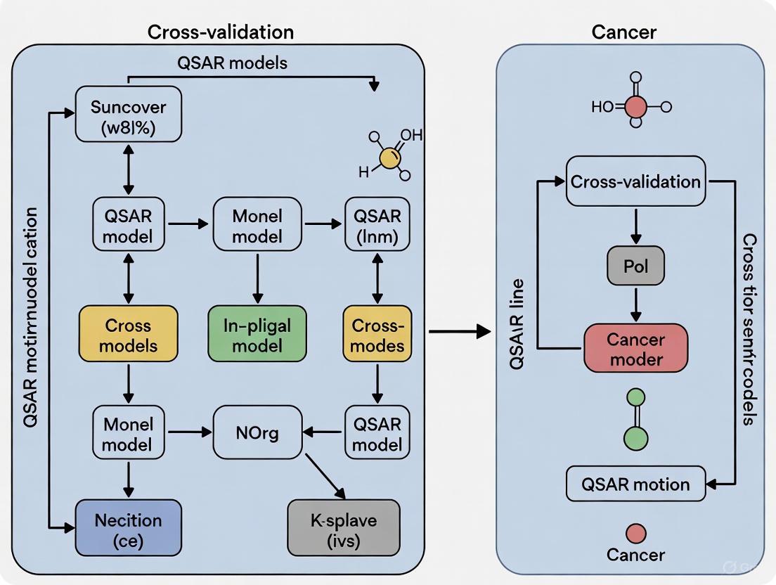
The following diagram illustrates the integrated workflow of QSAR model development and validation, culminating in experimental testing.
The Scientist's Toolkit: Essential Reagents & Software
This table details key resources used in the development and experimental validation of QSAR models, as featured in the cited research.
- Table 2: Essential Research Reagent Solutions for QSAR Studies
Item Name Function / Description Example Use in Protocol Alvadesc Software Calculates a wide range of molecular descriptors from chemical structures [14]. Phase 1, Step 2: Generating predictor variables (X) for the dataset. ChEMBL Database A large, open-access database of bioactive molecules with curated bioactivity data [14]. Phase 1, Step 1: Sourcing chemical structures and bioactivity values (Y). Multiple Linear Regression (MLR) A statistical algorithm used to construct a linear model between descriptors and activity [14] [13]. Phase 2, Step 5: Building the initial predictive model. MTT Assay Kit A colorimetric assay for assessing cell metabolic activity, used to determine cytotoxicity (IC₅₀) [14]. Phase 3, Step 8: Experimental validation of predicted compound activity on cancer cell lines. Molecular Docking Software Computationally simulates how a small molecule binds to a protein target [14]. Used to provide further in silico support for the model's predictions by analyzing binding modes.
Case Study: QSAR in Cancer Research for FGFR-1 Inhibitors
A recent study exemplifies the application of these core principles to a critical cancer target [14]. The study aimed to develop a predictive QSAR model for inhibitors of Fibroblast Growth Factor Receptor 1 (FGFR-1), a target associated with lung and breast cancers.
Methodology:
- Data and Descriptors: A dataset of 1,779 compounds from ChEMBL was curated. Molecular descriptors were calculated using Alvadesc software and refined with feature selection [14].
- Model Construction: A Multiple Linear Regression (MLR) model was constructed, resulting in a robust model with a training set R² of 0.7869 [14].
- Validation: The model was rigorously validated through 10-fold cross-validation and an external test set (R² = 0.7413). Furthermore, the model's predictions were supported by molecular docking and dynamics simulations [14].
- Experimental Cross-Validation: The model identified Oleic acid as a promising inhibitor. In vitro experiments on A549 (lung cancer) and MCF-7 (breast cancer) cell lines using MTT, wound healing, and clonogenic assays confirmed significant anticancer activity and selectivity, with low cytotoxicity on normal cell lines [14]. This provided a strong correlation between predicted and observed pIC₅₀ values.
Key Descriptors and Biological Insight: The study demonstrated that descriptors quantifying properties like polarizability, van der Waals volume, and electronegativity were critical for predicting FGFR-1 inhibitory activity. This provides medicinal chemists with tangible guidance for optimizing molecular structures.
The core principles of QSAR—linking numerical descriptors to bioactivity within a defined chemical space—provide a powerful framework for rational drug design. The case study on FGFR-1 inhibitors highlights how a rigorously developed and validated model, following a structured protocol, can successfully guide the identification of novel anticancer agents. The integration of computational predictions with experimental validation remains the gold standard for establishing model credibility.
Future directions in QSAR involve the increasing use of deep learning which can automatically learn relevant features from molecular structures or images, potentially uncovering complex patterns beyond traditional descriptors [16]. Furthermore, methods like q-RASAR are emerging, which merge traditional QSAR with similarity-based read-across techniques to enhance predictive reliability [9]. As these methodologies mature, they will continue to refine the precision and accelerate the pace of anticancer drug discovery.
The development of anticancer drugs is a complex process, often hampered by tissue-specific biological responses that can limit the generalizability of computational models. Quantitative Structure-Activity Relationship (QSAR) modeling serves as a powerful computational tool in early drug discovery, predicting the biological activity of compounds from their chemical structure [4]. However, most existing QSAR studies target single cancer cell lines, creating a knowledge gap in understanding pan-cancer activity profiles [2]. This case study addresses this limitation by systematically developing and validating foundational QSAR models across three distinct carcinoma types: HepG2 (hepatocellular carcinoma), A549 (lung carcinoma), and MOLT-3 (T-lymphoblastic leukemia). The research is framed within a broader thesis on cross-validation methodologies for QSAR models, emphasizing the critical importance of model robustness and transferability across different cancer lineages.
Experimental Design and Methodologies
Cell Line Selection and Biological Rationale
The selection of these three specific cell lines provides a representative spectrum of human cancers, encompassing carcinomas derived from different tissue origins (liver, lung, and blood). This diversity is crucial for evaluating the broader applicability of QSAR models beyond a single cancer type.
- HepG2 (Hepatocellular Carcinoma): This cell line represents one of the most common types of liver cancer and is widely used in hepatotoxicity and anticancer drug screening studies [7] [17].
- A549 (Lung Carcinoma): A non-small cell lung cancer model, A549 is characterized by its expression of the KRAS mutation, making it a relevant model for a significant subset of lung cancers [7] [17].
- MOLT-3 (T-Lymphoblastic Leukemia): As a lymphoblastic leukemia cell line, MOLT-3 provides insights into blood-borne cancers, which often exhibit different drug sensitivity profiles compared to solid tumors [7] [17].
Cytotoxic Activity Assay Protocols
A standardized experimental protocol is essential for generating consistent and comparable bioactivity data across different cell lines. The methodologies cited below form the cornerstone for generating the activity data used in QSAR model construction.
Table 1: Standardized Assay Protocols for Cytotoxic Activity Measurement
| Cell Line | Assay Type | Culture Medium | Seeding Density | Incubation Time | Key Readout |
|---|---|---|---|---|---|
| HepG2, A549, MRC-5 | MTT Assay [7] | DMEM or Hamm's F12 + 10% FBS [7] | 5x10³ - 2x10⁴ cells/well [7] | 48 hours [7] | Absorbance at 550 nm [7] |
| MOLT-3 | XTT Assay [7] | RPMI-1640 + 10% FBS [7] | N/A (suspension culture) [7] | 48 hours [7] | Absorbance at 550 nm [7] |
| General Protocol | Data points are typically performed in replicates, and IC₅₀ values (concentration causing 50% growth inhibition) are calculated from dose-response curves. Compounds with IC₅₀ > 50 μM are often classified as non-cytotoxic [7]. |
Dataset Curation for QSAR Modeling
The integrity of a QSAR model is directly dependent on the quality of the input data. The following workflow outlines the critical steps in dataset preparation, from biological testing to model-ready data.
The process begins with experimental bioactivity testing (e.g., GI₅₀ or IC₅₀ determination) against the target cell lines [7] [4]. Data standardization follows, which may include removing duplicates and averaging replicate values [4]. Subsequently, chemical structures (often from canonical SMILES) are standardized using tools like ChemAxon Standardizer to ensure consistency [4]. The crucial step of molecular descriptor calculation then takes place, generating quantitative representations of the molecules using software such as Dragon or PaDEL [4] [5]. Finally, the dataset is split into training and test sets, typically in a 70:30 to 80:20 ratio, to allow for model building and subsequent validation [4].
QSAR Model Construction and Performance Analysis
Machine Learning Algorithms and Descriptor Selection
The construction of predictive QSAR models involves the careful selection of machine learning algorithms and relevant molecular descriptors.
- Algorithm Selection: Studies have successfully employed a range of algorithms. Random Forest (RF) has shown strong performance, with models achieving high Positive Predictive Values (PPV > 0.85) for melanoma cell lines [4]. Multiple Linear Regression (MLR) is also widely used, providing good predictive performance (R training set = 0.89-0.97) for naphthoquinone derivatives [7]. For more complex relationships, Deep Neural Networks (DNN) have demonstrated exceptional performance, achieving an R² of 0.94 in combinational therapy models for breast cancer [5].
- Descriptor Analysis: Not all descriptors contribute equally. Research analyzing 29 different cancer cell lines found that quantum chemical descriptors (e.g., charge, polarizability) are the most important class, followed by electrostatic, constitutional, and topological descriptors [2]. Specifically, for 1,4-naphthoquinones, polarizability (
MATS3p), van der Waals volume (GATS5v), and dipole moment were key features influencing anticancer activity [7]. It is often observed that models with 3 descriptors can be sufficient for good correlation, avoiding the overfitting associated with more complex models [2].
Comparative Model Performance Across Carcinoma Lines
The true test of a foundational model lies in its performance across diverse biological systems. The following table summarizes the predictive performance of QSAR models built for the HepG2, A549, and MOLT-3 cell lines, based on studies of triazole and naphthoquinone derivatives.
Table 2: Cross-Carcinoma QSAR Model Performance Comparison
| Cancer Cell Line | Cancer Type | Example Compound Series | Best Model Performance (Reported) | Key Influencing Descriptors/Features |
|---|---|---|---|---|
| HepG2 | Hepatocellular Carcinoma | 1,2,3-Triazoles [17] | RCV: 0.80, RMSECV: 0.34 [17] | nR=O, nR-CR, lipophilicity, steric properties [17] |
| A549 | Lung Carcinoma | 1,2,3-Triazoles [17] | RCV: 0.60, RMSECV: 0.45 [17] | nR=O, nR-CR, lipophilicity, steric properties [17] |
| MOLT-3 | T-Lymphoblastic Leukemia | 1,2,3-Triazoles [17] | RCV: 0.90, RMSECV: 0.21 [17] | nR=O, nR-CR, lipophilicity, steric properties [17] |
| All Three Lines | Diverse Carcinomas | 1,4-Naphthoquinones [7] | R training: 0.89-0.97, R testing: 0.78-0.92 [7] | Polarizability, van der Waals volume, dipole moment [7] |
Abbreviations: RCV: Cross-validated Correlation Coefficient; RMSECV: Root Mean Square Error of Cross-Validation.
The data reveals a critical finding: the predictive performance of a QSAR model is highly dependent on the specific cancer cell line. For instance, in the study of triazole derivatives, the model for the MOLT-3 leukemia cell line (RCV = 0.90) was significantly more robust than the model for the A549 lung cancer cell line (RCV = 0.60), despite using the same set of molecular descriptors [17]. This underscores the impact of cell line-specific biological contexts on compound activity.
Framework for QSAR Model Validation
Ensuring that a QSAR model is reliable and not the result of chance correlation requires a rigorous validation framework. The following diagram outlines the key components of this process.
This framework consists of four pillars. Internal validation assesses model stability, often through techniques like Leave-One-Out (LOO) or k-fold cross-validation, which calculates metrics like RCV and Q² [17]. External validation is the most crucial step, evaluating the model's predictive power on a completely independent set of compounds that were not used in model training [7] [4]. Y-Scrambling is a sanity check where the model is rebuilt with randomly shuffled activity data; a significant drop in performance confirms the model is not based on chance correlation [4]. Finally, defining the Applicability Domain (AD) is essential to identify the region of chemical space where the model's predictions are reliable, preventing extrapolation beyond its scope [4].
The Scientist's Toolkit: Essential Research Reagents and Software
A successful QSAR modeling project relies on a suite of specialized software tools and reagents. The following table details key solutions used in the featured experiments and the broader field.
Table 3: Essential Research Reagent Solutions and Computational Tools
| Category | Item / Software | Primary Function | Specific Application in Case Studies |
|---|---|---|---|
| Bioassay Reagents | MTT/XTT Reagents [7] | Measure cell viability and proliferative inhibition. | Cytotoxic activity determination for adherent (MTT) and suspension (XTT) cell lines [7]. |
| Cell Culture | DMEM, RPMI-1640, F-12 Media [7] | Provide optimized nutrients and environment for specific cell line growth. | Culture of HepG2 (DMEM), MOLT-3 (RPMI-1640), and A549/HuCCA-1 (F-12) [7]. |
| Descriptor Calculation | Dragon [4] | Calculates thousands of molecular descriptors from chemical structures. | Used to generate 13 blocks of descriptors (e.g., topological, 2D-autocorrelation) for model building [4]. |
| Descriptor Calculation | PaDEL [18] [5] | An open-source alternative for calculating molecular descriptors and fingerprints. | Used in large-scale studies to compute fingerprints for classifying anticancer molecules [18]. |
| Cheminformatics | ChemAxon Standardizer [4] | Standardizes chemical structures (e.g., neutralization, aromatization) to ensure consistency. | Preprocessing of canonical SMILES from PubChem before descriptor calculation [4]. |
| Machine Learning | R (rminer, mlr packages) [4] | Statistical computing and environment for building and evaluating machine learning models. | Implementation of RF, SVM, and other classifiers for QSAR model development [4]. |
| Machine Learning | Python (Scikit-learn) [5] | A versatile programming language with extensive libraries for machine learning. | Used for developing DNN and other ML-based QSAR models [5]. |
| Chemical Visualization | SAMSON [19] | A molecular modeling platform with advanced visualization and color-coding capabilities. | Aiding in the interpretation of molecular properties and QSAR results through intuitive visual feedback [19]. |
This case study demonstrates a robust framework for building and validating foundational QSAR models across three biologically diverse carcinoma cell lines. The findings confirm that while shared molecular descriptors (e.g., polarizability, steric volume) can govern anticancer activity across cell lines, the predictive performance of a model is inherently context-dependent, varying significantly with the cellular environment [7] [17]. The cross-validation thesis underscores that models performing excellently on one cell line (e.g., MOLT-3) may show only moderate performance on another (e.g., A549), highlighting the danger of over-generalizing single-line models and the necessity for multi-target validation strategies.
Future research directions should focus on the development of hybrid models that integrate chemical descriptor data with genomic features of cancer cell lines to improve predictive accuracy and biological interpretability [18]. Furthermore, the adoption of advanced visualization tools like MolCompass for navigating chemical space and visually validating QSAR models will be crucial for identifying model weaknesses and "activity cliffs" [20] [21]. As the field moves towards the era of deep learning and Big Data, the combination of sophisticated algorithms, rigorous cross-carcinoma validation, and intuitive visual analytics will be paramount in accelerating the discovery of novel, broad-spectrum anticancer agents.
Identifying Key Molecular Descriptors Governing Anticancer Potency
Quantitative Structure-Activity Relationship (QSAR) modeling represents a fundamental computational approach in modern anticancer drug discovery, establishing mathematical relationships between chemical structures and their biological activity against cancer targets [22]. These models operate on the principle that variations in molecular structure quantitatively determine variations in biological properties, enabling researchers to predict the potency of novel compounds before synthesis and biological testing [22]. In the context of anticancer research, QSAR has emerged as an indispensable tool for accelerating the identification and optimization of lead compounds, ranging from natural product derivatives to synthetically designed molecules [23] [22].
The predictive capability of QSAR models hinges on molecular descriptors—quantitative representations of structural and chemical properties that serve as model inputs. These descriptors encompass topological, geometric, electronic, and physicochemical characteristics that numerically encode specific aspects of molecular structure [5]. With the integration of machine learning (ML) and deep learning (DL) algorithms, QSAR modeling has evolved into a more powerful and accurate predictive tool, capable of handling complex, high-dimensional data to identify subtle structure-activity patterns that might escape conventional analysis [23] [5]. This review comprehensively examines key molecular descriptors governing anticancer potency, their performance across diverse cancer models, and advanced methodological frameworks for model development and validation.
Molecular Descriptor Classes in Anticancer QSAR Models
Fundamental Descriptor Categories
Molecular descriptors utilized in anticancer QSAR modeling can be categorized into distinct classes based on the structural and chemical properties they represent. Quantum chemical descriptors, derived from quantum mechanical calculations, include electronic properties such as atomic charges, molecular orbital energies, and dipole moments that influence drug-receptor interactions [22]. Electrostatic descriptors characterize the charge distribution and potential fields around molecules, playing crucial roles in binding affinity [22]. Topological descriptors encode molecular connectivity patterns through graph-theoretical indices, while constitutional descriptors represent basic structural features like atom and bond counts [22]. Geometrical descriptors capture spatial molecular arrangements, and conceptual DFT descriptors theoretically describe chemical reactivity [22].
Performance Comparison of Descriptor Classes
A comprehensive analysis of QSAR models across 29 cancer cell lines revealed the relative importance and predictive performance of different descriptor classes [22]. Charge-based descriptors appeared in approximately 50% of significant models, valency-based descriptors in 36%, and bond order-based descriptors in 28% of models [22]. The study demonstrated that quantum chemical descriptors consistently provided the strongest predictive power for anticancer activity, followed by electrostatic, constitutional, geometrical, and topological descriptors [22]. Conceptual DFT descriptors showed limited improvement in statistical quality for most models despite their computational intensity [22].
Table 1: Performance of Molecular Descriptor Classes in Anticancer QSAR Models
| Descriptor Class | Representative Examples | Frequency in Significant Models | Key Applications in Cancer Research |
|---|---|---|---|
| Quantum Chemical | HOMO/LUMO energies, atomic charges, dipole moments | Highest | 20 out of 39 models (approx. 50%) [22] |
| Electrostatic | Partial charges, electrostatic potential surfaces | High | Charge-based descriptors most frequent [22] |
| Constitutional | Molecular weight, atom counts, bond counts | Moderate | Found in 36% of models [22] |
| Topological | Connectivity indices, path counts | Moderate | Molecular graph representations [24] |
| Geometrical | Molecular volume, surface area | Moderate | 3D spatial descriptors [22] |
| Conceptual DFT | Chemical potential, hardness | Lower | Limited improvement in most models [22] |
Key Molecular Descriptors Across Cancer Types
Descriptors for Specific Cancer Targets
Research has identified distinct descriptor profiles associated with anticancer potency against various cancer types. For colon cancer targeting HT-29 cell lines, hybrid descriptors combining SMILES notation and hydrogen-suppressed molecular graphs (HSG) demonstrated exceptional predictive capability in models developed using the Monte Carlo method [24]. These optimal descriptors achieved remarkable validation metrics (R² = 0.90, IIC = 0.81, Q² = 0.89) when applied to chalcone derivatives [24].
In breast cancer research, specifically against MCF-7 cell lines, machine learning-driven QSAR models for flavone derivatives identified critical descriptors including electronic properties and substituent characteristics that significantly influenced cytotoxicity [23]. Random Forest models achieved high predictive accuracy (R² = 0.820 for MCF-7, R² = 0.835 for HepG2) using these descriptor sets [23].
For leukemia targeting K562 cell lines, studies on C14-urea tetrandrine compounds revealed three key descriptors: AST4p (a 2D autocorrelation descriptor), GATS8v (Geary autocorrelation of lag 8 weighted by van der Waals volume), and MLFER (a molecular linear free energy relation descriptor) [25]. The resulting QSAR model showed strong predictive power with R²train = 0.910 and R²test = 0.644 [25].
Pan-Cancer Descriptor Analysis
Comparative analysis across multiple cancer types reveals both universal and context-dependent descriptor significance. A comprehensive study modeling 266 compounds against 29 different cancer cell lines found that optimal model performance typically required only 3-5 carefully selected descriptors, with additional descriptors providing diminishing returns [22]. The performance of descriptor classes varied significantly across cancer types, with models for nasopharyngeal cancer achieving the highest average R² values (0.90), followed by melanoma models (average R² = 0.81) [22].
Table 2: Key Molecular Descriptors and Their Predictive Performance Across Cancer Types
| Cancer Type | Cell Line/Model | Most Significant Descriptors | Predictive Performance (R²) |
|---|---|---|---|
| Colon Cancer | HT-29 | Hybrid SMILES-HSG descriptors [24] | R²_validation = 0.90 [24] |
| Breast Cancer | MCF-7 | Electronic, steric substituent descriptors [23] | R² = 0.820 [23] |
| Liver Cancer | HepG2 | SHAP-identified molecular descriptors [23] | R² = 0.835 [23] |
| Leukemia | K562 | AST4p, GATS8v, MLFER [25] | R²train = 0.910, R²test = 0.644 [25] |
| Nasopharyngeal | Multiple | Charge- and valency-based descriptors [22] | Average R² = 0.90 [22] |
| Melanoma | Multiple | Quantum chemical descriptors [22] | Average R² = 0.81 [22] |
Methodological Frameworks for Descriptor Identification
Machine Learning and Feature Selection Approaches
Modern QSAR methodologies employ sophisticated machine learning algorithms and feature selection techniques to identify the most relevant molecular descriptors. Genetic Function Algorithm (GFA) has proven effective for descriptor selection, as demonstrated in leukemia research where it identified three critical descriptors from a larger pool of potential variables [25]. Random Forest algorithms provide robust feature importance rankings through ensemble learning, successfully applied to flavone derivatives for breast and liver cancer models [23]. Deep Neural Networks (DNNs) have achieved exceptional performance (R² = 0.94) in combinational QSAR models for breast cancer, effectively capturing complex descriptor-activity relationships [5].
The Monte Carlo optimization method implemented in CORAL software offers an alternative approach using descriptor correlation weights (DCWs) derived from SMILES notation and molecular graphs [24]. This method has demonstrated particular utility for chalcone derivatives against colon cancer, with hybrid descriptors outperforming SMILES-only or graph-only approaches [24]. For combinatorial therapy prediction, studies have successfully calculated molecular descriptors for both anchor and library drugs using the Padelpy library, enabling the development of novel combinational QSAR models that account for drug interactions [5].
Validation Protocols and Applicability Domain
Robust validation frameworks are essential for establishing descriptor significance and model reliability. External validation using independent test sets remains the gold standard, with performance metrics including R²test, Q², and IIC (Index of Ideality of Correlation) [25] [24]. Cross-validation techniques (e.g., 5-fold cross-validation) assess model stability and prevent overfitting [23] [26]. Applicability domain (AD) analysis critically determines the structural space where models provide reliable predictions, addressing a fundamental limitation in QSAR modeling [27].
Recent research emphasizes that QSAR model reliability depends not only on statistical performance but also on transparent definition of applicability domains and chemical space coverage [27]. Studies investigating multiple QSAR models for carcinogenicity prediction have observed significant test-specificity and inconsistencies between models, highlighting the importance of applicability domain considerations and weight-of-evidence approaches when interpreting results [27].
Figure 1: Comprehensive QSAR model development workflow integrating descriptor calculation, machine learning, and rigorous validation protocols
Experimental Protocols and Computational Methodologies
Standard QSAR Development Pipeline
The development of robust QSAR models follows a systematic computational workflow beginning with data collection and curation. Researchers compile compound datasets with experimentally determined biological activities (e.g., IC₅₀ values) from reliable sources, ensuring structural diversity and activity range representation [24]. For anticancer applications, data typically originates from standardized assays like MTT assays measuring mitochondrial reduction in cancer cell lines [24].
Molecular structure optimization represents a critical preprocessing step, often performed using Density Functional Theory (DFT) methods at levels such as B3LYP/6-31G* to generate energetically minimized 3D structures [25]. Subsequent descriptor calculation employs specialized software including PaDEL, CORAL, or proprietary tools to generate comprehensive descriptor sets spanning multiple classes [25] [24] [5]. The resulting descriptor matrix undergoes feature selection using algorithms like Genetic Function Algorithm (GFA) or machine learning-based importance ranking to identify the most relevant variables [25].
Model building employs statistical or machine learning techniques including Multiple Linear Regression (MLR), Random Forest, or Deep Neural Networks to establish quantitative descriptor-activity relationships [23] [25] [5]. The final stage involves rigorous model validation through internal (cross-validation) and external (test set prediction) methods to assess predictive capability and generalizability [25] [24].
Advanced Integrative Approaches
Contemporary anticancer QSAR increasingly integrates complementary computational methods to enhance predictive accuracy and mechanistic understanding. Structure-based drug design combines QSAR with molecular docking to validate predicted activities through binding mode analysis, as demonstrated in studies targeting αβIII-tubulin isotype [26]. Molecular dynamics simulations provide additional validation of stability and interactions for predicted active compounds [26].
Combinational QSAR models represent a innovative extension, simultaneously modeling descriptor-activity relationships for drug pairs in combination therapies [5]. These approaches calculate molecular descriptors for both anchor and library drugs, then employ machine learning to predict combination effects across multiple cancer cell lines [5]. Multi-target QSAR models have also emerged for designing compounds against dual targets, such as HDAC/ROCK inhibitors for triple-negative breast cancer, incorporating both structure-based and ligand-based approaches [28].
Figure 2: Experimental QSAR validation protocol showing dataset division, model training, and comprehensive validation stages
Table 3: Essential Computational Tools and Resources for Anticancer QSAR Research
| Tool/Resource | Type | Primary Function | Application Examples |
|---|---|---|---|
| OECD QSAR Toolbox | Software | Chemical categorization, trend analysis, (Q)SAR model implementation [27] | Carcinogenicity prediction, hazard assessment [27] |
| Danish (Q)SAR Software | Online Database | Archive of model estimates from commercial, free, and DTU (Q)SAR models [27] | Carcinogenic potential prediction for pesticides and metabolites [27] |
| CORAL Software | Modeling Tool | QSAR modeling using Monte Carlo method with optimal descriptors [24] | SMILES-based QSAR for chalcone derivatives against colon cancer [24] |
| PaDEL-Descriptor | Descriptor Calculator | Calculation of molecular descriptors and fingerprints from chemical structures [25] [26] [5] | Generation of 797+ descriptors for machine learning-based QSAR [26] |
| AutoDock Vina | Docking Software | Structure-based virtual screening and binding affinity prediction [26] | Identification of natural inhibitors against αβIII-tubulin isotype [26] |
| GDSC2 Database | Data Resource | Drug sensitivity genomics data for cancer cell lines and drug combinations [5] | Combinational QSAR model development for breast cancer [5] |
| ZINC Database | Compound Library | Curated collection of commercially available compounds for virtual screening [26] | Natural compound screening for tubulin inhibitors [26] |
The identification of key molecular descriptors governing anticancer potency has evolved significantly with advances in computational power, machine learning algorithms, and multi-modal validation approaches. Quantum chemical descriptors consistently demonstrate superior predictive capability across diverse cancer types, while hybrid descriptor strategies that combine multiple representation methods often yield optimal performance [22] [24]. The emerging paradigm emphasizes context-specific descriptor relevance rather than universal solutions, with different descriptor combinations showing preferential performance for specific cancer types, molecular targets, and compound classes.
Future directions in descriptor research include the development of dynamic descriptors that capture conformational flexibility, integration of omics data with structural descriptors for systems pharmacology approaches, and application of explainable AI to elucidate complex descriptor-activity relationships. As QSAR modeling continues to integrate with structural biology, systems modeling, and clinical translation frameworks, refined molecular descriptor selection will remain fundamental to accelerating anticancer drug discovery and optimization. The cross-validation of descriptor significance across multiple cancer models and experimental systems represents a crucial strategy for developing robust, generalizable predictive models with genuine utility in therapeutic development.
Advanced Methodologies and Practical Applications in Anticancer QSAR
Machine Learning and Deep Learning Algorithms for Enhanced Predictions (RF, XGBoost, DNN)
The high failure rates and immense costs associated with traditional cancer drug development have necessitated more efficient and predictive approaches [29]. Quantitative Structure-Activity Relationship (QSAR) modeling provides a computational foundation for linking chemical structures to biological activity. The integration of modern machine learning (ML) and deep learning (DL) algorithms has significantly enhanced the predictive power and applicability of these models in oncology research [29] [30]. This guide objectively compares the performance of three prominent algorithms—Random Forest (RF), eXtreme Gradient Boosting (XGBoost), and Deep Neural Networks (DNN)—in the context of QSAR model cross-validation for different cancer cell lines.
Performance Comparison of ML/DL Algorithms in Cancer Research
The selection of an appropriate algorithm is critical for building robust predictive models in computational oncology. The table below summarizes the documented performance of RF, XGBoost, and DNN across various cancer research tasks.
Table 1: Comparative Performance of Machine Learning and Deep Learning Algorithms in Cancer Research
| Algorithm | Research Task / Target | Cancer Type | Key Performance Metrics | Reference / Context |
|---|---|---|---|---|
| Random Forest (RF) | Tankyrase (TNKS2) inhibitor classification | Colorectal Cancer | ROC-AUC: 0.98 | [31] |
| Random Forest (RF) | KRAS inhibitor pIC50 prediction | Lung Cancer | R²: 0.796 (on test set) | [6] |
| XGBoost | KRAS inhibitor pIC50 prediction | Lung Cancer | Performance was below PLS and RF | [6] |
| LightGBM (XGBoost variant) | Anticancer ligand prediction (ACLPred) | Pan-Cancer | Accuracy: 90.33%, AUROC: 97.31% | [30] |
| Deep Neural Network (DNN) | Nanoparticle tumor delivery efficiency (DE) prediction | Pan-Cancer (Multiple) | R² (Test set): 0.41 (Tumor), 0.87 (Lung) | [32] |
| PLS Regression | KRAS inhibitor pIC50 prediction | Lung Cancer | R²: 0.851, RMSE: 0.292 (Best in study) | [6] |
Key Performance Insights
- Random Forest (RF) demonstrates exceptional performance in classification tasks, as seen in its near-perfect AUC for identifying tankyrase inhibitors [31]. It also shows robust performance in regression tasks like pIC50 prediction [6].
- XGBoost/LightGBM excels in complex classification and ranking problems. The LightGBM model in ACLPred achieved high accuracy and AUROC, indicating its strength in distinguishing active anticancer compounds from inactive ones [30].
- Deep Neural Networks (DNN) offer advantages in modeling complex, non-linear relationships, such as predicting the biodistribution of nanoparticles. Their performance can vary significantly across different tissues, highlighting the context-dependency of model efficacy [32].
- No Single Best Algorithm: The optimal algorithm is highly dependent on the specific research question, data type, and dataset size. For instance, in a direct comparison for KRAS inhibitor prediction, simpler models like PLS and RF outperformed XGBoost [6].
Experimental Protocols for Model Development and Validation
A rigorous, standardized protocol is essential for developing reliable and interpretable QSAR models. The following workflow, synthesized from multiple studies [31] [6] [30], outlines the critical steps.
Diagram 1: Workflow for Robust QSAR Model Development
Detailed Methodological Breakdown
Data Curation and Preprocessing
The foundation of a reliable model is high-quality data. Standard protocols involve:
- Database Sourcing: Publicly accessible databases like ChEMBL and PubChem BioAssay are primary sources for experimentally validated bioactive molecules and their inhibitory concentrations (e.g., IC50) [31] [6] [30].
- Data Standardization: This includes removing duplicates and standardizing molecular representations (e.g., SMILE strings).
- Activity Conversion: IC50 values (nM) are typically converted to pIC50 (-log10(IC50 × 10⁻⁹)) for regression modeling, providing a more normalized scale for analysis [6].
- Similarity Filtering: To prevent model bias, compounds with high structural similarity (e.g., Tanimoto coefficient > 0.85) are often filtered out, ensuring a diverse and non-redundant dataset [30].
Feature Calculation and Selection
Molecular structures are translated into numerical descriptors that algorithms can process.
- Descriptor Calculation: Software like PaDELPy and RDKit are used to calculate thousands of 1D, 2D, and 3D molecular descriptors and fingerprints, encompassing topological, constitutional, geometrical, and electronic features [30].
- Multistep Feature Selection: To avoid overfitting and reduce computational load, a rigorous feature selection process is employed:
- Variance Filtering: Remove descriptors with near-zero variance (e.g., variance < 0.05) [30].
- Correlation Filtering: Eliminate highly correlated descriptors (e.g., |r| > 0.95) to reduce multicollinearity [6] [30].
- Advanced Selection: Algorithms like the Boruta (a random forest-based method) or Genetic Algorithms (GA) are used to identify the most statistically significant features for prediction [6] [30].
Model Training, Optimization, and Validation
This phase involves building, tuning, and critically assessing the models.
- Data Splitting: The curated dataset is split into a training set (e.g., 70%) for model building and a hold-out test set (e.g., 30%) for final evaluation [6] [33].
- Hyperparameter Tuning: Model hyperparameters (e.g., number of trees in RF, learning rate in XGBoost, layers in DNN) are optimized via techniques like cross-validation on the training set [31] [33].
- Validation Metrics: Models are evaluated using multiple metrics:
- Applicability Domain (AD): The model's domain of applicability is assessed using methods like Mahalanobis distance to identify compounds for which predictions are reliable [6].
- Model Interpretability: Tools like SHAP (SHapley Additive exPlanations) are increasingly used to interpret "black-box" models, quantifying the contribution of each molecular descriptor to the final prediction and providing biological insights [6] [30] [32].
Successful implementation of ML-driven QSAR projects relies on a suite of computational tools and data resources.
Table 2: Key Research Reagent Solutions for ML-based QSAR in Oncology
| Category | Item / Resource | Specific Examples & Functions |
|---|---|---|
| Bioactivity Databases | ChEMBL | A manually curated database of bioactive molecules with drug-like properties. Used to obtain experimentally determined IC50 values for targets like TNKS2 and KRAS [31] [6]. |
| PubChem BioAssay | A public repository containing biological activity data of small molecules. Serves as a source for active/inactive anticancer compounds for classification models [30]. | |
| Descriptor Calculation | PaDELPy | A software tool for calculating molecular descriptors and fingerprints. Used to generate 1D and 2D descriptors for ML model training [30]. |
| RDKit | An open-source cheminformatics toolkit. Used for descriptor calculation, fingerprint generation, and molecular operations [30]. | |
| Feature Selection | Boruta Algorithm | A random forest-based wrapper method for all-relevant feature selection, identifying descriptors statistically significant for prediction [30]. |
| Genetic Algorithm (GA) | An optimization technique used to select an optimal subset of molecular descriptors by mimicking natural selection [6]. | |
| Model Interpretation | SHAP (SHapley Additive exPlanations) | A unified framework for interpreting model predictions, explaining the output of any ML model by quantifying feature importance [6] [30] [32]. |
| Specialized Models | ACLPred | An open-source, LightGBM-based prediction tool for screening potential anticancer compounds, achieving high accuracy (90.33%) [30]. |
The cross-validation of QSAR models for different cancer cell lines is powerfully enhanced by machine and deep learning algorithms. Random Forest proves to be a robust and reliable choice for both classification and regression tasks. XGBoost and its variants (like LightGBM) can achieve state-of-the-art performance in complex classification problems, such as general anticancer ligand prediction. Deep Neural Networks show great potential for modeling intricate, non-linear phenomena like nanoparticle biodistribution. There is no universally superior algorithm; the optimal choice is problem-dependent. Therefore, researchers should employ a rigorous, multi-step workflow encompassing diligent data curation, rigorous feature selection, and thorough validation, including model interpretation with tools like SHAP, to develop predictive and trustworthy models that can accelerate oncology drug discovery.
Quantitative Structure-Activity Relationship (QSAR) modeling represents a cornerstone approach in rational drug design, enabling researchers to predict biological activity based on a compound's molecular structure. Over forty years have elapsed since Hansch and Fujita published their pioneering QSAR work, establishing a foundation that has evolved significantly with computational advances [34]. The fundamental premise of QSAR involves constructing mathematical models that correlate molecular descriptors (quantitative representations of structural features) with biological activity, creating predictive tools that guide structural optimization before resource-intensive chemical synthesis and biological testing.
Following the introduction of Comparative Molecular Field Analysis (CoMFA) by Cramer in 1998, numerous three-dimensional QSAR methodologies have emerged, greatly enhancing the field's predictive capabilities [34]. Currently, the integration of classical QSAR with advanced computational techniques represents the state-of-the-art in modern drug discovery. These models have proven indispensable not only for reliably predicting specific properties of new compounds but also for elucidating potential molecular mechanisms of receptor-ligand interactions [34]. Within oncology drug discovery, QSAR approaches provide a systematic framework for optimizing chemotherapeutic agents, particularly through cross-validation across different cancer cell lines to assess compound specificity and potential therapeutic windows.
QSAR Fundamentals and Molecular Descriptors
Core Principles and Workflow
QSAR modeling transforms chemical structural information into quantifiable parameters that can be statistically correlated with biological responses. The standard QSAR development workflow involves: (1) compound selection and dataset curation, (2) molecular structure representation and optimization, (3) molecular descriptor calculation, (4) statistical model development correlating descriptors with biological activity, (5) model validation, and (6) model application for predicting new compounds. The predictive performance of QSAR models relies heavily on appropriate descriptor selection and robust statistical methodologies.
Essential Molecular Descriptor Classes
Molecular descriptors quantitatively characterize aspects of molecular structure that influence biological activity and physicochemical properties. Key descriptor categories include:
- Electronic descriptors: Quantify electron distribution characteristics affecting molecular interactions (e.g., electronegativity, dipole moment) [13]
- Steric descriptors: Characterize molecular size, shape, and volume (e.g., van der Waals volume, molar refractivity) [13]
- Geometric descriptors: Describe three-dimensional molecular dimensions and arrangements
- Topological descriptors: Encode molecular connectivity patterns and branching characteristics
- Polarizability parameters: Measure how easily electron clouds distort under external fields (e.g., MATS3p) [13]
Table 1: Key Molecular Descriptor Categories in QSAR Modeling
| Descriptor Category | Representative Descriptors | Structural Properties Characterized |
|---|---|---|
| Electronic | Dipole moment, E1e, EEig15d | Charge distribution, electronegativity, molecular polarity |
| Steric | GATS5v, GATS6v, Mor16v | van der Waals volume, molecular size and bulk |
| Topological | RCI, SHP2 | Ring complexity, molecular shape, branching patterns |
| Polarizability | MATS3p, BELp8 | Electron cloud distortion potential |
Case Study: QSAR-Guided Optimization of Anticancer Flavones
Experimental Protocol and Compound Design
A recent investigation applied machine learning-driven QSAR modeling to optimize flavones, recognized as "privileged scaffolds" in drug discovery [23]. Researchers initially employed pharmacophore modeling against diverse cancer targets to design 89 flavone analogs featuring varied substitution patterns. These compounds were subsequently synthesized and evaluated biologically to identify promising candidates with enhanced cytotoxicity against breast cancer (MCF-7) and liver cancer (HepG2) cell lines, alongside low toxicity toward normal Vero cells [23].
The experimental protocol followed this standardized approach:
- Rational Design: Computational pharmacophore modeling identified key structural motifs for target interaction
- Chemical Synthesis: Preparation of 89 flavone analogs with systematic substitution variations
- Biological Evaluation:
- Cytotoxicity assessment against MCF-7 and HepG2 cancer cell lines
- Selectivity profiling using normal Vero cells
- Dose-response measurements for IC50 determination
- QSAR Model Development:
- Molecular descriptor calculation for all synthesized compounds
- Machine learning algorithm implementation (RF, XGBoost, ANN)
- Model training and validation using cross-validation techniques
- Model Interpretation: SHapley Additive exPlanations (SHAP) analysis to identify critical structural features
Machine Learning Model Performance
The research team compared multiple machine learning algorithms for QSAR model development, with the Random Forest (RF) model demonstrating superior performance [23]. The RF model achieved R² values of 0.820 for MCF-7 and 0.835 for HepG2, with cross-validation R² (R²cv) of 0.744 and 0.770, respectively. External validation using 27 test compounds yielded root mean square error test values of 0.573 (MCF-7) and 0.563 (HepG2), confirming model robustness and predictive capability [23].
Table 2: Machine Learning QSAR Model Performance for Anticancer Flavones
| Machine Learning Algorithm | R² (MCF-7) | R² (HepG2) | R²cv (MCF-7) | R²cv (HepG2) | RMSE Test (MCF-7) | RMSE Test (HepG2) |
|---|---|---|---|---|---|---|
| Random Forest | 0.820 | 0.835 | 0.744 | 0.770 | 0.573 | 0.563 |
| Extreme Gradient Boosting | Performance data not specified in source | |||||
| Artificial Neural Network | Performance data not specified in source |
Key Structural Features Influencing Anticancer Activity
SHAP analysis identified critical molecular descriptors significantly influencing flavone anticancer activity [23]. These descriptors provide concrete guidance for structural modifications:
- Electron density distribution at specific molecular positions
- Steric bulk and substitution patterns affecting target binding
- Hydrophobic character optimizing membrane permeability
- Hydrogen bonding capacity facilitating specific molecular interactions
- Molecular polarizability influencing binding affinity
Case Study: QSAR Modeling of 1,4-Naphthoquinone Anticancer Agents
Experimental Methodology
Another research effort explored substituted 1,4-naphthoquinones for potential anticancer therapeutics, investigating a series of 14 compounds (1-14) against four cancer cell lines: HepG2 (liver), HuCCA-1 (bile duct), A549 (lung), and MOLT-3 (leukemia) [13]. Compound 11 emerged as the most potent and selective anticancer agent across all tested cell lines (IC₅₀ = 0.15 – 1.55 μM, selectivity index = 4.14 – 43.57) [13].
The research methodology encompassed:
- Compound Screening: Cytotoxicity evaluation against four cancer cell lines
- Selectivity Assessment: Comparison with normal cell toxicity
- QSAR Model Construction: Four models developed using multiple linear regression (MLR) algorithm
- Model Validation: Rigorous training and testing set validation
- Virtual Expansion: QSAR model application to 248 structurally modified compounds
- ADMET Profiling: Prediction of pharmacokinetic properties and potential biological targets
QSAR Model Performance and Structural Insights
The four QSAR models demonstrated excellent predictive performance with correlation coefficients (R) for the training set ranging from 0.8928 to 0.9664 and testing set values between 0.7824 and 0.9157 [13]. RMSE values were 0.1755–0.2600 for training sets and 0.2726–0.3748 for testing sets, confirming model reliability [13].
The QSAR analysis revealed that potent anticancer activities of naphthoquinones were primarily influenced by:
- Polarizability (MATS3p and BELp8 descriptors)
- van der Waals volume (GATS5v, GATS6v, and Mor16v)
- Atomic mass (G1m)
- Electronegativity (E1e)
- Dipole moment (Dipole and EEig15d)
- Ring complexity (RCI)
- Molecular shape (SHP2) [13]
Table 3: Key Molecular Descriptors for Naphthoquinone Anticancer Activity
| Molecular Descriptor | Descriptor Category | Structural Interpretation | Biological Significance |
|---|---|---|---|
| MATS3p | Polarizability | Electron cloud distortion potential | Influences binding affinity |
| GATS5v, GATS6v | Steric | van der Waals volume | Affects target binding pocket fit |
| G1m | Mass | Atomic mass distribution | Impacts pharmacokinetics |
| E1e | Electronic | Electronegativity | Affects hydrogen bonding capacity |
| Dipole | Electronic | Molecular dipole moment | Influences interaction orientation |
| RCI | Topological | Ring complexity | Affects molecular rigidity |
| SHP2 | Topological | Molecular shape | Determines target complementarity |
Advanced QSAR Methodologies and Machine Learning Approaches
Predictive Distributions and Model Validation
Modern QSAR implementation emphasizes comprehensive validation approaches, including predictive distributions that represent QSAR predictions as probability distributions across possible property values [11]. This advanced framework acknowledges that both experimental measurements and QSAR predictions contain inherent uncertainties that should be quantitatively assessed.
The Kullback-Leibler (KL) divergence framework provides an information-theoretic approach for evaluating predictive distributions, measuring the distance between experimental measurement distributions and model predictive distributions [11]. This method enables more nuanced model assessment by simultaneously evaluating prediction accuracy and uncertainty estimation, addressing a critical need in pharmaceutical applications where understanding prediction reliability directly impacts decision-making in compound selection and prioritization.
Cross-Validation Across Cancer Cell Lines
Cross-validation represents an essential component of robust QSAR model development, particularly in anticancer drug discovery where compound specificity across different cancer types is both a practical concern and an opportunity for therapeutic optimization. The case studies demonstrate this approach through:
- Multiple cell line screening: Both flavones and naphthoquinones were evaluated across distinct cancer cell types
- Model transferability assessment: Determining whether structural features important for activity against one cancer type generalize to others
- Selectivity profiling: Identifying structural modifications that enhance cancer cell specificity while reducing normal cell toxicity
The Scientist's Toolkit: Essential Research Reagents and Materials
Table 4: Essential Research Reagents for QSAR-Guided Anticancer Drug Discovery
| Reagent/Material | Specification | Research Function |
|---|---|---|
| Cancer Cell Lines | MCF-7, HepG2, A549, HuCCA-1, MOLT-3 | In vitro models for cytotoxicity assessment and selectivity profiling |
| Normal Cell Lines | Vero cells | Control for determining selective toxicity and therapeutic index |
| Chemical Scaffolds | Flavone, 1,4-naphthoquinone core structures | Privileged structures for structural modification and optimization |
| Molecular Descriptor Software | Various computational packages | Calculation of electronic, steric, and topological descriptors |
| Machine Learning Platforms | Random Forest, XGBoost, ANN algorithms | QSAR model development and validation |
| ADMET Prediction Tools | In silico platforms | Virtual assessment of pharmacokinetics and toxicity profiles |
Visualizing QSAR-Guided Drug Design Workflows
QSAR-Guided Drug Design Workflow: This diagram illustrates the iterative process of rational drug design using QSAR modeling, highlighting the continuous cycle of synthesis, testing, modeling, and structural refinement that leads to optimized lead compounds.
Machine Learning Approaches in QSAR: This diagram compares machine learning methodologies used in modern QSAR modeling, demonstrating how different algorithms contribute to compound activity prediction and optimization prioritization.
QSAR modeling continues to evolve as an indispensable tool in rational anticancer drug design, successfully bridging computational predictions and experimental validation. The integration of machine learning algorithms has significantly enhanced predictive accuracy, enabling more reliable compound prioritization before synthesis. The cross-validation of QSAR models across multiple cancer cell lines provides critical insights into structural features governing both potency and selectivity, ultimately contributing to improved therapeutic indices. As demonstrated in the flavone and naphthoquinone case studies, QSAR-guided structural modifications systematically optimize critical molecular descriptors including polarizability, steric volume, electronic properties, and molecular shape. These approaches collectively advance the development of targeted anticancer therapeutics with enhanced efficacy and reduced toxicity profiles.
Quantitative Structure-Activity Relationship (QSAR) modeling represents a fundamental computational approach in ligand-based drug discovery that mathematically correlates a chemical compound's molecular structure with its biological activity [35] [36]. In breast cancer research, QSAR models have evolved from predicting activity of single drugs to complex combinational therapies, addressing the heterogeneous nature of this prevalent malignancy [5] [1]. Breast cancer remains the most common and heterogeneous form of cancer affecting women worldwide, with an estimated 300,590 new cases and 43,700 deaths annually in the United States alone [5]. The limitations of monotherapies and the emergence of drug resistance have accelerated research into combinational approaches, creating an urgent need for predictive computational models that can efficiently screen drug pairs for synergistic effects [5] [37].
This guide objectively compares the performance of various machine learning (ML) and deep learning (DL) algorithms in developing combinational QSAR models for breast cancer therapy, with particular emphasis on cross-validation methodologies essential for ensuring model reliability across different cancer cell lines. By examining experimental protocols, performance metrics, and implementation requirements, we provide researchers with a comprehensive framework for selecting appropriate modeling strategies in anti-cancer drug discovery.
Theoretical Foundation: Combinational QSAR Principles
Core Concepts in Combinational QSAR
Traditional QSAR modeling establishes relationships between molecular descriptors of single compounds and their biological activity, typically expressed as IC50 values (the concentration required for 50% inhibition) [35]. Combinational QSAR extends this principle to drug pairs, incorporating the concept of anchor drugs—well-established primary therapeutic agents with known efficacy for specific targets—and library drugs—supplementary compounds that enhance anchor drug efficacy and broaden the therapeutic approach [5] [1].
The fundamental equation for QSAR modeling can be represented as:
Biological Activity = f(Molecular Descriptors) + ε
Where molecular descriptors quantitatively represent structural, topological, geometric, electronic, and physicochemical characteristics of the compounds, and ε represents the error term not explained by the model [36]. In combinational QSAR, this relationship expands to incorporate descriptors from both anchor and library drugs, plus interaction terms that capture synergistic effects [5].
Molecular Descriptors and Feature Representation
Molecular descriptors serve as the predictive variables in QSAR models and can be categorized into several classes:
- Constitutional descriptors: Molecular weight, number of atoms, bonds, or rings
- Topological descriptors: Indices derived from molecular graph representation
- Geometric descriptors: 3D spatial molecular parameters
- Electronic descriptors: Electronegativity, polarizability, HOMO/LUMO energies
- Physicochemical descriptors: logP, solubility, molar refractivity [36]
In combinational QSAR, descriptors are calculated for both drugs separately, then combined through mathematical operations (e.g., averaging, summing, or more complex functions) to create interaction terms that potentially capture synergistic effects [5].
Experimental Protocols and Methodologies
Data Collection and Preprocessing
The foundational dataset for combinational QSAR development typically originates from large-scale drug sensitivity databases. The GDSC2 (Genomics of Drug Sensitivity in Cancer) combinations database provides breast cancer-specific data comprising 52 cell lines, 25 anchor drugs, and 51 library drugs, with combinational biological activity (Combo IC50) as the primary target variable [5] [1].
Data preprocessing workflow:
- Molecular descriptor calculation: Using software tools like PaDEL-Descriptor in Python to compute descriptors for both anchor and library drugs [5]
- Dimensionality reduction: Application of Principal Component Analysis (PCA) to reduce noise while retaining 95% of explained variance [5] [1]
- Distribution normalization: Addressing outliers through Boxcox, yeojohnsons, and logarithmic transformations to ensure normal distribution [5]
- Data encoding and standardization: Using Scikit-learn library in Python for data encoding and standardization [5]
- Dataset partitioning: Splitting data into training, testing, and validation sets in a 60:20:20 ratio [5] [1]
Algorithm Implementation and Training
Eleven regression-based machine learning and deep learning algorithms are commonly implemented for combinational QSAR model development [5] [1]:
Figure 1: Combinational QSAR Model Development Workflow
Implementation details:
- Programming environment: Python v3.12.0 with Scikit-learn library [5]
- Hyperparameter optimization: Grid search or random search with cross-validation [5]
- Training configuration: Deep Neural Networks typically require more epochs (100-500) compared to traditional ML algorithms [5]
- Validation framework: k-fold cross-validation (typically k=5 or k=10) during training, with hold-out validation set for final evaluation [5] [1]
Performance Comparison of QSAR Modeling Algorithms
Quantitative Performance Metrics
Table 1: Performance Comparison of Machine Learning Algorithms in Combinational QSAR Modeling [5] [1]
| Algorithm | R² (Coefficient of Determination) | RMSE (Root Mean Square Error) | MAE (Mean Absolute Error) | Training Speed | Interpretability |
|---|---|---|---|---|---|
| Deep Neural Networks (DNN) | 0.94 | 0.255 | 0.198 | Slow | Low |
| Random Forest (RF) | 0.89 | 0.301 | 0.235 | Medium | Medium |
| Extra Gradient Boost (XGB) | 0.88 | 0.315 | 0.241 | Medium | Medium |
| Support Vector Regressor (rbf-SVR) | 0.86 | 0.332 | 0.258 | Slow | Low |
| Wider Neural Network (WNN) | 0.85 | 0.341 | 0.263 | Slow | Low |
| k-Nearest Neighbours (kNN) | 0.82 | 0.368 | 0.285 | Fast | High |
| LASSO Regression | 0.79 | 0.392 | 0.301 | Fast | High |
| Ridge Regression | 0.78 | 0.401 | 0.312 | Fast | High |
| Elastic Net Regression | 0.77 | 0.408 | 0.319 | Fast | High |
| CART | 0.75 | 0.421 | 0.328 | Medium | High |
| Stochastic Gradient Descent Regressor (SGD) | 0.72 | 0.445 | 0.341 | Fast | Medium |
Algorithm-Specific Strengths and Limitations
Deep Neural Networks (DNN) demonstrated superior predictive performance (R² = 0.94) with strong generalization capabilities, making them particularly effective for capturing complex, non-linear relationships between molecular descriptors of drug pairs and their combined biological activity [5] [1]. However, DNNs require substantial computational resources, large datasets, and offer limited interpretability compared to simpler models.
Ensemble methods (Random Forest, XGBoost) provided a favorable balance between performance (R² = 0.89 and 0.88, respectively) and interpretability, with built-in feature importance metrics that help identify molecular descriptors most influential in predicting combinational efficacy [5].
Traditional linear models (LASSO, Ridge, Elastic Net) offered the advantages of high interpretability and computational efficiency, making them valuable for initial feature selection and baseline model establishment, though with reduced predictive power (R² = 0.77-0.79) compared to more complex algorithms [5].
Cross-Validation Strategies for Robust QSAR Models
Validation Methodologies in QSAR Development
Table 2: Cross-Validation Methods for QSAR Model Assessment [38] [36]
| Validation Method | Procedure | Advantages | Limitations |
|---|---|---|---|
| Train-Test Split | Single split (typically 70-80% training, 20-30% test) | Simple implementation, computational efficiency | High variance based on single split, may not represent full chemical space |
| k-Fold Cross-Validation | Data divided into k subsets; each subset serves as test set once | More reliable performance estimate, uses all data | Computational intensity, potential selection bias |
| Leave-One-Out (LOO) CV | Each compound serves as test set once | Maximum training data usage, low bias | High computational cost, high variance with outliers |
| External Validation | Completely independent test set not used in model development | Most realistic performance estimation | Requires large datasets, independent test set availability |
| Double Cross-Validation | Outer loop for performance estimation, inner loop for parameter tuning | Unbiased performance estimation with parameter optimization | High computational complexity |
Critical Considerations in QSAR Validation
Reliable QSAR model development requires rigorous validation strategies. Research has demonstrated that employing the coefficient of determination (r²) alone is insufficient to indicate QSAR model validity [38]. The established criteria for external validation have specific advantages and disadvantages that must be considered in QSAR studies, and these methods alone may not be enough to conclusively indicate model validity or invalidity [38].
For combinational QSAR models specifically, scaffold-based splitting is recommended, where drug pairs sharing structural similarities are kept together in training or test sets to avoid overoptimistic performance estimates. Additionally, cell-line stratified splitting ensures that all cell lines are represented in both training and test sets, facilitating generalizability across different biological contexts [5].
Research Reagent Solutions for Combinational QSAR
Table 3: Essential Research Reagents and Computational Tools for Combinational QSAR [5] [37] [36]
| Resource Category | Specific Tools/Resources | Primary Function | Application in Combinational QSAR |
|---|---|---|---|
| Bioactivity Databases | GDSC2 Combinations Database | Provides experimental combinational drug screening data | Source for anchor/library drug pairs and Combo IC50 values [5] |
| Descriptor Calculation | PaDEL-Descriptor, Dragon, RDKit | Computes molecular descriptors from chemical structures | Generation of predictive variables for QSAR models [5] [36] |
| Machine Learning Frameworks | Scikit-learn, TensorFlow, PyTorch | Implementation of ML/DL algorithms | Model development and training [5] [1] |
| Chemical Modeling | CORAL Software, Monte Carlo Optimization | Builds QSAR models using SMILES and graph-based descriptors | Alternative approach using balance of correlation techniques [39] |
| Validation Tools | Custom Python/R scripts | Statistical validation and performance metrics | Implementation of cross-validation and external validation [38] |
| Specialized Libraries | Padelpy, MolVS, OpenBabel | Chemical standardization and descriptor calculation | Data preprocessing and cheminformatics operations [5] |
Comparative Analysis of QSAR Approaches in Breast Cancer Research
Figure 2: Evolution of QSAR Modeling Approaches in Breast Cancer Research
Beyond combinational QSAR, several alternative modeling approaches have been developed for breast cancer drug discovery:
Monte Carlo-based QSAR utilizes CORAL software with Simplified Molecular Input Line Entry System (SMILES) notations and molecular hydrogen-suppressed graphs (HSG) to build predictive models. This approach has demonstrated effectiveness in predicting anti-breast cancer activity of naphthoquinone derivatives, with selected compounds showing stable interactions with topoisomerase IIα in molecular dynamics simulations spanning 300 ns [39].
Traditional single-drug QSAR continues to provide value, particularly in early-stage drug discovery. Recent studies on indolyl-methylidene phenylsulfonylhydrazones revealed selective cytotoxicity against MCF-7 cells (ER-α⁺), with compound 3b demonstrating the highest potency (IC50 = 4.0 μM) and a selectivity index of 20.975 [37]. Similarly, N-tosyl-indole based hydrazones showed promising activity against triple-negative breast cancer (TNBC) cell line MDA-MB-231, with compound 5p exhibiting an IC50 of 12.2 ± 0.4 μM [40].
Quantitative Structure-Property Relationship (QSPR) models using entire neighborhood topological indices have emerged for characterizing physicochemical properties of breast cancer drugs. These graph-based approaches compute topological indices from molecular structures to predict drug properties, offering complementary insights to traditional QSAR [41].
Implementation Guidelines and Best Practices
Algorithm Selection Framework
Choosing the appropriate algorithm for combinational QSAR depends on several factors:
- For maximum predictive accuracy: Deep Neural Networks consistently outperform other algorithms but require substantial computational resources and larger datasets (>10,000 data points recommended) [5] [1]
- For balanced performance and interpretability: Random Forest or XGBoost provide favorable accuracy with feature importance metrics [5]
- For limited computational resources or smaller datasets: LASSO or Ridge Regression offer computationally efficient baselines with high interpretability [5]
- For integration with experimental design: Monte Carlo-based approaches using CORAL software facilitate structural interpretation and guidance for chemical synthesis [39]
Validation and Applicability Domain Considerations
Robust QSAR implementation requires careful attention to model validation and applicability domain definition:
- Always use external validation with compounds not included in model development to obtain realistic performance estimates [38]
- Define applicability domains using approaches such as leverage analysis, distance-based methods, or PCA-based chemical space mapping to identify where models make reliable predictions [35] [36]
- Apply multiple validation metrics beyond R², including RMSE, MAE, and concordance correlation coefficients to comprehensively assess model performance [38]
- Implement y-randomization (scrambling response values) to confirm models don't capture chance correlations [39] [36]
Combinational QSAR modeling represents a significant advancement in computational approaches for breast cancer therapy development, directly addressing the clinical challenge of tumor heterogeneity and drug resistance. Through objective performance comparison, Deep Neural Networks emerge as the most predictive algorithm (R² = 0.94, RMSE = 0.255), though their implementation requires substantial computational resources and expertise [5] [1]. Ensemble methods like Random Forest and XGBoost offer favorable balances between predictive performance and interpretability, while traditional linear models provide computationally efficient baselines.
The integration of rigorous cross-validation strategies remains paramount, as R² alone proves insufficient for confirming model validity [38]. Future directions in combinational QSAR will likely involve multi-modal approaches integrating structural, genomic, and proteomic data, hybrid modeling combining machine learning with molecular simulations, and expanded applicability to in vivo systems. As these computational methods continue evolving, they promise to accelerate the identification of effective drug combinations while reducing development costs and experimental animal use in anti-cancer drug discovery.
Predicting Pharmacokinetic Profiles and Biological Targets with In-Silico Tools
The development of new cancer therapies remains a time-intensive and resource-heavy process, often requiring over a decade and billions of dollars to bring a single drug to market, with approximately 90% of oncology drugs failing during clinical development [42]. In this challenging landscape, in-silico tools have emerged as transformative technologies that accelerate drug discovery by predicting pharmacokinetic profiles and biological targets with increasing accuracy. These computational approaches leverage artificial intelligence (AI), machine learning (ML), and deep learning (DL) to process massive, multimodal datasets—from genomic profiles to clinical outcomes—generating predictive models that enhance target identification, compound optimization, and toxicity assessment [42] [43].
The broader context of cross-validation of Quantitative Structure-Activity Relationship (QSAR) models for different cancer cell lines research underscores the critical importance of robust validation frameworks in computational oncology. As these in-silico tools become more sophisticated, their integration into established research workflows enables more efficient and reliable drug discovery pipelines, particularly for molecularly complex diseases like cancer characterized by tumor heterogeneity and resistance mechanisms [42].
Foundational Concepts: QSAR and ADMET Prediction
Quantitative Structure-Activity Relationship (QSAR) Modeling
QSAR modeling establishes mathematical relationships between the chemical structure of compounds and their biological activity, creating predictive frameworks that can identify promising therapeutic candidates without exhaustive laboratory testing [44]. These computational models use molecular descriptors—quantitative representations of structural and physicochemical properties—to forecast bioactivity [31]. The "guilt-by-association" principle often underpins these approaches, assuming that structurally similar compounds are likely to exhibit similar biological activities [44].
In cancer research, QSAR modeling has been successfully applied to numerous molecular targets. For instance, researchers developed a predictive QSAR model for Fibroblast Growth Factor Receptor 1 (FGFR-1) inhibitors using a dataset of 1,779 compounds from the ChEMBL database. The model demonstrated strong predictive performance with an R² value of 0.7869 for the training set and 0.7413 for the test set, subsequently validated through in-vitro assays on cancer cell lines [14]. Similarly, a machine learning-assisted QSAR model for tankyrase inhibitors in colon adenocarcinoma achieved a high predictive performance (ROC-AUC of 0.98) through random forest classification and rigorous validation strategies [31].
ADMET Profiling
Absorption, Distribution, Metabolism, Excretion, and Toxicity (ADMET) profiling represents another crucial application of in-silico tools in drug discovery. Computational ADMET prediction enables researchers to evaluate key pharmacokinetic and safety parameters early in the development process, reducing late-stage failures due to unfavorable drug properties [45].
A comprehensive in-silico study evaluated ADMET profiles of 58 organic compounds using computational tools including SwissADME and PreADMET. The research established predictive models for toxicity, particularly LD₅₀ (lethal dose for 50% of subjects), using Random Forest regression (r² = 0.8410; RMSE = 0.1112), with five-fold cross-validation confirming robustness [45]. Such approaches facilitate early identification of compounds with favorable pharmacokinetic properties and selective inhibitory potential, supporting candidate selection for further experimental exploration [45].
Table 1: Key ADMET Parameters and Their Computational Prediction Approaches
| ADMET Parameter | Computational Prediction Method | Significance in Drug Discovery |
|---|---|---|
| Log P | SwissADME, PreADMET | Predicts lipophilicity and membrane permeability |
| Caco-2 Permeability | QSAR Models, Machine Learning | Indicates intestinal absorption potential |
| CYP450 Interactions | Molecular Docking, QSAR | Predicts drug metabolism and potential interactions |
| hERG Inhibition | Random Forest, Deep Learning | Assesses cardiotoxicity risk |
| LD₅₀ | Random Forest Regression | Estimates acute toxicity |
| DILI (Drug-Induced Liver Injury) | SwissADME, PreADMET | Predicts hepatotoxicity potential |
Comparative Analysis of In-Silico Tools and Methodologies
Tool Performance Across Different Applications
The landscape of in-silico tools for predicting pharmacokinetic profiles and biological targets encompasses diverse methodologies, each with distinct strengths and applications. The following table provides a comparative analysis of major computational approaches based on their primary functions, underlying algorithms, and performance characteristics.
Table 2: Comparative Performance of In-Silico Tools in Cancer Drug Discovery
| Tool/Method | Primary Function | Underlying Algorithm | Performance Metrics | Limitations |
|---|---|---|---|---|
| Molecular Docking | Drug-Target Interaction Prediction | Shape Complementarity, Scoring Functions | Binding Affinity Estimation | Dependent on protein 3D structures |
| QSAR Modeling | Bioactivity Prediction | MLR, Random Forest, Neural Networks | R²: 0.74-0.79 [14] | Limited to similar chemical spaces |
| Deep Learning (DGraphDTA) | Drug-Target Affinity Prediction | Graph Neural Networks | Improved Binding Affinity Prediction | Requires large training datasets |
| Random Forest ADMET | Toxicity Prediction | Ensemble Decision Trees | r²: 0.84, RMSE: 0.11 [45] | Dataset-dependent performance |
| Pharmacophore Modeling | Virtual Screening | 3D Chemical Feature Mapping | Enhanced Hit Identification | Limited to known active compounds |
| AI-Driven De Novo Design | Novel Compound Generation | VAEs, GANs, Reinforcement Learning | Reduced Discovery Timelines [43] | Synthetic accessibility challenges |
Cross-Cancer Lineage Validation of QSAR Models
A critical aspect of QSAR modeling in oncology involves validating predictive performance across different cancer cell lines, addressing the fundamental challenge of tumor heterogeneity. Successful cross-validation demonstrates model robustness and generalizability, essential for developing broadly effective cancer therapeutics.
In a study on liver cancer, researchers developed a statistically reliable QSAR model for Shikonin Oxime derivatives that identified structural features responsible for enhanced anticancer activity. The newly designed compounds exhibited improved inhibitory potential compared to the parent molecule, with molecular dynamics simulations confirming the stability of the ligand-receptor complexes [46]. Similarly, the previously mentioned FGFR-1 inhibitor model was validated across multiple cancer cell lines including A549 (lung cancer), MCF-7 (breast cancer), HEK-293 (normal human embryonic kidney), and VERO (normal African green monkey kidney) cell lines, confirming significant inhibitory effects on cancer cells with low cytotoxicity on normal cell lines [14].
These cross-validation approaches typically employ techniques such as 10-fold cross-validation, external validation with test sets, and experimental validation through biological assays including MTT, wound healing, and clonogenic assays [14]. The integration of computational predictions with experimental validation across diverse cellular contexts strengthens the reliability of QSAR models for oncology applications.
Experimental Protocols and Methodologies
Standard Workflow for QSAR Model Development
The development of robust QSAR models follows a systematic workflow that integrates computational and experimental components:
Data Curation and Preprocessing: Collect bioactivity data from databases like ChEMBL, including compounds with experimentally determined IC₅₀ values. For tankyrase inhibitor research, this involved retrieving 1,100 inhibitors from the ChEMBL database (Target ID: CHEMBL6125) [31].
Molecular Descriptor Calculation: Compute 2D and 3D structural and physicochemical molecular descriptors using software such as Alvadesc [14]. These descriptors quantitatively represent molecular properties relevant to biological activity.
Feature Selection: Apply feature selection techniques to identify the most relevant descriptors, reducing dimensionality and minimizing overfitting. This enhances model interpretability and predictive performance [31].
Model Training and Optimization: Implement machine learning algorithms such as multiple linear regression (MLR), random forest, or neural networks. Utilize internal cross-validation (e.g., 10-fold cross-validation) to optimize hyperparameters [14].
External Validation: Evaluate model performance on an independent test set not used during model training. Report metrics including R² for regression models or ROC-AUC for classification models [14] [31].
Experimental Validation: Conduct in-vitro assays such as MTT, wound healing, and clonogenic assays on relevant cancer cell lines to confirm predictive accuracy [14].
Integrated Computational Workflow for Target Identification and Validation
A comprehensive approach to identifying and validating biological targets combines multiple computational techniques:
Target Identification: Utilize AI to integrate multi-omics data (genomics, transcriptomics, proteomics) to uncover hidden patterns and identify promising targets. ML algorithms can detect oncogenic drivers in large-scale cancer genome databases such as The Cancer Genome Atlas (TCGA) [42].
Molecular Docking: Perform docking simulations to evaluate binding affinities and interaction patterns between candidate compounds and target proteins. Use software such as PyRx and Discovery Studio [45].
Molecular Dynamics Simulations: Conduct simulations to assess the stability and interaction dynamics of ligand-receptor complexes in physiological environments. Analyze root-mean-square deviation (RMSD) and other stability parameters [46] [31].
Pharmacokinetic Profiling: Predict ADMET properties using tools like SwissADME and PreADMET to evaluate drug-likeness and potential bioavailability [45].
Network Pharmacology: Contextualize targets within broader cancer biology by mapping disease-gene interactions and functional enrichment to uncover target-associated roles in oncogenic pathways [31].
Successful implementation of in-silico predictions requires integration with wet-lab experimental approaches. The following table details key research reagent solutions and computational resources essential for cross-validating QSAR models across different cancer cell lines.
Table 3: Essential Research Reagent Solutions for Experimental Validation
| Resource Category | Specific Examples | Function in Validation | Application Context |
|---|---|---|---|
| Cancer Cell Lines | A549 (lung), MCF-7 (breast), COLO-320 DM (colon) | In-vitro validation of predicted bioactivity | Cross-cancer lineage validation [14] [31] |
| Cell-Based Assays | MTT, wound healing, clonogenic assays | Quantify inhibitory effects and cell viability | Functional validation of lead compounds [14] |
| Bioactivity Databases | ChEMBL, PubChem, TCGA | Source of training data and compound structures | Model development and virtual screening [14] [31] [47] |
| Molecular Modeling Software | Alvadesc, PyRx, Discovery Studio | Descriptor calculation, docking simulations | Structural analysis and binding prediction [14] [45] |
| ADMET Prediction Platforms | SwissADME, PreADMET | Pharmacokinetic and toxicity profiling | Early safety and bioavailability assessment [45] |
| Omics Databases | The Cancer Genome Atlas (TCGA), Gene Expression Omnibus (GEO) | Multi-omics data for target identification | AI-driven target discovery [42] [47] |
The integration of in-silico tools for predicting pharmacokinetic profiles and biological targets represents a paradigm shift in oncology drug discovery. AI and ML technologies are increasingly being applied across the drug development pipeline, from target identification to clinical trial optimization, offering dramatic improvements in speed, cost-efficiency, and predictive power [43]. The emergence of AI-designed molecules like DSP-1181, which entered clinical trials in less than a year—compared to the typical 4-5 years—exemplifies this transformative potential [42].
Future directions in the field point toward more sophisticated integrative approaches. Multi-modal AI capable of integrating genomic, imaging, and clinical data promises more holistic insights, while digital twins of patients, simulated through AI models, may allow virtual testing of drugs before actual clinical trials [42]. Federated learning approaches, which train models across multiple institutions without sharing raw data, can overcome privacy barriers and enhance data diversity [42]. Additionally, the integration of large language models and AlphaFold-predicted protein structures is advancing feature engineering for drug-target interaction prediction [44].
In conclusion, in-silico tools for predicting pharmacokinetic profiles and biological targets have matured into indispensable components of modern oncology drug discovery. When developed with rigorous cross-validation across different cancer cell lines and integrated with experimental validation, these computational approaches significantly enhance the efficiency and success rate of identifying viable therapeutic candidates. As these technologies continue to evolve, they will play an increasingly central role in delivering personalized, effective cancer therapies to patients.
Troubleshooting and Optimizing QSAR Models for Robust Performance
Navigating Data Heterogeneity and Challenges in Cytotoxicity Modeling
In the field of computational drug discovery, cytotoxicity modeling represents a critical tool for predicting the adverse effects of chemical compounds on living cells. The development of robust Quantitative Structure-Activity Relationship (QSAR) models provides a cost-effective strategy for identifying promising candidate molecules while filtering out those with potential toxicity issues. However, the predictive accuracy and reliability of these models face a fundamental challenge: data heterogeneity. This term encompasses the substantial variations in toxicity responses observed across different biological systems, including diverse cell lines, experimental conditions, and measurement protocols [48]. The cytotoxicity of a compound is not an intrinsic property but rather a context-dependent phenomenon, influenced by cellular origin, physiological characteristics, and specific laboratory methodologies [7]. This article objectively compares the performance of QSAR modeling approaches when applied across different cancer cell lines, examining the experimental data and methodologies that both highlight the challenges and pave the way for potential solutions, framed within the broader thesis of cross-validation needs in computational toxicology.
Comparative Performance Across Cancer Cell Lines
Empirical Evidence of Model Performance Variation
The performance of QSAR models is highly dependent on the specific cell line for which they are developed, directly illustrating the impact of biological context on predictive accuracy. Research on 1,4-naphthoquinone derivatives demonstrates this variability clearly, where distinct QSAR models were required for different cancer cell lines, each exhibiting unique performance metrics and relying on different molecular descriptors [7]. The table below summarizes the performance of these cell-line-specific QSAR models built using multiple linear regression (MLR).
Table 1: Performance Metrics of QSAR Models for Different Cancer Cell Lines [7]
| Cancer Cell Line | R Training Set | R Testing Set | RMSE Training Set | RMSE Testing Set |
|---|---|---|---|---|
| HepG2 (Liver) | 0.8928 | 0.7824 | 0.2600 | 0.3748 |
| HuCCA-1 (Bile Duct) | 0.9664 | 0.9157 | 0.1755 | 0.2726 |
| A549 (Lung) | 0.9446 | 0.8716 | 0.2186 | 0.3279 |
| MOLT-3 (Blood) | 0.9498 | 0.8474 | 0.2268 | 0.3472 |
Cytotoxicity Profiles of 1,4-Naphthoquinone Derivatives
The experimental cytotoxicity data used to build the QSAR models further underscores the concept of data heterogeneity. Compound 11 from the naphthoquinone series emerged as the most potent and selective anticancer agent, but its effectiveness varied significantly across the different cell lines [7]. This variation highlights that a compound's cytotoxic profile is not absolute but relative to the biological context.
Table 2: Experimental Cytotoxicity Data (IC50 in μM) for Select 1,4-Naphthoquinone Compounds [7]
| Compound | HepG2 | HuCCA-1 | A549 | MOLT-3 | MRC-5 (Normal) |
|---|---|---|---|---|---|
| 1 | 17.48 | 19.61 | 23.90 | 8.27 | 27.47 |
| 5 | 2.44 | 3.34 | 4.56 | 1.66 | 6.19 |
| 11 | 1.55 | 0.15 | 0.68 | 0.27 | 6.57 |
| 14 | 25.92 | 19.84 | 23.12 | 7.55 | 31.42 |
| Doxorubicin (Control) | 1.27 | 1.91 | 2.21 | 0.48 | 2.18 |
Key Structural Descriptors in Cytotoxicity Models
The molecular descriptors that govern cytotoxic potency also vary by cell line, suggesting differences in the underlying mechanisms of action or cellular uptake in different biological contexts. The QSAR models for naphthoquinones identified distinct sets of critical descriptors for each cell line [7].
Table 3: Key Molecular Descriptors Influencing Cytotoxicity in Different Cell Lines [7]
| Cancer Cell Line | Critical Molecular Descriptors | Descriptor Interpretation |
|---|---|---|
| HepG2 | MATS3p, GATS5v, G1m, E1e, Dipole, RCI | Polarizability, van der Waals volume, mass, electronegativity, dipole moment, ring complexity |
| HuCCA-1 | BELp8, GATS6v, EEig15d, SHP2 | Polarizability, van der Waals volume, dipole moment, molecular shape |
| A549 | MATS3p, Mor16v, G1m, E1e, RCI | Polarizability, van der Waals volume, mass, electronegativity, ring complexity |
| MOLT-3 | GATS5v, BELp8, Mor16v, SHP2 | van der Waals volume, polarizability, molecular shape |
Experimental Protocols for Cytotoxicity Assessment
Cell Culture and Maintenance Protocols
Standardized cell culture protocols are fundamental to generating reproducible cytotoxicity data, yet variations in these protocols contribute significantly to data heterogeneity [7].
- Cell Line Sources and Authentication: HepG2 (ATCC: HB-8065), A549 (ATCC: CCL-185), MOLT-3 (ATCC: CRL-1552), and MRC-5 (ATCC: CCL-171) were procured from certified biological resource centers, while HuCCA-1 was obtained from an institutional laboratory [7].
- Culture Conditions:
- A549 and HuCCA-1: Grew in Hamm's F12 medium supplemented with 2mM L-glutamine, 100 U/mL penicillin-streptomycin, and 10% FBS [7].
- MOLT-3: Cultured in RPMI-1640 medium containing 2mM L-glutamine, 100 U/mL penicillin-streptomycin, sodium pyruvate, glucose, and 10% FBS [7].
- HepG2 and MRC-5: Maintained in DMEM medium with 100 U/mL penicillin-streptomycin and 10% FBS [7].
- Passage Control: Strict passage number ranges were maintained for each cell line (e.g., HepG2 at passage 20-23, HuCCA-1 at passage 115-121) to preserve phenotypic stability [7].
Cytotoxicity Assay Methodology
The experimental assessment of cytotoxicity followed standardized colorimetric assays with specific technical adaptations for different cell types [7].
- Cell Seeding and Treatment: Cell suspensions were seeded in 96-well plates at densities ranging from 5×10³ to 2×10⁴ cells per well and incubated for 24 hours at 37°C under 5% CO₂ before compound treatment [7].
- Compound Exposure: Test compounds and reference drugs (doxorubicin and etoposide) were dissolved in DMSO and serially diluted in culture medium to achieve desired final concentrations, followed by 48-hour incubation [7].
- Viability Assessment:
- MTT Assay: Used for adherent cell lines (A549, HuCCA-1, HepG2, MRC-5) to measure mitochondrial activity via conversion of yellow tetrazolium salt to purple formazan crystals [7].
- XTT Assay: Employed for suspended MOLT-3 cells, offering similar functionality with enhanced solubility of the formazan product [7].
- Data Analysis: Absorbance was measured at 550 nm using a microplate reader, and IC₅₀ values were calculated as the concentration causing 50% inhibition of cell growth, with values >50 μg/mL considered non-cytotoxic [7].
Workflow for Cross-Cell Line Cytotoxicity Modeling
The following diagram illustrates the integrated computational and experimental workflow for developing and validating cytotoxicity models across multiple cell lines, highlighting points where data heterogeneity can be addressed.
Integrated Workflow for Cross-Cell Line Cytotoxicity Modeling
The Scientist's Toolkit: Essential Research Reagents & Databases
Successful cytotoxicity modeling and prediction requires access to comprehensive data resources and specialized computational tools. The following table details key resources that support research in this field.
Table 4: Essential Research Resources for Cytotoxicity Modeling
| Resource Name | Type | Primary Function | Relevance to Cytotoxicity Modeling |
|---|---|---|---|
| TOXRIC [48] | Database | Comprehensive toxicity database | Provides large-scale toxicity data for model training, covering acute toxicity, chronic toxicity, and carcinogenicity |
| DrugBank [48] | Database | Drug and drug target information | Offers detailed drug data, pharmacological information, and adverse reaction data for contextualizing cytotoxicity findings |
| ChEMBL [48] | Database | Bioactive molecule properties | Manually curated database containing compound structures, bioactivity data, and ADMET (Absorption, Distribution, Metabolism, Excretion, Toxicity) properties |
| PubChem [48] | Database | Chemical substance repository | Massive database of chemical structures, biological activities, and toxicity data for expanding training datasets |
| OCHEM [48] | Platform | QSAR Modeling Environment | Web-based platform for building QSAR models to predict chemical properties and screen chemical libraries for various toxicity endpoints |
| FAERS [48] | Database | Adverse Event Reporting | Clinical database containing post-market adverse drug reaction reports for validating preclinical cytotoxicity predictions |
| MTT/XTT Assay [7] | Experimental Protocol | Cell Viability Assessment | Standardized colorimetric methods for quantifying compound cytotoxicity in various cell lines |
The empirical evidence presented in this comparison guide clearly demonstrates that data heterogeneity presents both a challenge and an opportunity in cytotoxicity modeling. The performance variation of QSAR models across different cancer cell lines, coupled with the shifting importance of molecular descriptors in different biological contexts, underscores the necessity for cell line-specific modeling approaches rather than one-size-fits-all solutions. The integrated workflow combining computational prediction with experimental validation offers a robust framework for addressing these challenges. For researchers and drug development professionals, this analysis highlights the critical importance of transparent experimental protocols, comprehensive model validation across diverse biological systems, and the utilization of curated data resources. Future advances in the field will likely come from approaches that explicitly account for and systematically investigate the sources of data heterogeneity, ultimately leading to more reliable and translatable cytotoxicity predictions in drug discovery pipelines.
In the field of Quantitative Structure-Activity Relationship (QSAR) modeling, researchers perpetually face a fundamental trade-off: increasing the number of molecular descriptors may capture more complex chemical information but risks overfitting, while using too few descriptors might oversimplify the model and reduce predictive accuracy. This balance is particularly crucial in cancer drug discovery, where reliable predictions can significantly accelerate the identification of promising therapeutic candidates. The essence of QSAR modeling involves developing mathematical relationships between chemical structures and their biological activities, enabling the prediction of compound behavior for drug design and optimization [35]. As computational methods advance, the availability of numerous molecular descriptors and complex machine learning algorithms has made model complexity optimization increasingly important yet challenging.
This guide objectively compares different modeling approaches by examining their performance on cancer drug response prediction tasks, with a specific focus on how descriptor selection and model complexity impact predictive power across different validation scenarios. By synthesizing evidence from recent studies and benchmark experiments, we provide a structured framework for researchers to make informed decisions about their QSAR modeling strategies.
Theoretical Foundation: Model Complexity in QSAR Context
The Descriptor-Complexity Relationship
Molecular descriptors quantitatively represent structural and physicochemical properties of compounds, ranging from simple atom counts to complex three-dimensional topological indices [49] [35]. As the number of descriptors increases, models gain potentially greater representational capacity but become more susceptible to learning noise rather than underlying structure-activity relationships. This phenomenon is particularly problematic with limited training data, a common scenario in early-stage drug discovery.
The relationship between descriptor number and model performance follows a nonlinear pattern: initial descriptor additions significantly improve predictive power by capturing essential chemical features, but beyond an optimal point, further additions provide diminishing returns or even degrade performance on external validation sets. This optimal point varies depending on the dataset size, descriptor type, and modeling algorithm, necessitating systematic evaluation approaches.
Cross-Validation Principles in QSAR
Cross-validation provides a crucial mechanism for estimating model performance on unseen data and guiding complexity optimization. For QSAR models, particularly in cancer research, several validation approaches are employed:
- Mixed Validation: Test sets are randomly selected from all possible drug-cell line pairs
- Drug-Blind Validation: Models predict activities for completely novel compounds not seen during training
- Cell-Blind Validation: Models predict activities for novel cell lines not included in training
Drug-blind validation represents the most challenging but practically relevant scenario, as it directly tests a model's ability to generalize to new chemical entities [50]. The choice of validation strategy significantly impacts the observed optimal model complexity, with more challenging validation protocols typically favoring more conservative descriptor selection.
QSAR Model Optimization Workflow: This diagram illustrates the iterative process of descriptor calculation, feature selection, and multi-stage validation used to identify optimal model complexity.
Comparative Analysis of Modeling Approaches
Performance Comparison Across Model Types
Recent studies on cancer drug response prediction, particularly using the NCI60 GI50 dataset (which assesses over 50,000 compounds across 59 cancer cell lines), provide empirical evidence for comparing modeling approaches [50]. The table below summarizes the performance of different algorithms under drug-blind validation conditions:
| Model Type | Key Characteristics | Descriptor Handling | Performance (NCI60) | Computational Efficiency |
|---|---|---|---|---|
| Adaptive Topological Regression (AdapToR) | Adaptive anchor selection, optimization-based reconstruction | Uses molecular fingerprints with adaptive selection | Outperforms other models | High (significantly lower cost than DL) |
| Transformer CNN | Deep learning, uses SMILES strings | Automatic feature learning from raw data | Performance degrades in drug-blind setting | Low (high computational cost) |
| Graph Transformer | Graph convolutional networks | Learns from molecular graphs | Performance degrades in drug-blind setting | Low (high computational cost) |
| Traditional TR | Similarity-based, random anchors | Fixed molecular fingerprints | Moderate performance | Moderate |
| Random Forest | Ensemble of decision trees | Feature importance for selection | Variable performance | Moderate |
| Ridge/Lasso Regression | Regularized linear models | Built-in descriptor selection via regularization | Robust performance with multicollinearity | High |
AdapToR represents an advancement in similarity-based approaches that specifically addresses limitations of traditional Topological Regression through adaptive anchor selection and optimized reconstruction, achieving superior performance while maintaining computational efficiency [50]. Regularized linear models (Ridge/Lasso) demonstrate particularly strong performance given their simplicity, achieving high R² scores (0.93-0.94) with effective descriptor selection in QSAR tasks [49].
Impact of Feature Selection Strategies
Feature selection methods directly control model complexity by identifying the most relevant descriptors. Comparative studies show significant performance differences based on selection strategy:
| Feature Selection Method | Mechanism | Impact on Model Complexity | Effectiveness |
|---|---|---|---|
| Variance Threshold | Removes low-variance features | Reduces dimensionality minimally | Moderate (initial cleaning) |
| Correlation Filtering | Eliminates highly correlated descriptors (r > 0.85) | Reduces multicollinearity | High for linear models |
| Boruta Algorithm | Random forest-based statistical testing | Selects robust features against shadow features | High (comprehensive) |
| Regularization (L1/L2) | Embedded in model training | Automatically controls feature weights | High (model-specific) |
| Recursive Feature Elimination | Iteratively removes weakest features | Targeted complexity reduction | Variable |
Studies on anticancer ligand prediction (ACLPred) demonstrate that multistep feature selection combining variance thresholding, correlation filtering, and Boruta algorithms successfully reduced descriptor sets from 2,536 to 21 highly relevant features while maintaining 90.33% prediction accuracy [30]. This highlights how strategic descriptor reduction can preserve predictive power while significantly simplifying models.
Experimental Protocols for Complexity Optimization
Benchmark Dataset Preparation
To ensure reproducible comparisons, researchers should follow standardized dataset preparation protocols:
Data Sourcing: Collect compound structures from reliable databases like PubChem, ChEMBL, or ChemSpider, ensuring appropriate representation of chemical space [30] [35].
Descriptor Calculation: Compute comprehensive descriptor sets using tools like PaDELPy or RDKit, including 1D/2D descriptors, topological indices, and fingerprints (e.g., ECFP4, MHFP6) [50] [30].
Data Splitting: Implement drug-blind splitting where test compounds are structurally distinct from training compounds, typically using clustering methods or time-based splits to simulate real-world prediction scenarios [51] [50].
Baseline Establishment: Compare proposed models against established baselines including Ridge Regression, Random Forest, and recent deep learning approaches to contextualize performance improvements [50] [49].
Model Training and Validation Protocol
A robust experimental protocol for complexity optimization should include:
Complexity Optimization Methodology: This workflow depicts the multi-stage process for identifying optimal descriptor complexity through iterative feature selection and validation.
Multi-Step Feature Selection:
- Apply variance threshold (e.g., <0.05) to remove uninformative descriptors
- Eliminate highly correlated features (Pearson r > 0.85) to reduce multicollinearity
- Implement Boruta algorithm with random forest to identify statistically significant features
- For comparison, include models with full descriptor sets and random subsets
Model Training with Complexity Variation:
- Train multiple model types (linear, ensemble, neural networks) with identical descriptor sets
- For each model type, systematically vary complexity parameters (number of trees, hidden layers, regularization strength)
- Implement appropriate cross-validation (5-fold or 10-fold) for hyperparameter tuning
Comprehensive Validation:
- Evaluate on both internal validation (cross-validation) and external test sets
- Assess performance across different validation strategies (mixed, drug-blind, cell-blind)
- Calculate multiple metrics (R², MSE, AUC) to capture different aspects of predictive performance
- Perform statistical significance testing between different complexity levels
Research Reagent Solutions for QSAR Experiments
Implementing robust QSAR model optimization requires specific computational tools and datasets. The following table details essential "research reagents" for this field:
| Resource Category | Specific Tools/Databases | Primary Function | Application in Complexity Optimization |
|---|---|---|---|
| Descriptor Calculation | RDKit, PaDELPy, Dragon | Compute molecular descriptors and fingerprints | Generate comprehensive feature sets for selection |
| Feature Selection | Scikit-learn VarianceThreshold, Boruta | Identify relevant descriptors while eliminating redundancy | Control model complexity, reduce overfitting |
| Model Interpretation | SHAP, LIME, ELI5 | Explain model predictions and feature importance | Understand which descriptors drive predictions |
| Benchmark Datasets | NCI60 GI50, ChEMBL | Standardized data for model comparison | Enable fair comparison across different approaches |
| Visualization Tools | TensorBoard, Yellowbrick, LIT | Visualize model architecture and performance | Diagnose complexity-related issues |
The NCI60 GI50 dataset has emerged as a particularly valuable benchmark due to its scale (over 50,000 drug responses across 59 cancer cell lines) and relevance to cancer drug discovery [50]. For model interpretation, SHAP analysis has proven effective in identifying which topological features contribute most to predictions in anticancer ligand classification [30].
Based on comparative analysis of current QSAR methodologies for cancer drug response prediction, we recommend the following practices for optimizing model complexity:
Prioritize Appropriate Validation: Use drug-blind validation protocols rather than simpler random splits, as this more accurately reflects real-world application requirements and provides a more reliable guide for complexity optimization.
Implement Multi-Stage Feature Selection: Combine filter methods (variance, correlation) with wrapper methods (Boruta) to systematically reduce descriptor sets while maintaining predictive power, typically achieving 10-50x reduction in descriptor count without significant performance loss.
Balance Model Sophistication with Interpretability: While deep learning models can capture complex relationships, similarity-based approaches like AdapToR and regularized linear models often provide competitive performance with greater computational efficiency and interpretability [50] [49].
Context-Dependent Complexity Targets: The optimal descriptor-to-sample ratio varies by application, but as a general guideline, start with 1:10 to 1:20 ratio of descriptors to samples for linear models, and 1:5 to 1:10 for ensemble methods, then refine based on validation performance.
The most effective QSAR modeling strategy employs systematic complexity optimization through iterative feature selection and rigorous validation, rather than defaulting to the most complex available model. This approach ensures robust predictive performance while maintaining model interpretability and computational efficiency—essential qualities for accelerating cancer drug discovery.
In Quantitative Structure-Activity Relationship (QSAR) modeling for anticancer research, the reliability of predictive models hinges on the quality and preparation of the underlying data. Data pre-processing is a critical first step that transforms raw, often messy data into a structured format suitable for machine learning algorithms. As one analysis notes, data preparation activities can account for 80% of an analyst's time, highlighting its importance in the research workflow [52]. Within the specific context of cross-validating QSAR models across different cancer cell lines, proper handling of outliers, skewness, and high dimensionality is paramount to developing robust, generalizable models that can accurately predict compound activity. This guide objectively compares the techniques and their impact on model performance, drawing from experimental data in recent anticancer QSAR studies.
The Impact of Pre-processing on QSAR Model Performance
Recent QSAR studies in anticancer research demonstrate how strategic data pre-processing directly enhances model robustness and predictive power across different experimental contexts.
Table 1: QSAR Model Performance with Comprehensive Pre-processing
| Study Focus | Pre-processing Techniques Employed | Cell Lines Tested | Model Performance (R²/R²cv) | Key Outcome |
|---|---|---|---|---|
| FGFR-1 Inhibitors [14] | Data curation, feature selection, dimensionality reduction | A549 (lung), MCF-7 (breast) | R²: 0.7869 (train), 0.7413 (test) | Strong correlation between predicted and observed pIC₅₀ values; oleic acid identified as promising inhibitor. |
| Synthetic Flavone Library [23] | Data transformation, feature encoding, scaling | MCF-7 (breast), HepG2 (liver) | R²: 0.820 (MCF-7), 0.835 (HepG2); R²cv: 0.744-0.770 | Random Forest model outperformed other ML algorithms; model guided rational design of flavone derivatives. |
The FGFR-1 inhibitor study utilized a rigorously curated dataset of 1,779 compounds from the ChEMBL database, calculating molecular descriptors with specialized software and applying feature selection before model construction [14]. The resulting model showed strong predictive performance, which was further validated through molecular docking and in vitro assays on cancer cell lines, confirming the biological relevance of the computational predictions [14].
Similarly, research on flavone derivatives for breast and liver cancer created a robust QSAR model by comparing multiple machine learning algorithms, with Random Forest achieving superior performance after appropriate data preparation [23]. The use of SHapley Additive exPlanations (SHAP) analysis helped interpret the model by identifying key molecular descriptors influencing anticancer activity, thereby supporting the rational design of more effective compounds [23].
Techniques for Addressing Outliers
Outliers are data points that deviate significantly from the general distribution and can skew statistical analysis and model training, leading to lower accuracy [53]. In QSAR modeling, outliers may arise from experimental errors, data entry mistakes, or genuinely rare biological activities.
Detection Methods
- Z-Score Method: This statistical technique flags data points that fall more than 3 standard deviations from the mean. It is simple and fast but is most reliable for normally distributed data [53] [54].
- Interquartile Range (IQR) Method: A more robust technique that defines outliers as values falling below Q1 - 1.5×IQR or above Q3 + 1.5×IQR. It is less influenced by extreme values and is suitable for non-normal distributions [53] [54].
- Model-Based Methods (Isolation Forest): This algorithm isolates outliers by randomly selecting features and splitting values, requiring fewer splits to isolate anomalous points. It works efficiently with high-dimensional data but requires setting a contamination parameter [54].
Table 2: Comparison of Outlier Detection Techniques
| Technique | Key Principle | Pros | Cons | Best For |
|---|---|---|---|---|
| Z-Score [53] [54] | Flags points based on standard deviations from mean. | Simple, fast, easy to implement. | Unreliable for skewed/non-normal data. | Normally distributed continuous data. |
| IQR [53] [54] | Flags points outside 1.5×IQR from quartiles. | Robust to extremes, non-parametric. | Less adaptable to very skewed distributions. | Univariate data, boxplot-based analysis. |
| Isolation Forest [54] | Isolates outliers via random splits in trees. | Efficient with high-dimensional data and large datasets. | Contamination parameter must be set. | High-dimensional datasets with many features. |
Treatment Protocols
Upon identifying outliers, researchers must decide on an appropriate treatment strategy:
- Trimming/Removal: Complete removal of outlier data points is straightforward but is recommended only for large datasets where removing a few entries will not significantly impact the data's representativeness [53].
- Imputation: Replacing outliers with a central tendency measure like the median is often preferable to removal, as it preserves data volume. The median is recommended over the mean, as the mean itself is influenced by outliers [53].
- Capping (Winsorizing): Limiting extreme values by setting them to a specified percentile (e.g., 90th or 10th percentile) reduces the impact of outliers without eliminating them entirely [53].
Techniques for Addressing Skewness
Skewness describes the asymmetry of a data distribution. In a QSAR context, skewed molecular descriptor data or biological activity measurements can violate the assumptions of many statistical models.
Detection and Transformation Strategies
Skewness is typically quantified using statistical measures and visualized through histograms. A positive skew (tail to the right) is common in biological data, such as compound potency values, while negative skew (tail to the left) might be seen in other metrics [55].
Table 3: Data Transformation Techniques for Skewed Data
| Transformation | Formula/Approach | Best for Skewness Type | Key Advantage | Note |
|---|---|---|---|---|
| Log Transformation | ( X_{new} = \log(X) ) | Positive | Effectively compresses large value ranges. | Values must be > 0. |
| Square Root | ( X_{new} = \sqrt{X} ) | Moderate Positive | Softer effect than log; good for moderate skew. | - |
| Box-Cox Transformation | ( X_{new} = \frac{X^\lambda - 1}{\lambda} \text{ for } \lambda \neq 0 ) | Positive | Finds optimal λ to maximize normality. | Values must be > 0. |
| Yeo-Johnson | Similar to Box-Cox but adaptable | Both Positive & Negative | Adaptable to zero and negative values. | More flexible than Box-Cox. |
| Quantile Transformation | Maps data to a specified distribution (e.g., normal) | Both | Forces data to a normal distribution. | Non-linear; may be hard to invert. |
Experimental data from a study on the Ames housing dataset demonstrates the effectiveness of these transformations: a positively skewed 'SalePrice' variable (original skewness of 1.76) was successfully normalized using these methods. The Box-Cox and Yeo-Johnson transformations were particularly effective, reducing the skewness to nearly zero (-0.004) [55].
Techniques for Addressing Dimensionality
High-dimensional data, such as large sets of molecular descriptors, poses challenges including multicollinearity, overfitting, and high computational cost. Dimensionality reduction techniques help mitigate these issues.
Dimensionality Reduction Protocols
- Principal Component Analysis (PCA): A linear technique that transforms the original variables into a new set of uncorrelated components (principal components) that are ordered by the amount of variance they explain from the original data. This allows for a reduced dataset that retains most of the important information [56].
- Autoencoders (AEs): A type of neural network used for non-linear dimensionality reduction. The autoencoder learns to compress data into a lower-dimensional representation (the "encoding") and then reconstruct the input data from this encoding as accurately as possible [56].
Experimental Validation in Cancer Research
A 2025 study on survival modeling in head and neck cancer (HNC) provides a direct comparison of these techniques when integrating high-dimensional patient-reported outcomes (PROs) with clinical data [56].
Table 4: Performance of Dimensionality Reduction in HNC Survival Modeling
| Model Type | Concordance Index (OS) | Concordance Index (PFS) | Key Findings |
|---|---|---|---|
| Clinical-Only (Baseline) | Lower than PCA/AE models | Lower than PCA/AE models | Served as a reference point. |
| PCA-Based | 0.74 | 0.64 | Achieved the highest predictive performance. |
| Autoencoder (AE)-Based | 0.73 | 0.63 | Captured complex, non-linear patterns effectively. |
| Patient Clustering-Based | 0.72 | 0.62 | Showed more limited improvement. |
The study concluded that models incorporating PROs processed through PCA and autoencoders significantly outperformed the clinical-only baseline model for predicting both overall survival (OS) and progression-free survival (PFS) [56]. This demonstrates the tangible benefit of dimensionality reduction in creating more accurate prognostic tools.
Integrated Workflow and Research Toolkit
A typical pre-processing pipeline for QSAR modeling integrates the techniques above into a logical, sequential workflow.
Experimental Pre-processing Workflow
The Scientist's Toolkit: Essential Research Reagents & Solutions
Table 5: Key Reagents and Computational Tools for QSAR Pre-processing
| Item/Tool | Function in Pre-processing | Example Use Case |
|---|---|---|
| Alvadesc Software | Calculates molecular descriptors from chemical structures. | Generating a suite of quantifiable features for a library of flavone analogs [14]. |
| ChEMBL Database | Provides curated bioactivity data for model development and validation. | Sourcing a robust training set of 1,779 compounds for an FGFR-1 inhibitor model [14]. |
| MDASI-HN Questionnaire | Captures patient-reported outcome (PRO) data on symptom severity. | Integrating PROs as high-dimensional input for survival models in head and neck cancer [56]. |
| Python/R Libraries (e.g., scikit-learn, SciPy) | Provides implemented algorithms for statistical tests, transformations, and dimensionality reduction. | Executing Z-score/IQR analysis, log/Box-Cox transformations, and PCA [53] [55] [54]. |
| SHAP (SHapley Additive exPlanations) | Interprets complex ML model outputs and identifies key feature contributors. | Revealing critical molecular descriptors that influence anticancer activity in a flavone QSAR model [23]. |
| Collaborative Filtering Imputation | Estimates missing values in longitudinal data by leveraging inter-feature similarities. | Handling missing symptom ratings in longitudinal PRO datasets for HNC [56]. |
The cross-validation of QSAR models across diverse cancer cell lines demands a rigorous and systematic approach to data pre-processing. As evidenced by experimental results, the strategic handling of outliers, skewness, and dimensionality is not a mere preliminary step but a foundational component that directly dictates model accuracy, interpretability, and translational potential. Techniques such as IQR for outlier detection, Box-Cox transformations for normalization, and PCA for dimensionality reduction have consistently proven their value in creating robust predictive models. For researchers in anticancer drug development, mastering this data pre-processing toolkit is essential for converting complex chemical and biological data into reliable, actionable insights that can accelerate the discovery of new therapeutic agents.
In the field of cancer research, Quantitative Structure-Activity Relationship (QSAR) models have become indispensable tools for accelerating drug discovery. These models enable researchers to predict the biological activity and toxicity of compounds based on their chemical structures, potentially saving years of experimental work. However, the machine learning (ML) models that deliver state-of-the-art predictive performance in QSAR modeling often operate as "black boxes"—their internal decision-making processes remain opaque to the scientists who rely on them. This opacity presents a critical challenge in fields like oncology, where understanding why a compound is predicted to be effective is just as important as the prediction itself. Model interpretability refers to the degree to which a human can understand how a machine learning model makes its decisions, while explainability focuses on justifying these decisions to stakeholders [57].
The need for interpretability is particularly acute in cancer research using QSAR models, where researchers must identify which molecular features contribute to anticancer activity across different cell lines. For instance, studies on flavone derivatives for breast cancer (MCF-7) and liver cancer (HepG2) cell lines have demonstrated promising results, but without interpretable models, researchers cannot rationally design improved compounds [23]. Similarly, research on naphthoquinones has revealed that polarizability, van der Waals volume, and dipole moment are critical structural features influencing anticancer activity, knowledge that would remain hidden with purely black-box approaches [13]. This comparative guide examines the landscape of interpretability techniques for QSAR modeling, providing cancer researchers with objective data to select appropriate methods for their specific cell line validation studies.
Comparative Analysis of Interpretability Techniques
Technical Comparison of Interpretability Methods
Interpretability techniques can be broadly categorized into intrinsically interpretable models and post-hoc explanation methods. Intrinsically interpretable models include linear regression, decision trees, and rule-based models whose internal logic is transparent by design [57]. For example, multiple linear regression (MLR) QSAR models have been successfully employed to predict FGFR-1 inhibition with good predictive performance (R² = 0.7869 training, 0.7413 test set) while maintaining inherent interpretability through coefficient analysis [14]. Similarly, topological regression (TR) has emerged as a similarity-based approach that provides intuitive interpretation by identifying structurally similar neighbors and achieving performance competitive with deep learning methods [58].
Post-hoc interpretability methods, in contrast, are applied to complex models after training to explain their predictions. Popular techniques include SHapley Additive exPlanations (SHAP), Local Interpretable Model-agnostic Explanations (LIME), Partial Dependence Plots (PDP), and permutation feature importance [57]. For instance, SHAP has been widely applied in QSAR studies, including models predicting acute inhalation toxicity of fluorocarbon insulating gases, where it helped identify key molecular descriptors influencing toxicity [59]. However, recent research cautions that SHAP can faithfully reproduce and even amplify model biases, struggles with correlated descriptors, and does not infer causality, despite its popularity [60].
Table 1: Comparison of Interpretability Techniques for QSAR Modeling
| Technique | Mechanism | Advantages | Limitations | Best Use Cases |
|---|---|---|---|---|
| Multiple Linear Regression | Linear combination of descriptor coefficients | Intrinsically interpretable, simple to implement | Limited capacity for complex structure-activity relationships | Initial screening studies, linear relationships [14] [13] |
| SHAP (SHapley Additive exPlanations) | Game theory-based feature attribution | Model-agnostic, provides both global and local explanations | Sensitive to model specification, may amplify biases [60] | Explaining individual predictions, identifying key descriptors [59] [23] |
| Functional Decomposition | Breaks down black-box predictions into subfunctions | Provides direction and strength of feature contributions | Computationally intensive for high-dimensional data [61] | Understanding complex feature interactions in lead optimization |
| Topological Regression | Similarity-based regression using learned metrics | Statistically grounded, provides instance-level interpretation | Performance depends on similarity metric learning [58] | Activity landscape analysis, lead hopping in anticancer series |
| Partial Dependence Plots (PDP) | Visualizes feature effect while marginalizing others | Intuitive visualization of feature relationships | Can be misleading with correlated features [61] [57] | Understanding univariate effects in early discovery |
Performance Metrics Across Cancer QSAR Studies
Different interpretability approaches have been validated across various cancer cell line studies, providing comparative data on their performance in real-world scenarios. For example, in developing QSAR models for FGFR-1 inhibitors—a target relevant to multiple cancers including lung and breast cancer—researchers employed multiple linear regression, achieving R² values of 0.7869 for the training set and 0.7413 for the test set, demonstrating that interpretable models can maintain good predictive power [14]. Similarly, in a study on naphthoquinones against four cancer cell lines (HepG2, HuCCA-1, A549, and MOLT-3), MLR-based QSAR models showed excellent predictive performance with correlation coefficients ranging from 0.8928 to 0.9664 for training sets and 0.7824 to 0.9157 for testing sets [13].
More complex approaches have also shown promise. Research on flavone derivatives against breast cancer (MCF-7) and liver cancer (HepG2) cell lines utilized random forest models with SHAP interpretation, achieving R² values of 0.820 for MCF-7 and 0.835 for HepG2, with cross-validation scores (R²cv) of 0.744 and 0.770 respectively [23]. The ProQSAR framework, which incorporates various interpretability components, attained state-of-the-art descriptor-based performance with the lowest mean RMSE across regression benchmarks (0.658 ± 0.12) and top ROC-AUC on ClinTox (91.4%) while providing uncertainty quantification and applicability domain assessment [62].
Table 2: Performance Metrics of Interpretable QSAR Models in Cancer Research
| Study Focus | Cell Lines/Targets | Model Type | Interpretability Approach | Performance Metrics | Key Structural Features Identified |
|---|---|---|---|---|---|
| FGFR-1 Inhibitors [14] | FGFR-1 (associated with lung, breast cancer) | Multiple Linear Regression | Coefficient analysis | R² training = 0.7869, R² test = 0.7413 | Molecular descriptors from Alvadesc software |
| Naphthoquinones [13] | HepG2, HuCCA-1, A549, MOLT-3 | Multiple Linear Regression | Descriptor coefficient analysis | R training = 0.8928-0.9664, R testing = 0.7824-0.9157 | Polarizability (MATS3p), van der Waals volume (GATS5v), dipole moment |
| Synthetic Flavones [23] | MCF-7, HepG2 | Random Forest | SHAP analysis | R² = 0.820 (MCF-7), 0.835 (HepG2); RMSE test = 0.573-0.563 | Key molecular descriptors influencing cytotoxicity |
| Various Targets [58] | 530 ChEMBL human targets | Topological Regression | Similarity-based interpretation | Competitive with deep learning models | Structural similarity neighborhoods in chemical space |
| Benchmark Compounds [62] | Clinical toxicity, BACE, BBBP | ProQSAR Framework | Multiple interpretability components | Mean ROC-AUC = 75.5 ± 11.4%, Best on ClinTox (91.4%) | Applicability domain assessment with conformal prediction |
Experimental Validation and Workflows
Standardized QSAR Development Protocol
Robust QSAR model development requires standardized protocols to ensure interpretability and reliability. The ProQSAR framework exemplifies such an approach with its modular, reproducible workbench that formalizes end-to-end QSAR development [62]. The workflow begins with molecular standardization and proceeds through feature generation, data splitting (including scaffold- and cluster-aware splits to avoid overoptimistic performance), preprocessing, outlier handling, scaling, feature selection, model training and tuning, statistical comparison, conformal calibration, and applicability-domain assessment. This comprehensive approach generates versioned artifact bundles including serialized models, transformers, split indices, and provenance metadata suitable for deployment and audit [62].
For cancer research specifically, validated experimental protocols typically include several key stages. First, compound libraries are designed and synthesized based on pharmacophore modeling against cancer targets. Biological evaluation follows, assessing cytotoxicity against relevant cancer cell lines (e.g., MCF-7 for breast cancer, HepG2 for liver cancer, A549 for lung cancer) alongside normal cell lines to determine selectivity. Subsequent QSAR modeling involves calculating molecular descriptors, feature selection to reduce dimensionality, model training with appropriate validation (e.g., 10-fold cross-validation, external test sets), and finally interpretation using selected explainability techniques [23]. This workflow ensures models are both predictive and interpretable, providing actionable insights for cancer drug discovery.
Advanced Interpretation Methodologies
Beyond standard SHAP and partial dependence plots, several advanced interpretation methodologies show particular promise for QSAR modeling in cancer research. Functional decomposition approaches break down black-box predictions into simpler subfunctions through a concept termed "stacked orthogonality," providing insights into the direction and strength of main feature contributions and their interactions [61]. This method combines neural additive modeling with an efficient post-hoc orthogonalization procedure to ensure main effects capture as much functional behavior as possible without being confounded by interactions.
Topological regression offers another innovative approach, creating a sparse model that achieves performance competitive with deep learning methods while providing better intuitive interpretation by extracting an approximate isometry between the chemical space of drugs and their activity space [58]. This method is particularly valuable for navigating activity cliffs—pairs of compounds with similar structures but large differences in potency—which traditionally challenge QSAR models. By learning a supervised similarity metric, topological regression creates smoother structure-activity landscapes that enable more reliable interpolation and design suggestions.
Unsupervised, label-agnostic descriptor prioritization methods (e.g., feature agglomeration, highly variable feature selection) followed by non-targeted association screening (e.g., Spearman correlation with p-values) provide model-agnostic alternatives that can improve stability and mitigate model-induced interpretative errors [60]. These approaches are particularly valuable for validating findings from supervised interpretation methods and ensuring that identified relationships reflect genuine biological patterns rather than model artifacts.
Visualization of Interpretability Approaches
Functional Decomposition Workflow
Functional decomposition represents a powerful approach to interpreting complex QSAR models by breaking down the prediction function into simpler, more interpretable components. This method decomposes the model's prediction function F(X) into an intercept term, main effects (functions of individual features), two-way interactions, and higher-order interactions, making it possible to visualize the direction and strength of feature contributions separately from their interactions [61].
The decomposition follows this mathematical representation: F(X) = μ + Σfθ(Xθ) + Σfθ(Xθ) + ... + Σfθ(Xθ) where μ is the intercept, and the subsequent sums represent main effects (|θ| = 1), two-way interactions (|θ| = 2), and higher-order interactions (|θ| > 2) [61]. This approach allows researchers to distinguish between the individual effects of molecular descriptors and their interactive effects, providing crucial insights for molecular design in cancer drug discovery.
The Scientist's Toolkit: Essential Research Reagent Solutions
Table 3: Essential Research Reagents and Computational Tools for Interpretable QSAR
| Tool/Reagent | Type | Primary Function | Application in Cancer QSAR |
|---|---|---|---|
| Alvadesc Software [14] | Computational Tool | Molecular descriptor calculation | Generates quantitative descriptors for chemical structures in FGFR-1 inhibitor studies |
| PaDEL, Mordred, RDKit [58] | Computational Tool | Molecular descriptor calculation and fingerprint generation | Provides comprehensive molecular representation for topological regression models |
| ProQSAR Framework [62] | Computational Platform | End-to-end QSAR modeling with interpretability components | Standardized workflow for reproducible model development across cancer targets |
| SHAP Library [57] [23] | Interpretability Tool | Model-agnostic prediction explanations | Identifies key molecular descriptors in flavone anticancer activity models |
| Cancer Cell Lines [13] [23] | Biological Reagent | Experimental validation of predictions | MCF-7, HepG2, A549, HuCCA-1, MOLT-3 for testing predicted anticancer compounds |
| Molecular Docking Software [14] | Computational Tool | Structure-based validation | Verifies predicted activities through binding mode analysis with cancer targets |
The move beyond black-box modeling in cancer QSAR research represents both a scientific imperative and an opportunity to accelerate drug discovery. As this comparison demonstrates, researchers now have multiple robust options for maintaining model interpretability without sacrificing predictive power. Intrinsically interpretable models like multiple linear regression continue to provide value, particularly in early-stage discovery where linear relationships dominate. Meanwhile, advanced interpretation methods like functional decomposition, topological regression, and model-agnostic explainers enable deeper insights from complex models.
The most effective approach often combines multiple techniques—using unsupervised descriptor prioritization to identify stable features, building models with inherent interpretability where possible, and applying post-hoc explanations with appropriate caution regarding their limitations. Frameworks like ProQSAR that embed interpretability throughout the modeling pipeline represent the future of responsible QSAR development in cancer research. As interpretable machine learning continues to evolve, cancer researchers should prioritize methods that provide not just explanations, but actionable insights that can guide the rational design of novel therapeutic compounds across multiple cancer cell lines.
Validation Frameworks and Comparative Analysis for Reliable QSAR Models
In the fields of quantitative structure-activity relationship (QSAR) modeling and prognostic prediction in medicine, the development of a mathematical model that fits the original data is only the first step. The true test of a model's utility lies in its ability to make accurate predictions for new, independent data—a process known as external validation [63]. While the coefficient of determination (R²) is commonly reported as a measure of model performance, relying on this single parameter provides an incomplete and potentially misleading picture of a model's predictive capability [38]. External validation is necessary to determine a prediction model's reproducibility and generalizability to new and different patients or chemical compounds [63]. This is particularly crucial in cancer research, where models must perform reliably across different cell lines and compound classes to be valuable in drug discovery pipelines.
The importance of external validation extends beyond academic interest. In clinical and pharmaceutical settings, implementing prediction models that have not been properly validated can lead to incorrect decisions with potentially adverse outcomes [63]. For instance, in anti-cancer drug development, a poorly validated QSAR model could misdirect synthetic efforts toward compounds with low actual efficacy, wasting valuable resources and delaying therapeutic advances. This review provides a comprehensive critical assessment of statistical parameters used for external validation, moving beyond R² to explore a suite of complementary metrics that together provide a more robust evaluation of model performance.
Key Statistical Parameters for External Validation
Beyond R²: Essential Metrics for Comprehensive Validation
While R² measures the proportion of variance explained by the model, it has significant limitations as a sole validation metric. It is sensitive to outliers and does not directly measure prediction accuracy [38]. A comprehensive external validation should therefore incorporate multiple statistical parameters that evaluate different aspects of model performance:
Predictive R² (R²pred): This is calculated using the test set data only and provides a direct measure of external predictive ability. Unlike the training set R², R²pred is not inflated by overfitting [64]. Models with R²pred > 0.6 are generally considered to have acceptable predictive capability, though this threshold varies by application [38].
Mean Absolute Error (MAE): The average absolute difference between predicted and observed values. MAE provides a direct interpretation of average prediction error in the original units of measurement [38].
Root Mean Square Error (RMSE): The square root of the average squared differences between predicted and observed values. RMSE gives greater weight to larger errors and is useful for identifying problems with outlier predictions [38].
Concordance Correlation Coefficient (CCC): Measures the agreement between two variables by accounting for both precision (how far observations deviate from the fitted line) and accuracy (how far the fitted line deviates from the 45° line through the origin) [38].
Slope and Intercept of Regression Line: For a model with perfect prediction, the regression of observed versus predicted values should have a slope of 1 and an intercept of 0 [38].
The limitations of relying solely on R² are clearly demonstrated in comparative studies. One analysis of 44 reported QSAR models found that using R² alone could not reliably indicate model validity, as some models with acceptable R² values showed poor performance when evaluated with other metrics [38].
Comparative Analysis of Validation Parameters Across Studies
Table 1: Comparison of External Validation Metrics in Published QSAR Studies
| Study Focus | R²pred Range | Additional Metrics Reported | Model Performance Assessment |
|---|---|---|---|
| Anti-melanoma compounds (SK-MEL-2) [64] | 0.706 | R² (0.864), R²adjusted (0.845), Q²cv (0.799) | Acceptable predictive ability with good internal validation |
| General QSAR models (44 studies) [38] | 0.088 - 0.963 | r₀², r'₀², AEE ± SD | High variability in predictive performance; R² alone insufficient |
| Anti-breast cancer compounds [35] | Varies by study | Q², RMSE, MAE, CCC | Comprehensive metrics provide more reliable validity assessment |
| Environmental fate of cosmetics [12] | Qualitative focus | Applicability Domain, classification accuracy | Qualitative predictions often more reliable than quantitative |
Methodological Framework for External Validation
Standard Protocols for External Validation Studies
A robust external validation study follows a structured methodology to ensure unbiased assessment of model performance:
Data Splitting: The original dataset is divided into training and test sets. The test set should be representative of the overall data distribution but completely separate from the training process. Common approaches include random splitting, time-based splitting, or clustering-based splitting [63] [38]. For cell line-based QSAR models in cancer research, it is crucial that compounds in the test set are structurally distinct from those in the training set to assess true generalizability.
Model Application: Apply the existing model (with fixed equation and coefficients) to the external validation dataset. Calculate predicted values using only the original model parameters—no recalibration or refitting should be performed at this stage [63].
Performance Calculation: Compute all relevant statistical parameters (R²pred, MAE, RMSE, CCC, etc.) by comparing predicted values with actual observed values in the test set [38].
Applicability Domain Assessment: Evaluate whether the compounds in the external validation set fall within the model's applicability domain—the chemical space in which the model can make reliable predictions [12]. This step is critical for interpreting validation results, as predictions for compounds outside the applicability domain may be unreliable regardless of statistical metrics.
Comparison with Internal Validation: Compare external validation metrics with internal validation results (e.g., cross-validated R² or Q²). A significant drop in performance from internal to external validation suggests potential overfitting or limited generalizability [63].
Advanced Validation Approaches
More sophisticated validation approaches are emerging to address specific challenges:
- Temporal Validation: Validating models on data collected at different time periods to assess performance consistency over time [63].
- Geographical Validation: Testing models on data from different institutions or regions to evaluate transportability across settings [63].
- Quantile Regression: Assessing whether predictive performance is consistent across different ranges of the response variable, which is particularly important for models that will be used for risk stratification [65].
Experimental Data and Case Studies
Comparative Performance in Anti-Cancer QSAR Models
In anti-breast cancer QSAR studies, comprehensive external validation has revealed significant variability in model performance. One analysis found that models with similar R² values showed markedly different predictive capabilities when assessed with multiple metrics [35]. For instance, a model with R² = 0.725 demonstrated poor external validation performance (R²pred = 0.310), while another with R² = 0.715 maintained better performance (R²pred = 0.715) [38]. This highlights why R² alone is insufficient for evaluating model utility.
In melanoma research, a QSAR model developed for SK-MEL-2 cell line inhibition showed acceptable predictive ability with R²pred of 0.706, which was consistent with its internal validation metrics (R² = 0.864, Q²cv = 0.799) [64]. The model's better performance was attributed to rigorous descriptor selection and applicability domain definition.
Impact of Modeling Approach on Validation Outcomes
The choice of modeling approach significantly influences external validation performance. Studies comparing different methodologies have found that:
- Machine learning approaches (Random Forests, Support Vector Machines) often show superior predictive performance compared to classical methods (Multiple Linear Regression, Partial Least Squares) for complex, non-linear relationships [66].
- Ensemble methods that combine multiple models tend to demonstrate more consistent performance across external validation sets [66].
- Model simplicity does not necessarily equate to better generalizability. While parsimonious models with fewer descriptors are preferred, over-simplification may miss important predictive features [35].
Table 2: External Validation Performance by Modeling Approach
| Modeling Approach | Typical External R² Range | Strengths | Limitations |
|---|---|---|---|
| Multiple Linear Regression | 0.6 - 0.8 | Simple, interpretable | Limited to linear relationships |
| Partial Least Squares | 0.65 - 0.85 | Handles correlated descriptors | Less interpretable than MLR |
| Random Forests | 0.7 - 0.9 | Captures complex patterns, robust to outliers | "Black box" nature |
| Support Vector Machines | 0.75 - 0.9 | Effective in high-dimensional spaces | Parameter sensitivity |
| Neural Networks | 0.8 - 0.95 | High predictive power for large datasets | Computational intensity, overfitting risk |
Visualizing External Validation Frameworks
External Validation Workflow
Statistical Parameter Relationships
Table 3: Key Research Reagent Solutions for QSAR Validation Studies
| Tool/Category | Specific Examples | Function in External Validation |
|---|---|---|
| Chemical Descriptor Software | DRAGON, PaDEL, RDKit [66] | Calculate molecular descriptors for new compounds in validation sets |
| Model Development Platforms | QSARINS, Build QSAR, scikit-learn [66] [35] | Implement various algorithms and maintain fixed parameters for validation |
| Validation Metric Calculators | Custom R/Python scripts, VEGA, EPI Suite [12] | Compute comprehensive statistical parameters beyond R² |
| Applicability Domain Tools | VEGA, AMBIT, OCHEM [12] | Define and assess chemical space coverage for reliable predictions |
| Curated Compound Databases | ChEMBL, PubChem, NCI databases [35] | Source diverse validation sets structurally distinct from training data |
| Visualization Packages | MATLAB, R/ggplot2, Python/Matplotlib | Create observed vs. predicted plots and diagnostic visualizations |
External validation remains the cornerstone of establishing predictive model credibility in QSAR research and beyond. While R² provides a useful starting point for evaluating model performance, this review demonstrates that a multifaceted approach incorporating multiple statistical parameters—including R²pred, MAE, RMSE, CCC, and regression parameters—is essential for comprehensive validation [38]. The case studies across different cancer cell lines reveal that models with similar R² values can show markedly different predictive capabilities when subjected to rigorous external validation [35].
For researchers developing QSAR models for anti-cancer drug discovery, the implications are clear: invest in robust external validation protocols using diverse chemical scaffolds and multiple statistical measures. Future directions should focus on standardizing validation reporting, developing more sophisticated applicability domain definitions, and creating benchmark datasets for cross-model comparisons [12]. Only through such rigorous validation practices can we advance reliable computational models that genuinely accelerate cancer drug discovery and development.
Comparative Analysis of Machine Learning Classifiers for Melanoma Cell Line (SK-MEL-5) Models
Within the field of oncology drug discovery, the SK-MEL-5 cell line—a human melanoma line derived from a metastatic axillary node and characterized by the V600E mutation of the B-Raf gene—serves as a critical experimental model for in vitro studies [67] [68] [4]. The development of Quantitative Structure-Activity Relationship (QSAR) models to predict the cytotoxic effect of chemical compounds on this cell line is a significant area of research. These models leverage molecular descriptors to forecast biological activity, providing a computational tool to prioritize compounds for laboratory testing [67]. Within this context, the choice of machine learning (ML) classifier is a pivotal decision that influences the predictive accuracy and reliability of the model. This guide provides a comparative analysis of multiple classifiers used in SK-MEL-5 QSAR modeling, detailing their performance, underlying methodologies, and practical implementation requirements to aid researchers in selecting the most appropriate algorithm for their work.
Classifier Performance Comparison
Different machine learning algorithms have been evaluated for their efficacy in classifying compounds as active or inactive against the SK-MEL-5 cell line based on molecular descriptors. The following table summarizes the performance of key classifiers as reported in the literature.
Table 1: Performance Metrics of Machine Learning Classifiers in SK-MEL-5 QSAR Models
| Classifier | Reported Performance Metrics | Key Findings / Strengths |
|---|---|---|
| Random Forest (RF) | Positive Predictive Value (PPV) > 0.85 in nested and external validation [67] [4]. | Top-performing algorithm; robust with topological and edge-adjacency descriptors [67] [4]. |
| Gradient Boosting (BST) | Evaluated but did not consistently outperform Random Forest [67] [4]. | A competent algorithm, though in direct comparisons on this specific task, it was not among the very top performers [67] [4]. |
| Support Vector Machine (SVM) | Evaluated but did not consistently outperform Random Forest [67] [4]. | Showed similar performance to other non-RF algorithms in this application [67] [4]. |
| k-Nearest Neighbors (KNN) | Evaluated but did not consistently outperform Random Forest [67] [4]. | Showed similar performance to other non-RF algorithms in this application [67] [4]. |
| Multiple Linear Regression (MLR) | R² = 0.864, Q²cv = 0.841, R²pred = 0.885 [68]. | Provides an interpretable linear model with good predictive power for regression tasks (pGI50) [68]. |
Detailed Experimental Protocols
The comparative performance of these classifiers is derived from standardized QSAR modeling protocols. The following workflow outlines the general process, with specifics for the SK-MEL-5 model detailed thereafter.
Data Collection and Curation
The foundational dataset for building SK-MEL-5 models is typically sourced from public repositories. One study downloaded 445 compounds with recorded GI50 (the molar concentration that causes 50% growth inhibition) from the PubChem database [67] [4]. After removing duplicates, 422 unique compounds remained, of which 174 were labeled 'active' (GI50 < 1 µM) and 248 'inactive' (GI50 > 1 µM) [67] [4]. Another study utilized 72 compounds with pGI50 (the negative log of GI50) data from the National Cancer Institute (NCI) database [68]. This initial curation ensures data quality and defines the binary classification target.
Descriptor Calculation and Feature Selection
Molecular descriptors are quantitative representations of a compound's structural and physicochemical properties. In the cited research, software like Dragon 7 was used to calculate a wide array of descriptors, including topological indices, information indices, 2D-autocorrelations, and edge-adjacency indices [67] [4]. A critical pre-processing step involves removing descriptors with constant or near-constant values, those with missing values, and those that are highly correlated (using a correlation coefficient threshold of 0.80) to reduce redundancy [67] [68]. Feature selection methods, such as Genetic Algorithms (GA) or Random Forest importance, are then employed to identify a compact set of the most relevant descriptors for model building [67] [68].
Model Training and Validation
The curated dataset is divided into a training set (typically 70-75% of the data) for model development and a test set (the remaining 25-30%) for evaluating predictive performance [67] [68]. The models are then built using the training set and the selected features. Performance is assessed through internal validation (e.g., cross-validation on the training set, yielding metrics like Q²cv) and external validation (using the held-out test set, yielding metrics like R²pred for regression or PPV for classification) [67] [68]. The y-scrambling test, where model performance is checked against models built with randomly shuffled activity data, is also conducted to confirm the non-random nature of the successful models [67] [4].
Table 2: Key Reagents and Computational Tools for SK-MEL-5 QSAR Modeling
| Resource | Type | Function in Research |
|---|---|---|
| SK-MEL-5 Cell Line | Biological Material | A human melanoma cell line with B-Raf V600E mutation used for in vitro cytotoxicity (GI50) assays [67] [68]. |
| PubChem / NCI Databases | Data Repository | Public databases providing chemical structures and corresponding GI50 bioactivity data for model building [67] [68]. |
| Dragon Software | Computational Tool | Calculates a wide range of molecular descriptors from chemical structures for use as model features [67] [4]. |
| PaDEL-Descriptor | Computational Tool | An open-source software for calculating molecular descriptors and fingerprint patterns [68]. |
| R Statistical Language | Computational Tool | A programming environment and ecosystem of packages (e.g., randomForest, mlr) used for data pre-processing, model building, and validation [67] [4]. |
Integrated Signaling Pathway and Rationale
Understanding the biological context of the molecular target can inform the QSAR modeling effort. The SK-MEL-5 cell line is characterized by its high expression of the V600E mutant B-Raf protein [68], a key player in the MAPK/ERK signaling pathway, which regulates cell growth and proliferation. This pathway's central role in melanoma makes it a prime target for therapeutic intervention.
The comparative analysis indicates that for QSAR classification models predicting cytotoxicity on the SK-MEL-5 melanoma cell line, the Random Forest algorithm has demonstrated superior and consistent performance, achieving the highest Positive Predictive Value in rigorous validation tests [67] [4]. This makes it a highly recommended classifier for this specific application. However, the success of any model is also profoundly dependent on rigorous data curation, prudent descriptor selection, and robust validation protocols. Researchers are encouraged to consider this entire pipeline, from high-quality data input to thorough validation, rather than focusing solely on the choice of algorithm. The continued integration of these computational models with experimental biology, as illustrated by the targeted signaling pathway, will be crucial for accelerating the discovery of new anti-melanoma agents.
Establishing the Applicability Domain for Reliable Virtual Screening
In the field of computer-aided drug discovery, virtual screening (VS) serves as a cornerstone technique for identifying promising hit compounds from vast chemical libraries [69]. However, the predictive performance of any VS model is not universal; it is intrinsically linked to the chemical space on which it was trained. This concept is formally recognized as the Applicability Domain (AD), which defines the scope of compounds for which the model can make reliable predictions [70]. Establishing a robust AD is not merely a supplementary step but a fundamental requirement for ensuring the reliability and interpretability of VS results, particularly in complex therapeutic areas like oncology.
The challenge is particularly acute in cancer research, where the same chemical scaffold can exhibit vastly different activities across various cancer cell lines [2]. A model developed for one cellular context may fail dramatically in another if its AD is not properly defined and respected. This guide provides a comparative analysis of modern methodologies for establishing the AD, equipping researchers with the protocols and knowledge to enhance the rigor of their virtual screening campaigns.
Methodological Approaches to Defining the Applicability Domain
Several computational strategies have been developed to quantify the AD of a machine learning model. The choice of method often involves a trade-off between computational simplicity, interpretability, and performance.
Kernel Density Estimation (KDE)
Kernel Density Estimation (KDE) has emerged as a powerful and flexible approach for AD determination. Unlike methods that assume a single, connected region in feature space, KDE can naturally account for data sparsity and define complex, potentially disjointed regions where the model is trustworthy [71].
- Core Principle: KDE models the underlying probability distribution of the training data in feature space. A new compound is considered in-domain if it falls within a region of high data density.
- Key Advantage: It avoids the pitfalls of convex hull methods, which may incorrectly label empty regions within a circle of data points as "in-domain" [71].
- Implementation: The density estimate for a new test compound is calculated. If this density falls below a predefined threshold, the compound is flagged as out-of-domain (OD), and its prediction is considered unreliable.
Similarity-Based Methods
Similarity-based methods operate on the intuitive principle that a model is more likely to make an accurate prediction for a compound that is highly similar to those in its training set.
- Core Principle: These methods quantify the distance or similarity of a new test compound to the compounds in the training set. Common metrics include the distance to the k-nearest neighbors or the average similarity to all training compounds [70] [71].
- Consideration: A significant limitation is the absence of a universal distance metric. Performance can vary depending on the choice of similarity measure (e.g., Euclidean vs. Mahalanobis distance) and the feature representation used [71].
Kernel-Based Formulations for Structured Data
For models using structured data representations (e.g., graph kernels for molecules), standard vector-based AD methods are not directly applicable. Specialized kernel-based AD formulations have been developed to address this need [70].
- Core Principle: These methods leverage the kernel function itself—which defines similarity in the model's feature space—to estimate the applicability of the model for a new compound.
- Utility: They are particularly valuable for support vector machine (SVM) models and other kernel-based QSAR techniques, allowing for AD estimation directly from the kernel matrix without requiring an explicit descriptor representation [70].
Comparative Analysis of AD Methods
The table below summarizes the key characteristics of these primary approaches.
Table 1: Comparison of Applicability Domain Determination Methods
| Method | Underlying Principle | Advantages | Limitations |
|---|---|---|---|
| Kernel Density Estimation (KDE) | Data density in feature space | Handles complex data geometries; accounts for sparsity | Requires selection of a kernel bandwidth parameter |
| Similarity-Based | Distance to training set compounds | Intuitive; computationally simple | No unique distance metric; performance metric-dependent |
| Kernel-Based Formulations | Kernel similarity in model space | Directly applicable to complex, structured kernels | Tied to the specific kernel used by the model |
Experimental Protocols for AD Validation and VS Workflow
Establishing an AD is only the first step; validating its effectiveness is crucial for building confidence in its use. The following protocol outlines a standard workflow for integrating and testing an AD within a virtual screening pipeline.
Protocol: Validating an Applicability Domain
Objective: To quantitatively demonstrate that a defined AD successfully identifies compounds for which the model's predictions are reliable.
- Data Splitting: Divide the available labeled data (e.g., compounds with known activity) into a training set and an external test set.
- Model Training: Train the virtual screening model (e.g., a QSAR regression model) exclusively on the training set.
- AD Definition: Apply an AD method (e.g., KDE) to the training set to establish the domain boundaries.
- Prediction and Categorization: Use the trained model to predict the activities of the external test set. Categorize each test compound as In-Domain (ID) or Out-of-Domain (OD) based on the established AD.
- Performance Analysis: Calculate model performance metrics (e.g., Root Mean Square Error (RMSE), enrichment factors) separately for the ID and OD subsets.
- Validation: A successful AD is indicated by significantly better model performance (e.g., lower RMSE, higher enrichment) on the ID subset compared to the OD subset [70] [71].
Integrated Virtual Screening Workflow with AD
The following diagram illustrates a robust VS workflow that incorporates AD validation to enhance the reliability of hit identification.
Diagram 1: A virtual screening workflow integrating an Applicability Domain check. Compounds flagged as "Out-of-Domain" have less reliable predictions and can be deprioritized or subjected to further scrutiny.
The Scientist's Toolkit: Essential Reagents and Software
A successful virtual screening campaign relies on a suite of computational tools and data resources. The table below catalogues key solutions used in modern, reliable VS pipelines.
Table 2: Key Research Reagent Solutions for Virtual Screening
| Category | Tool/Resource | Primary Function | Use in Context |
|---|---|---|---|
| Descriptor Calculation | RDKit [69] [72] | Open-source toolkit for cheminformatics; calculates molecular descriptors and fingerprints. | Generates feature representations for QSAR model training and similarity searches. |
| Conformer Generation | OMEGA [73] [69] | Commercial software for rapid generation of small molecule conformations. | Prepares 3D structures for docking and 3D pharmacophore-based screening. |
| Structure-Based Docking | AutoDock Vina [73] [74] | Widely used open-source program for molecular docking. | Predicts binding poses and scores for protein-ligand complexes. |
| FRED [73] | Rigid-body docking program using a shape-fitting algorithm. | Used in benchmarking studies for its fast and robust performance. | |
| PLANTS [73] | Docking tool utilizing a particle swarm optimization algorithm. | Recognized for high enrichment in specific targets like PfDHFR. | |
| Machine Learning Scoring | CNN-Score / RF-Score [73] | Pretrained machine-learning scoring functions. | Re-scoring docking outputs to improve enrichment and distinguish true binders. |
| Benchmarking Datasets | DEKOIS [73] | Public database of benchmark sets for VS, containing actives and decoys. | Used for evaluating and validating the performance of docking protocols. |
| DUD-E [74] [72] | Directory of Useful Decoys: Enhanced, a benchmark library for VS. | Provides a rigorous test set to avoid artificial enrichment and assess real-world performance. |
Performance Benchmarking: Quantitative Data and Analysis
The true value of establishing an AD is demonstrated through enhanced screening performance. The following data from recent studies provides empirical evidence.
Impact of AD on Virtual Screening Metrics
A study on kernel-based QSAR models demonstrated that applying an AD threshold can substantially improve the quality of virtual screening results. By removing the 50% of screening database compounds that were furthest from the model's domain, the virtual screening performance, as measured by the Boltzmann-Enhanced Discrimination (BEDROC) score, was considerably improved. This confirms that the AD successfully filtered out regions of chemical space where the model's ranking was unreliable [70].
Performance of Integrated Workflows in Cancer Research
The effectiveness of combining multiple VS methods is evident in specialized applications. Research focused on breast cancer combinational therapy developed a QSAR model to predict the combined biological activity (Combo IC50) of drug pairs. Among 11 machine learning and deep learning algorithms tested, a Deep Neural Network (DNN) achieved a coefficient of determination (R²) of 0.94 and a Root Mean Square Error (RMSE) of 0.255 on the test set, indicating a highly accurate model with strong generalization capabilities [5]. This underscores the potential of advanced ML techniques within their well-defined applicability domains.
Benchmarking Docking and ML Re-scoring
Comparative benchmarking is essential for selecting the right tool. A study on Plasmodium falciparum Dihydrofolate Reductase (PfDHFR) provides a clear example:
Table 3: Benchmarking Docking Tools with ML Re-scoring on PfDHFR (EF 1% Values)
| Target Variant | Docking Tool | Standard Docking | With RF-Score Re-scoring | With CNN-Score Re-scoring |
|---|---|---|---|---|
| Wild-Type (WT) | AutoDock Vina | Worse-than-random | Better-than-random | Better-than-random |
| Wild-Type (WT) | PLANTS | - | - | 28.0 |
| Quadruple-Mutant (Q) | FRED | - | - | 31.0 |
Data adapted from [73]. EF 1% = Enrichment Factor at top 1%, a measure of early enrichment. A higher value indicates better performance.
The data shows that re-scoring initial docking results with machine learning functions like CNN-Score consistently augments performance. Most notably, it can rescue a poor initial performance, as seen with AutoDock Vina on the wild-type target, lifting it from worse-than-random to better-than-random enrichment [73].
Establishing a rigorous Applicability Domain is a non-negotiable component of a reliable virtual screening workflow, especially in the nuanced field of cancer research involving diverse cell lines. As demonstrated, methods like Kernel Density Estimation offer a robust statistical framework for defining this domain, while consensus and hybrid approaches that combine ligand- and structure-based methods provide a practical path to improved predictive accuracy [75] [72].
The future of reliable virtual screening lies in the seamless integration of these elements: high-quality benchmark data, sophisticated docking and machine learning tools, and a disciplined approach to defining and respecting the model's applicability domain. By adhering to these principles, researchers can significantly de-risk the early drug discovery process, leading to more efficient identification of viable lead compounds with a higher probability of experimental validation.
In the field of Quantitative Structure-Activity Relationship (QSAR) modeling, particularly in cancer research, the reliability of a model is paramount. Robust validation determines whether a model can accurately predict the activity of new, untested compounds, guiding efficient resource allocation in drug discovery. Adherence to established validation principles is critical, as a model that performs well on its training data can still fail on novel compounds if not properly validated [76]. This guide compares the two cornerstone validation methodologies—internal and external validation—against the benchmark of the OECD principles to help researchers select and implement the most appropriate strategies for their work.
Understanding Internal and External Validation
Validation ensures that a QSAR model is both reliable and predictive. The process is broadly categorized into internal and external validation, each serving a distinct purpose.
- Internal validation assesses the performance of the model on cases drawn from a similar population as the original training data. It focuses on the model's goodness-of-fit (how well it reproduces the data it was trained on) and robustness (the stability of its parameters) [76]. The most common internal validation techniques are cross-validation methods, such as Leave-One-Out (LOO) and Leave-Many-Out (LMO) [38] [77].
- External validation is the ultimate test of a model's real-world applicability. It evaluates the predictivity of the model by testing it on a set of compounds that were not used in any part of the model development process [38] [78]. This provides an unbiased estimate of how the model will perform on new chemical entities [79].
The OECD's 4th principle explicitly calls for appropriate measures of all three categories: goodness-of-fit, robustness, and predictivity [76]. While internal validation checks the first two, external validation is the only way to fulfill the third.
A Comparative Analysis of Validation Strategies
The table below summarizes the core characteristics, strengths, and limitations of internal and external validation.
Table 1: A direct comparison of internal and external validation protocols in QSAR modeling.
| Feature | Internal Validation | External Validation |
|---|---|---|
| Primary Objective | Assess goodness-of-fit and model robustness [76]. | Quantify true predictive power for new compounds [78]. |
| Core Principle | Data splitting and resampling within the training set. | Strict separation of a portion of data before model development. |
| Common Techniques | Leave-One-Out (LOO), Leave-Many-Out (LMO) cross-validation [77]. | Training set / Test set split, often using scaffold-based splitting [62]. |
| Key Metrics | Q²LOO, Q²LMO, R² (training) [77]. | R²ext, Q²F1-F3, Concordance Correlation Coefficient (CCCext) [38] [77]. |
| Main Strength | Efficient use of available data; useful for model development and parameter tuning. | Provides an unbiased evaluation of a model's generalizability. |
| Critical Limitation | Can overestimate predictive ability; insufficient alone to confirm model utility [78]. | Requires a larger initial dataset; an improperly selected test set can skew results. |
A critical finding from benchmarking studies is that a high internal cross-validated correlation coefficient (e.g., q² > 0.5) is a necessary but not sufficient condition for a predictive model [78]. A model can have a high q² yet perform poorly on an external test set. Therefore, external validation is an absolute requirement for confirming the predictive power of a QSAR model [78].
Experimental Protocols for Validation
Implementing a rigorous validation process is key to developing trustworthy QSAR models. Below are detailed protocols for both internal and external validation.
Protocol for Internal Validation via Cross-Validation
Objective: To evaluate the robustness and internal predictive ability of a model during its development phase.
- Model Training: Develop the initial QSAR model using the entire training set.
- Data Omission: Remove one compound (LOO) or a group of compounds (LMO, typically 20-30%) from the training set.
- Model Rebuilding: Rebuild the QSAR model using the reduced training set.
- Prediction: Predict the activity of the omitted compound(s) using the new model.
- Iteration & Calculation: Repeat steps 2-4 until every compound in the training set has been omitted and predicted once. Calculate the cross-validated correlation coefficient Q² and the cross-validated root mean square error (RMSECV) from all the predictions [77].
- Y-Scrambling: Perform Y-scrambling (randomization of response variables) to rule out chance correlation. The R² and Q² of the true model should be significantly higher than those from the scrambled models [76] [77].
Protocol for External Validation
Objective: To provide a final, unbiased assessment of the model's predictive power on unseen data.
- Initial Data Partitioning: Before any model development, randomly split the full dataset into a training set (typically 70-80%) for model building and a test set (20-30%) for final validation [80]. For a more challenging and realistic assessment, use scaffold-based splitting to ensure the test set contains structurally novel compounds not represented in the training set [62].
- Model Development: Develop the final QSAR model using only the training set data.
- Blind Prediction: Use the finalized model to predict the activities of the compounds in the test set.
- Performance Calculation: Calculate external validation metrics by comparing the predicted versus experimental activities of the test set. Key metrics include [38] [77]:
- R²ext: The coefficient of determination for the test set predictions.
- RMSEext: The root mean square error for the test set.
- CCCext: The concordance correlation coefficient, which evaluates both precision and accuracy relative to the line of unity.
- Applicability Domain (AD) Assessment: Determine if the compounds in the test set fall within the model's Applicability Domain—the chemical space defined by the training set. Predictions for compounds outside the AD are considered unreliable [80] [62].
The following workflow diagram illustrates the relationship between these protocols and the OECD principles.
Diagram Title: QSAR validation workflow integrating OECD principles.
The Scientist's Toolkit: Key Reagents & Computational Solutions
Building and validating a QSAR model requires a suite of computational tools and statistical metrics.
Table 2: Essential computational tools and metrics for QSAR validation.
| Tool / Metric | Category | Function in Validation |
|---|---|---|
| CHEMOPY / Dragon | Descriptor Generator | Calculates molecular descriptors from chemical structures to quantify molecular properties [77]. |
| QSARINS / ProQSAR | Modeling Software | Provides specialized environments for model development, rigorous internal/external validation, and applicability domain assessment [77] [62]. |
| Cross-Validation (Q²) | Internal Metric | Measures model robustness; values > 0.5 are typically considered acceptable [78] [77]. |
| Concordance Correlation Coefficient (CCC) | External Metric | Assesses the agreement between predicted and observed values, superior to R² for assessing accuracy and precision [77]. |
| Applicability Domain (AD) | Domain Assessment | Defines the chemical space where the model's predictions are considered reliable, crucial for risk-aware decision making [80] [62]. |
| Y-Randomization | Statistical Test | Checks for chance correlation by scrambling the target activity values; validates the model's fundamental significance [76] [77]. |
The benchmarking data and protocols presented lead to a clear conclusion: both internal and external validation are indispensable, but they are not interchangeable. Internal validation is a diagnostic tool for use during model building, ensuring robustness and guarding against overfitting. External validation, however, is the definitive benchmark for predictive power. For QSAR models in critical fields like anti-cancer drug discovery, relying solely on internal validation metrics like q² is a high-risk practice. A rigorous, best-practice workflow mandates the use of both, culminating in an external test set validation with compounds that are structurally distinct from the training set, fully validating the model against the OECD principles before it is deployed.
Conclusion
The rigorous cross-validation of QSAR models across diverse cancer cell lines is paramount for building trust in their predictive capabilities and advancing their utility in oncology drug discovery. This synthesis of current research underscores that successful models integrate thoughtful cell line selection, advanced machine learning methodologies, meticulous troubleshooting, and stringent validation protocols. Key takeaways include the superiority of models incorporating quantum chemical and electrostatic descriptors, the demonstrated efficacy of ensemble methods and deep neural networks for complex prediction tasks, and the critical need to move beyond a single metric like R² for model validation. Future directions point towards the expansion of combinational therapy models, the integration of multi-omics data for enhanced specificity, and the development of standardized, transparent validation frameworks to bridge the gap between in-silico predictions and successful clinical outcomes. By adhering to these principles, QSAR modeling will continue to be an indispensable tool in the efficient design of novel, potent, and selective anticancer agents.
References
- 1. A breast cancer-specific combinational QSAR model ... [https://pmc.ncbi.nlm.nih.gov/articles/PMC10822965/]
- 2. Analogue-based approaches in anti-cancer compound ... [https://pmc.ncbi.nlm.nih.gov/articles/PMC3279142/]
- 3. Predicting and characterizing a cancer dependency map of ... [https://pmc.ncbi.nlm.nih.gov/articles/PMC8378822/]
- 4. Development of QSAR machine learning-based models to ... [https://www.spandidos-publications.com/10.3892/ol.2019.10068]
- 5. A breast cancer-specific combinational QSAR model ... [https://www.frontiersin.org/journals/bioinformatics/articles/10.3389/fbinf.2023.1328262/full]
- 6. QSAR-guided discovery of novel KRAS inhibitors for lung ... [https://www.frontiersin.org/journals/bioinformatics/articles/10.3389/fbinf.2025.1663846/full]
- 7. Substituted 1,4-naphthoquinones for potential anticancer ... [https://pmc.ncbi.nlm.nih.gov/articles/PMC12345971/]
- 8. Comparative study between deep learning and QSAR ... [https://www.nature.com/articles/s41598-020-73681-1]
- 9. Quantitative structure–activity relationship [https://en.wikipedia.org/wiki/Quantitative_structure%E2%80%93activity_relationship]
- 10. Descriptors and their selection methods in QSAR analysis [https://www.sciencedirect.com/science/article/abs/pii/S1359644616302318]
- 11. QSAR with experimental and predictive distributions [https://pmc.ncbi.nlm.nih.gov/articles/PMC3639359/]
- 12. Comparative Study of (Q)SAR Models for Predicting the ... [https://www.sciencedirect.com/science/article/pii/S3050620425000491]
- 13. Substituted 1,4-naphthoquinones for potential anticancer ... [https://www.sciencedirect.com/science/article/pii/S2001037025003046]
- 14. Developing a predictive QSAR model for FGFR-1 inhibitors [https://pubmed.ncbi.nlm.nih.gov/41045407/]
- 15. Molecular Descriptors, Structure Generation, and Inverse ... [https://pmc.ncbi.nlm.nih.gov/articles/PMC10286088/]
- 16. Quantitative structure–activity relationship analysis using ... [https://www.sciencedirect.com/science/article/pii/S0960894X18307078]
- 17. Discovery of novel 1,2,3-triazole derivatives as anticancer ... [https://pmc.ncbi.nlm.nih.gov/articles/PMC4628044/]
- 18. Prediction of anticancer molecules using hybrid model ... [https://bmccancer.biomedcentral.com/articles/10.1186/s12885-016-2082-y]
- 19. Choosing the Right Color Palette in Molecular Visualization [https://blog.samson-connect.net/choosing-the-right-color-palette-in-molecular-visualization]
- 20. MolCompass: multi-tool for the navigation in chemical space ... [https://jcheminf.biomedcentral.com/articles/10.1186/s13321-024-00888-z]
- 21. Chemical space visual navigation in the era of deep ... [https://www.sciencedirect.com/science/article/pii/S1359644625001059]
- 22. Analogue-based approaches in anti-cancer compound ... [https://orgmedchemlett.springeropen.com/articles/10.1186/2191-2858-1-3]
- 23. Machine Learning-Driven QSAR Modeling of Anticancer ... [https://pubmed.ncbi.nlm.nih.gov/40447459/]
- 24. SMILES-based QSAR and molecular docking studies of ... [https://www.nature.com/articles/s41598-025-91338-9]
- 25. Journal of the Turkish Chemical Society Section A: Chemistry »... [https://dergipark.org.tr/en/pub/jotcsa/issue/37193/457618]
- 26. Structure based drug design and machine learning ... [https://www.nature.com/articles/s41598-025-17708-5]
- 27. A Statistical Exploration of QSAR Models in Cancer Risk ... [https://pmc.ncbi.nlm.nih.gov/articles/PMC12030765/]
- 28. Molecular Docking, Machine Learning-Guided Design ... [https://chemrxiv.org/engage/chemrxiv/article-details/682c8b293ba0887c33dd1404]
- 29. Exploring Artificial Intelligence's Potential to Enhance ... [https://pmc.ncbi.nlm.nih.gov/articles/PMC12582507/]
- 30. ACLPred: an explainable machine learning and tree-based ... [https://www.nature.com/articles/s41598-025-16575-4]
- 31. A machine learning-Assisted QSAR and integrative ... [https://www.sciencedirect.com/science/article/abs/pii/S0010482525014209]
- 32. Predicting tissue distribution and tumor delivery of ... [https://pmc.ncbi.nlm.nih.gov/articles/PMC11886896/]
- 33. Assessment of modelling strategies for drug response ... [https://www.nature.com/articles/s41598-020-59656-2]
- 34. Development of quantitative structure-activity relationships ... [https://pubmed.ncbi.nlm.nih.gov/17168765/]
- 35. A (Comprehensive) Review of the Application ... [https://www.mdpi.com/2076-3417/15/3/1206]
- 36. A Beginner's Guide to QSAR Modeling in Cheminformatics ... [https://neovarsity.org/blogs/cheminformatics-qsar-beginner-guides]
- 37. Synthesis, In Vitro Evaluation, ADME, and QSAR Studies - PMC [https://pmc.ncbi.nlm.nih.gov/articles/PMC12389196/]
- 38. Comparison of various methods for validity evaluation ... [https://pmc.ncbi.nlm.nih.gov/articles/PMC9396839/]
- 39. Integrating QSAR modeling, ADMET screening, molecular ... [https://www.sciencedirect.com/science/article/abs/pii/S0010482525013241]
- 40. In-vitro and in-silico study to assess anti breast cancer ... [https://www.nature.com/articles/s41598-025-21326-6]
- 41. Exploring QSPR in breast cancer drugs via entire ... [https://www.nature.com/articles/s41598-025-12179-0]
- 42. Artificial Intelligence in Cancer Drug Discovery in 2025 [https://oncodaily.com/oncolibrary/artificial-intelligence-in-cancer-drug-discovery]
- 43. Integrating artificial intelligence into small molecule ... [https://www.nature.com/articles/s44386-025-00029-y]
- 44. In silico methods for drug-target interaction prediction - PMC [https://pmc.ncbi.nlm.nih.gov/articles/PMC12570315/]
- 45. In silico evaluation of pharmacokinetic properties and ... [https://www.sciencedirect.com/science/article/pii/S1476927125002877]
- 46. Design and Evaluation of Potential Anti-Liver Cancer Agents [https://www.sciencedirect.com/science/article/pii/S3050787125001295]
- 47. Developing angiogenesis-related prognostic biomarkers ... [https://www.nature.com/articles/s41598-025-08945-9]
- 48. Artificial Intelligence-Driven Drug Toxicity Prediction [https://pmc.ncbi.nlm.nih.gov/articles/PMC12299075/]
- 49. Improved QSAR methods for predicting drug properties utilizing topological indices and machine learning models | The European Physical Journal E [https://link.springer.com/article/10.1140/epje/s10189-025-00491-6]
- 50. AdapTor: Adaptive Topological Regression for quantitative ... [https://pmc.ncbi.nlm.nih.gov/articles/PMC12392520/]
- 51. Predictive QSAR modeling based on diversity sampling of experimental datasets for the training and test set selection | Molecular Diversity [https://link.springer.com/article/10.1023/A:1021372108686]
- 52. Steps, Techniques, and Importance in AI and ML [https://www.arkondata.com/en/post/data-preprocessing-in-machine-learning]
- 53. Detecting and Treating Outliers | Treating the odd one out! [https://www.analyticsvidhya.com/blog/2021/05/detecting-and-treating-outliers-treating-the-odd-one-out/]
- 54. How to Detect Outliers in Machine Learning [https://www.geeksforgeeks.org/machine-learning/machine-learning-outlier/]
- 55. Skewness Be Gone: Transformative Tricks for Data Scientists [https://machinelearningmastery.com/skewness-be-gone-transformative-tricks-for-data-scientists/]
- 56. Evaluating Dimensionality Reduction for Patient-Reported ... [https://pmc.ncbi.nlm.nih.gov/articles/PMC12529990/]
- 57. Model Interpretability Techniques Explained [https://builtin.com/articles/model-interpretability-techniques]
- 58. Topological regression as an interpretable and efficient ... [https://www.nature.com/articles/s41467-024-49372-0]
- 59. QSAR-Based Prediction of Acute Inhalation Toxicity and ... [https://pubmed.ncbi.nlm.nih.gov/40651761/]
- 60. Reassessing SHAP-Based Interpretations in QSAR: Model- ... [https://www.sciencedirect.com/science/article/abs/pii/S0013935125023783]
- 61. Achieving interpretable machine learning by functional ... [https://www.nature.com/articles/s44387-025-00033-7]
- 62. ProQSAR: A Modular and Reproducible Framework for ... [https://chemrxiv.org/engage/chemrxiv/article-details/68e6a58abc2ac3a0e0f1aaed]
- 63. External validation of prognostic models: what, why, how, ... [https://pmc.ncbi.nlm.nih.gov/articles/PMC7857818/]
- 64. QSAR modelling and molecular docking studies for anti- ... [https://www.sciencedirect.com/science/article/pii/S2405844020304850]
- 65. Beyond predictive R2: Quantile regression and non- ... [https://pmc.ncbi.nlm.nih.gov/articles/PMC12256909/]
- 66. AI-Integrated QSAR Modeling for Enhanced Drug Discovery [https://pmc.ncbi.nlm.nih.gov/articles/PMC12525248/]
- 67. Development of QSAR machine learning-based models to ... [https://pmc.ncbi.nlm.nih.gov/articles/PMC6466999/]
- 68. Design of potential anti-melanoma agents against SK-MEL ... [https://link.springer.com/article/10.1007/s42452-020-2620-8]
- 69. The Light and Dark Sides of Virtual Screening [https://pmc.ncbi.nlm.nih.gov/articles/PMC6470506/]
- 70. Estimation of the applicability domain of kernel-based ... [https://jcheminf.biomedcentral.com/articles/10.1186/1758-2946-2-2]
- 71. A general approach for determining applicability domain of ... [https://www.nature.com/articles/s41524-025-01573-x]
- 72. Consensus holistic virtual screening for drug discovery [https://jcheminf.biomedcentral.com/articles/10.1186/s13321-024-00855-8]
- 73. Benchmarking the Structure-Based Virtual Screening ... [https://pmc.ncbi.nlm.nih.gov/articles/PMC12363558/]
- 74. An artificial intelligence accelerated virtual screening ... [https://www.nature.com/articles/s41467-024-52061-7]
- 75. Combining Ligand- and Structure-Based Methods for More ... [https://www.genengnews.com/topics/drug-discovery/combining-ligand-and-structure-based-methods-for-more-effective-virtual-screening/]
- 76. The Relevance of Goodness‐of‐fit, Robustness and ... [https://pmc.ncbi.nlm.nih.gov/articles/PMC9787734/]
- 77. QSAR Modeling, Molecular Docking, and ADME Studies ... [https://www.jmchemsci.com/article_177388.html]
- 78. Beware of q2! [https://www.sciencedirect.com/science/article/abs/pii/S1093326301001231]
- 79. What is the difference between external and internal cross ... [https://stats.stackexchange.com/questions/492367/what-is-the-difference-between-external-and-internal-cross-validation]
- 80. Advancing QSAR models in drug discovery for best ... [https://www.sciencedirect.com/science/article/abs/pii/S0169743925002291]