Conquering High-Dimensionality in Gene Expression Data: From Foundational Concepts to Advanced AI Applications
This article provides a comprehensive guide for researchers and drug development professionals tackling the challenges of high-dimensional gene expression data from microarrays and single-cell RNA sequencing.
Conquering High-Dimensionality in Gene Expression Data: From Foundational Concepts to Advanced AI Applications
Abstract
This article provides a comprehensive guide for researchers and drug development professionals tackling the challenges of high-dimensional gene expression data from microarrays and single-cell RNA sequencing. It explores the foundational nature of data sparsity and dimensionality, reviews a spectrum of methods from traditional feature selection to modern foundation models and compositional data analysis, addresses common troubleshooting and optimization pitfalls, and establishes rigorous validation frameworks. By synthesizing current methodologies and emerging trends, this resource aims to equip scientists with the knowledge to extract robust biological insights and advance precision medicine.
Understanding the High-Dimensional Landscape: Data Sparsity, Compositionality, and Core Challenges
The Nature of High-Dimensionality in Microarray and Single-Cell RNA-seq Data
FAQs on High-Dimensional Data Challenges
What constitutes 'high-dimensionality' in gene expression data? High-dimensionality refers to a scenario where the number of features (genes) measured is vastly larger than the number of observations (samples or cells) [1] [2]. For example, a single-cell RNA-seq dataset might profile 20,000 genes across only 10,000 cells, creating a high-dimensional space where each gene represents a separate dimension [1]. This characteristic is central to the analysis challenges in the field.
Why is high-dimensionality a problem for analysis? High-dimensionality presents several computational and statistical challenges, often referred to as the "curse of dimensionality." It increases memory requirements and execution times for algorithms [1]. Moreover, it can make it difficult to identify truly informative genes amidst the thousands of measured features, potentially reducing the accuracy of models that classify tissues or cell types [2].
How do the sources of high-dimensionality differ between microarray and single-cell RNA-seq data? While both technologies generate high-dimensional data, their underlying structures differ. In microarray and bulk RNA-seq, the high dimensionality primarily stems from measuring thousands of genes across a limited number of tissue samples [2]. In single-cell RNA-seq, high-dimensionality arises from two axes: the high number of genes and the high number of cells isolated from a tissue sample [1]. Furthermore, scRNA-seq data is characterized by high sparsity due to an abundance of zero counts (dropout events), which adds another layer of complexity to the analysis [1].
What are the two main approaches to tackling high-dimensionality? The two principal approaches are feature selection and feature extraction [1].
- Feature Selection: This method identifies and keeps a subset of the most informative genes (features) and discards the rest. Techniques like the weighted Fisher score (WFISH) prioritize genes that show significant expression differences between classes or cell types [2].
- Feature Extraction: This method creates a new, smaller set of combined features (latent variables) from the original genes. Principal Component Analysis (PCA) is a classic example, transforming gene expression data into a set of uncorrelated principal components that capture the maximum variance in the data [1].
Troubleshooting Guides
Problem: Poor Cell Clustering or Classification Accuracy This issue often arises from uninformative genes obscuring the true biological signal.
- Potential Cause: The dataset contains a high number of genes that do not contribute to distinguishing between cell types or disease states [2].
- Solution: Implement feature selection before clustering or classification.
- Methodology: Apply a feature selection algorithm like the weighted Fisher score (WFISH) [2].
- Input your normalized gene expression matrix.
- For each gene, calculate a weight based on its expression differences between predefined classes (e.g., healthy vs. diseased samples).
- Prioritize genes with the highest weights, as they are the most biologically significant.
- Use the reduced gene set for downstream analysis with classifiers like Random Forest (RF) or k-Nearest Neighbors (kNN), which have been shown to achieve lower classification errors with this approach [2].
- Methodology: Apply a feature selection algorithm like the weighted Fisher score (WFISH) [2].
Problem: Inefficient or Slow Downstream Analysis The sheer size of the data can make visualization and analysis workflows prohibitively slow.
- Potential Cause: The full gene expression matrix is too large for efficient computation [1].
- Solution: Use feature extraction for dimensionality reduction.
- Methodology: Perform Principal Component Analysis (PCA) [1].
- Input your pre-processed and normalized scRNA-seq gene count matrix.
- PCA will perform an orthogonal linear transformation, creating new variables called Principal Components (PCs).
- Select the top PCs that explain a sufficient amount of the total variance (e.g., using the "elbow" method).
- The output is a lower-dimensional matrix of "latent genes" (PCs) that retains most of the biological information from the original dataset. This matrix can then be used for efficient clustering and visualization.
- Methodology: Perform Principal Component Analysis (PCA) [1].
Problem: Difficulty Visualizing Cell Populations Visualizing high-dimensional data directly is impossible; reducing dimensions to 2D or 3D is necessary for exploration.
- Potential Cause: The default parameters of a dimensionality reduction method are not suitable for your specific data.
- Solution: Utilize and tune non-linear dimensionality reduction methods designed for visualization.
- Methodology: Apply methods like t-SNE, UMAP, or PaCMAP [3] [1].
- It is often best practice to first reduce the data using PCA before applying these methods.
- These methods work by preserving the local and/or global structure of the data in a low-dimensional space.
- Be aware that standard parameter settings can limit performance. Experiment with hyperparameters such as perplexity (t-SNE) or number of neighbors (UMAP) to optimize the separation of cell populations [3].
- Note that while these methods are excellent for visualizing discrete cell types, some may struggle to capture subtle, continuous changes like dose-dependent transcriptomic shifts [3].
- Methodology: Apply methods like t-SNE, UMAP, or PaCMAP [3] [1].
Data Presentation: Dimensionality Characteristics & Methods
Table 1: Characteristics of High-Dimensionality in Transcriptomic Data
| Feature | Microarray / Bulk RNA-seq | Single-Cell RNA-seq |
|---|---|---|
| Primary Source of High Dimensionality | Many genes per sample [2] | Many genes and many cells [1] |
| Data Sparsity | Low | High (Many dropout events) [1] |
| Representative Data Structure | Samples x Genes | Cells x Genes |
| Typical Analytical Goal | Classify tissue samples [2] | Identify cell types and states [4] [1] |
Table 2: Comparison of Dimensionality Reduction Techniques
| Method | Category | Key Principle | Best Suited For |
|---|---|---|---|
| Principal Component Analysis (PCA) [1] | Linear Feature Extraction | Orthogonal linear transformation that maximizes variance | Data compression; initial step before non-linear visualization |
| t-SNE [3] [1] | Non-linear Visualization | Preserves local data structure and neighbors | Creating visualizations that separate distinct cell clusters |
| UMAP [3] [1] | Non-linear Visualization | Preserves both local and more of the global data structure | Visualization; often faster than t-SNE with similar results |
| PaCMAP [3] | Non-linear Visualization | Preserves both local and global structure | Grouping data with similar characteristics (e.g., drugs with same target) |
| Weighted Fisher Score (WFISH) [2] | Feature Selection | Selects genes with high expression differences between classes | Improving accuracy in classification tasks (e.g., tumor vs. normal) |
Experimental Protocols for Key Analyses
Protocol 1: Feature Selection using Weighted Fisher Score (WFISH) This protocol is designed to select the most influential genes for a classification task from high-dimensional gene expression data [2].
- Data Preparation: Begin with a normalized gene expression matrix (samples x genes) and class labels (e.g., healthy, diseased).
- Weight Calculation: For each gene, calculate a weight based on the difference in its expression levels between the classes. This weight prioritizes genes that are consistently and strongly differentially expressed.
- Score Integration: Incorporate these weights into the traditional Fisher score calculation. The WFISH score for each gene will reflect both its class separability and biological significance.
- Gene Selection: Rank all genes by their WFISH score and select the top-performing genes to create a reduced feature set.
- Validation: Use the reduced feature set to train a classifier, such as Random Forest or k-Nearest Neighbors. Validate performance using cross-validation to ensure lower classification error compared to other feature selection techniques.
Protocol 2: Dimensionality Reduction for scRNA-seq Visualization This protocol outlines the steps to reduce the dimensions of scRNA-seq data for the purpose of cell clustering and visualization [1].
- Pre-processing: Start with a cell-by-gene UMI count matrix. Perform quality control, normalization, and identification of highly variable genes.
- Primary Reduction (PCA): Apply PCA to the scaled data of highly variable genes. This linear transformation creates principal components (PCs).
- PC Selection: Determine the number of PCs to keep for downstream analysis. This can be done by using the "elbow" method in a scree plot or by selecting PCs that explain a predetermined percentage of cumulative variance (e.g., 90%).
- Non-linear Visualization: Use the top PCs as input to a non-linear dimensionality reduction algorithm like t-SNE, UMAP, or PaCMAP.
- Hyperparameter Tuning: Explore different hyperparameter settings (e.g., number of neighbors, minimum distance) for the visualization method to achieve the clearest separation of cell populations.
- Clustering and Annotation: Perform clustering on the reduced-dimensional space (either the PCs or the 2D/3D coordinates) to identify cell populations and annotate them based on marker genes.
Visualization of Analysis Workflows
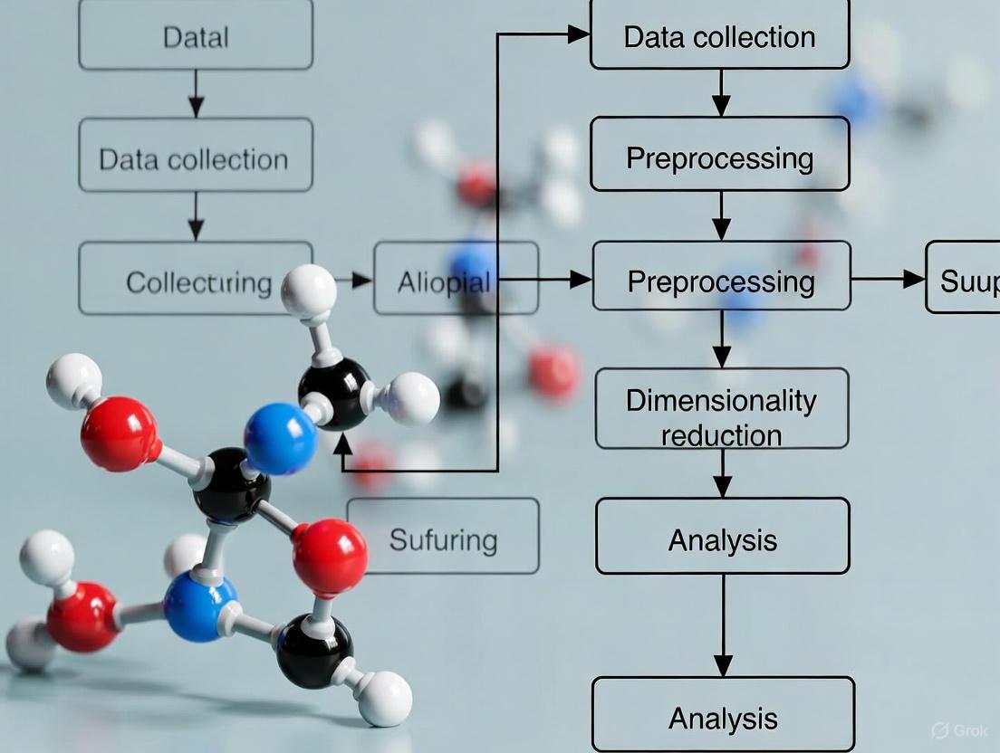
Analysis Workflow for scRNA-seq Data
Approaches to Tackle High-Dimensionality
The Scientist's Toolkit: Research Reagent Solutions
Table 3: Essential Computational Tools for Dimensionality Reduction
| Item | Function | Example Use Case |
|---|---|---|
| Principal Component Analysis (PCA) [1] | A linear feature extraction method that creates uncorrelated components capturing maximum variance in the data. | The foundational first step for compressing scRNA-seq data before non-linear visualization or clustering. |
| t-SNE / UMAP / PaCMAP [3] [1] | Non-linear dimensionality reduction methods that project high-dimensional data into 2D or 3D space for visualization. | Used to create scatter plots where each point is a cell, allowing researchers to visually identify distinct cell clusters and populations. |
| Weighted Fisher Score (WFISH) [2] | A feature selection algorithm that identifies and prioritizes the most informative genes based on class separability. | Applied before building a diagnostic model to classify tumor subtypes from bulk gene expression data, improving accuracy. |
| Variational Autoencoder (VAE) [1] | A deep learning technique that compresses data and can also generate synthetic gene expression profiles. | Used to augment scRNA-seq datasets by generating synthetic cell data, which can help overcome data sparsity and improve downstream analysis. |
Technical Support Center
Core Concepts: Understanding Dropouts
What is the "dropout problem" in single-cell RNA-seq data? The dropout problem refers to a phenomenon where a gene that is actively expressed in a cell fails to be detected during the sequencing process. This results in an observed zero count, which does not reflect the gene's true biological expression. These events occur due to the exceptionally low amounts of mRNA in individual cells, inefficient mRNA capture, and the inherent stochasticity of gene expression at the single-cell level. Consequently, scRNA-seq data become highly sparse, with often over 97% of the data matrix containing zeros [5] [6].
How do dropouts differ from true biological zeros? A biological zero represents a gene that is genuinely not expressed in a given cell. A dropout, however, is a technical artifact—a false negative where a truly expressed gene is not measured. It is often challenging to distinguish between the two without additional experimental or computational evidence. Dropouts are more prevalent for genes with low to moderate expression levels, which can include critical regulatory genes like transcription factors [7] [5].
Why is data sparsity a major challenge for analysis? Data sparsity, caused by high dropout rates, breaks fundamental assumptions of many standard bioinformatics tools. Specifically, it challenges the principle that "similar cells are close to each other" in the high-dimensional expression space. This can severely impact the reliability of downstream analyses, making it difficult to consistently identify cell sub-populations, infer accurate gene regulatory networks, and reconstruct developmental trajectories [6].
Troubleshooting Guides & FAQs
FAQ 1: My cell clusters are unstable between analysis runs. Could dropouts be the cause?
Yes, this is a documented effect of high data sparsity. While cluster homogeneity (cells within a cluster being of the same type) might remain high, cluster stability (the same cell pairs consistently clustering together) has been shown to decrease as dropout rates increase. This occurs because the apparent similarities between cells become inconsistent [6].
- Troubleshooting Steps:
- Assess Sparsity: First, calculate the sparsity of your count matrix (percentage of zeros). Sparsity exceeding 90-95% is common and warrants caution.
- Benchmark Methods: Do not rely on a single clustering pipeline. Run multiple analyses using different methods or parameters and compare the consensus.
- Consider Alternative Approaches: Explore methods that are more robust to dropouts. For example, some studies suggest that using the binary dropout pattern (0 for non-detection, 1 for detection) for co-occurrence clustering can be as informative as using quantitative expression for identifying major cell types [5].
- Validate Biologically: Always correlate your computational clusters with known biological markers to ensure the unstable clusters are not biologically meaningful rare populations.
FAQ 2: I need to accurately infer Gene Regulatory Networks (GRNs). How do I mitigate dropout effects?
Dropouts pose a significant challenge for GRN inference as they corrupt the observed gene-gene co-expression relationships. A common solution is data imputation, but this can introduce its own biases.
Recommended Protocol: Dropout Augmentation (DA) for GRN Inference A novel approach called Dropout Augmentation (DA) offers an alternative to imputation by focusing on model regularization. Instead of removing zeros, DA intentionally adds synthetic dropout noise during model training. This teaches the model to be robust to these events, preventing overfitting to the noisy, zero-inflated data. The DAZZLE model, which implements this concept, has shown improved performance and stability in inferring GRNs from scRNA-seq data [8].
Workflow for DA-enhanced GRN Inference:
FAQ 3: Should I use a whole transcriptome or a targeted approach for my drug development study?
The choice hinges on the trade-off between discovery and precision, heavily influenced by the dropout effect.
- Decision Guide:
| Factor | Whole Transcriptome Profiling | Targeted Gene Expression Profiling |
|---|---|---|
| Goal | Unbiased discovery, novel cell type/pathway identification [7] | Validating targets, quantifying specific pathways, clinical assay development [7] |
| Impact on Dropouts | High: Sequencing depth is spread thin, leading to frequent dropouts, especially for low-abundance genes [7] | Low: Sequencing resources are focused, achieving greater depth per gene and minimizing dropouts for targets [7] |
| Cost & Scalability | Higher cost per cell, less scalable for large cohorts [7] | More cost-effective, enables scaling to hundreds/thousands of samples [7] |
| Best Use Case | Initial target discovery and atlas building [7] | Target validation, mechanism of action studies, patient stratification, and biomarker development [7] |
FAQ 4: What normalization or transformation methods help with compositional data and zeros?
Recognizing that scRNA-seq data is compositional—where the value of each gene represents a proportion of the total transcripts in a cell—can guide better preprocessing. The Compositional Data Analysis (CoDA) framework is designed for this.
Methodology: CoDA-hd for High-Dimensional Data A high-dimensional adaptation of CoDA (CoDA-hd) has been explored for scRNA-seq. A key step is applying a Centered Log-Ratio (CLR) transformation after using a count addition scheme to handle zeros. This approach has shown advantages in providing well-separated clusters in dimensional reduction and producing more biologically plausible trajectories in inference tools like Slingshot, potentially by mitigating artifacts caused by dropouts [9].
Typical CoDA-hd Workflow:
The Scientist's Toolkit
Research Reagent & Computational Solutions
| Tool / Method | Function | Context of Use |
|---|---|---|
| Dropout Augmentation (DA) [8] | A model regularization technique that improves robustness to zeros by adding synthetic dropouts during training. | Gene Regulatory Network inference (e.g., in the DAZZLE model). |
| CoDA-hd & CLR Transformation [9] | A statistical framework and transformation that treats data as log-ratios, making it more robust for downstream analysis. | Data normalization and preprocessing for clustering and trajectory inference. |
| Co-occurrence Clustering [5] | A clustering algorithm that uses binary dropout patterns (non-zero vs. zero) instead of quantitative expression to identify cell types. | Cell type identification when dropouts are prevalent; can utilize pathway-level information. |
| Weighted Fisher Score (WFISH) [2] | A feature selection method that assigns weights based on gene expression differences between classes. | Identifying influential genes for classification tasks in high-dimensional gene expression data. |
| Targeted Gene Panels [7] | A focused sequencing approach that profiles a pre-defined set of genes to maximize sensitivity and quantitative accuracy. | Target validation, biomarker studies, and clinical assay development where specific genes are of interest. |
Disclaimer: The protocols and tools listed are based on current research literature. Performance may vary depending on specific dataset properties. Always validate computational findings with experimental evidence.
RNA sequencing (RNA-seq) data are fundamentally compositional, meaning the abundance of each transcript is only meaningfully interpreted relative to other transcripts within the same sample [10]. This property arises from the assay technology itself: the total number of counts recorded for each sample (the library size) is constrained by sequencing depth and is therefore arbitrary [10] [11]. Consequently, the data exist in a non-Euclidean space where each sample can be represented as a composition of parts (transcripts) that sum to a constant total [10] [12].
This compositional nature has critical implications. A large increase in a few transcripts will necessarily cause the relative proportions of all other transcripts to decrease, even if their absolute abundances remain unchanged [10]. This can lead to spurious findings if analyses designed for absolute data are applied [11]. Compositional Data Analysis (CoDA) provides a statistically rigorous framework to handle these relative properties, transforming the data to enable valid statistical inferences [10] [12].
Key Principles of Compositional Data Analysis (CoDA)
The CoDA framework is built upon core principles that acknowledge the relative nature of the data.
The Core Properties of Compositional Data
Compositional data have three key properties [9]:
- Scale Invariance: The total size of the composition (library size) is irrelevant. Only the relative proportions between components carry information.
- Sub-compositional Coherence: Conclusions drawn from a subset of components (e.g., a specific gene pathway) should be consistent with those from the full composition.
- Permutation Invariance: The results of an analysis do not depend on the order in which the components (genes) are listed.
The Simplex Space and the Constant-Sum Constraint
RNA-seq count data reside on a simplex space, a geometric representation where all points are vectors of positive values that sum to a constant [11]. This constant-sum constraint introduces dependencies; an increase in one component's proportion mathematically necessitates a decrease in one or more others [10] [12]. Applying standard Euclidean-based statistics (e.g., correlation, distance measures) directly to this constrained space is invalid and can produce misleading results [10] [11].
Essential CoDA Transformations and Workflows
To analyze compositional data correctly, log-ratio transformations are used to map data from the simplex to a real Euclidean space where standard statistical methods can be applied.
Core Log-Ratio Transformations
The following table summarizes the primary transformations used in CoDA.
Table 1: Key Log-Ratio Transformations for Compositional Data
| Transformation | Acronym | Formula (for a vector D) | Reference Used | Advantages | Limitations |
|---|---|---|---|---|---|
| Centered Log-Ratio | CLR | ( \text{CLR}(D) = \ln\left[\frac{D1}{g(D)}, \frac{D2}{g(D)}, ..., \frac{D_N}{g(D)}\right] ) | Geometric mean ( g(D) ) of all components [11]. | Preserves all components; symmetric [9] [11]. | Results in a singular covariance matrix, complicating some multivariate tests [12]. |
| Additive Log-Ratio | ALR | ( \text{ALR}(D) = \ln\left[\frac{D1}{DR}, \frac{D2}{DR}, ..., \frac{D{N-1}}{DR}\right] ) | A single, carefully chosen reference component ( D_R ) [11]. | Simple interpretation; avoids singular covariance [11]. | Not symmetric; results depend on choice of reference component [11]. |
| Isometric Log-Ratio | ILR | ( \text{ILR}(D) = \text{orthonormal basis coordinates on the simplex} ) | A set of orthogonal balances (contrasts) [12]. | Creates orthogonal, interpretable coordinates ideal for multivariate analysis [12]. | More complex to define and interpret; requires constructing a balance tree [12]. |
A Standard CoDA Workflow for RNA-seq
The following diagram illustrates a generalized CoDA-based analysis workflow for RNA-seq data, integrating these transformations.
Figure 1: A generalized CoDA workflow for RNA-seq analysis. The core CoDA steps transform data from a constrained simplex space to Euclidean space for valid analysis.
The Scientist's Toolkit: Research Reagent Solutions
Successful RNA-seq and CoDA require high-quality starting materials and specialized reagents. The table below lists essential items and their functions.
Table 2: Essential Research Reagents for RNA-seq Experiments
| Reagent / Kit | Primary Function | Key Considerations |
|---|---|---|
| RNA Extraction Kit | Isolate and purify intact total RNA from cells or tissues [13]. | Select kits designed for your sample source (e.g., tissue, blood). Assess RNA integrity (RIN >7.0) and purity [13] [14]. |
| RNase Inhibitors | Protect RNA samples from degradation by ubiquitous RNases [13] [15]. | Include in reverse transcription setup. Use nuclease-free water and tubes. Wear gloves [15]. |
| DNase I Treatment | Remove contaminating genomic DNA that can cause false positives [13] [15]. | Perform prior to reverse transcription. Select a protocol with minimal impact on RNA integrity [15]. |
| Poly(A) Selection or rRNA Depletion Kits | Enrich for messenger RNA (mRNA) from total RNA [14]. | Poly(A) selection is standard for eukaryotic mRNA. rRNA depletion is used for prokaryotes or degraded samples [14]. |
| cDNA Synthesis Kit (Reverse Transcriptase) | Synthesize complementary DNA (cDNA) from RNA templates [15]. | Choose a high-efficiency, thermostable enzyme. Use a mix of random hexamers and oligo(dT) for full transcriptome coverage [15]. |
| High-Performance Library Prep Kit | Prepare cDNA libraries for sequencing [14]. | Kits include reagents for fragmentation, adapter ligation, and index multiplexing. Follow manufacturer protocols rigorously [14]. |
| Unique Molecular Identifier (UMI) Kits | Tag individual mRNA molecules to correct for PCR amplification bias and accurately count transcripts [16]. | Essential for digital counting and reducing technical noise in single-cell RNA-seq [16]. |
Troubleshooting Common RNA-seq Experimental Issues
Pre-sequencing: RNA Extraction and QC
Table 3: Troubleshooting RNA Extraction and Quality Control
| Problem | Potential Cause | Solution |
|---|---|---|
| RNA Degradation | RNase contamination; improper sample storage; repeated freeze-thaw cycles [13] [15]. | Use RNase-free reagents and consumables. Store samples at -80°C in single-use aliquots. Include an RNase inhibitor [13]. |
| Low RNA Yield/Purity | Excessive sample input; incomplete homogenization; carryover of inhibitors (e.g., salts, proteins, polysaccharides) [13]. | Adjust sample input to protocol recommendations. Ensure complete tissue lysis. Increase wash steps during purification. Re-purify if necessary [13]. |
| Genomic DNA Contamination | Inefficient DNase digestion; high sample input [13] [15]. | Treat RNA with DNase I. Use reverse transcription reagents with genomic DNA removal modules. Design PCR primers spanning exon-exon junctions [15]. |
| Incomplete cDNA Synthesis / Poor Coverage | Poor RNA integrity; high GC content/secondary structures; suboptimal reverse transcriptase [15]. | Denature RNA at 65°C before reverse transcription. Use a thermostable, high-performance reverse transcriptase. Optimize primer mix (random hexamers vs. oligo(dT)) [15]. |
Post-sequencing: Data Analysis and CoDA
Q: My PCA plot seems to be driven by library size or a few highly expressed genes, not my experimental conditions. How can CoDA help? A: This is a classic sign of compositional data. The apparent "differences" are often artifacts of the relative nature of the data. Applying a CLR transformation before PCA is crucial. The CLR transforms the data so that the Euclidean distances in the new space correspond to Aitchison distances on the simplex, which are valid for compositional data. This often leads to more biologically meaningful clustering, as it reduces the dominance of a few variables and accounts for the data's relative structure [9] [11].
Q: My single-cell RNA-seq data is full of zeros (dropouts). Can I still use CoDA? A: Yes, but handling zeros is a key challenge since log-ratios are undefined for zero values. Recent research explores solutions such as:
- Count addition schemes: Adding a small, carefully chosen pseudo-count to all measurements to allow for log-ratio transformation [9].
- Imputation methods: Using algorithms to estimate the true expression value for zeros based on information from similar cells, though this must be done cautiously to avoid introducing false signals [9] [16]. Studies show that CoDA transformations like count-added CLR can provide advantages in downstream analyses like clustering and trajectory inference, even for sparse scRNA-seq data [9].
Q: Why does CoDA prevent more false positives in differential expression analysis? A: Traditional analyses that ignore compositionality can identify a gene as "increased" simply because other genes have decreased in relative proportion, even if its absolute abundance is unchanged. This is a spurious correlation induced by the constant-sum constraint [10] [11]. CoDA methods, by analyzing data in terms of log-ratios, are inherently immune to this effect. When combined with a scale uncertainty model (acknowledging that the total number of molecules can change between conditions), CoDA-based differential expression pipelines have been shown to effectively control false-positive rates while maintaining high sensitivity [11].
CoDA in the Context of High-Dimensionality
RNA-seq data is not only compositional but also high-dimensional, often profiling >20,000 genes from far fewer samples [17] [18]. This "curse of dimensionality" (COD) exacerbates analytical challenges.
The Dual Challenge: Compositionality and High Dimensions
High-dimensional data suffers from noise accumulation, where technical noise across thousands of genes distorts distances and statistical summaries [16]. This leads to:
- Loss of Closeness (COD1): Distances between samples become similar and uninformative, impairing clustering [16].
- Inconsistency of Statistics (COD2): Metrics like PCA contribution rates become unreliable [16].
- Inconsistency of Principal Components (COD3): PCA results can be driven by technical artifacts like sequencing depth instead of biology [16].
Compositionality and COD are intertwined. The relative nature of the data means that analyzing one gene in isolation is invalid, forcing a high-dimensional approach. Conversely, the high-dimensional noise can obscure the true relative signal.
Resolving the Dimensionality Curse with CoDA
The following diagram illustrates the relationship between data spaces and how CoDA interacts with the curse of dimensionality.
Figure 2: The analytical pathway from raw RNA-seq data to biological insight. CoDA transformations first resolve compositionality, creating valid data for subsequent dimensionality reduction techniques that tackle the curse of dimensionality.
CoDA does not eliminate high-dimensionality but creates a valid foundation for subsequent analysis. After CLR transformation, standard dimensionality reduction techniques like PCA can be applied more reliably because the data's relative structure is correctly represented [9] [11]. Furthermore, specialized noise-reduction methods like RECODE (Resolution of the Curse of Dimensionality) have been developed to directly address COD in high-dimensional data like scRNA-seq, working to recover true expression values without reducing the number of genes, thus enabling precise analysis of all gene information [16].
FAQ: Understanding the Core Challenges
What is the "curse of dimensionality" in the context of gene expression data? The curse of dimensionality describes the challenges that arise when analyzing data with a vast number of features (like thousands of genes) but a relatively small number of samples. In high-dimensional spaces, data becomes sparse, making it difficult to discover reliable patterns. For example, in genome-wide association studies (GWAS), evaluating all possible interactions among millions of genetic variants leads to a combinatorial explosion that diminishes the usefulness of traditional statistical methods [19] [20]. This sparsity also means that distance metrics become less meaningful, as most data points appear to be equally far apart [21] [20].
How does high dimensionality lead to overfitting? Overfitting occurs when a model learns not only the underlying biological signal but also the random noise or spurious correlations present in the training dataset. High-dimensional data intensifies this problem because the model has an immense number of features to use, allowing it to easily "memorize" noise. With more features, the model's capacity to learn increases, but so does the risk of fitting to random fluctuations that do not represent true biological mechanisms [22]. This is particularly problematic when the number of features far exceeds the number of samples, a common scenario in genomics [23].
What are batch effects, and why are they particularly problematic for omics studies? Batch effects are technical variations introduced into data due to differences in experimental conditions, such as processing time, reagent batches, different laboratories, or sequencing runs. These variations are unrelated to the biological question under study [24]. They are problematic because they can confound the real biological signals, leading to increased variability, reduced statistical power, or even completely incorrect conclusions. For instance, a change in RNA-extraction solution was shown to cause incorrect classification outcomes for patients in a clinical trial [24]. Batch effects are a paramount factor contributing to the irreproducibility of scientific findings [24].
Can these three problems be addressed simultaneously? Yes, a robust analytical pipeline must address all three issues. While distinct, these challenges are deeply interconnected. High-dimensional data is prone to overfitting, and batch effects can introduce structured technical variation that models may mistakenly learn as biological signal, thereby worsening overfitting. A comprehensive strategy involves careful experimental design, dimensionality reduction or feature selection to combat the curse of dimensionality, regularization to prevent overfitting, and the application of batch effect correction methods before key analyses [21] [22] [24].
Troubleshooting Guides
Guide 1: Diagnosing and Mitigating Overfitting
Symptoms: Your model achieves near-perfect accuracy on your training data but performs poorly on a separate validation set or new experimental data.
Methodology:
- Data Splitting (Hold-out): Start by splitting your dataset into a training set (e.g., 80%) and a testing set (e.g., 20%). The test set must never be used during model training or feature selection; it serves solely for the final evaluation of generalization performance [25].
- Cross-Validation: For a more robust assessment, use k-fold cross-validation. This involves splitting the data into k groups (folds), iteratively using k-1 folds for training and the remaining fold for validation, and then averaging the results. This allows all data to be used for validation while providing an estimate of model stability [22] [25].
- Apply Regularization Techniques: Introduce penalty terms to your model's objective function to constrain its complexity.
- Simplify the Model: Reduce the complexity of the model architecture itself. For neural networks, this can mean removing layers or reducing the number of units per layer. For tree-based models, limit the depth of the trees [25].
- Implement Early Stopping: When training iterative models (e.g., neural networks), monitor the model's performance on a validation set. Stop the training process as soon as the validation performance begins to degrade, even if training performance continues to improve [22] [25].
Table: Comparison of Techniques to Prevent Overfitting
| Technique | Methodology | Best Used When | Key Advantage |
|---|---|---|---|
| Hold-out / Cross-Validation | Data is split into training and validation sets. | You have a sufficiently large dataset. | Provides an unbiased estimate of model performance on unseen data. |
| L1/L2 Regularization | A penalty term is added to the model's loss function. | You have many potentially correlated features. | Constrains model complexity without reducing the number of features. |
| Dropout | Randomly "drops" a subset of model units during training. | Training deep neural networks. | Reduces interdependent learning among units, forcing robustness. |
| Early Stopping | Training is halted when validation error stops improving. | Training models for a large number of epochs is computationally expensive. | Prevents the model from over-optimizing on the training data. |
Guide 2: Overcoming the Curse of Dimensionality
Symptoms: Models fail to generalize, distance-based algorithms (e.g., clustering) perform poorly, and you have far more features (genes) than samples.
Methodology:
- Feature Selection: Identify and retain only the most informative features.
- Univariate Feature Selection: Tests the relationship between each feature and the response variable individually (e.g., using correlation or chi-squared tests). It is fast but ignores interactions between features [21].
- Feature Importance Ranking: Use model-based methods like Random Forests, which provide an intrinsic measure of feature importance based on the average decrease in impurity [21].
- Permutation-Based Feature Importance: A model-agnostic method where the performance decrease after randomly permuting a feature indicates its importance. It is flexible but can be computationally slow [21].
- Recursive Feature Elimination (RFE): Recursively removes the least important features and re-fits the model until the desired number of features is reached [21].
- Dimensionality Transformation: Create new, lower-dimensional features from the original high-dimensional space.
- Principal Component Analysis (PCA): A linear technique that projects data onto a set of orthogonal axes (principal components) that capture the maximum variance in the data [21] [23].
- t-SNE / UMAP: Non-linear manifold learning techniques that are particularly effective for visualization, as they can reveal complex cluster structures in 2D or 3D plots [21].
- Leverage Distributed Computing: For exhaustive searches in massive datasets (e.g., all possible gene-gene interactions), use frameworks like PySpark to distribute computations across multiple processors, dramatically improving processing speed [19].
The following diagram illustrates the logical workflow for tackling the curse of dimensionality:
Guide 3: Identifying and Correcting for Batch Effects
Symptoms: Samples cluster strongly by processing date, sequencing batch, or laboratory in a PCA plot, rather than by the biological groups of interest.
Methodology:
- Visual Diagnostics:
- Principal Component Analysis (PCA): Plot the first few principal components and color the points by known batch variables (e.g., lab, date) and by biological conditions. Strong clustering by batch is a clear indicator [24] [26].
- t-SNE Plots: Similarly, create t-SNE plots colored by batch and biological variables to check for confounding [26].
- Statistical Diagnostics: Use metrics like the F-test to assess the association between principal components and known experimental variables. A strong association between a PC and a batch variable indicates a significant batch effect [26].
- Apply Batch Effect Correction Algorithms (BECAs): If a batch effect is diagnosed, apply a correction method. The choice of method depends on the experimental design.
- Balanced Design: If biological groups are equally represented across batches, methods like Limma's
removeBatchEffectcan be effective [26]. - Unbalanced Design: For more complex scenarios, methods like ComBat or SVA are often used. Newer methods like NPmatch, which uses sample matching, have also shown promise [24] [26].
- Critical Note: Correction is extremely difficult or impossible in a "fully confounded" study where the biological variable of interest is perfectly aligned with batch (e.g., all controls were processed in one batch and all cases in another). This underscores the importance of a balanced experimental design from the outset [26].
- Balanced Design: If biological groups are equally represented across batches, methods like Limma's
Table: Common Batch Effect Correction Algorithms (BECAs)
| Algorithm | Methodology | Key Consideration |
|---|---|---|
| Limma (removeBatchEffect) | Fits a linear model to the data and removes the component associated with the batch. | Works well for balanced designs. A standard, widely-used tool. |
| ComBat | Uses an empirical Bayes framework to adjust for batch effects. Can be more powerful for small sample sizes. | Can over-correct and remove biological signal if not applied carefully. |
| SVA (Surrogate Variable Analysis) | Identifies and adjusts for unmeasured or "hidden" batch effects. | Useful when not all sources of technical variation are known. |
| NPmatch | Uses sample matching and pairing to correct for batch effects. | A newer method reported to show superior performance in some contexts [26]. |
The Scientist's Toolkit: Essential Research Reagents & Platforms
Table: Key Platforms for Profiling and Analysis
| Tool / Reagent | Function | Application in Research |
|---|---|---|
| L1000 Assay | A high-throughput gene expression profiling platform that measures the mRNA levels of ~978 "landmark" genes. | Captures transcriptional states from cells perturbed by chemicals or genetics. Used for large-scale screening [27] [28]. |
| Cell Painting | A high-content imaging assay that uses fluorescent dyes to stain cellular components, generating morphological profiles. | Extracts thousands of features related to cell shape, intensity, and texture to quantify phenotypic impact of perturbations [27] [28]. |
| CellProfiler | Open-source software for automated image analysis. | Used to extract quantitative morphological features from microscopy images generated in Cell Painting assays [27]. |
| PySpark | A Python API for Apache Spark, a distributed processing engine. | Enables scalable analysis of extraordinarily large genomic datasets, helping to overcome computational bottlenecks [19]. |
| Omics Playground | An integrated bioinformatics platform. | Provides a user-friendly interface for multiple batch effect correction algorithms (Limma, ComBat, SVA) and other analyses without requiring programming [26]. |
Core Concepts: The High-Dimensional Data Challenge
Modern genomic and proteomic technologies, such as gene expression microarrays and protein chips, present researchers with the task of extracting meaningful information from high-dimensional data spaces, where each sample is defined by hundreds or thousands of measurements obtained concurrently [29]. This data structure is common in studies seeking better predictive models for cancer diagnosis, prognosis, therapy response, and the identification of key signaling networks [29].
What defines a high-dimensional data space in genomics?
High-dimensionality arises when the number of features (e.g., genes, proteins) vastly exceeds the number of biological samples. For instance, a whole human genome expression array can probe for 47,000 transcripts from a single sample, while a study may include only several dozen to a few hundred patient samples [29]. This creates a scenario where the ratio of samples to features can be as low as 0.01, starkly contrasting with the conventional rule of thumb suggesting at least 10 training samples per feature dimension [30].
What are the principal analytical challenges?
Working in high-dimensional spaces introduces unique methodological problems [29] [30]:
- The Curse of Dimensionality: As the number of features increases, data becomes sparse, and the distance between a data point and its nearest and farthest neighbors can become nearly equidistant. This compromises the accuracy of distance-based analyses and predictive models [29] [30].
- Model Overfitting: Complex models learned from a small sample size may fit irreproducible noise in the training data, rather than the true biological signal, leading to poor performance on independent test data [30].
- Multiple Testing Problem: When testing thousands of genes simultaneously for differential expression, using a standard significance threshold (e.g., p < 0.05) will yield hundreds of false positives by chance alone [29].
- Spurious Correlations: High-dimensional data are rarely randomly distributed and often contain chance correlations that are not biologically meaningful [29].
Table 1: Common Challenges in High-Dimensional Genomic Studies
| Research Question | High-Dimensional Problems |
|---|---|
| Biomarker Selection | Trade-off between accuracy and computational complexity; spurious correlations; multiple testing; model overfitting [29]. |
| Cancer Classification | Curse of dimensionality; spurious clusters; small sample size; biased performance estimate [29]. |
| Cell Signaling Analysis | Confound of multimodality; spurious correlations; multiple testing [29]. |
Troubleshooting Guides & FAQs
FAQ: Why does my classifier perform well on training data but fail on new data?
This is a classic symptom of model overfitting [30]. In high-dimensional spaces, it is easy for a complex model to "memorize" the training data, including its noise, rather than learning the generalizable underlying pattern.
Solutions:
- Apply Feature Selection: Reduce the feature set to only the most informative genes before classifier training. With small sample sizes, univariate methods (e.g., t-test, signal-to-noise ratio) can perform comparably to more complex multivariate methods [30].
- Use Simpler Models: Employ classifiers with restricted complexity, such as naive Bayes models or linear Support Vector Machines (SVMs), which are less prone to overfitting [30].
- Implement Regularization: Use techniques that modify the training objective function to penalize model complexity, thereby limiting the learning of noisy patterns [30].
FAQ: How can I distinguish true biological signal from false positives in differential expression?
The multiple testing problem means that many seemingly significant results will occur by chance. If you test 10,000 genes with a p-value threshold of 0.05, you can expect 500 false positives [29].
Solutions:
- Control the False Discovery Rate (FDR): Methods like the Benjamini-Hochberg procedure control the expected proportion of false positives among the identified significant genes, which is less conservative than family-wise error rate control [29].
- Use Bayesian Methods: Hierarchical models can be used to compute the probability that a given SNP or gene is truly associated with a trait by incorporating prior probabilities and functional genomic annotations [31].
- Independent Validation: Always validate findings in an independent dataset or through functional experiments. For classification and prognostic studies, validation in independent data sets is essential [29].
FAQ: My co-expression analysis did not recover known pathway genes. What went wrong?
Co-expression is not universally informative for all biological processes. Genes in the same pathway may not have correlated transcript profiles due to post-transcriptional regulation, and the utility of co-expression depends heavily on the dataset and analytical parameters used [32].
Solutions:
- Optimize Dataset Selection: Larger datasets are not always better. Use expression datasets directly relevant to the biological process of interest (e.g., a drought stress dataset to find drought response genes) [32].
- Exhaustively Explore Parameters: Test multiple dataset combinations, similarity measures (e.g., Pearson correlation), and clustering algorithms (e.g., k-means, hierarchical) to maximize the recovery of functional associations [32].
- Validate with Independent Data: Confirm the biological relevance of co-expression clusters using an independent data type, such as phenomics or mutant data [32].
Key Analytical Workflows
The shift from one-at-a-time analyses to integrative models is crucial for robust discovery in high-dimensional biology. The diagram below illustrates a generalized workflow for joint modeling that leverages multiple data types to improve statistical power and biological insight.
Workflow: Joint Modeling of Multi-Omic Data for Target Identification
This workflow demonstrates a Bayesian approach to integrating different genomic data types to overcome the limitations of analyzing each dataset in isolation [31] [33].
Detailed Methodology:
- Data Input: Collect diverse genomic data types. For example, in a study of a transcription factor (TF) like Lrp in E. coli, this could include:
- DNA-Protein Binding (ChIP-chip) Data: Provides direct evidence of in vivo TF binding [33].
- Gene Expression Data: Identifies genes differentially expressed in a TF mutant vs. wild type, indicating functional impact [33].
- DNA Sequence Data: Scores upstream gene regions for the presence of TF binding motifs to predict affinity [33].
- Model Specification: Construct a hierarchical Bayesian model. In this framework, the binding data is often used as the primary data, while expression and sequence data are incorporated as secondary data to inform the priors [33].
- Prior Incorporation: Use functional genomic annotations (e.g., DNase-I hypersensitive sites, protein-coding exons, enhancers) to define prior probabilities that a genomic region or gene is functionally important. The model learns the relevance of these annotations from the data itself [31].
- Model Inference: Use computational methods like Markov Chain Monte Carlo (MCMC) or an empirical Bayes approach to draw inferences. This process calculates the posterior probability that a given gene is a true target of the TF, automatically weighting the contribution of each data type based on its correlation with the primary signal [31] [33].
- Output: Generate a list of high-confidence target genes, often with an increased number of loci with high-confidence associations compared to conventional approaches [31].
The Scientist's Toolkit: Research Reagent Solutions
Table 2: Essential Resources for Genomic Data Analysis
| Resource Category | Specific Examples & Functions |
|---|---|
| Public Data Repositories | Gene Expression Omnibus (GEO) & ArrayExpress: Central repositories for submitting and downloading high-throughput functional genomics data [34] [35]. |
| Analysis & Visualization Tools | GOEAST (Gene Ontology Enrichment Analysis Software): Identifies significantly enriched Gene Ontology terms among given gene lists [34]. |
| Reference Databases | The Cancer Genome Atlas (TCGA) Data Portal: Provides clinical and genomic characterization data from tumor samples for analysis and comparison [34]. |
| Experimental Reagents | Validated Antibodies (e.g., for IHC): Critical for experimental validation; require proper controls and titration to avoid background staining [36]. |
| Integrated Platforms | Expression Atlas (EMBL-EBI): Provides information on gene and protein expression patterns under different biological conditions [34]. |
A Methodologist's Toolkit: Feature Selection, Dimensionality Reduction, and AI-Driven Solutions
Troubleshooting Guide: Common Issues in High-Dimensional Gene Expression Analysis
1. My model is overfitting despite using regularization. What should I do?
- Problem: The "curse of dimensionality" means your high-dimensional data is sparse, causing the model to learn noise instead of true biological signals [37] [38].
- Solution: Apply robust feature selection before model training. Use filter methods like Weighted Fisher Score (WFISH) or Weighted Signal to Noise Ratio (WSNR) to remove irrelevant genes and reduce the feature set to the most informative ones [2] [39]. This directly tackles the root cause of overfitting in high-dimensional spaces.
2. I need to visualize cell clusters from my single-cell RNA-seq data. Which technique is best?
- Problem: Visualizing high-dimensional data to identify biological patterns like cell types.
- Solution: Use feature extraction methods like t-SNE or UMAP. These non-linear techniques are specifically designed to project data into 2D or 3D spaces while preserving the local structure and cluster separation, making them ideal for exploratory data analysis [40] [41].
3. My computational resources are limited, but I have a dataset with millions of features.
- Problem: Training models on a very high number of features (e.g., SNPs or genes) is slow and computationally expensive [37].
- Solution: Start with a fast filter method for feature selection (e.g., correlation-based, signal-to-noise ratio). These methods use statistical measures to rank features independently of a model, offering high computational efficiency and scalability [39] [42].
4. I want to know which specific genes are driving the classification of disease subtypes.
- Problem: The model is a "black box," and you need biological interpretability.
- Solution: Use feature selection. Methods like WFISH or Random Forest importance scores provide a ranked list of genes, allowing you to identify and validate specific biomarkers. This preserves the original biological meaning of the features [2] [41].
5. After integration, my single-cell reference atlas fails to detect a rare cell population in a new query sample.
- Problem: The features selected for building the reference atlas might be insensitive to certain biological variations.
- Solution: Re-evaluate your feature selection strategy for integration. Benchmarking studies suggest using batch-aware highly variable gene selection or lineage-specific feature selection to create a reference that is more robust and capable of detecting unseen populations [43].
Frequently Asked Questions (FAQs)
Q1: What is the fundamental difference between feature selection and feature extraction?
- Feature Selection reduces dimensionality by choosing a subset of the original features (e.g., selecting 500 informative genes from 50,000). It preserves the interpretability of the features [41] [42].
- Feature Extraction creates a smaller set of new features by transforming and combining the original ones (e.g., PCA creates principal components from linear combinations of all genes). This often improves model performance at the cost of direct interpretability [40] [44].
Q2: When should I prefer feature selection over feature extraction in my gene expression analysis? Prefer Feature Selection when:
- Interpretability is key: You need to identify specific genes or biomarkers for downstream validation (e.g., drug targets) [2] [39].
- You have domain knowledge: You want to use biological priors to guide the selection process [42].
- Computational efficiency is critical: Filter methods are fast and scalable [37].
Prefer Feature Extraction when:
- Predictive accuracy is the primary goal: Combining features can help capture complex interactions and improve performance [44].
- Visualization is needed: Methods like t-SNE and UMAP are excellent for visualizing high-dimensional data in 2D/3D [40] [41].
- Features are highly correlated: PCA can effectively handle multicollinearity by creating uncorrelated components [40] [38].
Q3: How does the "curse of dimensionality" affect my machine learning model, and how do these techniques help? The "curse of dimensionality" describes how, as the number of features grows, data becomes sparse, and model performance can degrade because it becomes easier to overfit to noise [37] [38]. Both techniques combat this:
- Feature Selection directly reduces
p(the number of features) to be closer ton(the number of samples), mitigating sparsity and overfitting [37]. - Feature Extraction projects the data into a lower-dimensional space that is denser and more manageable for learning algorithms [40].
Q4: Are there methods that combine the advantages of both strategies? Yes, some advanced workflows combine them. For instance, you can first use feature selection to filter out clearly irrelevant genes, reducing noise. Then, apply feature extraction (like PCA) on the reduced gene set to further compress the data and capture latent structures for a final classifier [44]. This hybrid approach balances interpretability with performance.
Q5: How many features should I finally select or extract for my model? There is no universal answer. The optimal number depends on your dataset and goal. It is determined empirically through:
- Cross-validation: Evaluating model performance with different numbers of features [37].
- Benchmarking: Using multiple metrics to find a point of diminishing returns [43].
- Stability analysis: Checking if the selected features are consistent across different data subsets [41]. Start with a few hundred top-ranked features and adjust based on validation results.
Comparative Analysis at a Glance
The table below summarizes the core characteristics of feature selection and feature extraction to guide your choice.
| Aspect | Feature Selection | Feature Extraction |
|---|---|---|
| Core Principle | Selects a subset of original features. [42] | Creates new features from original ones. [40] |
| Interpretability | High (preserves original feature meaning). [41] | Low (new features are transformations). [44] |
| Model Performance | Good; can be enhanced by removing noise. [2] | Often high; can capture complex patterns. [44] |
| Primary Methods | Filter (e.g., WSNR [39]), Wrapper, Embedded. [37] | PCA [40], LDA [41], t-SNE/UMAP [40]. |
| Handling Redundancy | Identifies and removes redundant features. [42] | Projects data into uncorrelated components. [40] |
| Ideal Use Case | Identifying biomarkers; resource-constrained environments. [2] | Data visualization; maximizing predictive accuracy. [44] |
Experimental Protocols for Key Methodologies
Protocol 1: Implementing Weighted Fisher Score (WFISH) for Gene Selection
- Objective: To identify the most informative genes in a high-dimensional gene expression dataset for classification tasks [2].
- Materials: Normalized gene expression matrix (samples x genes), class labels (e.g., diseased vs. healthy).
- Procedure:
- Data Preprocessing: Ensure data is normalized and log-transformed if necessary.
- Calculate Weights: For each gene, compute a weight based on the difference in its average expression between classes. This prioritizes genes with large inter-class differences [2].
- Compute Fisher Score: Calculate the traditional Fisher score for each gene, which measures the ratio of inter-class variance to intra-class variance.
- Apply Weight: Multiply the traditional Fisher score by the weight from step 2 to get the WFISH score [2].
- Rank Genes: Rank all genes in descending order of their WFISH score.
- Select Subset: Choose the top
kgenes for downstream model training. The value ofkcan be determined via cross-validation.
- Validation: Evaluate the selected gene subset by training a classifier (e.g., Random Forest or k-NN) and assessing accuracy on a held-out test set [2].
Protocol 2: Dimensionality Reduction Workflow with PCA and t-SNE
- Objective: To reduce the dimensionality of a gene expression dataset for visualization and exploratory analysis.
- Materials: Normalized gene expression matrix.
- Procedure:
- Optional Feature Selection: First, apply a variance filter or simple feature selection to remove very low-variance genes, reducing noise.
- Standardization: Standardize the data so each gene has a mean of 0 and a standard deviation of 1. This is critical for PCA [40].
- Principal Component Analysis (PCA):
- Compute the covariance matrix of the standardized data.
- Calculate the eigenvectors (principal components) and eigenvalues (explained variance) of this matrix.
- Project the data onto the first
mprincipal components. This step performs initial linear compression [40].
- t-SNE Projection: Use the PCA-reduced data (or the original standardized data for smaller datasets) as input for t-SNE.
- t-SNE calculates pairwise similarities between data points in the high-dimensional space.
- It then maps the data to a 2D/3D space, optimizing the layout to preserve these local similarities [40].
- Validation: Inspect the 2D/3D plot for clear cluster separation. Validate clusters using biological knowledge or cell-type labels if available.
Workflow Visualization: Strategy Selection
The following diagram illustrates a logical workflow for choosing between feature selection and feature extraction, based on your project's primary goal.
The Scientist's Toolkit: Essential Research Reagents & Materials
The table below lists key "reagents" in the computational workflow for handling high-dimensional gene expression data.
| Item / Solution | Function / Explanation |
|---|---|
| Normalized Expression Matrix | The fundamental input data. Raw counts are normalized (e.g., TPM for bulk RNA-seq) to make samples comparable and reduce technical bias. |
| Feature Selection Algorithms (e.g., WFISH, WSNR) | Act as molecular "filters" to isolate the most informative genes from a noisy background, much like a probe pulls down a specific target [2] [39]. |
| Dimensionality Reduction Tools (e.g., PCA, UMAP) | Serve as a "staining dye" for high-dimensional data, revealing underlying structures and patterns (like cell lineages) that are invisible in the raw data [40] [43]. |
| Cross-Validation Framework | The "quality control" step. It ensures that the selected features or the model built on reduced dimensions will generalize well to new, unseen data [37] [41]. |
| Benchmarking Metrics Suite | A set of quantitative measures (e.g., Batch ASW, ARI, mapping accuracy) used to objectively evaluate and compare the performance of different feature selection/extraction methods [43]. |
| Gene Set Enrichment Tools | Used post-feature selection to determine if the identified biomarker genes are enriched in known biological pathways (e.g., KEGG, GO), adding functional context to the results [41]. |
Frequently Asked Questions (FAQs) and Troubleshooting Guides
This technical support resource addresses common challenges researchers face when implementing optimization algorithms for feature selection in high-dimensional gene expression data.
Eagle Prey Optimization (EPO)
FAQ 1: What is the core biological inspiration behind the Eagle Prey Optimization algorithm? EPO is a novel nature-inspired optimization technique that mimics the hunting strategies of eagles, which exhibit unparalleled precision and efficiency in capturing prey. The algorithm simulates how an eagle descends from a height, formulating its trajectory to find the optimal solution (prey) by exploring and exploiting the search space [45] [46].
FAQ 2: Why is EPO particularly suitable for microarray gene expression data? Microarray data possesses high dimensionality, characterized by thousands of gene features but often with small sample sizes. EPO is designed to address this challenge by balancing global exploration and local exploitation, effectively identifying a small subset of informative genes that can discriminate between cancer subtypes with high accuracy and minimal redundancy [45].
Troubleshooting Guide: Handling Premature Convergence in EPO
| Problem | Potential Causes | Recommended Solutions |
|---|---|---|
| Algorithm converges too quickly to a suboptimal solution | Population diversity is too low; insufficient exploration | Increase population size; Adjust mutation rate parameters; Incorporate chaos techniques for initialization [45] [47] |
| Poor classification accuracy despite high fitness | Fitness function overfitting; redundant genes | Incorporate a fitness function that considers both discriminative power and gene diversity; Use penalized metrics to reduce redundancy [45] [48] |
Genetic Algorithms (GAs)
FAQ 3: How do Genetic Algorithms tackle the feature selection problem? GAs treat feature selection as a combinatorial optimization problem. Each potential feature subset is represented as a binary chromosome (1 for feature inclusion, 0 for exclusion). The algorithm evolves a population of these chromosomes over generations using selection, crossover, and mutation operators to find the subset that gives the best predictive performance [49] [50] [51].
FAQ 4: What are the common encoding schemes and genetic operators used in GAs for feature selection?
- Encoding Schemes: Binary encoding is most common, where each feature's presence is indicated by 1 or 0 [50].
- Selection: Roulette wheel or rank-based selection chooses fitter individuals for reproduction [49] [50].
- Crossover: One-point or uniform crossover combines genetic material from parents [50] [51].
- Mutation: Bit-wise mutation randomly flips genes to introduce diversity, with a typical rate of 1/m (where m is the number of features) [49] [50].
Troubleshooting Guide: Managing Computational Complexity in Genetic Algorithms
| Problem | Potential Causes | Recommended Solutions |
|---|---|---|
| Experiment is prohibitively slow | High dimensionality of gene data; Large population size; Many generations | Use parallel computing (2x-25x speedups reported) [51]; Employ internal holdout sets instead of nested resampling [48] |
| Overfitting to the training data | Overly aggressive optimization; Lack of validation | Use external resampling estimates not seen by the GA [48]; Apply penalized metrics like AIC; Implement early stopping [48] |
Algorithm Performance and Comparison
FAQ 5: How does the performance of EPO compare to other optimization algorithms? Extensive experiments on publicly available microarray datasets demonstrate that EPO consistently outperforms state-of-the-art gene selection methods in terms of classification accuracy, dimensionality reduction, and robustness to noise [45].
FAQ 6: What performance metrics are most appropriate for evaluating feature selection in a biological context? Common metrics include:
- Classification Accuracy: Measures the model's ability to correctly classify cancer subtypes [45].
- F1-Score and ROC-AUC: Provide a more balanced view of model performance, especially with imbalanced data [51].
- Internal vs. External Validation: Internal performance (e.g., OOB error) guides the search, while external validation on held-out data gives a better assessment of overfitting [48].
The table below summarizes quantitative results from benchmark studies as referenced in the search results.
Table 1. Performance Comparison of Feature Selection Algorithms on Gene Expression Data
| Algorithm | Reported Classification Accuracy | Key Strengths | Computational Cost |
|---|---|---|---|
| Eagle Prey Optimization (EPO) | Consistently outperforms state-of-the-art methods [45] | High accuracy, minimal redundancy, robust to noise [45] | Not specified |
| Genetic Algorithm (GA) | High (specific metrics depend on implementation) [51] [48] | Discovers optimal feature combinations, parallelizable [49] [51] | High, but reduced via parallelism [51] |
| Improved GA (IGA) + Improved BA (IBA) | Geometric mean: 0.99; Silhouette coefficient: 1.0 [47] | High inter-cluster variability, high intra-cluster similarity [47] | Higher convergence speed [47] |
Experimental Protocols
Protocol 1: Implementing a Genetic Algorithm for Feature Selection
This protocol outlines the key steps for applying a GA to gene expression data, based on established methodologies [50] [48].
Define Encoding Scheme: Use binary encoding. A chromosome is a binary vector where each gene (bit) represents the inclusion (1) or exclusion (0) of a feature.
Define Fitness Function: The function should evaluate the quality of the feature subset.
Configure Genetic Operators:
Set Termination Criteria: Define conditions to stop the algorithm, such as a maximum number of generations or convergence threshold [50].
The following workflow diagram illustrates the typical GA process for feature selection.
The Scientist's Toolkit: Essential Research Reagents and Computational Materials
The table below lists key resources for conducting optimization-driven feature selection experiments in bioinformatics.
Table 2. Key Research Reagent Solutions for Optimization Experiments
| Item Name | Function / Purpose | Example / Notes |
|---|---|---|
| Microarray Datasets | Provide high-dimensional gene expression data for algorithm validation | Publicly available datasets representing different cancer types [45] |
| scRNA-seq Datasets | Used for testing feature selection on sparse, high-dimensional data | Requires methods robust to dropout noise and sparsity [52] |
Python scikit-learn |
Machine learning library for model building and evaluation | Used to implement fitness functions (e.g., RandomForestClassifier) [50] [51] |
| CRISPR Screen Data (DepMap) | Large compendium of gene dependency data for functional network analysis | Used to test normalization and dimensionality reduction methods [53] |
| CORUM Database | Gold standard protein complex annotations | Used for benchmarking functional relationships in gene networks [53] |
| Parallel Computing Framework (e.g., PySpark, joblib) | Speeds up fitness evaluation in GAs by distributing computations | Enables parallel model training, reducing total GA time [51] |
In gene expression research, single-cell and bulk RNA-sequencing data present a fundamental challenge: each sample is characterized by tens of thousands of gene expression values, creating a high-dimensional space that is difficult to visualize and analyze directly [54]. Dimensionality reduction techniques are essential tools that address this by transforming such data into a lower-dimensional space (e.g., 2 or 3 dimensions), enabling researchers to identify sample clusters, uncover biological patterns, and detect technical artifacts like batch effects [55] [54].
This guide focuses on three foundational methods: Principal Component Analysis (PCA), a linear method; t-Distributed Stochastic Neighbor Embedding (t-SNE), a non-linear method excels at revealing local structure; and Uniform Manifold Approximation and Projection (UMAP), a non-linear method that balances local and global structure preservation [56] [57]. Understanding their operational principles, optimal applications, and common pitfalls is crucial for generating biologically meaningful insights.
Method Comparison and Selection Guide
FAQ: How do I choose the right dimensionality reduction method for my gene expression dataset?
The choice depends on your data's characteristics and your analytical goal. The table below summarizes the key differences to guide your selection.
Table 1: Key Characteristics of PCA, t-SNE, and UMAP
| Feature | PCA | t-SNE | UMAP |
|---|---|---|---|
| Linearity | Linear [56] [57] | Non-linear [56] [57] | Non-linear [56] [57] |
| Structure Preserved | Global variance and structure [56] | Primarily local structure and clusters [56] | Both local and global structure [56] [57] |
| Best For | Linearly separable data, feature extraction, noise reduction, fast preliminary analysis [56] [57] | Visualizing complex local clusters and relationships in small to medium-sized datasets [56] [57] | Visualizing data of all sizes while maintaining a balance between local and global structure [56] [57] |
| Computational Speed | Fast and computationally efficient [56] | Slow and computationally intensive, especially for large datasets [56] [57] | Faster than t-SNE and scalable to large datasets [56] [57] |
| Deterministic | Yes (same result every time) [56] | No (results vary between runs; use a random seed) [56] [57] | No (results vary between runs; use a random seed) [56] [57] |
| Handling Outliers | Highly affected by outliers [56] | Better at handling outliers [56] | Better at handling outliers [56] |
| Key Parameter(s) | Number of components [56] | Perplexity, number of iterations [56] | Number of neighbors, minimum distance [56] |
Standard Operating Procedure: A Combined Workflow for Single-Cell RNA-Seq Data
A common and powerful practice in single-cell genomics is to combine PCA and UMAP (or t-SNE) in a sequential workflow [57]. This leverages the strengths of both methods: PCA first reduces the high-dimensional gene expression matrix (e.g., 20,000 genes) to a smaller set of principal components (e.g., 50 PCs) that capture most of the biological variance and help denoise the data. Subsequently, UMAP is applied to these top PCs to generate a final 2D or 3D visualization where clusters of cells can be easily identified [57].
Experimental Protocols and Troubleshooting
FAQ: My UMAP/t-SNE plot looks different every time I run the analysis. Is this normal?
Yes, this is expected behavior. Unlike deterministic algorithms like PCA, both t-SNE and UMAP are stochastic, meaning they incorporate randomness during their optimization process [56] [57]. To ensure the reproducibility of your results, which is a cornerstone of scientific research, you must set a random seed every time you run the analysis. Most programming languages and software packages (e.g., R, Python, Scanpy, Seurat) allow you to do this. Using the same seed will guarantee that you get an identical embedding each time you rerun the code.
FAQ: The distances between clusters in my UMAP plot are large. Can I interpret this biologically?
Proceed with caution. In UMAP and t-SNE plots, the primary meaningful interpretation is the presence of groupings or clusters themselves. The relative sizes of clusters and the distances between different clusters are not directly interpretable in a quantitative biological sense [57]. A large distance between two clusters on a UMAP plot does not necessarily mean they are biologically "twice as different" as two other, closer clusters. The algorithms are designed primarily to preserve local neighborhoods, making the internal structure of a cluster more reliable than the global distances between clusters [56] [57].
FAQ: My dimensionality reduction runtime is too long. How can I speed it up?
Long runtimes are a common issue, especially with t-SNE on large datasets. Consider the following steps:
- Check Dataset Size: For very large datasets (e.g., >100,000 cells), UMAP is generally recommended over t-SNE due to its superior speed and scalability [56] [57].
- Use PCA Preprocessing: As outlined in the workflow above, first reducing your data to the top 50-100 principal components can significantly speed up subsequent UMAP or t-SNE calculations by reducing noise and the number of dimensions the non-linear method must process [57].
- Optimize Hyperparameters: Parameters like UMAP's
n_neighborsor t-SNE'sperplexitycan impact speed. Using a smaller value forn_neighborscan make UMAP run faster, though it will focus more on very local structure [56].
Protocol: Normalizing Bulk Transcriptomic Data to Enhance Biological Signals
In bulk transcriptomic analysis, such as with data from the Cancer Dependency Map (DepMap), dominant technical or biological signals (e.g., mitochondrial gene expression) can mask weaker but biologically important signals from other pathways [53]. Dimensionality reduction can be used for normalization to remove this dominant, confounding signal.
- Input: A gene expression matrix or genetic dependency matrix (e.g., CERES scores from DepMap).
- Method: Apply a dimensionality reduction technique like Robust PCA (RPCA) or an Autoencoder to learn the low-dimensional representation of the data that captures the dominant, confounding variation [53].
- Subtraction: Subtract this learned low-dimensional signal from the original dataset.
- Output: A normalized dataset where the confounding signal has been attenuated, thereby enhancing the visibility of other biological relationships [53].
- Validation: Construct gene-gene similarity networks from the normalized data and benchmark them against gold-standard databases (e.g., CORUM for protein complexes) to confirm improved detection of non-mitochondrial complexes [53].
Table 2: Key Research Reagent Solutions for Dimensionality Reduction Analysis
| Item / Resource | Function / Purpose |
|---|---|
| Computational Environment (R/Python) | Provides the foundational software ecosystem for implementing statistical and machine learning algorithms. |
| Analysis Libraries (Seurat, Scanpy, scikit-learn) | Software packages that contain pre-built, optimized functions for performing PCA, t-SNE, and UMAP, streamlining the analytical workflow. |
| Gold-Standard Annotations (e.g., CORUM) | Databases of known biological groupings (e.g., protein complexes) used to benchmark and validate the biological relevance of clusters found by dimensionality reduction. |
| CRISPR Screen Data (e.g., DepMap Portal) | Public resources providing high-dimensional genetic dependency data that can be mined using these techniques to uncover functional gene relationships. |
| Preprocessing Tools (e.g., scran) | Methods for normalizing single-cell RNA-seq data (e.g., cell-wise total normalization), which is a critical step before applying dimensionality reduction to handle technical noise. |
Single-cell RNA sequencing (scRNA-seq) generates high-dimensional gene expression data, presenting significant challenges in analyzing cellular heterogeneity and extracting biological meaning. Single-cell Foundation Models (scFMs), pre-trained on millions of cells, have emerged as powerful tools to address this complexity by learning unified, lower-dimensional representations of cellular states [58]. These models, including CellFM and scGPT, leverage transformer architectures to capture intricate gene-gene relationships and contextual patterns within the transcriptomic "language" of cells [59] [58]. This technical support article provides a structured guide for researchers applying these models, with a specific focus on navigating high-dimensional data challenges through practical troubleshooting and experimental protocols.
Model Specifications and Pre-training Data
Table 1: Key Specifications of CellFM and scGPT
| Feature | CellFM | scGPT |
|---|---|---|
| Pre-training Scale | 100 million human cells [59] | 33 million human cells [60] [61] |
| Model Parameters | 800 million [59] | Not specified in results |
| Core Architecture | Modified RetNet (ERetNet Layers) [59] | Transformer [60] |
| Tokenization Strategy | Value projection; preserves full expression resolution [59] | Binning of expression values into buckets [59] |
| Primary Embedding Dimension | Not specified in results | 512 [60] [62] |
| Key Innovation | Balance of efficiency and performance via linear complexity RetNet [59] | Self-supervised learning on non-sequential omics data [60] |
Understanding Tokenization: From Expression Data to Model Input
A critical step in managing high-dimensional data is tokenization—converting raw gene expression counts into a sequence of tokens the model can process. Different models use distinct strategies, which can impact performance and interpretability.
- CellFM employs a value projection strategy. It expresses the gene expression vector as a sum of a projection vector and a positional or gene embedding. This method aims to preserve the full, continuous resolution of the original expression data [59].
- scGPT and scBERT use a value categorization strategy. This involves binning continuous gene expression values into discrete "buckets," effectively transforming a regression problem into a classification task [59].
- Gene-ranking approaches, used by models like Geneformer and scPRINT, involve ranking genes by their expression levels within a cell and feeding this ordered list to the model [59] [63].
Diagram 1: Tokenization strategies for single-cell foundation models. The choice of strategy (red arrow) directly influences how the model interprets high-dimensional expression data.
Experimental Protocols and Benchmarking
Protocol: Generating Cell Embeddings with scGPT
This protocol is adapted from the scGPT quickstart guide for generating cell embeddings from a single-cell gene expression matrix (AnnData object) [62].
- Environment Setup: Create a Python environment with Python 3.9 and install scGPT. Critical dependency: PyTorch 2.1.2. Incompatible PyTorch versions are a common source of failure.
- Data Preparation: Load your dataset as an AnnData object. Ensure the model's required gene vocabulary (
id_in_vocab) is present inadata.var. Preprocess the data by selecting 2,000-3,000 highly variable genes to reduce memory usage and computational load. - Model Initialization: Download the pre-trained "scGPT-human" checkpoint and load the model and its associated configuration (
model_configs) and vocabulary (vocab). - Embedding Generation: Use the
get_batch_cell_embeddingsfunction to generate embeddings. Key parameters:cell_embedding_mode="cls": Uses the dedicated<cls>token's embedding to represent the entire cell.batch_size=64: Adjust based on available GPU memory.max_length=1200: The maximum sequence length (number of genes) per cell.
- Output: The function returns a NumPy array of shape
(n_cells, 512), where each cell is represented by a normalized 512-dimensional vector. These embeddings can be used for downstream tasks like clustering, visualization, or classification.
Protocol: Zero-Shot Performance Evaluation
Given the noted limitations of scFMs in zero-shot settings [61], rigorously evaluating a model's performance without fine-tuning is crucial. The following workflow outlines a standard evaluation procedure.
- Embedding Extraction: Generate cell embeddings from the target dataset using the pre-trained model without any fine-tuning.
- Baseline Establishment: Compare the model's embeddings against simple but strong baselines:
- Highly Variable Genes (HVG): A matrix of the top N highly variable genes.
- Established Methods: Embeddings from dedicated integration tools like Harmony or scVI.
- Task-Specific Evaluation:
- Cell Type Clustering: Perform clustering (e.g., Leiden, K-means) on the embeddings. Evaluate using metrics like Average BIO Score (AvgBIO) and Average Silhouette Width (ASW) against known cell type labels [61].
- Batch Integration: Visualize embeddings using UMAP, coloring by batch and cell type. Quantitatively assess batch mixing using metrics like the proportion of variance explained by batch [61].
- Interpretation: If the foundation model underperforms baselines, it indicates a need for task-specific fine-tuning or reliance on more specialized tools for that particular analysis.
Benchmarking Performance Across Tasks
Table 2: Comparative Model Performance on Key Downstream Tasks
| Task | Model | Reported Performance | Key Findings & Context |
|---|---|---|---|
| Cell Type Annotation | scKAN (Interpretable) | 6.63% improvement in macro F1 score over SOTA [64] | Knowledge distillation from scGPT into a Kolmogorov-Arnold network for interpretability. |
| Cell Type Clustering (Zero-shot) | scGPT & Geneformer | Underperform HVG, scVI, and Harmony on multiple datasets [61] | Highlights the importance of zero-shot evaluation; fine-tuning is often necessary for good performance. |
| Drug Response Prediction | scGPT + DeepCDR | Outperforms original DeepCDR and a scFoundation-based approach [60] | Demonstrates scGPT's utility in generating rich cell representations for therapeutic applications. |
| Perturbation Prediction | scGPT & scFoundation | Outperformed by a simple mean baseline and Random Forest with GO features [65] | Suggests current benchmarks may have low perturbation-specific variance, complicating evaluation. |
| Gene Network Inference | scPRINT | Superior performance to SOTA in GRN inference; competitive zero-shot abilities [63] | A foundation model specifically designed for gene network inference, trained on 50M cells. |
The Scientist's Toolkit: Essential Research Reagents
Table 3: Key Resources for scFM-Based Research
| Resource / Solution | Function / Description | Relevance to High-Dimensional Data |
|---|---|---|
| AnnData Object | Standard Python data structure for single-cell data (.X: matrix, .obs: cell metadata, .var: gene metadata) [62]. | The fundamental container for managing high-dimensional expression matrices and associated metadata. |
| Pre-trained Model Weights | Checkpoints for models like scGPT-human or CellFM, containing learned parameters from millions of cells [59] [62]. | Provide the pre-learned, compressed representation of the transcriptomic space, avoiding training from scratch. |
| CZ CELLxGENE | A unified data platform providing access to over 100 million curated single-cells for pre-training and validation [63] [58]. | A primary source of large-scale, diverse training data essential for learning robust, generalizable representations. |
| Gene Ontology (GO) Vectors | Feature vectors representing gene function and pathway annotations from the Gene Ontology resource [65]. | Provides structured biological prior knowledge that can augment expression data and improve model performance, e.g., in perturbation prediction [65]. |
| Graph Neural Networks (GNNs) | Neural networks that operate on graph structures, used in frameworks like DeepCDR for processing molecular drug graphs [60]. | Enables the integration of non-vectorial data (e.g., drug structures) with cell embeddings for multi-modal prediction tasks. |
Troubleshooting Guides and FAQs
FAQ 1: Why does my pre-trained scGPT model perform poorly in identifying cell types on my new dataset without any fine-tuning?
Answer: This is a recognized limitation. A 2025 zero-shot evaluation found that scGPT and Geneformer can underperform simpler methods like Highly Variable Genes (HVG) or specialized models like scVI and Harmony on cell type clustering [61]. This occurs because:
- Pretraining Objective Mismatch: The self-supervised pre-training task (e.g., masked gene prediction) does not directly optimize for cell type separation in the embedding space.
- Technical Variance: Your new dataset likely contains technical batch effects (from different sequencing platforms, protocols, etc.) not fully compensated for by the model's pre-training.
Solution: Do not rely on zero-shot embeddings for critical cell type annotation. Instead, use supervised fine-tuning on a small, annotated subset of your data to adapt the model's representations to your specific dataset and cell types.
FAQ 2: How do I choose between a massive model like CellFM and a more established model like scGPT for my project?
Answer: The choice involves a trade-off between scale, specialized functionality, and computational resources.
- Choose CellFM if your goal is to push the boundaries of state-of-the-art performance on standard tasks like gene function prediction or cell annotation, and you have the computational infrastructure to handle its 800 million parameters [59].
- Choose scGPT if you need a well-documented, widely adopted model with proven integration paths for downstream applications like drug response prediction [60] and accessible tutorials for embedding generation [62].
- Consider Specialized Models like scPRINT if your primary goal is gene network inference [63], or scKAN if model interpretability and discovering cell-type-specific genes are critical [64].
FAQ 3: My perturbation response predictions using a fine-tuned scGPT model are inaccurate. What could be wrong?
Answer: Benchmarking studies have revealed that foundation models like scGPT can be outperformed by simpler models on perturbation prediction tasks [65]. Potential reasons and solutions include:
- Problem: The benchmark datasets (e.g., Perturb-seq) may have low perturbation-specific variance, making it difficult for any model to learn strong signal-to-noise patterns [65].
- Solution: Augment the model's input with prior biological knowledge. A Random Forest regressor using Gene Ontology (GO) features has been shown to outperform fine-tuned scGPT in predicting post-perturbation expression profiles [65]. Consider using such features in conjunction with or instead of foundation model embeddings.
Diagram 2: A systematic troubleshooting workflow for addressing poor performance with single-cell foundation models, emphasizing validation and integration of biological knowledge.
FAQ 4: What are the best practices for tokenizing my single-cell data to use with these models?
Answer: Tokenization is model-specific, but general best practices exist:
- Follow Model Specifications: Use the tokenization method the model was pre-trained with (e.g., value binning for scGPT, ranking for Geneformer) to ensure compatibility.
- Preserve Information: Be aware that some tokenization strategies, like value categorization, discard the continuous nature of expression data. If preserving full resolution is critical, consider models like CellFM that use value projection [59].
- Standardize Gene Identifiers: Ensure your gene identifiers (e.g., HGNC symbols) match the model's vocabulary. Mismatches are a common source of errors and result in genes being treated as zero-expressed.
- Handle Sparsity: Use the model's recommended method for handling unexpressed genes and dropouts, which may involve padding or masking.
Compositional Data Analysis for high-dimensional data (CoDA-hd) is a statistical framework for analyzing single-cell RNA sequencing (scRNA-seq) data by treating the transcript abundances of every single cell as compositional in nature [9] [66]. This means the analysis focuses on the relative proportions of genes rather than their absolute counts. The Centered-Log-Ratio (CLR) transformation is a key technique within this framework that transforms raw count data into a Euclidean space compatible with common downstream analyses, often providing more distinct cell clusters and improved trajectory inference by mitigating artifacts caused by technical dropouts [9].
Frequently Asked Questions (FAQs)
Q1: Why should I use CoDA-hd CLR transformation instead of conventional log-normalization for my scRNA-seq data? Conventional log-normalization can sometimes lead to suspicious findings, such as biologically implausible trajectories, likely caused by dropout events [9]. The CoDA-hd CLR transformation is scale-invariant and more robust to these technical artifacts. It often results in better-defined clusters in dimensionality reduction visualizations and can eliminate spurious trajectories [9].
Q2: How does the CoDA-hd approach handle the excessive zeros in my sparse scRNA-seq matrix? Zeros are a key challenge for CLR transformation, as log-ratios cannot be computed for zero values [9]. The CoDA-hd framework explores strategies like:
- Count addition schemes: Adding a small, intelligent count to all genes in all cells (e.g., the SGM method) to enable log-ratio calculations [9].
- Imputation: Using methods like MAGIC or ALRA to estimate missing values before transformation [9]. The innovative count addition schemes are often a preferred and optimal method for handling sparsity in this context [9].
Q3: My data is already log-normalized. Can I still convert it to a CoDA-hd CLR representation? Yes, the CoDA-hd framework indicates that data which has undergone prior log-normalization can be converted into a CoDA log-ratio representation [9].
Q4: What are the main advantages of using CLR-transformed data for trajectory inference? When applied to trajectory inference tools like Slingshot, CLR-transformed data can improve the results by eliminating suspicious trajectories that were probably caused by dropouts. It provides a more reliable representation of cellular developmental paths [9].
Q5: Is there a software package available to implement CoDA-hd CLR transformations?
Yes, an R package named CoDAhd has been developed specifically for conducting CoDA log-ratio transformations on high-dimensional scRNA-seq data. The code and example datasets are available at: https://github.com/GO3295/CoDAhd [9].
Troubleshooting Guides
Issue 1: Poor Cell Separation in Dimensionality Reduction
Problem: After applying CLR transformation and PCA, the clusters of different cell types are not well-separated.
| Possible Cause | Diagnostic Checks | Solution |
|---|---|---|
| Inadequate handling of zeros | Check the percentage of zeros in your raw count matrix. | Experiment with different count addition schemes (e.g., SGM) or imputation methods (e.g., ALRA) instead of a simple pseudo-count [9]. |
| High technical noise masking signal | Compare the variance explained by the first few Principal Components (PCs) to a negative control. | Ensure the CLR transformation was applied correctly and consider integrating a batch correction tool if multiple samples are present [67]. |
| Incorrect number of highly variable genes | Verify the selection of highly variable genes (HVGs) before dimensionality reduction. | Re-run the HVG selection step, as using too many or too few can obscure the biological signal. |
Issue 2: Errors During CLR Transformation
Problem: The transformation fails or returns errors, often due to invalid values.
| Possible Cause | Diagnostic Checks | Solution |
|---|---|---|
| Presence of negative values | Inspect your input matrix. CLR requires non-negative input. | Ensure you are using a raw count matrix or a normalized matrix that does not contain negative values. Log-normalized counts should be exponentiated back to a linear scale before CLR [9]. |
| All-zero cells or genes | Filter out cells and genes with a sum of zero across all features or cells. | Implement a pre-processing step to remove cells with zero total counts and genes not expressed in any cell. |
Experimental Protocols & Data Presentation
Protocol 1: Applying CLR Transformation using the CoDA-hd Framework
This protocol details the steps to transform a raw UMI count matrix using the CLR transformation.
- Input: Raw UMI count matrix (cells x genes).
- Zero Handling: Choose and apply a method to handle zeros.
- Count Addition (SGM method): Add a small, gene-specific count to the entire matrix [9].
- Imputation: Use a method like ALRA to impute missing values.
- CLR Transformation: For each cell, transform the count vector ( x ) with ( G ) components (genes) as follows:
- Calculate the geometric mean of the cell's counts: ( g(x) = \sqrt[G]{x1 \cdot x2 \cdot ... \cdot xG} )
- Apply the CLR: ( clr(x) = \left[ \ln\frac{x1}{g(x)}, \ln\frac{x2}{g(x)}, ..., \ln\frac{xG}{g(x)} \right] ) This centers the log-ratio values around zero for each cell [9].
- Output: A transformed matrix of the same dimensions, now in Euclidean space, ready for downstream analysis.
Protocol 2: Validating Trajectory Inference Results
To assess if CLR transformation has improved trajectory inference.
- Apply Trajectory Tool: Run a trajectory inference algorithm (e.g., Slingshot) on both the CLR-transformed data and a log-normalized dataset using the same parameters [9].
- Biological Plausibility Check: Compare the inferred trajectories to known biology. A trajectory eliminated by the CLR method that lacks biological support is likely a spurious result of dropouts [9].
- Quantitative Evaluation (Optional): Use a trajectory-aware metric like the Trajectory-Aware Embedding Score (TAES), which jointly measures clustering accuracy and preservation of developmental trajectories, to quantitatively compare the outcomes [68].
Comparison of Zero-Handling Techniques for CLR Transformation
The table below summarizes different approaches to manage zeros in scRNA-seq data prior to CLR transformation.
| Method | Principle | Advantages | Limitations |
|---|---|---|---|
| Simple Pseudo-count | Adds a fixed small value (e.g., 1) to all counts. | Simple and fast to implement. | Can distort the compositional structure if not chosen carefully [9]. |
| SGM Count Addition | Adds a sophisticated, gene-specific count to the matrix [9]. | A more optimal and innovative scheme for high-dimensional sparse data. | Implementation details may be specific to the CoDAhd R package. |
| ALRA | Uses low-rank matrix approximation to impute missing values (zeros). | Can recover biological signal from dropouts. | Adds complexity to the workflow; results may depend on algorithm parameters. |
| MAGIC | A graph-based diffusion method for data imputation. | Effective for denoising and recovering gene-gene relationships. | Computationally intensive for very large datasets. |
The Scientist's Toolkit
Research Reagent Solutions
| Item | Function in CoDA-hd CLR Experiment |
|---|---|
| Raw UMI Count Matrix | The fundamental input data, representing the digital counts of transcripts per gene per cell. Essential for all compositional analysis [9]. |
CoDAhd R Package |
The specialized software tool for performing high-dimensional CoDA transformations, including CLR, on scRNA-seq data [9]. |
| Trajectory Inference Software (e.g., Slingshot) | Downstream analysis tool used to validate the performance of CLR transformation in revealing biologically plausible developmental paths [9]. |
| Dimensionality Reduction Tool (e.g., UMAP) | Used to visualize the CLR-transformed data in 2D or 3D to assess cluster separation and overall data structure [9] [68]. |
Workflow Visualization
CoDA-hd CLR Transformation and Analysis Workflow
The diagram below outlines the key steps in processing scRNA-seq data using the CoDA-hd CLR transformation.
Experimental Validation of CLR Transformation Benefits
This diagram illustrates the comparative experimental setup to validate the advantages of the CLR transformation.
Frequently Asked Questions (FAQs)
FAQ 1: What is the main advantage of using a hybrid biclustering approach over traditional methods for gene expression data?
Traditional clustering methods often group genes based on their expression across all conditions, which can be inaccurate for high-dimensional, complex data. Hybrid biclustering, specifically dual clustering, simultaneously groups genes and experimental conditions. This allows genes with similar expression patterns under specific conditions to be clustered together, directly addressing data high-dimensionality. The integration of improved algorithms like IGA and IBA results in higher inter-cluster variability and higher intra-cluster similarity [47].
FAQ 2: My biclustering results are noisy and inconsistent. How can I improve the reliability of my clusters?
Noise is a common challenge in high-dimensional gene expression data. To mitigate this:
- Preprocess Data Rigorously: Standardize and harmonize your data to ensure compatibility across measurements. This involves normalization, batch effect correction, and filtering to remove technical biases and low-quality data points [69].
- Utilize Robust Algorithms: The improved genetic algorithm (IGA) and improved bat algorithm (IBA) in the hybrid method are designed to handle noise. IGA provides strong local search capabilities, while IBA offers stronger global search, together improving convergence speed and optimal solution accuracy for more reliable results [47].
FAQ 3: What are the key metrics to evaluate the performance of a biclustering method?
Key quantitative metrics for evaluating biclustering performance include the Silhouette Coefficient, Davies-Bouldin Index, and Adjusted Rand Index. The table below summarizes ideal values and interpretations based on the performance of the IGA-IBA hybrid method [47].
| Metric | Ideal Value/Range | Interpretation |
|---|---|---|
| Silhouette Coefficient | Close to 1.0 | Indicates well-separated, distinct clusters. |
| Davies-Bouldin Index | Close to 0.2 | Signifies clusters are dense and well-separated (lower is better). |
| Adjusted Rand Index | Close to 0.92 | Measures similarity between computed and true clusters (higher is better). |
| Geometric Mean | Close to 0.99 | Provides a single-figure assessment of clustering quality. |
FAQ 4: How do I choose between different multi-omics data integration methods?
The choice depends on your biological question and data structure.
- MOFA (Multi-Omics Factor Analysis): An unsupervised method ideal for exploring dominant sources of variation across multiple omics datasets without pre-defined labels [70].
- DIABLO (Data Integration Analysis for Biomarker discovery using Latent cOmponents): A supervised method that should be used when you want to integrate datasets in relation to a specific categorical outcome variable, such as disease state [70].
- SNF (Similarity Network Fusion): A network-based method that constructs and fuses sample-similarity networks from each omics data type to identify shared patterns [70].
Troubleshooting Guides
Problem: Slow Algorithm Convergence and Poor Optimal Solutions
- Symptoms: The algorithm takes too long to run or gets stuck in sub-optimal clustering solutions.
- Solutions:
- Algorithm Tuning: The hybrid IGA-IBA method addresses this by combining the strong local search of IGA with the global search capability of IBA. Ensure parameters for both algorithms are properly calibrated [47].
- Initialize with Chaos Technology: Use chaos theory and binary coding for population initialization to improve the search capability and avoid local optima [47].
Problem: Inability to Identify Biologically Meaningful Clusters
- Symptoms: The resulting gene clusters do not correlate with known biological pathways or functions.
- Solutions:
- Validate with Linear Genome Annotations: Compare your clustering results with established linear annotations of the genome, such as histone modifications, chromatin states, or lamina-associated domains (LADs). Research shows that network properties like the local square clustering coefficient can be a strong classifier for biological features like LADs [71].
- Design from a User Perspective: When integrating multi-omics data, design your analysis pipeline and resource with the end biological question in mind, not just the data curation process. This ensures the results are interpretable and relevant [69].
Problem: Poor Visualization and Interpretation of Biclustering Results
- Symptoms: Clusters are difficult to distinguish in visual plots, or patterns are obscured.
- Solutions:
- Optimize Color Scales: Use perceptually uniform color spaces (like CIE L*a*b* or CIE L*u*v*) so that a change in value corresponds to a perceived change of the same magnitude. For gene expression plots, map low expression values to lighter colors and high expression to darker colors to prevent the numerous low-expression values from visually swamping the high-expression ones [72] [73].
- Ensure Sufficient Contrast: For any text or symbols in your visualizations, ensure a minimum contrast ratio of 4.5:1 against the background to make them readable by a wider audience, including users with low vision [74].
Experimental Protocol: Hybrid Biclustering with IGA and IBA
This protocol details the methodology for performing dual clustering on gene expression data using the improved genetic algorithm (IGA) and improved bat algorithm (IBA) as described in the research [47].
1. Dataset Preparation
- Input: Obtain a gene expression matrix where rows represent genes, columns represent samples/conditions, and values represent expression levels.
- Preprocessing: Perform standardization and normalization. Use public datasets like the Yeast Cell Cycle dataset or data from Gene Expression Omnibus (GEO) for validation. Store raw data to ensure full reproducibility [47] [69].
2. Algorithm Initialization
- Improved Bat Algorithm (IBA): Initialize the bat population, defining parameters for frequency, pulse rate, and loudness. The "improved" aspect involves optimizing these parameters to overcome shortcomings in the standard BA's optimization process [47].
- Improved Genetic Algorithm (IGA): Initialize the population of candidate solutions (clusters). The "improved" aspect involves enhancing selection, crossover, and mutation operations. Introduce chaos technology and binary coding to optimize the initialization and search capabilities [47].
3. Iterative Optimization and Biclustering
- Execute the hybrid IGA-IBA workflow as diagrammed below. The BA component performs a broad global search for potential cluster areas, while the GA component refines these clusters with strong local search. The process iterates until convergence criteria are met, outputting the final set of biclusters (subsets of genes exhibiting similar behavior under a subset of conditions) [47].
Research Reagent Solutions
The following table lists key computational tools and resources essential for implementing advanced biclustering and multi-omics integration analyses.
| Research Reagent | Function/Brief Explanation |
|---|---|
| Improved GA-BA Hybrid Algorithm | The core dual clustering method that groups genes and conditions simultaneously for high-accuracy analysis of gene expression data [47]. |
| mixOmics (R package) | A widely-used toolkit for the exploration and integration of multi-omics data, providing various statistical and visualization methods [69]. |
| INTEGRATE (Python package) | A tool for multi-omics data integration, offering another approach for combining different types of biological data [69]. |
| MOFA+ | An unsupervised factorization tool that infers latent factors capturing the principal sources of variation across multiple omics data types [70]. |
| TCGA2BED Database | Provides data from The Cancer Genome Atlas (TCGA) program in a standardized BED format, useful for accessing multi-omics data like DNA methylation and RNA sequencing [69]. |
Navigating Pitfalls and Enhancing Performance: A Troubleshooting Guide
Troubleshooting Guides & FAQs
Overfitting
Q: My predictive model performs excellently on my dataset but fails completely on a new validation set. What is the cause and how can I fix it?
- Problem Identified: This is a classic symptom of overfitting, where a model learns the noise and specific patterns of the training data rather than the underlying generalizable relationship. In high-dimensional gene expression data, where the number of features (p, genes) far exceeds the number of samples (n, patients), this is a pervasive risk [75] [76].
- Diagnostic Steps:
- Check Model Complexity: Compare your model's performance on training data versus a held-out test set. A significant performance drop on the test set indicates overfitting.
- Use Permutation Tests: Permute (scramble) your outcome variable and rerun your analysis. If your model still appears to find predictive patterns in the randomized data, your procedure is likely overfitting [77].
- Evaluate Data Hunger: Be cautious with minimal-assumption methods like Random Forests, which can be "data hungry" and may require as many as 200 events per candidate variable to maintain performance in new samples [75].
- Solutions:
- Apply Regularization: Use penalized regression methods like Lasso, Ridge, or Elastic-net. These methods constrain (shrink) the model coefficients, preventing any single variable from having an exaggerated influence. Ridge regression, in particular, is noted for likely having the highest predictive ability in this context [75].
- Incorporate Resampling: Use cross-validation (see Protocol 1 below) to tune model parameters and bootstrap resampling to estimate the confidence intervals for the rank importance of features. This provides a more honest assessment of which genes are reliably important [75].
- Reconsider Complexity: The traditional view that model error always decreases then increases with complexity is not always true. Modern phenomena like "double descent" show that sometimes larger models can generalize well. Let performance on a held-out test set, not a priori assumptions about complexity, guide model selection [78].
Double Dipping
Q: I first screened thousands of genes to find the most significant ones associated with an outcome, then built a predictive model using only those "winner" genes. A colleague warned me about "double dipping." What does this mean?
- Problem Identified: Double dipping refers to the error of using the same dataset for both feature selection (or hypothesis formulation) and model evaluation (or hypothesis testing). This creates a circular logic and capitalizes on chance associations, producing severely over-optimistic performance estimates that will not generalize [75] [77]. It is akin to "torturing the data until it confesses" [75].
- Diagnostic Steps:
- Random Data Test: Run your entire analysis pipeline on a dataset where the outcome variable is completely random. A procedure that reports high predictive accuracy on random data is definitively flawed by double dipping [77].
- Review Analysis Workflow: Scrutinize your workflow. Did you use the entire dataset to select features before splitting it into training and testing? If so, information from the test set has leaked into the training phase, invalidating its independence.
- Solutions:
- Use Strict Sample Splitting: Dedicate a portion of your data exclusively for feature selection and the rest exclusively for model evaluation [77].
- Implement Full Cross-Validation: In k-fold cross-validation, the feature selection process must be repeated afresh within each training fold. The test fold must never be used in any part of the feature selection process. Failing to inform the resampling procedure about the data mining steps is a common error [75] [77].
- Focus on Feature Selection Only: In some cases, the most robust approach is to frame the study entirely around feature selection using methods like Random Forests, and leave model evaluation for a future, independent study [77].
Regression to the Mean
Q: After identifying a set of "top hit" genes in one experiment, their effect sizes appear much weaker in follow-up studies. Why does this happen?
- Problem Identified: This is a manifestation of regression to the mean. When you select variables based on extreme statistical performance (e.g., smallest p-values or largest effect sizes) from a high-dimensional set, you are naturally selecting those that, by chance, performed well in that specific sample. Their performance will, on average, "regress" toward the true (and often weaker) mean effect in new samples [75].
- Diagnostic Steps:
- Assess Selection Bias: Acknowledge that any "winner-takes-all" selection process in a noisy, high-dimensional environment is susceptible to this bias. The more variables you screen, the more severe the overestimation of the top performers' effects can be.
- Solutions:
- Use Shrinkage Methods: Employ penalized estimation (Lasso, Ridge) or Bayesian methods with skeptical priors. These techniques inherently shrink estimated coefficients toward zero, providing more realistic and reliable effect sizes that are better suited for prediction [75].
- Report Confidence Intervals for Ranks: Instead of just presenting a list of top genes, use bootstrap resampling to calculate confidence intervals for the rank of each gene's importance. This honestly communicates the uncertainty in the selection process and reveals that many genes may not be reliably classified as "winners" or "losers" [75].
Detailed Experimental Protocols
Protocol 1: k-Fold Cross-Validation to Prevent Overfitting & Double Dipping
Objective: To obtain an unbiased estimate of a predictive model's performance on unseen data by rigorously separating data used for training and testing.
- Step 1 - Data Preparation: Standardize your gene expression matrix (features) and prepare the outcome vector (e.g., disease state). Ensure data quality and pre-processing.
- Step 2 - Partition Data: Randomly split the entire dataset into
kroughly equal-sized folds (common choices are k=5 or k=10). Withnsamples, each fold will contain aboutn/ksamples. - Step 3 - Iterative Training & Validation: For each of the
kiterations:- Designate one fold as the validation (test) set.
- Designate the remaining
k-1folds as the training set. - Crucial: Perform all steps of model building, including feature selection and parameter tuning, using only the training set.
- Use the trained model to predict the outcomes for the held-out validation set.
- Calculate the prediction error for the validation set.
- Step 4 - Performance Estimation: Average the prediction errors from the
kiterations to produce a single, robust estimate of the model's predictive accuracy. This final estimate is your cross-validated performance.
The following workflow ensures no data leakage between training and testing phases:
Protocol 2: Bootstrap Resampling to Assess Feature Stability
Objective: To quantify the uncertainty and stability of selected features (genes) in a high-dimensional analysis, directly addressing regression to the mean.
- Step 1 - Bootstrap Sampling: Generate a large number (e.g., B = 1000) of bootstrap samples by randomly sampling
nobservations from your original dataset with replacement. - Step 2 - Feature Analysis: For each bootstrap sample, rerun your entire feature selection or modeling procedure (e.g., calculate association measures, fit a Lasso model). Track the selected features and their estimated importance or rank in each iteration.
- Step 3 - Calculate Stability Metrics: For each gene, you can now calculate:
- Selection Frequency: The proportion of bootstrap samples in which the gene was selected.
- Confidence Interval for Rank: The 2.5th and 97.5th percentiles of the gene's rank distribution across all bootstrap samples.
- Step 4 - Interpret Results: A gene with a high selection frequency and a high lower confidence limit for its rank (e.g., consistently in the top 10) can be considered a stable and important feature. This method reveals a large middle ground of genes that cannot be confidently declared as winners or losers [75].
Table 1: Common Statistical Traps in High-Dimensional Data Analysis
| Trap | Core Problem | Primary Consequences | Recommended Solutions |
|---|---|---|---|
| Overfitting | Model learns noise instead of signal [77]. | Poor generalizability; inaccurate predictions on new data [75]. | Regularization (Lasso, Ridge); Cross-validation; Using simpler models [75]. |
| Double Dipping | Using same data for feature selection and model evaluation [75] [77]. | Circular analysis; massively over-optimistic performance estimates [77]. | Strict sample splitting; Nested cross-validation; Independent validation sets [77]. |
| Regression to the Mean | Overestimation of effect sizes for selected "winner" features [75]. | Failed replications; exaggerated belief in a feature's importance [75]. | Shrinkage methods; Reporting confidence intervals for ranks; Bootstrap resampling [75]. |
Table 2: Comparison of Modeling Approaches for High-Dimensional Data
| Method | Key Mechanism | Advantages | Disadvantages/Cautions |
|---|---|---|---|
| One-at-a-Time Screening | Selects features based on individual association with outcome. | Simple, intuitive. | Unreliable; ignores correlated features; high false negative rate; maximizes bias [75]. |
| Lasso Regression | Performs feature selection via a penalty on absolute coefficient size. | Creates parsimonious models. | Feature list can be highly unstable with small data changes; co-linear features cause random selection [75]. |
| Ridge Regression | Shrinks coefficients via a penalty on squared coefficient size. | Often has high predictive ability; handles co-linearity well. | Does not perform feature selection (keeps all variables) [75]. |
| Elastic-Net | Combines Lasso and Ridge penalties. | Good predictive ability; some parsimony. | Requires tuning two parameters [75]. |
| Random Forest | Averages many decision trees built on random data subsamples. | Handles complex interactions; internal error estimation. | Can be "data hungry"; may have poor calibration; internal protections can be bypassed, leading to double dipping [75] [77]. |
The Scientist's Toolkit: Key Reagent Solutions
Table 3: Essential Methodological "Reagents" for Robust Genomic Analysis
| Tool / Method | Function | Application Context |
|---|---|---|
| k-Fold Cross-Validation | Provides an unbiased estimate of model performance by rigorously rotating training and test data. | Model selection, hyperparameter tuning, and performance estimation [77]. |
| Bootstrap Resampling | Estimates the stability and confidence of feature selection and model parameters by simulating repeated experiments. | Assessing reliability of "top hit" genes; addressing regression to the mean [75]. |
| Permutation Test | Creates a null distribution by scrambling the outcome variable, used to test if observed results are better than chance. | Detecting double dipping and validating the significance of a model's performance [77]. |
| Penalized Regression (Lasso, Ridge) | Prevents overfitting by adding a constraint to the model, shrinking coefficients toward more realistic values. | Building predictive models from thousands of genes with a small sample size [75] [79]. |
| False Discovery Rate (FDR) | Controls the expected proportion of false positives among the declared significant features. | Multiple testing correction in genome-wide association studies or differential expression analysis [75] [76]. |
Frequently Asked Questions (FAQs)
FAQ 1: What are the main types of zeros in single-cell RNA-seq data, and why is distinguishing between them important?
In single-cell RNA-seq data, zeros are not all the same; they arise from distinct biological and technical processes. Correctly identifying their origin is crucial for choosing the right analysis strategy, as inappropriate handling can lead to high false-discovery rates or false-negative results [80].
- Biological Zeros: These represent a gene that is truly absent from a cell's transcriptome at the time of sequencing [80].
- Technical Zeros (Dropouts): These are false zeros. The gene was expressed in the cell, but due to technical limitations like low mRNA quantity or inefficient amplification, it was not detected. This phenomenon is also known as "dropout" [81].
- Sampling Zeros: These occur due to the finite sequencing depth. A lowly expressed gene might be present but not sampled in the counted reads [80].
FAQ 2: When should I use a zero-inflated model versus a standard negative binomial model for my count data?
The choice depends largely on your data quantification scheme (read counts vs. UMI counts). Research indicates that for UMI-count data, which is less prone to amplification noise, a standard Negative Binomial (NB) model is often sufficient and using a zero-inflated negative binomial (ZINB) model may be unnecessary and can increase false-negative rates [82]. In contrast, for traditional read-count data, which exhibits more technical noise and a distinct bimodal pattern, a zero-inflated model (ZINB) can be more appropriate [82] [83]. Before applying a complex model, always check the goodness-of-fit for your specific dataset [82].
FAQ 3: What are the best practices for handling zeros prior to compositional data analysis (CoDA)?
CoDA requires all data values to be non-zero. Simply adding a pseudo-count to all values is common, but novel count addition schemes have been developed for high-dimensional, sparse scRNA-seq data. A method called SGM (a specific count addition scheme) has been shown to enable the effective application of CoDA to scRNA-seq, leading to advantages in downstream analyses like clustering and trajectory inference [9]. The key is to use a method that minimizes distortion of the original data structure while making it compatible with log-ratio transformations.
FAQ 4: How does data normalization differ in a compositional data framework compared to conventional methods?
Conventional normalization methods (e.g., log-normalization) transform raw counts into a format assumed to exist in Euclidean space. In contrast, the Compositional Data Analysis (CoDA) framework explicitly treats the data as relative and operates in "simplex" geometry. It uses log-ratio (LR) transformations to project the data into a Euclidean space for analysis [9]. The core difference is that CoDA analyzes genes as log-ratios relative to other genes, which provides properties like scale invariance and sub-compositional coherence, making it more robust to the effects of dropouts in some downstream applications [9].
Troubleshooting Guides
Problem 1: High Disagreement in Differential Expression Results Between Different Models
- Symptoms: Your analysis identifies different sets of top differentially expressed genes when using a standard negative binomial model versus a zero-inflated model.
- Root Cause: This often occurs with genes showing "presence-absence" patterns (high counts in one condition, zero counts in another). The models interpret these patterns differently: the NB model typically attributes them to differential expression, while the ZINB model may attribute them to "differential zero-inflation" [80].
- Solution:
- Investigate the genes causing the discrepancy. Focus on those with presence-absence patterns.
- Be aware that ZINB models might have a higher false-negative rate for these genes [80].
- Validate key genes from your list using independent experimental methods.
- For UMI-count data, consider defaulting to a non-zero-inflated NB model, as it is often a good approximation [82].
Problem 2: Poor Cell Clustering or Suspicious Trajectory Inference Results
- Symptoms: Clusters are not well-separated, or a trajectory inference analysis suggests a cell differentiation path that is biologically implausible.
- Root Cause: These issues can be caused by a high number of dropout events (technical zeros), which obscure the true biological signal [9].
- Solution:
- Consider using imputation methods that preserve biological zeros while imputing technical zeros, such as ALRA (Adaptively Thresholded Low-Rank Approximation) [81].
- Alternatively, apply a Compositional Data Analysis (CoDA) approach with an appropriate count addition scheme (e.g., SGM). Research has shown that the centered-log-ratio (CLR) transformation can provide more distinct clusters and eliminate suspicious trajectories caused by dropouts [9].
Comparative Data Tables
Table 1: Comparison of Zero-Handling Models for Sequence Count Data
| Model | Key Principle | Best Suited For | Advantages | Limitations |
|---|---|---|---|---|
| Negative Binomial (NB) | Models counts with a mean-variance relationship. Does not specifically model excess zeros [84]. | UMI-count data from scRNA-seq; Bulk RNA-seq data [82]. | Simpler, computationally efficient; Good fit for UMI data; Lower false-negative rates for presence-absence patterns [80] [82]. | May not adequately capture technical noise in read-count data. |
| Zero-Inflated NB (ZINB) | Adds a second component to model excess zeros from a specific process (e.g., dropouts) [80]. | Read-count based scRNA-seq data where technical zeros are a major concern [82]. | Explicitly models dropout events; Can be more accurate for read-count data. | Can increase false-negative rates; May be unnecessary for UMI data; More complex [80] [82]. |
| ALRA (Imputation) | Uses low-rank matrix approximation to impute technical zeros while preserving biological zeros via thresholding [81]. | scRNA-seq data for downstream analyses like clustering and visualization. | Preserves biological zeros; Computationally efficient for large datasets. | Requires a low-rank assumption about the data. |
| CoDA with CLR | Treats data as compositions and uses log-ratios. Requires zeros to be handled via count addition or imputation first [9]. | Scenarios requiring scale-invariance and robustness to dropout effects, e.g., trajectory inference. | Scale-invariant; Reduces data skewness; Can improve clustering. | Requires all non-zero values; Choice of zero-handling method is critical. |
Table 2: Strategies for Handling Zeros in Compositional Data Analysis (CoDA)
| Strategy | Description | Use Case |
|---|---|---|
| Pseudo-Count Addition | Adding a small, uniform value (e.g., 1) to all counts in the matrix. | A simple baseline method, but can distort the original data structure. |
| Imputation (e.g., ALRA) | Using statistical models to predict and fill in likely technical zeros before CoDA transformation. | When you want to recover signal from technical dropouts while preserving true zeros. |
| Novel Count Addition (e.g., SGM) | Adding a sophisticated, non-uniform value designed for high-dimensional sparse data [9]. | Recommended for scRNA-seq. Optimized for applying CoDA-hd to gene expression data. |
Experimental Protocols
Protocol 1: Implementing the ALRA Method for Zero-Preserving Imputation
ALRA is designed to impute technical zeros while keeping biological zeros at zero, which is vital for downstream biological interpretation [81].
- Input: A normalized scRNA-seq count matrix (cells x genes).
- Low-Rank Approximation: Compute the singular value decomposition (SVD) of the observed matrix. The rank k is automatically determined.
- Matrix Denoising: Use the rank-k approximation to obtain a denoised matrix, which will generally have no zeros.
- Adaptive Thresholding: For each gene (row) in the denoised matrix, set all entries that are smaller than the magnitude of the most negative value for that gene to zero. This step leverages the theoretical property that values corresponding to true biological zeros are symmetrically distributed around zero, thus preserving them [81].
- Output: An imputed matrix ready for clustering, visualization, or other analyses.
The following workflow diagram outlines the key steps of the ALRA method:
Protocol 2: Differential Expression Analysis with a Negative Binomial Model
This protocol outlines a robust method for identifying differentially expressed genes in bulk RNA-seq data using a negative binomial model, as implemented in tools like DESeq2 [84].
- Count Matrix Input: Begin with a raw gene-level count matrix, typically generated from alignment tools (e.g., STAR) and count aggregators (e.g., featureCounts) [85].
- Estimate Size Factors: Calculate sample-specific size factors (e.g., using the median-of-ratios method) to account for differences in sequencing depth [84].
- Estimate Dispersions: Model the dependence of the variance on the mean. This is done by fitting a smooth curve representing the relationship between gene expression strength and variability across all genes [84].
- Statistical Testing: Fit a negative binomial generalized linear model (GLM) and test for differential expression using a Wald test or likelihood ratio test.
- Output: A list of genes with statistical measures (p-values, adjusted p-values) and effect sizes (log2 fold changes).
Key Signaling Pathways and Workflows
The following diagram illustrates the core logical workflow for pre-processing high-dimensional gene expression data, highlighting critical decision points for handling zeros and normalization.
The Scientist's Toolkit: Research Reagent Solutions
Table 3: Essential Computational Tools for Gene Expression Pre-processing
| Tool / Resource | Function | Key Application |
|---|---|---|
| DESeq2 / edgeR | Implements negative binomial models for differential expression analysis. | Identifying statistically significant differentially expressed genes from bulk or UMI-based RNA-seq data [84]. |
| ZINB-WaVE | Provides a framework for zero-inflated negative binomial models. | Modeling data with suspected high levels of technical zeros (e.g., read-count scRNA-seq) [80]. |
| ALRA | Performs zero-preserving imputation via low-rank approximation. | Denoising scRNA-seq data for improved clustering and visualization while preserving biological zeros [81]. |
| featureCounts | Summarizes aligned sequencing reads to genomic features. | Generating the raw count matrix from BAM files, a prerequisite for all statistical analysis [85]. |
| CoDAhd R package | Performs high-dimensional compositional data analysis transformations. | Applying CoDA methods (like CLR) to scRNA-seq data for robust, scale-invariant analysis [9]. |
Core Concepts and Methodologies
Understanding the Feature Selection Landscape
What is the fundamental challenge of high-dimensional gene expression data that feature selection aims to solve? Gene expression datasets typically contain thousands of genes (features) but far fewer samples, creating a "curse of dimensionality" scenario. This imbalance causes machine learning models to be prone to overfitting, reduces generalization capability, and significantly increases computational complexity. Feature selection addresses this by identifying the most informative genes while eliminating noisy, redundant, and irrelevant features. [86]
What are the main categories of feature selection methods? Feature selection approaches can be broadly categorized into four types [87]:
- Filter Methods: Use statistical measures (e.g., mutual information, correlation coefficients) to rank features independently of the classifier. They are computationally efficient but may overlook feature interactions.
- Wrapper Methods: Evaluate feature subsets using classification performance as a criterion (e.g., recursive feature elimination). They typically provide higher accuracy but at greater computational cost.
- Embedded Methods: Integrate feature selection within model training (e.g., LASSO, decision tree-based importance). They balance efficiency and performance but are classifier-specific.
- Hybrid Approaches: Combine multiple strategies to leverage their respective strengths while mitigating limitations.
Advanced Feature Selection Frameworks
What advanced frameworks are available for optimizing gene subset selection? Recent research has developed sophisticated frameworks that integrate multiple selection strategies:
Table 1: Advanced Feature Selection Frameworks
| Framework Name | Key Components | Primary Advantage | Reported Performance |
|---|---|---|---|
| BoMGene [86] | Boruta + mRMR | Global relevance with local refinement | Reduces features while maintaining or improving accuracy vs. individual methods |
| DBO-SVM [88] | Dung Beetle Optimizer + SVM | Balances exploration and exploitation in search | 97.4-98.0% accuracy on binary cancer classification |
| CEFS+ [87] | Copula Entropy + Rank Strategy | Captures full-order interaction gain between features | Highest accuracy in 10/15 benchmark scenarios |
| iSCALE [89] | Machine Learning + H&E Image Features | Predicts gene expression from histology images | Enables large tissue analysis beyond conventional ST platforms |
Troubleshooting Common Experimental Issues
Implementation and Performance Problems
Why does my feature selection method fail to identify biologically meaningful gene subsets? This common issue often stems from inadequate handling of feature interactions. Traditional filter methods like ReliefF and basic mRMR variants may miss genes that only demonstrate discriminative power through combinatorial effects. The CEFS+ framework specifically addresses this by using copula entropy to capture full-order interaction gains between features, which is particularly important in genetic data where certain diseases are jointly determined by multiple genes. [87] Additionally, ensure your method considers both relevance (gene-class relationship) and redundancy (gene-gene relationships) simultaneously.
How can I improve the stability and reproducibility of my feature selection results? Instability often arises from random initialization in stochastic algorithms or sensitivity to small data perturbations. The CEFS+ method incorporates a rank technique to overcome instability observed in its predecessor. [87] For wrapper methods like Boruta, ensure adequate iterations and consider ensemble approaches. The Dung Beetle Optimizer demonstrates strong convergence properties due to its balancing of exploration (global search) and exploitation (local refinement) behaviors. [88]
My feature selection process is computationally expensive—how can I optimize runtime? High computational complexity is particularly problematic with wrapper methods and large gene expression datasets. The BoMGene framework addresses this by using mRMR for initial large-scale reduction followed by Boruta for refinement, substantially lowering computational complexity. [86] For nature-inspired algorithms like DBO, proper parameter tuning and population sizing can significantly reduce iterations needed for convergence. [88] Parallel processing implementations, as used in GeneSetCluster 2.0, can also dramatically decrease execution time. [90]
Validation and Interpretation Challenges
How can I validate that my selected gene subset captures essential biological signals? Beyond standard cross-validation, consider spatial validation frameworks like iSCALE, which benchmarks predictions against ground truth datasets and pathologist annotations. [89] For methods generating spatial gene expression predictions, quantitative metrics like Root Mean Squared Error (RMSE), Structural Similarity Index Measure (SSIM), and Pearson correlation at multiple spatial resolutions provide robust validation. [89] Integration with known biological pathways through tools like GeneSetCluster 2.0 can further verify biological relevance. [90]
What approaches help interpret feature selection results in biologically meaningful contexts? GeneSetCluster 2.0 addresses interpretation challenges by clustering redundant gene-sets and associating clusters with relevant tissues and biological processes. [90] The "Unique Gene-Sets" methodology merges duplicated gene-sets with identical IDs but varying associated genes, creating more interpretable clusters. The BreakUpCluster function enables hierarchical exploration of large clusters into finer sub-clusters for detailed biological interpretation. [90]
Experimental Protocols and Workflows
Benchmarking Protocol for Feature Selection Methods
Comprehensive evaluation methodology adapted from recent studies [89] [86]:
Data Preparation
- Utilize publicly available gene expression datasets covering multiple cancer types
- Implement stratified train-test splits (typical: 70-30 or 80-20 ratio)
- Normalize data using standardized approaches (e.g., TPM for RNA-seq, RMA for microarrays)
Experimental Setup
- Apply multiple feature selection methods to identical datasets
- Evaluate selected features using diverse classifiers (SVM, Random Forest, XGBoost)
- Employ nested cross-validation to prevent overfitting
Performance Metrics
- Classification accuracy, precision, recall, F1-score
- Computational efficiency (training time, memory usage)
- Stability across multiple runs with different random seeds
- Biological interpretability through pathway enrichment analysis
Comparative Analysis
- Benchmark against baseline methods (no feature selection, individual methods)
- Statistical significance testing of performance differences
- Effect size measures for practical significance
iSCALE Protocol for Large Tissue Spatial Analysis
Spatial transcriptomics enhancement workflow for large tissues [89]:
Training Data Preparation
- Select regions from the same tissue block fitting standard ST platform capture areas ("daughter captures")
- Generate multiple training ST captures from adjacent sections
- Perform spatial clustering analysis on daughter ST data
Alignment and Integration
- Align daughter captures onto the full tissue "mother image" using semiautomatic process
- Integrate gene expression and spatial information across aligned daughter captures
- Extract global and local tissue structure information from mother H&E image
Model Training and Prediction
- Employ feedforward neural network to learn relationship between histological features and gene expression
- Predict gene expression for each 8-μm × 8-μm superpixel across entire mother image
- Annotate superpixels with cell types and identify enriched cell types in each region
Validation and Benchmarking
- Compare with manual pathologist annotations
- Benchmark against alternative methods (iStar, RedeHist)
- Quantitative evaluation using RMSE, SSIM, and Pearson correlation metrics
Research Reagent Solutions and Computational Tools
Table 2: Essential Research Reagents and Computational Tools
| Item Name | Type | Primary Function | Application Context |
|---|---|---|---|
| Visium Spatial Gene Expression | Wet-bench platform | Whole transcriptome spatial profiling | Generating training data for iSCALE; limited to 6.5×6.5mm or 11×11mm capture area [89] |
| Xenium In Situ | Wet-bench platform | Subcellular resolution spatial transcriptomics | Ground truth validation; 377 genes across 12×24mm area [89] |
| H&E Stained Histology Slides | Wet-bench preparation | Routine histology imaging | Mother images for iSCALE prediction; enables large tissue analysis (up to 25×75mm) [89] |
| Dung Beetle Optimizer (DBO) | Computational algorithm | Nature-inspired feature selection | Simulates foraging, rolling, breeding behaviors for optimization [88] |
| Boruta Algorithm | Computational algorithm | All-relevant feature selection | Compares original features with shadow features via Random Forest [86] |
| mRMR (Minimum Redundancy Maximum Relevance) | Computational algorithm | Filter-based feature selection | Maximizes feature-class relevance while minimizing feature-feature redundancy [86] |
| GeneSetCluster 2.0 | Software tool | Gene-set interpretation and clustering | Addresses redundancy in GSA results; R package and web application [90] |
| Copula Entropy Framework | Mathematical framework | Dependency measurement in features | Captures full-order interaction gains between genetic features [87] |
Performance Metrics and Validation Standards
What quantitative metrics should I use to evaluate feature selection performance? Comprehensive evaluation requires multiple metric types:
Table 3: Feature Selection Performance Metrics
| Metric Category | Specific Metrics | Optimal Values | Interpretation |
|---|---|---|---|
| Classification Performance | Accuracy, Precision, Recall, F1-score | Higher values better (context-dependent) | Measures predictive power of selected features [88] |
| Computational Efficiency | Training time, Memory usage, Feature reduction ratio | Lower time/memory, higher reduction | Practical implementation considerations [86] |
| Stability | Jaccard index across runs, Consistency index | Higher values indicate more stable selection | Reproducibility across similar datasets [87] |
| Biological Relevance | Pathway enrichment p-value, Known biomarker recovery | Lower p-values, higher recovery | Connection to established biological knowledge [90] |
| Spatial Prediction Quality | RMSE, SSIM, Pearson correlation | Lower RMSE, higher SSIM/correlation | Accuracy of spatial gene expression prediction [89] |
How do advanced methods perform relative to traditional approaches? Recent benchmarking experiments demonstrate clear advantages for hybrid approaches. The DBO-SVM framework achieves 97.4-98.0% accuracy on binary cancer classification and 84-88% on multiclass tasks. [88] BoMGene reduces the number of selected features compared to mRMR alone while maintaining or improving classification accuracy. [86] CEFS+ achieves the highest classification accuracy in 10 out of 15 benchmark scenarios, particularly excelling on high-dimensional genetic datasets. [87]
Batch effects are technical, non-biological variations introduced into high-throughput data due to differences in experimental conditions, such as the use of different labs, technicians, equipment, or reagent batches over time [91]. In gene expression research, these effects can manifest as systematic shifts in data distribution, altering not only the mean expression of individual genes but also the complex covariance relationships between them [92]. If left unaddressed, batch effects can obscure true biological signals, reduce statistical power, and lead to irreproducible and misleading conclusions, ultimately compromising the validity of research findings and drug development pipelines [91].
Frequently Asked Questions (FAQs)
Q1: My PCA plot shows samples clustering strongly by processing date rather than biological condition. What does this mean, and what should I do?
This is a classic sign of a dominant batch effect where the technical variation introduced by different processing dates is greater than the biological variation of interest [93]. Your analysis is confounded. You should:
- Verify the Design: Confirm that your biological conditions of interest are represented across the different processing dates (batches). A confounded design, where one batch contains only one biological condition, is extremely difficult to correct [93].
- Apply Batch Correction: Use a computational batch effect correction method such as ComBat-seq or ComBat-ref, which are specifically designed for RNA-seq count data [94].
- Re-run PCA: Perform PCA on the batch-corrected data to confirm that the batch-driven clustering has been reduced and biological patterns are more apparent [93].
Q2: After batch correction, my negative control genes now show differential expression. Is this a sign of over-correction?
Yes, this is a potential indicator of over-correction. A good batch correction method should preserve the biological signal while removing technical artifacts. When negative controls, which by definition should not change between biological conditions, appear differential, it suggests the method may have been too aggressive [91]. To diagnose this:
- Inspect Negative Controls: Always include a set of known housekeeping or negative control genes in your analysis to monitor for over-correction.
- Compare Methods: Try a different batch correction algorithm and compare the results on these control genes.
- Validate Biologically: Use a separate, uncorrected validation dataset or a different experimental method (like RT-qPCR) to confirm key biological findings.
Q3: Can I combine single-cell RNA-seq data from different platforms without introducing bias?
Yes, but it requires careful preprocessing and integration. Single-cell data presents additional challenges like high dropout rates and greater technical noise [91]. The process is successful when cells of the same type from different batches mix well in low-dimensional embeddings. Specialized methods like those implemented in the batchelor package (e.g., quickCorrect()) are designed for this task. They involve rescaling batches to adjust for sequencing depth differences and using features robust to batch-specific variations for integration [95].
Q4: What is the minimum sample size per batch required for effective batch correction?
There is no universal minimum, but the reliability of batch effect estimation improves with more samples. For methods that estimate a batch effect per gene (e.g., ComBat-seq), having a very small number of samples per batch (e.g., less than 3-5) can lead to unstable estimates. It is crucial that each biological condition is present in multiple batches to disentangle the batch effect from the biological signal. A balanced design with replicates in each batch-condition combination is ideal [94] [93].
Troubleshooting Guides
Problem: Poor Integration of Multiple Batches in Single-Cell Data
Symptoms: After integration, cell types that should be aligned remain separated by batch in UMAP/t-SNE plots [95].
Solutions:
- Check Feature Selection: The integration relies on highly variable genes (HVGs). Using too few HVGs might miss population-specific markers. Try increasing the number of HVGs used for integration [95].
- Adjust Algorithm Parameters: Methods like MNN correction have parameters such as the number of nearest neighbors (
k). Akthat is too small may not adequately correct the data, while one that is too large may over-correct and blur distinct cell populations. - Iterative Integration: For complex integrations with many batches, consider a step-wise pairwise integration strategy rather than attempting to correct all batches simultaneously.
Problem: Loss of Biological Signal After Correction
Symptoms: Known differentially expressed genes lose significance, or the separation between biological groups weakens after correction.
Solutions:
- Use a Reference Batch: Employ methods like ComBat-ref, which selects a high-quality, low-dispersion batch as a reference and adjusts other batches toward it. This approach has been shown to better preserve biological sensitivity and specificity [94].
- Avoid Over-Correction: Ensure that the biological variable of interest (e.g., disease state) is not included in the correction model. The model should only contain the batch covariate.
- Validate with a Hold-Out Set: If possible, keep one batch completely separate from the correction process and use it to validate the biological findings from the corrected data.
Problem: Batch Effect Persists After Applying Standard Correction
Symptoms: PCA still shows strong batch-driven clustering after applying a method like removeBatchEffect.
Solutions:
- Diagnose the Effect: The batch effect might be non-linear or affect the variance-covariance structure of the data, not just the mean. Standard linear methods may be insufficient [92].
- Use a More Advanced Model: Switch to a method that models count data directly (e.g., ComBat-seq's negative binomial model) or one that performs multivariate covariance adjustment [94] [92].
- Check for Unknown Covariates: Use methods like Surrogate Variable Analysis (SVA) or RUV to estimate and adjust for unknown sources of variation that may be correlated with batch [92].
Batch Correction Method Comparison
The table below summarizes key features and applications of popular batch correction methods to help you select an appropriate one.
Table 1: Comparison of Batch Effect Correction Methods
| Method Name | Core Model | Data Type (Best For) | Key Feature | Key Consideration |
|---|---|---|---|---|
| ComBat-ref [94] | Negative Binomial GLM | Bulk RNA-seq Counts | Selects a low-dispersion reference batch, preserving its data and adjusting others towards it. | Superior statistical power and controlled FPR, especially with dispersions vary across batches. |
| ComBat-seq [94] | Negative Binomial GLM | Bulk RNA-seq Counts | Preserves integer count data structure; uses an empirical Bayes framework. | Power can be lower than ComBat-ref when batch dispersions differ significantly. |
| rescaleBatches() [95] | Linear Regression | Single-cell RNA-seq (log-normalized) | Scales batch means to the lowest level; fast and efficient, preserving sparsity. | Assumes batch effect is additive and composition of cell populations is the same. |
| MNN Correction [95] | Mutual Nearest Neighbors | Single-cell RNA-seq | Identifies pairs of cells that are mutual nearest neighbors across batches; does not require identical population composition. | Can be sensitive to the choice of the k parameter (number of nearest neighbors). |
| Covariance Adjustment [92] | Factor Model & Hard-Thresholding | Microarray / Gene Expression | A multivariate approach that adjusts both mean and covariance structure between batches. | Designed for scenarios where one batch is from a superior condition and can serve as a target. |
Experimental Protocol: Correcting Batch Effects with ComBat-ref
This protocol provides a step-by-step guide for applying the ComBat-ref method to bulk RNA-seq count data, as described in the recent literature [94].
1. Data Preparation and Input
- Input Data: Your data should be a matrix of raw RNA-seq read counts, with genes as rows and samples as columns.
- Metadata: Prepare a data frame specifying the batch identifier (e.g., processing date, lab) and the biological condition (e.g., treatment, disease state) for each sample.
- Prerequisite: Ensure that each biological condition is present in at least two batches to allow the model to disentangle biological from technical effects.
2. Model Fitting and Dispersion Estimation
- The ComBat-ref algorithm fits a generalized linear model (GLM) with a negative binomial distribution for each gene. The model is:
log(μ_ijg) = α_g + γ_ig + β_cjg + log(N_j), where:μ_ijgis the expected count for genegin samplejfrom batchi.α_gis the global background expression for geneg.γ_igis the effect of batchion geneg.β_cjgis the effect of the biological conditioncon geneg.N_jis the library size for samplej[94].
- The method then pools gene count data within each batch to estimate a batch-specific dispersion parameter. The batch with the smallest dispersion is automatically selected as the reference batch.
3. Data Adjustment
- The count data for all non-reference batches are adjusted towards the reference batch. For a sample in batch
i(not the reference), the adjusted expression is calculated as:log(μ~_ijg) = log(μ_ijg) + γ_refg - γ_ig[94]. - The adjusted dispersion for all batches is set to that of the reference batch.
- Finally, adjusted counts are generated by matching the cumulative distribution function (CDF) of the original negative binomial distribution to the CDF of the new adjusted distribution, ensuring the output remains as integer counts suitable for tools like edgeR and DESeq2.
4. Output and Downstream Analysis
- The output of ComBat-ref is a corrected matrix of integer counts.
- This matrix can be directly used for downstream differential expression analysis. Studies have shown that using this corrected data with tools like edgeR maintains high sensitivity and specificity in detecting DE genes [94].
The following workflow diagram illustrates the ComBat-ref process:
The Scientist's Toolkit
Table 2: Essential Research Reagents and Computational Tools
| Item | Function / Application |
|---|---|
| Negative Binomial Model | The foundational statistical model for RNA-seq count data used by methods like ComBat-seq and ComBat-ref to accurately capture technical and biological variation [94]. |
| Housekeeping Genes | A set of genes known to be stably expressed across biological conditions and batches. Used as negative controls to diagnose over-correction after batch adjustment. |
| sva / ComBat-seq R Package | A Bioconductor package providing the implementation for the ComBat-seq and ComBat-ref algorithms, essential for correcting bulk RNA-seq data [94]. |
| batchelor R Package | A Bioconductor package providing multiple correction algorithms (e.g., rescaleBatches(), MNN) specifically designed for single-cell RNA-seq data integration [95]. |
| High-Variable Genes (HVGs) | A subset of genes with high cell-to-cell variation in expression, selected as informative features for integrating single-cell datasets from different batches [95]. |
| Uniform Manifold Approximation and Projection (UMAP) | A dimensionality reduction technique used to visualize the success of batch correction, where mixed batches indicate effective integration [95]. |
Frequently Asked Questions
| Question | Answer |
|---|---|
| Why do my analysis scripts run out of memory with large gene expression matrices? | Gene expression data (rows=samples, columns=genes) becomes high-dimensional. Load data in chunks or use specialized packages like SingleCellExperiment that leverage sparse matrix formats. |
| How can I improve the performance of data visualization for thousands of data points? | Implement downsampling techniques before plotting. For iterative analysis, precompute results and cache them to avoid recalculating expensive operations. |
| What is the best way to store large, processed datasets for quick retrieval? | Use binary file formats (e.g., HDF5, Feather) instead of plain text (CSV). These formats read/write data faster and are more efficient for storage. |
Troubleshooting Common Experimental Issues
| Problem | Possible Cause | Solution |
|---|---|---|
| Memory Allocation Error | Loading an entire dataset into memory. | Protocol: Use a memory-efficient data structure. Read large files in chunks with tools like Python's pandas with chunksize or R's data.table. |
| Analysis Pipeline is Too Slow | Inefficient algorithms or data structures. | Protocol: Profile code to find bottlenecks. Replace loops with vectorized operations. Use parallel processing for independent tasks. |
| Cannot Reproduce Analysis Results | Inconsistent computational environment or random number generation. | Protocol: Use containerization (Docker) or package management (conda). Explicitly set and record random seeds for any stochastic steps. |
Experimental Protocols for Key Analyses
Protocol 1: Efficient Dimensionality Reduction for High-Dimensional Gene Expression Data
Aim: To reduce the computational burden of analyzing genome-scale data without losing critical biological signals.
Methodology:
- Data Input: Load a gene expression matrix (cells x genes) from a 10X Genomics dataset or similar source.
- Quality Control: Filter out low-quality cells and genes. This step reduces noise and dataset size.
- Normalization: Normalize counts to account for library size differences.
- Feature Selection: Identify the top highly variable genes. This focuses subsequent analysis on the most informative features, drastically reducing dimensionality.
- Principal Component Analysis (PCA): Perform PCA on the scaled data of highly variable genes.
- Clustering & Visualization: Use the first principal components for graph-based clustering and non-linear dimensionality reduction.
Protocol 2: Managing Computational Workflows
Aim: To ensure computational efficiency and reproducibility.
Methodology:
- Version Control: Track all code and scripts using Git.
- Environment Management: Document all package versions.
- Resource Monitoring: Track memory and CPU usage during analysis to identify steps that require optimization.
Experimental Workflow Visualization
The following diagram illustrates the core computational workflow for analyzing large-scale gene expression data.
Analysis Workflow Overview
The Scientist's Toolkit: Essential Research Reagents & Computational Solutions
| Item | Function in Analysis |
|---|---|
| SingleCellExperiment Object (R/Bioconductor) | A specialized data container for managing single-cell genomics data, integrating counts, metadata, and dimensionality reductions efficiently. |
| Scanpy (Python) | A scalable toolkit for analyzing single-cell gene expression data built on AnnData objects. It includes preprocessing, visualization, clustering, and trajectory inference. |
| HDF5 File Format | A hierarchical data format ideal for storing large, complex datasets. It allows for partial reading of data from disk without loading the entire file into memory. |
| Seurat (R) | An R package for quality control, analysis, and exploration of single-cell RNA-seq data. It provides a comprehensive framework for statistical analysis. |
Benchmarking, Validation, and Clinical Translation: Ensuring Robust Results
FAQs and Troubleshooting Guides
FAQ: Core Concepts and Method Selection
1. What are the primary validation challenges with high-dimensional, low-sample-size gene expression data? In high-dimensional biological data (e.g., with 15,000 transcripts and only 50-100 samples), the enormous number of features (genes) relative to samples leads to high risk of overfitting, where models perform well on training data but fail to generalize. This complexity reduces model generalizability, increases noise, and complicates the identification of truly informative biomarkers. Furthermore, feature selection and model validation become unstable, meaning slight perturbations in the training data can lead to selecting completely different gene sets [96] [97].
2. When should I use k-fold cross-validation over the bootstrap, and vice versa? The choice depends on your goal and dataset size.
- K-Fold Cross-Validation is primarily used for model evaluation (estimating test error) and model selection (tuning hyperparameters). It is generally recommended for high-dimensional settings as it provides a good balance between bias and variance, especially with smaller sample sizes [97] [98]. Typical values are K=5 or K=10 [99] [100] [101].
- Bootstrap is primarily used for assessing the variability and accuracy of parameter estimates (e.g., confidence intervals for a coefficient) or a statistical learning method. It is computationally intensive but powerful for quantifying uncertainty [101]. However, standard bootstrap can be optimistic in high-dimensional settings, and the .632+ bootstrap variant may be overly pessimistic with small samples [97] [102].
3. Why are my cross-validation results different every time I run the analysis? This is a known reproducibility crisis in K-fold CV. The results are highly dependent on the random partitioning of data into folds. A different random seed can lead to different data splits, substantially varying performance estimates, and even opposite statistical conclusions [98]. To mitigate this, you can:
- Use a fixed random seed for reproducibility during final model reporting.
- Use stratified k-fold CV for classification to preserve class distribution in each fold [100].
- Consider advanced methods like exhaustive nested cross-validation which, while computationally intensive, account for all possible data divisions to eliminate partition dependency [98].
4. My feature selection results are unstable across different datasets from the same experiment. How can I improve stability? Instability in feature selection is a common issue in high-dimensional biology. To enhance robustness, consider ensemble feature selection methods. For instance, the MVFS-SHAP framework uses a majority voting strategy: it applies bootstrap sampling to generate multiple datasets, runs a base feature selector on each, and integrates the results using majority voting and SHAP importance scores. This approach has been shown to achieve high stability (exceeding 0.90 on some metabolomics datasets) [96].
Troubleshooting Common Experimental Issues
Problem: Over-optimistic model performance from bootstrap validation.
- Symptoms: The model performance (e.g., C-index, AUC) estimated via bootstrap is significantly higher than when evaluated on an independent test set.
- Causes: The standard bootstrap is known to be over-optimistic, particularly in high-dimensional, small-sample scenarios, as it tends to overfit the training data [97].
- Solutions:
- Use the .632+ bootstrap estimator, which combines the resampling error and bootstrap error with a weighting scheme to reduce optimism bias [97].
- Consider switching to k-fold cross-validation or nested cross-validation, which have demonstrated greater stability and reliability for internal validation in high-dimensional time-to-event settings and other omics analyses [97].
Problem: High variance in model performance estimates from a single train-test split.
- Symptoms: The performance metric changes drastically with different random splits of the data into training and test sets.
- Causes: The holdout method uses only a single subset of observations for training and testing. This makes the estimate highly variable and dependent on which samples end up in the training set [99] [101].
- Solutions:
- Replace the single train-test split with k-fold cross-validation. By performing multiple splits and averaging the results, you obtain a more robust and lower-variance estimate of model performance [101] [103].
- Ensure you are using a sufficient number of folds. K=10 is common, but with very small samples, K=5 might be more appropriate to ensure the training set is large enough [99].
Problem: Prohibitive computational time for leave-one-out cross-validation (LOOCV).
- Symptoms: The model takes an extremely long time to validate, as it requires fitting the model n times (once for each sample).
- Causes: LOOCV is computationally intensive because it requires fitting the model as many times as there are data points [100] [101]. For large n, this becomes infeasible.
- Solutions:
- Use k-fold cross-validation with a small K (e.g., 5 or 10). This is computationally much cheaper and often provides a more favorable bias-variance trade-off than LOOCV [99] [101].
- For linear models, some software packages offer efficient closed-form solutions to compute LOOCV error without needing to refit the model n times [103].
Problem: Unreliable feature selection when using a single feature selection method.
- Symptoms: The list of selected genes (biomarkers) varies significantly with small changes in the input data.
- Causes: In high-dimensional spaces, many feature selection methods are sensitive to noise and data perturbations, leading to low stability [96].
- Solutions:
- Implement a homogeneous ensemble feature selection strategy. Use bootstrap sampling to create multiple data subsets, apply your chosen feature selection method to each subset, and then aggregate the results using a robust method like majority voting [96].
- Integrate feature importance scores from multiple models (e.g., using SHAP values) to re-rank and select features based on a more reliable importance estimate [96].
Experimental Protocols and Data Presentation
The table below summarizes findings from a simulation study that compared internal validation methods for Cox penalized regression models on high-dimensional transcriptomic data with time-to-event outcomes. This provides a quantitative basis for selecting a validation strategy [97].
Table 1: Comparison of Internal Validation Methods for High-Dimensional Data
| Validation Method | Recommended Sample Size | Key Strengths | Key Limitations | Stability |
|---|---|---|---|---|
| Train-Test Split | Not Recommended | Simple, fast | High variability, overestimates test error | Unstable |
| Standard Bootstrap | n ≥ 500 | Useful for parameter uncertainty | Over-optimistic, particularly in small samples | Over-optimistic |
| 0.632+ Bootstrap | n ≥ 500 | Reduces optimism bias | Can be overly pessimistic in small samples (n=50-100) | Overly pessimistic |
| K-Fold Cross-Validation | n ≥ 100 | Good bias-variance trade-off, stable | Performance depends on K | Stable |
| Nested Cross-Validation | n ≥ 100 | Optimizes hyperparameters, good accuracy | Computationally intensive, performance fluctuates | Stable |
Detailed Protocol: Ensemble Feature Selection for Robust Biomarker Discovery
This protocol is adapted from the MVFS-SHAP framework designed for high-dimensional metabolomics data and is directly applicable to gene expression studies [96].
Objective: To identify a stable and reproducible set of biomarker genes from high-dimensional gene expression data.
Workflow Overview:
Step-by-Step Methodology:
Generate Multiple Datasets via Bootstrap Sampling:
Apply Base Feature Selection:
- For each of the
Bbootstrap datasets, apply the same base feature selection method (e.g., Lasso, Random Forest, or Ridge regression with Linear SHAP) to obtain a ranked list of features or a feature subset [96].
- For each of the
Aggregate Feature Subsets via Majority Voting:
- For each feature (gene), calculate its selection frequency across all
Bbootstrap runs. This frequency reflects its stability. - Retain features that appear in more than a predefined percentage of the runs (e.g., 50% for simple majority) [96].
- For each feature (gene), calculate its selection frequency across all
Re-rank Features using SHAP Values:
- For the features that passed the majority voting step, compute a more refined importance score.
- Using a model like Ridge regression, calculate the SHAP (SHapley Additive exPlanations) value for each feature in each bootstrap sample. This quantifies the contribution of each feature to the model's predictions [96].
- Re-rank the features based on their average absolute SHAP value across all bootstrap samples. This integrates both selection frequency and feature contribution.
Form the Final Feature Subset:
- Select the top
kfeatures from the re-ranked list to form the final, stable biomarker subset. The value ofkcan be based on domain knowledge or an performance elbow point.
- Select the top
Validation:
- Construct a predictive model (e.g., Partial Least Squares regression) using the final feature subset.
- Evaluate the stability of the selected features using an index like the extended Kuncheva index, which measures the consistency of feature subsets under data perturbations [96].
The Scientist's Toolkit: Research Reagent Solutions
This table details key computational "reagents" and their functions for establishing rigorous validation frameworks in computational biology.
Table 2: Essential Tools for High-Dimensional Data Validation
| Tool / Reagent | Type | Primary Function | Application Context |
|---|---|---|---|
| K-Fold Cross-Validation | Resampling Method | Reliable estimation of test error and model selection. | General purpose model evaluation; recommended for high-dimensional data [97] [98]. |
| Bootstrap (.632+) | Resampling Method | Estimating parameter uncertainty with reduced optimism bias. | Assessing stability of coefficients or other model parameters [97]. |
| Nested Cross-Validation | Resampling Method | Combining hyperparameter tuning and model assessment without bias. | Providing a nearly unbiased performance estimate when model selection is needed [97] [98]. |
| SHAP (SHapley Additive exPlanations) | Interpretation Framework | Explaining model output by quantifying each feature's contribution. | Feature re-ranking and importance validation in ensemble settings [96]. |
| SingleCellExperiment (Bioconductor) | Data Container | Standardized storage of single-cell genomics data and results. | Managing single-cell RNA-seq data, ensuring interoperability between analysis packages [104]. |
| Stratified K-Fold | Resampling Method | Maintaining class distribution in each fold during cross-validation. | Classification problems with imbalanced class labels [100]. |
| Majority Voting Aggregator | Ensemble Strategy | Integrating multiple feature subsets to improve selection stability. | Core component of stable biomarker discovery pipelines [96]. |
Modern gene expression research, particularly single-cell RNA sequencing (scRNA-seq), generates data with substantial technical noise and over 10,000 gene dimensions simultaneously [27] [16]. This high-dimensionality creates a statistical problem known as the "curse of dimensionality" (COD), which causes detrimental effects including loss of closeness among data points, inconsistency of statistical metrics, and impaired clustering capabilities [16]. Traditional optimization algorithms struggle with these complexities, often becoming trapped in local optima or requiring excessive computational resources. This technical support center provides practical guidance for researchers navigating these challenges, offering comparative analysis and troubleshooting for modern optimization approaches.
What is Evolutionary Policy Optimization (EPO)?
Evolutionary Policy Optimization (EPO) represents a hybrid approach that integrates the exploration strengths of evolutionary methods with the exploitation efficiency of policy gradient optimization [105]. In industrial process control applications, EPO utilizes an exploration network that dynamically adjusts based on discrepancies between predicted and actual state-action values, guiding agents toward underexplored regions of the solution space [106]. For high-dimensional biological data, this translates to more effective navigation of complex parameter landscapes.
Traditional Optimization Algorithms
Traditional approaches include several distinct algorithmic families:
Genetic Algorithms (GAs): A subset of evolutionary algorithms that emphasizes crossover operations and chromosomal representation [107] [108]. GAs encode solutions as chromosomes (typically binary strings) and apply selection, crossover, and mutation operators to evolve populations toward better solutions [107].
Evolutionary Algorithms (EAs): The broader family of population-based optimization techniques inspired by natural evolution [107] [108]. EAs encompass various representations beyond chromosomal encoding, including vectors, trees, and real-number arrays [107].
Policy Gradient (PG) Methods: Gradient-based reinforcement learning approaches that directly optimize policies through gradient ascent [105]. Advanced methods like Proximal Policy Optimization (PPO) increase sample efficiency but often struggle with exploration in high-dimensional spaces [105] [106].
Simulated Annealing (SA): A probabilistic technique inspired by thermodynamic processes that can be hybridized with other methods [109].
Table 1: Key Characteristics of Optimization Algorithm Families
| Algorithm Type | Core Mechanism | Representation | Strength | Weakness |
|---|---|---|---|---|
| Evolutionary Policy Optimization (EPO) | Hybrid neuroevolution & policy gradients | Neural network parameters | Balanced exploration/exploitation [105] | Complex implementation |
| Genetic Algorithms (GA) | Selection, crossover, mutation | Chromosomes (binary/real-valued) [107] | Escapes local optima [107] | Premature convergence |
| Policy Gradient (PG) | Gradient ascent on expected returns | Parameterized policy [105] | Sample efficiency [105] | Local optima trapping [105] |
| Evolutionary Strategies (ES) | Mutation & recombination | Real-number arrays [107] | Continuous optimization [107] | Limited discrete problem application |
| Simulated Annealing (SA) | Probabilistic acceptance | Various representations | Simple implementation [109] | Slow convergence [110] |
Technical Comparison: Quantitative Performance Analysis
Empirical Performance Metrics
Experimental evaluations across domains demonstrate significant performance differences:
Table 2: Empirical Performance Comparison Across Domains
| Application Domain | Algorithm | Performance Metrics | Results | Reference |
|---|---|---|---|---|
| Atari Game Benchmarks | EPO | Sample efficiency vs PPO | 26.8% improvement [105] | Mustafaoglu et al. |
| Atari Game Benchmarks | EPO | Sample efficiency vs pure evolution | 57.3% improvement [105] | Mustafaoglu et al. |
| Industrial Process Control | EPO | Production yield, product quality | Outperformed PPO, SAC [106] | Zhang et al. |
| Penicillin Production | EPO | Efficiency, stability, yield | Significant improvements [106] | Zhang et al. |
| Uncapacitated Facility Location | GA+SA Hybrid | Solution quality for large instances | Superior to standalone algorithms [109] | Kısaboyun & Sonuç |
| Standard Test Functions | VFSR vs GA | Optimization efficiency | Orders of magnitude better [110] | Ingber |
High-Dimensional Data Handling Capabilities
For gene expression data with >10,000 dimensions, algorithm selection critically impacts outcomes [21] [16]:
- EPO: Dynamically adjusts exploration strategies to handle unmodelled dynamics in high-dimensional spaces [106]
- Genetic Algorithms: Population-based search reduces local optima trapping but may require feature selection preprocessing [107]
- Policy Gradient Methods: Struggle with exploration in sparse reward environments common in high-dimensional biological data [105]
- Evolutionary Strategies: Excel at continuous optimization but less effective for combinatorial features in genomic data [107]
Frequently Asked Questions: Troubleshooting Guide
Q1: How do I choose between EPO, GA, and PG methods for my gene expression dataset?
Answer: Consider these key factors:
- Dataset Size: For large-scale datasets (>28,000 perturbations [27]), EPO's sample efficiency provides advantage
- Dimensionality: For extremely high dimensions (>10,000 genes), employ feature selection preprocessing [21] before optimization
- Exploration Needs: If navigating deceptive local optima, EPO's hybrid approach outperforms pure PG methods [105]
- Resource Constraints: For limited computational resources, traditional GA may be more practical despite lower performance
Q2: My optimization is converging too quickly to suboptimal solutions. What adjustments should I try?
Answer: Implement this troubleshooting protocol:
- Increase Exploration: Adjust EPO's exploration network to prioritize underexplored actions [106]
- Diversity Maintenance: For GA implementations, increase mutation rates or implement diversity preservation mechanisms [107]
- Parameter Tuning: Balance exploration-exploitation tradeoff by tuning the Temporal Difference (TD) index in advantage estimation [106]
- Hybrid Approach: Consider integrating simulated annealing with genetic algorithms to escape local optima [109]
Q3: How can I validate that my optimization algorithm is effectively handling high-dimensional curse of dimensionality?
Answer: Monitor these diagnostic metrics:
- Clustering Quality: Track Silhouette scores and dendrogram structure preservation [16]
- Distance Metrics: Verify Euclidean and correlation distances maintain meaningful separation [16]
- Statistical Consistency: Check PCA contribution rates for instability [16]
- Biological Plausibility: Ensure results align with known biological pathways and gene interactions [27]
Q4: What are the computational resource requirements for implementing EPO versus traditional methods?
Answer: Resource requirements vary significantly:
- EPO: Requires substantial computational investment initially but achieves faster convergence [105] [106]
- Genetic Algorithms: Moderate requirements, easily parallelizable [107]
- Policy Gradient: High sample requirements, computationally intensive per iteration [105]
- Practical Tip: For large-scale gene expression profiles [27], start with feature selection [21] to reduce dimensionality before optimization
Experimental Protocols: Methodologies for Comparative Analysis
Benchmarking Protocol: Algorithm Performance Evaluation
For rigorous comparison of optimization algorithms on high-dimensional biological data:
Data Preparation:
Experimental Setup:
- Configure EPO with exploration network and tuned TD index [106]
- Implement GA with tournament selection, uniform crossover, and bit-flip mutation [107]
- Set up PPO with conservative policy updates and advantage estimation [105]
- Use identical neural network architectures (e.g., 2 layers, 64 neurons [106]) where applicable
Evaluation Metrics:
- Convergence speed (iterations to threshold)
- Solution quality (reward maximization or loss minimization)
- Sample efficiency (interactions required)
- Computational resource consumption
- Biological validity through pathway analysis
Workflow Visualization: Experimental Pipeline
High-Dimensional Optimization Experimental Workflow
Research Reagent Solutions: Essential Materials for Implementation
Table 3: Essential Computational Tools for Optimization Experiments
| Tool Category | Specific Solution | Function | Application Context |
|---|---|---|---|
| Data Resources | Rosetta Multi-omics Datasets [27] | Provides gene expression & morphological profiles | Benchmarking optimization algorithms |
| Preprocessing | RECODE [16] | Resolves curse of dimensionality | scRNA-seq data preprocessing |
| Feature Selection | SelectKBest, RFE [21] | Reduces data dimensionality | Pre-optimization processing |
| Algorithm Implementation | Custom EPO framework [105] [106] | Hybrid evolutionary-policy optimization | High-dimensional biological data |
| Benchmarking | Scikit-learn, Custom evaluation | Performance metrics calculation | Algorithm comparison |
Decision Framework: Algorithm Selection Guide
Algorithm Selection Decision Framework
This technical support resource provides researchers with evidence-based guidance for selecting and troubleshooting optimization algorithms in high-dimensional gene expression research. The comparative analysis demonstrates EPO's advantages for complex, exploration-dependent problems while acknowledging the continued relevance of traditional methods for well-defined optimization landscapes with limited computational resources.
Frequently Asked Questions
Q: My single-cell clustering results are inconsistent between transcriptomic and proteomic data from the same cells. Which algorithms are most robust for cross-modal use? A: This is a common challenge due to the different data distributions and feature dimensions of these modalities. Based on a comprehensive benchmark of 28 clustering algorithms, scAIDE, scDCC, and FlowSOM consistently achieve top performance across both transcriptomic and proteomic data. FlowSOM is particularly noted for its excellent robustness. If memory efficiency is a priority, consider scDCC and scDeepCluster. For time-efficient analysis, TSCAN, SHARP, and MarkovHC are recommended [111] [112].
Q: For analyzing drug-induced transcriptomic data, which dimensionality reduction methods best preserve both local and global biological structures? A: When working with data like the CMap dataset to study drug responses, t-SNE, UMAP, PaCMAP, and TRIMAP have been shown to outperform other methods in preserving both local and global structures. They are particularly effective at separating distinct drug responses and grouping drugs with similar molecular targets. However, for detecting subtle, dose-dependent transcriptomic changes, Spectral, PHATE, and t-SNE demonstrate stronger performance [113].
Q: What metrics should I prioritize to evaluate clustering performance in single-cell analysis when I have ground truth labels? A: The most commonly used and informative metrics are the Adjusted Rand Index (ARI) and Normalized Mutual Information (NMI). ARI measures the similarity between the predicted clustering and the ground truth, ranging from -1 to 1, while NMI measures their mutual information, normalized to [0, 1]. For both metrics, values closer to 1 indicate better performance. These should be your primary metrics for quantifying clustering quality [112].
Q: I need to classify cancer types from RNA-seq data. Which machine learning classifier has shown the highest accuracy? A: In a recent benchmark evaluating eight classifiers on the PANCAN RNA-seq dataset, the Support Vector Machine (SVM) achieved the highest classification accuracy of 99.87% under 5-fold cross-validation. This demonstrates the strong potential of machine learning for accurate cancer type classification from gene expression data [114].
Troubleshooting Guides
Issue: Poor Clustering Results on High-Dimensional Gene Expression Data
Problem Identification: Clustering algorithms are failing to identify distinct cell populations or tissue domains, resulting in low ARI/NMI scores or biologically incoherent clusters.
Resolution Steps:
- Apply Dimensionality Reduction (DR): First, reduce the feature space using an appropriate DR method.
- For general purpose use on transcriptomic data: PCA provides a fast, linear baseline [115].
- For identifying additive, parts-based gene programs: NMF often yields more interpretable gene signatures due to its non-negativity constraint [115].
- For capturing complex, nonlinear manifolds: VAE balances reconstruction accuracy and interpretability, while Autoencoders (AE) offer a middle ground [115].
- Select a Robust Clustering Algorithm: Choose an algorithm based on your data modality and priorities. Refer to the following performance guide from a large-scale benchmark [111] [112]:
Table: Single-Cell Clustering Algorithm Performance Guide
| Algorithm | Best For | Transcriptomics Ranking | Proteomics Ranking | Key Strength |
|---|---|---|---|---|
| scAIDE | Top Overall Performance | 2nd | 1st | High accuracy across modalities |
| scDCC | Top Performance & Memory Efficiency | 1st | 2nd | High accuracy, low memory use |
| FlowSOM | Robustness & Speed | 3rd | 3rd | Excellent robustness, fast |
| TSCAN/SHARP | Time Efficiency | High | High | Fast running time |
| PARC | Transcriptomics-specific | 5th | Low | Good for transcriptomics |
- Post-Processing Refinement: Implement a post-clustering step to reassign misallocated cells. The Marker Exclusion Rate (MER)-guided reassignment algorithm has been shown to improve biological coherence. This method reassigns any cell that shows higher aggregate marker expression for a different cluster, improving Cluster Marker Coherence (CMC) scores by up to 12% on average [115].
Verification: Recalculate ARI and NMI after applying the new DR and clustering pipeline. Biologically, clusters should show high expression of known, distinct marker genes.
Issue: Dimensionality Reduction Method Fails to Capture Biologically Relevant Patterns
Problem Identification: The low-dimensional embedding from your DR technique does not separate samples by known conditions (e.g., drug treatment, cell type) or the visualizations are misleading.
Resolution Steps:
- Method Selection: Match the DR method to your biological question. The table below summarizes top-performing methods for different scenarios in transcriptomic data [113]:
Table: Dimensionality Reduction Method Selection Guide
| Scenario | Recommended Methods | Preservation Focus | Notes |
|---|---|---|---|
| General Drug Response | t-SNE, UMAP, PaCMAP, TRIMAP | Local & Global Structure | Good for separating distinct drug classes |
| Subtle Dose-Response | Spectral, PHATE, t-SNE | Local Structure | Better for continuous gradients |
| Interpretable Features | NMF | Global Structure | Parts-based, additive components |
| Fast Baseline | PCA | Global Variance | Linear, fast, and simple |
- Hyperparameter Tuning: Do not rely solely on standard parameters. Systematically explore key parameters (e.g., perplexity for t-SNE, neighbors for UMAP, dimensions for all methods) as they can dramatically impact performance [113].
- Multi-Metric Evaluation: Assess DR output with multiple metrics. Beyond standard metrics like Silhouette score, use biologically-motivated metrics like Cluster Marker Coherence (CMC) and Marker Exclusion Rate (MER) to ensure the resulting clusters are biologically meaningful [115].
Verification: Visualize the embedding in 2D/3D and color points by known labels or key marker gene expression. A good embedding will show clear separation of known groups.
Experimental Protocols
Protocol 1: Benchmarking Clustering Algorithms for Single-Cell Omics Data
Objective: To systematically evaluate and select the optimal clustering algorithm for a given single-cell transcriptomic or proteomic dataset.
Materials:
- Datasets: 10 paired transcriptomic and proteomic datasets from SPDB and Seurat v3, covering over 50 cell types and 300,000 cells [112].
- Computational Environment: High-performance computing cluster with sufficient memory (≥64 GB RAM recommended).
Methodology:
- Data Preprocessing:
- Follow standard preprocessing pipelines for your data type (e.g., normalization, log-transformation for RNA-seq).
- For transcriptomic data, consider the impact of selecting Highly Variable Genes (HVGs) on clustering performance.
- Algorithm Execution:
- Apply a suite of clustering algorithms. The benchmark included 28 methods spanning classical machine learning (e.g., SC3, CIDR), community detection (e.g., Leiden, PARC), and deep learning (e.g., DESC, scGNN) [112].
- Run each algorithm with its recommended default parameters and multiple random seeds to ensure stability.
- Performance Evaluation:
- Calculate Adjusted Rand Index (ARI) and Normalized Mutual Information (NMI) by comparing cluster assignments to ground truth cell type labels [112].
- Monitor Peak Memory Usage and Total Running Time for each run.
- Robustness Assessment:
- Evaluate robustness on simulated datasets with varying noise levels and cell population sizes [112].
Expected Output: A ranked list of clustering algorithms for your specific dataset, balanced by accuracy, resource usage, and robustness.
Protocol 2: Evaluating Dimensionality Reduction for Drug-Induced Transcriptomics
Objective: To identify the optimal dimensionality reduction technique for preserving drug response signatures in transcriptomic data (e.g., from CMap).
Materials:
- Dataset: Connectivity Map (CMap) transcriptomic profiles. A typical benchmark used 2,166 profiles from 9 cell lines (A549, HT29, etc.) [113].
- Software: Implementations of 30 DR methods (e.g., PCA, UMAP, t-SNE, PHATE, PaCMAP).
Methodology:
- Define Benchmark Conditions: Test DR methods under four conditions [113]:
- Condition i: Different cell lines treated with the same compound.
- Condition ii: Single cell line treated with different compounds.
- Condition iii: Single cell line treated with compounds of different MOAs.
- Condition iv: Single cell line treated with the same compound at varying dosages.
- Generate Embeddings: Apply each DR method to reduce the data to a target dimension (e.g., 2-32 dimensions).
- Quantitative Assessment: Use internal/external clustering validation metrics to score how well the embedding separates known biological groups [113].
- Biological Validation: For dose-response studies (Condition iv), specifically assess the embedding's ability to preserve a continuous gradient of transcriptomic change [113].
Expected Output: Identification of the best-performing DR method for your specific analytical goal (e.g., discrete class separation vs. continuous trajectory analysis).
Workflow Visualizations
Benchmarking Workflow for Single-Cell Clustering
The Scientist's Toolkit
Table: Essential Computational Tools for Gene Expression Analysis
| Tool Name | Category | Primary Function | Key Application |
|---|---|---|---|
| scAIDE [111] [112] | Clustering Algorithm | Deep learning-based cell grouping | Top-performing for both transcriptomic & proteomic data |
| FlowSOM [111] [112] | Clustering Algorithm | Self-organizing map for cell clustering | Excellent robustness and speed for large datasets |
| UMAP [113] | Dimensionality Reduction | Non-linear manifold learning | Preserving local & global structure in drug response data |
| PHATE [113] | Dimensionality Reduction | Trajectory and gradient visualization | Ideal for analyzing dose-dependent transcriptomic changes |
| t-SNE [113] | Dimensionality Reduction | Non-linear visualization | Preserving local neighborhood structure effectively |
| PaCMAP [113] | Dimensionality Reduction | Balanced structure preservation | Strong performance on both local & global patterns |
| SVM [114] | Classifier | Supervised classification | High-accuracy cancer type classification from RNA-seq |
| CMC/MER [115] | Evaluation Metric | Biological coherence assessment | Measuring cluster quality based on marker gene expression |
Frequently Asked Questions (FAQs)
Q1: My differential expression analysis yields thousands of significant genes. How can I prioritize which ones are biologically important for further experimental validation? Prioritizing genes involves moving beyond p-values to consider effect size and biological context. Focus on genes with large fold changes that are also key players in relevant biological pathways. Pathway enrichment analysis can identify if these genes cluster in specific processes. Using a ranked list of genes based on a combination of statistical significance and magnitude of change for Gene Set Enrichment Analysis (GSEA) is also highly recommended, as it can reveal subtle but coordinated changes in biologically meaningful gene sets.
Q2: What are the common pitfalls in functional enrichment analysis, and how can I avoid them? A major pitfall is using an inappropriate background list; your background should reflect all genes accurately measured in your assay, not the entire genome. Failure to correct for multiple testing leads to false positives, so always use adjusted p-values (e.g., FDR). Another issue is interpreting results without considering gene set redundancy; use tools that cluster similar gene sets or provide hierarchical views. Over-interpreting results from a single database can also be misleading; cross-reference findings across multiple resources like GO, KEGG, and Reactome for robust conclusions.
Q3: How do I handle high dimensionality in gene expression data when performing pathway analysis? High dimensionality can be addressed through dimensionality reduction or gene set-based methods. Dimensionality reduction techniques like PCA can be used to summarize gene expression before analysis. Methods designed for high-dimensional data, such as GSEA or over-representation analysis (ORA), aggregate gene-level statistics into pathway-level statistics, reducing the multiple testing burden. Some advanced pathway methods incorporate network information or model inter-gene correlations to improve stability and power in high-dimensional settings.
Q4: My pathway analysis results seem to contradict known biology from the literature. What should I do? First, verify the quality of your data and the parameters used in your analysis. Check the version of the pathway database, as they are frequently updated. The contradiction could also be a novel finding. Examine the specific genes driving the enrichment in your data—are they the usual key players or different members of the pathway? Consider conducting a sensitivity analysis with different gene set libraries or statistical cutoffs. If the contradiction persists, it may warrant further experimental investigation.
Q5: What is the difference between Over-Representation Analysis (ORA) and Gene Set Enrichment Analysis (GSEA), and when should I use each? ORA tests whether genes in a pre-defined set (e.g., a pathway) are over-represented in a list of significant genes (e.g., genes with p-value < 0.05). It requires a binary threshold to define significance. GSEA, on the other hand, uses all genes ranked by a metric like fold change and tests whether the genes in a pre-defined set are found at the top or bottom of the ranked list without needing a significance threshold. Use ORA when you have a clear list of differentially expressed genes. Use GSEA when you want to detect subtle shifts in expression across an entire gene set, which might be missed by a hard threshold.
Experimental Protocols
Protocol 1: RNA-seq Differential Expression and Functional Enrichment Analysis
Purpose: To identify genes differentially expressed between two conditions and interpret the results in a biological context.
Steps:
- Quality Control & Alignment: Process raw FASTQ files. Use FastQC for quality assessment. Align reads to a reference genome using a splice-aware aligner like STAR.
- Quantification: Generate gene-level counts using featureCounts or HTSeq-Count.
- Differential Expression: In R, use the
DESeq2oredgeRpackage to perform statistical testing. Key steps include:- Creating a
DESeqDataSetobject from the count matrix and sample information. - Running the
DESeq()function, which performs estimation of size factors, dispersion estimation, and negative binomial generalized linear model fitting. - Extracting results using the
results()function. Genes with an FDR-adjusted p-value (padj) < 0.05 and absolute log2 fold change > 1 are typically considered significant.
- Creating a
- Functional Enrichment Analysis:
- For ORA: Extract the list of significant gene symbols. Use the
clusterProfilerR package to perform over-representation analysis against the Gene Ontology (GO) or KEGG databases. The key function isenrichGO()orenrichKEGG(). - For GSEA: Create a ranked list of all genes based on their log2 fold change or another signed statistic. Use the
gseGO()orgseKEGG()functions fromclusterProfilerto run the analysis.
- For ORA: Extract the list of significant gene symbols. Use the
Troubleshooting:
- Low Number of DEGs: Consider relaxing the FDR and fold-change thresholds. Ensure there is sufficient statistical power (replicates per condition).
- Enrichment Analysis Returns No Results: Verify that your gene identifiers (e.g., Ensembl IDs) are correctly mapped to the identifiers used by the pathway database (e.g., Entrez IDs). Use the
bitr()function inclusterProfilerfor ID conversion.
Protocol 2: Performing Gene Set Enrichment Analysis (GSEA) using Pre-Ranked Input
Purpose: To identify coordinated, subtle expression changes in pre-defined gene sets without applying a hard significance threshold.
Steps:
- Generate a Pre-Ranked Gene List: From your differential expression analysis, create a list of all genes ranked by a metric such as log2 fold change or the product of the fold change and the inverse of the p-value (e.g., -log10(p-value)*sign(FC)). The ranking metric should reflect the strength and direction of the association.
- Prepare Gene Sets: Download gene sets of interest in GMT format from the MSigDB (Molecular Signatures Database) or another source.
- Run GSEA: Use the GSEA desktop application from the Broad Institute or the
clusterProfilerR package.- In R: The
GSEA()function fromclusterProfilerrequires the ranked gene list and the GMT file. It will calculate an enrichment score (ES) for each gene set, normalize the ES to account for gene set size, and compute a false discovery rate (FDR).
- In R: The
- Interpret Results: Focus on gene sets with a normalized FDR q-value < 0.25 (as suggested by the GSEA documentation). Examine the enrichment plot to understand the distribution of the gene set members across your ranked list.
Data Presentation
Table 1: Key Quantitative Thresholds for Gene Expression Analysis This table summarizes common thresholds and standards used in the analysis of high-dimensional gene expression data.
| Analysis Stage | Parameter | Typical Threshold or Standard | Rationale |
|---|---|---|---|
| Differential Expression | Adjusted P-value (FDR) | < 0.05 | Controls the false discovery rate among significant tests. |
| Absolute Log2 Fold Change | > 1 (or 0.585) | Filters for a biologically meaningful effect size (equivalent to 2x change). | |
| Pathway Enrichment | Enrichment FDR/Q-value | < 0.05 | Standard significance cutoff for over-representation analysis. |
| GSEA FDR/Q-value | < 0.25 | A more lenient cutoff recommended by the GSEA method to avoid false negatives. | |
| Data Quality | RNA Integrity Number (RIN) | > 8 | Ensures high-quality, non-degraded RNA for sequencing. |
Table 2: Essential Research Reagent Solutions for RNA-seq Workflows This table details key reagents and materials used in a standard RNA-seq experiment, from sample preparation to analysis.
| Reagent/Material | Function | Example Product/Kit |
|---|---|---|
| RNA Stabilization Reagent | Preserves RNA integrity immediately after sample collection, preventing degradation. | RNAlater |
| Total RNA Isolation Kit | Extracts high-purity total RNA from cells or tissues; often based on spin-column technology. | Qiagen RNeasy Kit |
| Poly-A Selection Beads | Enriches for messenger RNA (mRNA) by binding the poly-adenylated tail, crucial for standard RNA-seq. | NEBNext Poly(A) mRNA Magnetic Isolation Module |
| cDNA Synthesis Kit | Reverse transcribes RNA into complementary DNA (cDNA) for downstream library construction. | SuperScript IV Reverse Transcriptase |
| Stranded RNA-seq Library Prep Kit | Prepares sequencing libraries where the strand of origin of the transcript is maintained. | Illumina Stranded mRNA Prep |
| Sequence Alignment Software | Aligns sequenced reads to a reference genome to determine the origin of each read. | STAR (Spliced Transcripts Alignment to a Reference) |
| Differential Expression Tool | Statistical software/R package to identify genes differentially expressed between conditions. | DESeq2, edgeR |
Diagram Specifications and Workflows
The following diagrams are generated using the DOT language with the specified color palette (#4285F4, #EA4335, #FBBC05, #34A853, #FFFFFF, #F1F3F4, #202124, #5F6368). All text within nodes is explicitly set to have high contrast against the node's background color (fillcolor), with light-colored backgrounds using dark text (#202124) and dark-colored backgrounds using light text (#FFFFFF). This ensures compliance with WCAG enhanced contrast requirements [116]. The calculation for text color follows established methods, choosing black or white based on the perceived brightness of the background color [117].
Diagram 1: RNA-seq Data Analysis Workflow This diagram outlines the logical flow from raw sequencing data to biological insight.
Diagram 2: Pathway Enrichment Concepts This diagram illustrates the conceptual difference between Over-Representation Analysis (ORA) and Gene Set Enrichment Analysis (GSEA).
Diagram 3: High-Dimensional Data Analysis Strategy This diagram shows a strategic approach to handling high dimensionality in gene expression data.
The path from genomic discoveries to clinical applications is fraught with a central, technical challenge: high-dimensionality. Gene expression datasets, particularly from technologies like microarrays and RNA sequencing, often measure tens of thousands of genes from a relatively small number of samples [118]. This "p >> n" problem (where the number of features, p, far exceeds the number of observations, n) creates a significant risk of overfitting, where models perform well on the data they were trained on but fail to generalize to new patient populations [2]. Furthermore, the presence of a vast number of non-informative genes obscures the crucial biological signals necessary for robust biomarker discovery and therapeutic target identification [2]. Successfully navigating this complexity requires a sophisticated toolkit of computational methods, rigorous validation protocols, and a clear understanding of the common pitfalls that can derail a promising discovery. This technical support guide is designed to help researchers and drug development professionals troubleshoot specific issues encountered when working with high-dimensional gene expression data in a translational context.
Frequently Asked Questions (FAQs) & Troubleshooting Guides
FAQ 1: Data Quality & Preprocessing
Q: My biomarker signature fails to validate in an independent cohort. Could the issue be data quality rather than the biology?
A: Yes, this is a common and often overlooked problem. Inconsistent data quality is a major source of failed validation.
- Problem Description: A model or signature developed in one dataset performs poorly when applied to another, independent dataset. This can stem from batch effects, platform-specific biases, or inadequate normalization.
- Solution & Troubleshooting Steps:
- Check for Batch Effects: Use PCA to visualize your data. If samples cluster by batch (e.g., processing date, sequencing run) rather than by biological group, you have a significant batch effect.
- Implement Correction: Apply batch effect correction algorithms like ComBat. For integrated analyses, use platforms that offer automated data harmonization to standardize data from diverse microarray or RNA-seq platforms [118].
- Validate Preprocessing: Ensure your normalization and transformation protocols (e.g., log-transformation) are consistently applied across all datasets. Use standardized protocols for data collection and preprocessing to ensure consistency and minimize errors [118].
- Re-test Your Model: After correcting for technical artifacts, rebuild and re-validate your biomarker model.
FAQ 2: Feature Selection in High-Dimensional Spaces
Q: With thousands of genes, how do I reliably select the most informative features for my diagnostic biomarker panel without introducing false positives?
A: Effective feature selection is critical for building interpretable and generalizable models.
- Problem Description: Traditional univariate statistical tests can have high false discovery rates in high-dimensional data. The selected feature set may be unstable and not biologically coherent.
- Solution & Troubleshooting Steps:
- Employ Advanced Feature Selection Methods: Move beyond simple differential expression analysis. Use methods specifically designed for high-dimensional data, such as the Weighted Fisher Score (WFISH), which assigns weights based on gene expression differences between classes to prioritize informative genes [2]. Other machine learning techniques like LASSO regression or random forests can also perform robust feature selection [119].
- Incorporate Biological Knowledge: Use pathway analysis or gene set enrichment analysis (GSEA) to check if your shortlisted genes are enriched in biologically relevant processes. This adds a layer of validation beyond pure statistical measures.
- Perform Stability Analysis: Use resampling techniques (e.g., bootstrapping) to assess the stability of your selected feature set. A robust biomarker should be consistently selected across multiple resampled datasets.
FAQ 3: Dimensionality Reduction for Visualization & Analysis
Q: When should I use t-SNE, UMAP, or PCA for visualizing my spatial transcriptomics or drug response data?
A: The choice of dimensionality reduction (DR) method depends heavily on your data type and the biological question. The table below summarizes the performance of various DR methods based on a recent benchmark study on drug-induced transcriptomic data [120].
Table 1: Benchmarking of Dimensionality Reduction Methods for Transcriptomic Data
| Method | Key Strength | Performance in Preserving Structure | Ideal Use Case |
|---|---|---|---|
| t-SNE | Excellent at preserving local cluster structures [120] | High | Exploring discrete cell populations or drug response clusters [120]. |
| UMAP | Balances local and global structure preservation [120] | High | General-purpose visualization where both fine detail and broad topology are needed [120]. |
| PaCMAP | Preserves both local and long-range relationships [120] | Top Performer | Tasks requiring a faithful global structure, like trajectory inference. |
| PCA | Global structure preservation, computationally efficient [120] | Moderate | Initial data exploration, noise reduction, and as a preprocessing step for other DR methods. |
| SpaSNE | Integrates both molecular and spatial information [121] | High (for spatial data) | Spatially resolved transcriptomics where spatial organization is key [121]. |
| PHATE | Models diffusion-based geometry for gradual transitions [120] | Strong for subtle changes | Detecting subtle, dose-dependent transcriptomic changes [120]. |
- Problem Description: A DR method is chosen by default, leading to a visualization that obscures the biological pattern of interest (e.g., failing to show a dose-response relationship).
- Solution & Troubleshooting Steps:
- Define Your Goal: Is it to find tight clusters (use t-SNE), see a continuum (use PHATE), or integrate spatial location (use SpaSNE)?
- Benchmark Multiple Methods: Don't rely on a single method. As shown in Table 1, run multiple DR algorithms and compare the results using internal validation metrics like the Silhouette score [121] [120].
- Optimize Hyperparameters: The performance of methods like t-SNE and UMAP is highly sensitive to parameters like perplexity and number of neighbors. Explore different settings beyond the default values [120].
FAQ 4: Integrating Multi-Omics Data
Q: How can I effectively combine genomic, transcriptomic, and proteomic data to discover more robust therapeutic targets?
A: Single-omics approaches often give an incomplete picture. Multi-omics integration provides a holistic view of disease mechanisms.
- Problem Description: Findings from one omics layer (e.g., genomics) do not translate to another (e.g., proteomics), leading to poorly validated targets.
- Solution & Troubleshooting Steps:
- Use Network-Based Integration: Employ tools that use protein-protein interaction networks or other biological networks to combine multi-omics data. For example, the Target and Biomarker Exploration Portal (TBEP) harnesses machine learning to mine and combine multimodal datasets, including human genetics and functional genomics, to decode causal disease mechanisms [122].
- Leverage Machine Learning: Use ML models capable of handling mixed data types. Neural networks and transformers can integrate diverse and high-volume data types, such as genomics, transcriptomics, proteomics, and clinical records, to identify reliable biomarkers [119].
- Focus on Functional Outcomes: Move beyond correlation to causality. Prioritize targets that sit at the convergence of multiple omics layers and have a clear link to functional pathways or outcomes, such as biosynthetic gene clusters crucial for drug discovery [119].
FAQ 5: Validation & Clinical Translation
Q: What are the key barriers to clinical translation of biomarkers discovered from high-dimensional data, and how can I address them?
A: The gap between computational discovery and clinical application is wide, with several key barriers.
- Problem Description: A computationally robust biomarker fails to gain traction for clinical use due to validation, regulatory, or practical concerns.
- Solution & Troubleshooting Steps:
- Ensure Robust Validation: Biomarkers must be validated in independent, diverse cohorts that represent the target patient population. This is critical to ensure generalizability and is a major hurdle noted by experts [123].
- Address Data Sharing and Standardization: A lack of high-quality, shared data hampers validation. Advocate for and adhere to FAIR (Findable, Accessible, Interoperable, Reusable) data principles. Utilize federated data portals and standardized protocols to improve data harmonization [123].
- Demonstrate Clinical Utility: A biomarker must provide clinically actionable insights. It should reliably inform diagnosis, prognosis, or treatment selection in a way that improves patient outcomes, a core challenge for biomarkers of aging and other fields [123].
- Plan for Regulatory Requirements: Understand that biomarkers used for patient stratification or therapeutic decision-making must comply with rigorous standards set by regulatory bodies like the FDA. This requires rigorous validation and often, explainable AI methods [119].
Experimental Protocols
Protocol 1: Implementing Spatially-Aware Dimensionality Reduction with SpaSNE
Purpose: To generate a low-dimensional visualization of spatially resolved transcriptomics data that faithfully represents both gene expression patterns and spatial tissue organization.
Background: Standard methods like t-SNE use only molecular information. SpaSNE adapts the t-SNE algorithm by introducing new loss functions that integrate spatial distances, leading to visualizations where clusters reflect both molecular similarity and spatial proximity [121].
Methodology:
- Data Preprocessing: Normalize the unique molecular identifier (UMI) count matrix using a standard pipeline (e.g., Scanpy). Normalize counts per cell to the median of total counts and transform to a natural log scale [121].
- Input Data Preparation: You will need two input matrices:
- Gene Expression Matrix: A normalized matrix of genes (features) by cells/spots (observations).
- Spatial Coordinates Matrix: A matrix listing the X and Y spatial locations for each cell/spot.
- Dimensionality Reduction (Optional): For large datasets, perform an initial PCA to reduce the gene expression data to the top 200 principal components to speed up computation [121].
- Apply SpaSNE Algorithm: The core algorithm minimizes a total loss function that combines three components [121]:
- Local Molecular Structure (
C_mol_local): Standard t-SNE loss based on gene expression. - Global Molecular Structure (
C_mol_global): Preserves large-scale intercluster gene expression structure. - Global Spatial Structure (
C_spatial_global): Preserves large-scale spatial distances from the image. The total loss is:C_total = C_mol_local + α * C_mol_global + β * C_spatial_global, where α and β are weighting parameters.
- Local Molecular Structure (
- Optimization: Use gradient descent to minimize
C_totaland obtain the final 2-dimensional embedding. - Quality Control: Quantitatively evaluate the embedding quality using:
- Pearson correlation (
R_gene): Between pairwise gene expression and embedding distances. - Pearson correlation (
R_spatial): Between pairwise spatial and embedding distances. - Silhouette Score (s): Measures cluster consistency with ground-truth annotations [121].
- Pearson correlation (
Protocol 2: Workflow for Biomarker Discovery & Validation Using Machine Learning
Purpose: To provide a standardized, robust pipeline for discovering and validating molecular biomarkers from high-dimensional gene expression data using machine learning.
Background: ML models can identify complex, multi-feature patterns in omics data that are missed by univariate analyses. This protocol outlines a supervised learning approach for building a diagnostic or prognostic biomarker classifier.
Methodology:
- Cohort Selection and Splitting: Divide data into independent Training, Validation, and Hold-out Test sets. Ensure splits are balanced for key clinical variables.
- Data Preprocessing and Quality Control:
- Apply stringent QC filters to remove low-quality samples and genes.
- Normalize data and correct for batch effects.
- Perform feature pre-selection to reduce dimensionality (e.g., retain highly variable genes).
- Model Training with Feature Selection:
- On the training set, use a feature selection method embedded within a cross-validation loop to avoid overfitting. Suitable methods include WFISH [2], LASSO, or feature importance from a Random Forest.
- Train multiple classifier models (e.g., Support Vector Machine, Random Forest, XGBoost) using the selected features.
- Hyperparameter Tuning: Use the validation set to tune model hyperparameters and select the final model.
- Rigorous Validation:
- Evaluate the final model's performance on the held-out test set.
- Seek validation in one or more completely independent external cohorts.
- Biological Interpretation and Functional Validation:
- Perform pathway analysis on the biomarker features.
- Where possible, plan for experimental (wet-lab) validation to confirm biological mechanism.
The Scientist's Toolkit: Research Reagent Solutions
Table 2: Essential Tools for Managing High-Dimensional Gene Expression Data
| Tool / Resource Name | Type | Primary Function | Key Application in Translational Research |
|---|---|---|---|
| SpaSNE | Algorithm / Software | Dimensionality reduction integrating spatial and molecular data. | Visualization and analysis of spatially resolved transcriptomics data from platforms like Visium and MERFISH [121]. |
| Weighted Fisher Score (WFISH) | Algorithm / Method | Feature selection for high-dimensional classification. | Identifying the most influential genes in high-dimensional gene expression datasets for building robust diagnostic classifiers [2]. |
| Target and Biomarker Exploration Portal (TBEP) | Web-Based Tool | Integrates multi-omics data with network analysis and an LLM. | Accelerating drug discovery by decoding causal disease mechanisms and uncovering novel therapeutic targets [122]. |
| Elucidata Platform | Data Management Platform | Automated harmonization and management of heterogeneous omics data. | Integrating and standardizing legacy microarray data with modern datasets to enhance data quality and research impact [118]. |
| Scanpy | Python Toolkit | Preprocessing and analysis of single-cell and spatial omics data. | A standard pipeline for data normalization, PCA, and differential expression analysis in Python-based workflows [121]. |
| UMAP / t-SNE / PaCMAP | Dimensionality Reduction Tools | Visualization of high-dimensional data in 2D/3D. | Exploring cell populations, drug responses, and other clusters in transcriptome data; choice depends on need for local/global structure [120]. |
Conclusion
The effective handling of high-dimensional gene expression data requires a nuanced understanding of its compositional nature and a sophisticated methodological toolkit that spans from robust feature selection algorithms like Eagle Prey Optimization to transformative AI foundation models such as CellFM. The integration of Compositional Data Analysis (CoDA-hd) principles offers a statistically sound framework for normalization, while rigorous validation remains paramount to avoid overfitting and ensure biological relevance. Future progress hinges on enhancing model interpretability, improving cross-dataset generalization, and deepening the integration of multi-omics data. By adopting these advanced computational strategies, researchers can unlock the full potential of gene expression data, accelerating the discovery of novel biomarkers and therapeutic targets to advance the frontiers of precision medicine.
References
- 1. Single‑cell RNA sequencing data dimensionality reduction ... [https://www.spandidos-publications.com/10.3892/wasj.2025.315]
- 2. Optimized feature selection in high-dimensional gene ... [https://www.sciencedirect.com/science/article/pii/S1568494625006404]
- 3. Benchmarking of dimensionality reduction methods to ... [https://pubmed.ncbi.nlm.nih.gov/40890192/]
- 4. Global trends in machine learning applications for single-cell ... [https://pmc.ncbi.nlm.nih.gov/articles/PMC12357469/]
- 5. Embracing the dropouts in single-cell RNA-seq analysis [https://www.nature.com/articles/s41467-020-14976-9]
- 6. The impact of dropouts in scRNAseq dense neighborhood ... [https://www.sciencedirect.com/science/article/pii/S2001037025001023]
- 7. Whole Transcriptome vs. Targeted Gene Expression Profiling [https://missionbio.com/company/blog/comparison-whole-transcriptome-vs-targeted-gene-expression-profiling/]
- 8. Improved gene regulatory network inference from single cell ... [https://pmc.ncbi.nlm.nih.gov/articles/PMC12574904/]
- 9. Compositional data modeling of high-dimensional single cell ... [https://translational-medicine.biomedcentral.com/articles/10.1186/s12967-025-07157-z]
- 10. Understanding sequencing data as compositions [https://pmc.ncbi.nlm.nih.gov/articles/PMC6084572/]
- 11. Compositional data analysis enables statistical rigor in ... [https://www.nature.com/articles/s41467-025-56249-3]
- 12. Editorial: Compositional data analysis and related methods ... [https://pmc.ncbi.nlm.nih.gov/articles/PMC7724639/]
- 13. Troubleshooting Guide: RNA Extraction for Sequencing [https://rna.cd-genomics.com/resource/troubleshooting-rna-extraction.html]
- 14. A Beginner's Guide to Analysis of RNA Sequencing Data [https://pmc.ncbi.nlm.nih.gov/articles/PMC6096346/]
- 15. Reverse transcription troubleshooting guide [https://www.thermofisher.com/us/en/home/life-science/cloning/cloning-learning-center/invitrogen-school-of-molecular-biology/rt-education/reverse-transcription-troubleshooting.html]
- 16. Resolution of the curse of dimensionality in single-cell RNA ... [https://pmc.ncbi.nlm.nih.gov/articles/PMC9363502/]
- 17. Why Next Generation Sequencing Data Is High ... [https://www.biostars.org/p/65725/]
- 18. How to tame the curse of dimensionality in RNA-seq data [https://www.linkedin.com/posts/%F0%9F%8E%AF-ming-tommy-tang-40650014_ever-wondered-why-analyzing-rna-seq-data-activity-7382040911871975424--Oep]
- 19. Gene-gene interaction: the curse of dimensionality - PMC [https://pmc.ncbi.nlm.nih.gov/articles/PMC6989881/]
- 20. Curse of dimensionality [https://en.wikipedia.org/wiki/Curse_of_dimensionality]
- 21. Working with high dimensional data — Bioinforomics [https://introduction-to-bioinformatics.dev.maayanlab.cloud/lectures/08-dimensionality-reduction/dimensionality-reduction.html]
- 22. The Relationship Between High Dimensionality and ... [https://www.geeksforgeeks.org/machine-learning/the-relationship-between-high-dimensionality-and-overfitting/]
- 23. Curse of Dimensionality - an overview [https://www.sciencedirect.com/topics/mathematics/curse-of-dimensionality]
- 24. Assessing and mitigating batch effects in large-scale omics ... [https://pmc.ncbi.nlm.nih.gov/articles/PMC11447944/]
- 25. 8 Simple Techniques to Prevent Overfitting [https://medium.com/data-science/8-simple-techniques-to-prevent-overfitting-4d443da2ef7d]
- 26. What are Batch Effects in Omics Data and How to Correct ... [https://bigomics.ch/blog/the-tricky-problem-of-batch-effects-in-biological-data/]
- 27. High-dimensional gene expression and morphology ... [https://www.nature.com/articles/s41592-022-01667-0]
- 28. High-Dimensional Gene Expression and Morphology ... [https://pmc.ncbi.nlm.nih.gov/articles/PMC10012424/]
- 29. The properties of high-dimensional data spaces: implications for exploring gene and protein expression data - PMC [https://www.ncbi.nlm.nih.gov/pmc/articles/PMC2238676/]
- 30. Approaches to working in high-dimensional data spaces: gene expression microarrays | British Journal of Cancer [https://www.nature.com/articles/6604207]
- 31. Joint Analysis of Functional Genomic Data and Genome-wide ... [https://pmc.ncbi.nlm.nih.gov/articles/PMC3980523/]
- 32. Utility and Limitations of Using Gene Expression Data to ... [https://pmc.ncbi.nlm.nih.gov/articles/PMC5147789/]
- 33. A Bayesian Approach to Joint Modeling of Protein-DNA Binding, Gene Expression and Sequence Data - PMC [https://pmc.ncbi.nlm.nih.gov/articles/PMC3341088/]
- 34. Bioinformatics Tools: Expression Analysis [https://guides.library.yale.edu/bioinformatics/expression-analysis]
- 35. Gene Expression Omnibus - NCBI - NIH [https://www.ncbi.nlm.nih.gov/geo/]
- 36. IHC Troubleshooting Guide [https://www.thermofisher.com/us/en/home/life-science/protein-biology/protein-biology-learning-center/protein-biology-resource-library/pierce-protein-methods/ihc-troubleshooting-guide.html]
- 37. A Review of Feature Selection Methods for Machine Learning ... [https://pmc.ncbi.nlm.nih.gov/articles/PMC9580915/]
- 38. What is Dimensionality Reduction? [https://www.ibm.com/think/topics/dimensionality-reduction]
- 39. Feature selection for high dimensional microarray gene ... [https://pubmed.ncbi.nlm.nih.gov/37098036/]
- 40. Top 12 Dimensionality Reduction Techniques for Machine ... [https://encord.com/blog/dimentionality-reduction-techniques-machine-learning/]
- 41. 8.4 Feature selection and dimensionality reduction [https://fiveable.me/bioinformatics/unit-8/feature-selection-dimensionality-reduction/study-guide/WHighN3iGITJUud1]
- 42. A Review of Feature Selection and Feature Extraction ... [https://pmc.ncbi.nlm.nih.gov/articles/PMC4480804/]
- 43. Feature selection methods affect the performance of ... [https://www.nature.com/articles/s41592-025-02624-3]
- 44. Research Article Benchmarking feature selection and ... [https://www.sciencedirect.com/science/article/pii/S2001037024000692]
- 45. Cancer classification in high dimensional microarray gene ... [https://www.frontiersin.org/journals/genetics/articles/10.3389/fgene.2025.1528810/full]
- 46. Eagle perching optimizer for the online solution of ... [https://www.sciencedirect.com/science/article/pii/S2773064622000159]
- 47. Gene expression data mining by hybrid biclustering with ... [https://www.nature.com/articles/s41598-025-18326-x]
- 48. 21 Feature Selection using Genetic Algorithms | The caret ... [https://topepo.github.io/caret/feature-selection-using-genetic-algorithms.html]
- 49. Genetic algorithms for feature selection in machine learning [https://www.neuraldesigner.com/blog/genetic_algorithms_for_feature_selection/]
- 50. Mastering Feature Selection with Genetic Algorithms in ... [https://medium.com/@pinakdatta/mastering-feature-selection-with-genetic-algorithms-in-machine-learning-10056781a9b8]
- 51. Distributed Genetic Algorithm for Feature Selection [https://arxiv.org/html/2401.10846v1]
- 52. Redefining the high variable genes by optimized LOESS ... [https://pmc.ncbi.nlm.nih.gov/articles/PMC12001687/]
- 53. Dimensionality reduction methods for extracting functional ... [https://pmc.ncbi.nlm.nih.gov/articles/PMC10632734/]
- 54. Chapter 7 Dimension Reduction - Shirley Liu Lab [https://liulab-dfci.github.io/bioinfo-combio/dr.html]
- 55. Dimensionality reduction by UMAP reinforces sample ... [https://www.sciencedirect.com/science/article/pii/S2211124721008597]
- 56. Understanding Dimensionality Reduction: PCA vs t-SNE vs ... [https://carnotresearch.medium.com/understanding-dimensionality-reduction-pca-vs-t-sne-vs-umap-vs-fit-sne-vs-largevis-vs-laplacian-13d0be9ef7f4]
- 57. PCA vs UMAP vs t-SNE [https://biostatsquid.com/pca-umap-tsne-comparison/]
- 58. Single-cell foundation models: bringing artificial ... [https://pmc.ncbi.nlm.nih.gov/articles/PMC12586647/]
- 59. CellFM: a large-scale foundation model pre-trained on ... [https://www.nature.com/articles/s41467-025-59926-5]
- 60. Integrating Single-Cell Foundation Models with Graph ... [https://arxiv.org/html/2504.14361v2]
- 61. Zero-shot evaluation reveals limitations of single-cell ... [https://pmc.ncbi.nlm.nih.gov/articles/PMC12007350/]
- 62. scgpt quickstart - qmd version [https://gene46100.hakyimlab.org/post/2025-05-12-unit04/scgpt/scgpt-quickstart]
- 63. scPRINT: pre-training on 50 million cells allows robust gene ... [https://pmc.ncbi.nlm.nih.gov/articles/PMC12003772/]
- 64. scKAN: interpretable single-cell analysis for cell-type-specific ... [https://pmc.ncbi.nlm.nih.gov/articles/PMC12462335/]
- 65. Benchmarking foundation cell models for post-perturbation ... [https://bmcgenomics.biomedcentral.com/articles/10.1186/s12864-025-11600-2]
- 66. Compositional data modeling of high-dimensional single ... [https://pubmed.ncbi.nlm.nih.gov/41121316/?utm_source=FeedFetcher&utm_medium=rss&utm_campaign=None&utm_content=10MSL6XygFtCE0XpaQpog-pEt8342fXkY9lzrR7q6H0wVRyEa2&fc=None&ff=20251024055221&v=2.18.0.post22+67771e2]
- 67. Structure-preserving visualization for single-cell RNA-Seq ... [https://www.nature.com/articles/s42003-023-04662-z]
- 68. A comparative study of manifold learning methods for ... [https://www.nature.com/articles/s41598-025-14301-8]
- 69. Ten quick tips for avoiding pitfalls in multi-omics data ... [https://pmc.ncbi.nlm.nih.gov/articles/PMC10325053/]
- 70. Multi-Omics: Challenges and Solutions [https://bigomics.ch/blog/key-challenges-in-multi-omics-data-integration/]
- 71. Node features of chromosome structure networks and their ... [https://www.sciencedirect.com/science/article/pii/S2001037024001715]
- 72. Ten simple rules to colorize biological data visualization - PMC [https://pmc.ncbi.nlm.nih.gov/articles/PMC7561171/]
- 73. Better Color Scales for Gene Expression Plots [https://singlecell.broadinstitute.org/single_cell/features/2021-12-09-better-color-scales-for-genetic-expression-plots]
- 74. Understanding Success Criterion 1.4.3: Contrast (Minimum) [https://www.w3.org/WAI/WCAG21/Understanding/contrast-minimum.html]
- 75. 20 Challenges of Analyzing High-Dimensional Data [https://hbiostat.org/bbr/hdata]
- 76. The properties of high-dimensional data spaces: implications ... [https://pmc.ncbi.nlm.nih.gov/articles/PMC2238676/]
- 77. Double Dipping in Machine Learning: Problems and ... [https://pmc.ncbi.nlm.nih.gov/articles/PMC7422774/]
- 78. Overfitting to theories of overfitting - by Ben Recht [https://www.argmin.net/p/overfitting-to-theories-of-overfitting]
- 79. High-Dimensional Regression Adjustment Estimation for ... [https://www.mdpi.com/2227-7390/10/24/4715]
- 80. Naught all zeros in sequence count data are the same - PMC [https://pmc.ncbi.nlm.nih.gov/articles/PMC7568192/]
- 81. Zero-preserving imputation of single-cell RNA-seq data [https://www.nature.com/articles/s41467-021-27729-z]
- 82. UMI-count modeling and differential expression analysis for ... [https://genomebiology.biomedcentral.com/articles/10.1186/s13059-018-1438-9]
- 83. ZIFA: Dimensionality reduction for zero-inflated single-cell ... [https://genomebiology.biomedcentral.com/articles/10.1186/s13059-015-0805-z]
- 84. Differential expression analysis for sequence count data [https://pmc.ncbi.nlm.nih.gov/articles/PMC3218662/]
- 85. Introduction to RNA-Seq using high-performance computing [https://hbctraining.github.io/Intro-to-rnaseq-hpc-O2/lessons/05_counting_reads.html]
- 86. BoMGene: Integrating Boruta–mRMR feature selection for ... [https://arxiv.org/html/2510.00907v1]
- 87. An efficient and interactive feature selection approach ... [https://www.nature.com/articles/s41598-025-15068-8]
- 88. Optimizing accuracy and dimensionality: a swarm intelligence ... [https://pmc.ncbi.nlm.nih.gov/articles/PMC12625619/]
- 89. Scaling up spatial transcriptomics for large-sized tissues [https://www.nature.com/articles/s41592-025-02770-8]
- 90. GeneSetCluster 2.0: a comprehensive toolset for summarizing ... [https://bmcbioinformatics.biomedcentral.com/articles/10.1186/s12859-025-06249-3]
- 91. Assessing and mitigating batch effects in large-scale omics ... [https://genomebiology.biomedcentral.com/articles/10.1186/s13059-024-03401-9]
- 92. Covariance Adjustment for Batch Effect in Gene Expression ... [https://pmc.ncbi.nlm.nih.gov/articles/PMC4065794/]
- 93. Batch Correction Analysis [https://rnabio.org/module-03-expression/0003/06/02/Batch-Correction/]
- 94. Highly effective batch effect correction method for RNA-seq ... [https://pmc.ncbi.nlm.nih.gov/articles/PMC11718288/]
- 95. Chapter 1 Correcting batch effects | Multi-Sample Single- ... [https://bioconductor.org/books/3.16/OSCA.multisample/integrating-datasets.html]
- 96. A Stable Feature Selection Method Based on Majority ... [https://www.sciencedirect.com/science/article/abs/pii/S0169260725005863]
- 97. Internal validation strategy for high dimensional prognosis ... [https://pmc.ncbi.nlm.nih.gov/articles/PMC12451366/]
- 98. Predictive Performance Test based on the Exhaustive ... [https://arxiv.org/html/2408.03138v2]
- 99. Resampling Techniques for Cross-Validation in Machine ... [https://yeesabella.medium.com/resampling-techniques-for-cross-validation-in-machine-learning-a3bc6e93dbf9]
- 100. Cross Validation in Machine Learning [https://www.geeksforgeeks.org/machine-learning/cross-validation-machine-learning/]
- 101. ISLR Chapter 5 - Resampling Methods [https://www.bijenpatel.com/guide/islr/resampling-methods/]
- 102. On the high dimensional bootstrap - Cross Validated [https://stats.stackexchange.com/questions/615642/on-the-high-dimensional-bootstrap]
- 103. Cross-validation (statistics) [https://en.wikipedia.org/wiki/Cross-validation_(statistics)]
- 104. Orchestrating Single-Cell Analysis with Bioconductor - PMC [https://pmc.ncbi.nlm.nih.gov/articles/PMC7358058/]
- 105. Evolutionary Policy Optimization [https://arxiv.org/html/2504.12568v1]
- 106. Explorative Policy Optimization for industrial-scale ... [https://www.sciencedirect.com/science/article/abs/pii/S095915242500099X]
- 107. Evolutionary algorithms vs genetic algorithms | by Hey Amit [https://medium.com/data-scientists-diary/evolutionary-algorithms-vs-genetic-algorithms-5f015eed4b45]
- 108. What is the difference between genetic and evolutionary ... [https://stackoverflow.com/questions/2890061/what-is-the-difference-between-genetic-and-evolutionary-algorithms]
- 109. A Hybrid Genetic Algorithm and Simulated Annealing ... [https://comdem.org/index.php/comdem/article/view/13781]
- 110. Genetic Algorithms and Very Fast Simulated Reannealing [https://www.sciencedirect.com/science/article/pii/089571779290108W]
- 111. Comparative benchmarking of single-cell clustering ... [https://pubmed.ncbi.nlm.nih.gov/40903792/]
- 112. Comparative benchmarking of single-cell clustering ... [https://genomebiology.biomedcentral.com/articles/10.1186/s13059-025-03719-y]
- 113. Benchmarking of dimensionality reduction methods to ... [https://pmc.ncbi.nlm.nih.gov/articles/PMC12402208/]
- 114. Machine learning approach to identify significant genes ... [https://pubmed.ncbi.nlm.nih.gov/41142322/]
- 115. Benchmarking Dimensionality Reduction Techniques ... [https://arxiv.org/html/2509.13344v1]
- 116. Text has enhanced contrast | ACT Rule | WAI [https://www.w3.org/WAI/standards-guidelines/act/rules/09o5cg/]
- 117. Change text color based on brightness of the covered ... [https://stackoverflow.com/questions/11867545/change-text-color-based-on-brightness-of-the-covered-background-area]
- 118. Microarray Data Management Solutions [https://www.elucidata.io/blog/microarray-data-management-solutions-to-enhance-data-quality-and-research-impact]
- 119. Machine Learning in Biomarker Discovery for Precision ... [https://esmed.org/machine-learning-in-biomarker-discovery-for-precision-medicine/]
- 120. Benchmarking of dimensionality reduction methods to ... [https://www.nature.com/articles/s41598-025-12021-7]
- 121. Dimensionality reduction for visualizing spatially resolved ... [https://pmc.ncbi.nlm.nih.gov/articles/PMC11831803/]
- 122. Target and Biomarker Exploration Portal for Drug Discovery [https://pubmed.ncbi.nlm.nih.gov/41234055/]
- 123. Challenges and recommendations for the translation of ... [https://pmc.ncbi.nlm.nih.gov/articles/PMC12262637/]