Circulating Tumor DNA Clearance: From Biological Mechanisms to Clinical Applications in Oncology Drug Development
This comprehensive review examines the half-life and clearance dynamics of circulating tumor DNA (ctDNA) and their critical implications for cancer research and drug development.
Circulating Tumor DNA Clearance: From Biological Mechanisms to Clinical Applications in Oncology Drug Development
Abstract
This comprehensive review examines the half-life and clearance dynamics of circulating tumor DNA (ctDNA) and their critical implications for cancer research and drug development. Covering foundational biology, methodological approaches, current challenges, and clinical validation, we synthesize evidence that ctDNA has a remarkably short half-life ranging from 16 minutes to 2.5 hours, enabling real-time monitoring of tumor dynamics. We explore how ctDNA clearance serves as a predictive biomarker for treatment response, particularly in immunotherapy contexts, while addressing technical limitations and standardization needs. This resource provides researchers and drug development professionals with practical insights for implementing ctDNA monitoring in clinical trials and advancing personalized oncology approaches.
The Biology of ctDNA Clearance: Understanding Fundamental Mechanisms and Kinetics
Circulating tumor DNA (ctDNA) refers to the fraction of cell-free DNA (cfDNA) in the bloodstream that originates from tumor cells, released through processes including apoptosis, necrosis, and active secretion [1] [2]. The half-life of ctDNA—the time required for its concentration in plasma to reduce by half—is a fundamental pharmacokinetic property that underpins its clinical utility. This transient nature allows ctDNA to serve as a dynamic biomarker for real-time monitoring of tumor burden and treatment response [3]. Understanding the reported ranges and factors influencing ctDNA half-life is crucial for optimizing its application in precision oncology.
The reported half-life of ctDNA exhibits a consistent range across multiple studies, establishing a core kinetic profile for this biomarker.
Table 1: Reported Half-Life Ranges of ctDNA
| Half-Life Range | Reported Context | Key Implications |
|---|---|---|
| 16 minutes to 2.5 hours [1] | Characteristic of ctDNA in cancer patients' plasma [1]. | Enables real-time monitoring of tumor dynamics; distinguishes it from slower-turnover protein biomarkers [4] [3]. |
| ~2 hours [4] | General property of ctDNA released from tumor cells [4]. | Facilitates assessment of minimal residual disease (MRD) and early recurrence detection post-surgery [4]. |
| Approximately one to two hours [3] | Described in context of plasma cell-free DNA (cfDNA) fragments [3]. | Allows for early on-treatment response assessment (molecular response) weeks before radiographic changes are evident [3]. |
Biological Mechanisms of ctDNA Clearance
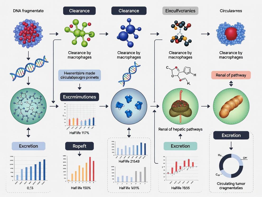
The short half-life of ctDNA is a consequence of its rapid elimination from the bloodstream via two primary mechanisms: enzymatic degradation in the blood and clearance by organs such as the liver and kidneys [5]. The graphic below illustrates the complete journey of ctDNA, from its release to its clearance.
The fragmentation pattern of ctDNA provides clues about its origin. Apoptotic cells typically release shorter DNA fragments (~166 base pairs, associated with nucleosomal DNA), while necrotic cells release longer, more variable fragments [6] [2]. These fragments are rapidly cleared from circulation, primarily by the liver and kidneys, and also undergo enzymatic degradation in the blood [5]. This efficient clearance system is what allows ctDNA levels to reflect the current tumor burden so accurately.
Experimental Protocols for Half-Life and Kinetic Analysis
Determining ctDNA half-life and its kinetic changes in response to treatment requires carefully designed experiments and sensitive detection methodologies.
Core Experimental Workflow
A standard protocol for monitoring ctDNA kinetics involves a structured workflow from sample collection to data analysis, as depicted below.
Key Methodologies for ctDNA Quantification
Different technologies are employed to detect and quantify ctDNA, each with specific strengths and sensitivities.
Table 2: Key Methodologies for ctDNA Detection and Kinetic Analysis
| Methodology | Principle | Sensitivity | Application in Kinetic Studies |
|---|---|---|---|
| Droplet Digital PCR (ddPCR) [1] [3] | Partitions sample into thousands of droplets for individual PCR reactions; directly counts mutant molecules. | ~0.1% [1] | Ideal for tracking known mutations over time; cost-effective and rapid turnaround [3]. |
| Next-Generation Sequencing (NGS) [1] [7] [3] | High-throughput sequencing of a large gene panel or entire exome/genome. | Varies; can be <0.1% with error correction [7] | Provides a broad picture of tumor clones; tracks multiple mutations simultaneously [3]. |
| BEAMing Technology [1] [7] | Combines beads, emulsion, amplification, and magnetics (flow cytometry) for highly sensitive detection. | ~0.02% [1] | Used for ultra-sensitive detection of low-frequency variants in clinical studies [1] [7]. |
Calculating Molecular Response
In clinical studies, ctDNA kinetics are often expressed as "molecular response" rather than a direct half-life measurement. This involves calculating the change in ctDNA levels between baseline and a pre-specified early on-treatment timepoint (e.g., 2-4 weeks after treatment initiation) [3]. Common metrics include:
- ctDNA Clearance: A binary assessment of whether baseline mutations become undetectable [3].
- Variant Allele Frequency (VAF) Change: Calculated as the difference (delta VAF) or ratio (ratio VAF) of mutant allele frequencies between timepoints [3]. A >50% reduction in ctDNA (VAF) is a commonly used threshold to define molecular response and is associated with improved survival [5].
The Scientist's Toolkit: Essential Reagents and Materials
Robust analysis of ctDNA half-life and kinetics relies on a suite of specialized reagents and materials to ensure precision and accuracy.
Table 3: Essential Research Reagents for ctDNA Analysis
| Reagent / Material | Function | Key Features & Examples |
|---|---|---|
| Cell-Free DNA Blood Collection Tubes | Stabilizes nucleated blood cells to prevent genomic DNA contamination and preserve cfDNA profile for up to several days at room temperature [6]. | Streck Cell-Free DNA BCT, CellSave CTCTM, ACD tubes [6]. |
| cfDNA Extraction Kits | Isolates and purifies cfDNA from plasma. Magnetic bead-based methods are favored for high-throughput, automatable workflows [6]. | Magnetic bead-based silica chemistry (e.g., Promega, Qiagen, Roche kits) [6]. |
| Reference Standard Materials | Acts as a process control for evaluating cfDNA extraction efficiency, assay performance, and variant detection accuracy [6]. | Commercially available cfDNA/ctDNA controls (e.g., Seraseq ctDNA, AcroMetrix, nRichDx) with known mutation concentrations and VAFs [6]. |
| Quantitative Assays | Detects and quantifies tumor-specific mutations in extracted cfDNA. | ddPCR assays for known mutations; NGS panels for broader profiling [3]. |
| Fragment Analysis Kits | Assesses the size distribution and quantity of extracted cfDNA, ensuring it matches the expected mononucleosomal peak (~167 bp) and checks for gDNA contamination [6]. | Agilent TapeStation, Bioanalyzer; assays based on fluorometry or qPCR [6]. |
Clinical Implications and Research Context
The short half-life of ctDNA is the critical property that enables its use for real-time monitoring of disease status. In contrast to traditional protein biomarkers like CEA (carcinoembryonic antigen), which have longer half-lives, ctDNA can provide almost a "real-time" snapshot of tumor burden [4]. This makes it exceptionally valuable for several clinical applications within a drug development framework:
- Monitoring Treatment Response: A rapid decline in ctDNA levels after treatment initiation (molecular response) is strongly correlated with improved progression-free survival (PFS) and overall survival (OS) across various cancers, including non-small cell lung cancer (NSCLC), colorectal cancer (CRC), and breast cancer [7] [3].
- Detecting Minimal Residual Disease (MRD): After curative-intent surgery, the persistence or subsequent reappearance of ctDNA, despite being at very low levels, is a highly specific predictor of future clinical recurrence [4] [7]. This can identify patients who might benefit from additional or escalated therapy.
- Understanding Resistance Mechanisms: The rapid clearance of ctDNA also means that the emergence of new resistance mutations can be detected in the blood long before clinical or radiographic progression, allowing for earlier intervention and therapy modification [7].
The concentration of circulating tumor DNA (ctDNA) in the bloodstream is a dynamic equilibrium determined by the balance between its release from tumor cells and its subsequent clearance from the body [8] [2]. Understanding the primary clearance mechanisms—hepatic metabolism, renal excretion, and nuclease degradation—is therefore fundamental to interpreting liquid biopsy results and leveraging ctDNA as a robust biomarker for cancer detection, prognosis, and treatment monitoring [9] [10]. A precise understanding of these pathways is essential for contextualizing the half-life of ctDNA and its fluctuations in response to disease progression or therapeutic intervention. This review synthesizes current evidence on these clearance mechanisms, integrating quantitative data and methodological approaches to provide a technical resource for researchers and drug development professionals.
The Triad of ctDNA Clearance
Cell-free DNA (cfDNA), the total pool of circulating DNA of which ctDNA is a tumor-derived fraction, is efficiently cleared from circulation under normal physiological conditions [11] [9]. The body employs a multi-organ system to achieve this, primarily involving the liver and kidneys, complemented by enzymatic degradation in the blood [9] [10].
Hepatic Metabolism: The liver acts as a primary filter for cfDNA. Kupffer cells, the resident macrophages in the liver, play a key role in phagocytosing and clearing long DNA fragments from the bloodstream [11] [9]. Experimental evidence from mouse models indicates that transiently blocking the liver's clearance function can increase cfDNA levels in circulation by up to 10-fold, underscoring its critical role [10] [12].
Renal Excretion: The kidneys contribute to clearance by filtering smaller DNA fragments from the plasma. This process involves the breakdown of DNA fragments through deoxyribonuclease activity within the kidneys [11] [9]. DNA fragments detected in urine are typically shorter than 100 base pairs (bp), having been passed from plasma through the glomeruli [9].
Nuclease Degradation: Circulating nucleases in the blood, such as DNase1, actively cleave DNA into smaller fragments [9] [13]. This enzymatic degradation shapes the fragmentomic landscape of cfDNA, producing characteristic fragment sizes and end motifs. The activity of nucleases like DNase1L3 is crucial for digesting DNA within apoptotic bodies after phagocytosis [13] [8].
Table 1: Key Organs and Mechanisms in ctDNA Clearance
| Clearance Organ/Mechanism | Primary Cell Type/Enzyme | Function | Evidence and Impact |
|---|---|---|---|
| Liver | Kupffer cells (macrophages) | Phagocytosis of long DNA fragments [11] [9] | Liver blockade in mice raised cfDNA 10-fold; key for large fragment removal [10] [12]. |
| Kidneys | Glomeruli (filtration), Renal nucleases | Filtration and enzymatic degradation of small fragments [11] [9] | Urinary ctDNA fragments are typically <100 bp [9]. |
| Nuclease Degradation | DNase1, DNase1L3, DFFB | Enzymatic cleavage in bloodstream [9] [13] | Shapes fragment size distribution and end motifs; critical post-phagocytosis [13]. |
Quantitative Clearance Kinetics and Influencing Factors
Half-Life of ctDNA
The half-life of ctDNA is a critical parameter for its use in monitoring disease burden and treatment response. Studies report that the half-life of cfDNA/ctDNA is relatively short, enabling real-time monitoring of tumor dynamics.
Table 2: Experimentally Determined Half-Life of Cell-Free DNA
| Study Model | Measured DNA Species | Reported Half-Life | Key Experimental Context |
|---|---|---|---|
| Healthy men post-exercise [11] | 100-250 bp cfDNA fragments | 24.2 minutes | Blood collected in PAXgene tubes to inhibit nucleases; measured via electrophoresis. |
| Post-delivery in women [11] | Fetal-derived cfDNA (SRY gene) | 16.3 minutes (range: 4-30 min) | Blood in EDTA tubes; measured via PCR. Lacked nuclease inhibition. |
| General estimate (review) [10] | ctDNA in healthy individuals | ~2 hours | Cited in human cohort study of colorectal cancer patients. |
Impact of Organ Function on Clearance
The potential influence of impaired liver or kidney function on ctDNA levels is a key consideration for clinical testing. Recent large-scale human studies provide clarifying evidence.
Table 3: Impact of Liver and Kidney Function on ctDNA Detection in Human Studies
| Study & Population | Key Findings on Liver Function | Key Findings on Kidney Function | Clinical Implication |
|---|---|---|---|
| 846 Stage I-III Colorectal Cancer Patients [10] [12] | No significant association between bilirubin, alkaline phosphatase, or alanine transaminase levels and ctDNA detection/level. | No significant association between creatinine, eGFR, sodium, or potassium levels and ctDNA detection/level. | ctDNA test results are stable and reliable across a wide range of liver and kidney function in early-stage CRC. |
| 276 Muscle-Invasive Bladder Cancer Patients [14] | No significant associations observed between liver function markers and ctDNA detection. | No significant associations observed between kidney function markers and ctDNA detection. | Plasma ctDNA detection is not affected by kidney/liver function, ensuring reliability for monitoring. |
Experimental Protocols for Studying Clearance
Protocol: Measuring cfDNA Half-Life After Physiological Stimulus
The following methodology, adapted from a 2025 study, details a robust approach for investigating cfDNA kinetics and half-life using exercise as a physiological stimulus [11].
- 1. Participant Preparation: Recruit eligible participants (e.g., healthy adult men with no chronic conditions). Obtain informed consent and baseline characteristics.
- 2. Pre-Exercise Baseline Sample: Collect a blood sample (e.g., 10 mL) while participants are in a resting, sitting position. Use specialized blood collection tubes designed to stabilize cfDNA, such as PAXgene Blood ccfDNA Tubes, which inhibit nuclease activity and minimize genomic DNA contamination [11].
- 3. Controlled Exercise Intervention: Subject participants to a standardized exercise regimen to induce cfDNA release. For example, a 30-minute treadmill exercise at a controlled speed of 8 km/h [11].
- 4. Post-Exercise Time-Series Sampling: Collect blood samples at multiple time points immediately after exercise cessation (e.g., 0, 5, 10, 15, 30, and 60 minutes). All samples should be drawn with participants in the same resting position [11].
- 5. Sample Processing and Storage: Centrifuge blood samples at 4°C (e.g., 1900× g for 15 minutes). Carefully aspirate the plasma supernatant and perform a second centrifugation under the same conditions to remove residual cells. Aliquot plasma and store at 4°C if DNA extraction will occur within 1-3 days [11].
- 6. cfDNA Extraction: Isolate cfDNA from plasma using a dedicated kit (e.g., QuickGene cfDNA isolation kit) and extraction system (e.g., QuickGene-Mini8L) according to the manufacturer's instructions. The protocol typically involves adding a lysis buffer and ethanol to the plasma, followed by passing the mixture through a filter cartridge and washing with buffer solutions [11].
- 7. cfDNA Quantification and Fragment Analysis: Quantify cfDNA concentration using an electrophoresis-based system (e.g., 4150 TapeStation system). This technique allows for quantification based on specific fragment sizes (e.g., 100-250 bp), providing a more accurate assessment than PCR, which can be biased by genomic DNA amplification [11].
- 8. Data Analysis and Half-Life Calculation: Analyze the concentration of the target cfDNA fragment size over time. The half-life can be calculated by determining the time required for the peak concentration to decrease by 50% after exercise [11].
The Scientist's Toolkit: Essential Reagents and Materials
Table 4: Key Research Reagents for ctDNA Clearance and Half-Life Studies
| Reagent / Material | Specific Example | Critical Function in Protocol |
|---|---|---|
| Blood Collection Tube | PAXgene Blood ccfDNA Tubes (QIAGEN) | Stabilizes cfDNA immediately upon draw; inhibits nuclease degradation and reduces gDNA contamination [11]. |
| cfDNA Extraction Kit | QuickGene cfDNA Isolation Kit (KURABO) | Efficiently isolates pure cfDNA from plasma samples for downstream analysis [11]. |
| Automated Extraction System | QuickGene-Mini8L (KURABO) | Automates the cfDNA extraction process from plasma, improving reproducibility and yield [11]. |
| Fragment Analyzer | 4150 TapeStation System (Agilent) | Electrophoresis-based quantification of cfDNA concentration and size distribution (e.g., 100-250 bp fragments) [11]. |
Integrated Clearance Pathways
The following diagram summarizes the coordinated interplay of the primary clearance mechanisms for circulating tumor DNA (ctDNA).
The diagram illustrates how ctDNA in the bloodstream is processed through three concurrent pathways. Nuclease degradation in the blood enzymatically cleaves DNA. The liver primarily clears long DNA fragments via phagocytosis by Kupffer cells, while the kidneys filter short fragments for excretion. This multi-mechanism system maintains low background cfDNA levels under physiological conditions [11] [9] [10].
Discussion and Future Directions
The body's clearance mechanisms maintain a tight regulatory control over cfDNA levels, resulting in a short half-life that enables real-time monitoring of tumor dynamics [11] [10]. While the liver, kidneys, and nucleases are established as the primary clearance routes, recent evidence from large human cohorts suggests that variations in liver and kidney function within typical clinical ranges do not significantly confound ctDNA detection in early-stage cancers [14] [10] [12]. This finding strengthens the reliability of ctDNA as a robust biomarker across diverse patient populations.
Future research should focus on elucidating the clearance dynamics in patients with severe organ dysfunction, such as end-stage renal or liver disease, which were not fully represented in the cited studies. Furthermore, a deeper understanding of how nuclease activities vary between individuals and disease states could unlock new diagnostic and therapeutic applications. As liquid biopsy technology evolves toward detecting minimal residual disease and earlier-stage cancers, a refined, quantitative model of ctDNA clearance that integrates all contributing factors will be indispensable for accurate clinical interpretation.
Circulating tumor DNA (ctDNA) has emerged as a pivotal biomarker in liquid biopsies, enabling non-invasive cancer diagnosis, prognosis, and therapy monitoring. The biological origins of ctDNA—primarily apoptosis, necrosis, and active secretion via extracellular vesicles (EVs)—fundamentally shape its molecular characteristics and clearance kinetics [9] [2]. A thorough understanding of these release mechanisms is essential for interpreting ctDNA dynamics, including its half-life, which is estimated between 16 minutes and several hours [7]. This guide provides a technical overview of ctDNA sources, their experimental investigation, and their implications for ctDNA clearance research, tailored for researchers, scientists, and drug development professionals.
Biological Release Mechanisms of ctDNA
Passive Release via Apoptosis
Apoptosis, a form of programmed cell death, is a significant source of ctDNA, characterized by a highly regulated and systematic DNA fragmentation process.
- Molecular Mechanism: The execution is carried out by caspase-activated DNase (CAD) and other nucleases like DNaseI L-3, NM23-H1, and EndoG. These enzymes cleave DNA at internucleosomal regions, resulting in DNA fragments that are wrapped around histone proteins and packaged into apoptotic bodies [9].
- Fragment Characteristics: Apoptosis produces short, mononucleosomal DNA fragments with a dominant peak at 167 base pairs (bp), corresponding to the length of DNA wrapped around a single nucleosome (147 bp) plus a linker DNA (20 bp) [9] [2]. This process creates a distinctive "ladder-like" pattern on gel electrophoresis [9].
- Clearance Implications: Apoptotic bodies are swiftly cleared by phagocytosis, primarily by macrophages. The DNA within these bodies is subsequently enzymatically digested and released as soluble cfDNA into circulation. This efficient, packaged clearance mechanism may influence the observed half-life of ctDNA in the bloodstream [9].
Passive Release via Necrosis
Necrosis, often a result of pathological cell death due to factors like hypoxia or metabolic stress in the tumor microenvironment, contributes to a different pool of ctDNA.
- Molecular Mechanism: In contrast to apoptosis, necrosis involves uncontrolled cell death with plasma membrane rupture, leading to the random and disorganized release of cellular contents, including large, irregular DNA fragments, into the extracellular space [9] [2].
- Fragment Characteristics: Necrosis is associated with the release of longer DNA fragments, often ranging up to many kilo-base pairs (kbp), due to the non-systematic digestion of genomic DNA [9]. The cfDNA integrity (ratio of long to short fragments) is often higher in cancer patients, suggesting a significant role for necrotic cell death, particularly in advanced or aggressive tumors [2].
- Clearance Implications: The clearance of necrotic debris involves a robust inflammatory response and attractants for immune cells. The larger DNA fragments released during necrosis are exposed to extracellular nucleases and degradative agents, potentially altering their stability and half-life in circulation [9].
Active Secretion via Extracellular Vesicles (EVs)
A crucial non-passive mechanism involves the active secretion of DNA by viable tumor cells through extracellular vesicles, which protects the DNA from immediate degradation.
- Molecular Mechanism: EVs are lipid-bilayer enclosed particles released by cells. DNA can be associated with different EV subtypes:
- Small EVs (sEVs/exosomes): Generated through the endosomal pathway, these vesicles (40-150 nm) can carry DNA, potentially as a mechanism to maintain cellular homeostasis by removing cytoplasmic DNA or as a form of intercellular communication [15] [16].
- Microvesicles (MVs): Produced by direct outward budding of the plasma membrane (150-1000 nm), MVs have been shown to carry abundant DNA, including oncogene sequences [15].
- DNA Localization and Characteristics: EV-associated DNA can be found both on the external surface of the vesicle and enclosed within it [16]. The DNA within EVs is often of higher molecular weight, with fragments up to 4 kb reported, and is protected from circulating nucleases [16]. This DNA accurately reflects the tumor's mutational status [15] [16].
- Clearance Implications: The vesicular membrane shields EV-DNA from nucleases, potentially conferring a longer half-life compared to freely circulating ctDNA. The clearance pathways for EVs themselves (e.g., via the liver and spleen) will govern the persistence of EV-associated DNA in the bloodstream [15].
Table 1: Comparative Characteristics of ctDNA from Different Biological Sources
| Feature | Apoptosis | Necrosis | Active EV Secretion |
|---|---|---|---|
| Primary Trigger | Programmed cell death | Pathological cell death (e.g., hypoxia) | Cellular homeostasis / Communication |
| Key Enzymes/Proteins | Caspases, CAD, EndoG | DNases (non-specific) | ESCRT, nSMase, ARF6 |
| Dominant Fragment Size | ~167 bp (mononucleosomal) | >200 bp, up to many kbp | Longer fragments, up to 4 kb |
| DNA Integrity | Low | High | High |
| Protection from Nucleases | Moderate (in apoptotic bodies) | Low | High (within lipid bilayer) |
| Impact on Half-life | Shorter (efficient clearance) | Shorter (exposed to degradation) | Potentially longer (vesicle-protected) |
Investigating the biological sources of ctDNA requires integrated experimental approaches, from sample collection to advanced molecular analysis. The workflow below outlines the pathway from hypothesis to data interpretation.
Diagram 1: Experimental workflow for ctDNA source investigation, covering key stages from sample collection to data interpretation.
Sample Collection and Pre-analytical Processing
The integrity of pre-analytical steps is critical for accurate ctDNA analysis.
- Blood Collection: It is recommended to collect 2 × 10 mL of blood using butterfly needles, avoiding thin needles and prolonged tourniquet use. Blood should be drawn into specialized blood collection tubes (BCTs) containing cell-stabilizing preservatives (e.g., Streck cfDNA BCT, PAXgene Blood ccfDNA tubes) [17]. These tubes prevent the release of genomic DNA from white blood cells, allowing sample stability for up to 3-7 days at room temperature, unlike conventional EDTA tubes which require processing within 2-6 hours [17].
- Plasma Separation and cfDNA Extraction: Plasma must be separated via a two-step centrifugation protocol to efficiently remove cells and debris [17]. Subsequent cfDNA extraction from the plasma can be performed using commercial kits optimized for low-concentration, fragmented DNA.
Source-Specific Analytical Techniques
Different methodologies are employed to attribute ctDNA to its biological source.
- Fragment Size Analysis: Analyzing the size distribution of cfDNA fragments is a direct method to infer origin. Tools like the Bioanalyzer or TapeStation can reveal the dominant ~167 bp peak indicative of apoptosis, while a significant proportion of longer fragments (>200 bp) suggests a contribution from necrosis [9] [2]. EV-derived DNA often appears as a smear of high-molecular-weight fragments when analyzed without vesicle lysis [16].
- EV Isolation and DNA Extraction: To study EV-associated DNA, EVs must first be isolated from plasma. Common methods include:
- Ultracentrifugation: The traditional gold standard, which pellets EVs through high-speed spins [15] [16].
- Commercial Kits: Polymer-based precipitation kits (e.g., ExoQuick, Total Exosome Isolation Reagent) offer a more accessible alternative [16].
- To determine if DNA is inside EVs or surface-bound, treatments with DNase I are used. DNase will degrade external DNA, while internal DNA is protected and can be recovered after EV lysis (e.g., with proteinase K and detergent) [16].
- Mutation Detection and Quantification: The presence of tumor-specific mutations in total cfDNA versus the EV-DNA fraction can be assessed using highly sensitive techniques like droplet digital PCR (ddPCR) or next-generation sequencing (NGS) with unique molecular identifiers (UMIs) for error correction [7] [16]. The ratio of mutant to wild-type alleles (variant allele frequency, VAF) can provide insights into the relative abundance of ctDNA from different sources.
Table 2: The Scientist's Toolkit: Key Reagents and Methods for ctDNA Source Analysis
| Tool Category | Specific Examples | Function in ctDNA Research |
|---|---|---|
| Blood Collection Tubes | Streck cfDNA BCT, PAXgene Blood ccfDNA Tube | Stabilizes nucleated blood cells to prevent background gDNA release during sample transport/storage. |
| EV Isolation Kits | ExoQuick, Total Exosome Isolation Reagent | Precipitates EVs from biofluids for subsequent analysis of vesicle-protected DNA. |
| Nuclease Enzymes | DNase I | Digests DNA outside of EVs (surface-bound or free-floating) to confirm intravesicular DNA localization. |
| Ultracentrifugation | Protocol for 100,000 × g spins | Pellet-based isolation of EVs from cell-conditioned media or plasma. |
| Size Analysis Instruments | Agilent Bioanalyzer, TapeStation | Precisely determines the fragment size profile of cfDNA to infer apoptotic vs. necrotic origin. |
| High-Sensitivity DNA Assays | ddPCR, NGS with UMIs (e.g., Safe-SeqS, CAPP-Seq) | Detects and quantifies rare tumor-specific mutations in complex cfDNA or EV-DNA backgrounds. |
Interplay with ctDNA Clearance Kinetics
The mechanism of ctDNA release is intrinsically linked to its fate in the circulatory system. The following diagram synthesizes the relationships between source mechanisms, ctDNA properties, and clearance pathways.
Diagram 2: Logical relationship between ctDNA release mechanisms, physical properties, clearance routes, and resulting half-life.
- Clearance Pathways: The body eliminates ctDNA through two primary routes: (1) enzymatic degradation by circulating nucleases in the blood, and (2) phagocytosis by macrophages, primarily in the liver and spleen [7] [17]. The accessibility of ctDNA to these systems determines its half-life.
- Source Dictates Fate:
- Apoptotic and Necrotic DNA: Free-floating ctDNA from apoptosis and necrosis is directly exposed to circulating nucleases. While apoptotic DNA is already fragmented, necrotic DNA is degraded into smaller pieces over time. Both are susceptible to rapid clearance by hepatic macrophages, leading to a short half-life [9] [7].
- EV-Associated DNA: The lipid bilayer of EVs acts as a physical barrier, shielding the enclosed DNA from nucleases [15] [16]. Consequently, the clearance of EV-DNA is tied to the metabolic fate of the vesicles themselves, which may be filtered by organs like the liver and spleen. This protection mechanism could potentially prolong the half-life of EV-DNA compared to free ctDNA, a critical consideration for pharmacokinetic models in drug development [15].
- Influencing Factors: Pre-analytical variables can impact clearance measurements. For instance, surgical trauma or other inflammatory conditions can cause a transient increase in total cfDNA, complicating the interpretation of ctDNA levels [17]. Furthermore, experimental strategies to slow ctDNA clearance by interfering with liver macrophages or nucleases are being explored to improve the sensitivity of liquid biopsies [17].
The biological origins of ctDNA—apoptosis, necrosis, and active EV secretion—are not merely alternative pathways but are fundamental determinants of its physical state and persistence in circulation. Apoptosis provides a predictable, fragmented source, necrosis contributes larger, more heterogeneous molecules, and EV secretion offers a protected pool of DNA with potentially distinct kinetics. For researchers focused on the half-life and clearance of ctDNA, ignoring these origins risks misinterpretation of data. Future work must continue to refine methods for source-specific ctDNA analysis and integrate these insights into robust pharmacokinetic and disease-monitoring models, ultimately advancing the application of liquid biopsy in precision oncology.
The characterization of cell-free DNA (cfDNA) fragmentomics has emerged as a critical field in molecular biology, with profound implications for cancer diagnostics and therapeutic monitoring. Within liquid biopsy approaches, circulating tumor DNA (ctDNA) represents a fraction of total cfDNA that originates from tumor cells, carrying genetic and epigenetic information about the malignancy. The half-life and clearance kinetics of ctDNA are directly influenced by its molecular origin—specifically, whether it is derived via apoptotic or necrotic cell death pathways. These distinct death mechanisms impart characteristic fragmentation patterns to the resulting DNA, which in turn affect its stability, clearance, and ultimate detectability in circulation. This technical review examines the fundamental differences between apoptotic and necrotic DNA fragmentation patterns, their formation mechanisms, and the direct implications for ctDNA persistence in biological fluids. Understanding these relationships provides a crucial foundation for optimizing liquid biopsy applications, interpreting ctDNA kinetics in response to therapy, and developing advanced fragmentomic-based cancer diagnostics.
Biochemical Mechanisms of DNA Fragmentation
Apoptotic DNA Fragmentation
Apoptosis, or programmed cell death, represents a highly regulated process characterized by specific biochemical events that lead to controlled cellular dismantling. A defining biochemical hallmark of apoptosis is the systematic fragmentation of nuclear DNA into oligonucleosomal units through the activation of specific endonucleases [18].
The central mechanism involves caspase-activated DNase (CAD), which exists in proliferating cells as an inactive complex with its inhibitor, ICAD (inhibitor of caspase-activated DNase). During apoptosis, the apoptotic effector caspase, caspase-3, cleaves ICAD, thereby dissociating the CAD:ICAD complex and activating CAD's endonucleolytic function [18]. The activated CAD enzyme then cleaves chromosomal DNA at internucleosomal linker sites—the regions between nucleosomes that are exposed and accessible in chromatin. Nucleosomes, the fundamental repeating units of chromatin, consist of approximately 147 base pairs of DNA wrapped around a histone core, connected by linker DNA of varying lengths [18].
This specific cleavage pattern results in DNA fragments that are multiples of approximately 180-200 base pairs, corresponding to single nucleosomes (approximately 147 bp plus linker DNA) and their oligomers [18]. The fragmentation occurs in a discontinuous manner, likely reflecting different levels of restriction in DNA accessibility to DNases imposed by the supranucleosomal and nucleosomal levels of chromatin structure [18].
Necrotic DNA Fragmentation
In contrast to apoptosis, necrosis represents a premature form of cell death resulting from overwhelming cellular injury, characterized by mitochondrial dysfunction, ATP depletion, and loss of plasma membrane integrity. The DNA degradation in necrosis lacks the ordered progression seen in apoptosis and occurs through more stochastic processes [19] [9].
During necrosis, cellular components, including DNA, are released randomly into the extracellular space due to plasma membrane rupture [9]. This exposes the genetic material to intracellular and extracellular degradative agents, including nucleases, free radicals, and factors from the tissue microenvironment. Research comparing early necrosis (1 hour after cell death) with apoptosis in rat thymus and Jurkat cell models has revealed distinct differences in the DNA ends generated [19].
Analysis of double-strand DNA breaks shows that early necrotic cells predominantly contain 5' overhangs, with an absence of blunt ends or 3' overhangs that are characteristic of apoptosis [19]. This selective generation of 5' overhangs suggests the involvement of a specific 3'→5' exonuclease activity in early necrotic DNA degradation. The researchers hypothesized that this damage pattern could be attributed to the ubiquitous 3'→5' proofreading exonuclease activity associated with cellular polymerases [19]. The fragmentation pattern is non-systematic, resulting in a broader size distribution of DNA fragments, including larger fragments up to many kilobases in length [9].
Figure 1: Biochemical Pathways of DNA Fragmentation in Apoptosis and Necrosis
Comparative Fragment Characteristics
The distinct mechanisms of DNA fragmentation in apoptosis and necrosis produce characteristic fragment patterns that serve as molecular signatures of their cellular origins. These differences are evident in multiple aspects of DNA structure and size distribution.
DNA Fragment Size Distribution
Apoptotic DNA demonstrates a highly regular fragmentation pattern resulting from enzymatic cleavage at internucleosomal sites. This produces DNA fragments that are multiples of approximately 180-200 base pairs, creating the classic "DNA ladder" pattern when separated by agarose gel electrophoresis [18] [20]. The predominant cfDNA fragment size in apoptosis is approximately 167 bp, corresponding to the length of DNA wrapped around a single nucleosome (147 bp) plus a linker DNA (20 bp) [9]. This specific size reflects protection from cleavage by nucleosomal structures.
Necrotic DNA displays a more heterogeneous size distribution due to random digestion by nucleases. The fragments typically show a "smear" pattern on agarose gels, with a broad size range extending from small fragments to large fragments of many kilobase pairs [9]. The absence of controlled enzymatic cleavage results in this non-specific fragmentation pattern without the regular periodicity observed in apoptotic DNA.
DNA End Structure Characteristics
The structural characteristics of DNA ends differ significantly between the two cell death pathways:
Apoptotic DNA ends typically include a combination of blunt ends and 3' overhangs, which are characteristic of DNA cleaved by the major apoptotic nucleases DNase I and caspase-activated deoxyribonuclease (CAD) [19]. These specific end structures reflect the precise enzymatic mechanism of CAD-mediated cleavage.
Necrotic DNA ends in early necrosis predominantly feature 5' overhangs, with an absence of blunt ends or 3' overhangs [19]. This distinct end structure suggests the involvement of a 3'→5' exonuclease activity in early necrotic DNA degradation and provides a potential mechanism for distinguishing necrosis-derived DNA from apoptosis-derived DNA in clinical samples.
Table 1: Comparative Characteristics of Apoptotic and Necrotic DNA Fragmentation
| Characteristic | Apoptotic DNA | Necrotic DNA |
|---|---|---|
| Primary Mechanism | Caspase-activated DNase (CAD) | Random nuclease activity |
| DNA End Structures | Blunt ends and 3' overhangs [19] | Predominantly 5' overhangs [19] |
| Electrophoretic Pattern | Regular "ladder" at ~180bp intervals [18] | Heterogeneous "smear" [9] |
| Fragment Size Range | Narrow (multiples of ~180bp) [18] | Broad (up to kilobases) [9] |
| Predominant cfDNA Size | ~167 bp [9] | Variable, often larger fragments |
| Chromatin Protection | Nucleosomal protection evident [9] | Limited nucleosomal protection |
| Associated Conditions | Programmed cell death, homeostasis | Cellular injury, inflammation |
Detection Methodologies and Experimental Protocols
DNA Fragmentation Analysis via Agarose Gel Electrophoresis
The detection of apoptotic DNA fragmentation through agarose gel electrophoresis remains a fundamental methodology for identifying programmed cell death. This protocol enables visualization of the characteristic internucleosomal DNA cleavage pattern [20].
Protocol Stages:
Cell Harvesting and Lysis
- Pellet approximately 1-5 × 10^6 cells by centrifugation
- Lyse cells in 0.5 mL detergent buffer (10 mM Tris pH 7.4, 5 mM EDTA, 0.2% Triton X-100)
- Vortex and incubate on ice for 30 minutes
- Centrifuge at 27,000 × g for 30 minutes to separate fragmented DNA (supernatant) from intact chromatin (pellet)
DNA Precipitation
- Divide supernatants into two 250 μL aliquots
- Add 50 μL ice-cold 5 M NaCl to each aliquot and vortex
- Add 600 μL ethanol and 150 μL 3 M sodium-acetate (pH 5.2) and mix
- Incubate at -80°C for 1 hour
- Centrifuge at 20,000 × g for 20 minutes and discard supernatants carefully
- Pool DNA extracts by re-dissolving pellets in 400 μL extraction buffer (10 mM Tris, 5 mM EDTA)
DNA Purification and Analysis
- Add 2 μL of 10 mg/mL DNase-free RNase and incubate for 5 hours at 37°C
- Add 25 μL proteinase K (20 mg/mL) and 40 μL buffer (100 mM Tris pH 8.0, 100 mM EDTA, 250 mM NaCl)
- Incubate overnight at 65°C
- Extract DNA with phenol/chloroform/isoamyl alcohol (25:24:1) and precipitate with ethanol
- Air-dry pellet and resuspend in 20 μL Tris-acetate EDTA buffer with loading dye
- Separate DNA electrophoretically on a 2% agarose gel containing ethidium bromide
- Visualize by ultraviolet transillumination [20]
Advanced Detection Techniques
Several sophisticated methodologies have been developed to enhance sensitivity and specificity in detecting apoptotic and necrotic DNA fragmentation:
TUNEL Assay (Terminal deoxynucleotidyl transferase dUTP Nick End Labeling) This method detects DNA strand breaks by utilizing terminal deoxynucleotidyl transferase (TdT) to add labeled dUTP to the 3'-ends of DNA fragments. The fluorochrome-based TUNEL assay can be correlated with cellular DNA content and cell cycle position via flow cytometry, while the avidin-peroxidase labeling TUNEL assay is applicable for light absorption microscopy [18].
In Situ Ligatio This technique enables selective detection of double-strand DNA breaks with specific end structures. Using hairpin-shaped oligonucleotide probes with defined ends, in situ ligation can distinguish between blunt ends, 3' overhangs, and 5' overhangs characteristic of different cell death pathways [19]. When combined with Klenow enzyme pretreatment to modify DNA ends, this method can detect specific exonuclease activities in situ.
Flow Cytometric Analysis of Sub-G1 Cells Analysis of DNA content by flow cytometry can identify apoptotic cells with fragmented DNA as populations with fractional DNA content (sub-G1 cells). The Nicoletti assay utilizes propidium iodide staining and flow cytometry to rapidly measure thymocyte apoptosis based on reduced DNA content [18].
Figure 2: Methodologies for Detecting DNA Fragmentation Patterns
Clearance Kinetics and Biological Implications
Half-Life and Clearance Mechanisms of Cell-Free DNA
The clearance kinetics of cell-free DNA, including ctDNA, are influenced by multiple physiological factors, with fragment characteristics playing a significant role in determining half-life. Recent research has provided quantitative data on cfDNA clearance rates using improved methodological approaches.
A 2025 study employing fragment size-specific measurement reported a cfDNA half-life of approximately 24.2 minutes following exercise-induced elevation in healthy individuals [21] [22]. This study utilized specialized blood collection tubes (PAXgene Blood ccfDNA tubes) to prevent nuclease-mediated degradation ex vivo and focused specifically on 100-250 bp fragments, similar in size to apoptosis-derived DNA. The transient increase in cfDNA fragments post-exercise rapidly returned to baseline within 60 minutes, demonstrating efficient clearance mechanisms [21].
The metabolism and clearance of cfDNA occur primarily in the liver, kidneys, and spleen through multiple pathways [21]. Kupffer cells in the liver play a key role in removing longer DNA fragments, while the kidneys contribute to fragmentation through deoxyribonuclease activity. Clearance involves degradation by nucleases, phagocytosis, and immune complex formation [21].
Earlier studies reported variable half-life estimates ranging from 15 minutes to 2 hours, depending on methodological approaches [21]. For example, Lo et al. reported an average half-life of 16.3 minutes (range 4-30 minutes) for fetal DNA in maternal circulation based on SRY gene detection [21]. The variability in reported half-lives underscores the influence of measurement techniques, fragment sizes, and physiological context on clearance kinetics.
Table 2: Circulating DNA Half-Life Estimates Across Studies
| Study Context | Half-Life Estimate | Measurement Method | Key Factors |
|---|---|---|---|
| Post-exercise (2025) | 24.2 minutes [21] | Electrophoresis (100-250 bp fragments) | Nuclease activity, fragment size |
| Fetal DNA in maternal circulation | 16.3 minutes (range 4-30) [21] | Real-time PCR (SRY gene) | Physiological clearance mechanisms |
| Ancient DNA in bone | 521 years (242 bp mtDNA) [23] | Quantitative PCR | Deposition environment, temperature |
| General cfDNA clearance | 15 minutes - 2 hours [21] | Various methods | Physiological state, nuclease activity |
Biological Implications for ctDNA Research
The fragment characteristics and clearance kinetics of tumor-derived DNA have profound implications for liquid biopsy applications and cancer management:
Tumor Microenvironment Interpretation: The relative proportion of apoptosis-derived versus necrosis-derived ctDNA fragments provides insight into tumor biology and treatment response. Tumors with predominant apoptotic signatures may respond differently to therapies than those with significant necrosis, which often indicates more aggressive disease or treatment-induced cytotoxicity [9].
Detection Sensitivity Considerations: The rapid clearance of cfDNA (half-life of approximately 24.2 minutes) supports its utility as a real-time biomarker for monitoring tumor dynamics and treatment response [21]. However, the short half-life also necessitates optimized sampling protocols to capture transient changes in ctDNA levels.
Fragment Size as a Diagnostic Parameter: The distinct size distributions of apoptosis-derived and necrosis-derived DNA fragments can be leveraged to improve the specificity of ctDNA detection. Size selection approaches may enhance tumor DNA detection in backgrounds of normal cfDNA, particularly since apoptosis-derived fragments from healthy cells tend to demonstrate more uniform nucleosomal protection [9].
Therapeutic Response Monitoring: Changes in ctDNA fragmentation patterns following treatment may provide early indicators of therapeutic efficacy. For example, shifts from necrosis-dominated to apoptosis-dominated patterns (or vice versa) could reflect specific drug mechanisms of action and tumor responses [9].
The Scientist's Toolkit: Essential Research Reagents
Table 3: Key Research Reagents for DNA Fragmentation Analysis
| Reagent/Kit | Primary Function | Application Context |
|---|---|---|
| PAXgene Blood ccfDNA Tubes | Stabilizes cfDNA and inhibits nuclease degradation ex vivo [21] | Blood collection for cfDNA half-life studies |
| QuickGene cfDNA Isolation Kit | Extracts cell-free DNA from plasma samples [21] | cfDNA purification for fragment analysis |
| DNase-free RNase | Degrades RNA to prevent interference in DNA analysis [20] | Sample preparation for DNA fragmentation assays |
| Proteinase K | Digests nucleoproteins and enhances DNA recovery [20] | DNA extraction from cells and tissues |
| T4 DNA Ligase | Joins DNA ends with specific terminal structures [19] | In situ ligation assays for DNA end characterization |
| Klenow Enzyme | Fills in or removes DNA overhangs to create blunt ends [19] | Modification of DNA ends for structural analysis |
| SYBR-green Detection Chemistry | Fluorescent DNA binding for quantitative PCR applications [23] | Quantification of specific DNA fragments |
The fragment size characteristics of DNA derived from apoptotic versus necrotic cell death pathways represent fundamental biological signatures with direct implications for circulating tumor DNA research. The systematic, enzyme-mediated fragmentation in apoptosis produces regular ~180bp multiples with blunt and 3' overhang ends, while necrosis generates heterogeneous fragments with predominant 5' overhangs. These distinct patterns influence DNA clearance kinetics, with recent studies demonstrating a cfDNA half-life of approximately 24 minutes under physiological conditions. Understanding these relationships enhances our ability to interpret ctDNA profiles in cancer patients, providing insights into tumor biology, treatment responses, and disease dynamics. As liquid biopsy technologies continue to evolve, incorporating fragmentomic analyses that distinguish apoptotic and necrotic signatures will undoubtedly refine the diagnostic, prognostic, and predictive utility of ctDNA in clinical oncology.
Circulating tumor DNA (ctDNA) has emerged as a transformative biomarker in oncology, enabling non-invasive tumor genotyping and monitoring of treatment response. The clinical utility of ctDNA is fundamentally governed by its clearance kinetics, which determine its presence and concentration in the bloodstream. This technical review examines the complex interplay of tumor location, vascularity, and host metabolic factors that modulate ctDNA clearance rates. Within the context of broader ctDNA half-life research, we synthesize current understanding of how anatomical compartmentalization, vascular architecture, and physiological clearance mechanisms collectively influence ctDNA dynamics. Through structured data presentation, experimental protocols, and visual schematics, this review provides researchers and drug development professionals with a comprehensive framework for interpreting ctDNA kinetics across varying biological contexts.
Circulating tumor DNA (ctDNA) comprises fragments of tumor-derived nucleic acids released into the bloodstream through various biological processes, including apoptosis, necrosis, and active secretion [8] [24]. These fragments typically range from 70-200 base pairs in length, with a characteristic peak at approximately 166 bp corresponding to nucleosome-associated DNA [8] [1]. The half-life of ctDNA is remarkably short, estimated between 16 minutes to 2.5 hours, necessitating careful timing of blood collection for clinical applications [1] [11]. This rapid clearance occurs primarily through degradation by nucleases, hepatic uptake, renal excretion, and immune complex formation [11] [24].
Understanding ctDNA clearance kinetics is paramount for optimizing its clinical applications, including minimal residual disease detection, therapy monitoring, and treatment response assessment. The concentration of ctDNA detected in circulation represents a dynamic equilibrium between the rates of release from tumor cells and elimination by clearance mechanisms [24]. This review systematically examines how tumor location, vascularity, and metabolic factors perturb this equilibrium, thereby influencing measurable ctDNA levels and interpretation of liquid biopsy results.
Tumor Location and Anatomical Considerations
The anatomical origin of a tumor significantly influences the concentration and fragmentation patterns of ctDNA detected in peripheral blood due to variations in release mechanisms and direct drainage pathways. Tumors in direct contact with certain body fluids can shed ctDNA more readily than those requiring passage through multiple tissue barriers before entering systemic circulation.
Table 1: Impact of Tumor Location on ctDNA Detection and Characteristics
| Tumor Location/Body Fluid | ctDNA Concentration | Fragment Size Characteristics | Primary Release Mechanisms |
|---|---|---|---|
| Plasma (Systemic Circulation) | Low to Moderate (1-10 ng/mL) [1] | ~166 bp (nucleosomal) [8] | Apoptosis, Necrosis, Active Release [24] |
| Cerebrospinal Fluid (CSF) | Higher than plasma [24] | Not specified | Direct shedding from CNS tumors [24] |
| Pleural/Peritoneal Effusions | Higher than plasma [24] [8] | Not specified | Necrosis, Secretion from metastatic lesions [24] |
| Urine | Variable | Shorter fragments (<100 bp) [24] | Glomerular filtration of plasma ctDNA [24] |
| Saliva | Lower than plasma | Very short (40-60 bp) [24] | Direct shedding from oral tumors [24] |
The blood-brain barrier represents a particularly significant anatomical constraint, limiting the passage of ctDNA from central nervous system tumors into peripheral circulation. Consequently, cerebrospinal fluid (CSF) often contains substantially higher concentrations of ctDNA than plasma for CNS malignancies, making CSF a superior liquid biopsy source for these tumors [24]. Similarly, malignant effusions (pleural, peritoneal, pericardial) constitute rich reservoirs of ctDNA due to their proximity to tumor surfaces and relatively contained anatomical spaces [8] [24].
The anatomical compartmentalization of ctDNA has profound implications for clinical assay sensitivity. For tumors draining directly into contained spaces (e.g., ovarian cancer cells shedding into peritoneal fluid or glioblastoma cells into CSF), local fluid sampling often detects ctDNA when peripheral blood testing yields false-negative results [24]. Furthermore, fragment size analysis can provide clues about the tissue of origin, with different bodily fluids exhibiting characteristic fragmentation patterns due to their distinct filtration mechanisms and nuclease activities [24].
Tumor Vascularity and Perfusion Dynamics
Tumor vascular architecture represents a critical determinant of ctDNA release and clearance, governing both the delivery of tumor DNA to circulation and the efficiency of its elimination. The development of tumor vasculature occurs through multiple mechanisms, including angiogenesis, vascular co-option, and vasculogenic mimicry, each creating distinct patterns of vascular efficiency [25].
Vascular Phases of Tumor Growth
Solid tumor growth progresses through distinct vascular phases that dramatically influence ctDNA shedding:
- Avascular Phase: Early tumors (≤1-2 mm³) lack intrinsic vasculature and depend on diffusion for nutrient exchange, resulting in limited ctDNA shedding due to barriers to DNA entry into circulation [25].
- Angiogenic Switch: Tumors activate the "angiogenic switch" by releasing factors like VEGF and FGF-2, initiating neovascularization and enabling transition to the vascular phase [25].
- Vascular Phase: Once vascularized, tumors exhibit exponential growth and significantly increased ctDNA release due to enhanced vascular permeability and improved access to circulation [25].
The tumor vasculature that forms is typically disorganized, leaky, and dysfunctional, characterized by heterogeneous blood flow, increased vascular permeability, and irregular branching patterns [26] [25]. This abnormal vascular network creates regions of hypoperfusion and hypoxia, which promote necrotic cell death—a process associated with release of larger, more irregular DNA fragments compared to the orderly fragmentation pattern of apoptotic DNA [24].
Quantitative Models of Vascular Efficiency
The relationship between tumor vascularization and metabolic rate can be described mathematically. West et al. proposed a quantitative theory linking tumor vascular networks to metabolic rate, stating that tumor metabolic rate (Bₜ) is proportional to the total blood volume flow rate to the tumor (Qₜ) [26]. This relationship follows:
Bₜ ∝ Qₜ
This model enables predictions of how vascular efficiency influences not only tumor growth but also the release of tumor components into circulation. The theory further proposes that vascular inefficiencies necessarily lead to necrotic tissue formation, with the necrotic mass fraction increasing with both tumor and host size [26].
Table 2: Tumor Vascularization Patterns and Impact on ctDNA
| Vascularization Mechanism | Vascular Characteristics | Impact on ctDNA Release | Association with Tumor Type |
|---|---|---|---|
| Angiogenesis | Disorganized, leaky, permeable vessels [25] | Increased shedding, necrotic fragments [24] | Most solid tumors [25] |
| Vascular Co-option | Utilization of existing host vessels [25] | Moderate, apoptosis-derived fragments [24] | Early metastatic lesions [25] |
| Vasculogenic Mimicry | Vessel-like structures lined by tumor cells [25] | Variable, depends on perfusion [25] | Melanoma, glioblastoma [25] |
| Intussusceptive Microvascular Growth | Vascular splitting and remodeling [25] | Moderate, efficient perfusion [25] | Various solid tumors [25] |
The efficiency of tumor vascularization directly impacts ctDNA detection in clinical settings. Highly vascularized tumors typically yield higher ctDNA concentrations in plasma, while hypovascular tumors (e.g., pancreatic ductal adenocarcinoma or prostate cancer) often present challenges for liquid biopsy due to limited DNA shedding [25] [26]. Additionally, anti-angiogenic therapies can transiently "normalize" tumor vasculature, potentially altering ctDNA release patterns in ways that could serve as early biomarkers of treatment response [25].
Metabolic Factors and Host Physiology
Host metabolic processes and physiological conditions significantly influence ctDNA clearance kinetics through multiple pathways, including nuclease activity, hepatic and renal function, and systemic inflammatory responses. Understanding these factors is essential for interpreting inter-patient variability in ctDNA levels and clearance rates.
Clearance Mechanisms and Metabolic Pathways
The primary routes for ctDNA elimination include:
- Hepatic Clearance: Kupffer cells in the liver phagocytose long DNA fragments, while hepatocytes contribute to nuclease-mediated degradation [11] [24].
- Renal Clearance: The kidneys filter smaller DNA fragments through glomeruli, with deoxyribonuclease activity in the renal tubules further degrading ctDNA [11] [27].
- Nuclease Activity: Circulating DNases in blood plasma, including DNase I and II, progressively shorten ctDNA fragments, influencing both their detectability and clearance kinetics [11] [24].
- Immune-Mediated Clearance: Phagocytic cells throughout the reticuloendothelial system clear ctDNA complexes, particularly those associated with proteins or vesicles [24].
The metabolic rate of these clearance processes appears to scale with host body size, as predicted by allometric scaling laws. West et al. proposed that tumor metabolic rate depends on both tumor mass and host mass, with implications for how ctDNA kinetics might differ across species and individuals of different sizes [26].
The Tumor Microenvironment and Metabolic Stress
The tumor microenvironment (TME) imposes intense metabolic stress through nutrient competition, lactate-driven acidification, and hypoxia [28]. These conditions profoundly influence both ctDNA release and clearance:
- Hypoxia: Regions of low oxygen tension within tumors promote necrotic cell death and alter DNA fragmentation patterns [24].
- Acidosis: The acidic TME (pH ~6.5-6.9) can influence nuclease activity and cellular release mechanisms [28].
- Nutrient Competition: Tumor cells and immune cells compete for glucose, amino acids, and other metabolites, affecting cell turnover rates and consequent DNA shedding [28] [29].
The metabolic interplay within the TME extends to immune cells, whose antitumor functions are critically dependent on their metabolic states. Tumor-infiltrating lymphocytes often exhibit metabolic exhaustion, limiting their capacity to clear cellular debris, including ctDNA [28] [29].
Table 3: Metabolic Factors Influencing ctDNA Clearance
| Metabolic Factor | Biological Process | Impact on ctDNA Clearance | Experimental Assessment Methods |
|---|---|---|---|
| Hepatic Function | Enzyme production, phagocytosis [11] | Clearance of long DNA fragments [11] | Liver enzyme tests, functional imaging [11] |
| Renal Function | Glomerular filtration, tubular degradation [11] | Clearance of short DNA fragments [11] | eGFR, creatinine clearance [11] |
| Body Size/Mass | Allometric scaling of metabolic rate [26] | Influences overall clearance capacity [26] | Body surface area calculations [26] |
| Systemic Inflammation | Cytokine release, immune activation [24] | Variable impact based on inflammatory state [24] | CRP, cytokine profiling [24] |
| TME Acidosis | Altered enzyme kinetics, cell death pathways [28] | May slow degradation, alter release [28] | pH probes, metabolic imaging [28] |
Experimental Protocols and Methodological Considerations
Standardized methodologies are essential for reliable investigation of ctDNA clearance factors. This section outlines key experimental approaches for studying clearance kinetics and the critical technical considerations for interpreting results.
Half-Life Determination Protocol
Recent research has established optimized protocols for measuring cfDNA/ctDNA half-life:
Blood Collection and Stabilization
- Use PAXgene Blood ccfDNA tubes or similar specialized collection systems that immediately stabilize samples and inhibit nuclease activity [11].
- Process samples within 1-3 days of collection, with storage at 4°C [11].
- Employ double centrifugation protocols (1900× g for 15 minutes at 4°C) to obtain platelet-poor plasma and minimize cellular DNA contamination [11].
cfDNA Quantification
- Utilize electrophoresis-based techniques (e.g., TapeStation systems) that provide fragment size resolution rather than PCR-based methods alone [11].
- Focus analysis on the 100-250 bp size range corresponding to mononucleosomal and dinucleosomal DNA [11].
- Calculate half-life using serial measurements after a ctDNA-releasing stimulus (e.g., exercise, therapeutic intervention) [11].
Clinical Study Design for Clearance Assessment
- The ctMoniTR project established standardized timepoints for ctDNA monitoring in clinical trials: baseline (0-14 days pre-treatment), early window (T1: up to 7 weeks post-treatment), and late window (T2: 7-13 weeks post-treatment) [30].
- Define molecular response (MR) using specific ctDNA reduction thresholds: MR50 (≥50% decrease), MR90 (≥90% decrease), and clearance (100% decrease) [30].
- For accurate kinetic studies, collect frequent early timepoints (0, 5, 10, 15, 30, 60 minutes) after a defined stimulus [11].
The Scientist's Toolkit: Essential Research Reagents
Table 4: Key Research Reagents for ctDNA Clearance Studies
| Reagent/Kit | Primary Function | Key Features | Application in Clearance Studies |
|---|---|---|---|
| PAXgene Blood ccfDNA Tubes | Blood collection and stabilization [11] | Inhibits nuclease activity, reduces gDNA contamination [11] | Preserves in vivo fragmentation patterns for accurate half-life measurement [11] |
| QuickGene cfDNA Isolation Kit | cfDNA extraction [11] | Compatible with automated systems, maintains fragment integrity [11] | High-quality DNA extraction for fragmentation analysis [11] |
| TapeStation System | Electrophoresis-based quantification [11] | Size resolution of 100-250 bp fragments, quantitative [11] | Direct measurement of cfDNA concentration by fragment size [11] |
| Digital PCR Platforms | Absolute quantification of tumor-specific mutations [1] | High sensitivity (0.1% VAF), absolute quantification [1] | Tracking specific mutant alleles during clearance studies [30] |
| Next-Generation Sequencing Panels | Comprehensive mutation profiling [1] | Targeted capture, error correction, broad coverage [1] | Monitoring complex clearance kinetics across multiple mutations [30] |
Research Implications and Future Directions
The complex interplay between tumor location, vascularity, and metabolic factors in determining ctDNA clearance rates has profound implications for both basic research and clinical applications. Understanding these relationships enables more accurate interpretation of liquid biopsy results and informs the development of next-generation biomarkers.
From a clinical development perspective, clearance kinetics should inform optimal timing for ctDNA assessment in therapeutic trials. The differential clearance patterns between treatment modalities—with immunotherapy potentially exhibiting different kinetics compared to chemotherapy or targeted therapy—necessitate tailored monitoring schedules [30] [27]. Additionally, the growing interest in artificial intelligence applications for analyzing ctDNA fragmentation patterns and nucleosome positioning may uncover deeper relationships between clearance mechanisms and tumor biology [27].
Future research directions should include:
- Comprehensive mapping of clearance kinetics across different cancer types and anatomical locations
- Investigation of how targeted therapies affect both ctDNA release and clearance mechanisms
- Development of integrated pharmacokinetic-pharmacodynamic models that incorporate ctDNA clearance parameters
- Exploration of how modulators of host metabolism might influence ctDNA half-life
As ctDNA analysis progresses toward broader clinical adoption, particularly for minimal residual disease monitoring and early cancer detection, accounting for the factors influencing clearance rates will be essential for proper test interpretation and clinical decision-making [30] [27]. The research framework and methodological considerations outlined in this review provide a foundation for these advancing applications.
The clearance of ctDNA represents a critical determinant of its detectability and clinical utility as a cancer biomarker. Tumor location dictates accessibility to biological fluids and drainage pathways, with contained compartments often yielding higher local concentrations than peripheral blood. Tumor vascularity governs the efficiency of ctDNA release into circulation, with disorganized vasculature promoting necrotic shedding while limiting uniform distribution. Host metabolic factors, including hepatic and renal function, establish the baseline clearance capacity, while the tumor microenvironment creates local conditions that modulate DNA release and degradation. Together, these factors create a complex kinetic profile that researchers must consider when designing studies and interpreting liquid biopsy results. As the field advances, standardized protocols and sophisticated modeling of these interrelated factors will enhance both our fundamental understanding of ctDNA biology and its clinical applications in precision oncology.
Measuring ctDNA Dynamics: Technical Approaches and Clinical Applications in Drug Development
The analysis of circulating tumor DNA (ctDNA) has emerged as a transformative paradigm in oncology, enabling real-time, non-invasive assessment of tumor burden, genetic heterogeneity, and therapeutic response [31]. The core challenge, however, lies in the inherently low concentration of ctDNA within the total cell-free DNA (cfDNA) pool—sometimes constituting less than 0.1%—and its rapid clearance from the bloodstream [31] [32]. The half-life of ctDNA is estimated to range from minutes to a few hours [32]. This rapid turnover means that ctDNA levels provide a near real-time snapshot of tumor activity but also that the target analyte is exceptionally scarce and transient, particularly in early-stage disease and minimal residual disease (MRD) [31]. This biological context makes the development of ultrarapid and ultrasensitive detection technologies not merely an analytical improvement but a fundamental necessity for capturing this elusive biomarker. This whitepaper examines advanced technologies that address this challenge, focusing on ultrarapid sensitivity assays and fragment-enriched library preparation, and frames their utility within the context of ctDNA kinetics.
Fragmentomics: Exploiting the Physical Characteristics of ctDNA
A significant advancement in ctDNA detection leverages the biological property that tumor-derived DNA fragments are often shorter than those from non-tumor cells. Research has consistently shown that mutant ctDNA is more fragmented than non-mutant cfDNA, with a pronounced enrichment in the 90–150 base pair (bp) size range [33] [34]. These fragments are approximately 20–40 bp shorter than the canonical nucleosomal DNA length of ~167 bp [33]. This physical difference provides a powerful, orthogonal method to enhance detection sensitivity independent of sequencing depth.
Methodologies for Fragment Size Selection
Two primary approaches are employed to harness the power of fragmentomics:
- In Silico Size Selection: This bioinformatics-based method utilizes read-pair positioning from standard next-generation sequencing (NGS) data. After sequencing and alignment, bioinformatic filters selectively analyze paired-end reads corresponding to pre-defined fragment length ranges (e.g., 90–150 bp) [33]. This method is convenient but may offer less enrichment than physical selection.
- In Vitro Size Selection: This wet-lab technique involves physical separation of short cfDNA fragments prior to library preparation or sequencing. This is typically achieved using automated liquid handlers (e.g., NIMBUS Select) or specialized electrophoresis systems (e.g., PippinHT/Blue Pippin) [33] [34]. This method actively enriches the ctDNA fraction, leading to superior signal-to-noise ratios in subsequent analyses.
Table 1: Impact of Fragment Size Selection on ctDNA Detection
| Selection Method | Median Enrichment Factor | Cases with >2-fold Enrichment | Cases with >4-fold Enrichment | Key Application |
|---|---|---|---|---|
| In Vitro Selection (90-150 bp) | >2-fold | >95% of cases | >10% of cases | Enhancing SCNA detection in low-ctDNA scenarios [33] |
| In Silico Selection (90-150 bp) | Less than in vitro | Data not specified | Data not specified | Post-hoc analysis of standard sWGS data [33] |
| Single-Stranded DNA (ssDNA) Library Prep | Higher ctDNA content vs. dsDNA | Data not specified | Data not specified | Managing degraded samples; inherently captures shorter fragments [34] |
Experimental Protocol: Short Fragment Enrichment via Magnetic Bead-Based Size Selection
The following protocol, adapted from a study by Huang et al., details a method for constructing ssDNA libraries with a large proportion of magnetic beads to enrich for shorter cfDNA fragments [34].
- Library Preparation: Generate single-stranded DNA libraries using a kit such as the Accel-Ngs 1S Plus DNA Library Kit. The process includes denaturation, adaptase reaction, extension, adaptor ligation, and amplification.
- Bead-Based Cleanup and Size Selection: Modify standard protocols by using a larger proportion of VAHTS DNA Clean Beads for post-extension, post-ligation, and post-PCR cleanup steps.
- Use a bead-to-sample ratio of 1.8:1 for the post-extension cleanup.
- Use a bead-to-sample ratio of 1.6:1 for the post-ligation and post-PCR cleanups.
- These increased ratios preferentially recover smaller DNA fragments.
- Target Enrichment and Sequencing: Employ 500 ng of the pre-library for target enrichment using a customized hybrid-capture panel (e.g., from IDT). After hybridization and pull-down with streptavidin beads, amplify the enriched library and sequence on a platform such as the Illumina NovaSeq 6000 with 150 bp paired-end runs.
This method demonstrated an increased proportion of short fragment cfDNA and improved the sensitivity of ctDNA detection without requiring additional specialized equipment [34].
Figure 1: Experimental workflow for fragment-enriched ctDNA detection, highlighting key steps of size selection and specialized library preparation.
Ultrarapid and Ultrasensitive Detection Assays
Pushing the limits of detection sensitivity to attomolar concentrations requires innovations beyond conventional NGS. Several promising technologies are emerging.
Electrochemical Biosensors Based on Nanomaterials
These sensors utilize the high surface area and conductive properties of nanomaterials to transduce DNA-binding events into recordable electrical signals, enabling extremely rapid assays [31].
- Methodology: Platforms using magnetic nanoparticles coated with gold and conjugated with complementary DNA probes can capture and enrich target ctDNA fragments near an electrode surface. This configuration has demonstrated attomolar limits of detection within 20 minutes [31]. Alternatively, materials like graphene or molybdenum disulfide (MoS₂) facilitate label-free sensing methods where ctDNA hybridization is detected through changes in impedance or current-voltage characteristics [31].
Magnetic Nano-Electrode Systems
These hybrid systems combine the sensitivity of nucleic acid amplification with the speed and simplicity of electrochemical detection [31].
- Methodology: The system employs superparamagnetic Fe₃O₄–Au core–shell particles. Following PCR amplification, the ctDNA products are held on these nanoparticles for electrochemical probe readout. This integration allows for a high signal-to-noise ratio at concentrations as low as three attomolar within 7 minutes of PCR amplification [31].
Structural Variant (SV)-Based ctDNA Assays
Moving beyond traditional single nucleotide variant (SNV) detection, SV-based assays target tumor-specific chromosomal rearrangements (e.g., translocations, insertions, deletions) [31].
- Methodology: Using multiplexed PCR panels or hybrid-capture probes personalized to individual tumor breakpoints, these assays can achieve parts-per-million sensitivity. The key advantage is that these breakpoint sequences are unique to the tumor, virtually eliminating background noise from sequencing errors or PCR artifacts that plague SNV-based methods [31]. In early-stage breast cancer, one such assay detected ctDNA in 96% (91/95) of patients at baseline, with 10% of positives having a variant allele frequency below 0.01% [31].
Table 2: Comparison of Ultrarapid and Ultrasensitive ctDNA Detection Platforms
| Technology Platform | Key Principle | Reported Limit of Detection | Assay Time | Key Advantage |
|---|---|---|---|---|
| Nanomaterial Electrochemical Sensor [31] | Electrical signal transduction from DNA hybridization | Attomolar | ~20 minutes | Potential for point-of-care devices |
| Magnetic Nano-Electrode System [31] | PCR combined with electrochemical readout | Three attomolar | ~7 minutes (post-PCR) | Extreme speed and sensitivity |
| SV-Based NGS Assay [31] | Tracking tumor-specific structural variants | <0.01% VAF | Hours (sequencing-dependent) | High specificity; low false-positive rate |
| Methylation-Based Tumor Fraction [35] | Epigenetic profiling of ctDNA | More sensitive than genomic VAF | Hours (sequencing-dependent) | Tumor-agnostic; detects low-shedding tumors |
Figure 2: Schematic of a nanomaterial-based electrochemical biosensor for rapid ctDNA detection, showing capture, enrichment, and signal transduction.
The Scientist's Toolkit: Essential Research Reagents and Materials
The successful implementation of the described protocols relies on specific reagents and tools. The following table details key components for a research toolkit.
Table 3: Research Reagent Solutions for Advanced ctDNA Detection
| Item | Function/Application | Example Product(s) |
|---|---|---|
| cfDNA Library Prep Kit | Optimized for converting scarce, fragmented cfDNA into NGS libraries; high conversion rate is critical. | Twist cfDNA Library Preparation Kit [36] |
| ssDNA Library Prep Kit | Particularly useful for managing degraded DNA; improves library efficiency from short fragments. | Accel-Ngs 1S Plus DNA Library Kit [34] |
| Size Selection Instrument | Physical separation of DNA fragments by size (e.g., 90-150 bp) prior to library prep. | PippinHT/Blue Pippin (Sage Bioscience); NIMBUS Select (Hamilton) [33] |
| Magnetic Beads | For post-reaction cleanup and size selection; varying ratios recover different fragment sizes. | VAHTS DNA Clean Beads [34]; M270 Dynabeads [34] |
| Hybrid-Capture Panels | Target enrichment for sequencing; can be customized for specific genes or structural variants. | Customized panels (e.g., from IDT) [34] |
| Reference Standard | Validating assay sensitivity, specificity, and limit of detection using samples with known VAF. | Multiplex I cfDNA Reference Standard Set (Horizon Discovery) [34] |
Clinical Utility and Future Directions in ctDNA Detection
These advanced technologies are demonstrating significant clinical impact across the cancer care continuum.
In early-stage disease and MRD detection, the correlation between ctDNA detection after curative-intent therapy and subsequent clinical relapse is well-established [31] [37]. For instance, in colorectal cancer, longitudinal ctDNA monitoring provides earlier detection of molecular relapse than traditional carcinoembryonic antigen (CEA) and imaging assessments [31]. Furthermore, an analysis from the CROWN study in ALK-positive non-small cell lung cancer (NSCLC) revealed that methylation-based tumor fraction measurement was more sensitive in detecting ctDNA than standard genomic methods, allowing for better stratification of patient progression-free survival [35].
In advanced disease, ctDNA analysis enables non-invasive genotyping and therapy selection. The SERENA-6 clinical trial, presented at ASCO 2025, demonstrated that switching therapies based on the emergence of ESR1 mutations in ctDNA—before radiographic progression—improved progression-free survival and quality of life in patients with advanced breast cancer [38]. This is a landmark example of ctDNA guiding treatment in a clinically meaningful way.
Despite this progress, barriers to widespread clinical application remain, including pre-analytical variability, analytical platform differences, cost, and the need for further large-scale, prospective validation [31]. The future horizon for ctDNA technology points toward multiplexed CRISPR-Cas assays, microfluidic point-of-care devices, and AI-based error suppression methods to further enhance speed, sensitivity, and accessibility [31].
The paradigm of early oncology drug development is shifting from the historical focus on identifying the maximum tolerated dose (MTD) towards determining the optimal biological dose (OBD). This evolution is particularly critical for molecularly targeted therapies and immunotherapies, which often exhibit therapeutic plateaus below the MTD. Circulating tumor DNA (ctDNA) has emerged as a powerful, dynamic pharmacodynamic biomarker that enables real-time assessment of molecular response and biological activity. The short half-life of ctDNA (approximately 16 minutes to 2 hours) permits rapid detection of treatment-induced changes, making it exceptionally suitable for early dose-response assessment and OBD determination in Phase I trials. This technical guide explores the implementation of ctDNA kinetics to optimize dose selection, patient stratification, and early decision-making in oncology drug development.
From maximum tolerated dose to optimal biological dose
Traditional Phase I oncology trials primarily aimed to identify the MTD of cytotoxic chemotherapies, based on the principle that increasing doses would lead to increased tumor killing [39]. However, this paradigm has proven inadequate for molecular targeted therapies (MTTs) and immunotherapies, which often have different mechanisms of action, toxicity profiles, and dose-response relationships [40]. The U.S. Food and Drug Administration's Project Optimus has highlighted the need for reform in oncology dose optimization and selection, emphasizing that doses for targeted therapies should be selected based on a favorable risk-benefit profile rather than solely on toxicity endpoints [39] [41].
The concept of OBD has subsequently been introduced, defined as the dose that provides the optimal balance between biological efficacy and acceptable toxicity [40]. For molecularly targeted agents, the OBD is typically the lowest dose that provides maximal target inhibition or biological effect, while for immunotherapies, it may represent the dose that induces optimal immune activation without excessive toxicity [42].
The challenge of pharmacodynamic assessment in Phase I trials
A significant obstacle in Phase I trial design has been the difficulty in obtaining repeated longitudinal measurements of pharmacodynamic (PD) biomarkers. Traditional tissue biopsies are invasive, not always feasible for serial assessment, and may not fully capture tumor heterogeneity [39]. This limitation has created a critical need for non-invasive approaches to assess molecular response and target engagement—a need that ctDNA-based liquid biopsies are uniquely positioned to address.
Biological foundation of ctDNA: Implications for pharmacodynamic monitoring
Origins and clearance mechanisms
ctDNA originates from tumor cells through various mechanisms, primarily apoptosis and necrosis, with potential contributions from active secretion via extracellular vesicles [43] [24]. Apoptosis produces characteristic DNA fragments of approximately 167 base pairs, corresponding to DNA wrapped around nucleosomes, while necrosis tends to release larger, more random fragments [24]. The short half-life of ctDNA—estimated between 16 minutes and 2 hours—makes it exceptionally responsive to changes in tumor burden, enabling near real-time monitoring of treatment effects [43] [3].
Factors influencing ctDNA levels
The concentration of ctDNA in circulation varies significantly among patients and cancer types, influenced by:
- Tumor burden and location: Advanced or metastatic tumors typically shed more ctDNA, though tumors behind biological barriers (e.g., brain tumors) may release less [44]
- Tumor metabolism and proliferation: ctDNA levels may reflect metabolic activity rather than simply tumor size [44]
- Clearance mechanisms: Renal excretion, nuclease degradation, and hepatic uptake contribute to ctDNA clearance, with potential modifications in patients with organ dysfunction [43]
Table 1: Biological Characteristics of ctDNA Relevant to Pharmacodynamic Monitoring
| Characteristic | Description | Implication for PD Monitoring |
|---|---|---|
| Half-life | 16 min - 2 hours | Enables rapid assessment of treatment response; early changes detectable within days of treatment initiation |
| Primary Release Mechanisms | Apoptosis (167 bp fragments), necrosis (variable larger fragments), active secretion | Fragment size analysis may provide information on cell death mechanisms induced by treatment |
| Clearance Routes | Renal excretion, nuclease degradation, hepatic uptake | Consider renal/hepatic function when interpreting ctDNA levels; potential for false negatives with impaired clearance |
| Tumor Fraction | Proportion of total cfDNA derived from tumor cells (0.1-89% in advanced cancers) | Baseline tumor fraction correlates with prognosis; higher levels may provide more robust signal for monitoring |
Methodological approaches for ctDNA analysis in Phase I trials
Analytical technologies
The selection of appropriate analytical methods is crucial for reliable ctDNA monitoring in the Phase I setting:
- Droplet digital PCR (ddPCR): Ideal for tracking known mutations with high sensitivity; cost-effective and rapid turnaround (2-3 days) [3]
- Next-generation sequencing (NGS): Enables broader genomic assessment, including unknown mutations; suitable for target-agnostic approaches and comprehensive genomic profiling [3]
- Low-pass whole genome sequencing: Cost-effective method for detecting copy number alterations and aneuploidy; useful for tumors without known driver mutations [39]
- Fragmentomics analysis: Examines DNA fragmentation patterns; emerging approach that may provide additional biological insights [39]
Molecular response assessment methods
Several methodologies have been developed to quantify ctDNA dynamics for response assessment:
Table 2: Methods for Calculating Molecular Response Using ctDNA
| Method | Calculation | Advantages | Limitations |
|---|---|---|---|
| ctDNA Clearance | Binary assessment: presence/absence of detectable ctDNA after treatment | Simple to calculate; strong prognostic validation in multiple trials | Does not account for partial responses; may miss clinically meaningful reductions |
| Delta VAF (dVAF) | Difference in variant allele frequency between timepoints: VAF~baseline~ - VAF~on-treatment~ | Accounts for degree of change; continuous variable | Does not consider residual disease burden; equal changes from different baselines treated similarly |
| Ratio Methods | Ratio of on-treatment to baseline VAF (e.g., MinerVa-Delta [45]) | Accounts for both relative change and residual disease; better performance for predicting outcomes | More complex calculation; requires standardization |
| VAF Mean Ratio | Mean of individual variant ratios rather than ratio of means [3] | Reduces impact of outlier variants; more robust to clonal hematopoiesis | Computationally more intensive |
The MinerVa-Delta approach exemplifies advanced ratio methods, calculating weighted mutation changes in samples with multiple tracked variants, with a <30% cutoff defining molecular response in lung squamous cell carcinoma [45].
Implementation framework: ctDNA for OBD determination in Phase I trials
Dose-finding trial designs incorporating ctDNA
Innovative trial designs are emerging to formally incorporate ctDNA endpoints into dose optimization:
ITIT Design (Interval design based on Toxicity, Immune response, and Tumor response) This design incorporates three endpoints for immunotherapy trials: toxicity, immune response (e.g., immune cell counts), and tumor response (including ctDNA dynamics) [42]. The dose allocation algorithm prioritizes safety while incorporating biological and molecular response data to identify the OBD.
Backfilling Cohorts Utilizing patients treated at lower dose levels during dose escalation to collect additional PK/PD and ctDNA data, providing richer information across the dose range [39].
Randomized Fractional Factorial Design A three-step approach (dose escalation, RDE selection, dose optimization) that explores multiple recommended doses for expansion (RDEs) across tumor cohorts to identify the optimal dose for registration trials [41].
Temporal considerations for ctDNA sampling
Optimal timing of ctDNA assessment is critical for meaningful OBD determination:
- Baseline: Essential for determining tumor fraction and establishing mutation profile for tracking
- Early on-treatment: 2-4 weeks after treatment initiation appears most practical for early decision-making
- Cycle-based monitoring: Aligned with treatment cycles to assess durability of molecular response
- End-of-treatment: Correlate with radiographic assessments and clinical outcomes
The rapid clearance of ctDNA enables detection of molecular responses often weeks before radiographic assessments [3].
Practical implementation: The researcher's toolkit
Essential research reagents and materials
Table 3: Essential Research Reagents for ctDNA Analysis in Phase I Trials
| Reagent/Material | Function | Technical Considerations |
|---|---|---|
| Cell-free DNA Blood Collection Tubes | Stabilizes blood samples to prevent genomic DNA contamination and cfDNA degradation | Critical for pre-analytical standardization; enables sample shipping and batch processing |
| DNA Extraction Kits | Isolation of high-quality cfDNA from plasma | Efficiency and purity critical for downstream analysis; must optimize for low DNA concentrations |
| PCR Reagents | Amplification of target sequences (for ddPCR) | Digital PCR methods preferred for absolute quantification of rare variants |
| NGS Library Preparation Kits | Preparation of sequencing libraries | Target capture efficiency and unique molecular identifiers reduce errors and improve sensitivity |
| Reference Standards | Quality control and assay validation | Synthetic ctDNA controls with known mutations essential for validating assay sensitivity and specificity |
Key methodological protocols
Protocol 1: Baseline Tumor Fraction Assessment
- Collect plasma at baseline (2 × 10 mL blood in cfDNA BCT tubes)
- Extract cfDNA using validated methods
- Perform low-pass whole genome sequencing (0.5-1x coverage)
- Calculate tumor fraction from genome-wide copy number alterations or aneuploidy [39]
- Stratify patients based on tumor fraction threshold (e.g., high vs. low shedder)
Protocol 2: Molecular Response Assessment
- Identify tumor-specific mutations from baseline sample using targeted NGS
- Design patient-specific ddPCR assays for 2-3 top mutations
- Analyze on-treatment sample (day 14-21) using ddPCR
- Calculate molecular response using MinerVa-Delta or ratio method
- Classify as molecular responder (≥30% reduction) vs. non-responder [45]
Visualizing the role of ctDNA in Phase I trial development
Conceptual framework for ctDNA in OBD determination
Diagram 1: Integration of ctDNA Analysis in Phase I Trial Design for OBD Determination
Biological foundation of ctDNA as a PD biomarker
Diagram 2: Biological Foundations of ctDNA as a Pharmacodynamic Biomarker
The integration of ctDNA as a pharmacodynamic biomarker represents a transformative approach for OBD determination in Phase I oncology trials. The unique biological properties of ctDNA—particularly its short half-life and direct derivation from tumor tissue—enable real-time assessment of molecular response that complements traditional safety and efficacy endpoints. As the field moves beyond the MTD paradigm towards more nuanced dose optimization, ctDNA kinetics provide critical insights into biological activity across the dose range. Implementation of standardized methodologies for ctDNA analysis, coupled with innovative trial designs that formally incorporate molecular response endpoints, promises to enhance the efficiency of early drug development and increase the likelihood of identifying doses with optimal therapeutic indices for subsequent development.
The measurement of circulating tumor DNA (ctDNA) clearance represents a paradigm shift in the assessment of cancer treatment response. Framed within the broader thesis of ctDNA half-life and clearance research, this approach leverages the fundamental biological principle that ctDNA possesses a short half-life—estimated between 16 minutes and several hours [7]. This rapid turnover enables ctDNA levels to serve as a real-time indicator of tumor cell death and therapeutic efficacy. Unlike traditional imaging methods that detect anatomical changes over weeks or months, ctDNA clearance provides a dynamic, molecular-level assessment of treatment response within days to weeks of therapy initiation [31] [7]. The underlying pharmacokinetic principle is straightforward: maintaining detectable steady-state ctDNA concentrations requires continuous tumor cell death to balance rapid elimination; effective therapy disrupts this balance, leading to measurable clearance that precedes radiographic changes [46] [47].
Technical Foundations of ctDNA Detection
Advanced Detection Technologies
Accurately measuring ctDNA clearance requires highly sensitive technologies capable of detecting trace amounts of tumor-derived DNA amidst a background of normal cell-free DNA. The field has evolved from detecting variant allele frequencies (VAF) of ~0.1% to achieving parts-per-million (PPM) sensitivity, enabling earlier response assessment and minimal residual disease (MRD) detection [31] [48].
Table 1: Comparison of ctDNA Detection Technologies
| Technology | Mechanism | Sensitivity Range | Key Applications | Limitations |
|---|---|---|---|---|
| PCR-based (ddPCR, BEAMing) | Targeted amplification of specific mutations | 0.01%-0.1% VAF | Treatment monitoring, resistance mutation detection | Limited to known mutations; low multiplex capability |
| Structural Variant (SV)-based Assays | Detection of tumor-specific chromosomal rearrangements | <0.01% VAF (100 PPM) | MRD detection, early recurrence monitoring | Requires prior tumor sequencing |
| Next-Generation Sequencing (NGS) | High-throughput sequencing of multiple genomic regions | 0.01%-0.1% VAF | Comprehensive genomic profiling, heterogeneous tumors | Higher cost; computational complexity |
| Personalized Tumor-Informed (NeXT Personal) | Whole genome sequencing to create patient-specific panels (~1,800 variants) | 1.67 PPM (0.000167%) | Ultra-sensitive MRD detection, recurrence monitoring | Requires tumor-normal sequencing; longer turnaround |
| Nanomaterial-based Electrochemical Sensors | Magnetic nanoparticles with DNA probes for electrical signal transduction | Attomolar concentrations | Rapid point-of-care testing; results within 20 minutes | Still in development; limited clinical validation |
The exceptional sensitivity of newer platforms like the NeXT Personal assay demonstrates the rapid technological advances, with a detection threshold of 1.67 PPM and limit of detection at 95% (LOD95) of 3.45 PPM, coupled with high quantitative precision (Pearson correlation coefficient = 0.9998) across a broad dynamic range [48]. These ultrasensitive methods are particularly crucial for detecting ctDNA clearance in early-stage disease or MRD settings where tumor DNA shedding is minimal.
Fragmentomics and Analytical Considerations
Beyond mutation detection, ctDNA analysis leverages several biological properties to enhance sensitivity and specificity:
- Fragment Size Analysis: ctDNA is more fragmented (90-150 base pairs) than non-tumor cfDNA [31]. Enrichment for these shorter fragments through bead-based or enzymatic size selection can increase the fractional abundance of ctDNA in sequencing libraries by several folds, significantly improving detection limits for low-frequency variants [31].
- Phased Variant Approaches: Techniques like PhasED-seq improve sensitivity by targeting multiple single-nucleotide variants (SNVs) on the same DNA fragment, creating more specific biomarkers than single mutations [31].
- Error Suppression Methods: Unique molecular identifiers (UMIs) and consensus sequencing techniques (Duplex Sequencing, SaferSeqS, CODEC) filter true mutations from PCR and sequencing artifacts, enabling ultra-sensitive detection [7].
Experimental Protocols for ctDNA Clearance Assessment
Standardized Sample Collection and Processing Protocol
Proper pre-analytical handling is critical for reliable ctDNA measurement:
- Blood Collection: Draw 10-20 mL of peripheral blood into cell-stabilizing tubes (e.g., Streck Cell-Free DNA BCT or PAXgene Blood cDNA tubes) to prevent genomic DNA contamination from white blood cell lysis [49].
- Processing Timeline: Process samples within 2-6 hours of collection by double centrifugation (first at 1,600×g for 10 minutes, then 16,000×g for 10 minutes) to isolate platelet-free plasma [49].
- cfDNA Extraction: Use commercial cfDNA extraction kits (QIAamp Circulating Nucleic Acid Kit, Maxwell RSC ccfDNA Plasma Kit, or MagMAX Cell-Free DNA Isolation Kit) with elution in low-EDTA TE buffer to maintain DNA stability [49].
- Quality Control: Quantify cfDNA using fluorometric methods (Qubit dsDNA HS Assay) and assess fragment size distribution (Bioanalyzer 2100 or TapeStation) to confirm expected size distribution centered around 167 bp [49].
Longitudinal Monitoring Study Design
Robust assessment of ctDNA clearance requires strategic timing of blood collections:
- Baseline: Pre-treatment sample (within 2 weeks of therapy initiation)
- Early On-Treatment: Day 14-21 of first treatment cycle
- Mid-Treatment: Before each subsequent treatment cycle (every 2-3 weeks)
- Post-Treatment: After completion of therapy (2-4 weeks post-treatment)
- Long-term Surveillance: Every 3-6 months for recurrence monitoring [7] [47]
This sampling design enables quantification of both rapid early clearance (predicting response) and delayed clearance (suggesting resistance).
Clinical Validation of ctDNA Clearance
Correlation with Treatment Outcomes
Evidence supporting ctDNA clearance as an early response biomarker spans multiple cancer types and therapeutic modalities:
Table 2: ctDNA Clearance as a Predictive Biomarker Across Cancers
| Cancer Type | Therapeutic Context | ctDNA Clearance Metric | Clinical Correlation |
|---|---|---|---|
| Non-Small Cell Lung Cancer (NSCLC) | Targeted therapy, immunotherapy | Clearance of EGFR mutations; >50% reduction in mutant molecules/mL | More accurate prediction of radiographic response than imaging; detection of resistance mutations weeks before clinical progression [31] [47] |
| Colorectal Cancer | Surgical resection, adjuvant chemotherapy | Undetectable ctDNA post-resection (MRD-negative status) | 2-year recurrence-free survival non-inferior in ctDNA-negative patients spared adjuvant chemotherapy (DYNAMIC trial) [48] |
| Breast Cancer | Neoadjuvant/adjuvant therapy | Clearance of PIK3CA, ESR1 mutations | Detection of molecular recurrence >1 year before clinical recurrence; prediction of distant metastasis [31] [7] |
| Aggressive B-cell Lymphoma | Immunochemotherapy | Undetectable ctDNA during/after treatment | More sensitive than PET/CT imaging for MRD detection; prediction of relapse [31] |
| Cholangiocarcinoma | Targeted therapy | Clearance of IDH1/2, FGFR2 fusion | Correlation with treatment response; emerging biomarker [50] |
A pooled analysis of 454 patients across three clinical studies demonstrated that early changes in ctDNA levels strongly predicted overall survival, with ctDNA reduction within the first treatment cycle associated with significantly longer survival across NSCLC and melanoma [47].
Quantitative Framework for ctDNA Clearance
The pharmacokinetic principles underlying ctDNA dynamics can be modeled mathematically. Maintaining steady-state ctDNA concentration (Css) requires a balance between tumor cell death (infusion rate) and elimination:
Css = Infusion rate / Elimination rate [46]
Since elimination follows first-order kinetics with a half-life of approximately 0.58 hours (35 minutes), the clearance (CL) of ctDNA can be approximated as:
CL = 0.692 × Vd / t½ [46]
Where Vd represents the volume of distribution (approximately 5L for the blood compartment). Effective anticancer therapy reduces the infusion rate by decreasing tumor cell death, leading to measurable ctDNA clearance that can be quantified as:
% ctDNA clearance = [1 - (ctDNApost/ctDNApre)] × 100 [47]
Studies have established that a >50% reduction in ctDNA levels within the first 3 weeks of treatment predicts radiographic response with higher accuracy than conventional tumor markers [47] [31].
The Scientist's Toolkit: Essential Research Reagents
Table 3: Key Research Reagents for ctDNA Clearance Studies
| Reagent/Category | Specific Examples | Function/Application | Considerations |
|---|---|---|---|
| Blood Collection Tubes | Streck Cell-Free DNA BCT, PAXgene Blood cDNA tubes | Preserves blood cell integrity; prevents contamination | Critical for pre-analytical standardization; impacts cfDNA yield |
| cfDNA Extraction Kits | QIAamp Circulating Nucleic Acid Kit, Maxwell RSC ccfDNA Plasma Kit | Isolate high-quality cfDNA from plasma | Recovery efficiency varies; impacts downstream sensitivity |
| Library Preparation | AVENIO, Safe-SeqS, CAPP-Seq, TEC-Seq kits | Prepare sequencing libraries from low-input cfDNA | Incorporation of UMIs essential for error correction |
| qPCR/dPCR Reagents | ddPCR Supermix, BEAMing beads | Absolute quantification of specific mutations | Ideal for tracking known mutations during therapy |
| Reference Materials | SeraCare Reference Standards, Horizon Multiplex I gDNA | Assay validation and quality control | Enables cross-laboratory standardization |
| Bioinformatic Tools | NeXT SENSE, MuTect, VarScan2 | Variant calling; noise suppression | Algorithm selection impacts sensitivity/specificity balance |
Challenges and Future Directions
Despite promising clinical applications, several challenges remain for widespread implementation of ctDNA clearance as a standardized biomarker:
- Pre-analytical Variability: Differences in blood collection, processing, and storage can significantly impact ctDNA measurements [31] [49]. Implementation of standardized protocols across institutions is essential.
- Tumor Shedding Heterogeneity: Some tumors (e.g., gliomas, cholangiocarcinomas) release less ctDNA than others, creating detection challenges in low-shedding cancers [49] [50].
- Technical Limitations: While sensitivity continues to improve, even current ultrasensitive assays (≤3.45 PPM LOD95) may miss MRD in some patients who later relapse [48].
- Interpretative Challenges: Distinguishing tumor-derived fragments from clonal hematopoiesis and other non-tumor sources requires careful bioinformatic analysis [7].
Future research directions include integrating multi-omic approaches (combining mutational analysis with methylation profiling and fragmentomics), developing point-of-care microfluidic devices, implementing AI-based error suppression methods, and validating ctDNA clearance as a surrogate endpoint in large-scale prospective trials [31] [7]. As these advances mature, monitoring ctDNA clearance is poised to become a standard approach for real-time assessment of treatment efficacy across the spectrum of malignant disease.
Minimal residual disease (MRD), often referred to as measurable residual disease, represents the small number of cancer cells that can remain in the body after treatment, even when a patient is in clinical remission. These residual cells are a primary cause of disease recurrence. The half-life of circulating tumor DNA (ctDNA), which is approximately two hours, enables it to reflect real-time tumor burden dynamically, making it an ideal biomarker for detecting MRD. This technical guide explores the methodologies for MRD detection, its profound prognostic value across solid tumors and hematological malignancies, and the detailed experimental protocols that allow for the prediction of clinical recurrence months before traditional imaging or symptom onset. Integration of these advanced liquid biopsy techniques into clinical trials and practice is paving the way for more personalized and pre-emptive cancer management.
The detection of minimal residual disease (MRD) has revolutionized the post-treatment monitoring of cancer. MRD refers to the small population of cancer cells that persist after curative-intent treatment in patients who have achieved clinical and hematological remission. These cells are the latent reservoir from which clinical relapse originates.
The analysis of circulating tumor DNA (ctDNA) has emerged as a powerful, minimally invasive tool for MRD assessment. ctDNA are small DNA fragments containing tumor-specific genetic or epigenetic alterations that are released into the bloodstream through apoptosis or necrosis of tumor cells. A critical property of ctDNA is its remarkably short half-life of approximately 2 hours. This rapid clearance allows ctDNA levels to accurately reflect the real-time tumor burden in the body, providing a dynamic biomarker for tracking treatment response and the emergence of resistance. In the context of MRD, the "clearance" of ctDNA—a shift from detectable to undetectable levels—after therapy is a significant indicator of treatment efficacy and is strongly associated with improved long-term outcomes.
Methodologies for MRD Detection
A variety of sophisticated techniques are employed for MRD detection, each with distinct principles, sensitivities, and clinical applications. The following table summarizes the key methodologies.
Table 1: Comparison of Major MRD Detection Methodologies
| Method | Principle | Sensitivity | Key Advantages | Key Limitations |
|---|---|---|---|---|
| Next-Generation Sequencing (NGS) | High-throughput sequencing to identify somatic mutations or clonal rearrangements [51]. | 10⁻² to 10⁻⁶ [51] | Comprehensive; can detect novel alterations; broad applicability [51]. | Complex data analysis; high cost; slower turnaround time [51]. |
| Digital Droplet PCR (ddPCR) | Partitions sample into thousands of droplets for absolute quantification of known mutations [52]. | ~98.15% Sensitivity, 88.66% Specificity [52] | High sensitivity for known targets; absolute quantification without standard curves. | Limited to a small number of pre-defined mutations per assay. |
| Flow Cytometry (FCM) | Detects abnormal cell immunophenotypes using fluorescently labeled antibodies [51] [53]. | 10⁻³ to 10⁻⁶ (depends on panel) [51] | Widely used; fast; can profile millions of cells [51]. | Lack of standardization; affected by immunophenotype changes [51] [53]. |
| Real-time Quantitative PCR (RQ-PCR) | Quantifies specific DNA targets like immunoglobulin gene rearrangements or fusion genes [51] [54]. | 10⁻⁴ to 10⁻⁶ [51] | Highly standardized for specific targets; lower cost [51] [54]. | Requires a pre-identified, stable marker; limited to one target per assay [51]. |
These methods can be further categorized by their approach:
- Tumor-Informed Assays: Require prior sequencing of the patient's tumor tissue to identify patient-specific mutations, which are then tracked in the blood. This approach enhances sensitivity, especially in early-stage disease [52] [55].
- Tumor-Agnostic Assays: Use a preselected panel of common mutations across all patients without prior tumor tissue analysis, facilitating a more rapid turnaround but potentially limiting personalization [52].
Prognostic Value and Clinical Evidence
Extensive clinical data validate the power of MRD detection in predicting patient outcomes across various cancer types. The quantitative data below highlights its prognostic strength.
Table 2: Prognostic Value of ctDNA-Based MRD Detection Across Cancers
| Cancer Type | Study / Context | Finding | Statistical Strength (Hazard Ratio - HR) |
|---|---|---|---|
| Colorectal Cancer (CRC) | GALAXY (CIRCULATE-Japan) [52] | 78% of MRD+ patients had recurrence vs. 13% of MRD-; 36-month DFS was 16% (MRD+) vs. 83% (MRD-). | ctDNA positivity was the single most significant prognostic factor for lower DFS [52]. |
| Esophageal Cancer (EC) | Meta-analysis (22 studies) [56] | ctDNA positivity post-neoadjuvant therapy predicted poorer Progression-Free Survival (PFS). | HR = 3.97 (95% CI: 2.68-5.88) for PFS [56]. |
| Esophageal Cancer (EC) | Meta-analysis (22 studies) [56] | ctDNA positivity during follow-up predicted poorer Overall Survival (OS). | HR = 4.93 (95% CI: 3.31-7.34) for OS [56]. |
| Solid Tumors | Meta-analysis on Neoadjuvant Immunotherapy [55] | Lack of ctDNA clearance post-therapy identified patients unlikely to achieve pathologic complete response (pCR). | Pooled Sensitivity: 0.98, Specificity: 0.53 for predicting pCR [55]. |
| General | Multiple Cancers [56] | ctDNA detection predicted clinical recurrence earlier than conventional radiological imaging. | Average lead time: 4.53 months (Range: 0.98 - 11.6 months) [56]. |
Lead Time for Clinical Intervention
A critical advantage of MRD monitoring is the significant lead time it provides for potential clinical intervention. A systematic review of esophageal cancer studies found that ctDNA test positivity predicted clinical recurrence an average of 4.53 months earlier than conventional radiological imaging techniques, with a range of up to 11.6 months [56]. This window offers a crucial opportunity to initiate earlier salvage therapies or enroll patients in clinical trials before the disease becomes radiologically evident.
Experimental Protocols for ctDNA-Based MRD Detection
A robust MRD detection protocol involves multiple meticulous steps. The following workflow outlines a standardized tumor-informed approach using NGS.
Detailed Methodology: Tumor-Informed NGS
1. Patient Sample Collection
- Tissue Biopsy: Collect a formalin-fixed paraffin-embedded (FFPE) tumor tissue block from the primary tumor or metastasis, ensuring a tumor cell content of >20%.
- Blood Samples: Draw blood into dedicated cell-free DNA collection tubes (e.g., Streck Cell-Free DNA BCT). These tubes preserve nucleated blood cells and prevent genomic DNA contamination. Process samples within 48-72 hours of collection.
- Centrifugation Protocol: Perform two-step centrifugation. First, centrifuge at 1600-1900 RCF for 10 minutes at 4°C to separate plasma from blood cells. Transfer the supernatant (plasma) to a new tube and centrifuge again at 16,000 RCF for 10 minutes at 4°C to remove any remaining cellular debris. Aliquot and store plasma at -80°C.
2. DNA Extraction and Sequencing
- Tumor DNA: Extract DNA from FFPE tissue sections using a commercially available kit. Perform whole-exome sequencing (WES) or whole-genome sequencing (WGS) to a high coverage depth (e.g., 100x for WES).
- cfDNA Extraction: Extract cfDNA from 2-4 mL of plasma using a silica membrane-based kit optimized for low DNA concentrations. Elute in a low-volume elution buffer (e.g., 20-50 µL). Quantify using a fluorometer sensitive to low DNA concentrations (e.g., Qubit).
3. Assay Design and Library Preparation
- Bioinformatic Analysis: Analyze WES/WGS data from the tumor and a matched normal sample (e.g., patient's buffy coat) to identify 8-16 high-confidence, clonal somatic mutations (e.g., single nucleotide variants, small insertions/deletions).
- Custom Panel Design: Design a multiplex PCR primer panel targeting the selected patient-specific mutations.
- Library Preparation: Construct sequencing libraries from the extracted cfDNA using the custom panel. Amplify with a unique molecular identifier (UMI) to correct for PCR amplification errors and enable accurate quantification.
4. Sequencing and Data Analysis
- Sequencing: Sequence the libraries on a high-throughput sequencer to achieve a minimum coverage of 50,000x-100,000x per amplicon to detect variants at very low allele frequencies.
- Bioinformatic Pipeline:
- Alignment: Align sequencing reads to the reference genome.
- UMI Consensus: Group reads by UMI and generate a consensus sequence to eliminate PCR and sequencing errors.
- Variant Calling: Use a specialized variant caller for low-frequency variants. The MRD status is determined positive if the variant allele frequency for one or more tracked mutations exceeds a predefined threshold (e.g., 0.003% or 2 mutant molecules per 100,000 genome equivalents) [52].
The Scientist's Toolkit: Essential Research Reagents
Successful MRD research relies on a suite of specialized reagents and tools.
Table 3: Key Research Reagent Solutions for MRD Detection
| Reagent / Tool | Function | Example Products / Notes |
|---|---|---|
| Cell-Free DNA Blood Collection Tubes | Preserves blood sample integrity by stabilizing nucleated cells, preventing lysis and release of genomic DNA. | Streck Cell-Free DNA BCT, Roche Cell-Free DNA Collection Tube |
| cfDNA Extraction Kits | Isolate and purify low-concentration cfDNA from plasma with high efficiency and minimal contamination. | QIAamp Circulating Nucleic Acid Kit, MagMAX Cell-Free DNA Isolation Kit |
| NGS Library Prep Kits | Prepare sequencing libraries from low-input cfDNA, often incorporating UMIs for error correction. | AVENIO ctDNA Library Prep Kits, NEBNext Ultra II DNA Library Prep |
| Tumor-Informed Assay Services | End-to-end services for designing and running patient-specific MRD assays. | Signatera (Natera), Guardant Reveal (Guardant Health) |
| Multiplex PCR Panels | Pre-designed panels for tumor-agnostic detection of common cancer mutations in ctDNA. | Guardant360 CDx, FoundationOne Liquid CDx |
| Digital Droplet PCR (ddPCR) Systems | Absolute quantification of specific mutations without the need for NGS; high sensitivity for known targets. | Bio-Rad QX600, QIAGEN QIAcuity |
Clinical Applications and Future Directions
The ability to predict recurrence months in advance is reshaping clinical paradigms in oncology. Key applications include:
- Risk Stratification: MRD status post-treatment is a powerful tool for distinguishing patients at high versus low risk of relapse [57] [52]. This enables treatment de-intensification for MRD-negative patients to avoid unnecessary toxicity, and treatment intensification or change for MRD-positive patients [57].
- Therapy Monitoring: Dynamic changes in ctDNA levels during treatment can provide an early readout of therapeutic efficacy, often far ahead of radiographic changes [52] [55].
- Clinical Trial Endpoints: MRD is increasingly used as a surrogate endpoint in clinical trials to accelerate the development of new therapies, particularly in hematologic malignancies and early-stage solid tumors [51].
Despite its promise, challenges remain, including the need for greater standardization of assays, reducing costs, and generating more data from large, prospective clinical trials to firmly establish the clinical utility of MRD-guided intervention strategies [53] [55]. International consortia like the EuroMRD Foundation are critical for developing quality assessment schemes and unified interpretation guidelines to ensure reproducible and accurate MRD data worldwide [54].
The detection of minimal residual disease through the analysis of circulating tumor DNA represents a paradigm shift in cancer management. Leveraging the short half-life of ctDNA allows for a real-time, dynamic assessment of tumor burden that is not possible with traditional methods. The high sensitivity of modern sequencing and PCR techniques provides a critical window for intervention, predicting recurrence months before clinical evidence emerges. As standardization improves and evidence from ongoing trials accumulates, MRD-guided therapy is poised to become a cornerstone of precision oncology, enabling more personalized and effective treatment strategies for cancer patients.
The study of circulating tumor DNA (ctDNA) has emerged as a fundamental component of precision oncology, providing a non-invasive means to monitor tumor dynamics, treatment response, and minimal residual disease. A critical biological characteristic that makes ctDNA particularly valuable for clinical applications is its short half-life, estimated to be between 16 minutes and 2.5 hours [1] [7]. This rapid clearance from the bloodstream enables ctDNA levels to reflect real-time tumor burden, offering a dynamic biomarker that can inform clinical decision-making. However, this very characteristic also presents significant analytical challenges, primarily due to the low abundance of ctDNA in blood, often constituting less than 1% of total cell-free DNA (cfDNA) in early-stage cancers [7] [24].
To overcome these limitations, the field has turned to two transformative technologies: artificial intelligence (AI) and microfluidics. AI-based error suppression methods enhance the sensitivity and specificity of ctDNA detection, enabling the identification of rare mutations against a background of wild-type DNA. Simultaneously, microfluidic point-of-care (POC) devices miniaturize and automate complex laboratory processes, facilitating rapid, accessible, and cost-effective ctDNA analysis. When integrated, these technologies create powerful diagnostic systems capable of supporting the precise monitoring required for personalized cancer therapy, directly addressing the challenges posed by the transient nature and low concentration of ctDNA [58] [7].
AI-Based Error Suppression for Enhanced ctDNA Detection
The Need for Error Suppression in ctDNA Analysis
The detection of ctDNA is fundamentally limited by technical artifacts introduced during laboratory processing and sequencing. Without error suppression, background noise dominates at the low allele frequencies (below 0.2%) typical of ctDNA in early-stage cancers, making true signal detection impossible [59]. Error suppression strategies are therefore essential to distinguish biological variants from technical artifacts.
Key Methodologies and Experimental Protocols
Molecular Barcoding Strategies
Integrated Digital Error Suppression (iDES) combines molecular barcoding with in silico elimination of stereotypical background artifacts [59]. The experimental workflow involves:
Library Preparation: DNA sequencing adapters contain multiple unique identifiers (UIDs):
- Index Barcodes (4-base degenerate): Incorporated into sample indices for single-strand identification.
- Insert Barcodes (2×2 bp): Flank the inserted DNA fragment, enabling double-stranded duplex identification when concatenated.
Duplicate Consolidation: Bioinformatic pipelines group sequencing reads originating from the same original DNA molecule using the UIDs.
Consensus Generation: A consensus base call is generated for each molecular family, effectively filtering out random PCR and sequencing errors.
This dual barcoding approach reduces error rates by approximately 15-fold compared to conventional sequencing, enabling detection of ctDNA down to 4 variant molecules per 100,000 cfDNA molecules [59].
Nanopore Consensus Sequencing
CyclomicsSeq utilizes Oxford Nanopore sequencing of concatenated copies of a single DNA molecule to improve accuracy [60]. The protocol involves:
- Circularization: Target DNA fragments (e.g., TP53 amplicons) are circularized using specialized DNA adapters (backbones).
- Rolling Circle Amplification (RCA): Circularized molecules undergo RCA to generate long concatemers containing multiple copies of the original insert.
- Nanopore Sequencing: Long reads are sequenced on MiniON platforms.
- Data Processing: Copies within concatemers are aligned to generate consensus sequences, improving base-calling accuracy by approximately 60-fold and enabling detection of mutations at 0.02% allele frequency [60].
Figure 1: Workflow comparison of AI-based error suppression methods for ctDNA analysis.
Performance Comparison of Error Suppression Techniques
Table 1: Quantitative Performance Metrics of Error Suppression Methods
| Method | Principle | Error Reduction | Detection Sensitivity | Key Advantage |
|---|---|---|---|---|
| iDES [59] | Molecular barcoding + in silico filtering | ~15-fold | 0.004% (4 in 10^5 molecules) | Combines physical + computational error suppression |
| CyclomicsSeq [60] | Nanopore sequencing of concatemers | ~60-fold | 0.02% | Suitable for long fragments; minimal equipment |
| Duplex Sequencing [7] | Complementary strand verification | >1000-fold | <0.001% | Highest accuracy (gold standard) |
| CODEC [7] | Concatenating original duplex | 1000× higher than NGS | Ultra-sensitive | 100× fewer reads than duplex sequencing |
Microfluidic Point-of-Care Devices for ctDNA Analysis
Fundamentals and Design Principles
Microfluidics technology manipulates small fluid volumes (microliter to picoliter range) within channels less than 1 millimeter wide, creating miniaturized "lab-on-a-chip" systems [61]. These devices offer significant advantages for ctDNA analysis, particularly given the time-sensitive nature imposed by ctDNA's short half-life. Key benefits include:
- Minimal reagent consumption and reduced sample requirements [61]
- Faster analysis times due to small dimensions and high surface-to-volume ratios [61]
- Portability and compact design enabling deployment at point-of-care [61]
- Integrated, automated workflows that minimize manual processing steps [61]
AI Integration for Enhanced Microfluidic Diagnostics
Artificial intelligence significantly enhances the analytical capabilities of microfluidic POC devices. Comparative evaluations have demonstrated the performance of various AI models in interpreting complex microfluidic data:
Table 2: Performance of AI Models in Microfluidics Testing for POC Diagnostics [62] [63]
| AI Model | Type | Sensitivity (%) | Specificity (%) | AUC (%) | Suitability for POC |
|---|---|---|---|---|---|
| Random Forest | Machine Learning | 95.52 | 82.57 | 97 | High (moderate computational needs) |
| DenseNet169 | Deep Learning | 92.63 | 92.22 | 92 | High (mobile-compatible) |
| Logistic Regression | Machine Learning | >90 | 76.34 | 89 | Excellent (low resource needs) |
| K-Nearest Neighbors | Machine Learning | 85.29 | 85.39 | 85 | Excellent (simple algorithm) |
| Support Vector Machine | Machine Learning | 83.52 | 81.73 | 82 | Moderate (kernel-dependent) |
| Naive Bayes | Machine Learning | 71.43 | 80.43 | 76 | Excellent (minimal resources) |
Implementation Framework for AI-Enhanced Microfluidic ctDNA Analysis
The integration of AI with microfluidics creates a powerful synergistic system for ctDNA analysis. A typical implementation framework includes:
Figure 2: Workflow of an integrated AI-microfluidic system for ctDNA analysis.
Sample Processing: Microfluidic devices efficiently extract and prepare cfDNA from blood plasma, with specific designs optimized for handling the characteristic fragment sizes of ctDNA (70-200 base pairs) [1] [61].
Target Enrichment: Integrated microfluidic chambers perform PCR or isothermal amplification of target genomic regions, with some systems implementing digital droplet PCR for absolute quantification [61].
Signal Detection: Optical (fluorescence, colorimetry) or electrical sensors capture raw data from the microfluidic device [62].
AI Analysis: Machine learning models (particularly Random Forest or optimized deep learning networks like DenseNet169) process the sensor data to classify results, quantify ctDNA, and suppress background noise [62] [63].
This integrated approach addresses the challenge of ctDNA's short half-life by enabling rapid, on-site analysis that can be completed within hours rather than days, making it possible to act on real-time tumor information before biological clearance occurs.
The Scientist's Toolkit: Essential Research Reagents and Materials
Table 3: Key Research Reagent Solutions for AI-Enhanced Microfluidic ctDNA Analysis
| Category | Specific Material/Reagent | Function in Experimental Workflow |
|---|---|---|
| Microfluidic Materials | Poly(dimethylsiloxane) (PDMS) | Flexible, transparent elastomer for rapid device prototyping [61] |
| Flexdym | Thermoplastic alternative to PDMS with improved biocompatibility [61] | |
| Paper substrates | Ultra-low-cost matrix for disposable diagnostic chips [61] | |
| Molecular Biology Reagents | Unique Molecular Identifiers (UMIs) | Short degenerate oligonucleotide barcodes for error suppression [59] [7] |
| Hybrid capture baits | Biotinylated oligonucleotides for target enrichment in CAPP-Seq [59] | |
| Rolling circle amplification enzymes | Generate concatemers for CyclomicsSeq nanopore sequencing [60] | |
| AI Training Resources | TensorFlow/PyTorch frameworks | Open-source libraries for developing custom deep learning models [63] |
| Pre-trained CNN models (DenseNet169, MobileNetV2) | Transfer learning starting points for image-based classification [62] [63] |
Future Perspectives and Concluding Remarks
The convergence of AI-based error suppression and microfluidic POC devices represents a paradigm shift in ctDNA analysis, directly addressing the analytical challenges posed by ctDNA's short half-life and low abundance. As these technologies continue to evolve, several emerging trends promise to further enhance their clinical utility:
- Increased integration of multi-omic approaches combining ctDNA with other circulating biomarkers such as ctRNA and extracellular vesicles [7] [24]
- Development of sustainable microfluidic materials that reduce environmental impact while maintaining performance [61]
- Federated learning approaches for AI model development that preserve patient privacy while improving algorithm performance [58]
- Standardized regulatory frameworks that accelerate the translation of research innovations into clinically validated diagnostics [64]
The rapid clearance of ctDNA from circulation, while presenting analytical challenges, ultimately enables these technologies to provide real-time snapshots of tumor dynamics. By leveraging AI-enhanced microfluidic platforms, clinicians can monitor treatment response and disease progression with unprecedented speed and precision, facilitating more responsive and personalized therapeutic interventions. As these technologies become increasingly accessible and validated through clinical trials, they hold the potential to transform cancer management across diverse healthcare settings, from specialized academic centers to resource-limited environments.
Addressing Challenges in ctDNA Analysis: Technical Limitations and Optimization Strategies
The analysis of circulating tumor DNA (ctDNA) has emerged as a transformative tool in precision oncology, enabling non-invasive monitoring of tumor dynamics, treatment response, and resistance mechanisms. The biological foundation of this approach rests upon key characteristics of ctDNA: its short half-life (16 minutes to 2.5 hours) and rapid clearance from circulation [1] [65]. This rapid turnover makes ctDNA an excellent dynamic biomarker, reflecting real-time tumor activity [7]. However, this same characteristic renders ctDNA measurements exquisitely sensitive to pre-analytical variables, as delays or improper handling can significantly alter concentration and fragment integrity before analysis.
The accurate measurement of ctDNA clearance kinetics—a parameter increasingly recognized for its prognostic and predictive value in treatment monitoring—is entirely dependent on standardized collection and processing protocols [66] [7]. Variations in these pre-analytical phases can introduce artifacts that obscure true biological signals, compromising data reliability and clinical utility. This technical guide details evidence-based protocols for blood collection tube validation and sample handling, providing the methodological foundation required for robust ctDNA analysis within clearance and half-life research.
Blood Collection Tube Selection and Validation
The choice of blood collection tube is a primary determinant of sample quality, as it directly influences cellular integrity and cfDNA stability during the interval between venipuncture and plasma processing.
Table 1: Comparison of Blood Collection Tubes for ctDNA Analysis
| Tube Type | Anticoagulant/Preservative | Maximum Storage Before Processing | Key Advantages | Key Limitations |
|---|---|---|---|---|
| K2/K3 EDTA | EDTA | 4-6 hours at 4°C or room temperature [67] | Inhibits DNase activity; does not inhibit PCR; widely available [67] [68] | Risk of leukocyte lysis and gDNA contamination if processing is delayed >6 hours [67] [68] |
| Cell Stabilizer Tubes | Proprietary reagents that stabilize nucleated blood cells | 5-7 days at room temperature [67] [68] | Prevents leukocyte lysis and preserves original cfDNA profile for extended periods; enables shipping [67] [68] | Higher cost; processing protocols may vary by manufacturer [67] |
| Heparin Tubes | Heparin | Not recommended for ctDNA analysis | - | Can inhibit PCR and interfere with downstream molecular analysis [68] |
Tube Validation Framework
Validation of any blood collection tube for a specific ctDNA assay should confirm that it maintains the analytic stability and fragment profile of ctDNA for the intended pre-processing storage duration. The following protocol provides a framework for experimental validation:
Experimental Protocol: Blood Collection Tube Comparison
- Objective: To evaluate the performance of different blood collection tubes over time for ctDNA yield, fragment size distribution, and variant allele frequency (VAF) stability.
- Materials: Paired blood samples from healthy donors and cancer patients, candidate collection tubes (e.g., K2EDTA, Streck, Roche), plasma separation equipment, DNA extraction kits, and a sensitive detection method (e.g., dPCR or NGS).
- Methodology:
- Collect blood from each donor/patient directly into multiple tube types.
- Store tubes under intended conditions (room temperature for stabilizer tubes, 4°C for EDTA).
- Process aliquots from each tube type at pre-defined time points (e.g., 0, 6, 24, 48, 72 hours post-collection).
- Isolve cfDNA from all plasma samples using a standardized method.
- Quantify total cfDNA yield and analyze fragment size distribution (e.g., via Bioanalyzer).
- For patient samples, measure VAF for a known mutation using a validated assay.
- Validation Metrics: A tube is considered valid if no statistically significant increase in total cfDNA concentration (indicating leukocyte lysis) and no significant change in the mutant allele frequency are observed over the claimed storage period [67] [69] [68].
Sample Handling and Plasma Processing Protocols
After blood collection, the subsequent steps of plasma separation and handling are critical to minimize the introduction of non-tumor DNA, which can dilute the ctDNA fraction below the limit of detection.
Centrifugation Protocols
A two-step centrifugation process is universally recommended to ensure the removal of cells and cellular debris, yielding cell-free plasma.
Table 2: Standardized Two-Step Centrifugation Protocols
| Step | Centrifugation Force & Duration | Temperature | Objective | Critical Considerations |
|---|---|---|---|---|
| First Centrifugation | 800-1,600 × g for 10 minutes [67] [68] | 4°C (recommended) or Room Temperature [67] [68] | Separate plasma from cellular components (RBCs, WBCs, platelets) | Use a brake-off setting to avoid disturbing the cell pellet. Transfer the supernatant (plasma) carefully to avoid buffy coat [67]. |
| Second Centrifugation | 14,000-16,000 × g for 10 minutes [67] [68] | 4°C (recommended) or Room Temperature [67] [68] | Remove remaining cellular debris and platelets | After this step, the supernatant is cell-free plasma. For best practices, aliquot into low-protein-binding tubes [67]. |
Diagram 1: Plasma Processing Workflow for ctDNA Analysis
Plasma Quality Control and Storage
Following separation, plasma must be subjected to quality control and stored under conditions that preserve ctDNA integrity.
- Quality Control: Visually inspect plasma for hemolysis, indicated by an orange or red color. Hemolysis suggests leukocyte lysis and contamination of the sample with wild-type genomic DNA, which can drastically reduce the mutant allele fraction [67]. Icterus (yellow) and lipemia (opaque) should also be noted, as they may affect cfDNA concentration [67].
- Plasma Storage: To minimize nuclease activity, plasma should be immediately frozen if cfDNA extraction cannot be performed promptly.
- Short-term: Store at -20°C for up to 3 months for mutation detection [67] [68].
- Long-term: For archiving or longer stability, store at -80°C. Samples are stable for up to 9 months at -20°C to -80°C for mutation detection, but quantification and fragmentation analysis are best performed within 3 months [67] [68].
- Freeze-Thaw Cycles: Limit freeze-thaw cycles to a maximum of three to prevent cfDNA fragmentation [68].
The Scientist's Toolkit: Essential Research Reagent Solutions
Table 3: Key Reagents and Materials for Pre-analytical ctDNA Workflows
| Item | Function/Application | Examples & Technical Notes |
|---|---|---|
| Cell-Free DNA BCT (Streck) | Stabilizes blood cells for up to 14 days at room temperature, preventing gDNA release. | Enables standardized multi-center studies and sample shipping. Validation required for specific assay timepoints [67] [68]. |
| PAXgene Blood ccfDNA Tubes (Qiagen) | Chemically stabilizes blood cells to prevent lysis and nuclease degradation. | Alternative to Streck tubes; allows plasma separation up to 5 days post-collection [68]. |
| Magnetic Bead-based cfDNA Kits | High-efficiency extraction of short-fragment cfDNA. | Kits from QIAGEN, Circulating Nucleic Acid Kit (Roche), and similar providers often show superior recovery of short cfDNA fragments vs. silica-column methods [65] [68]. |
| Silica Membrane-based Kits | Reliable DNA binding via spin columns; effective for variable-sized DNA. | A widely used, reliable method. May be less efficient for fragments <150 bp compared to magnetic beads [65] [68]. |
| Contrived Reference Materials | Analytical controls with known VAF for assay validation. | Commercially available cell line DNA or synthetic controls spiked into healthy donor plasma. Critical for determining sensitivity and specificity [69]. |
Impact of Pre-analytical Variables on Clearance and Half-Life Studies
The investigation of ctDNA clearance kinetics—a powerful prognostic indicator—demands meticulous pre-analytical rigor. Studies have demonstrated that patients achieving ctDNA clearance during treatment experience significantly longer progression-free and overall survival [66] [7]. However, artifactual changes in ctDNA levels due to poor sample handling can mimic or mask true biological clearance.
- Half-Life Artifacts: The short half-life of ctDNA (16 min - 2.5 hours) means that delays in processing or improper temperature control can lead to significant and variable degradation of ctDNA between serial samples, distorting the calculated clearance rate [1] [65]. For accurate kinetics, the handling of serial samples from the same patient must be identical.
- False Negatives and MRD: Contamination of plasma with genomic DNA from lysed leukocytes, a direct consequence of delayed processing in EDTA tubes or excessive agitation during transport, dilutes the ctDNA fraction. This can cause a false-negative result or fail to detect minimal residual disease (MRD) [67] [68].
- Temporal Considerations for Collection: Blood for baseline mutation identification should be collected before surgery or initiation of therapy. To assess treatment response or MRD, collection should occur at least 1-2 weeks after surgery or chemotherapy to avoid false positives from DNA released due to tissue injury rather than active tumor burden [67].
Diagram 2: Impact of Pre-analytical Errors on ctDNA Kinetics
The pursuit of precise ctDNA half-life and clearance research is fundamentally rooted in standardized pre-analytical practices. The validation of appropriate blood collection tubes, coupled with strict adherence to documented protocols for plasma separation, quality control, and storage, is not merely a preliminary step but a foundational component of data integrity. As ctDNA continues to solidify its role as a critical biomarker for monitoring treatment efficacy and predicting outcomes, the commitment to rigorous and reproducible pre-analytical workflows will be paramount in generating reliable, clinically actionable data.
The analysis of circulating tumor DNA (ctDNA) has emerged as a revolutionary approach in oncology, enabling non-invasive monitoring of tumor dynamics through a simple blood draw. However, a significant challenge in this field is the reliable detection of ctDNA when it constitutes less than 0.1% of the total cell-free DNA (cfDNA) pool, a scenario common in early-stage cancers, minimal residual disease (MRD), and low-shedding tumors [70] [7]. This technical barrier is intrinsically linked to the fundamental biology of ctDNA, particularly its short half-life, which is estimated to be between 16 minutes and several hours [7] [49].
This remarkably brief half-life means ctDNA is cleared from the bloodstream within hours, offering a dynamic, real-time snapshot of tumor burden [71] [7]. While this enables rapid monitoring of treatment response, it also imposes critical pre-analytical constraints. The rapid clearance necessitates optimized and standardized sample collection and processing protocols to prevent the degradation of the already scarce ctDNA fraction before analysis [72]. The low abundance of ctDNA in these contexts demands exceptionally sensitive and specific methodological approaches to distinguish true tumor-derived signals from background noise and technical artifacts introduced during sample handling and analysis [73].
Methodological Approaches for Ultra-Sensitive Detection
Overcoming the challenge of low VAF requires a multi-faceted strategy involving pre-analytical optimization, advanced molecular techniques, and sophisticated bioinformatics. The table below summarizes the core technological approaches used to achieve this high sensitivity.
Table 1: Core Methodologies for Detecting Low-Frequency ctDNA
| Method Category | Key Principle | Example Techniques | Reported Sensitivity | Primary Applications |
|---|---|---|---|---|
| PCR-Based Methods | Amplification and precise quantification of known mutations using specialized chemistry. | Droplet Digital PCR (ddPCR) with SuperSelective primers [73], BEAMing [7] | VAFs as low as 0.01%-0.1% [73] | Orthogonal validation, MRD monitoring, tracking specific mutations |
| Targeted Next-Generation Sequencing (NGS) | Deep sequencing of a targeted gene panel using error-correction methods. | CAPP-Seq [7], TEC-Seq [7], Safe-SeqS [7], using Unique Molecular Identifiers (UMIs) [7] | VAFs down to 0.01% [7] | Tumor-informed MRD detection, therapy selection |
| Whole-Genome/Exome Approaches | Broad analysis of the genome or exome to detect mutations and other alterations. | Whole-Genome Sequencing (WGS) of ctDNA [74], Whole-Exome Sequencing (WES) of ctDNA [74] | VAFs down to 0.1% (requires high coverage) [74] | Discovery of novel alterations, comprehensive profiling |
| Multi-Modal Analysis | Leveraging non-mutational features of ctDNA to increase specificity. | Fragmentomics (size selection) [70] [7], DNA methylation profiling [70] | Enhances specificity of other methods [70] | Early cancer detection, distinguishing ctDNA from cfDNA |
Pre-Analytical Optimization
The journey to sensitive detection begins long before sequencing, with sample collection and processing being critical. Research has demonstrated that the choice of blood collection tube significantly impacts cfDNA yield and quality. Streck Cell-Free DNA BCT tubes have been shown to provide superior stability for cfDNA compared to conventional EDTA tubes, preserving sample integrity for up to 14 days at room temperature [72]. Furthermore, the cfDNA extraction method influences results; manual protocols like the Zymo Quick cfDNA serum and plasma kit were found to be superior for yield and stability compared to other manual and automated systems [72]. These optimized pre-analytical conditions can increase sensitivity for ctDNA detection by up to 1.5-fold [72].
Advanced Wet-Lab Techniques
In the laboratory, several advanced techniques are employed to enhance sensitivity:
- Error-Reduced NGS: Standard NGS suffers from PCR amplification errors that can be mistaken for low-frequency variants. Utilizing proofreading polymerases (e.g., Q5 High-Fidelity) and Unique Molecular Identifiers (UMIs) is essential. UMIs are molecular barcodes ligated to individual DNA fragments before amplification, allowing bioinformatic pipelines to group sequencing reads originating from the same original molecule and create a consensus sequence, thereby filtering out random errors [72] [7].
- SuperSelective ddPCR: For validating known mutations, the SP-ddPCR method uses specialized primers. These primers contain a long 5' "anchor" sequence for strong binding and a very short 3' "foot" sequence that differentiates a single-nucleotide variant, enabling highly selective amplification of mutant alleles in a vast excess of wild-type DNA. This method has demonstrated linear quantification of variants at frequencies as low as 0.1% [73].
- Duplex Sequencing: Considered a gold standard for accuracy, this method sequences both strands of a DNA duplex independently. A true mutation must be present in the same position on both strands, drastically reducing false-positive rates. Newer methods like CODEC have been developed to improve the efficiency of this approach [7].
Experimental Protocols for Low-Frequency ctDNA Analysis
Protocol: Error-Reduced Targeted NGS for ctDNA
This protocol is adapted from studies achieving high sensitivity in colorectal and lung cancer [72] [7].
- Blood Collection and Plasma Separation: Collect peripheral blood in Streck Cell-Free DNA BCT tubes. Centrifuge at 300 × g for 20 minutes to separate plasma. Transfer the supernatant and perform a second centrifugation at 5,000 × g for 10 minutes to remove residual cells. Store plasma at -80°C until extraction.
- cfDNA Extraction: Extract cfDNA from 1-3 mL of plasma using the Zymo Quick cfDNA serum and plasma kit (or similar optimized manual protocol). Elute the cfDNA in a small volume (e.g., 60 μL) of nuclease-free water or TE buffer.
- Library Preparation and UMI Tagging: Quantify cfDNA using a sensitive method like digital PCR. Construct sequencing libraries using a kit such as the ThruPLEX-FD Prep Kit, which includes a step for ligating UMIs to the DNA fragments. Use a high-fidelity, proofreading polymerase for all PCR amplification steps.
- Target Enrichment and Sequencing: Perform hybrid capture-based enrichment using a targeted gene panel (e.g., a 139-gene lung cancer panel). Sequence the enriched libraries on an appropriate NGS platform to achieve ultra-deep coverage (e.g., >30,000x mean coverage) [75].
Protocol: SP-ddPCR for Orthogonal Validation
This protocol is used to confirm specific low-VAF mutations identified by NGS [73].
- Primer Design: Design a SuperSelective primer for the target mutation, comprising a 20-nucleotide 5' "anchor," a non-homologous "bridge," and a 3' "foot" containing the variant nucleotide.
- Reaction Setup: Prepare the ddPCR reaction mix containing the SuperSelective primer, a standard reverse primer, and the cfDNA sample.
- Droplet Generation and PCR: Generate droplets using a droplet generator. Perform PCR amplification with a standardized thermal cycling profile.
- Droplet Reading and Analysis: Read the droplets on a droplet reader. The presence of mutant DNA is indicated by a fluorescent signal in droplets containing the amplified mutant allele. The VAF is calculated as the ratio of mutant-positive droplets to the total number of positive droplets (mutant + wild-type).
The Scientist's Toolkit: Essential Research Reagent Solutions
Table 2: Key Reagents and Kits for Low-Abundance ctDNA Research
| Research Reagent / Kit | Function | Key Feature / Rationale |
|---|---|---|
| Streck Cell-Free DNA BCT Tubes [72] | Blood collection and stabilization | Preserves cfDNA integrity by preventing white blood cell lysis and release of genomic DNA, critical for preventing contamination. |
| Zymo Quick cfDNA Serum and Plasma Kit [72] | Manual cfDNA extraction from plasma | Provides high yield and purity of cfDNA without the use of carrier RNA, which can interfere with downstream assays. |
| ThruPLEX-FD Prep Kit [74] | NGS library preparation from low-input cfDNA | Optimized for low DNA inputs, includes UMI tagging, and allows for real-time PCR monitoring to minimize amplification biases. |
| Q5 High-Fidelity DNA Polymerase [72] | PCR amplification during library prep | Proofreading enzyme with very low error rates, reducing the introduction of sequencing artifacts mistaken for mutations. |
| SuperSelective Primers [73] | Selective amplification for ddPCR | Enables discrimination of single-nucleotide variants at very low frequencies in a wild-type background without requiring two specific probes. |
| Agilent SureSelectXT2 Target Enrichment [74] | Hybrid capture for NGS | Allows for customizable or comprehensive panel-based enrichment of genomic regions of interest prior to deep sequencing. |
Visualization of Experimental and Analytical Workflows
The following diagram illustrates the integrated workflow from sample collection to data analysis, highlighting key decision points and quality control checkpoints for reliable low-frequency ctDNA detection.
The ability to accurately detect ctDNA at frequencies below 0.1% is no longer an insurmountable challenge but a achievable goal through integrated methodological rigor. Success hinges on a foundational understanding of ctDNA biology, particularly its short half-life, which informs every step from sample collection to analysis. By combining optimized pre-analytical protocols to maximize sample quality, advanced molecular techniques like UMI-based NGS and SuperSelective ddPCR to enhance specificity, and sophisticated bioinformatic error correction, researchers can reliably access the rich information carried by even the scarcest ctDNA molecules. As these strategies continue to be refined and standardized, they pave the way for transformative applications in early cancer detection, minimal residual disease monitoring, and the precise guidance of cancer therapy.
Biological variability presents a fundamental challenge in cancer research, particularly in the interpretation of circulating tumor DNA (ctDNA) dynamics across diverse cancer types and stages. The half-life and clearance of ctDNA are not fixed biological constants; they are influenced by a complex interplay of tumor biology, host factors, and disease progression status. Understanding this variability is critical for accurate interpretation of liquid biopsy results in both clinical practice and drug development. ctDNA, the tumor-derived fraction of cell-free DNA, is released into the bloodstream through various mechanisms including apoptosis, necrosis, and active secretion [1] [24]. Its rapid clearance from circulation, with a half-life ranging from 16 minutes to 2.5 hours, enables real-time monitoring of tumor dynamics [1]. However, this advantage is counterbalanced by significant biological variability that must be accounted for when interpreting results across different clinical contexts.
The clinical utility of ctDNA analysis extends across multiple applications including treatment response monitoring, assessment of minimal residual disease, and tracking clonal evolution [1] [76]. Yet, each cancer type and stage exhibits distinct ctDNA release patterns, shedding rates, and clearance kinetics that influence the interpretation of liquid biopsy results. This technical guide examines the sources of biological variability in ctDNA analysis, provides quantitative frameworks for interpretation across cancer types and stages, and offers methodological guidance for robust experimental design in ctDNA research.
Biological Foundations of ctDNA Variability
Molecular Origins and Release Mechanisms
The biological variability of ctDNA begins at its source, with multiple mechanisms contributing to its release into circulation. Understanding these origins is essential for contextualizing inter-patient and inter-cancer differences in ctDNA profiles.
Passive Release Mechanisms: Apoptosis, a programmed cell death process, represents a major source of ctDNA release. ctDNA derived from apoptosis typically demonstrates a characteristic ladder-like fragmentation pattern, with a peak fragment size of approximately 167 base pairs, corresponding to the length of DNA wrapped around a nucleosome plus linker DNA [24]. Necrosis, in contrast, results from uncontrolled cell death due to factors such as hypoxia and metabolic stress in the tumor microenvironment. This process releases larger, more random DNA fragments that can extend to kilo-base pair sizes [24]. The relative contribution of apoptosis versus necrosis to total ctDNA burden varies by cancer type, stage, and treatment exposure.
Active Release Mechanisms: Emerging evidence indicates that viable tumor cells can actively release DNA through extracellular vesicles (EVs) and protein complexes, although this mechanism is less well-characterized than passive release pathways [24]. The balance between these release mechanisms contributes significantly to the biological variability observed in ctDNA levels and characteristics across different cancer types.
Clearance Dynamics: The clearance of ctDNA from circulation occurs primarily through hepatic metabolism and renal excretion, with contributions from nuclease degradation [1]. This rapid turnover enables dynamic monitoring of tumor burden, but also introduces variability based on individual differences in organ function and metabolic status.
The following diagram illustrates the primary release mechanisms and clearance pathways that govern ctDNA biology:
Tumor-Intrinsic Factors Influencing Variability
The relationship between tumor characteristics and ctDNA shedding is complex and non-uniform across cancer types. Several tumor-intrinsic factors significantly influence this relationship:
Tumor Burden and Volume: While larger tumors generally release more ctDNA, the relationship is not linear or consistent across cancer types. The concept of shedding rate—the amount of ctDNA released per tumor cell—varies significantly between cancer types and even between molecular subtypes of the same cancer [24]. Pancreatic cancers, for instance, may show variable ctDNA detectability despite substantial tumor burden, while some prostate cancers may shed minimal ctDNA despite extensive metastatic disease [76].
Anatomic Location and Vascularity: Tumors in highly vascularized organs or those with direct drainage into the portal circulation may demonstrate higher ctDNA levels compared to tumors in poorly vascularized sites or those with complex drainage patterns [24]. For example, colorectal cancers with hepatic metastases typically show higher ctDNA levels than those with peritoneal-only disease.
Tumor Histology and Biology: The underlying biology of different cancer types significantly influences ctDNA characteristics. For instance, the chromatin structure, apoptotic index, and necrotic fraction all contribute to variability in ctDNA yield and fragment patterns [24].
Quantitative Assessment of Variability Across Cancer Types and Stages
Analytical Frameworks for Variability Measurement
The accurate interpretation of ctDNA dynamics requires robust analytical frameworks that account for both technical and biological variability. Multiple approaches exist for quantifying and contextualizing this variability:
Background Variability Assessment: A critical approach involves analyzing ctDNA fluctuations in the absence of treatment intervention. A 2025 study of 360 patients with advanced EGFR-mutant non-small cell lung cancer (NSCLC) quantified this background variability by measuring ctDNA changes between paired pretreatment samples [77]. The study revealed that ≥20% reductions in ctDNA levels occurred in 18.9-23.5% of patients even without therapeutic intervention, highlighting the importance of accounting for this background noise when interpreting molecular response [77].
Variant Allele Frequency (VAF) Considerations: The interpretation of ctDNA levels must account for the dynamic range of detection. Low VAF (often below 1%) presents analytical challenges, particularly in early-stage disease or low-shedding tumors [1]. The quantitative relationship between VAF and tumor volume is influenced by cancer type, with different cancers demonstrating distinct shedding efficiencies.
Molecular Response Definitions: Various thresholds have been proposed to define molecular response to therapy, including ≥20%, ≥30%, ≥50% reductions, or 100% clearance (complete molecular response) of ctDNA [77]. The optimal threshold varies by cancer type and clinical context, requiring cancer-specific validation.
Cancer-Type Specific Variability Patterns
Different cancer types exhibit distinct patterns of ctDNA release and clearance, necessitating disease-specific interpretation frameworks:
Table 1: ctDNA Characteristics Across Cancer Types
| Cancer Type | Key Variability Considerations | Shedding Efficiency | Prognostic HR for OS (High vs Low ctDNA) | Evidence Level |
|---|---|---|---|---|
| Non-Small Cell Lung Cancer (EGFRm) | Pretreatment variability observed; ≥20% fluctuation in 18.9-23.5% of patients without treatment | Moderate to High | N/A | Prospective analysis of 360 patients from FLAURA/AURA3 trials [77] |
| Pancreatic Ductal Adenocarcinoma | High baseline ctDNA associated with poorer prognosis; strong prognostic value for OS and PFS | Variable | HR = 2.3 (95% CI: 1.9-2.8) | Meta-analysis of 64 studies (n=5,652) [76] |
| Colorectal Cancer | Used in quantitative modeling of cancer-immunity cycle; interindividual differences in treatment responses | Moderate to High | N/A | QCIC modeling of mCRC progression [78] |
Stage-Dependent Variability Considerations
Cancer stage significantly influences ctDNA detectability and interpretation:
Table 2: ctDNA Variability by Disease Stage
| Disease Stage | ctDNA Detectability | Key Variability Challenges | Clinical Utility |
|---|---|---|---|
| Early-Stage (I-II) | Low (often <0.1% VAF) | Limited by tumor shedding and low DNA yield; higher false-negative rates | Minimal residual disease detection; recurrence monitoring |
| Locally Advanced (III) | Moderate (0.1%-1% VAF) | Heterogeneous shedding patterns; variable clearance kinetics | Treatment response assessment; recurrence risk stratification |
| Metastatic (IV) | High (often >1% VAF) | Background variability can confound response assessment; anatomic site-specific differences | Therapy selection; response monitoring; resistance mechanism identification |
The variability observed in advanced cancers is particularly relevant for response interpretation. In the NSCLC pretreatment variability study, factors associated with larger ctDNA fluctuations included low VAF and low cfDNA input, suggesting that quantitative interpretations must account for these preanalytical and biological factors [77].
Methodological Considerations for Variability Assessment
Experimental Protocols for Variability Quantification
Robust assessment of biological variability requires standardized experimental approaches:
Paired Pretreatment Sampling Protocol: To quantify background ctDNA variability, collect paired blood samples during the screening period and immediately before treatment initiation (C1D1) [77].
- Sample Collection: Draw approximately 20mL blood per time point into appropriate collection tubes (e.g., Streck Cell-Free DNA BCT or EDTA tubes) [77].
- Processing Timeline: Process samples within 4-6 hours of collection to prevent leukocytic DNA contamination and fragment length bias.
- Plasma Separation: Centrifuge at 1600-2000 × g for 10 minutes, collect supernatant, followed by high-speed centrifugation at 16,000 × g for 10 minutes to remove cellular debris.
- cfDNA Extraction: Use commercial cfDNA extraction kits (e.g., QIAamp Circulating Nucleic Acid Kit) with elution in low-EDTA TE buffer.
- Quantification Methods: Utilize either targeted (digital PCR, BEAMing) or untargeted (NGS panel) approaches for ctDNA quantification [1] [77]. For NGS approaches, apply molecular barcoding to reduce technical errors.
Longitudinal Monitoring Protocol: For assessing ctDNA kinetics during treatment:
- Baseline Sampling: Collect pre-treatment sample as close to treatment initiation as possible.
- Early-On-Treatment Sampling: Collect at 2-4 weeks after treatment initiation to capture early molecular response.
- Subsequent Monitoring: Schedule additional timepoints aligned with radiographic assessments (typically every 6-12 weeks).
- Consistent Timing: Collect samples at consistent times of day to minimize diurnal variation.
- Standardized Processing: Maintain identical processing protocols across all timepoints.
The Scientist's Toolkit: Essential Research Reagents and Platforms
Table 3: Essential Research Tools for ctDNA Variability Studies
| Category | Specific Tools/Platforms | Key Function in Variability Assessment |
|---|---|---|
| Blood Collection Systems | Streck Cell-Free DNA BCT, EDTA tubes | Preserve ctDNA integrity; prevent background DNA release |
| cfDNA Extraction Kits | QIAamp Circulating Nucleic Acid Kit, Maxwell RSC ccfDNA Plasma Kit | Isolve ctDNA with consistent yield and fragment preservation |
| Quantification Platforms | Droplet digital PCR (ddPCR), BEAMing technology | Absolute quantification of specific mutations; high sensitivity (0.02%) [1] |
| NGS Approaches | Guardant360, GuardantOMNI panels, CAPP-Seq | Comprehensive mutation profiling; mean VAF calculation for burden assessment [77] |
| Bioinformatic Tools | Guardant360 Response algorithm, custom R/Python pipelines | ctDNA level calculation; background variability quantification [77] |
Data Visualization and Statistical Approaches
Effective visualization and statistical analysis are essential for interpreting biological variability in ctDNA studies:
Longitudinal Plots: Display individual patient ctDNA trajectories over time, with annotated treatment periods and clinical events, to visualize patterns of response, resistance, and background fluctuation [77] [79].
Kaplan-Meier Curves: Illustrate the association between ctDNA dynamics (e.g., molecular response) and clinical outcomes such as overall survival or progression-free survival [79] [76].
Violin Plots: Combine box plots with density traces to visualize the distribution of ctDNA levels or changes across patient subgroups, effectively displaying the shape of variability [79].
Statistical Considerations: Account for both technical and biological variability in sample size calculations. Incorporate random effects for patients in mixed models when analyzing longitudinal ctDNA data. Establish thresholds for molecular response that exceed background variability levels observed in pretreatment paired samples [77].
The following workflow diagram outlines the comprehensive process for assessing and accounting for biological variability in ctDNA studies:
Clinical Implications and Future Directions
The interpretation of ctDNA dynamics must evolve from a universal, one-size-fits-all approach to context-specific frameworks that acknowledge the biological variability inherent across cancer types and stages. Key implications for clinical research and drug development include:
Cancer-Type Specific Thresholds: Molecular response thresholds should be validated for specific cancer types rather than applied universally. The observed background variability in NSCLC (≥20% fluctuation without treatment) suggests that response thresholds may need to exceed this background noise level to reliably indicate true treatment effect [77].
Timing Considerations: The documented variability in paired pretreatment samples supports the recommendation to obtain baseline samples as close as possible to treatment initiation to minimize the impact of background variability [77].
Integrated Response Assessment: ctDNA dynamics should be interpreted in conjunction with radiographic and clinical assessment, particularly in cancers with known variable shedding or when ctDNA changes appear discordant with clinical status.
Future research directions should focus on developing more comprehensive models of ctDNA kinetics that incorporate cancer-specific shedding rates, clearance mechanisms, and tumor microenvironment interactions. Additionally, standardized approaches for reporting and accounting for biological variability will enhance the comparability of results across studies and facilitate the clinical translation of ctDNA-based biomarkers.
As the field advances, the systematic quantification and incorporation of biological variability into ctDNA interpretation frameworks will be essential for realizing the full potential of liquid biopsies in precision oncology.
The study of circulating tumor DNA (ctDNA) has emerged as a transformative approach in oncology, enabling non-invasive monitoring of tumor dynamics, treatment response, and resistance mechanisms. This DNA fragment, shed into the bloodstream by tumor cells, provides a real-time snapshot of tumor heterogeneity and evolution. The clinical utility of ctDNA is profoundly influenced by its unique biological characteristics, most notably its short half-life of approximately 16 minutes to 2.5 hours [80]. This rapid clearance enables ctDNA levels to reflect real-time tumor dynamics, making it an exceptionally responsive biomarker for monitoring therapeutic efficacy [81]. However, this same transient nature demands exceptionally reliable and reproducible assay systems; otherwise, valuable biological signals can be lost amidst technical noise.
Platform variability and reproducibility issues present significant barriers to realizing the full potential of ctDNA in both clinical research and precision oncology. Inconsistent results across platforms or even between batches on the same platform can compromise data integrity, hinder cross-study comparisons, and ultimately delay the adoption of ctDNA-based endpoints in clinical trials. The broader thesis of this whitepaper is that effective assay standardization is not merely a technical exercise but a fundamental prerequisite for generating clinically actionable insights from ctDNA research, particularly given the biomarker's dynamic behavior in circulation. This guide provides researchers, scientists, and drug development professionals with a comprehensive framework for overcoming these challenges through rigorous validation, process automation, and standardized analytical approaches.
The Standardization Imperative: Consequences of Platform Variability
The imperative for assay standardization becomes starkly evident when examining case studies where platform variability has compromised research outcomes. A validation study of a multiplex immunoassay platform revealed critical deficiencies that would severely impact its utility in clinical research. When researchers compared the Searchlight multiplex platform to singleplex immunoassays from R&D Systems, they discovered substantial differences in performance metrics that directly affect data reliability [82].
Table 1: Comparative Assay Performance Metrics Between Platforms
| Performance Metric | Searchlight Multiplex Platform | R&D Systems Singleplex Assays |
|---|---|---|
| Spike Recovery Efficiency | Inefficient recovery compared to singleplex | Efficient recovery of recombinant proteins |
| Intra-assay CV% Range | 9.1–13.7 | 1.6–6.4 |
| Inter-assay CV% Range | 16.7–119.3 | 3.8–7.1 |
| Major Issues Identified | Plate-to-plate variability, spotting irregularities | Consistent performance across runs |
The dramatically elevated inter-assay coefficient of variation (CV%) observed with the Searchlight platform – reaching as high as 119.3% for some analytes – indicates unacceptable plate-to-plate variability that would make longitudinal monitoring unreliable [82]. Such performance is particularly problematic for ctDNA research, where detecting meaningful biological changes depends on distinguishing true signal variation from technical artifacts. The study authors attributed these deficiencies to potential irregularities in spotting of capture antibodies during manufacturing, highlighting how upstream production inconsistencies can propagate through the entire analytical workflow [82].
These variability issues transcend mere technical inconvenience; they directly impact the ability to generate clinically meaningful data. In the context of ctDNA monitoring, where studies have demonstrated that ctDNA clearance within 10 weeks of treatment initiation correlates significantly with improved overall survival (adjusted HR = 2.12, p < 0.001) in non-small cell lung cancer [81], assay variability could obscure critical early response signals. The consequences include an inability to replicate findings across sites, reduced statistical power requiring larger sample sizes, and ultimately, diminished confidence in ctDNA as a reliable biomarker for drug development.
Fundamental Concepts: ctDNA Biology and Analytical Challenges
To appreciate the standardization challenges in ctDNA analysis, one must first understand the fundamental biology of circulating nucleic acids and the technical hurdles in their measurement. Cell-free DNA (cfDNA) refers to fragmented DNA found in the cell-free fraction of whole blood and other bodily fluids, typically ranging from 40-1000 base pairs with an average size of approximately 166 bp [83]. Circulating tumor DNA (ctDNA) represents a subset of cfDNA specifically derived from tumor cells, characterized by the presence of tumor-specific mutations, epigenetic alterations, or other molecular features that distinguish it from background cfDNA derived from healthy cells [83].
The analytical challenge in ctDNA research stems from its typically low abundance in total cfDNA, especially in early-stage disease or minimal residual disease settings. The ctDNA fraction can constitute less than 1% of total cfDNA in early-stage cancers, creating a significant signal-to-noise challenge for detection technologies [83]. This problem is compounded by the rapid clearance of ctDNA from circulation, with a half-life ranging from 16 minutes to 2.5 hours [80]. This dynamic range means that sampling timing and processing consistency become critical factors in assay reproducibility.
Table 2: Key Biological Characteristics of ctDNA
| Characteristic | Description | Implication for Assay Development |
|---|---|---|
| Origin | Apoptosis, necrosis, or active release from tumor cells [44] | Represents heterogeneous tumor cell populations |
| Half-life | 16 minutes - 2.5 hours [80] | Requires standardized sample processing timelines |
| Fraction in cfDNA | <0.1% to >89% depending on tumor burden [44] | Demands high-sensitivity detection methods |
| Size profile | Typically shorter than non-mutant cfDNA [83] | Size selection can enrich for tumor-derived fragments |
| Clearance mechanism | Primarily hepatic and renal clearance [44] | Patient-specific factors may influence baseline levels |
The following diagram illustrates the journey of ctDNA from release to clearance and the corresponding critical control points in the assay workflow that must be standardized to ensure reproducible results:
This dynamic biological context underscores why standardization is particularly crucial for ctDNA research. Without strict controls throughout the pre-analytical, analytical, and post-analytical phases, the rapidly changing signal of interest can be easily obscured by technical artifacts.
Experimental Protocols for Validation and Standardization
Establishing robust, standardized protocols is essential for generating reproducible ctDNA data. The following sections detail critical methodological approaches for assay validation, emphasizing the control points that minimize variability.
Pre-analytical Phase: Sample Collection and Processing
The pre-analytical phase represents the most vulnerable stage for introducing variability, as inconsistencies in early sample handling can irrevocably compromise downstream analyses. Standardized protocols must be established for:
- Blood Collection: Draw blood into consistent volumes of specified collection tubes (e.g., CellSave for circulating tumor cells, Streck for cfDNA, or EDTA for standard processing). The blood collection tube validation should be documented as part of the assay validation [80]. Maintain consistent order of draw during phlebotomy to avoid contaminants.
- Plasma Separation: Process samples within a strict timeframe (e.g., within 2 hours of collection for EDTA tubes, up to 72-96 hours for specialized cfDNA tubes). Centrifuge at 1600-2000 × g for 10-20 minutes at room temperature to separate plasma from cellular components. Perform a second centrifugation step at 16,000 × g for 10 minutes to remove residual cells [83].
- Plasma Storage: Aliquot plasma to avoid freeze-thaw cycles and store at -80°C in low-protein-binding tubes. Document storage duration and conditions, as cfDNA stability under various conditions should be validated [80].
- cfDNA Extraction: Use validated extraction kits specifically designed for cfDNA (e.g., MagMAX Cell-Free DNA Isolation Kit) with consistent elution volumes. Automated extraction systems (e.g., KingFisher instruments) enhance reproducibility [83]. Include control samples with known cfDNA concentrations to monitor extraction efficiency.
Analytical Validation: Establishing Assay Performance
Rigorous analytical validation establishes the performance characteristics of the ctDNA assay and identifies potential sources of variability. The following protocol outlines key experiments:
Spike and Recovery Experiments:
- Prepare a dilution series of reference DNA with known mutations in wild-type background DNA or plasma.
- Process these samples through the entire workflow alongside unspiked controls.
- Calculate percent recovery for each sample: (Measured Concentration / Expected Concentration) × 100.
- Acceptable recovery typically falls between 85-115%, though broader ranges may be acceptable for low-abundance targets [82].
Limit of Detection (LoD) and Limit of Blank (LoB) Determination:
- Test a dilution series of mutant DNA in wild-type background with at least 20 replicates per concentration near the expected detection limit.
- Include at least 60 blank samples (wild-type only) to establish LoB.
- Calculate LoB as the 95th percentile of blank measurements: LoB = μblank + 1.645σblank.
- Determine LoD as the lowest concentration where ≥95% of replicates are detected above the LoB [80].
Precision and Reproducibility Testing:
- Run multiple replicates (n≥3) of identical patient samples across different days, operators, and instrument lots.
- Calculate coefficient of variation (CV%) for each analyte: (Standard Deviation / Mean) × 100.
- Establish acceptable CV% thresholds based on biological variation and clinical requirements. For ctDNA monitoring, inter-assay CV% should ideally be <15% [82].
Cross-Platform Comparison:
- Assay identical patient samples on different technology platforms (e.g., ddPCR, NGS, RT-PCR).
- Calculate concordance metrics including percent agreement, Cohen's kappa, and correlation coefficients.
- Investigate discrepant results through orthogonal methods to identify platform-specific biases [80].
The following workflow diagram illustrates the comprehensive validation process and its key checkpoints:
Laboratory Automation: Enhancing Reproducibility and Throughput
Laboratory automation represents a powerful strategy for reducing variability in ctDNA analysis by minimizing manual intervention and standardizing liquid handling. Automated systems demonstrate particular value in addressing the reproducibility challenges evident in platforms with high inter-assay variability [82] [84].
Implementation Strategies for Automated Systems
Successful implementation of laboratory automation for assay standardization requires careful planning and execution:
- Task Selection: Prioritize automating repetitive, high-volume tasks prone to human error, such as liquid handling, sample preparation, and plate washing [85]. These steps significantly impact assay variability when performed manually.
- System Integration: Ensure seamless compatibility between new automated systems and existing equipment. Choose automation software with broad compatibility, support for open standards, and robust APIs for seamless integration [86].
- Process Optimization: Adapt manual protocols for automated execution through systematic optimization. For example, in implementing an automated ELISpot assay, researchers achieved a maximum mean delta difference of 6% with lower dispersion compared to manual assays through step-by-step process adaptation [84].
- Quality Control: Implement automated tracking systems that monitor performance metrics in real-time. This includes monitoring pipetting accuracy, reagent volumes, and incubation times to identify deviations before they impact results [85].
Overcoming Automation Challenges
Several common challenges can undermine the effectiveness of laboratory automation if not properly addressed:
- Personnel Training: Invest in comprehensive training programs that transition operators from manual execution to system supervision. Inadequate training can lead to misuse, errors, and underutilization of automated technology [86].
- Maintenance Scheduling: Establish proactive maintenance schedules including regular software updates, calibration checks, and preventive maintenance to prevent system failures and downtime [85].
- Data Management: Implement robust data management systems capable of handling large volumes of generated data while ensuring integrity, accuracy, and security. Automated systems should include features for secure data storage, real-time tracking, and comprehensive audit trails [86].
The Researcher's Toolkit: Essential Reagents and Technologies
Selecting appropriate reagents and technologies with well-documented performance characteristics is fundamental to assay standardization. The following table catalogues essential components for standardized ctDNA analysis:
Table 3: Research Reagent Solutions for ctDNA Analysis
| Category | Specific Product/Technology | Function & Standardization Role |
|---|---|---|
| cfDNA Extraction | MagMAX Cell-Free DNA Isolation Kit [83] | Magnetic bead-based isolation for reproducible recovery of high-quality cfDNA |
| Automated Purification | KingFisher Instruments [83] | Automated nucleic acid purification for consistent sample processing |
| Blood Collection | Cell-free DNA Blood Collection Tubes [80] | Preserve blood samples for standardized transport and processing |
| Mutation Detection | Cobas EGFR Mutation Test v2 [80] | FDA-approved RT-PCR test for specific mutations with validated performance |
| Digital PCR | ddPCR Systems [83] | Absolute quantification of rare mutations with high precision |
| NGS Library Prep | FoundationOne Liquid CDx [80] | Comprehensive NGS-based panel with validated analytical accuracy |
| Methylation Analysis | Epi ProColon [80] | PCR-based detection of methylated Septin 9 DNA for colorectal cancer screening |
| Data Analysis | Custom Bioinformatics Pipelines [80] | Standardized variant calling and interpretation across samples |
Each component should be selected based on rigorous validation data demonstrating minimal lot-to-lot variability, consistent performance across operators, and compatibility with other system components. For regulated environments, FDA-approved or CE-IVD marked products provide established performance characteristics [80]. The reagent lot interchangeability should be formally validated as part of the overall assay validation [80].
Assay standardization is not merely a technical requirement but a fundamental enabler of robust, clinically relevant ctDNA research. The rapid clearance and dynamic nature of ctDNA – with a half-life as short as 16 minutes – creates a biological imperative for highly reproducible analytical systems [80]. Platform variability, evidenced by inter-assay CV% exceeding 100% in some cases, poses a substantial threat to the reliability of research findings and their translation to clinical practice [82].
The strategies outlined in this technical guide – rigorous analytical validation, strategic implementation of automation, standardized experimental protocols, and careful reagent selection – provide a comprehensive framework for overcoming reproducibility challenges. By adopting these practices, researchers can generate ctDNA data of sufficient quality to detect the clinically significant early clearance associated with improved survival outcomes (HR = 2.12, p < 0.001) in advanced NSCLC [81].
As the field of liquid biopsy continues to evolve, commitment to standardization across laboratories will be essential for realizing the full potential of ctDNA as a sensitive, dynamic biomarker for cancer detection, monitoring, and treatment response assessment. Only through concerted efforts to minimize technical variability can researchers confidently detect the biologically meaningful signals that reflect underlying tumor dynamics and therapeutic efficacy.
The research into circulating tumor DNA (ctDNA) has ushered in a transformative era for precision oncology. The unique characteristics of ctDNA—its short half-life (approximately 2 hours) and dynamic clearance—make it an exceptionally powerful tool for real-time monitoring of tumor burden and treatment response [7]. This biological reality positions ctDNA as the ideal biomarker for detecting minimal residual disease (MRD) and guiding adaptive therapy strategies, forming the core of a broader thesis on its clinical value [4] [87] [7].
However, the path from robust research to routine clinical implementation is fraught with significant challenges. This guide details the primary barriers—cost and infrastructure—that researchers, scientists, and drug development professionals must navigate to successfully integrate ctDNA technologies into clinical trials and practice. Understanding these practical constraints is essential for designing feasible studies and building the systems necessary to deliver on the promise of liquid biopsy.
Cost Considerations in Clinical Implementation
The financial burden of clinical development is substantial, and the integration of advanced technologies like ctDNA analysis adds layers of complexity and expense. A clear understanding of these costs is critical for budgeting and resource allocation.
Foundational Clinical Trial Costs
Before layering in ctDNA-specific costs, it is important to understand the baseline financial outlay for clinical trials. These costs escalate significantly with each progressive phase and are influenced by therapeutic area and geographic location [88].
Table 1: Average Clinical Trial Costs by Phase [88]
| Trial Phase | Typical Participant Number | Average Cost Range (USD) | Primary Cost Drivers |
|---|---|---|---|
| Phase I | 20 - 100 | $1 - $4 million | Investigator fees, specialized safety monitoring, pharmacokinetic/dynamic studies. |
| Phase II | 100 - 500 | $7 - $20 million | Increased participant numbers, longer study duration, detailed endpoint analyses. |
| Phase III | 1,000+ | $20 - $100+ million | Large-scale recruitment, multiple trial sites, comprehensive data collection/analysis, regulatory submissions. |
Trials in complex therapeutic areas like oncology, which is the primary focus of ctDNA applications, typically reside at the higher end of these cost ranges due to intricate protocols and harder-to-recruit patient populations [88]. Furthermore, trials conducted in the United States are among the most expensive globally, with costs driven by high labor rates, stringent regulatory compliance, and advanced infrastructure needs [88].
ctDNA-Specific Program Costs
Implementing a ctDNA component within a trial introduces significant, recurring expenses. The operational budget must account for the following key areas:
- Technology and Assay Costs: The choice between PCR-based (e.g., dPCR, BEAMing) and next-generation sequencing (NGS) methods has major cost implications. While PCR is rapid and cost-effective for tracking a few known mutations, more comprehensive NGS approaches (e.g., CAPP-Seq, TEC-Seq, Whole-Exome/Genome Sequencing) are necessary for tumor-informed analyses, MRD detection, and identifying resistance mechanisms [7]. These require sophisticated error-correction techniques like Unique Molecular Identifiers (UMIs) and Duplex Sequencing to ensure accuracy, adding to the expense [7].
- Data Management and Analysis: The volume of data generated by NGS-based ctDNA analysis is massive. Costs are associated with secure data storage, high-performance computing resources for complex bioinformatic analyses, and the specialized personnel (bioinformaticians, data scientists) required for interpretation [89] [7].
- Operational and Logistical Expenses: A successful ctDNA program involves costs for sample collection kits, standardized shipping and handling of biospecimens (often across multiple international sites), and long-term storage of samples in biobanks [88].
One industry analysis suggests that implementing a complex digital endpoint program, analogous to a sophisticated ctDNA monitoring protocol, can exceed $500,000 per trial in direct technology and integration costs [90].
Infrastructure and Workflow Requirements
The reliable and scalable implementation of ctDNA analysis demands a robust technical and operational infrastructure. This framework ensures data integrity, security, and interoperability from sample collection to clinical decision-making.
Technical Infrastructure and Data Governance
A modern, integrated infrastructure is the backbone of any ctDNA-enabled clinical trial or clinical care pathway.
- Data Integration and Interoperability: The seamless flow of information between clinical data (EHRs), trial data, and ctDNA results is paramount. Life sciences organizations should prioritize data standards like FHIR (Fast Healthcare Interoperability Resources) for healthcare data exchange and CDISC (Clinical Data Interchange Standards Consortium) standards for clinical research data to create integrated research-care systems [89]. Implementing standardized APIs is essential for this interoperability.
- Computing and Security: The infrastructure must include a scalable cloud-based architecture for data ingestion and storage, high-performance computing resources for analytics, and end-to-end encryption with strong identity and access management to protect sensitive patient data [89].
- Governance Frameworks: Robust governance is required to maintain compliance and data quality. This includes defined data stewardship roles, standard operating procedures for data handling, comprehensive audit trails, and privacy impact assessments that adhere to regulations like HIPAA and GDPR [89].
Operational Workflow for ctDNA Analysis
The journey from a patient's blood draw to a clinical report involves a multi-step, highly coordinated workflow. The following diagram maps this critical path, highlighting the key stages and decision points.
Figure 1: ctDNA Analysis and Clinical Integration Workflow. This diagram outlines the key steps from sample acquisition to clinical reporting, culminating in a critical decision point based on the presence or absence of ctDNA.
An Implementation Framework for ctDNA Technologies
Navigating the barriers to implementation requires a structured, phased approach that mirrors the rigorous methodology of clinical trials themselves. This framework ensures patient safety, demonstrates efficacy, and validates real-world effectiveness before full-scale deployment [91].
Table 2: Phased Clinical Trial-Informed Implementation Framework for ctDNA [91]
| Phase | Primary Goal | Key Activities for ctDNA Implementation | Feasibility & Cost Focus |
|---|---|---|---|
| Phase 1: Safety | Assess foundational safety and analytical validity. | Perform retrospective or "silent" analyses on banked samples; validate assay performance; conduct bias analyses across patient demographics. | Initial investment in assay validation and bioinformatic pipeline development. |
| Phase 2: Efficacy | Test performance under ideal, real-time conditions. | Integrate ctDNA analysis into live clinical environments in "background" mode; results not used for clinical decisions; optimize data pipelines and workflow integration. | Costs associated with prospective sample collection and real-time data processing. |
| Phase 3: Effectiveness | Evaluate impact on clinical decisions and outcomes vs. standard of care. | Deploy ctDNA tool more broadly; use results to guide clinical decisions (e.g., adjuvant therapy) in a controlled setting; measure impact on patient outcomes and clinician workflows. | Higher operational costs of interventional use; cost-effectiveness analyses begin. |
| Phase 4: Monitoring | Ensure sustained performance and safety post-deployment. | Conduct ongoing surveillance for model/assay "drift"; monitor long-term clinical utility; establish feedback loops for continuous improvement. | Long-term budgetary commitment for monitoring and maintenance. |
The Scientist's Toolkit: Essential Reagents and Materials
The experimental protocols for ctDNA analysis rely on a suite of specialized reagents and technologies to achieve the required sensitivity and specificity.
Table 3: Research Reagent Solutions for ctDNA Analysis
| Reagent / Material | Function | Application in ctDNA Workflow |
|---|---|---|
| Cell-Free DNA Blood Collection Tubes | Stabilizes nucleated blood cells to prevent genomic DNA contamination during shipment and storage. | Critical for pre-analytical phase; ensures sample integrity from remote collection sites [7]. |
| cfDNA Extraction Kits | Isolves cell-free DNA from plasma components with high efficiency and minimal fragmentation. | Foundational step for preparing analyte for downstream analysis [7]. |
| PCR Reagents (for dPCR/qPCR) | Enables highly sensitive amplification and quantification of specific mutant DNA sequences. | Used for tracking known, recurrent mutations (e.g., KRAS, ESR1) in a rapid, cost-effective manner [7]. |
| NGS Library Preparation Kits | Prepares DNA fragments for sequencing by adding adapters and, crucially, Unique Molecular Identifiers (UMIs). | Essential for NGS-based methods; UMIs enable error correction and accurate variant calling by distinguishing true mutations from PCR/sequencing artifacts [7]. |
| Targeted Capture Panels | A set of probes designed to enrich for a pre-defined set of cancer-related genes or patient-specific mutations. | Used in tumor-informed (e.g., CAPP-Seq) and tumor-agnostic approaches to sequence relevant genomic regions deeply and cost-effectively [7]. |
| Bioinformatic Pipelines | Software tools for processing raw sequencing data, aligning reads, error correction with UMIs, and variant calling. | The computational engine that transforms raw data into actionable clinical information [7]. |
The integration of ctDNA monitoring into clinical practice and drug development represents a paradigm shift in precision oncology, directly leveraging the biomarker's short half-life and rapid clearance for real-time patient management. However, this guide demonstrates that overcoming the associated cost and infrastructure barriers is a complex, multi-faceted challenge. Success requires meticulous planning, strategic investment in interoperable technical systems, and a commitment to a phased implementation approach that rigorously demonstrates value. By proactively addressing these considerations, researchers and drug developers can fully unlock the potential of ctDNA to refine clinical trials, guide therapeutic decisions, and ultimately improve patient outcomes.
Clinical Validation and Comparative Analysis: Establishing ctDNA Clearance as a Robust Biomarker
The U.S. Food and Drug Administration (FDA) has formalized its stance on using circulating tumor DNA (ctDNA) in oncology drug development through the November 2024 final guidance, "Use of Circulating Tumor DNA for Curative-Intent Solid Tumor Drug Development." This guidance provides a critical framework for sponsors employing ctDNA as a biomarker in clinical trials for early-stage solid tumors, with a particular focus on detecting Molecular Residual Disease (MRD). The utility of ctDNA is intrinsically linked to its biological characteristics, especially its short half-life, which enables real-time monitoring of tumor dynamics and early assessment of treatment response. This document explores the regulatory, biological, and methodological considerations for integrating ctDNA into drug development, positioning it within the broader scientific context of ctDNA kinetics and clearance mechanisms.
FDA Guidance Scope and Purpose
The FDA's final guidance, issued in November 2024, is intended to assist sponsors planning to use circulating cell-free plasma derived tumor DNA (ctDNA) in cancer clinical trials conducted under an Investigational New Drug Application (IND) and/or to support the marketing approval of drugs and biological products for early-stage, curative-intent solid tumors [92] [93]. This guidance reflects the FDA's current thinking on drug development and clinical trial design issues related to ctDNA, emphasizing the importance of standardization and harmonization of ctDNA assays and methodologies [92].
A key clarification in the final guidance is that while a major focus pertains to the assessment of MRD, the document also encompasses other potential uses of ctDNA in the early-stage disease setting [93]. This recognizes the multifaceted potential of ctDNA, which includes detecting targetable alterations, enriching study populations based on risk, reflecting patient response to treatment, and serving as a potential early marker of efficacy.
CtDNA Biology: Half-Life and Clearance
The clinical and regulatory utility of ctDNA is fundamentally grounded in its biological properties. ctDNA consists of small, double-stranded DNA fragments, typically 150-200 base pairs in length, that are shed into the bloodstream by tumor cells through various mechanisms, including apoptosis, necrosis, and active secretion [8] [24]. A defining characteristic that makes ctDNA a powerful dynamic biomarker is its relatively short half-life in circulation, estimated to be between 16 minutes and several hours [7] [24].
This rapid clearance rate means that changes in ctDNA levels can reflect real-time shifts in tumor burden and cell turnover. The short half-life allows for rapid assessment of treatment response, as a decrease in ctDNA levels can indicate effective tumor cell killing shortly after therapy initiation. Furthermore, the detection of ctDNA after curative-intent therapy (e.g., surgery) suggests the presence of residual disease that may not yet be detectable by conventional imaging [94]. The clearance of ctDNA from the bloodstream is a complex process involving mechanisms such as nuclease degradation, renal clearance, and uptake by the liver and spleen [8] [24].
Table 1: Fundamental Characteristics of Circulating Tumor DNA
| Characteristic | Description | Regulatory/Laboratory Implication |
|---|---|---|
| Typical Fragment Size | 150-200 base pairs (with a peak at ~166 bp) [8] | Informs assay design and bioinformatics processing for optimal ctDNA capture. |
| Primary Release Mechanisms | Apoptosis, necrosis, active secretion via extracellular vesicles [8] [24] | Underlies ctDNA levels and fragmentomics patterns; different mechanisms may dominate based on tumor type and treatment. |
| Estimated Half-Life | 16 minutes to several hours [7] | Enables real-time monitoring of tumor dynamics and treatment response. |
| Key Advantage over Tissue Biopsy | Captures tumor heterogeneity, minimally invasive, allows for serial sampling [8] [95] | Supports use as a longitudinal biomarker in clinical trials for monitoring. |
Regulatory Framework and Clinical Applications
Core Principles of the FDA Guidance
The FDA guidance outlines several critical considerations for the use of ctDNA in early-stage solid tumor drug development. A central premise is the recognition that ctDNA quantity is highly variable and depends on multiple factors, including tumor type, location, stage, tumor burden, and response to therapy [93]. This variability necessitates careful consideration of pre-analytical and analytical factors to ensure reliable results.
The guidance also highlights the potential for ctDNA to serve as an early efficacy endpoint in clinical trials. The FDA has acknowledged that ctDNA may function as an early surrogate marker "reasonably likely to predict clinical benefit" in early-stage solid tumor drug development [95]. However, the agency also notes that further clinical validation is required before ctDNA can be fully established as a validated efficacy-response biomarker to support regulatory decision-making [94]. This underscores the need for robust trial designs and analytically validated assays.
Clinical Applications in Drug Development
The guidance delineates several specific applications for ctDNA in the early-stage setting, which are actively being implemented in clinical trials:
- Detection of Targetable Alterations: Identifying specific genomic alterations in ctDNA that can guide the use of targeted therapies, even in early-stage disease [93].
- Risk Stratification and Trial Enrichment: Using the presence or absence of ctDNA after curative-intent therapy to identify high-risk or low-risk populations for clinical trial enrollment. Patients with detectable ctDNA (MRD-positive) post-surgery have a significantly higher risk of recurrence, making them a target population for adjuvant therapy trials [94]. This enrichment strategy can lead to more efficient trials with smaller sample sizes and shorter follow-up periods.
- Monitoring Treatment Response: Serial ctDNA monitoring to assess molecular response to therapy. A rapid decline or clearance of ctDNA (often termed " molecular response") has been correlated with improved survival outcomes in various cancers [7] [95].
- Early Detection of Recurrence: ctDNA-based MRD testing can detect recurrent disease months before it becomes apparent through radiographic imaging, with studies reporting lead times of 1.7 to 18.9 months [94]. This allows for potential early intervention.
Table 2: Key Clinical Applications of ctDNA in Early-Stage Solid Tumor Drug Development
| Application | Mechanism | Regulatory Status & Evidence |
|---|---|---|
| Molecular Residual Disease (MRD) Detection | Detects tumor-derived DNA fragments remaining in bloodstream after curative-intent therapy, indicating residual disease [96] [94]. | Strong prognostic biomarker; associated with worse DFS. Used for patient enrichment in trials. Not yet a validated surrogate endpoint for approval [94]. |
| Monitoring Treatment Response | Quantitative tracking of ctDNA allele frequency over time during treatment. A drop indicates effective cell kill [7] [95]. | Emerging data show correlation with PFS and OS. FDA recognizes potential as an early surrogate marker. Requires predefined molecular response thresholds [95]. |
| Guiding Adjuvant Therapy | Identifying MRD-positive patients who are most likely to benefit from additional (adjuvant) therapy post-surgery [96] [97]. | Being tested in prospective trials. Recent studies show ctDNA can identify patients with limited-stage SCLC who benefit from consolidation immunotherapy [97]. |
| Targetable Alteration Detection | Identifying somatic mutations (e.g., in EGFR, PIK3CA, KRAS) from plasma to guide use of targeted therapies [8] [94]. | Multiple FDA-approved liquid biopsy companion diagnostic tests exist for advanced cancers; use in early-stage is an area of active development [94]. |
The following diagram illustrates the logical workflow for utilizing ctDNA in early-stage solid tumor drug development, from patient testing to clinical decision-making, as conceptualized under the FDA guidance.
Experimental Protocols and Methodologies
Analytical Techniques for ctDNA Detection
The detection and analysis of ctDNA require highly sensitive techniques due to its low abundance in a high background of normal cell-free DNA, especially in early-stage cancers. The FDA guidance emphasizes the need for rigorous analytical validation of ctDNA assays. The main technological approaches are summarized below.
- PCR-Based Methods: Techniques like digital PCR (dPCR) and BEAMing offer high sensitivity for detecting known mutations. dPCR partitions a sample into thousands of droplets, allowing for absolute quantification of target molecules and is effective for identifying rare variants [95]. These methods are ideal for tumor-informed monitoring of a limited set of mutations.
- Next-Generation Sequencing (NGS): NGS-based approaches allow for a broader interrogation of the genome without a priori knowledge of tumor mutations. Key methods include:
- CAPP-Seq (Cancer Personalized Profiling by Deep Sequencing): A targeted NGS method for ultradeep sequencing of selected genomic regions [7].
- TEC-Seq (Targeted Error Correction Sequencing): Uses deep sequencing and statistical methods to identify low-frequency mutations [7].
- Whole-Genome/Exome Sequencing (WGS/WES): Provides a comprehensive view but is less commonly used for ctDNA detection due to cost and depth constraints.
- Error Correction Techniques: A major challenge in NGS is distinguishing true low-frequency mutations from PCR/sequencing errors. Methods employing Unique Molecular Identifiers (UMIs) are critical. Duplex Sequencing (tagging and sequencing both strands of DNA) is considered a gold standard, with newer methods like SaferSeqS and CODEC offering improved efficiency and accuracy [7].
The following diagram illustrates a generalized workflow for ctDNA analysis, from sample collection to data interpretation, highlighting key steps where methodological rigor is critical.
The Scientist's Toolkit: Essential Reagents and Materials
Successful ctDNA analysis requires careful selection of reagents and materials throughout the workflow to ensure sample integrity and data quality.
Table 3: Research Reagent Solutions for ctDNA Analysis
| Item | Function | Key Considerations |
|---|---|---|
| Cell-Stabilizing Blood Collection Tubes | Preserve blood sample by preventing white blood cell lysis and release of genomic DNA, which dilutes ctDNA [95]. | Tubes containing K2EDTA require processing within ~6 hours. Tubes with preservatives (e.g., Streck, PAXgene) allow for longer storage/transport (up to several days). |
| cfDNA Extraction Kits | Isolate and purify cell-free DNA from plasma. | Kits based on silica columns or magnetic beads are common. Yield and recovery of short fragments are critical performance metrics. |
| PCR/NGS Library Preparation Kits | Prepare sequencing libraries from low-input cfDNA. | Must be optimized for fragmented DNA. Inclusion of UMI (Unique Molecular Identifier) tagging is essential for error correction in NGS workflows [7]. |
| Targeted Hybrid Capture Panels | Enrich for genomic regions of interest (e.g., cancer genes) prior to NGS. | Panel size and design (e.g., focus on frequently mutated genes) impact sensitivity and cost. |
| dPCR/ddPCR Assays | Absolute quantification of specific mutations with high sensitivity. | Requires pre-designed probe-based assays (e.g., TaqMan) for known mutations. Ideal for longitudinal monitoring of a specific variant [95]. |
| Bioinformatic Pipelines | Analyze sequencing data, including alignment, UMI consensus building, and variant calling. | Must be calibrated for high specificity to distinguish true low-frequency variants from sequencing artifacts [7]. |
Current Challenges and Future Directions
Barriers to Clinical and Regulatory Adoption
Despite its promise, several challenges must be addressed before ctDNA can be fully integrated into regulatory decision-making as a validated endpoint.
- Lack of Standardization: Pre-analytical variables (blood collection tube type, time to processing, centrifugation protocols), analytical factors (assay sensitivity/specificity), and bioinformatic pipelines lack harmonization across platforms [95]. This makes cross-trial comparisons difficult.
- Biological and Technical Limitations: In early-stage disease or low-shedding tumors, ctDNA abundance can be very low, challenging even the most sensitive assays [7]. Factors like clonal hematopoiesis (CHIP) can also be a source of false-positive results, as mutations from blood cells can be misattributed to the tumor [95].
- Need for Clinical Validation: While ctDNA is a strong prognostic biomarker, more prospective data are needed to validate its utility as a predictive biomarker. Specifically, trials must demonstrate that therapeutic interventions guided by ctDNA status actually improve patient outcomes like overall survival [94].
Emerging Trends and Regulatory Evolution
The field of ctDNA analysis is rapidly evolving, with several trends shaping its future regulatory use:
- Dose Optimization: The FDA is encouraging the use of biomarkers, including ctDNA, in early-phase trials to help establish the Biologically Effective Dose (BED) range, moving beyond the traditional focus on Maximum Tolerated Dose (MTD) [98]. Changes in ctDNA levels can provide early evidence of biological activity to inform dosing decisions.
- Advanced Analytical Techniques: Innovations are moving beyond simple mutation detection. Analysis of methylation patterns, fragmentomics (fragmentation size and patterns), and end motifs of ctDNA are being explored to enhance cancer detection, determine tissue of origin, and improve specificity [8] [7].
- Co-development of Diagnostics and Therapeutics: The FDA's Breakthrough Device Designation for tests like Quest's Haystack MRD for stage II colorectal cancer highlights a regulatory pathway to accelerate the availability of validated ctDNA tests alongside new therapies [96].
- Collaborative Initiatives: Efforts like the ctDNA to Monitor Treatment Response (ctMoniTR) Project by Friends of Cancer Research aim to create a unified approach for generating data to support ctDNA as an early endpoint for treatment response in regulatory decision-making [95].
The FDA's 2024 guidance on ctDNA use in early-stage solid tumor drug development marks a significant milestone in the integration of liquid biopsies into oncology drug development. This document provides a crucial framework for sponsors, emphasizing the need for robust assay validation and careful clinical trial design. The guidance recognizes the transformative potential of ctDNA, particularly for MRD detection and as an early efficacy endpoint, while also acknowledging the need for further standardization and clinical validation.
The effective use of ctDNA is fundamentally rooted in an understanding of its biology, especially its short half-life and clearance dynamics, which enable it to act as a real-time, dynamic biomarker. As technologies advance and collaborative efforts generate the necessary evidence, ctDNA is poised to become an integral tool for accelerating the development of more effective, personalized cancer therapies, ultimately improving outcomes for patients with early-stage solid tumors.
The half-life and clearance of circulating tumor DNA (ctDNA) are foundational to its utility as a predictive biomarker in cancer immunotherapy. Circulating tumor DNA (ctDNA) refers to the fraction of cell-free DNA in a cancer patient's blood that is derived from tumor cells, released through mechanisms including apoptosis, necrosis, and active secretion [2]. A critical characteristic of ctDNA is its remarkably short half-life, ranging from 16 minutes to 2.5 hours in circulation [1]. This rapid clearance enables researchers to use changes in ctDNA levels as a near real-time indicator of therapeutic efficacy, a concept termed "molecular response" [3]. When a treatment effectively kills tumor cells, the subsequent release and rapid clearance of ctDNA can be measured as a decrease in ctDNA variant allele frequency (VAF), providing an early and sensitive readout of drug activity long before traditional radiological assessments can detect tumor size changes [3].
Framing predictive performance metrics within this context is essential. The sensitivity of a ctDNA-based assay reflects its ability to correctly identify patients who will respond to immunotherapy by detecting a true molecular response. Its specificity confirms the assay's ability to correctly identify non-responders by confirming the continued presence of ctDNA. The diagnostic odds ratio (DOR), which combines sensitivity and specificity into a single metric, ultimately quantifies how effectively ctDNA kinetics can stratify patients, guiding critical treatment decisions in drug development and clinical practice [99]. This technical guide explores these metrics, the methodologies behind them, and their application in advancing immunotherapy.
Quantitative Performance of Predictive Biomarker Assays
A systematic review and network meta-analysis directly compared the diagnostic accuracy of seven major biomarker testing assays for predicting response to anti-PD-1/PD-L1 monotherapy [99]. The analysis, encompassing 144 diagnostic tests from 49 studies and over 5,300 patients, provides a robust hierarchy of assay performance.
Table 1: Predictive Performance of Biomarker Assays for Anti-PD-1/PD-L1 Response [99]
| Assay | Sensitivity | Specificity | Diagnostic Odds Ratio (DOR) |
|---|---|---|---|
| Multiplex IHC/IF (mIHC/IF) | 0.76 (0.57-0.89) | 0.67 (0.47-0.82) | 5.09 (1.35-13.90) |
| Microsatellite Instability (MSI) | 0.42 (0.31-0.55) | 0.90 (0.85-0.94) | 6.79 (3.48-11.91) |
| Tumor Mutational Burden (TMB) | 0.46 (0.38-0.54) | 0.79 (0.72-0.85) | 3.10 (1.95-4.72) |
| PD-L1 IHC | 0.52 (0.45-0.59) | 0.77 (0.71-0.82) | 3.56 (2.44-5.11) |
| Gene Expression Profiling (GEP) | 0.66 (0.53-0.77) | 0.67 (0.54-0.78) | 3.66 (1.73-7.18) |
| Combined Assays (e.g., PD-L1 + TMB) | 0.89 (0.82-0.94) | 0.57 (0.46-0.67) | 9.92 (5.45-17.20) |
The data reveals that mIHC/IF assays, which simultaneously characterize multiple cell types and functional states within the tumor microenvironment, exhibited the highest sensitivity. Conversely, MSI status showed the highest specificity, meaning it is excellent at ruling in responders, though it misses many who would benefit (low sensitivity). Critically, combined assays, such as the integration of PD-L1 IHC with TMB, demonstrated a dramatically improved sensitivity of 0.89 and the highest overall DOR of 9.92, highlighting the power of multi-analyte approaches for optimal predictive performance [99].
Performance of ctDNA for Minimal Residual Disease Detection
The predictive performance of ctDNA-based assays is further illustrated in its application for detecting Minimal Residual Disease (MRD) in early-stage non-small cell lung cancer (NSCLC) after surgery. A 2025 meta-analysis of 30 studies (3,287 patients) compared two primary ctDNA detection strategies [100].
Table 2: Performance of ctDNA MRD Strategies in Early-Stage NSCLC [100]
| Detection Scenario | Assay Type | Sensitivity | Specificity | AUC |
|---|---|---|---|---|
| Landmark (Single Post-Op Test) | Tumor-Informed | 0.42 | 0.97 | 0.81 |
| Tumor-Agnostic | 0.44 | 0.93 | 0.70 | |
| Longitudinal Monitoring (Multiple Tests) | Tumor-Informed | 0.76 | 0.96 | 0.86 |
| Tumor-Agnostic | 0.79 | 0.88 | 0.91 |
This data shows that tumor-informed assays (which use patient-specific mutations identified from tumor tissue) consistently achieve higher specificity, making them valuable for confirming recurrence and avoiding false positives. Tumor-agnostic approaches (using fixed gene panels) show slightly higher sensitivity in some settings. Most importantly, longitudinal monitoring significantly outperforms single time-point testing, with sensitivity improvements of over 30%, directly leveraging the dynamic and rapid clearance of ctDNA for superior predictive power [100].
Experimental Protocols for Predictive Biomarker Analysis
Protocol 1: Tumor-Informed ctDNA MRD Detection
This protocol is designed for ultra-sensitive tracking of patient-specific mutations after curative-intent surgery [100].
Tissue Sequencing and Panel Design:
- Extract genomic DNA from formalin-fixed paraffin-embedded (FFPE) tumor tissue and matched normal blood or saliva.
- Perform whole-exome sequencing (WES) on both samples to identify all somatic single nucleotide variants (SNVs) and small insertions/deletions (indels) unique to the tumor.
- Select 16-50 clonal, high-confidence mutations to create a patient-specific custom sequencing panel.
Blood Collection and Plasma Processing:
- Collect 10-20 mL of peripheral blood into cell-stabilizing tubes (e.g., Streck Cell-Free DNA BCT).
- Process within 6 hours of collection to minimize background cfDNA release.
- Centrifuge to separate plasma from blood cells. Perform a second high-speed centrifugation to remove residual cells and debris.
- Isolate cfDNA from plasma using a silica-membrane or magnetic bead-based kit.
Library Preparation and Ultra-Deep Sequencing:
- Construct sequencing libraries from the plasma cfDNA.
- Use the custom panel to perform hybrid capture-based enrichment of the patient-specific mutations.
- Sequence to an ultra-deep coverage of >100,000x using next-generation sequencing (NGS) to detect mutant allele frequencies as low as 0.01%.
Bioinformatic Analysis and MRD Calling:
- Align sequences to the reference genome and call variants.
- Use a personalized error-correction model that accounts for sequencing artifacts and clonal hematopoiesis.
- A sample is classified as MRD-positive if ≥2 unique patient-specific mutations are detected with a VAF above a statistically defined threshold (e.g., based on genome-equivalents sequenced).
Protocol 2: Multiplex Immunofluorescence (mIHC/IF) for Tumor Microenvironment Profiling
This protocol details the steps for quantifying immune cell subsets in the tumor microenvironment, a high-performance predictive assay [99].
Tissue Sectioning and Deparaffinization:
- Cut 5-μm thick sections from FFPE tumor tissue blocks.
- Deparaffinize by incubating at 60°C, followed by xylene and a graded ethanol series.
Multiplexed Immunostaining:
- Perform cyclic immunohistochemistry/immunofluorescence staining. Each cycle involves: a. Antigen Retrieval: Use heat-induced epitope retrieval (HIER) with a citrate or EDTA-based buffer. b. Protein Blocking: Incubate with a serum block to prevent non-specific antibody binding. c. Primary Antibody Incubation: Apply a primary antibody targeting a specific marker (e.g., CD8 for cytotoxic T-cells, FoxP3 for regulatory T-cells, CK for tumor cells, PD-1). d. Secondary Antibody Incubation: Apply a fluorophore-conjugated secondary antibody. e. Image Acquisition: Image the entire slide at high resolution using a multispectral microscope. f. Antibody Stripping: Gently remove the primary-secondary antibody complex using a low-pH buffer or detergent, without damaging the tissue or other fluorophores.
Image and Data Analysis:
- Use multispectral imaging analysis software to unmix the fluorescent signals from each cycle and remove tissue autofluorescence.
- Perform cell segmentation to identify individual nuclei and cytoplasm.
- Phenotype each cell based on its multi-marker expression profile (e.g., CD8+PD-1+ T-cell).
- Quantify the density, location (e.g., intra-tumoral vs. stromal), and spatial relationships (e.g., proximity between CD8+ T-cells and tumor cells) of each immune cell subset.
Visualizing Experimental Workflows and Biological Relationships
Workflow for Comparative Biomarker Testing
ctDNA Half-Life and Molecular Response Logic
The Scientist's Toolkit: Essential Research Reagents and Materials
Table 3: Key Reagents for Predictive Immunotherapy Biomarker Research
| Item | Function/Application | Key Characteristics |
|---|---|---|
| Cell-Free DNA Blood Collection Tubes | Stabilizes blood cells to prevent genomic DNA contamination and preserve ctDNA profile post-phlebotomy. | Contains cell-stabilizing reagents (e.g., formaldehyde-free). Critical for reproducible ctDNA analysis. |
| Hybrid Capture-Based NGS Panels | Enriching for genomic regions of interest (e.g., cancer-associated genes or patient-specific mutations) from cfDNA libraries. | Can be fixed (for TMB) or custom (for MRD). High capture efficiency is vital for sensitivity. |
| ddPCR Probe Assays | Absolute quantification of specific mutant alleles (e.g., EGFR p.L858R) in ctDNA. | High sensitivity (0.1% VAF), fast turnaround. Ideal for tracking known mutations. |
| Validated IHC/IF Antibodies | Detecting protein expression (PD-L1) and immune cell markers (CD8, CD3, FoxP3) in FFPE tissue. | High specificity, validated for use on FFPE tissue. Crucial for mIHC/IF and PD-L1 CPS. |
| Multispectral Imaging System | Capturing and unmixing fluorescent signals from multiplexed antibody panels on a single tissue section. | Enables spatial phenotyping of the tumor immune microenvironment. |
| Bioinformatic Pipelines for ctDNA | Aligning NGS data, calling variants, filtering errors, and calling MRD status. | Includes error-suppression models (e.g., UMIs) and clonal hematopoiesis filters. |
The integration of liquid biopsy into clinical oncology represents a paradigm shift in cancer management, moving away from sole reliance on invasive tissue sampling. Circulating tumor DNA (ctDNA) analysis enables real-time monitoring of tumor dynamics through detection of tumor-derived DNA fragments in blood and other bodily fluids [52] [101]. Concordance studies between tissue and liquid biopsies serve as fundamental validation steps, establishing the analytical and clinical validity of ctDNA-based approaches by demonstrating their ability to detect the same genomic alterations identified in traditional tissue biopsies [102]. These studies are particularly crucial within research contexts investigating ctDNA half-life and clearance, as they provide the methodological foundation for understanding temporal dynamics of tumor DNA in circulation following treatment interventions [103].
The clinical imperative for liquid biopsy integration stems from several limitations inherent to tissue sampling, including insufficient tissue quantity or quality (affecting up to 26% of solid tumor samples), procedural complexities, extended turnaround times, and inability to capture tumor heterogeneity across metastatic sites [104]. Liquid biopsies address these limitations while enabling serial monitoring of treatment response and disease progression through non-invasive blood draws [101] [104]. The establishment of robust concordance between tissue and liquid biopsy results therefore represents a critical milestone in validating liquid biopsy as a reliable tool for precision oncology.
Methodological Frameworks for Concordance Studies
Experimental Design Considerations
Well-designed concordance studies require careful consideration of multiple experimental parameters to ensure scientifically valid results. The pre-analytical phase demands standardized protocols for sample collection, processing, and storage to preserve ctDNA integrity [101]. Blood samples should be collected in specialized tubes containing stabilizers to prevent cell lysis and genomic DNA contamination, with plasma separation typically performed within 2-6 hours of collection through double-centrifugation to eliminate residual cells [101] [102]. Isolated plasma should be stored at -80°C until DNA extraction to minimize fragment degradation.
The selection of an appropriate patient cohort represents another critical consideration. Studies should include patients with confirmed advanced or metastatic solid tumors across multiple cancer types, with matched tissue and blood samples collected within a defined timeframe (ideally ≤30 days apart) to minimize discordance due to tumor evolution [102]. Sample size calculations should account for expected concordance rates, with larger cohorts providing greater statistical power for subgroup analyses across different tumor types, variant classes, and allele frequency ranges.
Analytical Methods and Platforms
Multiple technological platforms are employed in concordance studies, each with distinct strengths and limitations for ctDNA detection:
PCR-based methods: Digital droplet PCR (ddPCR) and BEAMing (beads, emulsion, amplification, magnetics) technologies offer high sensitivity for detecting single or few well-characterized mutations, making them ideal for orthogonal validation [52] [101]. These methods are particularly valuable for quantifying variant allele frequencies (VAF) in samples with known mutations.
Next-generation sequencing (NGS): Targeted NGS panels provide comprehensive genomic profiling across dozens to hundreds of genes simultaneously [52] [101]. Two primary approaches exist: tumor-agnostic methods using preselected mutation panels across all patients, and tumor-informed assays that analyze patient-specific mutations based on prior tissue sequencing [52]. Common NGS-based platforms include the Oncomine Precision Assay (Thermo Fisher), Custom Solid Tumor Panels (SOPHiA Genetics), and the recently developed Northstar Select assay [104] [102].
Emerging approaches: Fragmentomics analysis examines the structural and sequential characteristics of plasma cell-free DNA, while methylomics focuses on DNA methylation patterns [101]. Multimodal approaches that combine genomic, epigenomic, and fragmentomic analyses demonstrate enhanced sensitivity compared to single-modality approaches [101].
Figure 1: Experimental workflow for tissue-liquid biopsy concordance studies, highlighting critical steps from patient selection through analytical phases.
Orthogonal Validation Methods
Principles and Applications
Orthogonal validation refers to the process of confirming results obtained from one methodological platform using an alternative technological approach with different underlying biochemical principles. This validation strategy is essential for establishing the analytical validity of liquid biopsy assays, particularly for variants detected at low allele frequencies where false positives may occur [104] [105]. Common orthogonal methods include ddPCR, BEAMing, and alternative NGS platforms with different chemistry or bioinformatic approaches [104] [106].
The selection of appropriate orthogonal methods depends on the variant type being validated. For single nucleotide variants (SNVs) and small insertions/deletions (Indels), ddPCR provides exceptional sensitivity and specificity for known mutations, capable of detecting variants at frequencies as low as 0.001% in some cases [104]. For structural variants including gene fusions and copy number variations (CNVs), alternative NGS approaches or multiplex PCR methods may be required [104] [106].
Implementation in Validation Studies
In practice, orthogonal validation should be performed on a representative subset of variants detected in concordance studies, with particular emphasis on clinically actionable findings and those detected at low VAF. The recent validation study of Northstar Select exemplifies this approach, where orthogonal confirmation via ddPCR demonstrated 98% concordance with NGS results, providing compelling evidence that additional alterations identified by the more sensitive assay represented true positives rather than technical artifacts [104] [105].
A critical consideration in orthogonal validation is distinguishing true tumor-derived variants from clonal hematopoiesis (CH) of indeterminate potential, which represents age-related mutations in hematopoietic stem cells that can be detected in plasma and mistakenly attributed to malignancy [104]. Matched buffy coat testing (analysis of white blood cell DNA) should be performed to identify and filter CH-related variants, ensuring accurate interpretation of liquid biopsy results [104].
Figure 2: Orthogonal validation workflow for confirming liquid biopsy findings, illustrating pathways for different variant types and critical exclusion of clonal hematopoiesis.
Quantitative Concordance Data Across Studies
Tissue-Liquid Biopsy Concordance Rates
Recent concordance studies demonstrate variable but generally high agreement between tissue and liquid biopsy profiling, with rates influenced by tumor type, disease burden, analytical sensitivity of the ctDNA assay, and time between sample collections. The following table summarizes key concordance metrics from recent clinical studies:
Table 1: Tissue-Liquid Biopsy Concordance Rates Across Cancer Types and Platforms
| Cancer Type | Study/Platform | Sample Size | Overall Concordance | Gene-Specific Concordance | Key Observations | Citation |
|---|---|---|---|---|---|---|
| Multiple Solid Tumors | Northstar Select vs. standard CGP | 182 patients | 93% PPA* above 0.5% VAF | 51% more SNV/Indels detected vs. comparators | 91% of additional actionable variants found below 0.5% VAF | [104] [105] |
| Lung Cancer | Tertiary Cancer Center (India) | 96 matched samples | 37.5% (36/96 cases) | EGFR: 44% detection in liquid biopsy | TP53 mutations in 43% of lung cases | [102] |
| Gastrointestinal Cancers | Tertiary Cancer Center (India) | 236 samples | Not specified | TP53: 51%, KRAS: 25% | High concordance with MSKCC datasets | [102] |
| Pan-Cancer | Hedera Profiling 2 (HP2) Assay | 137 samples | 94% for ESMO Level I variants | 96.92% sensitivity for SNV/Indels | 100% sensitivity for fusion detection | [106] |
| Merkel Cell Carcinoma | Tumor-informed assay | 48 patients | 90% NPV* for recurrence | Not specified | ctDNA clearance post-RT predicts treatment response | [103] |
PPA: Positive Percent Agreement; ESMO: European Society for Medical Oncology; *NPV: Negative Predictive Value
Analytical Performance Metrics
Beyond clinical concordance, comprehensive validation requires detailed assessment of analytical performance using standardized metrics and reference materials. The following table summarizes analytical validation data from recent liquid biopsy assays:
Table 2: Analytical Performance Metrics of Liquid Biopsy Assays
| Performance Metric | Northstar Select | Hedera Profiling 2 (HP2) | Standard CGP Assays | Measurement Context |
|---|---|---|---|---|
| SNV/Indel LOD₉₅ | 0.15% VAF | 0.5% VAF | 0.2-0.5% VAF | 95% Limit of Detection for single nucleotide variants |
| CNV LOD₉₅ | 2.11 copies (amplifications)1.80 copies (losses) | Not specified | 2.46-3.83 copies (amplifications)≥20-30.4% tumor fraction (losses) | Detection threshold for copy number variations |
| Fusion LOD₉₅ | 0.30% tumor fraction | 100% sensitivity at 0.5% VAF | Varies by assay | Detection threshold for gene fusions |
| MSI-H LOD₉₅ | 0.07% tumor fraction | Not specified | Varies by assay | Detection threshold for microsatellite instability |
| Specificity | >99.9% | 99.67% for SNV/Indels | Typically >99% | Rate of true negative detection |
| Orthogonal Concordance | 98% with ddPCR | 94% with orthogonal methods | Varies by platform | Agreement with alternative detection methods |
Implications for ctDNA Half-Life and Clearance Research
Concordance studies provide essential methodological foundations for investigating ctDNA kinetics, particularly half-life and clearance dynamics following therapeutic interventions. The high sensitivity and specificity demonstrated in modern liquid biopsy assays enable precise quantification of temporal changes in ctDNA levels, which serve as powerful biomarkers for treatment response monitoring [52] [103].
Research in Merkel cell carcinoma exemplifies this application, where ctDNA levels demonstrated rapid decline during and after definitive radiation therapy, with clearance patterns strongly associated with clinical outcomes [103]. Patients achieving undetectable ctDNA levels following treatment demonstrated significantly improved outcomes, while persistent or rising ctDNA levels were associated with recurrence [103]. Similarly, the CIRCULATE-Japan trial demonstrated that molecular residual disease (MRD) detected by ctDNA following resection of stage II-IV colorectal cancer was strongly prognostic, with 78% of MRD-positive patients experiencing recurrence compared to only 13% in the ctDNA-negative cohort [52].
These findings highlight the critical importance of reliable detection at low variant allele frequencies for accurate assessment of ctDNA clearance. Assays with enhanced sensitivity, such as those achieving limits of detection below 0.2% VAF, enable more precise characterization of ctDNA kinetics and earlier detection of residual disease following treatment [104] [105]. This capability is particularly valuable for monitoring treatment response in tumors with low shedding rates or early-stage disease where ctDNA concentrations are minimal.
Essential Research Reagents and Platforms
Successful implementation of concordance studies requires access to specialized reagents, platforms, and analytical tools. The following table catalogues essential components of the liquid biopsy research toolkit:
Table 3: Research Reagent Solutions for Liquid Biopsy Concordance Studies
| Category | Specific Products/Platforms | Key Applications | Performance Considerations |
|---|---|---|---|
| Blood Collection Systems | Cell-free DNA blood collection tubes with stabilizers | Sample preservation during transport and storage | Prevents genomic DNA contamination and ctDNA degradation |
| DNA Extraction Kits | QIAamp Circulating Nucleic Acid Kit, Maxwell RSC ccfDNA Plasma Kit | Isolation of high-quality cfDNA from plasma | Maximize yield while maintaining fragment integrity |
| NGS Library Prep | Oncomine Precision Assay, Custom Solid Tumor Panels, Northstar Select | Target enrichment and sequencing library construction | Capture efficiency, uniformity, and minimal bias |
| Reference Standards | Seraseq ctDNA Reference Materials, Horizon Multiplex I cfDNA Reference Sets | Analytical validation and quality control | Well-characterized variants at known allele frequencies |
| PCR-based Platforms | Bio-Rad ddPCR, BEAMing technologies | Orthogonal validation of specific variants | Ultra-sensitive detection of known mutations |
| Bioinformatic Tools | SOPHiA DDM, CLC Genomics Server, custom pipelines | Variant calling, annotation, and interpretation | Sensitivity/specificity balance, artifact filtering |
| CH Exclusion Methods | Matched buffy coat sequencing, CH-specific panels | Discrimination of clonal hematopoiesis | Identification of hematopoietic-derived variants |
Concordance studies between tissue and liquid biopsies represent a methodological cornerstone in the validation of ctDNA-based approaches for cancer management. Through rigorous experimental design, appropriate technological selection, and comprehensive orthogonal validation, these studies establish the analytical and clinical validity necessary for liquid biopsy integration into precision oncology frameworks. The continuous refinement of assay sensitivity—exemplified by platforms achieving detection limits below 0.2% VAF—enables increasingly precise characterization of ctDNA kinetics, including half-life and clearance dynamics following therapeutic interventions. As liquid biopsy technologies evolve toward detection of minimal residual disease and earlier cancer detection, robust concordance studies will remain essential for translating technological advances into clinically actionable insights that ultimately improve patient outcomes.
The advent of liquid biopsy, particularly the analysis of circulating tumor DNA (ctDNA), represents a paradigm shift in cancer monitoring, offering a non-invasive, dynamic, and highly specific biomarker. This whitepaper provides a technical comparison between ctDNA, traditional imaging, and serum protein biomarkers, with a specific focus on the implications of the short half-life of ctDNA (approximately 16 minutes to 2.5 hours) for real-time treatment response monitoring and minimal residual disease (MRD) detection. We detail advanced experimental protocols for ctDNA analysis, visualize core workflows and concepts, and catalog essential research tools, providing a comprehensive resource for drug development professionals and researchers navigating the evolving landscape of precision oncology.
Cancer management has long relied on traditional biomarkers, including protein-based serum markers like carcinoembryonic antigen (CEA) and cancer antigen 19-9 (CA19-9), and anatomic imaging techniques such as computed tomography (CT) and magnetic resonance imaging (MRI) assessed via Response Evaluation Criteria in Solid Tumors (RECIST). While useful, these methods have significant limitations. Serum biomarkers often lack specificity and sensitivity for early-stage disease [107], while imaging cannot detect microscopic disease and may lag weeks or months behind molecular response [7].
In contrast, ctDNA—a subset of cell-free DNA (cfDNA) released into the bloodstream by tumor cells through apoptosis, necrosis, or active secretion—offers a direct window into the tumor's genetic landscape [107] [83]. Its defining characteristic is its remarkably short half-life, enabling it to provide a near-real-time "snapshot" of tumor burden and genomic heterogeneity [80] [71]. This transient nature, juxtaposed with the relative stability of protein biomarkers and the static anatomical pictures from imaging, forms the core thesis of its superior dynamism for monitoring treatment efficacy and detecting relapse.
Technical Performance and Clinical Utility
The following tables provide a quantitative and qualitative comparison of the three biomarker classes across key parameters.
Table 1: Analytical and Performance Characteristics
| Parameter | Circulating Tumor DNA (ctDNA) | Traditional Serum Biomarkers (e.g., CEA, CA19-9) | Anatomic Imaging (CT, MRI) |
|---|---|---|---|
| Measured Entity | Tumor-specific genetic/epigenetic alterations (mutations, methylation) [107] | Proteins or antigens | Anatomical size and morphology of lesions |
| Half-Life/Dynamic Range | Short (16 min - 2.5 hours) [80] [71] | Longer (days to weeks) | Stable (no biological half-life) |
| Limit of Detection (LOD) | Can detect mutant allele frequencies <0.01% (ultrasensitive assays); attomolar sensitivity with nanosensors [31] | Typically ng/mL concentrations; limited sensitivity for low tumor burden [107] | Typically 5-10 mm lesion size; cannot detect microscopic disease [7] |
| Key Strength | High specificity for tumor-derived changes; real-time monitoring of tumor dynamics and evolution [7] | Low cost; widely available; standardized assays | Standard of care; provides anatomical context; assesses non-metabolic disease |
| Key Limitation | Low abundance in early-stage disease; pre-analytical and technical variability [31] | Low specificity and sensitivity; can be elevated in benign conditions [107] | Inability to assess molecular response or detect MRD; exposure to radiation (CT) [7] |
Table 2: Clinical Application Across the Cancer Care Continuum
| Clinical Scenario | ctDNA Utility | Imaging & Serum Biomarkers Utility |
|---|---|---|
| Early Detection / Diagnosis | Detects tumor-specific mutations; emerging role in pan-cancer screening [31] [107] | Limited by low sensitivity and specificity of protein markers; imaging not suitable for population screening [107] |
| Treatment Response Monitoring | Early ctDNA clearance correlates strongly with pathologic complete response (pCR) [55]. Declining levels predict radiographic response earlier than imaging [31] [7]. | RECIST criteria on imaging is gold standard but lags molecular response. Protein markers are not reliably quantitative for response [7]. |
| Minimal Residual Disease (MRD) & Recurrence | Highly effective. Detects molecular recurrence a median of 4.53 months earlier than imaging [71]. Positive post-treatment ctDNA strongly predicts relapse (HR for PFS up to 5.42) [71]. | Ineffective for MRD. Imaging can only detect macroscopic recurrence. Protein markers may rise late in recurrence [7] [71]. |
| Therapy Selection & Resistance | Identifies actionable mutations and emerging resistance mechanisms (e.g., EGFR T790M in NSCLC) from a single blood draw [31] [7] | Tissue biopsy remains gold standard for initial genotyping. Imaging cannot identify resistance mechanisms. |
Experimental Protocols for ctDNA Analysis
Robust ctDNA analysis requires meticulous protocol execution. Key methodologies are detailed below.
Pre-Analytical Phase: Blood Collection and Plasma Processing
- Blood Collection: Draw blood (typically 10-20 mL) into specialized cell-stabilizing tubes (e.g., Streck, PAXgene) to prevent lysis of nucleated blood cells and preserve the native profile of cfDNA [80].
- Plasma Separation: Process blood within 2-6 hours of collection. Centrifuge at 800-1600 × g for 10-20 minutes at 4°C to separate plasma from cellular components. Transfer the supernatant to a new tube and perform a second, high-speed centrifugation (16,000 × g for 10 min) to remove residual cells and platelets.
- cfDNA Extraction: Isolate cfDNA from plasma using magnetic bead-based kits (e.g., MagMAX Cell-Free DNA Isolation Kit) or silica-membrane columns. These methods are optimized for the recovery of short DNA fragments (90-150 bp) that are characteristic of ctDNA [31] [83]. Elute in a low-volume buffer compatible with downstream applications.
- Quality Control and Quantification: Quantify the extracted cfDNA using fluorometry (e.g., Qubit dsDNA HS Assay). The average yield from 4-5 mL of plasma from a cancer patient is typically 5-10 ng/μL [80].
Analytical Phase: Core Detection Methodologies
Tumor-Informed, Error-Corrected Next-Generation Sequencing (NGS)
This is the gold-standard for sensitive MRD detection [31] [7].
- Tumor Whole Exome/Genome Sequencing: First, sequence the patient's matched tumor tissue and normal blood DNA to identify a set of 16-50 tumor-specific, clonal mutations (SNVs, indels).
- Custom Panel Design: Synthesize a personalized hybrid-capture probe panel targeting the identified mutations.
- Library Preparation & Unique Molecular Indexing (UMI): Prepare sequencing libraries from plasma cfDNA. During this step, each DNA molecule is tagged with a unique molecular identifier (UMI) before PCR amplification. This allows for the bioinformatic correction of PCR and sequencing errors.
- Hybrid Capture and Sequencing: Enrich the libraries for the personalized targets using the custom probes, then sequence on an NGS platform to high depth (>50,000x).
- Bioinformatic Analysis: Generate consensus sequences from read families sharing the same UMI. True mutations are those present in the original DNA fragment and will be found in both strands of the duplex. Variant calling is performed against the patient-specific mutation panel. Methods like CAPP-Seq and PhasED-seq (targeting multiple SNVs on a single fragment) further enhance sensitivity [31] [7].
Digital PCR (dPCR) for Target Monitoring
For tracking known, recurrent mutations (e.g., KRAS, EGFR).
- Assay Design: Design and validate fluorescent probe-based assays (e.g., TaqMan) specific for the mutant and wild-type alleles.
- Partitioning and Amplification: Partition the cfDNA sample and PCR reagents into thousands to millions of individual reactions (droplets or wells). Each partition contains either zero, one, or a few DNA molecules.
- Endpoint PCR and Droplet Reading: Perform PCR amplification. Partitions containing the mutant allele will fluoresce in the mutant channel.
- Quantitative Analysis: Count the positive and negative partitions. Using Poisson statistics, calculate the precise variant allele frequency (VAF) of the mutation in the original sample. Droplet Digital PCR (ddPCR) is a widely used implementation of this technology [50] [80].
ctDNA Analysis Workflow
The following diagram illustrates the complete journey of a liquid biopsy sample from blood draw to clinical report, integrating the protocols described above.
The Scientist's Toolkit: Essential Research Reagents and Platforms
Table 3: Key Research Reagent Solutions for ctDNA Analysis
| Reagent / Platform | Function | Key Characteristics |
|---|---|---|
| Cell-Free DNA Collection Tubes (e.g., Streck) | Stabilize blood samples post-phlebotomy | Prevents white blood cell lysis and release of genomic DNA, preserving original ctDNA profile [80]. |
| Magnetic Bead-based cfDNA Kits (e.g., MagMAX Cell-Free DNA Isolation Kit) | Isolation and purification of cfDNA from plasma | High recovery efficiency of short DNA fragments; amenable to automation on platforms like KingFisher [83]. |
| Unique Molecular Indexes (UMIs) | Tagging individual DNA molecules pre-amplification | Enables bioinformatic error-correction by differentiating true mutations from PCR/sequencing errors [7]. |
| Hybrid-Capture Probes (Custom or Pan-Cancer) | Enriching libraries for genomic regions of interest | Tumor-informed panels (custom) offer highest sensitivity; tumor-naive panels (fixed gene sets) offer convenience [31] [7]. |
| Ultra-Sensitive NGS Assays (e.g., Guardant360 CDx, FoundationOne Liquid CDx) | Comprehensive genomic profiling of ctDNA | FDA-approved assays; detect SNVs, indels, fusions, and copy number alterations from blood [80]. |
| Digital PCR Systems (e.g., Bio-Rad ddPCR) | Absolute quantification of low-frequency mutations | High sensitivity and precision for monitoring specific known mutations; no UMI required [50] [80]. |
Temporal Dynamics: The Half-Life Advantage
The short half-life of ctDNA is the fundamental property that enables its dynamic monitoring capabilities. The following diagram contrasts the timeline of biomarker detection across the three modalities, highlighting ctDNA's lead time in predicting recurrence and its rapid clearance upon effective treatment.
The integration of ctDNA into cancer biomarker analysis represents a significant advancement over traditional imaging and serum protein markers. Its short half-life and tumor-specific nature provide an unprecedented ability for real-time assessment of tumor dynamics, enabling earlier detection of treatment response, resistance, and minimal residual disease. While challenges in standardization and sensitivity for very low-burden disease remain, ongoing technological innovations in sequencing, bioinformatics, and sensor technology are rapidly addressing these hurdles. For researchers and drug developers, ctDNA offers a powerful tool for accelerating precision oncology, facilitating more adaptive clinical trial designs, and ultimately guiding more personalized and effective cancer therapies.
Circulating tumor DNA (ctDNA), the fraction of cell-free DNA in plasma originating from tumor cells, has emerged as a transformative biomarker in oncology. Its short half-life, estimated between 16 minutes and several hours, enables real-time monitoring of tumor burden and treatment response [7]. The analysis of ctDNA clearance—the shift from detectable to undetectable levels—is under intensive investigation as a potential predictor of treatment efficacy, particularly for predicting pathologic complete response (pCR) following neoadjuvant therapy [55]. pCR, defined as the absence of invasive cancer at surgery, represents a critical surrogate endpoint for long-term clinical outcomes, including survival [55]. This whitepaper synthesizes evidence from recent meta-analyses and clinical trials evaluating ctDNA clearance as a predictive biomarker for pCR, providing technical guidance for researchers and drug development professionals navigating this evolving landscape.
Quantitative Evidence from Meta-Analyses
Recent large-scale meta-analyses have provided robust, quantitative assessments of the relationship between ctDNA clearance and pCR across multiple solid tumor types. The table below summarizes key findings from these comprehensive reviews.
Table 1: Meta-Analysis Evidence for ctDNA Clearance Predicting Pathologic Complete Response
| Cancer Type | Number of Studies/Patients | Pooled Sensitivity (95% CI) | Pooled Specificity (95% CI) | Key Predictive Relationship | Reference |
|---|---|---|---|---|---|
| Solid Tumors (treated with neoadjuvant ICIs) | 13 trials, 380 patients [55] | 0.98 (0.86-1.00) | 0.53 (0.37-0.69) | Lack of clearance predicts absence of pCR [55] | Annals of Oncology, 2025 |
| Breast Cancer (during NAT) | 12 studies, 13 reports [108] | Not pooled | Not pooled | ctDNA negativity at T1, MT, EOT associated with pCR (OR = 0.34-0.38) [108] | BMC Cancer, 2025 |
| Esophageal Cancer (post-neoadjuvant therapy) | 22 studies, 1519 patients [71] | Not pooled | Not pooled | ctDNA detection post-therapy associated with poorer PFS (HR=3.97) and OS (HR=3.41) [71] | Frontiers in Oncology, 2025 |
The data consistently demonstrates that ctDNA clearance possesses high sensitivity for identifying patients who achieve pCR. However, the moderate specificity highlights a significant limitation: a substantial proportion of patients without pCR may still show ctDNA clearance, potentially limiting its utility as a standalone biomarker for de-escalation strategies [55].
Methodological Protocols for Key Studies
The validity of ctDNA analysis depends critically on standardized methodologies. The following section details the experimental protocols from pivotal meta-analyses and the ctMoniTR project, which are foundational to this field.
Protocol for Meta-Analysis in Solid Tumors (Annals of Oncology, 2025)
This meta-analysis established a rigorous framework for assessing ctDNA clearance as a predictor of pCR in patients with solid tumors receiving neoadjuvant immune checkpoint inhibitors (ICIs) [55].
- Search Strategy and Study Identification: Researchers performed a systematic search of PubMed and EMBASE databases, along with conference proceedings up to August 5, 2024, with no date restrictions. The search followed PRISMA guidelines and was registered in PROSPERO (CRD42024586152). The initial search yielded 2512 publications, from which 13 trials involving 380 patients with detectable baseline ctDNA were included in the final analysis [55].
- Eligibility Criteria: Included studies were phase Ib, II, or III clinical trials investigating ctDNA clearance and pCR in patients with solid tumors and detectable baseline ctDNA who were undergoing neoadjuvant therapy with ICIs, with or without other anticancer agents [55].
- Data Extraction and Statistical Analysis: Two independent reviewers extracted data using a standardized form. A bivariate model was used to estimate the pooled sensitivity, specificity, positive and negative likelihood ratios (LR+/-), and diagnostic odds ratio (DOR) with 95% confidence intervals. Heterogeneity was assessed using the I² statistic, which was approximately 70% for all metrics, indicating considerable variability across studies [55].
Protocol for the ctMoniTR Project (Lung Cancer, 2025)
The ctDNA for Monitoring Treatment Response (ctMoniTR) project provides critical insights into defining molecular response (MR).
- Data Aggregation and Objectives: The project aggregated patient-level data from four randomized clinical trials (RCTs) of patients with advanced non-small cell lung cancer (aNSCLC). Its primary goal was to evaluate different percent-change thresholds in ctDNA levels for defining MR and their association with overall survival (OS) [30].
- ctDNA Assessment and MR Definitions: The maximum variant allele frequency (VAF) from each sample was used to calculate percent change from baseline. Three predefined MR thresholds were evaluated: a ≥50% decrease, a ≥90% decrease, and 100% clearance (undetectable) of ctDNA [30].
- Timing of Blood Collection: Plasma collection timepoints were categorized into an early window (T1, up to 7 weeks post-treatment initiation) and a later window (T2, 7-13 weeks post-treatment initiation). The analysis used multivariable Cox models to assess associations between MR and OS [30].
Visualizing Concepts and Workflows
The following diagrams illustrate the core predictive relationship and a generalized experimental workflow for ctDNA analysis in the neoadjuvant setting, based on the reviewed evidence.
Predictive Value of ctDNA Clearance for pCR
Experimental Workflow for ctDNA Analysis
The Scientist's Toolkit: Essential Reagent Solutions
Successful implementation of ctDNA analysis requires specific reagents and platforms. The following table details key solutions used in the studies cited in this review.
Table 2: Essential Research Reagent Solutions for ctDNA Analysis
| Reagent / Solution | Primary Function | Technical Specification & Application Notes |
|---|---|---|
| Cell-Free DNA Blood Collection Tubes (e.g., Streck Tubes) | Stabilizes nucleated blood cells to prevent genomic DNA contamination and preserve cfDNA profile. | Enables ambient temperature shipment; critical for multi-center trials. Used in the SWOG S1403 trial [109]. |
| ctDNA Extraction Kits | Isulates high-purity cfDNA from plasma for downstream molecular analysis. | Yield and fragment size preservation are key quality metrics. Compatible with low-input volumes [7]. |
| Tumor-Informed NGS Assays (e.g., Guardant360, CAPP-Seq) | Detects and quantifies tumor-specific mutations in plasma using a patient-specific panel. | Achieves high sensitivity (LOD ~0.01% VAF) for MRD detection. Used in 85% of trials in the 2025 meta-analysis [55]. |
| Tumor-Naive NGS Panels | Detects ctDNA without prior tumor sequencing using predefined gene panels. | Covers recurrent alterations (e.g., EGFR, KRAS, PIK3CA). Lower sensitivity in low-disease-burden settings [7]. |
| Unique Molecular Identifiers (UMIs) | Tags individual DNA molecules pre-amplification to correct for PCR errors and sequencing artifacts. | Essential for distinguishing low-frequency variants from technical noise; foundation of duplex sequencing [7]. |
| Bioinformatic Analysis Pipelines | Analyzes NGS data for variant calling, clonal hematopoiesis filtering, and VAF calculation. | Critical for calculating maximum VAF and defining molecular response per ctMoniTR criteria [30]. |
Discussion and Future Directions
The collective evidence firmly establishes that ctDNA clearance is a powerful exclusionary biomarker; the lack of clearance after neoadjuvant therapy robustly identifies patients highly unlikely to achieve pCR [55]. This has profound implications for clinical trial design, potentially enabling early adaptation of treatment strategies for non-responders. However, current limitations, particularly the low to moderate specificity and significant heterogeneity across studies, preclude its use as a standalone decision-making tool for therapy de-escalation or omitting surgery in routine practice [55] [110].
Future research must focus on standardizing key variables, including the timing of blood collection and definitions of molecular response, which the ctMoniTR project has begun to address [30]. Furthermore, integrating ctDNA dynamics with other biomarkers, such as radiographic imaging and immunohistochemistry, may improve predictive accuracy [110]. The ongoing development of more sensitive and standardized assays, coupled with prospective validation in clinical trials, will be crucial for establishing ctDNA clearance as a regulatory-grade endpoint capable of accelerating drug development and personalizing cancer therapy.
Conclusion
The rapid clearance kinetics of ctDNA, with a half-life of just 16 minutes to 2.5 hours, position it as a dynamic biomarker for real-time tumor monitoring in oncology research and drug development. While ctDNA clearance shows exceptional sensitivity (98%) in predicting treatment response to immune checkpoint inhibitors, its limited specificity (53%) and significant heterogeneity across studies highlight the need for continued assay standardization and validation. Future directions should focus on establishing analytical standards through regulatory guidance, integrating multi-analyte approaches combining ctDNA with other biomarkers, and developing standing clinical trial platforms that leverage ctDNA's predictive power for molecular recurrence. As technological advances improve detection sensitivity to attomolar levels, systematic implementation of ctDNA monitoring in early-phase trials promises to accelerate oncology drug development and advance personalized cancer therapy.
References
- 1. Clinical Circulating Tumor DNA Testing for Precision Oncology [https://pmc.ncbi.nlm.nih.gov/articles/PMC10101787/]
- 2. Circulating Tumor DNA as a Cancer Biomarker [https://www.frontiersin.org/journals/oncology/articles/10.3389/fonc.2022.943253/full]
- 3. Molecular response assessment using circulating tumor ... [https://www.nature.com/articles/s41416-023-02445-1]
- 4. Research trends and prospects of Circulating tumor DNA ... [https://pmc.ncbi.nlm.nih.gov/articles/PMC12360996/]
- 5. Circulating tumor DNA: Opportunities and challenges for ... [https://pmc.ncbi.nlm.nih.gov/articles/PMC10030934/]
- 6. Standardized Workflow and Analytical Validation of Cell ... [https://pmc.ncbi.nlm.nih.gov/articles/PMC12293894/]
- 7. Circulating tumor DNA to monitor treatment response in ... [https://www.nature.com/articles/s41698-025-00876-y]
- 8. Circulating Tumor DNA as a Cancer Biomarker [https://pmc.ncbi.nlm.nih.gov/articles/PMC9350013/]
- 9. Circulating tumor nucleic acids: biology, release ... [https://pmc.ncbi.nlm.nih.gov/articles/PMC9862574/]
- 10. The role of renal and liver function in clinical ctDNA testing [https://pmc.ncbi.nlm.nih.gov/articles/PMC11856342/]
- 11. Dynamics and Half-Life of Cell-Free DNA After Exercise [https://pmc.ncbi.nlm.nih.gov/articles/PMC11720216/]
- 12. The role of renal and liver function in clinical ctDNA testing [https://journals.plos.org/plosone/article?id=10.1371/journal.pone.0319194]
- 13. Cell-Free DNA: Features and Attributes Shaping the Next ... [https://pmc.ncbi.nlm.nih.gov/articles/PMC12062165/]
- 14. The Impact of Kidney, Liver, and Immune Function on ... [https://www.sciencedirect.com/science/article/pii/S2588931125002469]
- 15. Extracellular vesicle-associated DNA: ten years since its ... [https://www.nature.com/articles/s41419-024-07003-y]
- 16. DNA-Loaded Extracellular Vesicles in Liquid Biopsy [https://pmc.ncbi.nlm.nih.gov/articles/PMC7872099/]
- 17. Improvement of the sensitivity of circulating tumor DNA- ... [https://www.explorationpub.com/Journals/etat/Article/1002333]
- 18. Apoptotic DNA fragmentation [https://en.wikipedia.org/wiki/Apoptotic_DNA_fragmentation]
- 19. Early Necrotic DNA Degradation: Presence of Blunt-Ended ... [https://pmc.ncbi.nlm.nih.gov/articles/PMC1851179/]
- 20. Apoptosis DNA fragmentation analysis protocol [https://www.abcam.com/en-us/technical-resources/protocols/apoptosis-dna-fragmentation]
- 21. Dynamics and Half-Life of Cell-Free DNA After Exercise [https://www.mdpi.com/2075-4418/15/1/109]
- 22. Dynamics and Half-Life of Cell-Free DNA After Exercise [https://pubmed.ncbi.nlm.nih.gov/39795637/]
- 23. The half-life of DNA in bone: measuring decay kinetics ... [https://pmc.ncbi.nlm.nih.gov/articles/PMC3497090/]
- 24. Circulating tumor nucleic acids: biology, release mechanisms ... [https://molecular-cancer.biomedcentral.com/articles/10.1186/s12943-022-01710-w]
- 25. Overview on the Different Patterns of Tumor Vascularization [https://pmc.ncbi.nlm.nih.gov/articles/PMC8000806/]
- 26. A Quantitative Theory of Solid Tumor Growth, Metabolic Rate ... [https://pmc.ncbi.nlm.nih.gov/articles/PMC3182997/]
- 27. Experts Forecast Cancer Research and Treatment ... [https://www.aacr.org/blog/2025/01/10/experts-forecast-cancer-research-and-treatment-advances-in-2025/]
- 28. Metabolism in the tumor microenvironment - PubMed Central [https://pmc.ncbi.nlm.nih.gov/articles/PMC12605492/]
- 29. Immunometabolic Interplay in the Tumor Microenvironment [https://www.sciencedirect.com/science/article/pii/S1535610820304785]
- 30. Molecular response cutoffs and ctDNA collection ... [https://pmc.ncbi.nlm.nih.gov/articles/PMC12458711/]
- 31. Monitoring and Assessment of Circulating Tumor DNA in ... [https://pmc.ncbi.nlm.nih.gov/articles/PMC12550271/]
- 32. a roadmap for DNA methylation biomarkers in liquid biopsies [https://www.nature.com/articles/s41388-025-03624-5]
- 33. Enhanced detection of circulating tumor DNA by fragment size ... [https://pmc.ncbi.nlm.nih.gov/articles/PMC6483061/]
- 34. Increased detection of circulating tumor DNA by short ... [https://tlcr.amegroups.org/article/view/50525/html]
- 35. CROWN Analysis Evaluates Sensitive ctDNA Detection ... [https://www.cancernursingtoday.com/post/crown-analysis-the-role-of-ctdna-in-predicting-improved-survival-outcomes-in-patients-with-alk-nsclc]
- 36. What's The Key To ctDNA Detection? Good Preparation [https://www.twistbioscience.com/blog/science/cfDNA-library-preparation-kit]
- 37. Circulating Tumor DNA as a Prognostic and Predictive ... [https://pmc.ncbi.nlm.nih.gov/articles/PMC12564818/]
- 38. Circulating tumour DNA assays in focus - Nature [https://www.nature.com/articles/s44276-025-00181-y]
- 39. A perspective review on the systematic implementation of ... [https://pmc.ncbi.nlm.nih.gov/articles/PMC11871688/]
- 40. Optimal biological dose: a systematic review in cancer phase I ... [https://bmccancer.biomedcentral.com/articles/10.1186/s12885-021-07782-z]
- 41. Three steps toward dose optimization for oncology ... [https://www.sciencedirect.com/science/article/pii/S2451865424000760]
- 42. Interval design to identify the optimal biological dose for ... [https://pmc.ncbi.nlm.nih.gov/articles/PMC9520219/]
- 43. Circulating Tumor DNA as a Cancer Biomarker [https://pmc.ncbi.nlm.nih.gov/articles/PMC5326709/]
- 44. Life and death of circulating cell-free DNA - PMC [https://pmc.ncbi.nlm.nih.gov/articles/PMC6606043/]
- 45. Quantitative and Dynamic ctDNA as a Biomarker of ... [https://pubmed.ncbi.nlm.nih.gov/40473106/]
- 46. The Correlation between Plasma Circulating Tumor DNA and ... [https://pmc.ncbi.nlm.nih.gov/articles/PMC11524323/]
- 47. Circulating tumor DNA: Opportunities and challenges for ... [https://www.frontiersin.org/journals/pharmacology/articles/10.3389/fphar.2022.1058220/full]
- 48. Analytical validation of NeXT Personal ® , an ultra ... [https://www.oncotarget.com/article/28565/text/]
- 49. Using cfDNA and ctDNA as Oncologic Markers [https://pmc.ncbi.nlm.nih.gov/articles/PMC10487653/]
- 50. Circulating tumor DNA in cholangiocarcinoma: current ... [https://www.frontiersin.org/journals/cell-and-developmental-biology/articles/10.3389/fcell.2025.1616064/full]
- 51. Minimal Residual Disease Detection: Implications for ... [https://pmc.ncbi.nlm.nih.gov/articles/PMC12079024/]
- 52. Integrating Circulating Tumor DNA into Clinical Management ... [https://pmc.ncbi.nlm.nih.gov/articles/PMC12346868/]
- 53. Standardization of Measurable Residual Disease in Acute ... [https://pmc.ncbi.nlm.nih.gov/articles/PMC12519880/]
- 54. Analysis of measurable residual disease by IG/TR gene ... [https://www.nature.com/articles/s41375-024-02272-0]
- 55. Circulating tumor DNA clearance as a predictive biomarker ... [https://www.sciencedirect.com/science/article/abs/pii/S0923753425001309]
- 56. Circulating tumor DNA predicts prognosis at different time ... [https://www.frontiersin.org/journals/oncology/articles/10.3389/fonc.2025.1608872/full]
- 57. 2148 MRD Study Reaches Milestone With First Patient In ... [https://www.eortc.org/blog/2025/08/04/2148-mrd-study-first-patient-in/]
- 58. The Convergence of Artificial Intelligence and Microfluidics ... [https://www.sciencedirect.com/science/article/pii/S2095809925004357]
- 59. Integrated digital error suppression for improved detection ... [https://pmc.ncbi.nlm.nih.gov/articles/PMC4907374/]
- 60. Accurate detection of circulating tumor DNA using ... [https://www.nature.com/articles/s41525-021-00272-y]
- 61. Microfluidics in 2025: Applications, Trends & How It Work [https://eden-microfluidics.com/news-events/microfluidics-overview/]
- 62. Artificial intelligence performance in testing microfluidics for ... [https://pmc.ncbi.nlm.nih.gov/articles/PMC11448392/]
- 63. Artificial Intelligence Performance in Testing Microfluidics ... [https://www.medrxiv.org/content/10.1101/2024.06.24.24309435v1.full-text]
- 64. Healthcare Microfluidics Market Global Outlook 2025 [https://www.linkedin.com/pulse/healthcare-microfluidics-market-global-outlook-2025-trends-aff5e/]
- 65. Techniques of using circulating tumor DNA as a liquid ... [https://pmc.ncbi.nlm.nih.gov/articles/PMC6197739/]
- 66. Circulating tumor DNA clearance predicts prognosis across ... [https://pmc.ncbi.nlm.nih.gov/articles/PMC7225135/]
- 67. Clinical Practice Guideline for Blood-based Circulating Tumor ... [https://pmc.ncbi.nlm.nih.gov/articles/PMC10813828/]
- 68. Circulating tumor DNA laboratory processes and clinical ... [https://www.frontiersin.org/journals/oncology/articles/10.3389/fonc.2025.1520733/full]
- 69. Understanding BLOODPAC's Analytical Validation Protocols [https://www.bloodpac.org/bloodpac-blog/understanding-bloodpacs-analytical-validation-protocols-for-ctdna-assays]
- 70. Advances and challenges in circulating tumor DNA-based ... [https://pmc.ncbi.nlm.nih.gov/articles/PMC12605629/]
- 71. Circulating tumor DNA predicts prognosis at different time ... [https://pmc.ncbi.nlm.nih.gov/articles/PMC12507613/]
- 72. Sensitive Quantification of Cell-Free Tumor DNA for Early ... [https://pmc.ncbi.nlm.nih.gov/articles/PMC8766716/]
- 73. Quantification of rare somatic single nucleotide variants by ... [https://www.nature.com/articles/s41598-023-39874-0]
- 74. Low Input Whole-Exome Sequencing to Determine the ... [https://pmc.ncbi.nlm.nih.gov/articles/PMC4987014/]
- 75. Circulating Tumor DNA May Guide Immunotherapy Use in ... [https://www.iaslc.org/iaslc-news/press-release/circulating-tumor-dna-may-guide-immunotherapy-use-limited-stage-sclc-new]
- 76. Circulating tumour DNA (ctDNA) as a predictor of ... [https://www.sciencedirect.com/science/article/pii/S2950195425001572]
- 77. Variability of Circulating Tumor DNA Levels in Plasma ... [https://pmc.ncbi.nlm.nih.gov/articles/PMC12591551/]
- 78. Quantitative cancer-immunity cycle modeling for predicting ... [https://www.nature.com/articles/s41540-025-00513-1]
- 79. Current and Evolving Methods to Visualize Biological Data in ... [https://pmc.ncbi.nlm.nih.gov/articles/PMC5017943/]
- 80. Clinical Circulating Tumor DNA Testing for Precision ... [https://www.e-crt.org/journal/view.php?number=3469]
- 81. ctDNA Clearance as an Early Indicator of Improved Clinical ... [https://pubmed.ncbi.nlm.nih.gov/40261185/]
- 82. Accuracy and Reproducibility of a Multiplex Immunoassay Platform: A Validation Study - PMC [https://pmc.ncbi.nlm.nih.gov/articles/PMC3108329/]
- 83. Cell-Free DNA (cfDNA) vs. Circulating Tumor DNA (ctDNA) ... [https://www.thermofisher.com/blog/life-in-the-lab/cfdna-vs-ctdna/]
- 84. Towards standardized automated immunomonitoring: an automated ELISpot assay for safe and parallelized functionality analysis of immune cells - PMC [https://pmc.ncbi.nlm.nih.gov/articles/PMC5264623/]
- 85. 5 Tips for Maximizing Efficiency in Lab Automation [https://tubewriter.com/5-tips-for-maximizing-efficiency-in-lab-automation/]
- 86. Avoiding Common Pitfalls in Lab Automation Best Practices [https://www.wakoautomation.com/avoiding-common-pitfalls-in-laboratory-automation-best-practices-and-solutions]
- 87. Surveillance of the Genetic Signature in Circulating Tumor ... [https://pubmed.ncbi.nlm.nih.gov/40857715/]
- 88. The Ultimate Guide to Clinical Trial Costs in 2025 [https://www.sofpromed.com/ultimate-guide-clinical-trial-costs]
- 89. Oracle exec on clinical trials infrastructure in 2025 and beyond [https://www.drugdiscoverytrends.com/oracle-exec-maps-data-integration-strategy-for-modern-clinical-trials-in-2025-and-beyond/]
- 90. Bridging Implementation Gaps in Digital Health [https://pmc.ncbi.nlm.nih.gov/articles/PMC12494540/]
- 91. Clinical trials informed framework for real world ... [https://www.nature.com/articles/s41746-025-01506-4]
- 92. Use of Circulating Tumor Deoxyribonucleic Acid for Early- ... [https://www.fda.gov/regulatory-information/search-fda-guidance-documents/use-circulating-tumor-deoxyribonucleic-acid-early-stage-solid-tumor-drug-development-guidance]
- 93. Use of Circulating Tumor Deoxyribonucleic Acid for ... [https://www.federalregister.gov/documents/2024/11/29/2024-28033/use-of-circulating-tumor-deoxyribonucleic-acid-for-curative-intent-solid-tumor-drug-development]
- 94. Regulatory implications of ctDNA in immuno-oncology for ... [https://pmc.ncbi.nlm.nih.gov/articles/PMC9936292/]
- 95. Drug Discovery News — ctDNA monitoring is providing a ... [https://friendsofcancerresearch.org/news/drug-discovery-news-ctdna-monitoring-is-providing-a-smarter-way-to-track-and-treat-cancer/]
- 96. FDA Grants Breakthrough Device Designation for Haystack ... [https://ir.questdiagnostics.com/press-releases/press-release-details/2025/FDA-Grants-Breakthrough-Device-Designation-for-Haystack-MRD-Circulating-Tumor-DNA-Test-from-Quest-Diagnostics/default.aspx]
- 97. ctDNA May Guide Immunotherapy in Limited-Stage SCLC [https://ascopost.com/news/september-2025/ctdna-may-guide-immunotherapy-in-limited-stage-sclc/]
- 98. FDA-AACR Strategies for Optimizing Dosages for Oncology ... [https://pmc.ncbi.nlm.nih.gov/articles/PMC12614637/]
- 99. Comparison of different predictive biomarker testing assays ... [https://www.frontiersin.org/journals/immunology/articles/10.3389/fimmu.2023.1265202/full]
- 100. Predictive Effectiveness of Circulating Tumor DNA in ... [https://pmc.ncbi.nlm.nih.gov/articles/PMC12513050/]
- 101. A Review of Circulating Tumor DNA (ctDNA) and the Liquid ... [https://pmc.ncbi.nlm.nih.gov/articles/PMC12032849/]
- 102. Implementation of Circulating Tumor DNA (ctDNA) Testing ... [https://www.sciencedirect.com/science/article/pii/S2950195425000359]
- 103. Circulating tumor DNA predicts response after definitive ... [https://www.sciencedirect.com/science/article/pii/S2452109425002052]
- 104. Validation of a liquid biopsy assay with increased sensitivity ... [https://pmc.ncbi.nlm.nih.gov/articles/PMC12447423/]
- 105. BillionToOne's Northstar Select® Demonstrates Superior ... [https://www.billiontoone.com/news-media/billiontoones-northstar-select-r-demonstrates-superior-sensitivity-in-prospective-head-to-head-validation-study-publication]
- 106. Analytical Validation of a Pan-Cancer NGS Assay for In- ... [https://www.sciencedirect.com/science/article/pii/S152515782500220X]
- 107. Circulating Tumor DNA—A Novel Biomarker of Tumor ... [https://pmc.ncbi.nlm.nih.gov/articles/PMC9775401/]
- 108. Multiple time points for detecting circulating tumor DNA to ... [https://pubmed.ncbi.nlm.nih.gov/39844103/]
- 109. Circulating tumor DNA (ctDNA) kinetics predict progression ... [https://pmc.ncbi.nlm.nih.gov/articles/PMC9444942/]
- 110. Utility and Future Perspectives of Circulating Tumor DNA ... [https://www.mdpi.com/2073-4409/14/17/1312]