Preamplification Methods for Limited Cancer Samples: Maximizing Molecular Data from Minimal Input
This article provides a comprehensive overview of preamplification techniques designed to overcome the critical challenge of low DNA yield in cancer sample analysis, particularly for liquid biopsies and limited tissue...
Preamplification Methods for Limited Cancer Samples: Maximizing Molecular Data from Minimal Input
Abstract
This article provides a comprehensive overview of preamplification techniques designed to overcome the critical challenge of low DNA yield in cancer sample analysis, particularly for liquid biopsies and limited tissue specimens. Aimed at researchers and drug development professionals, it explores the foundational principles of preamplification, details specific methodologies like multiplex PCR and T-Oligo Primed PCR, and offers practical troubleshooting and optimization strategies. The content further examines validation frameworks and comparative analyses with direct detection methods, synthesizing key insights to guide robust assay development for sensitive cancer genotyping and biomarker detection in clinical and research settings.
The Critical Need for Preamplification in Modern Cancer Analysis
Addressing the Challenge of Low Input in Liquid Biopsies and Limited Samples
The analysis of circulating tumor DNA (ctDNA) has fundamentally transformed precision oncology by enabling non-invasive detection of actionable mutations [1]. However, a significant limitation impedes its broader utility: the inherently low concentration of tumor-derived nucleic acids in circulation, especially in early-stage disease or minimal residual disease settings [2] [3]. In these scenarios, the mutant allele fraction in total cell-free DNA (cfDNA) can plummet to less than 0.1%, pushing standard detection technologies beyond their limits [2]. This challenge of "low input" is compounded when dealing with precious, volume-limited clinical samples, such as blood draws from pediatric patients or serial monitoring samples, where the total available cfDNA is restricted. Overcoming this bottleneck is critical for expanding the clinical utility of liquid biopsies into early cancer detection, treatment response monitoring, and comprehensive profiling of heterogeneous tumors. This application note addresses this challenge by evaluating and providing a detailed protocol for cfDNA pre-amplification, a key pre-analytical step designed to enhance the sensitivity of downstream mutation detection assays.
Quantitative Analysis of Pre-Amplification Performance
Evaluating the performance of pre-amplification methods requires careful consideration of multiple quantitative metrics. The following table summarizes key data from an optimization study of T-Oligo Primed PCR (TOP-PCR), a commercial cfDNA pre-amplification method, providing a benchmark for expected outcomes [2].
Table 1: Performance Metrics of TOP-PCR Pre-Amplification for cfDNA
| Performance Metric | Input cfDNA | PCR Cycles | Result / Output | Key Finding |
|---|---|---|---|---|
| Yield vs. Input Correlation | 0.5 to 20 ng | 15 cycles (manufacturer) | Yield: 443–1237 ng (inverse correlation, r=-0.9027) | High cycle count leads to reaction saturation with higher inputs. |
| Yield vs. Input Correlation | 0.5 to 20 ng | 5 cycles (optimized) | Yield: Linear increase (strong correlation, r=0.9882) | Reduced cycles prevent saturation and maintain linearity. |
| Amplification Efficiency | 5 ng | 4-7 cycles | PCR Efficiency: ~116% per cycle | Near-doubling of product per cycle achieved. |
| Amplification Efficiency | 20 ng | 4-7 cycles | PCR Efficiency: ~90% per cycle | Slightly lower but consistent amplification efficiency. |
| Size Profile Alteration | N/A | 5-7 cycles | Mono-nucleosomal peak: +22 bp (from ~193 bp to ~215 bp) | Size increase is predictable due to adapter ligation. |
| Size Profile Alteration | N/A | 5-7 cycles | Di-nucleosomal DNA proportion: Increased from 12.1% to 36.1% | Pre-amplification enriches for longer fragments. |
A critical trade-off identified in this optimization process is between sensitivity and specificity. The study underscores that while pre-amplification enhances ctDNA detection sensitivity, the PCR process itself can introduce errors [2]. These artefactual mutations can compromise specificity, making the establishment of robust negative controls and stringent, empirically determined mutation-calling thresholds an indispensable part of the workflow [2].
Detailed Experimental Protocol: cfDNA Pre-Amplification using TOP-PCR
This protocol is adapted from a study evaluating TOP-PCR for the analysis of melanoma patient samples and is designed to maximize yield while minimizing amplification artifacts [2].
Sample Collection and cfDNA Extraction
- Blood Collection: Collect peripheral blood into 10 mL EDTA vacutainer tubes.
- Plasma Processing: Process samples within 4 hours of collection. Perform a first centrifugation at 800 × g for 15 minutes at room temperature. Transfer the supernatant to a new tube and perform a second centrifugation at 1600 × g for 10 minutes to remove residual cells. Aliquot the double-spun plasma and store at -80°C until extraction.
- cfDNA Extraction: Extract cfDNA from 2-4 mL of plasma using the QIAamp Circulating Nucleic Acid Kit, following the manufacturer's instructions. Elute the purified cfDNA in 100 µL of sterile distilled water.
- Quantification and Quality Control: Quantify the cfDNA using a fluorometric method (e.g., Qubit dsDNA HS Assay Kit). Analyze the fragment size profile using a high-sensitivity platform (e.g., Agilent TapeStation with Cell-free DNA ScreenTape). A typical mono-nucleosomal peak is expected at ~167 bp [3].
TOP-PCR Pre-Amplification Protocol
Principle: TOP-PCR utilizes a highly efficient "half-adapter" ligation design followed by a single-primer-based PCR strategy to non-selectively amplify the entire cfDNA library [2].
Recommended Input: 20 ng of cfDNA. If necessary, concentrate low-volume samples using a vacuum concentrator to ≥3 ng/µL.
Reaction Setup:
- End Repair and A-tailing: Combine 20 ng of cfDNA in a 6.6 µL volume with the end repair and A-tailing master mix from the TOP-PCR kit. Incubate according to the manufacturer's specifications.
- Adapter Ligation: Add the half-adapter ligation mix to the reaction. This step attaches a single, linear adapter to both ends of the cfDNA fragments.
- PCR Amplification: Add the T-oligo primer and PCR master mix. The T-oligo primer is complementary to the half-adapter, enabling amplification of all ligated fragments.
- Thermal Cycling: Amplify for 5-7 cycles. The optimal cycle number should be determined empirically to balance yield and error introduction, but 5-7 cycles is a validated starting point [2].
- Product Purification: Purify the amplified product using AMPure XP beads at a bead-to-sample ratio of 1.93. Elute in 30 µL of sterile distilled water.
- Post-Amplification QC: Quantify the final yield (expected several hundred nanograms) and re-analyze the size profile to confirm the characteristic ~22 bp shift.
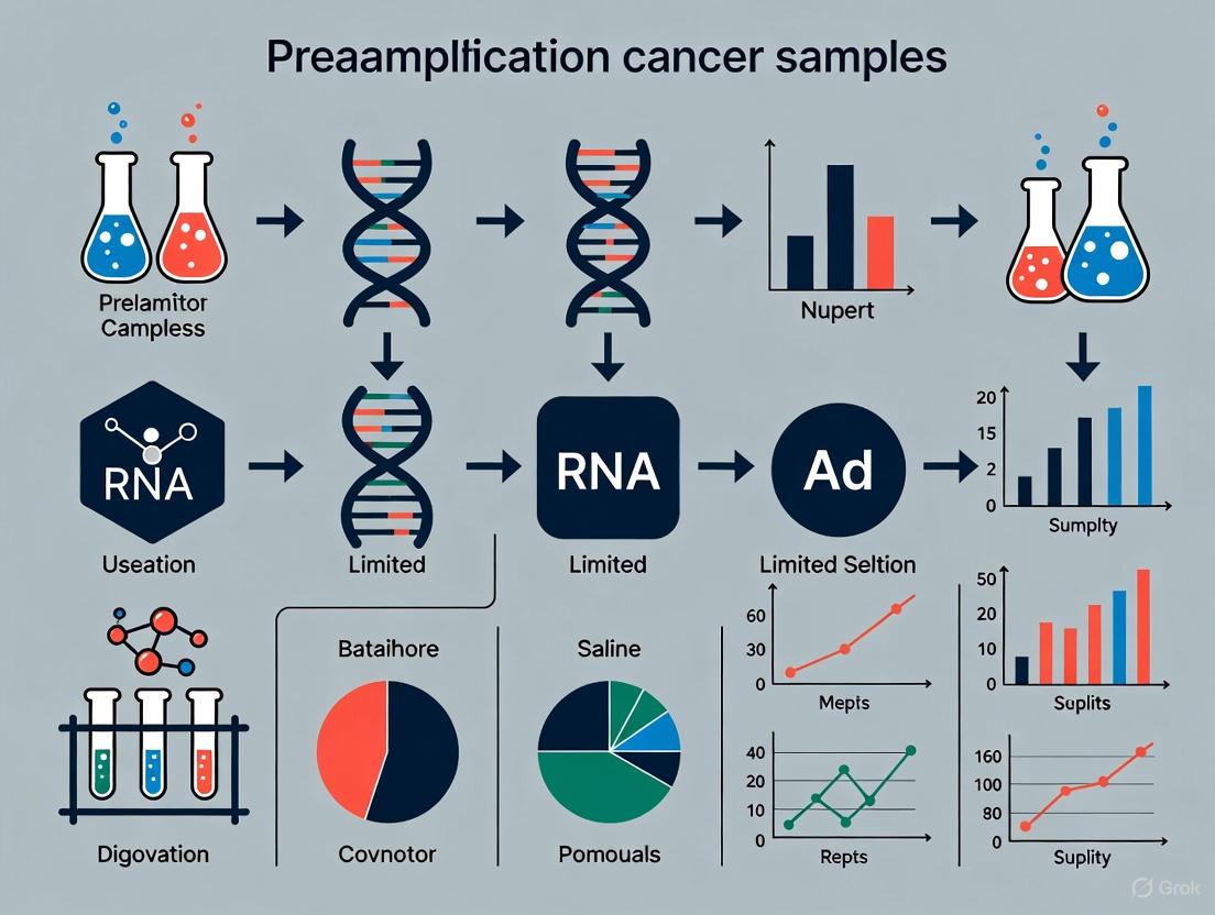
Workflow Visualization: Pre-Amplification for Low Input Samples
The following diagram illustrates the complete experimental and decision-making workflow for applying pre-amplification to low-input liquid biopsy samples.
The Scientist's Toolkit: Essential Reagents and Materials
Successful implementation of a robust pre-amplification strategy requires carefully selected reagents and tools. The following table lists key solutions used in the featured protocol and their critical functions.
Table 2: Research Reagent Solutions for cfDNA Pre-Amplification
| Reagent / Material | Specific Example | Function in Protocol |
|---|---|---|
| Blood Collection Tube | 10 mL EDTA Vacutainer | Prevents coagulation and preserves cell-free DNA integrity before processing. |
| cfDNA Extraction Kit | QIAamp Circulating Nucleic Acid Kit | Isulates high-purity, short-fragment cfDNA from plasma while removing inhibitors. |
| DNA Quantification Kit | Qubit dsDNA HS Assay Kit | Provides highly accurate fluorometric quantification of low-concentration dsDNA. |
| Fragment Analyzer | Agilent TapeStation 4150 | Assesses cfDNA quality and confirms the characteristic nucleosomal size profile. |
| Pre-Amplification Kit | DNA TOP-PCR Kit | Enables non-selective, whole-genome amplification of cfDNA via adapter ligation. |
| Post-Amplification Cleanup | AMPure XP Beads | Purifies the amplified DNA library, removing enzymes, primers, and salts. |
| ddPCR Supermix | ddPCR Supermix for Probes | Provides the optimized environment for droplet digital PCR-based mutation detection. |
| Mutation Detection Probes | FAM/HEX-labeled probes | Enable specific detection and quantification of wild-type and mutant alleles in ddPCR. |
The pre-amplification of cfDNA represents a vital methodological advancement for addressing the pervasive challenge of low input in liquid biopsies. As demonstrated, optimized protocols like TOP-PCR can significantly enhance detection sensitivity, making it possible to interrogate multiple tumor-informed mutations from limited samples that would otherwise be inadequate for analysis [2]. However, this enhanced sensitivity must be vigilantly balanced against the risk of introducing amplification errors, necessitating rigorous validation and the use of appropriate controls [2]. The ongoing work by international consortia, such as the International Society of Liquid Biopsy (ISLB), to define minimal requirements and standardize pre-analytical phases will be crucial for ensuring the reliability and reproducibility of these sensitive methods across laboratories [1]. As the field progresses, integrating such pre-amplification strategies with other emerging biomarkers, like stable circular RNAs [4], and leveraging highly multiplexed assays will further unlock the potential of liquid biopsies to guide personalized cancer care, even from the most challenging of samples.
In the genomic analysis of cancer, particularly from limited samples such as liquid biopsies or small tissue specimens, researchers consistently face two interconnected technical challenges: the detection of mutations present at very low allele fractions and the accurate determination of absolute copy number alterations. The low abundance of tumor-derived DNA in a high background of wild-type DNA makes the confident identification of somatic mutations technically demanding [5]. Concurrently, the presence of non-cancerous cells and the complex aneuploidy of cancer genomes complicate the conversion of relative copy number data into absolute, per-cell integer copy numbers, which is essential for understanding tumor biology and clonal architecture [6]. These challenges are especially pronounced in the context of preamplification methods for limited cancer samples, where the goal is to amplify the scarce genetic material without introducing biases or errors that would preclude accurate downstream analysis. This application note details the specific hurdles, provides quantitative comparisons of the technologies designed to overcome them, and offers detailed protocols for robust mutation detection and copy number analysis.
The Challenge of Low Mutant Allele Fraction
Nature of the Problem
The detection of low-frequency mutant alleles is a cornerstone of liquid biopsy and minimal residual disease (MRD) monitoring. The core problem is one of signal-to-noise ratio. Mutant allele fractions can be very low (frequently below 0.1%) in a large background of wild-type circulating, cell-free DNA (ccfDNA) [5]. Furthermore, the absolute amount of ccfDNA obtainable from plasma or serum is low, typically less than 20 ng/mL of plasma, which translates to approximately only 6,000 haploid genome equivalents per milliliter [5]. This combination of a low mutant allele fraction and low absolute mutant copy number presents a significant challenge for conventional mutation detection methods.
Technological Solutions and Their Performance
Several advanced methods have been developed to push the boundaries of detection sensitivity. The table below summarizes the key characteristics and performance metrics of these technologies.
Table 1: Comparison of Technologies for Detecting Low Allele Fraction Mutations
| Technology | Key Principle | Reported Sensitivity (VAF) | Key Enabling Reagents/Instruments |
|---|---|---|---|
| Multiplex Preamplification + dPCR [5] | Multiplexed preamplification using a high-fidelity polymerase to increase DNA template before digital PCR (dPCR) analysis. | 0.01% - 0.05% | RainDance dPCR platform, Q5 Hot Start High-Fidelity Master Mix |
| smCounter [7] | Multiplex PCR enrichment with molecular barcodes (UMIs) and a Bayesian probabilistic model for variant calling. | 1% | Custom molecular barcode adapters, smCounter software |
| QBDA [8] | Integration of UMI-based quantitation with Blocker Displacement Amplification (BDA) for variant enrichment. | 0.001% - 0.01% | QBDA blockers, internal positive control amplicons |
| Standard UMI Methods [8] | Redundant sequencing of all template molecules using Unique Molecular Identifiers (UMIs) for error correction. | ~0.1% | Standard UMI adapter kits |
Detailed Protocol: Multiplex Preamplification for dPCR
This protocol is adapted from a study that enhanced the detection of cancer-relevant mutations in patient serum [5].
1. DNA Extraction:
- Extract ccfDNA from patient serum or plasma (1-3 mL) using the QIAamp Circulating Nucleic Acid Kit.
- Elute DNA in 45 µL of TE buffer (pH 8.0).
- Quantify concentration using a fluorometer (e.g., Qubit 2.0 Fluorometer).
2. Multiplex Preamplification Reaction:
- Prepare a 10 µL reaction mixture containing:
- 50 ng of extracted ccfDNA.
- 1x Q5 Hot Start High-Fidelity Master Mix.
- 50 nM each of forward and reverse primers for the target genes (e.g., SMAD4, TP53, KRAS). Primer sequences are provided in the source literature [5].
- Perform thermal cycling. A typical program is:
- Initial Denaturation: 98°C for 30 seconds.
- 15-20 Cycles of:
- Denaturation: 98°C for 10 seconds.
- Annealing/Extension: 72°C for 1 minute.
- Final Extension: 72°C for 2 minutes.
- Hold: 4°C.
3. Digital PCR Analysis:
- Dilute the preamplified product as needed.
- Analyze the diluted product on a dPCR platform (e.g., RainDance dPCR) according to the manufacturer's instructions for the specific mutation assays.
- Use Poisson statistics on the partitioned reactions to calculate the absolute copy number and allele frequency of the mutant and wild-type alleles.
Key Advantages: This protocol increases the effective amount of template DNA, allowing for technical replicates and the assessment of multiple targets from a single, limited sample. The use of a high-fidelity polymerase during preamplification is critical to minimize PCR-introduced errors that can create false-positive signals [5].
The Challenge of Absolute Copy Number Determination
Nature of the Problem
Inferring absolute copy number per cancer cell from bulk sequencing data is complex because the measured DNA is a mixture of cancer and normal cells. The relative copy number profile obtained from a sequencer is a function of both the tumor's purity (α), the fraction of cancer cells in the sample, and its ploidy (τ), the average number of copies per cancer cell [6]. A sample with 50% purity and a near-diploid cancer genome can have a similar relative copy number profile to a sample with 100% purity and a tetraploid genome, leading to misinterpretation if not properly modeled.
Computational Frameworks and Signatures
Computational methods have been developed to solve this problem by jointly estimating purity and ploidy.
Table 2: Methods for Absolute Copy Number and CNA Signature Analysis
| Method / Concept | Primary Function | Key Input Data | Application Context |
|---|---|---|---|
| ABSOLUTE [6] | Infers tumor purity, ploidy, and absolute somatic copy-number. | Segmented copy-number data (e.g., from microarrays, WES); optional somatic point mutations. | Bulk tissue analysis; pan-cancer studies. |
| Copy Number Signatures [9] | Decomposes copy-number profiles into 21 distinct signatures of mutational processes. | Allele-specific copy-number profiles from WGS, WES, or SNP arrays. | Understanding patterns of genomic instability across cancer types. |
| HiScanner [10] | Detects high-resolution, allele-specific copy number alterations in single cells. | Read depth, B-allele frequency (BAF), and haplotype phasing from scWGS. | Single-cell analysis of non-neoplastic and neoplastic cells. |
The ABSOLUTE algorithm rescales relative copy-number data ((R(x))) into absolute copy-number per cell ((q(x))) using the relationship: [ R(x) = [αq(x) + 2(1-α)] / D ] where (D = ατ + 2(1-α)) is the total average ploidy of the sample [6]. The algorithm examines possible mappings to find the most plausible integer copy-number solution across the genome.
This protocol outlines the steps for performing absolute copy-number analysis on bulk tumor sequencing data.
1. Data Preprocessing and Segmentation:
- Process raw sequencing data (from WES or SNP arrays) using a standard copy-number calling pipeline (e.g., using tools like PENNCNV for arrays or Sequenza for WES).
- This step generates a segmented copy-number profile, which defines genomic regions with constant relative copy-number.
2. Running ABSOLUTE:
- Install ABSOLUTE (available from http://broadinstitute.org/software/ABSOLUTE).
- Prepare the required input files: the segmented copy-number data and, if available, a file of somatic mutation allele frequencies.
- Execute the ABSOLUTE algorithm. The core function will:
- Jointly optimize the parameters for tumor purity (α) and ploidy (τ).
- Use a database of recurrent cancer karyotypes to help resolve ambiguous cases.
- Model and account for subclonal copy-number alterations.
- The output provides the estimated purity and ploidy, and the absolute, integer copy-number for each segment.
3. Interpretation and Validation:
- Review the model fit. ABSOLUTE provides a goodness-of-fit metric.
- Where possible, validate purity estimates against an orthogonal method (e.g., pathological review or fluorescence-activated cell sorting data) [6].
- Use the absolute copy-number profiles for downstream analyses, such as identifying clonal events or determining the multiplicity of somatic point mutations.
The Scientist's Toolkit: Essential Research Reagents and Materials
Table 3: Key Reagent Solutions for Overcoming Detection and Copy Number Hurdles
| Item | Function / Application | Specific Examples / Notes |
|---|---|---|
| High-Fidelity Polymerase | Reduces PCR errors during preamplification, crucial for low-frequency variant detection. | Q5 Hot Start High-Fidelity Master Mix [5] |
| Circulating Nucleic Acid Kit | Optimized for extraction of low-concentration, fragmented ccfDNA from plasma/serum. | QIAamp Circulating Nucleic Acid Kit [5] |
| Digital PCR System | Provides absolute quantification of nucleic acids by partitioning samples into thousands of reactions. | RainDance dPCR platform [5] |
| Molecular Barcodes (UMIs) | Unique sequences ligated to individual DNA molecules pre-amplification to correct for sequencing errors and PCR duplicates. | Custom adapter designs [7] [8] |
| Blocker Oligonucleotides | Suppress amplification of wild-type sequences to enrich for variant alleles during PCR. | QBDA blockers [8] |
| Computational Tools | Analyze sequencing data to infer absolute copy number, call low-VAF variants, or decipher copy-number signatures. | ABSOLUTE [6], smCounter [7], Copy Number Signature frameworks [9] |
Workflow and Relationship Diagrams
Diagram 1: Overcoming Low Mutant Allele Fraction with Preamplification and dPCR
Diagram 2: Resolving Absolute Copy Number from Mixed Tumor Samples
The analysis of circulating tumor DNA (ctDNA) from liquid biopsies presents a transformative opportunity for non-invasive cancer diagnosis, monitoring therapeutic response, and tracking tumor evolution. However, this promise is challenged by the inherently low concentration of ctDNA in a high background of wild-type circulating cell-free DNA (ccfDNA), especially in early-stage tumors where mutant allele fractions can be less than 0.1% [5]. Achieving robust detection of these rare mutations requires pushing analytical sensitivity to its limits. This application note details a methodology centered on a multiplexed preamplification PCR step performed prior to digital PCR (dPCR) analysis. This approach effectively mitigates the limitations of sample volume and instrument noise, facilitating reliable detection of mutant alleles at frequencies as low as 0.01%, thereby framing a crucial strategy for research on limited cancer samples [5].
The Sensitivity Challenge in ctDNA Analysis
The reliable detection of rare somatic mutations in patient blood samples is technically demanding due to two primary constraints: the low total mass of ccfDNA (typically <20 ng/mL of plasma, or approximately 6000 genome equivalents/mL) and the minute fraction of this DNA that is tumor-derived [5]. In early-stage cancers, the absolute number of mutant copies can be fewer than six per milliliter of plasma [5]. Digital PCR, while a powerful tool for rare allele detection, can be hindered by technical noise and the practical impossibility of running technical replicates when sample material is scarce [5]. The protocol described herein is designed to overcome these hurdles by incorporating a targeted preamplification step to increase the amount of available template for analysis.
Performance of Preamplification-Enhanced dPCR
The following table summarizes the key quantitative improvements in sensitivity and signal quality achieved by implementing a multiplex preamplification step before dPCR analysis.
Table 1: Sensitivity Enhancement with Multiplex Preamplification
| Parameter | Standard dPCR (without Preamplification) | dPCR with Multiplex Preamplification |
|---|---|---|
| Typical Lower Limit of Detection | ~0.1% variant allele frequency [11] | 0.01% variant allele frequency [5] |
| Mutant Alleles Detected | Not specified | One mutant allele in a background of 10,000 wild-type alleles [5] |
| Signal-to-Noise Ratio | Challenging due to false-positive partitions [5] | Improved for all preamplified targets, allowing easier discrimination of low-abundance mutations [5] |
| Multiplexing Capability | Requires substantial optimization, can increase noise [5] | Enabled; multiple targets (e.g., SMAD4, TP53, KRAS) can be assessed from a single sample [5] |
Experimental Protocol: Multiplex Preamplification for dPCR
This section provides a detailed methodology for enhancing rare mutation detection in ctDNA, adapted from the research by PMC4851734 [5].
Materials and Reagents
- Sample Material: ccfDNA extracted from patient serum or plasma using the QIAamp Circulating Nucleic Acid Kit (Qiagen). Elute in a minimal volume (e.g., 45 μL of TE buffer, pH 8.0) to maximize concentration.
- Enzymes and Master Mix: Q5 Hot Start High-Fidelity Master Mix (New England Biolabs) or TaqMan Genotyping Master Mix (Life Technologies).
- Primers: Sequence-specific forward and reverse primers for target mutations (e.g., in genes like SMAD4, TP53, KRAS). In the referenced study, primers were used at 50 nmol/L each in the preamplification reaction [5].
- dPCR System: RainDance dPCR platform or comparable system (e.g., QuantStudio Absolute Q Digital PCR System).
- Equipment: Thermal cycler, fluorometer for DNA quantification (e.g., Qubit 2.0).
Step-by-Step Procedure
ccfDNA Extraction and Quantification:
- Extract ccfDNA from 1-3 mL of patient serum or plasma using a specialized kit for circulating nucleic acids.
- Precisely quantify the extracted ccfDNA using a fluorescence-based method. The typical low yield necessitates highly accurate measurement.
Multiplex Preamplification Reaction:
- Assemble a 10-μL preamplification reaction containing:
- 50 ng of ccfDNA (or the entire eluate if the yield is low).
- 1X High-Fidelity Master Mix.
- 50 nmol/L of each forward and reverse primer for every mutation target being interrogated (e.g., KRAS, TP53, SMAD4) [5].
- Perform PCR amplification using a cycling program optimized for the primer sets and master mix. The use of a high-fidelity polymerase is critical to minimize the introduction of PCR errors that could be misinterpreted as rare mutations.
- Assemble a 10-μL preamplification reaction containing:
Digital PCR Analysis:
- Dilute the preamplified product as needed and use it as the input for a probe-based dPCR assay according to the manufacturer's instructions for your specific dPCR platform.
- Partition the reaction into thousands of individual droplets or microchambers.
- Run the dPCR cycle and analyze the endpoint fluorescence data to count the number of partitions positive for wild-type and mutant alleles.
Data Analysis:
- Use Poisson statistics to calculate the absolute copy number and allele frequency of the mutant target in the original sample.
- Compare the results to negative controls to account for any background signal.
Workflow Visualization
The following diagram illustrates the logical flow and key steps of the multiplex preamplification dPCR protocol.
The Scientist's Toolkit: Essential Research Reagents
Successful implementation of this sensitive detection method relies on several key reagents and instruments.
Table 2: Essential Research Reagents and Materials
| Item | Function/Description | Example Product/Catalog |
|---|---|---|
| Circulating Nucleic Acid Kit | Specialized silica-membrane-based extraction of short-fragment ccfDNA from plasma/serum. | QIAamp Circulating Nucleic Acid Kit (Qiagen) [5] |
| High-Fidelity DNA Polymerase | Enzyme for accurate preamplification PCR; reduces PCR-induced errors that confound rare mutation detection. | Q5 Hot Start High-Fidelity Master Mix (NEB) [5] |
| TaqMan dPCR Assays | Predesigned, validated probe-based assays for specific mutation detection on dPCR platforms. | Absolute Q Liquid Biopsy dPCR Assays (Thermo Fisher) [11] |
| Digital PCR System | Instrument that partitions samples for absolute quantification of nucleic acids; essential for rare allele detection. | QuantStudio Absolute Q Digital PCR System [11] |
| Fluorometric Quantifier | Instrument for sensitive, specific quantification of low-concentration DNA samples. | Qubit Fluorometer (Life Technologies) [5] |
Preamplification has emerged as a critical preparatory technique in molecular biology, enabling comprehensive genetic analysis from limited and precious biological samples. In oncology research, where samples often consist of minute quantities of circulating tumor DNA (ctDNA), fine-needle aspirates, or formalin-fixed paraffin-embedded (FFPE) tissues, preamplification bridges the gap between sample scarcity and analytical requirements [12] [13]. This technique employs a limited number of PCR cycles to preferentially amplify target sequences before subsequent quantitative PCR (qPCR) or next-generation sequencing (NGS) analysis, thereby maximizing the amount of genetic information that can be obtained from limited starting material [14].
The fundamental challenge in preamplification lies in balancing two competing priorities: achieving sufficient yield to enable multi-target detection while maintaining fidelity to preserve the original quantitative relationships between targets. Bias introduced during preamplification can compromise data accuracy, potentially leading to incorrect biological conclusions [12] [15]. This application note examines the core principles of preamplification, provides optimized protocols for cancer research applications, and presents a framework for validating preamplification performance to ensure reliable results.
Core Principles and Technical Considerations
Yield and Amplification Dynamics
The primary objective of preamplification is to generate sufficient template for subsequent analysis while preserving the original relative abundances of targets. The theoretical yield from preamplification can be calculated using the formula:
Nf = No (1 + Y)n
Where Nf is the final copy number, No is the initial copy number, Y is the PCR efficiency per cycle, and n is the number of preamplification cycles [2]. In practice, amplification efficiency is rarely perfect, and the relationship between input and output can become non-linear at high input concentrations due to early saturation of reaction components [2].
Cycle number optimization is critical for balancing yield and fidelity. Studies demonstrate that 10-14 cycles typically provide optimal enrichment for most applications, with 14 cycles potentially necessary for extremely limited samples [14] [15]. Excessive cycling (e.g., 20 cycles) can lead to dynamic range bias where highly abundant targets produce extremely low Cq values that are difficult to properly baseline [14]. Input DNA quantity also significantly impacts performance; for cDNA preamplification, inputs as low as 10-100 pg can be successfully amplified with proper optimization [15].
The preservation of original quantitative relationships between targets—referred to as fidelity—is paramount for accurate downstream analysis. Three primary types of bias can be introduced during preamplification:
Amplification Bias: Occurs when PCR efficiency is suboptimal for some targets, leading to their under- or over-representation in the preamplified sample [14] [15]. This form of bias is influenced by factors including primer design, template sequence, and reaction conditions.
Fold-Change Bias: Manifests when the measured fold difference in target abundance between samples deviates from the actual biological difference [14]. This bias is particularly problematic in gene expression studies and cancer biomarker research where accurate quantification of differential expression is critical.
Dynamic Range Bias: Arises when highly abundant targets amplify so efficiently that they reach detection thresholds too early for accurate quantification [14].
GC-rich regions present particular challenges for preamplification fidelity. Studies evaluating T-Oligo Primed Polymerase Chain Reaction (TOP-PCR) demonstrated lower efficiency for GC-rich TERT promoter amplicons compared to BRAF and TP53 amplicons, highlighting how sequence composition can affect amplification uniformity [2].
Quantitative Comparison of Preamplification Performance
Performance Metrics Across Methods
Table 1: Comparative Performance of Preamplification Methods and Parameters
| Method/Parameter | Optimal Input | Cycle Range | Key Advantages | Limitations |
|---|---|---|---|---|
| TOP-PCR | 5-20 ng cfDNA | 5-7 cycles | Preserves DNA size profiles; 22 bp size increase from adaptor [2] | Inverse yield-input correlation; PCR artifacts with higher cycles [2] |
| Targeted Preamplification (Multiplex PCR) | 100 pg-20 ng cDNA | 14-18 cycles | Suitable for 10-400 targets; maintains quantitative relationships [12] [14] | Efficiency depends on primer concentration, annealing time/temperature [12] |
| abSLA PCR | Low template DNA | 15 cycles | Reduces stutter artifacts; improves STR locus recovery [16] | Requires specialized abasic site-containing primers [16] |
| superRCA | Low frequency mutations | ~10 pre-PCR cycles | Detects 1 variant in 100,000 wild-type molecules; flow cytometry compatible [17] | Two-step RCA process increases complexity [17] |
Commercial Kits and Bias Performance
Table 2: Commercial Preamplification Master Mix Performance Comparison
| Master Mix | Unbiased Amplification Rate | Percent Bias | Recommended Input | Key Features |
|---|---|---|---|---|
| Prelude PreAmp | 92/96 assays (96%) | 4% | 10 pg-100 pg | Optimized polymerase and buffer for maximum efficiency [15] |
| TaqMan PreAmp | 87/96 assays (91%) | 9% | Not specified | Established technology; moderate performance [15] |
| SsoAdvanced PreAmp | 72/96 assays (75%) | 25% | 100 pg cDNA/gDNA | Sso7d fusion polymerase for processivity [14] [15] |
| PerfeCTa PreAmp | 73/96 assays (76%) | 24% | Not specified | Competitive alternative for standard applications [15] |
Experimental Protocols
Targeted cDNA Preamplification for Gene Expression Analysis
This protocol is optimized for preamplifying cDNA from limited cancer samples, such as FFPE tissues or liquid biopsies, prior to high-throughput qPCR analysis on platforms such as the Fluidigm BioMark system [13] [15].
Reagents and Materials
- Prelude PreAmp Master Mix (or equivalent)
- Primer pool (50-400 assays, each at 40-50 nM final concentration)
- cDNA template (10 pg-20 ng)
- Nuclease-free water
- AMPure XP beads or equivalent purification system
Procedure
- Reaction Setup: Prepare preamplification mix in a total volume of 10-20 µL:
- 5-10 µL 2X PreAmp Master Mix
- 2-4 µL Primer pool (total concentration not exceeding 2 µM)
- 2-5 µL cDNA template
- Nuclease-free water to final volume
Thermal Cycling: Perform amplification using the following parameters:
- Initial denaturation: 95°C for 2 minutes
- 10-18 cycles of:
- Denaturation: 95°C for 15 seconds
- Annealing/Extension: 60°C for 2-4 minutes
- Final hold: 4°C
Product Purification: Dilute preamplified products 1:20 to 1:40 in 10 mM Tris-HCl, 1 mM EDTA, pH 8.0 [12] [13]. Alternatively, purify using AMPure XP beads at a 1.93:1 beads-to-sample ratio [2].
Quality Assessment: Verify preamplification success using:
- Quantitative PCR with housekeeping genes
- Bioanalyzer/TapeStation for size distribution
- Control assays specifically designed for preamplification validation [14]
Troubleshooting Notes
- If amplification bias is observed (>1.5 ΔΔCq from expected), reduce cycle number by 2-4 cycles [13]
- For low yields with high input DNA, reduce input amount to prevent reaction component saturation [2]
- Include no-template controls and genomic DNA controls to identify contamination sources [13]
ctDNA Preamplification for Mutation Detection
This protocol utilizes TOP-PCR for preamplification of circulating tumor DNA from plasma samples, enhancing detection sensitivity for rare mutations in cancer monitoring [2].
Reagents and Materials
- DNA TOP-PCR Kit (Top Science Biotechnologies Inc.)
- 20 ng cfDNA input (equivalent to ~6000 haploid genome copies)
- AMPure XP beads
- Qubit dsDNA HS Assay Kit for quantification
Procedure
- End Repair and A-Tailing: Perform end repair and A-tailing of cfDNA according to manufacturer's instructions.
Adapter Ligation: Ligate half-adaptors to DNA using the provided ligation buffer and enzyme mix.
Limited Cycle PCR: Amplify ligated cfDNA using:
- 5-7 cycles of PCR with T-oligo primer only
- Initial denaturation: 95°C for 2 minutes
- Cycling: 95°C for 15 seconds, 60°C for 30 seconds, 72°C for 30 seconds
Purification: Purify with AMPure XP beads (1.93:1 ratio) and elute in 30 µL sterile-distilled water [2].
Downstream Analysis: Use preamplified product for droplet digital PCR or NGS analysis of tumor-informed mutations.
Validation Metrics
- Expect 22 bp size increase in mono-nucleosomal DNA peak (from ~193 bp to ~215 bp)
- Di-nucleosomal DNA percentage should increase from ~12% to ~36% of total cfDNA
- Over 90% of TOP-PCR product should be within 100-700 bp size range [2]
The Scientist's Toolkit
Table 3: Essential Research Reagent Solutions for Preamplification
| Reagent/Category | Function | Examples & Applications |
|---|---|---|
| Specialized Polymerases | High processivity for multiplex amplification; some engineered for translesion synthesis | Sso7d fusion polymerase [14]; B-family DNA polymerases blocked by abasic sites [16] |
| Bias Control Reagents | Minimize amplification bias through optimized buffer systems | Prelude PreAmp Master Mix [15]; Additives like BSA, glycerol, formamide [12] |
| Library Preparation Kits | Adaptor ligation for non-selective whole genome amplification | DNA TOP-PCR Kit with half-adapter ligation design [2] |
| Ultra-Sensitive Detection Master Mixes | Enable detection of preamplified products with minimal input | SmartChip TB Green Gene Expression Master Mix [15]; Digital PCR supermixes [2] |
| Quality Control Assays | Validate preamplification efficacy and measure bias | PrimePCR PreAmp Control Assay [14]; ERCC RNA spike-in controls [14] |
Workflow Visualization
Preamplification Workflow for Limited Samples
Preamplification Method Selection Framework
Validation and Quality Control
Robust validation is essential for ensuring preamplification fidelity. The following approaches are recommended:
ΔΔCq Analysis for Bias Assessment Calculate the difference between theoretical and observed ΔCq values:
- ΔCqexperimental = Cqnon-preamp – Cqpreamp
- ΔCqtheoretical = number of pre-amplification cycles – log2 (all dilutions)
- ΔΔCq = ΔCqtheoretical - ΔCqexperimental
A ΔΔCq value of < 1.5 is generally considered acceptable, with values < 0.75 representing excellent uniformity [13] [15].
Control Strategies
- Include no-template controls (NTC) to detect contamination or primer-dimer formation
- Use positive controls with known mutation frequencies
- Implement reference assays with consistent expression across samples
- Utilize spike-in controls like ERCC RNA standards for absolute quantification [14]
- Perform melt curve analysis when using SYBR Green chemistry to verify specificity [12] [14]
Successful preamplification requires meticulous optimization of cycle numbers, input amounts, and reaction conditions to balance yield and fidelity. The protocols and quality control measures outlined herein provide a framework for implementing preamplification in cancer research applications where sample material is limited. As technologies evolve, emerging methods like abSLA PCR and superRCA offer promising alternatives to traditional approaches, particularly for challenging applications such as low-frequency mutation detection and forensic analysis of minimal samples [17] [16]. By adhering to these principles and validation strategies, researchers can confidently employ preamplification to maximize the scientific value derived from precious clinical specimens.
Core Preamplification Technologies and Workflow Integration
Multiplexed Preamplification with High-Fidelity Polymerases
The analysis of limited cancer samples, such as circulating tumor DNA (ctDNA) or small tissue biopsies, is often constrained by the low abundance of nucleic acids. Detecting tumor-specific mutations in plasma is particularly challenging, as mutant allele fractions are typically very low (often below 0.1%) amidst a large background of wild-type DNA, and the total amount of obtainable cell-free DNA is limited [5]. Multiplexed preamplification using high-fidelity polymerases addresses this fundamental limitation by enabling a specific, unbiased increase in the concentration of multiple target sequences prior to final analysis. This methodology is essential for robust mutation detection, monitoring tumor progression, and assessing therapeutic resistance through liquid biopsies, providing a clinically relevant alternative to invasive tissue biopsies [5].
The Scientist's Toolkit: Essential Research Reagents
The successful implementation of a multiplexed preamplification protocol relies on a set of core reagents, each fulfilling a specific role to ensure sensitivity and fidelity.
Table 1: Key Research Reagent Solutions for Multiplexed Preamplification
| Reagent | Function | Example Product & Specifications |
|---|---|---|
| High-Fidelity DNA Polymerase | Catalyzes DNA synthesis with ultra-low error rates, critical for accurate mutation detection. | Q5 High-Fidelity DNA Polymerase (NEB #M0491); ~280x higher fidelity than Taq [18]. |
| Preamplification Master Mix | A ready-to-use solution optimized for multiplex PCR, containing buffer, dNTPs, and polymerase. | SsoAdvanced PreAmp Supermix; enables preamplification of up to 400 targets from limited cDNA or DNA [14]. |
| Nucleic Acid Isolation Kit | Purifies high-quality DNA or RNA from complex biological samples like plasma or serum. | QIAamp Circulating Nucleic Acid Kit; designed for efficient recovery of cell-free DNA [5]. |
| Target-Specific Primer Pools | A multiplexed set of forward and reverse primers designed to preamplify genes of interest. | Custom primer pools (e.g., for SMAD4, TP53, KRAS); used at low concentrations (e.g., 50 nM each) [5]. |
Quantitative Performance of Preamplification Strategies
Preamplification significantly enhances the detectability of low-abundance targets. The following table summarizes key performance metrics from various studies, highlighting the utility of this approach in sensitive detection scenarios.
Table 2: Quantitative Performance of Preamplification in Research Applications
| Application Context | Key Quantitative Findings | Impact on Detection |
|---|---|---|
| ctDNA Mutation Detection [5] | Preamplification enabled detection of multiple cancer-relevant mutations down to an allele frequency of 0.01% from 50 ng of tumor-derived DNA. | Without preamplification, mutations at this level were not detectable; the signal-to-noise ratio was improved for all targets. |
| Targeted mRNA Quantification [19] | Global preamplification (Smart-Seq2) generated a 9.3-fold lower yield but allowed expression analysis of 90 genes from single cells. | Provides a flexible workflow for profiling small samples, though with slightly lower reproducibility than target-specific methods. |
| miRNA Analysis in Plasma [20] | Preamplification improved the cycle threshold (C_T) by 6.6 ± 0.89 on average, facilitating the detection of low-expressed miRNAs like miR-1537 and miR-190b. | The success rate for detecting miR-1537 in maternal plasma increased from 5/19 to 18/19 samples after preamplification. |
| Multiplexed cDNA Preamp for TaqMan Arrays [21] | Preamplification of 47 genes resulted in a mean C_T decrement of 3.85 cycles, allowing a ~30-fold greater effective cDNA load on the array. | Enabled reliable multi-gene expression profiling from scarce RNA samples, including degraded material from clinical fluids. |
Experimental Protocol: Multiplexed Preamplification for ctDNA Analysis
This protocol is adapted from a study that successfully detected mutations in patient serum and is designed for use with the RainDance digital PCR platform or similar systems [5].
Sample Collection and DNA Extraction
- Sample Collection: Collect blood in appropriate vacutainers. For serum, allow samples to clot for 1 hour, then centrifuge at 2,500 × g for 5 minutes. Aliquot and store serum at -80°C [5].
- DNA Extraction: Isolate circulating cell-free DNA (ccfDNA) from plasma or serum using the QIAamp Circulating Nucleic Acid Kit (or equivalent). Elute DNA in a small volume (e.g., 45 µL) of TE buffer (pH 8.0). Quantify DNA using a fluorescence-based method (e.g., Qubit Fluorometer) [5].
Multiplexed Preamplification Reaction Setup
This step uses a high-fidelity polymerase to amplify multiple targets simultaneously with minimal introduction of errors.
- Reaction Composition: Prepare a 10 µL reaction mixture containing [5]:
- Template DNA: 50 ng of gDNA or the entire yield of ccfDNA from a limited sample.
- Master Mix: 1× Q5 Hot Start High-Fidelity Master Mix.
- Primers: 50 nM each of forward and reverse primers for every target (e.g., KRAS, TP53, SMAD4).
- Thermal Cycling:
Post-Preamplification Processing and Downstream Analysis
- Dilution: Dilute the preamplified product appropriately (e.g., 1:20 to 1:50) in nuclease-free water or TE buffer before the subsequent quantification step [14] [21].
- Analysis by Digital PCR: Use the diluted preamplified product as a template for probe-based digital PCR. The increased target concentration allows for robust detection and absolute quantification of mutant alleles, even at very low frequencies [5].
Workflow and Bias Assessment Diagram
The following diagram illustrates the complete experimental workflow and the key types of bias that must be monitored during the preamplification process.
Validation and Troubleshooting
Preamplification Validation
- Amplification Bias: Compare the Cq values of preamplified samples against non-preamplified controls for the same input amount. The observed ΔCq should be within ±0.75 of the value expected from the number of preamplification cycles [14].
- Fold-Change Bias: Use synthetic controls or RNA spike-ins (e.g., ERCC controls) with known concentration ratios to confirm that preamplification does not distort quantitative relationships between targets [14].
- Specificity: Include a no-template control (NTC) and perform melt curve analysis (for SYBR Green assays) to ensure the absence of non-specific amplification products or primer-dimers [14] [19].
Troubleshooting Common Issues
- Low Yield or Poor Sensitivity: Verify the quality and quantity of the input DNA. Ensure the high-fidelity polymerase and master mix are suitable for multiplex reactions. Consider using a master mix specifically formulated for multiplex digital PCR [22].
- High Background or Non-specific Amplification: Optimize primer concentrations and thermal cycling conditions (particularly the annealing/extension temperature). Use in silico tools to check for primer-dimer formation and re-design primers if necessary [22].
- Introduction of Bias: Strictly limit the number of preamplification cycles (typically 10-14) to avoid entering the non-exponential phase of PCR, which can cause dynamic range bias [14].
The analysis of cell-free DNA (cfDNA), particularly circulating tumor DNA (ctDNA), represents a transformative approach for non-invasive cancer diagnostics, treatment monitoring, and minimal residual disease detection [2]. However, the clinical utility of liquid biopsies is often constrained by the limited quantity and quality of DNA obtainable from body fluids [23]. ctDNA fragments constitute only a small fraction of total cfDNA, especially in early-stage disease where mutant allele fractions can be less than 0.1% [2].
To overcome these limitations, T-Oligo Primed Polymerase Chain Reaction (TOP-PCR) has been developed as a robust whole genome amplification method specifically designed for minute DNA quantities [23]. This method utilizes an efficient "half-adapter" (HA) ligation design followed by single-primer PCR amplification, enabling full-length, non-selective amplification of trace DNA fragments from clinical samples such as plasma, saliva, and urine [23] [24]. For cancer researchers and drug development professionals working with limited samples, TOP-PCR provides a valuable tool to enhance detection sensitivity and expand material availability for downstream genomic analyses.
Technical Principle: The Half-Adapter Ligation Design
TOP-PCR employs a unique homogeneous "half adaptor" (HA) structure generated by annealing two oligonucleotides: the P oligo (carrying a phosphate group at the 5′ end) and the T oligo (carrying a T-tail at the 3′ end) [23]. This design prevents adaptor self-ligation and maximizes ligation efficiency to an unprecedented level.
The fundamental innovation of TOP-PCR lies in addressing a key inefficiency of conventional paired-adapter systems. In traditional approaches where two different adaptors ligate to DNA termini, approximately 50% of fragments ligate to only one adaptor type, resulting in substantial sequence information loss [23]. TOP-PCR circumvents this through its single HA design, where every successful ligation event enables subsequent amplification.
The following diagram illustrates the core TOP-PCR mechanism and workflow:
Figure 1: TOP-PCR Workflow and Half-Adapter Mechanism. The process begins with formation of the half-adapter structure through annealing of P and T oligos, followed by ligation to prepared DNA fragments, and culminates in amplification using only the T oligo primer.
After ligation, PCR amplification proceeds using the T oligo alone, which selectively amplifies only DNA fragments successfully ligated to the HA [23] [2]. This streamlined approach significantly enhances amplification efficiency for low-abundance molecules while maintaining the original size distribution of the input DNA.
Application Notes: Performance in Body Fluid DNA Analysis
Preservation of DNA Size Profiles
TOP-PCR demonstrates exceptional capability in maintaining the original size characteristics of input DNA, a critical factor for analyzing apoptosis-derived cfDNA fragments which exhibit characteristic nucleosomal patterns.
Table 1: DNA Size Profile Preservation by TOP-PCR
| DNA Source | Characteristic Size Profile | TOP-PCR Effect | Research Significance |
|---|---|---|---|
| Plasma cfDNA | Mono-nucleosomal (~166 bp) & di-nucleosomal fragments [23] | Maintains profile; accentuates di-nucleosomal peak [23] | Enhances detection of apoptosis-derived fragments; reveals tumor-associated fragmentation patterns |
| Saliva cfDNA | Similar to plasma but with more large-sized DNA [23] | Maintains profile while enhancing nucleosomal-sized fragments [23] | Improves resolution for oral cancer diagnostics and microbiota studies |
| Urine cfDNA | 150-250 bp fragments; no nucleosomal pattern [23] | Maintains non-nucleosomal profile [23] | Facilitates urological cancer detection despite different filtration biology |
Recent studies have quantified this size preservation, showing that TOP-PCR-amplified cfDNA from melanoma patients exhibits the expected ~22 bp size increase due to adaptor ligation, with mono-nucleosomal peaks increasing from a median of 193 bp to 208 bp [2]. Notably, TOP-PCR accentuates di-nucleosomal DNA, which increases from a median of 12.1% in unamplified cfDNA to 36.1% after amplification [2].
Enhanced Sensitivity for ctDNA Detection
In clinical oncology applications, TOP-PCR pre-amplification significantly enhances ctDNA detection sensitivity. A 2025 study demonstrated that optimized TOP-PCR conditions (20 ng cfDNA input with 5-7 amplification cycles) improved detection of tumor-informed mutations in melanoma patients [2]. This enhancement is particularly valuable for early-stage disease monitoring and minimal residual disease detection where ctDNA fractions are exceptionally low.
The pre-amplification step expands sample availability for multiple downstream analyses, enabling researchers to profile numerous tumor-associated mutations from limited clinical material [2]. However, the same study highlighted that PCR errors can emerge in pre-amplified samples, necessitating appropriate negative controls and stringent mutation calling thresholds to maintain specificity [2].
Experimental Protocol: Step-by-Step Methodology
TOP-PCR Amplification Procedure
Recommended Input and Reaction Setup:
- Input DNA: 0.5-20 ng cfDNA [2]
- Reaction volume: 6.6 μL containing DNA sample [2]
- Optimal performance: 20 ng input cfDNA with 5-7 amplification cycles [2]
Step 1: End Repair and A-Tailing Convert DNA fragments to blunt-ended, 5'-phosphorylated DNA with 3'-dA overhangs using standard end-repair and A-tailing enzymes according to manufacturer specifications [2].
Step 2: Half-Adapter Ligation
- Prepare HA by annealing P oligo (5'-phosphate) and T oligo (3'-T-tail) at room temperature [23]
- Ligate HA to both termini of target DNA using T4 DNA ligase
- Incubate at appropriate temperature (typically 20-25°C) for 15-30 minutes
Step 3: PCR Amplification
- Amplify ligated DNA using T oligo primer only [23] [2]
- Cycle conditions: 5-7 cycles as optimal for 20 ng input [2]
- Standard thermocycler parameters: Denaturation at 95°C, annealing at 60°C, extension at 72°C
Step 4: Purification
- Purify amplified product using AMPure XP beads at 1.93:1 beads-to-sample ratio [2]
- Elute in 30 μL sterile distilled water [2]
Optimization Guidelines
Table 2: TOP-PCR Optimization Parameters
| Parameter | Recommended Range | Optimization Notes | Impact on Results |
|---|---|---|---|
| Input DNA | 0.5-20 ng [2] | Higher inputs (20 ng) preferred for rare variant detection [2] | 20 ng enables detection of 0.02% MAF (1 in 6000 copies) [2] |
| PCR Cycles | 5-7 cycles [2] | 15 cycles recommended by manufacturer but shows yield inversion [2] | 5-7 cycles maintains linearity; 15 cycles causes reaction saturation [2] |
| DNA Quality | Degraded/fragmented samples suitable | Specifically designed for suboptimal samples [23] | Effectively amplifies apoptosis-derived fragments in cfDNA [23] |
| Yield Efficiency | 90-116% per cycle [2] | 90% for 20 ng input; 116% for 5 ng input [2] | Near-doubling of product achieved with optimal conditions [2] |
Critical optimization findings reveal that TOP-PCR yield with 15 cycles (manufacturer's recommendation) is inversely correlated with cfDNA input, while reducing to 5-7 cycles maintains a linear increase relative to input [2]. This adjustment is essential for maximizing amplification efficiency while minimizing artifacts.
The Scientist's Toolkit: Essential Research Reagents
Table 3: Key Research Reagent Solutions for TOP-PCR
| Reagent/Component | Function | Specification Notes |
|---|---|---|
| P Oligo | Provides 5'-phosphate for ligase recognition [23] | Contains phosphate group at 5' end; forms double-stranded backbone of HA [23] |
| T Oligo | Priming site for amplification; provides 3'-T-tail for sticky-end ligation [23] | Contains extra T at 3' end; later serves as universal primer for PCR [23] |
| Half-Adapter (HA) | Unified adapter for both DNA ends [23] | 10 bp double-stranded backbone with T-tail on one side, gcgc-tail on other [23] |
| DNA TOP-PCR Kit | Commercial implementation | Available from Top Science Biotechnologies Inc. [2] |
| AMPure XP Beads | Post-amplification purification [2] | Used at 1.93:1 beads-to-sample ratio [2] |
Technical Considerations and Limitations
While TOP-PCR significantly enhances detection sensitivity, researchers must account for its specific technical characteristics:
Size Range Limitations: TOP-PCR faithfully amplifies fragments up to approximately 1.5 kb under standard conditions [23]. Larger fragments can be isolated by agarose gel electrophoresis prior to sequencing if needed.
Amplification Artifacts: PCR errors emerge in pre-amplified cfDNA samples, requiring implementation of negative controls and establishment of stringent mutation positivity thresholds [2].
GC Content Bias: Amplification efficiency varies by genomic region, with lower efficiency observed for GC-rich targets like the TERT promoter amplicon compared to more balanced regions like BRAF and TP53 [2].
Yield Characteristics: TOP-PCR yield demonstrates an inverse correlation with input DNA at higher cycle numbers (15 cycles), but maintains linearity with optimized cycling (5-7 cycles) [2].
TOP-PCR represents a significant advancement in pre-amplification technology for limited cancer samples, effectively addressing the critical challenge of insufficient DNA material for comprehensive genomic analyses. Its unique half-adapter ligation design enables unprecedented amplification efficiency while preserving the original size profiles of cfDNA fragments - a crucial feature for cancer biomarker research.
For researchers and drug development professionals, TOP-PCR offers a robust method to enhance ctDNA detection sensitivity, particularly valuable for early-stage disease monitoring and longitudinal treatment response assessment. When implemented with appropriate controls and optimization, this technology expands the potential of liquid biopsy approaches in personalized oncology, enabling more reliable genomic analyses from minute biological samples.
The analysis of circulating tumour DNA (ctDNA) from liquid biopsies presents a transformative opportunity for personalised oncology, enabling real-time monitoring of tumour dynamics, minimal residual disease detection, and assessment of treatment response [2]. However, the clinical utility of ctDNA is often limited by the low abundance of tumour-derived DNA within the total cell-free DNA (cfDNA) population, especially in early-stage disease or after curative-intent therapy where mutant allele fractions can be less than 0.1% [2]. This challenge necessitates the development of robust, integrated workflows that begin with sample collection and proceed through pre-analytical processing, pre-amplification, and culminate in highly sensitive downstream analysis using digital PCR (dPCR) or Next-Generation Sequencing (NGS). This application note details a validated protocol that incorporates T-Oligo Primed PCR (TOP-PCR) pre-amplification to enhance ctDNA detection sensitivity, providing researchers with a comprehensive framework for analysing limited cancer samples.
The following tables summarize key quantitative findings from the optimization and validation of the TOP-PCR pre-amplification workflow for ctDNA analysis.
Table 1: Performance Metrics of TOP-PCR Pre-Amplification with Variable Input and Cycle Number
| Input cfDNA (ng) | PCR Cycles | Amplification Efficiency (%) | Average Yield (ng) | Linearity (Pearson r) |
|---|---|---|---|---|
| 0.5 - 20.0 | 15 | Highly Variable | 443 - 1237 | -0.9027 (p=0.0054) |
| 5.0 | 5 - 7 | 116% per cycle | Linear Increase | 0.9882 (p<0.001) |
| 20.0 | 5 - 7 | 90% per cycle | Linear Increase | Consistent |
Table 2: Impact of TOP-PCR Pre-Amplification on cfDNA Size Profile (n=21 stage IV melanoma samples)
| Size Profile Parameter | Unamplified cfDNA | TOP-PCR-Amplified cfDNA | p-value |
|---|---|---|---|
| Mono-nucleosomal Peak Median | 193 bp (183-205 bp) | 208 bp (195-218 bp) | - |
| Di-nucleosomal DNA Proportion | 12.1% (8.6-17.7%) | 36.1% (29.6-43.7%) | < 0.001 |
| DNA within 100-700 bp | 86% (74-96%) | >90% (86-97%) | < 0.001 |
Table 3: Target Amplification Efficiency and Error Considerations
| Gene Target | Amplification Efficiency | Key Consideration |
|---|---|---|
| TERT promoter (GC-rich) | Lower | Reduced efficiency for GC-rich targets |
| BRAF & TP53 | Higher | Reliable for mutant detection |
| All Targets | - | PCR errors necessitate negative controls & stringent mutation positivity thresholds |
Experimental Protocols
Sample Collection, Processing, and cfDNA Extraction
Principle: To obtain high-quality, uncontaminated cfDNA from blood samples, preserving the integrity of the native ctDNA fragment profile [2].
Materials:
- Blood Collection Tubes: 10 mL EDTA vacutainer tubes (e.g., Becton Dickinson) [2].
- Centrifuges: Capable of 800× g and 1,600× g [2].
- cfDNA Extraction Kit: QIAamp Circulating Nucleic Acid Kit (Qiagen) [2].
- Elution Buffer: Sterile distilled water [2].
- Quantification: Qubit High Sensitivity dsDNA Kit and Qubit 3.0 Fluorometer (Thermo Fisher Scientific) [2].
- Quality Control: Cell-free DNA ScreenTape and TapeStation 4150 (Agilent Technologies) [2].
Procedure:
- Collection: Collect whole blood from patients using 10 mL EDTA tubes.
- Processing: Process blood samples within four hours of collection.
- Centrifuge at 800× g for 15 minutes to separate plasma from blood cells.
- Transfer the supernatant (plasma) to a new tube and perform a second centrifugation at 1,600× g for 10 minutes to remove any remaining cellular debris [2].
- Storage: Aliquot the double-spun plasma and store at -80°C until extraction.
- Extraction: Extract cfDNA from 2-4 mL of plasma using the QIAamp Circulating Nucleic Acid Kit, strictly following the manufacturer's instructions [2].
- Elution: Elute the purified cfDNA in 100 µL of sterile distilled water.
- QC: Quantify the cfDNA using the Qubit dsDNA HS Assay. Analyze the fragment size distribution using the TapeStation system to confirm the characteristic nucleosomal ladder (~166 bp mono-nucleosomal peak) [2].
Pre-Amplification using T-Oligo Primed PCR (TOP-PCR)
Principle: To uniformly amplify limited input cfDNA via a three-step process involving end repair, ligation of a single "half-adapter," and PCR amplification with a single primer, thereby increasing the amount of available material for downstream mutation detection assays [2].
Materials:
- TOP-PCR Kit: DNA TOP-PCR Kit (Top Science Biotechnologies Inc., Taiwan, China) [2].
- Thermal Cycler
- Purification Beads: AMPure XP beads (Beckman Coulter) [2].
- Concentrator: Eppendorf Concentrator Plus (optional, for sample concentration) [2].
Procedure:
- Input DNA Preparation: Use a defined input of 20 ng of cfDNA in a 6.6 µL volume. If necessary, concentrate low-yield samples using a centrifugal concentrator to achieve a minimum concentration of ≥3 ng/µL [2].
- TOP-PCR Reaction Setup: Perform the TOP-PCR reaction as per the manufacturer's instructions. The process consists of:
- End Repair and A-tailing: To create blunt-ended, 5'-phosphorylated, and 3'-dA-tailed DNA fragments.
- Adapter Ligation: A single, linear "half-adapter" is ligated to the prepared DNA fragments.
- PCR Amplification: Amplify the ligated DNA using a single T-oligo primer for 5-7 cycles [2].
- Product Purification: Purify the amplified product using AMPure XP beads at a bead-to-sample ratio of approximately 1.93:1 [2].
- Elution and QC: Elute the final pre-amplified DNA in 30 µL sterile distilled water. Quantify the yield using the Qubit dsDNA HS Assay.
Downstream Analysis by Droplet Digital PCR (dPCR)
Principle: To detect and absolutely quantify specific tumour-informed mutations in pre-amplified cfDNA with high sensitivity and precision [2].
Materials:
- ddPCR Supermix: ddPCR Supermix for Probes (no dUTP; Bio-Rad Laboratories) [2].
- Assays: Wild-type and mutant-specific probes (FAM/HEX) for target mutations [2].
- Droplet Generator: QX200 AutoDG (Bio-Rad Laboratories) [2].
- Thermal Cycler: C1000 Touch Thermocycler (Bio-Rad Laboratories) [2].
- Droplet Reader and Software: QX600 Droplet Reader and QX Manager 2.0 software (Bio-Rad Laboratories) [2].
Procedure:
- Reaction Setup: Prepare the dPCR reaction mix according to the manufacturer's instructions, using the ddPCR Supermix, pre-amplified DNA (or unamplified cfDNA for comparison), and the appropriate mutant and wild-type probes [2].
- Controls: Include in each run:
- A no-template control (NTC).
- A positive control (e.g., a known positive sample or gBlock mutant spike-in).
- A negative control (e.g., cfDNA from human dermal fibroblasts) [2].
- Droplet Generation: Generate droplets using the QX200 AutoDG.
- PCR Amplification: Perform PCR amplification on the C1000 Touch Thermocycler using the optimized cycling conditions for the assay.
- Analysis: Read the droplets using the QX600 Droplet Reader and analyze the data with QX Manager software. Apply identical manual fluorescence thresholds across all samples within a run to determine the mutant allele frequency [2].
Workflow Visualization
The Scientist's Toolkit: Research Reagent Solutions
Table 4: Essential Reagents and Kits for the Integrated ctDNA Workflow
| Product Name | Vendor | Function in Workflow |
|---|---|---|
| QIAamp Circulating Nucleic Acid Kit | Qiagen | Extraction of high-quality, proteinase-free cfDNA from plasma samples [2]. |
| DNA TOP-PCR Kit | Top Science Biotechnologies | Uniform, non-selective pre-amplification of limited input cfDNA to enhance sensitivity for rare variant detection [2]. |
| AMPure XP Beads | Beckman Coulter | Solid-phase reversible immobilization (SPRI) for post-amplification clean-up and size selection of DNA fragments [2]. |
| ddPCR Supermix for Probes (no dUTP) | Bio-Rad Laboratories | Optimized reaction mix for probe-based digital PCR assays, enabling absolute quantification of mutant alleles [2]. |
| KAPA HyperPrep Kit | Roche | Efficient, high-yield library construction for NGS from low-input and degraded DNA samples [25]. |
| KAPA Library Quantification Kits | Roche | Accurate qPCR-based quantification of NGS libraries to ensure optimal sequencing cluster density [25]. |
| AVENIO Edge System | Roche | Automated, walk-away solution for NGS library preparation, reducing hands-on time and improving reproducibility [25]. |
The molecular profiling of tumor-derived material in patient blood, known as liquid biopsy, represents a transformative approach in oncology. This methodology is particularly vital for analyzing limited cancer samples, such as serum, where the low abundance of circulating tumor DNA (ctDNA) necessitates highly sensitive preamplification and detection techniques. This case study focuses on the successful detection of key driver mutations in KRAS, TP53, and SMAD4—genes frequently altered in pancreatic ductal adenocarcinoma (PDAC) and other solid tumors [26] [27]. The ability to reliably identify these mutations from blood samples provides a minimally invasive alternative to tissue biopsy, enabling improved early detection, disease monitoring, and personalized treatment strategies.
Background and Genetic Alterations
Pancreatic ductal adenocarcinoma is characterized by a high frequency of specific genetic alterations. The core tumor suppressor genes and oncogenes drive tumor development and progression.
Table 1: Key Genetic Alterations in Pancreatic Ductal Adenocarcinoma (PDAC)
| Gene | Function | Mutation Prevalence in PDAC | Common Mutation Types |
|---|---|---|---|
| KRAS | Oncogene (GTPase signaling) | ~88% - >90% [26] [27] | Point mutations (e.g., G12D, G12V, G13D) [27] |
| TP53 | Tumor Suppressor (Cell cycle, DNA damage response) | ~70% - 77% [26] [27] | Missense, truncating mutations [26] |
| SMAD4 | Tumor Suppressor (TGF-β signaling pathway) | ~29% - 30% [26] [27] | Homozygous deletion, point mutations [27] |
| CDKN2A | Tumor Suppressor (Cell cycle regulation) | ~18% - 98% (inactivated) [26] [27] | Loss of heterozygosity, homozygous deletion, promoter silencing [27] |
The detection of these mutations in serum ctDNA reflects the tumor's genetic profile and offers prognostic insights. For instance, TP53 mutations are associated with a poorer prognosis and can promote metastasis, while the co-occurrence of mutations in KRAS and TP53 is common and suggests early events in pancreatic carcinogenesis [26].
Methodological Approaches for Mutation Detection from Serum
The isolation and analysis of tumor-derived nucleic acids from serum require specialized techniques to overcome challenges of low concentration and high fragmentation.
Target Enrichment and Preamplification Strategies
The analysis of ctDNA from limited serum samples hinges on sophisticated preamplification methods to enrich low-abundance targets before detection.
- BEAMing PCR (Beads, Emulsion, Amplification, and Magnetics): This method was used in a study of 100 NSCLC patients to detect EGFR mutations in ctDNA with high concordance to tissue-based PCR (98.8% for exon 19) [28]. BEAMing encapsulates individual DNA fragments in water-in-oil emulsion droplets with magnetic beads, allowing clonal amplification of single DNA molecules. This effectively enriches rare mutant alleles from a background of wild-type DNA, making it exceptionally suitable for analyzing limited and low-concentration serum samples [28].
- Digital Droplet PCR (ddPCR): A similar principle of sample partitioning, ddPCR is widely used for its high sensitivity in detecting and quantifying rare mutations in ctDNA.
- Next-Generation Sequencing (NGS) with Target Capture: While not a preamplification method per se, NGS panels use probe-based hybridization to capture and enrich specific genomic regions of interest (e.g., all exons of KRAS, TP53, SMAD4) from a sample, enabling broad mutation profiling from limited DNA input.
Emerging and Complementary Techniques
- Exosome Analysis: Exosomes, nanoscale vesicles released by cells, carry proteins, DNA, and RNA fragments. A novel CDEXO (Circular Dichroism detection of EXOsomes) microfluidic chip uses chirality of surface proteins on captured exosomes to distinguish between healthy and lung cancer patients with high sensitivity, showing promise for detecting tumor-specific signatures [29].
- Mass Spectrometry-Based Metabolomics: While not a genetic test, liquid chromatography-mass spectrometry (LC-MS/MS) platforms can identify specific serum metabolic biomarker panels associated with cancers, providing functional insights into tumor biology [30]. This approach was used to identify a diagnostic metabolite panel for breast cancer with an area under the curve (AUC) of 0.995 [30].
Detailed Experimental Protocol: BEAMing PCR for Serum ctDNA
The following protocol, adapted from a study comparing EGFR mutations in ctDNA, outlines the core steps for detecting mutations like KRAS G12D/V or TP53 missense mutations from patient serum [28].
Workflow Overview:
Sample Collection and ctDNA Isolation
- Blood Collection: Draw peripheral blood into 10 mL tubes containing EDTA as an anticoagulant. Process samples within one hour of collection to prevent lysis of blood cells and contamination of plasma with genomic DNA.
- Plasma Separation: Centrifuge blood tubes at 820 × g for 10 minutes at room temperature. Carefully transfer the supernatant (plasma) to a fresh tube without disturbing the buffy coat. Centrifuge the plasma again at 16,000 × g for 10 minutes to pellet any remaining cellular debris.
- ctDNA Extraction: Extract total cell-free DNA from 1 mL of the clarified plasma using a commercial kit (e.g., Qiagen DNA Micro Kit), following the manufacturer's instructions. Elute the DNA in a small volume (e.g., 20-50 µL) of the provided elution buffer. Quantify the extracted ctDNA using a spectrophotometer (e.g., Nanodrop ND1000) [28].
BEAMing PCR Assay
Primary PCR Amplification:
- Set up eight separate 25 µL PCR reactions per sample.
- Each reaction should contain: template DNA (from ~250 µL of original plasma), high-fidelity PCR buffer, HotStart Phusion polymerase, primers specific to the target genomic regions (e.g., KRAS exon 2, TP53 exon 5-8), dNTPs, and MgCl₂.
- Use the following cycling conditions: 98°C for 30 s; 35 cycles of (98°C for 10 s, primer-specific annealing temp for 10 s, 72°C for 10 s); final extension at 72°C [28].
- Pool the amplification products from the eight reactions and quantify the total DNA.
Emulsion PCR (Microemulsion Preparation):
- Prepare a 150 µL PCR mixture containing: the pooled primary PCR product (e.g., 18 pg), Platinum Taq DNA polymerase, PCR buffer, dNTPs, MgCl₂, Tag1 and Tag2 oligonucleotides, and approximately 6 × 10^7 magnetic streptavidin beads pre-coated with a Tag1-capture oligonucleotide.
- Add this mixture to an oil/emulsifier mixture (e.g., 7% ABIL WE09, 20% mineral oil, 73% TegoSoft DEC) in a deep-well plate.
- Shake the plate vigorously on a TissueLyser to create a stable water-in-oil microemulsion, where each aqueous droplet contains, on average, less than one bead and one DNA molecule [28].
Emulsion PCR (Thermal Cycling):
- Transfer the emulsion to a PCR plate and run the following profile: 94°C for 2 min; several cycles with decreasing annealing temperatures (e.g., 3 cycles at 68°C, 3 cycles at 65°C, 3 cycles at 62°C); followed by 50 cycles with a constant annealing temperature (e.g., 57°C) [28].
Bead Recovery and Denaturation:
- Break the emulsions by adding a breaking buffer (e.g., containing Triton-X-100 and SDS) and shaking.
- Pellet the beads by centrifugation and remove the oil phase.
- Wash the beads to remove PCR components. Denature the DNA on the beads with 0.1 M NaOH to create single-stranded DNA for hybridization [28].
Mutation Detection by Allele-Specific Hybridization:
- Hybridize the beads with fluorescently labeled probes complementary to the wild-type and specific mutant sequences (e.g., KRAS G12D). Use probes of 15-18 nucleotides for high specificity.
- After hybridization and washing, analyze the beads using a flow cytometer (e.g., FACSArray III). Beads will be classified as mutant, wild-type, or unlabeled based on their fluorescence [28].
- The result is expressed as the percentage of mutant beads, providing a highly sensitive and quantitative measure of the mutant allele fraction in the original serum sample.
The Scientist's Toolkit: Essential Research Reagents
Table 2: Key Reagents and Materials for BEAMing PCR ctDNA Analysis
| Item | Function/Description | Example Product/Catalog |
|---|---|---|
| EDTA Blood Collection Tubes | Prevents coagulation and preserves cell-free DNA in blood samples. | K2EDTA or K3EDTA tubes |
| DNA Extraction Kit | Isolation of high-purity, short-fragment ctDNA from plasma. | Qiagen DNA Micro Kit [28] |
| High-Fidelity DNA Polymerase | Accurate initial amplification of target regions from ctDNA template. | HotStart Phusion Polymerase (NEB) [28] |
| Magnetic Streptavidin Beads | Solid support for clonal amplification in emulsion; enables post-PCR separation and analysis. | MyOne Streptavidin C1 Beads (Invitrogen) [28] |
| Emulsification Reagents | Creates stable water-in-oil microemulsion for compartmentalized PCR. | ABIL WE09, Mineral Oil, TegoSoft DEC [28] |
| Platinum Taq Polymerase | Robust amplification performance within the emulsion droplets. | Platinum Taq DNA Polymerase (Invitrogen) [28] |
| Fluorescently Labeled Probes | Allele-specific oligonucleotides for discriminating mutant and wild-type sequences via flow cytometry. | Custom-designed, dye-labeled probes (e.g., FAM, PE) [28] |
The successful application of BEAMing PCR for detecting KRAS, TP53, and SMAD4 mutations from serum exemplifies the power of advanced preamplification strategies in managing the analytical challenges of limited cancer samples. These sensitive, liquid biopsy-based methods provide a robust and minimally invasive framework for molecular profiling, which is crucial for advancing precision oncology, monitoring treatment response, and understanding tumor evolution in real-time.
Optimizing Performance and Mitigating Preamplification Artifacts
Determining Optimal Input DNA Mass and PCR Cycle Number
In cancer research, the analysis of clinical samples is often constrained by the limited quantity and quality of obtainable DNA. Pre-amplification methods are therefore critical for generating sufficient genetic material from these scarce samples for reliable next-generation sequencing (NGS) and downstream molecular analyses. The success of these methods hinges on two fundamental parameters: the input DNA mass and the PCR cycle number. Excessive amplification can introduce biases and reduce the fidelity of sequencing libraries, while insufficient amplification yields inadequate material for analysis. This application note provides a detailed, evidence-based protocol for determining these optimal parameters within the context of pre-amplifying limited cancer samples, such as those derived from formalin-fixed paraffin-embedded (FFPE) tissue or liquid biopsies.
Background and Significance
Targeted sequencing using PCR-based library preparation has become a cornerstone in clinical oncology for identifying diagnostically and prognostically significant mutations [31]. While hybridization capture is efficient for large panels, multiplex PCR-based enrichment offers a cost-effective, simpler, and more accessible alternative for routine biomarker screening, especially with limited samples [31]. The adaptation of these methods for platforms like MGI's DNBSEQ has further expanded their utility, demonstrating performance on par with established systems when protocols are correctly optimized [31].
The primary challenge with limited samples—such as FFPE-derived DNA, circulating tumor DNA (ctDNA), or samples from fine-needle aspirations—is balancing the need for sufficient amplification yield against the risk of introducing amplification bias and losing sequence coverage uniformity. As shown in Table 1, different sample types present unique challenges that directly influence pre-amplification strategy.
Table 1: Common Limited Sample Types in Cancer Research and Their Challenges
| Sample Type | Key Characteristics | Primary Pre-amplification Challenges |
|---|---|---|
| FFPE Tissue | Cross-linked, fragmented DNA; variable integrity [32] | High risk of allelic dropout; lower amplification efficiency [31] |
| Liquid Biopsy (ctDNA) | Very low input mass; short, fragmented DNA [32] | Stochastic PCR effects; risk of losing low-frequency variants |
| Fine-Needle Aspirates | Extremely low cellularity | Minimal total DNA yield; potential co-extraction of PCR inhibitors |
| Microdissected Samples | Low cell count, pure cell populations | Minimal total DNA yield; high sensitivity to amplification bias |
Establishing Optimal Input DNA Mass
The optimal mass of input DNA is a compromise between providing enough template to minimize stochastic amplification artifacts and avoiding an excess that promotes nonspecific background. Based on validated NGS workflows, a general guideline for PCR-based library preparation is to use 5–50 ng of genomic DNA in a 50 µL reaction [33]. However, for severely limited samples, inputs as low as 1 ng can be successful with highly sensitive polymerases.
Recent research on adapting amplicon-based libraries for cancer mutation testing successfully utilized inputs as low as 100 ng for challenging oncology samples, demonstrating that with optimized reagent composition, excellent results can be achieved even from low-input masses [31] [34]. The relationship between input mass and PCR outcomes is summarized in Table 2.
Table 2: Effects of Input DNA Mass on PCR Amplification
| Input DNA Mass | Expected Outcome | Recommended Use Case |
|---|---|---|
| Too High (>100 ng) | Increased nonspecific amplification and primer-dimers; reduced uniformity [33] | Generally not recommended for multiplex PCR |
| Optimal (10–50 ng) | High specificity and yield; uniform coverage [31] [33] | Standard for most genomic DNA samples |
| Low (1–10 ng) | Reduced yield; potential for allelic dropout and coverage gaps [31] | Acceptable for highly sensitive polymerases with limited samples |
| Very Low (<1 ng) | High risk of stochastic effects and significant bias; poor reproducibility | Last resort; requires digital PCR or other single-molecule methods for validation |
Figure 1: Decision workflow for determining input DNA mass based on available sample quantity. WGA: Whole Genome Amplification.
Determining the Optimal PCR Cycle Number
The number of PCR cycles must be sufficient to generate adequate product for library construction without reaching the reaction plateau, where amplification efficiency drops dramatically and bias is introduced. For most pre-amplification applications from limited cancer samples, the optimal cycle number typically falls between 18 and 25 cycles.
The precise optimal value depends on the input DNA mass. As shown in Figure 2, lower input masses require higher cycle numbers, but this increases the risk of amplifying background artifacts and errors. It is critical to perform a pilot experiment titrating cycle numbers with your specific sample type to identify the point of diminishing returns.
Figure 2: Relationship between PCR cycle number, amplification phase, and resulting product characteristics. The mid-cycle range (16-25 cycles) typically offers the best balance for limited cancer samples.
Integrated Experimental Protocol
This section provides a step-by-step protocol for a pilot experiment to simultaneously determine the optimal input DNA mass and PCR cycle number for your specific limited cancer samples.
Sample Quality Control and Preparation
- Extraction and QC: Extract DNA using methods optimized for your sample type (e.g., FFPE, plasma). Assess DNA quality and quantity using a fluorometric method and fragment analyzers. For FFPE DNA, an RNA Integrity Number (RIN) equivalent >7 is ideal [35].
- DNase Treatment: Treat purified RNA with DNase I to remove contaminating genomic DNA, which can act as a template and lead to spurious results [35].
- Template Dilution: Prepare a dilution series of your DNA sample in nuclease-free water or TE buffer to achieve the following concentrations: 1 ng/µL, 5 ng/µL, 10 ng/µL, and 50 ng/µL.
Pilot PCR Setup and Cycling
- Reaction Master Mix: Prepare a master mix for 12 reactions for each input mass to be tested. The components listed in Table 3 are critical for success with limited samples.
- Experimental Setup: Aliquot the master mix into 12 PCR tubes. Add the respective DNA template to achieve final input masses of 1, 5, 10, and 50 ng (in triplicate for each mass).
- Thermal Cycling: Run the PCR using the following program, removing one set of triplicates for each input mass at 18, 22, and 25 cycles.
Table 3: Recommended PCR Master Mix Components for Limited Samples
| Component | Final Concentration/Amount | Function & Rationale |
|---|---|---|
| Hot-Start DNA Polymerase | 1–2 units [33] | Prevents nonspecific amplification during reaction setup; enhances specificity [36] |
| Primers | 0.1–1 µM each [33] | Sequence-specific primers; higher concentrations risk mispriming [33] |
| dNTPs | 0.2 mM each [33] | Building blocks for new DNA strands; balanced concentrations ensure fidelity |
| MgCl₂ | 1.5–2.5 mM (optimize) | Essential DNA polymerase cofactor; concentration critical for efficiency [33] |
| PCR Buffer | 1X (as supplied) | Provides optimal salt and pH conditions for polymerase activity |
| Template DNA | Variable (e.g., 1–50 ng) | The target DNA to be amplified; mass is the key variable being tested |
| PCR Enhancers (Optional) | e.g., DMSO, Betaine | Can help amplify GC-rich regions or difficult templates [36] |
Post-Amplification Analysis
- Product Quantification: Quantify the PCR products from each condition using a fluorescent DNA quantification assay.
- Fragment Analysis: Analyze 1 µL of each product on a Bioanalyzer or TapeStation to assess amplicon size distribution and detect primer-dimer formation.
- Data Interpretation: Plot the yield against cycle number for each input mass. The optimal condition is the lowest input mass and cycle number that consistently produces a yield ≥ 10 ng/µL with a single, sharp peak of the expected size.
The Scientist's Toolkit
Successful pre-amplification of limited cancer samples relies on a combination of optimized reagents and specialized instruments.
Table 4: Essential Research Reagent Solutions for Limited Sample Pre-amplification
| Tool Category | Specific Examples | Function in Workflow |
|---|---|---|
| Specialized DNA Polymerases | Platinum II Taq Hot-Start, SuperScript III [36] [35] | Engineered for high sensitivity, processivity, and tolerance to inhibitors common in clinical samples. |
| Target Enrichment Panels | xGen Hyb Panels, Custom Multiplex PCR Panels [31] [34] | Enable focused amplification of cancer-relevant gene panels from low-input DNA (e.g., 100 ng). |
| NGS Library Prep Kits | xGen Hybridization and Wash v3 Kit [34] [37] | Streamlined workflows for converting PCR amplicons into sequencing-ready libraries with minimal hands-on time. |
| Nucleic Acid Preservation | RNAlater, specialized FFPE fixatives [38] | Stabilize nucleic acids at collection to minimize degradation, a critical first step for sample integrity. |
| Optimized Homogenization | Bead Ruptor Elite systems [38] | Provide controlled, efficient lysis of tough samples (e.g., bone, FFPE) while minimizing DNA shearing. |
Determining the optimal input DNA mass and PCR cycle number is a critical, foundational step in constructing robust and unbiased sequencing libraries from limited cancer samples. The synergistic relationship between these two parameters means they must be optimized in tandem, not in isolation. The protocol outlined herein, which leverages a systematic pilot experiment, provides a clear pathway to establishing these conditions for any given sample type and assay. By adhering to these guidelines and utilizing the recommended tools, researchers can maximize the scientific value derived from precious clinical specimens, thereby accelerating discoveries in cancer genomics and the development of personalized therapeutic strategies.
Managing PCR Errors and Establishing Robust Positivity Thresholds
In the field of cancer research, particularly when working with limited samples such as circulating tumor DNA (ctDNA), the fidelity of polymerase chain reaction (PCR) amplification is paramount. Even minimal errors introduced during amplification can obscure true low-frequency somatic mutations, leading to both false-positive and false-negative results. These preanalytical errors become particularly consequential in liquid biopsy applications and minimal residual disease monitoring, where the accurate detection of extremely rare mutations (<0.1%) against a high background of wild-type DNA is required [39]. Understanding, quantifying, and mitigating PCR errors is therefore not merely a technical consideration but a fundamental prerequisite for obtaining clinically actionable data from limited cancer samples.
PCR errors originate from multiple sources, including DNA polymerase misincorporation, template switching, PCR-mediated recombination, and DNA damage induced during thermal cycling [40]. The exponential nature of PCR amplification means that errors occurring in early cycles are propagated and amplified throughout the reaction, potentially overwhelming the signal from genuine low-abundance variants. This application note examines the primary sources of PCR errors, provides methodologies for error quantification, and establishes robust frameworks for setting positivity thresholds in rare mutation detection assays, with particular emphasis on applications in cancer research using limited sample material.
Polymerase Misincorporation and Sequence-Dependent Errors
DNA polymerases introduce errors primarily through misincorporation of nucleotides during DNA synthesis. The intrinsic error rate of a polymerase is determined by its ability to select correct nucleotides and its capacity for proofreading through 3'→5' exonuclease activity [41]. Error rates vary significantly between polymerases, spanning several orders of magnitude as shown in Table 1. Beyond these intrinsic error rates, certain sequence contexts are particularly prone to amplification errors. Mononucleotide and dinucleotide repeats demonstrate markedly decreased amplification fidelity, with error rates increasing substantially as repeat length exceeds 11 base pairs [42]. These repetitive sequences, common in many disease genes and genetic markers, undergo contraction during PCR amplification, creating artifacts that can be mistaken for genuine polymorphisms or mutations in cancer genes.
Table 1: Polymerase Fidelity Comparison Using Different Measurement Methods
| Polymerase | Substitution Rate (errors/base/doubling) | Accuracy (1 base error per X bases) | Fidelity Relative to Taq | Measurement Method |
|---|---|---|---|---|
| Taq | 1.5 × 10⁻⁴ | 6,456 | 1× | PacBio SMRT Sequencing |
| Q5 | 5.3 × 10⁻⁷ | 1,870,763 | 280× | PacBio SMRT Sequencing |
| Phusion | 3.9 × 10⁻⁶ | 255,118 | 39× | PacBio SMRT Sequencing |
| Pfu | 5.1 × 10⁻⁶ | 195,275 | 30× | PacBio SMRT Sequencing |
| Deep Vent (exo-) | 5.0 × 10⁻⁴ | 2,020 | 0.3× | PacBio SMRT Sequencing |
Template Switching and PCR-Mediated Recombination
In addition to single-base misincorporations, larger-scale errors occur through template switching and PCR-mediated recombination. These events generate chimeric sequences that can be particularly problematic in multiplex amplification systems and when amplifying homologous sequences. Single-molecule sequencing studies have revealed that PCR-mediated recombination occurs with frequency comparable to polymerase base substitution errors, suggesting it represents a significant but underappreciated source of amplification artifacts [40]. These recombination events are especially problematic in cancer research when amplifying gene families or multiple homologous regions, as they can create artificial hybrid sequences that may be misinterpreted as translocation events or complex structural variations.
DNA Damage and Non-Enzymatic Errors
Thermal cycling itself can induce DNA damage that contributes to PCR errors. For high-fidelity polymerases with proofreading capability, DNA damage introduced during temperature cycling may represent the major contributor to mutations in amplification products rather than polymerase base substitution errors [40]. This damage includes deamination of cytosine to uracil and oxidation of guanine to 8-oxoguanine, which can lead to G→T transversions during amplification. In the context of ancient or formalin-fixed paraffin-embedded (FFPE) cancer samples, where template DNA may already be damaged, this additional cycling-induced damage compounds pre-existing lesions, further complicating mutation detection.
Quantitative Error Assessment Methodologies
Digital PCR for Error Rate Quantification
Digital PCR (dPCR) provides a powerful approach for quantifying PCR error rates with exceptional sensitivity. By partitioning reactions into thousands of individual reactions, dPCR enables absolute quantification of target sequences without the need for standard curves and allows detection of rare variants present at frequencies below 0.1% [39]. The precision of dPCR quantification, however, depends critically on accurate partition classification—the dichotomization of partitions into positive and negative categories based on their fluorescence intensities [43]. Recent advancements in dPCR analysis include the development of tools like dipcensR, which automates the assessment of partition classification accuracy and flags classifications with low robustness that require manual review [43]. This is particularly important when working with limited cancer samples where every template molecule counts and inaccurate thresholding could significantly impact variant frequency estimates.
Table 2: Comparison of PCR Error Detection Methodologies
| Method | Detection Limit | Advantages | Limitations | Best Applications |
|---|---|---|---|---|
| Sanger Sequencing | ~15-20% variant frequency | Low cost, widely available | Low sensitivity | Verification of high-frequency mutations |
| Next-Generation Sequencing (Illumina) | ~1-5% variant frequency | High throughput, multiplexing | Amplification bias, sequencing errors | Panel-based mutation screening |
| Digital PCR (dPCR) | 0.01-0.1% variant frequency | Absolute quantification, high precision | Limited multiplexing | Rare variant detection, validation |
| Single-Molecule Sequencing (PacBio) | <0.1% variant frequency | Long reads, detects structural variants | Higher cost, lower throughput | Comprehensive error profiling |
Single-Molecule Sequencing for Comprehensive Error Profiling
Single-molecule sequencing technologies, such as PacBio SMRT sequencing, enable comprehensive characterization of PCR errors by directly sequencing amplification products without intermediary amplification steps [40] [41]. This approach provides a complete spectrum of errors, including base substitutions, indels, template switching, and PCR-mediated recombination. The background error rate for SMRT sequencing fidelity assays has been measured at 9.6 × 10⁻⁸ errors/base, making it suitable for quantifying the fidelity of proofreading polymerases [41]. For cancer researchers validating assays for rare mutation detection, this method provides the most complete assessment of potential amplification artifacts that might interfere with true mutation detection.
Experimental Protocols for Error Suppression in Limited Samples
Multiplex Preamplification for Enhanced Rare Variant Detection
For limited cancer samples with scant DNA, such as ctDNA or microdissected tumor material, a multiplex preamplification step prior to target-specific amplification significantly improves mutation detection sensitivity. The following protocol has been validated for detecting cancer-relevant mutations down to 0.01% variant allele frequency [39]:
Template Preparation: Extract cell-free DNA from plasma or serum using a method that maximizes recovery of short fragments (typically 160-180 bp for ctDNA). Quantify using fluorometry rather than spectrophotometry to accurately measure low concentrations.
Multiplex Preamplification Reaction:
- Use a high-fidelity DNA polymerase with proofreading activity (e.g., Q5, Pfu)
- Combine 1-10 ng of template DNA with 0.5 μM of each primer in a multiplex pool
- Include dNTPs at 200 μM each in a total reaction volume of 25 μL
- Thermal cycling conditions: Initial denaturation at 98°C for 30s; 10-15 cycles of: 98°C for 10s, 60°C for 30s, 72°C for 30s; Final extension at 72°C for 2 minutes
Dilution and Cleanup: Dilute the preamplification product 1:10 to 1:100 in molecular grade water or use bead-based cleanup to remove excess primers and dNTPs.
Target-Specific Digital PCR: Use the preamplified product as template for droplet digital PCR or chip-based dPCR with mutation-specific probes. Include no-template controls and wild-type-only controls to establish background error rates.
This preamplification approach increases the total DNA template and the number of targets that can be assayed from a single limited sample, improving the signal-to-noise ratio for all preamplified targets and enabling more reliable discrimination of low-abundance mutations against false-positive signals [39].
Blocker Strand-Mediated Error Suppression
The use of blocker strands (also known as clamps) provides a simple yet effective method for suppressing PCR errors by targeting the specificity of primer hybridization [44]. Blocker strands bind to unwanted target sequences, preventing primer mishybridization through a combination of energetic destabilization and kinetic suppression. The protocol for implementing blocker strands is as follows:
Blocker Strand Design:
- Design chimeric DNA-LNA (locked nucleic acid) oligonucleotides complementary to the unwanted target sequence
- Position LNA bases in the vicinity of the mismatch position to increase hybridization specificity
- Include two floating bases at the 3′ end to prevent the blocker from acting as a primer
- Ensure blocker concentration is approximately 20× the primer concentration (typically 2,000 nM)
PCR with Blocker Strands:
- Set up standard PCR reactions with the addition of blocker strands at 2,000 nM final concentration
- Use hot-start DNA polymerase to prevent nonspecific amplification during reaction setup
- Thermal cycling conditions can utilize standard parameters for the target amplicon
- The presence of blockers extends the viable range of annealing temperatures, reducing design constraints
This method broadens the range of viable annealing temperatures, which reduces design constraints of the primer sequence and extends the applicability of PCR for challenging targets [44].
Diagram 1: Comprehensive workflow for managing PCR errors in limited cancer samples, incorporating multiplex preamplification and multiple error suppression strategies.
Establishing Robust Positivity Thresholds
Determining Limits of Detection and Quantification
For rare variant detection in cancer research, establishing statistically robust positivity thresholds is essential to distinguish true mutations from background amplification errors. The limit of detection (LOD) and limit of quantification (LOQ) must be empirically determined for each assay using the following approach:
Dilution Series Preparation: Create a dilution series of known mutant allele frequency using synthetic mutants or characterized cell line DNA diluted into wild-type DNA. Include points at 0.1%, 0.05%, 0.01%, and 0% (wild-type only).
Replicate Measurements: Perform a minimum of 12 replicates at each dilution level, particularly around the anticipated LOD.
Statistical Analysis: Calculate mean, standard deviation, and coefficient of variation for mutant copies detected at each level. The LOD is typically defined as the lowest concentration where ≥95% of replicates are positive, while the LOQ is the lowest concentration where the coefficient of variation is ≤35%.
Background Error Determination: Analyze at least 24 no-template controls and wild-type-only controls to establish the background false-positive rate of the assay.
Threshold Robustness in Digital PCR Analysis
The accuracy of dPCR quantification depends on robust threshold setting between positive and negative partitions. The dipcensR method provides a systematic approach to evaluate threshold robustness by assessing the stability of target concentration estimates to threshold perturbation [43]. Implementation involves:
Raw Intensity Analysis: Start with raw partition-level fluorescence intensities and a predetermined linear threshold.
Intensity Trimming: Trim partitions with the most extreme fluorescence intensities to increase range estimate stability.
Threshold Perturbation: Calculate a sequence of perturbed thresholds based on the range of the remaining partition fluorescence intensities.
Robustness Quantification: For each perturbed threshold, calculate the change in partition occupancy relative to the original threshold.
Flagging System: Assign reaction-color pairs flags based on robustness metrics: "green" (no review needed), "orange" (needs review), or "red" (needs review and likely threshold adjustment).
This systematic approach to threshold verification is particularly valuable in high-throughput dPCR applications common in cancer biomarker studies, where manual review of every reaction becomes impractical [43].
Diagram 2: Threshold robustness analysis workflow for establishing statistically valid positivity thresholds in digital PCR experiments.
The Scientist's Toolkit: Essential Reagents and Materials
Table 3: Research Reagent Solutions for PCR Error Management
| Reagent/Material | Function | Application Notes | Example Products |
|---|---|---|---|
| High-Fidelity DNA Polymerases | DNA amplification with proofreading | Select enzymes with 3'→5' exonuclease activity for lowest error rates; 280X fidelity improvement over Taq possible | Q5 High-Fidelity, Pfu, Phusion |
| Blocker Strands (LNA) | Suppress primer mishybridization | Chimeric DNA-LNA design with floating 3' bases; use at 20× primer concentration | Custom LNA oligos |
| Digital PCR Systems | Absolute quantification of rare variants | Enables detection down to 0.01% VAF; requires robust threshold setting | Bio-Rad ddPCR, Thermo Fisher QuantStudio |
| Multiplex Preamplification Kits | Increase template for limited samples | 10-15 cycles recommended before target-specific amplification | Various commercial kits |
| Unique Molecular Identifiers (UMIs) | Distinguish true mutations from errors | Molecular barcoding of original templates before amplification | Custom UMI adapters |
| Cleanup Beads | Remove primers and enzymes post-preamplification | Critical before proceeding to downstream amplification | SPRI beads, AMPure XP |
Effective management of PCR errors and establishment of robust positivity thresholds are indispensable components of reliable rare variant detection in cancer research, particularly when working with limited sample material. Through the combined implementation of high-fidelity amplification systems, multiplex preamplification strategies, blocker-mediated error suppression, and rigorous threshold setting in digital PCR, researchers can achieve the sensitivity and specificity required for confident detection of low-frequency mutations. The protocols and methodologies outlined in this application note provide a comprehensive framework for optimizing preamplification approaches in limited cancer samples, ultimately supporting the generation of reproducible and clinically meaningful data in cancer biomarker research and liquid biopsy applications.
Controlling for Background Noise and False Positives
The analysis of circulating tumor DNA (ctDNA) has emerged as a major minimally invasive biomarker in oncology, offering applications across the cancer care continuum from early detection to minimal residual disease (MRD) monitoring [45]. However, the reliable detection of ctDNA is technically challenging due to its extremely low concentration in a large background of wild-type circulating, cell-free DNA, particularly in early-stage cancers and MRD settings [45] [46]. These challenges are compounded by various sources of biological and technical background noise that can generate false-positive results, potentially leading to inappropriate clinical decisions [45]. This application note addresses these critical challenges within the context of preamplification methods for limited cancer samples, providing detailed methodologies to enhance signal-to-noise ratio and ensure robust, reliable mutation detection.
The primary sources of background noise in ctDNA analysis include biological phenomena such as clonal hematopoiesis (CH), where hematopoietic clones accumulate somatic mutations in cancer-associated genes as part of the normal aging process [45]. These CH-associated mutations present a significant source of biological background noise within the cell-free DNA signal, leading to false positives if incorrectly classified as tumor-derived [45]. Technical noise arises from factors such as errors introduced during template preparation, preamplification steps, instrument calibration, base misincorporation, and base-calling errors [45]. When analyzing limited samples, these challenges are exacerbated by the minimal input material available, necessitating specialized approaches to maximize information yield while controlling for error sources.
Quantitative Comparison of Noise Control Strategies
The following tables summarize key quantitative data on background noise sources and the performance of various control strategies.
Table 1: Sources and Impact of Background Noise in ctDNA Analysis
| Noise Source | Description | Impact on Detection | Frequency/Occurrence |
|---|---|---|---|
| Clonal Hematopoiesis (CH) | Age-related accumulation of somatic mutations in blood cells [45] | Significant confounding factor leading to false positives [45] | Part of normal aging process [45] |
| Technical Errors (NGS) | Errors from template prep, amplification, base-calling [45] | Reduced detection sensitivity; false positives/negatives [45] | Sequencing errors typically 0.1%-0.05% with WGS [45] |
| Low Input DNA | Limited template material from scarce samples [46] | Challenges in detecting mutations <0.1%; limits technical replicates [46] | Common in plasma-derived cfDNA [45] |
Table 2: Performance of Preamplification and Co-detection Methods
| Method | Key Innovation | Detection Limit | Signal/Noise Improvement | Applications |
|---|---|---|---|---|
| Multiplex Preamplification + dPCR [46] | Multiplexed preamplification with high-fidelity polymerase before dPCR [46] | 0.01% mutant allele fraction [46] | Improved signal/noise ratio for all preamplified targets [46] | Robust, sensitive ctDNA detection in low-input clinical samples [46] |
| Mutation-Methylation Co-detection (MM-seq) [47] | Co-detection of mutations and methylation using dpyCTP for strand synthesis [47] | ~12% improvement in ctDNA detection sensitivity [47] | C-to-T substitution rate of 0.15% (vs. 2.33% with dmCTP) [47] | Improved tumor DNA detection and therapy monitoring [47] |
| Biotinylated Amplicon Sequencing (BAmSeq) [48] | Targeted sequencing allowing recovery of original DNA template [48] | Comparable genomic distributions and allelic frequencies post-recovery [48] | Prevents sample exhaustion; enables validation studies [48] | Preservation of limited DNA samples for multiple assays [48] |
Experimental Protocols
Protocol 1: Multiplex Preamplification for Enhanced Digital PCR
This protocol enables reliable detection of extremely rare cancer mutations (as low as 0.01%) in circulating DNA by improving the signal-to-noise ratio through targeted preamplification [46].
Workflow Diagram: Multiplex Preamplification for dPCR
Detailed Methodology:
- Sample Preparation: Extract cell-free DNA from patient serum or plasma using standard methods. Quantify DNA using fluorometric methods suitable for low-concentration samples [46].
- Multiplex Preamplification:
- Design primer panels targeting multiple cancer-relevant mutations (e.g., KRAS, TP53, SMAD4) [46].
- Set up preamplification reactions using a high-fidelity polymerase to minimize introduction of errors during amplification [46].
- Use the following reaction conditions:
- Template DNA: 1-10 ng cfDNA
- Primers: 0.1-0.5 µM each primer
- dNTPs: 200 µM each
- High-Fidelity Polymerase: As per manufacturer's instructions
- Buffer: Compatible with the polymerase
- Thermal cycling: 98°C for 30 sec; 15-20 cycles of [98°C for 10 sec, 60-65°C for 30 sec, 72°C for 30 sec]; 72°C for 5 min [46].
- Digital PCR Analysis:
- Purify preamplification products using bead-based clean-up.
- Set up digital PCR reactions using mutation-specific probes and partitions according to manufacturer's protocols.
- Run amplification and analyze using droplet reader or chip-based systems.
- Noise Control: The preamplification step increases total DNA input, allowing for analysis of more targets and technical replicates, which improves discrimination of low-abundance mutations against false-positive signals [46].
Protocol 2: Mutation-Methylation Co-detection (MM-seq)
This protocol enables simultaneous detection of DNA mutations and methylation patterns from a single workflow without splitting samples, improving ctDNA detection sensitivity by approximately 12% [47].
Workflow Diagram: Mutation-Methylation Co-detection
Detailed Methodology:
- Adapter Ligation:
- Ligate adapters with 5-methylcytosine (5mC)-modified sequences (excluding UMI regions) to both ends of double-stranded DNA through a two-step ligation process. This ensures adapter sequences remain unchanged during subsequent deamination steps [47].
- Protective Strand Synthesis:
- Perform complementary strand synthesis for both Watson and Crick strands using 5-propynyl-dCTP (dpyCTP). This cytosine analog demonstrates superior deamination resistance (95.45% perfectly matched UMIs) and low C-to-T substitution rates (0.15%) compared to other analogs [47].
- This step copies genetic alterations to the newly synthesized strand while protecting this information from subsequent conversion steps [47].
- Enzymatic Conversion:
- Treat samples with enzymatic conversion (e.g., TET-assisted) that deaminates unmodified cytosines to uracils while leaving modified cytosines (5mC and those from dpyCTP) intact [47].
- During this process: UMI sequences on the original strand undergo C-to-T conversion, while UMI sequences on the synthesized protective strand remain unchanged [47].
- Library Preparation and Sequencing:
- Perform PCR amplification according to standard library preparation protocols.
- Subject resulting libraries to high-throughput sequencing [47].
- Bioinformatic Analysis and Read Separation:
- Segregate sequencing reads into two groups based on UMI sequence patterns:
- MM-genet reads: UMI sequences perfectly match the primary UMI pool (derived from protective strand, carrying mutation information).
- MM-meth reads: UMI sequences match C-to-T converted versions (derived from original strand, carrying methylation information) [47].
- Analyze separated datasets for genomic alterations and methylation patterns respectively.
- Segregate sequencing reads into two groups based on UMI sequence patterns:
The Scientist's Toolkit: Research Reagent Solutions
Table 3: Essential Reagents for Background Noise Controlled Preamplification
| Reagent/Category | Specific Example | Function & Importance in Noise Control |
|---|---|---|
| High-Fidelity Polymerase | Various commercial high-fidelity PCR enzymes | Minimizes errors introduced during preamplification; crucial for maintaining sequence accuracy with limited templates [46]. |
| Modified Nucleotides | 5-propynyl-dCTP (dpyCTP) | Deamination-resistant cytosine analog for protective strand synthesis; reduces C-to-T errors (0.15% vs 2.33% with dmCTP) in co-detection methods [47]. |
| Biotinylated Primers | Target-specific primers with 5' biotin modification | Enables template recovery after sequencing; prevents sample exhaustion for validation studies [48]. |
| UMI Adapters | Adapters with unique molecular identifiers | Allows bioinformatic error correction and distinction between true mutations and amplification errors [47]. |
| Enzymatic Conversion Mix | TET-based conversion enzymes | Enables bisulfite-free methylation profiling while preserving genetic information through protective strand synthesis [47]. |
| Magnetic Beads | Streptavidin-coated magnetic beads | Facilitates recovery of original DNA template after biotinylated amplicon sequencing [48]. |
Preserving cfDNA Size Profiles and Fragment Representation
Circulating cell-free DNA (cfDNA) analysis has emerged as a cornerstone of liquid biopsy in oncology, enabling non-invasive cancer detection, monitoring, and personalized treatment strategies. The size profiles and fragmentation patterns of cfDNA carry crucial biological information reflecting their cellular origins, as nucleosomal positioning and transcription factor binding create distinct fragmentation signatures in DNA derived from tumor cells versus healthy cells. Preserving these native fragment characteristics during pre-amplification steps is particularly critical when working with limited clinical samples, where DNA quantity is often a constraint. This Application Note provides detailed methodologies for maintaining cfDNA size profiles and fragment representation during pre-amplification, specifically addressing the challenges of working with low-input cancer samples.
Quantitative Analysis of cfDNA Preamplification Performance
TOP-PCR Amplification Efficiency
Table 1: Performance Metrics of TOP-PCR Preamplification with Varying Input cfDNA
| Input cfDNA (ng) | PCR Cycles | Amplification Efficiency | Yield Range (ng) | Linearity (Pearson r) |
|---|---|---|---|---|
| 0.5-20.0 | 15 | Variable, decreasing with higher input | 443-1,237 | -0.9027 (p=0.0054) |
| 0.5-20.0 | 5 | Consistent across inputs | N/A | 0.9882 (p<0.001) |
| 5.0 | 7 | 116% per cycle | N/A | N/A |
| 20.0 | 7 | 90% per cycle | N/A | N/A |
Data adapted from TOP-PCR optimization studies showing inverse correlation between input amount and yield at high cycle numbers, but maintained linearity with reduced cycling [2].
cfDNA Size Profile Preservation
Table 2: Size Distribution Changes in cfDNA After TOP-PCR Preamplification
| Size Parameter | Unamplified cfDNA | TOP-PCR Amplified cfDNA | Change | Statistical Significance |
|---|---|---|---|---|
| Mono-nucleosomal peak median | 193 bp (183-205 bp) | 208 bp (195-218 bp) | +15 bp (expected +22 bp) | N/A |
| Di-nucleosomal DNA proportion | 12.1% (8.6-17.7%) | 36.1% (29.6-43.7%) | +24.0% | p < 0.001 |
| DNA within 100-700 bp | 86% (74-96%) | >90% (86-97%) | +4% minimum | p < 0.001 |
Size profiles demonstrate that TOP-PCR with half-adapter ligation preserves fragment length patterns while expectedly increasing absolute size due to adapter addition [2].
Experimental Protocol: TOP-PCR Preamplification for Limited Cancer Samples
Reagents and Equipment
- DNA TOP-PCR Kit (Top Science Biotechnologies Inc., Taiwan, China)
- QIAamp Circulating Nucleic Acid Kit (Qiagen, Hilden, Germany)
- Qubit High Sensitivity dsDNA Kit and Qubit 3.0 Fluorometer (Thermo Fisher Scientific)
- Cell-free DNA ScreenTape and TapeStation 4150 (Agilent Technologies)
- AMPure XP beads (Beckman Coulter)
- Eppendorf Concentrator Plus (Eppendorf)
- Thermal cycler with precise temperature control
Step-by-Step Procedure
Step 1: cfDNA Extraction and Quality Control
- Extract cfDNA from 2-4 mL plasma or cell culture medium using QIAamp Circulating Nucleic Acid Kit following manufacturer's instructions.
- Elute in 100 µL sterile distilled water.
- Quantify using Qubit High Sensitivity dsDNA Kit.
- Analyze cfDNA profiles using Cell-free DNA ScreenTape on TapeStation 4150 to establish baseline size distribution.
Step 2: cfDNA Input Concentration (Critical Step)
- Concentrate cfDNA using Eppendorf Concentrator Plus to ≥3 ng/µL when necessary.
- Use input amounts between 5-20 ng cfDNA for optimal balance between yield and representation.
- For limited samples, 5 ng input provides higher amplification efficiency (116% per cycle) while 20 ng enables lower limit of detection (0.02%) [2].
Step 3: TOP-PCR Reaction Setup
- Prepare TOP-PCR reaction mix according to manufacturer specifications (DNA TOP-PCR Kit).
- The three-step process consists of:
- End repair and A-tailing of DNA fragments
- Ligation of half-adapters to DNA fragments
- PCR amplification using only the T-oligo primer to selectively amplify ligated DNA
- Use 6.6 µL of concentrated cfDNA in the reaction mixture.
Step 4: Optimized Thermal Cycling
- Cycle Optimization: For 20 ng cfDNA input, use 5-7 amplification cycles.
- Thermal Profile:
- Initial denaturation: 94°C for 3 min
- Cycling (5-7 cycles):
- Denaturation: 94°C for 30 sec
- Annealing: 63°C for 20 sec
- Extension: 72°C for 60 sec
- Final extension: 72°C for 7 min
- Post-Amplification Purification: Purify using AMPure XP beads with a beads-to-sample ratio of approximately 1.93.
- Elute in 30 µL sterile distilled water.
Step 5: Quality Assessment of Amplified cfDNA
- Quantify TOP-PCR yield using Qubit High Sensitivity dsDNA Kit.
- Re-analyze size distribution using Cell-free DNA ScreenTape to verify preservation of fragment profiles.
- Calculate amplification efficiency using the formula: Nf = No (1 + Y)^n - 1, where No and Nf are initial and final DNA copy numbers, n is the number of PCR cycles, and Y is the efficiency of primer extension per cycle [2].
Critical Optimization Parameters
PCR Cycle Number: Limit to 5-7 cycles to maintain linear amplification and minimize artifacts. Higher cycles lead to reaction component saturation and reduced efficiency, particularly with higher input DNA [2].
Input DNA Quality: Ensure cfDNA integrity prior to amplification. Degraded samples will yield biased amplification regardless of optimization.
Negative Controls: Include pre-amplification negative controls (HDF1314 human dermal fibroblast cfDNA) to identify potential contamination and establish background mutation levels [2].
Analysis Workflow for Fragmentomics in Cancer Detection
The following diagram illustrates the integrated workflow for cfDNA analysis combining size profiling and fragmentomics for cancer detection:
Advanced Fragmentomics Metrics for Cancer Detection
Comprehensive Fragmentomics Feature Panel
Table 3: Fragmentomics Metrics for Cancer Detection in Targeted Sequencing Panels
| Metric Category | Specific Features | Performance (AUROC) | Application Notes |
|---|---|---|---|
| Normalized Depth Metrics | Depth at all exons | 0.943 (UW), 0.964 (GRAIL) | Best overall performance for cancer typing |
| Depth at first exons (E1) | 0.930 | Strong performance, slightly inferior to all exons | |
| Depth at full genes | 0.919 | Combines all exons from one gene | |
| Size-Based Metrics | Fragment length proportions | Variable by cancer type | Fraction of small fragments (<150 bp) |
| Fragment size distribution (FSD) | 0.917 with end motifs | Shannon entropy of size distribution | |
| Sequence-Based Metrics | End motif diversity (MDS) | 0.888 (SCLC-specific) | 4-mer end motif variation |
| Transcription factor binding sites | Variable | Fragment size diversity around TFBS | |
| Open chromatin sites | Variable | Cancer-specific ATAC-seq regions |
Data from fragmentomics analysis across multiple cohorts demonstrating that normalized read depth at individual exons provides strongest predictive power for cancer detection [49].
The Scientist's Toolkit: Essential Research Reagents
Table 4: Key Research Reagent Solutions for cfDNA Preamplification Studies
| Reagent/Category | Specific Product Examples | Function/Application | Optimization Notes |
|---|---|---|---|
| cfDNA Extraction Kits | QIAamp Circulating Nucleic Acid Kit (Qiagen) | Isolation of intact cfDNA from plasma | Critical for preserving native fragment profiles |
| Preamplification Systems | DNA TOP-PCR Kit (Top Science) | Non-selective whole cfDNA amplification | Half-adapter design preserves size information |
| DNA Quantification | Qubit HS dsDNA Kit (Thermo Fisher) | Accurate concentration measurement | Fluorometric method preferred over spectrophotometry |
| Size Profiling | Cell-free DNA ScreenTape (Agilent) | Fragment size distribution analysis | Essential for QC of native and amplified cfDNA |
| PCR Enzymes for GC-Rich Targets | PrimeSTAR GXL DNA Polymerase | Amplification of challenging templates | Optimized for GC-rich promoter regions (e.g., EGFR) |
| PCR Additives | DMSO (2.5-5%) | Improved amplification of GC-rich templates | Reduces secondary structure formation |
| Magnesium Optimization | MgCl₂ (1.5-2.0 mM) | Cofactor for DNA polymerase | Concentration critical for specificity and yield |
| Blood Collection | EDTA vacutainer tubes (Becton Dickinson) | Plasma preparation for cfDNA analysis | Process within 4 hours of collection |
Technical Considerations for Challenging Templates
GC-Rich Target Amplification
For amplification of GC-rich regions such as the EGFR promoter (75.45% GC content), specific optimization is required:
- Additives: Include 5% DMSO in reaction mix to reduce secondary structure formation [50].
- Annealing Temperature: Optimize using gradient PCR. For EGFR promoter, optimal annealing was 63°C despite calculated Tm of 56°C [50].
- MgCl₂ Concentration: Titrate between 1.5-2.0 mM for optimal results [50].
- DNA Input: Ensure sufficient template concentration (≥2 μg/mL) for reliable amplification [50].
- Polymerase Selection: Use polymerases specifically designed for GC-rich templates (PrimeSTAR series) with short annealing times (5-15 sec) to reduce mispriming [51].
Error Mitigation Strategies
- Duplicate Removal: cfDNA exhibits higher duplication rates than genomic DNA, requiring appropriate bioinformatic processing [52].
- Background Mutation Filtering: Establish stringent mutation positivity thresholds using negative controls.
- Unique Molecular Identifiers: Implement molecular barcoding to distinguish true mutations from amplification artifacts.
Preserving cfDNA size profiles and fragment representation during preamplification is achievable through optimized TOP-PCR protocols that maintain the native biological information embedded in fragmentation patterns while enabling analysis of limited cancer samples. The integration of fragment size, end motifs, and coverage metrics provides a multi-dimensional approach to cancer detection that outperforms single-metric models. As fragmentomics continues to evolve, these preservation techniques will be essential for unlocking the full potential of liquid biopsy in precision oncology, particularly for early cancer detection and monitoring of minimal residual disease.
Assaying Performance: Sensitivity, Specificity, and Method Comparison
The detection of ultra-rare mutations is a cornerstone of modern precision oncology, particularly for applications in liquid biopsy and minimal residual disease (MRD) monitoring. In these scenarios, tumor-derived DNA fragments exist in a vast background of wild-type DNA, often at variant allele frequencies (VAF) of 0.01% or lower. Traditional next-generation sequencing (NGS) methods, with a typical limit of detection (LoD) around 1-2% VAF, and even standard digital PCR (dPCR), are often insufficient for this challenge. This Application Note details advanced preamplification and enrichment techniques that enable reliable mutation detection at a 0.01% VAF, directly addressing the critical need for high-sensitivity analysis of limited cancer samples.
Performance Comparison of Ultra-Sensitive Detection Methods
The following table summarizes two advanced methodologies that achieve the required sensitivity for detecting mutations at 0.01% VAF, highlighting their core principles, performance, and ideal use cases.
Table 1: Comparison of Ultra-Sensitive Mutation Detection Techniques
| Method | Core Principle | Reported Limit of Detection (VAF) | Key Advantages | Key Limitations |
|---|---|---|---|---|
| Quantitative Blocker Displacement Amplification (QBDA) [53] | Integrates unique molecular identifiers (UMIs) with blocker displacement amplification for hypothesis-free variant enrichment and calibration-free quantitation. | < 0.01% [53] | Ultra-sensitive; does not require prior knowledge of specific driver mutations; enables mutation burden (UMB) analysis. | Not easily applicable to large indels (e.g., FLT3-ITD); requires specialized panel design. |
| Multiplex Preamplification + Digital PCR [46] | Uses a high-fidelity polymerase for targeted, multiplexed preamplification of circulating tumor DNA (ctDNA) to increase template input for subsequent dPCR analysis. | 0.01% [46] | Increases effective DNA input; improves signal-to-noise ratio; facilitates the detection of multiple targets from a single, limited sample. | Requires prior knowledge of target mutations; involves an additional enzymatic step. |
Detailed Experimental Protocols
Protocol 1: Ultra-Sensitive Mutation Burden (UMB) Sequencing with QBDA
This protocol is adapted from a recent study on MRD assessment in Acute Myeloid Leukemia (AML) and is designed for the detection of ultra-low frequency mutations without requiring a priori knowledge of the specific driver mutations present at diagnosis [53].
1. Sample Preparation and DNA Extraction
- Obtain bone marrow aspirate or peripheral blood samples collected in appropriate anticoagulant tubes.
- Extract genomic DNA (gDNA) from patient samples using a validated method (e.g., column-based or magnetic bead-based kits). Quantify DNA using a fluorescence-based assay for high accuracy.
- For calibration: Create a synthetic positive control by mixing gDNA from healthy donor peripheral blood mononuclear cells (PBMCs) with commercially available human genomic DNA reference standards containing known mutations.
2. QBDA Library Preparation and Sequencing
- Panel Design: Design a QBDA panel to cover genomic hotspots commonly mutated in the cancer of interest. For example, the AML panel cited covers 738 nucleotide sites across 28 hotspots in 22 genes [53].
- QBDA Reaction: For each sample, set up the QBDA reaction according to the established method [53]. The process integrates:
- Unique Molecular Identifiers (UMIs): To tag and track individual DNA molecules, mitigating PCR amplification bias and errors.
- Blocker Displacement Amplification (BDA): Uses sequence-specific blockers to suppress wild-type amplification, thereby enriching for variant-containing sequences without the need for a calibration curve.
- Sequencing: Purify the final QBDA amplicons and proceed to next-generation sequencing on a platform such as an Illumina MiSeq or HiSeq, following the manufacturer's instructions.
3. Data Analysis and UMB Calculation
- Bioinformatic Processing: Process raw sequencing data to demultiplex samples, align reads to the reference genome, and perform UMI-based consensus calling to eliminate PCR and sequencing errors.
- Variant Calling: Identify mutations and calculate their individual VAFs using a bioinformatics pipeline designed for QBDA data.
- UMB Calculation: Calculate the Ultra-sensitive Mutation Burden (UMB) for each sample by summing the VAFs of all mutations detected by the panel above the LoD. To reduce noise from age-related clonal hematopoiesis, exclude mutations in genes like DNMT3A, TET2, and ASXL1 (DTA genes) from the sum [53].
Protocol 2: Robust ctDNA Mutation Detection via Multiplex Preamplification and dPCR
This protocol enhances the reliability of dPCR for detecting known, low-frequency mutations in circulating cell-free DNA (cfDNA) by incorporating a targeted preamplification step [46].
1. Plasma Isolation and cfDNA Extraction
- Collect whole blood in EDTA or CellSave tubes. Process within a few hours by double centrifugation to isolate platelet-poor plasma.
- Extract cfDNA from 1-5 mL of plasma using a commercially available cfDNA extraction kit. Elute the cfDNA in a low-volume, low-EDTA or EDTA-free buffer to avoid inhibiting subsequent enzymatic reactions.
2. Multiplex Preamplification
- Design and validate primer pools for the multiplexed preamplification of specific target mutations (e.g., in KRAS, TP53, SMAD4) and reference genes.
- Set up the preamplification reaction using a high-fidelity DNA polymerase to minimize introduction of errors during amplification.
- Reaction Mix:
- cfDNA template (up to 40 μL eluate)
- High-fidelity polymerase master mix
- Multiplex primer assay pool
- Thermocycling Conditions:
- Initial denaturation: 95°C for 2 minutes
- 15-20 cycles of:
- Denaturation: 95°C for 15 seconds
- Annealing/Extension: 60-65°C for 4-10 minutes
- Final hold: 4°C
- Dilute the preamplified product in a buffer compatible with the downstream dPCR system.
3. Droplet Digital PCR (ddPCR) Analysis
- Droplet Generation: For each mutation assay, create a ddPCR reaction mix containing the diluted preamplified product, mutation-specific primers, and fluorescent probes (FAM for mutant, HEX/VIC for wild-type). Generate droplets using a droplet generator.
- PCR Amplification: Transfer the emulsified samples to a 96-well plate and perform PCR amplification to endpoint.
- Thermocycling Conditions:
- Hold: 95°C for 10 minutes
- 40 cycles of: 94°C for 30 seconds and 55-60°C (assay-specific) for 60 seconds
- Signal stabilization: 98°C for 10 minutes (optional, platform-dependent)
- Thermocycling Conditions:
- Droplet Reading and Analysis: Read the plate on a droplet reader. Analyze the data using the associated software to determine the concentration (copies/μL) of mutant and wild-type alleles in the original sample. The VAF is calculated as [mutant concentration / (mutant + wild-type concentration)].
Experimental Workflow Visualization
The following diagram illustrates the logical sequence and decision points for the two primary protocols described in this note.
Diagram 1: Method Selection Workflow for Ultra-Sensitive Detection.
Research Reagent Solutions
The following table lists the essential reagents and materials required to implement the described protocols successfully.
Table 2: Key Research Reagents and Materials
| Item | Function / Description | Example / Note |
|---|---|---|
| High-Fidelity DNA Polymerase | Essential for accurate preamplification to minimize errors during initial target enrichment [46]. | Select a polymerase with proofreading activity. |
| QBDA Panel | A custom-designed set of primers and blockers for hypothesis-free enrichment of mutations in target hotspots [53]. | Designed to cover common cancer-related hotspots (e.g., 28 hotspots in 22 genes for AML). |
| Unique Molecular Identifiers (UMIs) | Short random nucleotide sequences used to tag individual DNA molecules for error correction and accurate quantification [53]. | Incorporated during the QBDA library preparation step. |
| Digital PCR System | Platform for partitioning and amplifying single DNA molecules to achieve absolute quantification and detect rare variants [54] [46]. | Includes droplet-based (ddPCR) or chip-based systems. |
| Circulating Tumor DNA Reference Standard | Commercially available synthetic controls with known, low VAF mutations for assay validation and calibration [53]. | Used to verify a LoD of 0.01% and assay precision. |
| cfDNA Extraction Kit | Specialized kit for the isolation of short-fragment, low-concentration DNA from plasma samples [46]. | Optimized for yields from 1-5 mL of plasma. |
Comparing Preamplification-ddPCR to Direct Detection Methods
The analysis of limited cancer samples, such as liquid biopsies containing circulating tumor DNA (ctDNA) or scarce formalin-fixed paraffin-embedded (FFPE) tissues, presents significant challenges for molecular diagnostics due to the low abundance of target nucleic acids [55] [56]. Digital PCR (dPCR), particularly droplet digital PCR (ddPCR), has emerged as a powerful technology for absolute quantification of nucleic acids by partitioning samples into thousands of nanoliter-sized reactions, enabling precise single-molecule detection [57] [56]. This application note provides a comparative analysis of preamplification-ddPCR versus direct detection methods, offering detailed protocols and performance data to guide researchers in selecting optimal approaches for their specific applications in cancer research and drug development.
ddPCR Fundamentals: ddPCR operates by partitioning a PCR reaction into approximately 20,000 water-in-oil droplets, effectively creating individual microreactors [57]. Following end-point amplification, droplets are classified as positive or negative based on fluorescence signals, and absolute target concentrations are calculated using Poisson statistics without requiring standard curves [58]. This partitioning strategy significantly enhances quantification accuracy, sensitivity, and reproducibility compared to traditional quantitative PCR (qPCR) [57].
Comparative Performance Analysis
Technical Comparison of Detection Approaches
Table 1: Characteristics of Preamplification-ddPCR vs. Direct Detection Methods
| Parameter | Preamplification-ddPCR | Direct ddPCR |
|---|---|---|
| Minimum Input DNA | Effective input enhanced by preamplification; can work with sub-nanogram amounts [55] | 2 ng determined as minimum reliable input in validated assays [58] |
| Sensitivity | Enables robust analysis of low-abundance ctDNA shed by localized tumors [55] | High sensitivity for targets above LOQ; LOD approximately 0.17 copies/µL input [59] |
| Accuracy & Precision | Dependent on preamplification specificity and efficiency [55] | High concordance (95%) with gold standard methods; intra-assay CV 2.58-3.75% [57] [58] |
| Dynamic Range | Extends effective dynamic range for rare targets [55] | Linear range from <0.5 copies/µL to >3000 copies/µL input [59] |
| Practical Throughput | Additional processing step increases hands-on time | Direct workflow suitable for higher throughput |
| Implementation Complexity | Requires optimization of emulsion stabilization and amplicon recovery [55] | Simplified workflow with standardized commercial kits available |
Comparison to Alternative Technologies
Table 2: Performance Comparison Across Nucleic Acid Detection Platforms
| Technology | Quantification Method | Limit of Detection | Precision (CV) | Best Application Context |
|---|---|---|---|---|
| Preamplification-ddPCR | Absolute (Poisson statistics) | Enhanced for low-abundance targets [55] | Dependent on preamp efficiency | Limited cancer samples, liquid biopsies [55] |
| Direct ddPCR | Absolute (Poisson statistics) | 0.17 copies/µL [59] | 2.58-3.75% intra-assay [58] | FFPE samples, moderate-copy-number targets [58] |
| qPCR | Relative (standard curves) | Varies with target and inhibitors | 6-13% [59] | High-abundance targets, routine screening [60] |
| RT-LAMP | Endpoint detection | Moderate (80-90% sensitivity vs. RT-qPCR) [61] | Not typically reported | Rapid testing, resource-limited settings [61] |
Experimental Protocols
Standardized Droplet Preamplification Protocol
This protocol is adapted from Skeen et al. for pre-PCR enrichment of liquid biopsy samples [55]:
3.1.1 Sample Preparation
- Extract cell-free DNA from plasma using specialized kits for circulating nucleic acids.
- Quantify DNA using fluorometric methods; typically 1-10 ng total input is used for preamplification.
3.1.2 Preamplification Reaction Setup
- Prepare preamplification mix containing:
- 1× ddPCR Supermix for Probes
- 900 nM forward and reverse primers for target genes
- 250 nM target-specific probes
- 1-10 ng template DNA
- Nuclease-free water to adjust volume
- Emulsify the reaction using droplet generator oil and appropriate surfactants for emulsion stabilization.
3.1.3 Limited-Cycle Preamplification
- Perform 10-15 cycles of amplification with the following conditions:
- 95°C for 10 minutes (enzyme activation)
- 95°C for 30 seconds (denaturation)
- 55-60°C for 1 minute (annealing/extension)
- 72°C for 30 seconds (extension)
3.1.4 Droplet Breakage and Amplicon Recovery
- Carefully break emulsions using perfluorocarbon alcohol or proprietary breaking buffers.
- Recover aqueous phase containing amplified products.
- Purify amplicons using silica-based columns to remove excess primers, salts, and enzymes.
3.1.5 Digital PCR Analysis
- Dilute preamplified products 10-50 fold in nuclease-free water.
- Set up ddPCR reaction using:
- 1× ddPCR Supermix for Probes
- 450 nM forward and reverse primers
- 125-250 nM target-specific probes
- 2-5 µL of diluted preamplification product
- Generate droplets using appropriate droplet generators.
- Perform 40 cycles of amplification with endpoint fluorescence reading.
- Analyze using Poisson statistics to determine absolute target concentration.
Direct ddPCR Detection Protocol
This protocol for direct FRS2 copy number analysis in bladder cancer FFPE samples is adapted from [58]:
3.2.1 DNA Preparation
- Extract DNA from FFPE tissues using specialized FFPE DNA kits.
- Quantify DNA using spectrophotometry (NanoDrop) or fluorometry.
- Assess DNA purity; A260/A280 ratio should be 1.8-2.0.
3.2.2 ddPCR Reaction Setup
- Prepare reaction mix containing:
- 10 µL 2× Digital PCR Mix
- 2 µL DNA template (2-20 ng total)
- 1 µL each of forward and reverse primers (10 µM) for target (FRS2) and reference (RPP30) genes
- 0.8 µL each of target and reference probes (10 µM)
- 2.4 µL DEPC-treated water
- Transfer 20 µL of reaction mix to droplet generator cartridges.
- Generate droplets using appropriate oil and droplet generators.
3.2.3 PCR Amplification
- Perform amplification with the following conditions:
- 50°C for 10 minutes (reverse transcription if needed)
- 95°C for 10 minutes (enzyme activation)
- 40 cycles of:
- 95°C for 10 seconds (denaturation)
- 58°C for 45 seconds (annealing/extension)
- 98°C for 10 minutes (enzyme deactivation)
- 4°C hold
3.2.4 Droplet Reading and Analysis
- Transfer droplets to droplet reader.
- Measure fluorescence in each droplet for target and reference channels.
- Analyze data using manufacturer's software.
- Calculate copy number variation using the formula: Ratio = Target gene copy number / Reference gene copy number.
Workflow Visualization
Research Reagent Solutions
Table 3: Essential Reagents for Preamplification and Direct ddPCR Workflows
| Reagent/Material | Function | Application Notes |
|---|---|---|
| ddPCR Supermix for Probes | Provides optimized buffer, enzymes, and dNTPs for probe-based detection | Critical for assay performance; validation required as different mixes affect accuracy [62] |
| Target-Specific Primers/Probes | Enables specific amplification and detection of target sequences | FAM-labeled for target genes, HEX/VIC/ROX-labeled for reference genes [58] |
| Droplet Generation Oil | Creates stable water-in-oil emulsions for partitioning | Must include appropriate surfactants to prevent coalescence during thermal cycling [56] |
| Nuclease-Free Water | Maintains reaction integrity by preventing nucleic acid degradation | Essential for all reaction preparation and dilution steps |
| DNA Extraction Kits (FFPE) | Optimized nucleic acid extraction from challenging samples | Specialized kits required for FFPE tissues [58] |
| cfDNA Extraction Kits | Isolation of circulating tumor DNA from plasma | Designed for low-concentration, fragmented DNA in liquid biopsies |
| Restriction Enzymes | Improve DNA accessibility for amplification | Enzyme choice affects precision; HaeIII recommended over EcoRI for some applications [59] |
| Emulsion Stabilizers | Maintain droplet integrity during preamplification | Critical for recovery efficiency in preamplification protocols [55] |
Application Considerations
Selection Guidelines
The choice between preamplification-ddPCR and direct detection depends on several factors:
Sample Characteristics: Preamplification-ddPCR is particularly suited for limited cancer samples where target abundance is low, such as liquid biopsies containing ctDNA, minute tumor specimens, or samples with significant degradation [55]. Direct ddPCR is appropriate for samples with adequate nucleic acid quantity and quality, including fresh frozen tissues or cell lines.
Target Abundance: For rare targets (<1% mutant allele frequency) or low-copy-number genes (<10 copies per reaction), preamplification provides enhanced detection capability [55] [56]. For moderate to high abundance targets, direct detection offers superior precision and simpler implementation.
Throughput Requirements: Direct detection workflows are more amenable to higher throughput applications due to fewer processing steps. Preamplification approaches require additional hands-on time but may be necessary for challenging samples.
Quality Control Considerations: Preamplification introduces additional variability that must be controlled through careful optimization and validation. Include multiple negative controls and reference samples to monitor amplification specificity and efficiency.
Troubleshooting Common Issues
Poor Droplet Generation: Ensure proper sample preparation and avoid contaminants that might interfere with emulsion formation. Verify that reagents are at room temperature before droplet generation.
High Background Signal: Optimize probe and primer concentrations through titration. Include appropriate negative controls to distinguish specific from non-specific amplification.
Inconsistent Results Between Replicates: Verify input DNA quality and quantity. For preamplification protocols, ensure consistent emulsion stability and amplicon recovery.
Low Positive Droplet Count: For direct detection, increase input DNA if possible. For preamplification approaches, optimize the number of preamplification cycles to avoid saturation while maintaining sufficient signal enhancement.
Both preamplification-ddPCR and direct detection methods offer powerful approaches for nucleic acid quantification in cancer research, with distinct advantages for specific applications. Preamplification-ddPCR extends the sensitivity frontier for analyzing limited samples and low-abundance targets, while direct ddPCR provides robust, precise quantification for samples with adequate nucleic acid material. The choice between these approaches should be guided by sample characteristics, target abundance, and specific research requirements. As ddPCR technology continues to evolve with improved sensitivity and multiplexing capabilities, both approaches will play increasingly important roles in cancer diagnostics, monitoring, and drug development.
The analysis of limited cancer samples, such as liquid biopsies or low-input formalin-fixed paraffin-embedded (FFPE) tissue, presents significant challenges for molecular diagnostics due to low concentrations of circulating tumor DNA (ctDNA). This application note details a validation framework for preamplification-enhanced digital PCR (dPCR) methods against established next-generation sequencing (NGS) and standard PCR platforms. By incorporating a targeted preamplification step, the protocol significantly enhances the detection of low-frequency variants (<0.1% mutant allele fraction) in samples with low DNA yield, while maintaining high concordance (>97%) with comparator methods. The methodologies, data, and reagents described herein provide researchers with a robust workflow for sensitive and specific biomarker analysis in cancer research and drug development.
In oncology research, the reliable detection of somatic mutations from minimally invasive samples is crucial for personalized medicine. Circulating tumor DNA (ctDNA) serves as a valuable biomarker but is often present at very low concentrations (<0.1%) amidst a high background of wild-type DNA, especially in early-stage disease or when sample input is limited [5] [63]. While next-generation sequencing (NGS) offers comprehensive genomic coverage, its application is often hindered by long turnaround times, high sample input requirements, and complex data analysis [64]. Standard PCR methods, though faster, lack the sensitivity for very rare allele detection and can consume precious sample material when multiple targets are analyzed.
This creates a critical need for methods that maximize information from limited samples. This document outlines a validated approach that combines multiplex preamplification with dPCR to enhance sensitivity while rigorously demonstrating concordance with standard PCR and NGS. The protocol is designed within the broader research context of advancing preamplification methods to rescue samples that would otherwise be deemed quantity not sufficient (QNS) for comprehensive analysis [64].
Experimental Protocols
Multiplex Preamplification of Circulating Cell-Free DNA
This protocol is adapted from a study that developed a multiplexed preamplification step using a high-fidelity polymerase to increase the total DNA amount and the number of targets that can be assayed from a single, low-input sample [5].
Materials:
- Source DNA: Circulating cell-free DNA (ccfDNA) extracted from plasma or serum using a kit such as the QIAamp Circulating Nucleic Acid Kit. Elute in a small volume (e.g., 45 µL) of low-EDTA TE buffer.
- Enzymes: Q5 Hot Start High-Fidelity Master Mix (or another high-fidelity polymerase).
- Primers: A pool of primer pairs targeting relevant cancer genes (e.g., SMAD4, TP53, KRAS). In the referenced study, each primer pair was used at a final concentration of 50 nmol/L [5].
- Equipment: Standard thermal cycler.
Method:
- Reaction Setup: Prepare a 10 µL multiplex preamplification reaction containing:
- 50 ng of ccfDNA (or the entire elution if the yield is lower).
- 1x High-Fidelity Master Mix.
- 50 nmol/L of each forward and reverse primer for all targets.
- Thermocycling: Perform PCR amplification with a cycling program optimized for the selected high-fidelity polymerase. A typical program may include:
- Initial denaturation: 98°C for 30 seconds.
- Cycling (5-7 cycles [63]): Denaturation at 98°C for 10 seconds, Annealing/Extension at 60°C for 30 seconds.
- Final extension: 72°C for 2 minutes.
- Product Handling: The preamplified product can be diluted or used directly in the subsequent dPCR assay. It is recommended to proceed to dPCR analysis immediately or store the product at -20°C for short-term storage.
Digital PCR Analysis of Preamplified Products
This protocol uses the preamplified DNA from Section 2.1 for highly sensitive mutation detection.
Materials:
- dPCR System: RainDance dPCR platform, QIAcuity (Qiagen), or QX200 Droplet Reader (Bio-Rad).
- dPCR Master Mix: Compatible with the chosen system (e.g., ddPCR Supermix for Probes, no dUTP).
- Assays: Fluorescently labeled (FAM/HEX) probe-based assays for wild-type and mutant alleles.
- Consumables: Appropriate plates or cartridges for the dPCR system.
Method:
- Reaction Assembly: Prepare the dPCR reaction mix according to the manufacturer's instructions. A typical 20-22 µL reaction may contain:
- 1x dPCR Supermix.
- 1x primer-probe assay mix.
- 2-5 µL of the preamplified DNA product.
- Partitioning and Amplification: Load the reaction mix into the dPCR instrument to generate partitions (droplets or nanowells). Perform PCR amplification with a standard probe-based thermocycling protocol.
- Data Analysis: Read the partitions and analyze the endpoint fluorescence data. Use the instrument's software or a cloud-based analysis platform (e.g., ChromaCode Cloud [64]) to quantify the absolute copy number of wild-type and mutant alleles and calculate the mutant allele frequency.
Concordance Validation Workflow
To validate the preamplification-dPCR method, its performance must be compared against a reference method, such as NGS or standard PCR, using a set of characterized samples.
Materials:
- Reference Samples: A panel of DNA samples with known mutation status. This can include:
- Characterized FFPE Samples: Remnant human FFPE specimens previously characterized by an NGS method (e.g., Oncomine Precision Assay) [64].
- Contrived Samples: Wild-type DNA spiked with synthetic DNA (e.g., plasmids, gBlocks) containing specific mutations at defined allele frequencies (e.g., 0.01% to 10%) [5] [64].
- Methods: The established reference method (NGS or standard PCR) and the preamplification-dPCR method described above.
Method:
- Sample Selection: Select a cohort of samples (e.g., n=77 FFPE samples) that represent a range of mutations and allele fractions relevant to the cancer type under investigation [64].
- Parallel Testing: Analyze all samples using both the reference method and the preamplification-dPCR method. Ensure that operators are blinded to the results of the other method during analysis.
- Resolution of Discordance: For any sample where the two methods yield conflicting results, use a third, orthogonal method (e.g., Archer FusionPlex or Sanger sequencing) to determine the true status [64].
- Concordance Calculation: Calculate the positive percent agreement, negative percent agreement, and overall percent concordance between the preamplification-dPCR method and the reference method.
Results and Data Presentation
Analytical Sensitivity and Limit of Detection
Preamplification prior to dPCR significantly lowers the limit of detection (LOD), enabling the identification of very rare mutant alleles.
Table 1: Sensitivity of Mutation Detection with and without Preamplification
| Target Gene | Mutation | Input DNA | Without Preamplification | With Preamplification | Citation |
|---|---|---|---|---|---|
| SMAD4 | R361G | 50 ng | Not Detected at 0.05% | Detected at 0.01% | [5] |
| TP53 | R273C | 50 ng | Not Detected at 0.05% | Detected at 0.05% | [5] |
| KRAS | G12S | 50 ng | Not Detected at 0.05% | Detected at 0.05% | [5] |
| EGFR | G719S | 7.5-40 ng | N/A | LOD: 0.1% MAF | [64] |
| Various DNA | Multiple | 20 ng | N/A | LOD: 0.1% - 0.9% MAF | [64] |
Concordance with Reference Methods
Studies demonstrate that highly multiplexed dPCR assays, which may incorporate preamplification, show excellent agreement with NGS.
Table 2: Concordance of a Multiplex dPCR Panel with NGS
| Metric | Result | Notes | Citation |
|---|---|---|---|
| Overall Concordance | > 97% | Compared to Oncomine Precision Assay (NGS) on 77 FFPE samples | [64] |
| Analytical Reactivity (Coverage) | > 99% (in aggregate) | Population prevalence-based coverage across targets in NSCLC | [64] |
| Input Requirement | Low DNA/RNA Input | 15-40 ng DNA & 5 ng RNA total input, suitable for QNS samples | [64] |
The Scientist's Toolkit: Research Reagent Solutions
Table 3: Essential Materials for Preamplification and Concordance Studies
| Item | Function/Application | Example Products & Kits |
|---|---|---|
| ccfDNA Extraction Kit | Isolation of high-quality circulating cell-free DNA from plasma/serum. | QIAamp Circulating Nucleic Acid Kit (Qiagen) [5] [63] |
| High-Fidelity Polymerase | Multiplex preamplification with low error rates to minimize false positives. | Q5 Hot Start High-Fidelity Master Mix (NEB) [5] |
| Digital PCR System | Absolute quantification and rare allele detection in partitioned samples. | QIAcuity (Qiagen), QX200/x600 (Bio-Rad), RainDance [5] [64] |
| dPCR Master Mix | Optimized reagents for probe-based amplification in partitions. | ddPCR Supermix for Probes (Bio-Rad), QIAcuity Probe Master Mix (Qiagen) [64] [63] |
| NGS Library Prep Kit | Used as a reference method for comprehensive mutation profiling. | Oncomine Precision Assay [64] |
| Characterized Reference Samples | For analytical validation and concordance testing. | Horizon Discovery FFPE references, patient-derived specimens [64] |
Workflow and Pathway Diagrams
Diagram 1: Overall workflow for concordance study validation.
Diagram 2: Detailed steps for the multiplex preamplification protocol.
Advantages and Limitations vs. Direct NGS for Multi-Gene Panels
Next-generation sequencing (NGS) has revolutionized genomic analysis in oncology, enabling comprehensive tumor profiling that guides precision medicine. Two primary approaches have emerged for implementing this technology: targeted multi-gene panels and direct NGS of whole genomes or exomes. For researchers working with limited cancer samples—including low-input DNA, formalin-fixed paraffin-embedded (FFPE) tissues, or liquid biopsies—the choice between these approaches carries significant implications for success rates, data quality, and clinical utility. Targeted panels employ customized enrichment of specific genomic regions, while direct NGS approaches sequence without prior selection. This application note examines the technical and practical considerations for both methodologies within the specific context of preamplification requirements for compromised cancer specimens, providing structured comparisons and detailed protocols to inform research and drug development decisions.
Technical Comparison: Multi-Gene Panels vs. Direct NGS
Key Characteristics and Workflow Differences
Targeted multi-gene panels utilize customized probe sets to enrich specific genomic regions of interest prior to sequencing, while direct NGS approaches sequence samples without targeted enrichment. This fundamental difference creates distinct workflow architectures and performance characteristics, particularly impactful when dealing with limited or degraded cancer samples.
Multi-gene panels employ either amplicon-based or hybridization-capture-based enrichment strategies. Amplicon-based panels use polymerase chain reaction (PCR) with primers flanking regions of interest, making them highly efficient for small genomic targets but susceptible to amplification biases. Hybridization-capture panels use biotinylated oligonucleotide probes to pull down target sequences, offering more uniform coverage and better performance for larger target regions but requiring more input DNA and longer processing times [65]. Both approaches enable deep sequencing coverage (>500×) of clinically relevant mutations while conserving sequencing resources.
Direct NGS methods (whole-genome and whole-exome sequencing) provide unbiased sequencing without prior target selection. While this offers a comprehensive view of the genome, it typically results in lower coverage (30-100×) of any specific region unless significantly more sequencing resources are allocated. For cancer samples with limited material, this can reduce sensitivity for detecting low-frequency variants [66].
Table 1: Technical Comparison of Multi-Gene Panels and Direct NGS Approaches
| Parameter | Targeted Multi-Gene Panels | Direct NGS (Whole Exome/Genome) |
|---|---|---|
| Target Region | 50-500 genes (customizable) | Entire exome (~1-2% of genome) or whole genome |
| Typical Coverage | 500-2000× | 30-100× |
| Input DNA Requirements | 10-100 ng (can be lower with pre-amplification) | 50-1000 ng |
| Turnaround Time | 2-7 days [65] | 1-3 weeks |
| Variant Detection Sensitivity | ~2.9% VAF (can be lower with duplicates) [65] | ~5-10% VAF |
| Primary Applications | Diagnostic variant detection, therapy selection | Discovery research, novel biomarker identification |
| Sample Compatibility | Compatible with FFPE, liquid biopsies | Requires high-quality DNA |
Advantages of Multi-Gene Panels for Limited Samples
Targeted panels offer several distinct advantages when working with challenging cancer specimens:
Enhanced Sensitivity for Low-Input Samples: By concentrating sequencing power on relevant regions, multi-gene panels achieve higher coverage depths, enabling detection of variants present at low allele frequencies (2.9% VAF or lower) [65]. This is particularly valuable for analyzing liquid biopsies where circulating tumor DNA (ctDNA) represents a small fraction of total cell-free DNA.
Superior Performance with Degraded DNA: Multi-gene panels demonstrate higher success rates with FFPE-derived DNA, which is often fragmented and cross-linked. The smaller target regions (typically 150-200 bp) are more compatible with degraded samples compared to the larger continuous regions required for whole exome sequencing [67].
Reduced Sequencing Costs and Data Storage: By sequencing only targeted regions, panels generate significantly less data (typically 50-500 Mb versus 30-100 Gb for WES/WGS), reducing computational infrastructure requirements and analysis time while maintaining clinical utility [65].
Faster Turnaround Time: The focused nature of panel testing enables more rapid results, with some validated assays achieving reporting within 4 days compared to weeks for comprehensive sequencing approaches [65]. This acceleration can be critical for clinical decision-making in oncology.
Limitations of Multi-Gene Panels
Despite their advantages for targeted analysis, multi-gene panels have several important limitations:
Restricted Genomic View: Panels are inherently limited to known genomic regions and may miss novel biomarkers, structural variants outside targeted regions, or mutations in genes not included on the panel [66]. This can be particularly limiting in rare cancers or for discovery-phase research.
Panel Design Challenges: The rapidly evolving understanding of cancer genomics necessitates frequent panel updates, creating challenges for assay validation and longitudinal data comparison. One study demonstrated that commercial multi-gene panels for pediatric cancer predisposition syndromes showed significant heterogeneity in gene content and a trend toward expansion over a 4-year period [68].
Amplification Artifacts: PCR-based enrichment approaches can introduce amplification biases and errors, particularly when dealing with low-input samples or those requiring pre-amplification [63]. These artifacts can mimic true variants and require careful bioinformatic filtering and validation.
Limited Detection of Structural Variants: While excellent for detecting single nucleotide variants and small indels, most targeted panels have limited capability to detect complex structural variants, gene fusions, or copy number alterations unless specifically designed for these applications.
Preamplification Strategies for Limited Cancer Samples
The Role of Preamplification in NGS Workflows
Preamplification technologies have emerged as critical tools for enhancing the success of NGS with limited cancer samples. These methods selectively or non-selectively amplify input DNA prior to library preparation, enabling analysis of samples that would otherwise fail quality control metrics. For multi-gene panels, preamplification can be integrated at different workflow stages: whole-genome amplification before target enrichment, or target-specific preamplification using primers complementary to panel regions.
Recent studies have demonstrated that 40-60% of clinical tumor samples that fail standard QC metrics for direct NGS can yield clinically actionable results when analyzed with methods incorporating preamplification [67]. This capability is particularly valuable for precious samples with limited archival material or longitudinal monitoring studies where sample quantity is restricted.
Technical Approaches and Optimization
T-Oligo Primed PCR (TOP-PCR) is a commercial preamplification approach that utilizes efficient "half-adapter" ligation and single-primer-based amplification. This method has shown particular utility for ctDNA analysis, where it can enhance detection sensitivity for multiple tumor-informed mutations. Optimization studies indicate that 20 ng of input cfDNA with 5-7 amplification cycles provides optimal balance between yield and artifact generation [63].
Stem-Loop Inhibition-Mediated Amplification (SLIMamp) technology specifically addresses challenges of low tumor purity, poor-quality DNA, and low-input DNA. Incorporated into commercially available NGS testing kits, this approach has demonstrated the ability to generate clinical reports for 77% of samples that had previously failed standard QC metrics, with 60% containing clinically actionable variants that would otherwise have been missed [67].
Target-Specific Preamplification uses primer pools complementary to regions of interest to selectively amplify target sequences before library preparation. This approach preserves limited sample material for relevant genomic regions while minimizing the sequencing of non-informative areas.
Table 2: Preamplification Methods for Challenging Cancer Samples
| Method | Mechanism | Optimal Input | Key Advantages | Limitations |
|---|---|---|---|---|
| TOP-PCR | Half-adapter ligation followed by single-primer PCR | 20 ng cfDNA, 5-7 cycles | Preserves fragment size distribution; enhances sensitivity for ctDNA | PCR errors emerge with excessive cycles (>7) [63] |
| SLIMamp | Stem-loop structure inhibition during amplification | 10-50 ng FFPE DNA | High success with degraded samples; reduces false positives | Commercial kit dependency; limited customization |
| Target-Specific Preamplification | Multiplex PCR with target-specific primers | 1-10 ng DNA | Maximizes target coverage; minimizes off-target amplification | Primer design complexity; potential coverage unevenness |
| Whole Genome Amplification | Isothermal or PCR-based genome amplification | <1 ng DNA | Requires minimal input; preserves entire genome | Amplification biases; high artifact rates |
Experimental Protocol: TOP-PCR Preamplification for ctDNA Analysis
Principle: TOP-PCR utilizes a single small linear adaptor for enhanced ligation efficiency, enabling non-selective amplification of cfDNA while preserving fragment size distribution.
Materials:
- DNA TOP-PCR Kit (Top Science Biotechnologies)
- QIAamp Circulating Nucleic Acid Kit (Qiagen)
- AMPure XP beads (Beckman Coulter)
- Qubit dsDNA HS Assay Kit (Thermo Fisher Scientific)
- Agilent TapeStation 4150 with Cell-free DNA ScreenTape
Procedure:
- cfDNA Extraction: Extract cfDNA from 2-4 mL plasma using QIAamp Circulating Nucleic Acid Kit according to manufacturer's instructions. Elute in 100 µL sterile distilled water.
- Quantification and Quality Control: Quantify cfDNA using Qubit HS dsDNA assay. Analyze fragment size distribution using TapeStation with Cell-free DNA ScreenTape.
- Input DNA Preparation: Adjust input cfDNA to 20 ng in 6.6 µL volume. Concentrate if necessary using a vacuum concentrator to achieve ≥3 ng/µL concentration.
- End Repair and A-Tailing: Incubate cfDNA with end repair and A-tailing reagents from TOP-PCR kit according to manufacturer's protocol.
- Half-Adaptor Ligation: Add ligation mixture containing T-oligo half-adaptors to the end-repaired DNA. Incubate at appropriate temperature (typically 20-25°C) for 30 minutes.
- PCR Amplification: Set up PCR reactions using T-oligo primer only. Amplify for 5-7 cycles using the following conditions:
- Initial denaturation: 95°C for 3 minutes
- Cycling (×5-7): 95°C for 15 seconds, 60°C for 30 seconds, 72°C for 30 seconds
- Final extension: 72°C for 5 minutes
- Product Purification: Clean amplified DNA using AMPure XP beads at 1.93:1 beads-to-sample ratio. Elute in 30 µL sterile distilled water.
- Quality Assessment: Quantity amplified DNA using Qubit HS assay. Verify size distribution using TapeStation (expected mono-nucleosomal peak shift from ~193 bp to ~215 bp including adaptors).
Critical Considerations:
- Include negative controls (water) and positive controls (reference cfDNA) with each preamplification batch
- Establish mutation positivity thresholds using dilution series of reference standards
- Limit amplification cycles to 5-7 to minimize PCR errors while maintaining sufficient yield
- Calculate amplification efficiency using the formula: Nf = No(1+Y)^n, where No and Nf are initial and final DNA copy numbers, n is number of PCR cycles, and Y is primer extension efficiency per cycle [63]
Visualization of Method Selection and Workflow
The Scientist's Toolkit: Essential Research Reagents
Table 3: Key Research Reagent Solutions for Preamplification and Panel Testing
| Reagent/Category | Specific Examples | Function/Application | Key Considerations |
|---|---|---|---|
| Specialized DNA Extraction Kits | QIAamp Circulating Nucleic Acid Kit (Qiagen) | Optimized recovery of short-fragment cfDNA from plasma | Higher yields from limited volumes; preserves fragment integrity |
| Preamplification Systems | TOP-PCR Kit (Top Science); SLIMamp technology (Pillar Biosciences) | Whole-genome or target-specific amplification of limited DNA | Balance between yield and artifacts; optimal cycle determination |
| Hybridization Capture Reagents | xGen Lockdown Probes (IDT); SureSelectXT (Agilent) | Target enrichment for hybridization-based panels | Probe design flexibility; off-target rate control |
| Amplicon Panel Systems | Ion AmpliSeq (Thermo Fisher); TruSeq Amplicon (Illumina) | PCR-based target enrichment for amplicon panels | Coverage uniformity; primer design constraints |
| Library Preparation Master Mixes | KAPA HyperPrep (Roche); NEBNext Ultra II (NEB) | Efficient library construction from limited inputs | Compatibility with preamplified DNA; minimal bias |
| Automated Library Preparation | MGI SP-100RS (MGI Tech); Apollo (WaferGen) | Standardized library prep reducing human error | Throughput flexibility; hands-on time reduction |
| Targeted Sequencing Panels | OncoReveal Solid Tumor Panel (Pillar); TTSH-oncopanel [65] | Clinically validated multi-gene panels | Actionability of included genes; validation requirements |
The choice between targeted multi-gene panels and direct NGS approaches represents a fundamental strategic decision in cancer genomics, particularly when working with limited samples requiring preamplification. Multi-gene panels offer significant advantages in sensitivity, turnaround time, and cost-effectiveness for focused molecular profiling, while direct NGS provides comprehensive genomic coverage essential for discovery research. Preamplification methods like TOP-PCR and SLIMamp have dramatically improved success rates with challenging samples, enabling clinical analysis of specimens that would previously have failed quality metrics. As sequencing technologies continue to evolve and costs decrease, the integration of these approaches through sophisticated bioinformatics and validation protocols will further enhance personalized oncology, enabling more patients to benefit from genomically guided treatments regardless of sample limitations.
Conclusion
Preamplification methods are indispensable for unlocking the full potential of limited cancer samples, enabling the detection of rare, tumor-informed mutations that are critical for personalized oncology. By carefully optimizing input DNA, cycle number, and utilizing high-fidelity enzymes, researchers can significantly enhance detection sensitivity for liquid biopsy and minimal residual disease monitoring while managing amplification artifacts. The successful integration of these techniques with powerful downstream platforms like ddPCR and NGS paves the way for their expanded role in clinical diagnostics. Future directions will focus on standardizing protocols, further improving multiplexing capabilities, and validating these approaches in large-scale clinical trials to solidify their place in routine cancer management.
References
- 1. International society of liquid biopsy (ISLB) perspective on ... [https://www.sciencedirect.com/science/article/pii/S2950195425000177]
- 2. Balancing Amplification Errors with Enhanced Sensitivity - PMC [https://pmc.ncbi.nlm.nih.gov/articles/PMC12191314/]
- 3. Quantitative and Qualitative Analysis of Blood-based Liquid ... [https://pmc.ncbi.nlm.nih.gov/articles/PMC8941682/]
- 4. Circular RNA-based liquid biopsy: a promising approach for ... [https://pmc.ncbi.nlm.nih.gov/articles/PMC12635984/]
- 5. Multiplex Preamplification of Serum DNA to Facilitate ... [https://pmc.ncbi.nlm.nih.gov/articles/PMC4851734/]
- 6. Absolute quantification of somatic DNA alterations in ... [https://pmc.ncbi.nlm.nih.gov/articles/PMC4383288/]
- 7. Detecting very low allele fraction variants using targeted DNA ... [https://bmcgenomics.biomedcentral.com/articles/10.1186/s12864-016-3425-4]
- 8. Calibration-free NGS quantitation of mutations below 0.01 ... [https://www.nature.com/articles/s41467-021-26308-6]
- 9. Signatures of copy number alterations in human cancer [https://www.nature.com/articles/s41586-022-04738-6]
- 10. High-resolution detection of copy number alterations in ... [https://www.nature.com/articles/s41467-025-60446-5]
- 11. Digital PCR for Rare Mutation Detection [https://www.thermofisher.com/us/en/home/life-science/pcr/digital-pcr/mutation-detection.html]
- 12. Properties of targeted preamplification in DNA and cDNA ... [https://pmc.ncbi.nlm.nih.gov/articles/PMC4673511/]
- 13. Pre-amplification in the context of high-throughput qPCR gene ... [https://pmc.ncbi.nlm.nih.gov/articles/PMC4365555/]
- 14. Preamplification and Maximizing qPCR Data Generation [https://www.bioradiations.com/preamplification-and-how-it-can-be-used-to-maximize-qpcr-data-generation-from-limited-samples/]
- 15. Achieve the most unbiased, target-specific preamplification ... [https://www.takarabio.com/learning-centers/real-time-pcr/technical-notes/unbiased-preamplification-of-limited-samples?srsltid=AfmBOoqjjplsqvzt1LycM_DOn8lDOoZiJYDD6Dom4sP-ybO_FTt2eKfu]
- 16. Improving STR typing for low template DNA by abasic-site- ... [https://www.nature.com/articles/s41598-025-19847-1]
- 17. Taking cancer monitoring and care to the next level [https://myadlm.org/cln/articles/2025/septemberoctober/taking-cancer-monitoring-and-care-to-the-next-level]
- 18. Q5® High-Fidelity DNA Polymerase [https://www.neb.com/en-us/products/m0491-q5-high-fidelity-dna-polymerase?srsltid=AfmBOorTC-lSXCx8GPYgDOvGgHwFW0qCvnm7atLSEvIrbCIW1k0pjNGU]
- 19. Global preamplification simplifies targeted mRNA ... [https://www.nature.com/articles/srep45219]
- 20. Pre-amplification as a method for improvement of ... [https://pmc.ncbi.nlm.nih.gov/articles/PMC7745165/]
- 21. Multiplex preamplification of specific cDNA targets prior to ... [https://pmc.ncbi.nlm.nih.gov/articles/PMC2518557/]
- 22. Guidelines for 6-color multiplex assay design for optimized ... [https://www.stillatechnologies.com/digital-pcr/naica-system-analysis/guidelines-for-6-color-multiplex-assay-design-for-optimized-performance-with-crystal-digital-pcr/]
- 23. T Oligo-Primed Polymerase Chain Reaction (TOP-PCR) [https://pmc.ncbi.nlm.nih.gov/articles/PMC5240146/]
- 24. T Oligo-Primed Polymerase Chain Reaction (TOP-PCR) [https://escholarship.org/uc/item/0m93v9mt]
- 25. NGS Sample Preparation Solutions [https://sequencing.roche.com/global/en/article-listing/sample-prep-solutions.html]
- 26. Significance of TP53, CDKN2A, SMAD4 and KRAS in ... [https://pmc.ncbi.nlm.nih.gov/articles/PMC11049225/]
- 27. KRAS, TP53, CDKN2A, SMAD4, BRCA1, and BRCA2 ... [https://www.mdpi.com/2072-6694/9/5/42]
- 28. Comparative analysis of EGFR mutations in circulating ... [https://www.nature.com/articles/s41598-025-85160-6]
- 29. Faster, more sensitive lung cancer detection from a blood ... [https://news.umich.edu/faster-more-sensitive-lung-cancer-detection-from-a-blood-draw/]
- 30. LC-MS/MS platform-based serum untargeted screening ... [https://www.sciencedirect.com/science/article/abs/pii/S1046202324000215]
- 31. Adaptation of PCR-based library preparation for MGI platform ... [https://journals.plos.org/plosone/article?id=10.1371/journal.pone.0323685]
- 32. Sample Preparation Solutions for Cancer Research [https://www.thermofisher.com/us/en/home/life-science/dna-rna-purification-analysis/sample-preparation-solutions-cancer-research.html]
- 33. PCR Setup—Six Critical Components to Consider [https://www.thermofisher.com/us/en/home/life-science/cloning/cloning-learning-center/invitrogen-school-of-molecular-biology/pcr-education/pcr-reagents-enzymes/pcr-component-considerations.html]
- 34. Hybrid Capture Solution to Hasten Cancer Research | IDT [https://www.idtdna.com/pages/about/news/2025/04/21/integrated-dna-technologies-introduces-transformative-hybridization-capture-solution-to-accelerate-cancer-research]
- 35. Eleven Golden Rules of Quantitative RT-PCR - PMC [https://pmc.ncbi.nlm.nih.gov/articles/PMC2518243/]
- 36. PCR Methods - Top Ten Strategies [https://www.thermofisher.com/us/en/home/life-science/cloning/cloning-learning-center/invitrogen-school-of-molecular-biology/pcr-education/pcr-reagents-enzymes/pcr-methods.html]
- 37. Integrated DNA Technologies Introduces New ... [https://www.clinicallab.com/integrated-dna-technologies-introduces-new-hybridization-capture-solution-to-accelerate-cancer-research-28256]
- 38. How to Work with Challenging or Limited Genomic ... [https://blog.omni-inc.com/blog/how-to-work-with-challenging-or-limited-genomic-samples-updated-for-2025]
- 39. Multiplex Preamplification of Serum DNA to Facilitate Reliable... [https://pubmed.ncbi.nlm.nih.gov/26752305/]
- 40. Examining Sources of Error in PCR by Single-Molecule ... [https://journals.plos.org/plosone/article?id=10.1371/journal.pone.0169774]
- 41. What does DNA polymerase fidelity mean? [https://www.neb.com/en-us/tools-and-resources/feature-articles/polymerase-fidelity-what-is-it-and-what-does-it-mean-for-your-pcr?srsltid=AfmBOoqIznh4R-P1VEgIYI5C2Vd0PbzLgUgmPk7kZAnAx8mj_Wn9PmnN]
- 42. PCR amplification introduces errors into mononucleotide ... [https://pmc.ncbi.nlm.nih.gov/articles/PMC1187094/]
- 43. Digital PCR threshold robustness analysis and ... [https://pmc.ncbi.nlm.nih.gov/articles/PMC11472245/]
- 44. Error-suppression mechanism of PCR by blocker strands - PMC [https://pmc.ncbi.nlm.nih.gov/articles/PMC10111364/]
- 45. Circulating Tumor DNA detection in cancer - PubMed Central [https://pmc.ncbi.nlm.nih.gov/articles/PMC12450317/]
- 46. Multiplex Preamplification of Serum DNA to Facilitate ... [https://www.sciencedirect.com/science/article/pii/S1525157815002603]
- 47. A DNA alteration and methylation co-detection method for ... [https://pmc.ncbi.nlm.nih.gov/articles/PMC12254327/]
- 48. A method for preserving DNA samples of limited quantity [https://www.sciencedirect.com/science/article/pii/S2352551718300507]
- 49. Analysis of cfDNA fragmentomics metrics and commercial ... [https://www.nature.com/articles/s41467-025-64153-z]
- 50. Optimization of PCR Conditions for Amplification of GC‐ ... [https://pmc.ncbi.nlm.nih.gov/articles/PMC6807403/]
- 51. Optimizing your PCR [https://www.takarabio.com/learning-centers/pcr/faq/optimization?srsltid=AfmBOoqHmWuefUW0J8KZ2NXq1IA5MvdFSZXWhRpn8GGSQ9ZER4ZxsFZg]
- 52. Cell-free DNA as a potential alternative to genomic ... [https://pmc.ncbi.nlm.nih.gov/articles/PMC12408905/]
- 53. Longitudinal ultra-sensitive mutation burden sequencing ... [https://www.nature.com/articles/s41467-024-54254-6]
- 54. Analytical techniques for nucleic acid and protein detection ... [https://pmc.ncbi.nlm.nih.gov/articles/PMC12130535/]
- 55. Paper accepted to BioTechniques [https://ericaprattlab.com/post/2025-05-24-paper-biotechniques/]
- 56. Digital PCR: from early developments to its future application ... [https://pubs.rsc.org/en/content/articlehtml/2025/lc/d5lc00055f]
- 57. Digital droplet PCR is an accurate and precise method to ... [https://www.nature.com/articles/s41598-025-20944-4]
- 58. Droplet digital PCR assay for precise determination of ... [https://pmc.ncbi.nlm.nih.gov/articles/PMC12291395/]
- 59. Comparing the precision of two digital PCR applications for ... [https://www.nature.com/articles/s41598-025-13143-8]
- 60. Quantitative or digital PCR? A comparative analysis for ... [https://www.sciencedirect.com/science/article/abs/pii/S0165993624001584]
- 61. Cross comparison of alternative diagnostic protocols ... [https://www.frontiersin.org/journals/molecular-biosciences/articles/10.3389/fmolb.2024.1445142/full]
- 62. In-house validation of a droplet digital PCR system using ... [https://www.sciencedirect.com/science/article/pii/S0003267025005665]
- 63. Balancing Amplification Errors with Enhanced Sensitivity [https://www.mdpi.com/2218-273X/15/6/883]
- 64. Analytical Performance and Concordance with Next ... [https://pmc.ncbi.nlm.nih.gov/articles/PMC10650055/]
- 65. A targeted next-generation sequencing panel for ... [https://www.nature.com/articles/s41598-025-08039-6]
- 66. Next-generation sequencing in cancer diagnosis and ... [https://pmc.ncbi.nlm.nih.gov/articles/PMC12009796/]
- 67. Minimizing Sample Failure Rates for Challenging Clinical ... [https://www.sciencedirect.com/science/article/pii/S1525157823000247]
- 68. Changes in multi‐gene cancer panels for children: A 4‐year ... [https://pmc.ncbi.nlm.nih.gov/articles/PMC12203036/]