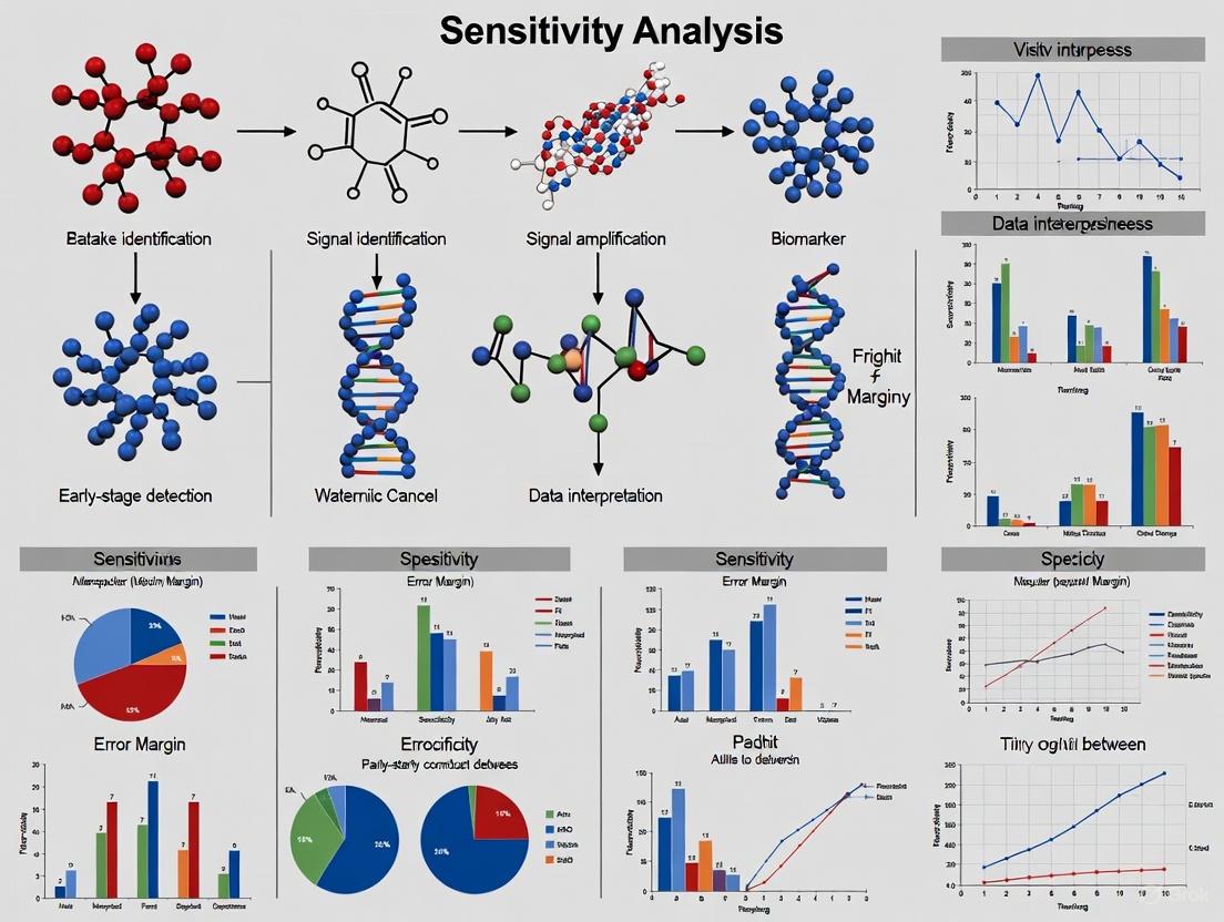
Optimizing Sensitivity in Early-Stage Cancer Detection: Methodologies, Models, and Clinical Validation
This article provides a comprehensive analysis of sensitivity optimization for early-stage cancer detection models, a critical frontier in oncology.
Optimizing Sensitivity in Early-Stage Cancer Detection: Methodologies, Models, and Clinical Validation
Abstract
This article provides a comprehensive analysis of sensitivity optimization for early-stage cancer detection models, a critical frontier in oncology. Aimed at researchers and drug development professionals, it explores the foundational importance of sensitivity-specificity trade-offs, reviews innovative machine learning methodologies like SMAGS-LASSO for feature selection, and addresses key challenges in model generalization and data aggregation. The content further synthesizes recent clinical validation data from multi-cancer early detection (MCED) tests, liquid biopsies, and AI-driven imaging analysis, offering a comparative evaluation of performance metrics across different technologies and their implications for accelerating diagnostic development and improving patient outcomes.
The Critical Imperative of Sensitivity in Early Cancer Detection
Defining Sensitivity and Specificity in Clinical Diagnostics
The Critical Role of Diagnostic Accuracy in Early Cancer Detection
In clinical diagnostics, sensitivity and specificity are foundational metrics for evaluating a test's performance. Sensitivity measures the proportion of true positive cases a test correctly identifies, crucial for ensuring diseases like cancer are not missed. Specificity measures the proportion of true negative cases correctly identified, minimizing false alarms and unnecessary, invasive follow-ups [1].
For multi-cancer early detection (MCED) technologies, these metrics determine clinical utility. High sensitivity is vital for early intervention, while high specificity is necessary to avoid burdening the healthcare system and causing patient anxiety from false positives [2] [1]. This guide objectively compares the performance and methodologies of leading MCED tests, providing researchers with a clear framework for evaluation.
Comparative Performance of MCED Tests
The table below summarizes published performance data for several advanced multi-cancer early detection tests, illustrating the balance between sensitivity and specificity across different technological approaches.
| Test Name | Technology / Biomarker | Reported Sensitivity (%) | Reported Specificity (%) | Number of Cancers Detected | Key Study/Cohort |
|---|---|---|---|---|---|
| Galleri (GRAIL) | Targeted Methylation of Cell-Free DNA | 51.5% (All cancers, all stages) [1] | 99.6% [1] | >50 types [1] | CCGA3 Validation Substudy [1] |
| 76.3% (12 deadly cancers, all stages) [1] | |||||
| OncoSeek | AI + 7 Protein Tumor Markers (PTMs) | 58.4% (All cohorts) [3] | 92.0% (All cohorts) [3] | 14 types [3] | Multi-Cohort Study (n=15,122) [3] |
| Carcimun | Conformational Changes in Plasma Proteins | 90.6% [4] | 98.2% [4] | 9 types (in study) [4] | Prospective Single-Blinded Study (n=172) [4] |
Note on Comparative Analysis: Test performance metrics do not represent results of a head-to-head comparative study. Separate studies have different designs, objectives, and participant populations, which limits the ability to draw conclusions about comparative performance [1].
Detailed Experimental Protocols and Workflows
Understanding the detailed methodology behind each test is critical for interpreting performance data and assessing technological validity.
Galleri Test: Targeted Methylation Sequencing
The Galleri test by GRAIL is based on sequencing cell-free DNA (cfDNA) to identify cancer-specific methylation patterns.
Workflow Diagram: Galleri Test Methodology
Key Experimental Steps:
- Sample Collection & Processing: A single blood draw (30-40 ml) is collected from the patient. Plasma is separated via centrifugation, and cell-free DNA is extracted [2].
- Library Preparation & Sequencing: The extracted cfDNA undergoes bisulfite conversion, which differentiates methylated from unmethylated cytosines. Targeted sequencing is then performed, focusing on a pre-defined panel of genomic regions with high diagnostic potential for cancer [1].
- Bioinformatic Analysis & Classification: The sequencing data is analyzed using a proprietary machine-learning classifier. This model, trained on methylation patterns from thousands of cancer and non-cancer samples, evaluates the cfDNA to determine two outputs:
- Cancer Signal Detection: A "Cancer Signal Detected" or "No Cancer Signal Detected" result.
- Cancer Signal Origin (CSO): For positive results, the model predicts the tissue or organ where the cancer originated with high accuracy (93.4%) to guide diagnostic workup [1].
OncoSeek Test: AI with Protein Biomarkers
The OncoSeek test employs a different strategy, combining the measurement of protein tumor markers (PTMs) with artificial intelligence.
Workflow Diagram: OncoSeek Test Methodology
Key Experimental Steps:
- Multiplex Immunoassay: A blood sample is analyzed on standard clinical immunoassay platforms (e.g., Roche Cobas, Bio-Rad Bio-Plex) to quantify the levels of seven pre-selected protein tumor markers [3].
- Data Integration: The concentrations of the seven PTMs are combined with the individual's basic clinical data, such as age and gender.
- AI-Powered Risk Assessment: An artificial intelligence algorithm processes the integrated data (PTM levels + clinical data) to generate a probability score for the presence of cancer. This approach is designed to be cost-effective and accessible, especially for low- and middle-income countries [3].
Carcimun Test: Plasma Protein Conformational Changes
The Carcimun test uses a biophysical method based on optical measurements of plasma proteins.
Workflow Diagram: Carcimun Test Methodology
Key Experimental Steps:
- Sample Preparation: A defined volume of blood plasma is diluted with a 0.9% sodium chloride solution and incubated at 37°C for thermal equilibration [4].
- Acidification and Measurement: A solution of acetic acid is added to the prepared sample. The final solution's optical extinction (absorbance) is measured at a wavelength of 340 nm using a clinical chemistry analyzer [4].
- Interpretation: The measured extinction value is compared against a pre-defined cut-off (determined in prior studies to be 120). Values above this threshold indicate a high probability of malignancy. The test detects conformational changes in plasma proteins that occur in the presence of cancer or acute inflammation [4].
The Scientist's Toolkit: Essential Research Reagents and Materials
The following table details key reagents and materials essential for developing and conducting MCED tests, based on the featured methodologies.
| Item Name | Function / Application in MCED Research |
|---|---|
| Cell-Free DNA (cfDNA) Extraction Kits | Isolate and purify fragmented DNA circulating in blood plasma, the primary analyte for sequencing-based liquid biopsies [2]. |
| Bisulfite Conversion Reagents | Chemically convert unmethylated cytosines in DNA to uracils, allowing for the precise mapping of methylation patterns via subsequent sequencing [1]. |
| Targeted Sequencing Panels | Pre-designed sets of probes to enrich and sequence specific genomic regions known to harbor informative methylation sites for cancer detection [1]. |
| Multiplex Immunoassay Panels | Enable the simultaneous quantification of multiple protein tumor markers (PTMs) from a single, small-volume blood sample [3]. |
| Clinical Chemistry Analyzers | Automated platforms used to perform precise photometric measurements, such as the extinction value readout in the Carcimun test [4]. |
| AI/ML Model Training Datasets | Large, well-annotated datasets of cancer and non-cancer samples used to train and validate classifiers for predicting cancer signals and tissue of origin [3] [1]. |
Why Sensitivity is Paramount for Early-Stage Disease and Low-Prevalence Cancers
Cancer remains a leading cause of mortality worldwide, with early detection representing one of the most powerful strategies for improving patient outcomes. For early-stage diseases and low-prevalence cancers, the diagnostic sensitivity of a test—its ability to correctly identify those with the disease—becomes not merely a performance metric but a crucial determinant of clinical utility. High sensitivity is particularly paramount in these contexts because missed diagnoses can lead to delayed interventions, significantly reducing treatment efficacy and survival probabilities. The fundamental challenge lies in the typically lower biomarker shed in early-stage malignancies, coupled with the higher prior probability of false negatives in low-prevalence settings, creating a diagnostic environment where only tests with exceptional sensitivity profiles demonstrate meaningful clinical value.
The statistical reality of screening low-prevalence populations further amplifies the critical importance of sensitivity. Even tests with seemingly high specificity can generate overwhelming numbers of false positives when applied to large screening populations, leading to unnecessary invasive procedures, patient anxiety, and increased healthcare costs. This review examines the central role of sensitivity in early cancer detection through a comparative analysis of emerging technologies, with particular focus on their performance characteristics and methodological approaches for evaluating robustness in low-prevalence clinical scenarios.
Performance Comparison of Cancer Detection Modalities
The evolving landscape of cancer detection technologies spans traditional imaging, liquid biopsy approaches, and artificial intelligence (AI)-enhanced diagnostic methods. The table below summarizes the published performance characteristics of various diagnostic modalities for different cancer types and clinical applications.
Table 1: Diagnostic Performance of Cancer Detection Technologies
| Technology | Cancer Type/Application | Sensitivity | Specificity | AUC | Sample Size | Reference |
|---|---|---|---|---|---|---|
| AI Deep Learning (DL) Models | Lymph Node Metastasis in T1-T2 CRC | 0.87 (95% CI: 0.76-0.93) | 0.69 (95% CI: 0.52-0.82) | 0.88 (95% CI: 0.84-0.90) | 8,540 patients | [5] |
| Galleri MCED Test | Multi-Cancer Detection | CSDR: 0.91% (Overall) | N/R | N/R | 111,080 individuals | [6] |
| Galleri MCED Test | Asymptomatic Patients (ePPV) | N/R | N/R | 49.4% (95% CI: 43.2-55.7%) | 259 patients | [6] |
| Enhanced CT | Colorectal Tumors | 76% (95% CI: 70-79%) | 87% (95% CI: 84-89%) | 0.89 (95% CI: 0.85-0.92) | 4,857 patients | [7] |
| Carcimun Test | Multiple Cancer Types | 90.6% | 98.2% | 95.4% (Accuracy) | 172 participants | [4] |
| AI Imaging Models | Esophageal Cancer | 90-95% | 80-93.8% | N/R | 158 studies (Umbrella Review) | [8] |
| AI Imaging Models | Breast Cancer | 75.4-92% | 83-90.6% | N/R | 158 studies (Umbrella Review) | [8] |
| AI Imaging Models | Ovarian Cancer | 75-94% | 75-94% | N/R | 158 studies (Umbrella Review) | [8] |
Abbreviations: CI: confidence interval; CRC: colorectal cancer; CSDR: cancer signal detection rate; ePPV: empirical positive predictive value; N/R: not reported; MCED: multi-cancer early detection
The comparative data reveals significant variability in sensitivity profiles across diagnostic approaches. AI-based models demonstrate particularly strong sensitivity for detecting lymph node metastasis in colorectal cancer (87%) and for identifying esophageal cancer (90-95%) through imaging analysis [5] [8]. The high specificity (98.2%) of the Carcimun test is notable, though its generalizability requires validation in larger cohorts [4]. For multi-cancer early detection, the Galleri test demonstrates a real-world cancer signal detection rate of 0.91%, consistent with prior clinical studies [6].
Methodological Approaches for Sensitivity Analysis
Sensitivity Analysis in Diagnostic Research
Sensitivity analysis represents a crucial methodological framework for assessing the robustness of research findings, particularly in observational studies using routinely collected healthcare data (RCD). These analyses systematically examine how variations in study definitions, designs, or statistical models impact effect estimates and conclusions. In cancer detection research, sensitivity analyses typically focus on three key dimensions:
- Alternative study definitions: Using different coding algorithms or thresholds to identify exposures, outcomes, or confounders [9] [10]
- Alternative study designs: Modifying inclusion criteria, time periods, or data sources [9] [10]
- Alternative statistical models: Employing different approaches for handling missing data, adjusting for confounding, or modeling associations [9] [10]
Recent evidence indicates that sensitivity analyses frequently produce meaningfully different results compared to primary analyses. A meta-epidemiological study of 256 observational studies found that 54.2% showed significant differences between primary and sensitivity analyses, with an average difference in effect size of 24% (95% CI: 12% to 35%) [9] [10]. Despite these discrepancies, only 9 of 71 studies with inconsistent results discussed the potential implications, highlighting a critical gap in current reporting practices [9] [10].
Experimental Protocols for Model Validation
Robust validation of sensitivity in cancer detection models requires meticulous methodological approaches. For AI-based prediction of lymph node metastasis in colorectal cancer, the following protocol represents current best practices:
Table 2: Experimental Protocol for AI Model Validation in Cancer Detection
| Protocol Phase | Key Procedures | Quality Control Measures |
|---|---|---|
| Study Registration | A priori registration of study protocol with repositories (e.g., PROSPERO: CRD42024607756) | Ensures transparency and reduces reporting bias [5] |
| Literature Search | Comprehensive search across multiple databases (PubMed, EMBASE, Web of Science, Cochrane, Scopus) using MeSH and free-text terms | Follows PRISMA guidelines; minimizes selection bias [5] |
| Study Selection | Dual-independent screening based on predetermined inclusion/exclusion criteria | Uses standardized tools (QUADAS-2) to assess risk of bias [5] |
| Data Extraction | Independent extraction by multiple researchers using piloted forms | Includes resolution of disagreements through consensus [5] |
| Statistical Analysis | Mixed-effects models to integrate diagnostic accuracy data | Calculates sensitivity, specificity, likelihood ratios, and diagnostic odds ratios with confidence intervals [5] |
| Heterogeneity Assessment | Evaluation of threshold effects using Spearman correlation | Quantifies inconsistency with I² statistic [5] |
This rigorous methodology enabled the meta-analysis to determine that AI-based models achieve a sensitivity of 0.87 (95% CI: 0.76-0.93) for predicting lymph node metastasis in T1-T2 colorectal cancer lesions, significantly outperforming traditional assessment methods [5].
Visualizing Diagnostic Pathways and Workflows
Multi-Cancer Early Detection Diagnostic Pathway
The following diagram illustrates the clinical workflow and decision pathway for multi-cancer early detection tests, based on real-world implementation data:
MCED Clinical Decision Pathway: This workflow illustrates the patient journey through multi-cancer early detection testing, showing key decision points and outcomes based on real-world data from 111,080 individuals [6]. CSDR: Cancer Signal Detection Rate; CSO: Cancer Signal Origin.
AI-Assisted Histopathological Analysis Workflow
For AI-based prediction of lymph node metastasis in colorectal cancer, the following diagram outlines the complex analytical workflow:
AI Histopathology Analysis Workflow: This diagram outlines the technical process for AI-based prediction of lymph node metastasis (LNM) in T1/T2 colorectal cancer (CRC), achieving 87% sensitivity in validation studies [5].
The Scientist's Toolkit: Essential Research Reagents and Materials
Table 3: Key Research Reagents and Materials for Cancer Detection Studies
| Reagent/Material | Specifications | Research Application |
|---|---|---|
| Cell-free DNA Collection Tubes | Streck Cell-Free DNA BCT or equivalent | Stabilizes nucleated blood cells for accurate cfDNA analysis in MCED tests [6] |
| Targeted Methylation Sequencing Panel | Bisulfite conversion reagents; methylation-specific primers | Enables detection of cancer-specific methylation patterns in cfDNA [6] |
| Whole Slide Imaging Scanners | High-resolution slide scanners (20x-40x magnification) | Digitizes histopathology slides for AI-based feature extraction [5] |
| Convolutional Neural Network Frameworks | TensorFlow, PyTorch, or specialized medical imaging libraries | Enables development of deep learning models for pattern recognition in medical images [5] [8] |
| Enhanced CT Contrast Agents | Iodinated contrast (IV); barium-based suspensions (oral) | Improves tissue characterization and tumor delineation for diagnostic imaging [7] |
| Optical Extinction Measurement System | UV-Vis spectrophotometer; microplate readers | Measures conformational changes in plasma proteins for novel cancer detection approaches [4] |
The critical importance of sensitivity in early-stage disease and low-prevalence cancers continues to drive methodological innovations across detection technologies. The emerging evidence demonstrates that AI-enhanced diagnostic approaches can achieve sensitivies exceeding 85% for challenging detection scenarios such as lymph node metastasis prediction, substantially outperforming conventional assessment methods [5]. Similarly, blood-based multi-cancer early detection tests now provide a viable approach for detecting malignancies that lack recommended screening modalities, which account for approximately 83% of cancer-related deaths [6].
Future progress will require intensified focus on validating sensitivity claims in real-world clinical settings, with particular attention to methodological robustness through comprehensive sensitivity analyses. As these technologies evolve, their integration into standardized clinical pathways promises to transform early cancer detection, ultimately enabling intervention at disease stages when treatments are most effective and survival outcomes most favorable. The continuing refinement of sensitivity profiles across detection platforms represents one of the most promising frontiers in oncology research, with profound implications for cancer mortality reduction worldwide.
In clinical practice, a diagnostic error represents a critical failure with profound implications for patient outcomes. The National Academies of Sciences, Engineering, and Medicine defines diagnostic error as "the failure to (a) establish an accurate and timely explanation of the patient's health problem(s) or (b) communicate that explanation to the patient" [11]. This dual definition captures both the cognitive and systems dimensions of diagnostic failure, emphasizing that accuracy and timeliness are equally crucial in the diagnostic process. Within oncology, these failures manifest specifically as false negatives (where cancer is present but not detected), missed diagnoses (where no diagnosis is established despite presenting symptoms), and delayed diagnoses (where the identification occurs after the optimal intervention window) [11].
The diagnostic process encompasses a sequence of stages where errors can occur: initial patient presentation, history taking, physical examination, diagnostic testing, assessment, referral, and follow-up [11]. A failure at any point in this cascade can lead to diagnostic harm. When cancer evades timely detection, the clinical consequences are often severe: progression to more advanced stages, reduced treatment options, diminished survival probabilities, and increased healthcare costs [12]. For researchers and drug development professionals, understanding the mechanisms, frequencies, and impacts of these diagnostic failures provides crucial insights for developing more robust detection technologies and therapeutic interventions.
Quantifying the Problem: Diagnostic Error Rates and Performance Metrics
The Scope of Diagnostic Error
Diagnostic errors affect a significant proportion of patients, with studies suggesting that approximately 11% of medical conditions may be misdiagnosed [12]. The likelihood of misdiagnosis varies substantially by condition type, with those presenting overlapping symptoms with other disorders being particularly vulnerable to diagnostic inaccuracy. Analyses of successful medical claims related to diagnostic failures reveal that the primary resulting harms include:
- Fatality
- Cancer progression
- Unnecessary pain
- Unnecessary operations
- Fracture complications [12]
The complexity of certain medical conditions represents a fundamental challenge to diagnostic accuracy. Diseases with diverse and overlapping symptoms, or those presenting atypical manifestations, pose considerable difficulties for clinicians [12]. This is particularly relevant in oncology, where early-stage cancers often present with non-specific symptoms that may be mistaken for benign conditions.
Sensitivity Metrics in Cancer Detection
The sensitivity of a cancer screening biomarker to detect prevalent preclinical cancer drives screening benefit, yet this metric must be interpreted with careful attention to how it was derived [13]. Different study designs produce meaningfully different sensitivity estimates:
- Clinical sensitivity: Generally optimistic, estimated from clinically diagnosed cases
- Archived-sample sensitivity: Variable bias depending on look-back intervals and test specificity
- Prospective empirical sensitivity: Optimistic when sojourn time is long relative to screening interval [13]
These distinctions in sensitivity measurement highlight the importance of clear terminology when evaluating and comparing the performance of cancer detection technologies. Preclinical sensitivity - the ability to detect cancer during the preclinical sojourn time - represents the ideal metric but is challenging to measure directly [13].
Comparative Performance of Cancer Detection Modalities
Traditional vs. AI-Enhanced Detection Methods
Cancer detection methodologies have evolved substantially, with machine learning (ML) and deep learning (DL) approaches now demonstrating remarkable capabilities. The table below summarizes the performance ranges of these approaches across multiple cancer types based on recent studies (2018-2023) [14].
Table 1: Performance Comparison of ML and DL Models in Cancer Detection
| Cancer Type | Best ML Accuracy | Best DL Accuracy | Lowest ML Accuracy | Lowest DL Accuracy |
|---|---|---|---|---|
| Lung Cancer | 99.7% [15] | 99.7% [15] | 74.4% [15] | 74.4% [15] |
| Breast Cancer | 99.04% [15] | 99.6% [15] | 75.48% [14] | 70% [14] |
| Prostate Cancer | Not specified | Not specified | Not specified | Not specified |
| Colorectal Cancer | 75% [15] | 75% [15] | 73% [15] | 73% [15] |
| Osteosarcoma | 97.8% [16] | Not specified | Not specified | Not specified |
The performance variation across cancer types reflects differences in data availability, imaging clarity, and biological complexity. For breast cancer detection, models using the Wisconsin breast cancer dataset have achieved exceptional accuracy (up to 99.6%) through both convolutional neural networks and traditional machine learning classifiers like Multilayer Perceptron models [15].
Multi-Cancer Early Detection (MCED) Tests
Innovative blood-based multi-cancer early detection tests represent a promising approach for detecting multiple cancer types from a single blood draw. The Carcimun test, which detects conformational changes in plasma proteins through optical extinction measurements, demonstrates the potential of this approach [4].
Table 2: Performance Metrics of the Carcimun MCED Test
| Metric | Value | Comparative Context |
|---|---|---|
| Overall Accuracy | 95.4% | Higher than many single-cancer biomarkers |
| Sensitivity | 90.6% | Effectively identifies cancer patients |
| Specificity | 98.2% | Minimizes false positives |
| PPV/NPV | Not specified | Not specified |
| Distinction Capability | Significantly different mean extinction values between cancer patients (315.1), healthy individuals (23.9), and those with inflammatory conditions (62.7); p<0.001 [4] | Addresses key limitation of inflammatory confounders |
This performance is particularly notable given the test's ability to differentiate cancer patients from those with inflammatory conditions (fibrosis, sarcoidosis, pneumonia) or benign tumors, a significant challenge for many cancer detection methods [4].
Methodological Approaches: Experimental Protocols in Cancer Detection Research
Protocol for MCED Test Validation
The validation of the Carcimun test followed a rigorous experimental protocol [4]:
- Study Design: Prospective, single-blinded study including 172 participants (80 healthy volunteers, 64 cancer patients with various types, 28 individuals with inflammatory conditions or benign tumors)
- Sample Preparation: 70 µl of 0.9% NaCl solution added to reaction vessel, followed by 26 µl of blood plasma, resulting in total volume of 96 µl with final NaCl concentration of 0.9%
- Testing Process: Addition of 40 µl distilled water, incubation at 37°C for 5 minutes, blank measurement at 340 nm, addition of 80 µl of 0.4% acetic acid solution, final absorbance measurement at 340 nm using Indiko Clinical Chemistry Analyzer
- Analysis: Measurements performed blinded to clinical status, using predetermined cut-off value of 120 to differentiate between healthy and cancer subjects
- Statistical Analysis: One-way ANOVA with Tukey and Games-Howell post-hoc tests, with p-values ≤ 0.05 considered statistically significant
This methodology highlights the importance of including patients with inflammatory conditions in validation cohorts to assess real-world performance and minimize false positives [4].
DNA Sequencing and Sentence Transformer Approach
Innovative approaches using DNA sequences with sentence transformer models represent cutting-edge methodology in cancer detection [15]:
- Input Data: Raw DNA sequences of matched tumor/normal pairs as sole input
- Feature Extraction: Sentence transformers (SBERT and SimCSE) to compute dense vector representations of DNA sequences
- Classification: Embeddings fed to machine learning algorithms (XGBoost, Random Forest, LightGBM, and CNNs) for classification
- Performance: XGBoost achieved highest overall accuracy (73 ± 0.13% with SBERT embeddings; 75 ± 0.12% with SimCSE embeddings)
This approach demonstrates how natural language processing techniques can be adapted to genomic data, though current accuracy rates remain lower than imaging-based methods [15].
ML-Based Clinical Decision Support System Development
The development of machine learning-based clinical decision support systems for emergency departments demonstrates a structured approach to predictive model creation [17]:
- Data Collection: 27 fixed features (age, sex, medical history, etc.) and 93 observation features (vital signs, laboratory results, mental status changes) extracted from electronic health records
- Data Preprocessing: Resampling into 1-hour units, carry-forward method for missing data, randomized under-sampling in training set to address class imbalance
- Model Development: XGBoost algorithm with hyperparameter tuning via tenfold cross-validation
- Temporal Modeling: Development of 25 predictive models with varying lagging (previous information) and leading (future prediction) time windows
- Outcome Prediction: Four critical outcomes - intubation, ICU admission, inotrope/vasopressor administration, and in-hospital cardiac arrest
This methodology emphasizes the importance of aligning model development with clinical decision-making frameworks and workflow considerations [17].
Diagnostic Process and Failure Points
The diagnostic process involves multiple stages where errors can occur, potentially leading to false negatives or missed diagnoses. The following diagram illustrates this pathway and critical failure points:
The Researcher's Toolkit: Essential Materials and Reagents
Table 3: Key Research Reagent Solutions in Cancer Detection Studies
| Reagent/Technology | Function/Application | Example Use Cases |
|---|---|---|
| Cell-free DNA (cfDNA) Isolation Kits | Extraction of circulating tumor DNA from blood samples | Liquid biopsy applications, multi-cancer early detection tests [4] |
| Sentence Transformers (SBERT/SimCSE) | Dense vector representation of DNA sequences | DNA sequence classification for cancer detection [15] |
| Optical Extinction Measurement Reagents | Detection of conformational changes in plasma proteins | Carcimun test for cancer identification [4] |
| Methylation-Specific PCR Reagents | Detection of epigenetic modifications in cancer cells | Analysis of DNA methylation patterns in circulating tumor DNA [13] |
| XGBoost Algorithm | Machine learning classification of complex medical data | Clinical decision support systems, outcome prediction [17] |
| Immunohistochemistry Kits | Tissue-based protein detection and classification | Tumor subtype classification, biomarker validation [14] |
The clinical consequences of false negatives and missed diagnoses in cancer care represent a critical challenge with significant implications for patient outcomes. Diagnostic errors, affecting an estimated 11% of medical conditions, can lead to progression to more advanced disease stages, reduced treatment options, and increased mortality [12].
Advances in detection methodologies, particularly machine learning and deep learning approaches, have demonstrated remarkable accuracy improvements, with some models achieving up to 99.7% accuracy in specific cancer types [15]. The emergence of multi-cancer early detection tests offers promising avenues for identifying cancers at earlier, more treatable stages, with tests like Carcimun demonstrating 90.6% sensitivity and 98.2% specificity while effectively differentiating cancer from inflammatory conditions [4].
For researchers and drug development professionals, these technological advances create opportunities for developing more robust detection systems and targeted therapies. However, challenges remain in standardizing sensitivity metrics across study designs, addressing spectrum bias in validation, and ensuring that improved diagnostic accuracy translates to meaningful clinical benefit [13]. Future research should focus on refining detection methodologies, expanding validation across diverse populations, and integrating artificial intelligence systems into clinical workflows in ways that enhance rather than disrupt the diagnostic process [18] [17].
Current Landscape and Unmet Needs in Early Detection Technologies
Cancer remains a leading cause of mortality worldwide, with early detection representing a critical factor in improving patient survival outcomes [19]. Conventional screening methods are predominantly limited to single-cancer detection—such as mammography for breast cancer and colonoscopy for colorectal cancer—and cover only a limited number of cancer types [19]. This approach leaves significant gaps, as approximately 45.5% of cancer cases occur in cancers without recommended screening protocols [19]. The prognosis for cancers typically diagnosed at advanced stages, such as pancreatic and ovarian cancers, remains particularly poor, underscoring the substantial unmet need for more comprehensive detection technologies [19].
The emerging landscape of early detection technologies is being reshaped by two transformative approaches: Multi-Cancer Early Detection (MCED) tests and Artificial Intelligence (AI)-powered diagnostic tools. MCED technologies, often utilizing liquid biopsy, analyze circulating tumor DNA (ctDNA) and other biomarkers in blood to simultaneously screen for multiple cancers [19]. Concurrently, AI and machine learning algorithms are demonstrating remarkable capabilities in analyzing complex datasets—from medical images to genomic sequences—to identify subtle cancer signals often missed by conventional methods [20] [21]. These technological advances are creating a paradigm shift from reactive cancer diagnosis to proactive, pre-symptomatic detection, potentially revolutionizing oncology care and prevention strategies.
Technology Comparison: Performance Metrics of Emerging Detection Platforms
Multi-Cancer Early Detection (MCED) Tests
MCED tests represent a groundbreaking approach to cancer screening by enabling the detection of multiple cancer types through a single blood draw. These tests primarily analyze cell-free DNA (cfDNA) using various methodological approaches, including assessment of DNA mutations, abnormal methylation patterns, and fragmentation profiles [19]. The table below summarizes the performance characteristics of leading MCED platforms under development.
Table 1: Comparative Performance of Multi-Cancer Early Detection Tests
| Test Name | Company/Developer | Detection Method | Reported Sensitivity | Reported Specificity | Detectable Cancer Types |
|---|---|---|---|---|---|
| Galleri | GRAIL | Targeted methylation sequencing | 51.5% (all cancers); 73.7% (for 12 high-mortality cancers) [22] | 99.5% [19] | >50 cancer types [19] |
| CancerSEEK | Exact Sciences | Multiplex PCR + protein immunoassay | 62% (across 8 cancer types) [19] | >99% [19] | Breast, colorectal, pancreatic, gastric, hepatic, esophageal, ovarian, lung [19] |
| Shield | Guardant Health | Genomic mutations, methylation, DNA fragmentation | 65% (Stage I CRC); 100% (Stages II-IV CRC) [19] | 99% [19] | Colorectal cancer (with MCED potential) [19] |
| DEEPGENTM | Quantgene | Next-generation sequencing (NGS) | 43% [19] | 99% [19] | Lung, breast, colorectal, prostate, bladder, pancreatic, liver [19] |
| DELFI | Delfi Diagnostics | cfDNA fragmentation profiles + machine learning | 73% [19] | 98% [19] | Lung, breast, colorectal, pancreatic, gastric, bile duct, ovarian [19] |
| PanSeer | Singlera Genomics | Semi-targeted PCR libraries and sequencing | 87.6% [19] | 96.1% [19] | Lung, colorectal, gastric, liver, esophageal [19] |
Recent data from large-scale interventional studies demonstrate the real-world performance of these technologies. The PATHFINDER 2 study, the largest U.S. MCED interventional study to date with 35,878 participants, evaluated Galleri's performance in a screening population [22]. When added to standard USPSTF-recommended screenings (breast, cervical, colorectal, and lung cancers), Galleri increased the cancer detection rate more than seven-fold [22]. Notably, 53.5% of cancers detected by Galleri were early-stage (I or II), and approximately three-quarters were cancer types without recommended screening tests [22]. The test demonstrated a 92% accuracy in predicting the cancer signal origin (CSO), facilitating efficient diagnostic workups with a median time to diagnostic resolution of 46 days [22].
Artificial Intelligence and Machine Learning Platforms
AI-based technologies are revolutionizing cancer detection across multiple modalities, including medical imaging, histopathology, and genomic data analysis. These tools leverage sophisticated algorithms to identify subtle patterns indicative of early-stage malignancies.
Table 2: AI-Powered Detection Platforms and Performance Characteristics
| AI Platform | Developer/Institution | Application | Reported Performance |
|---|---|---|---|
| DeepHRD | University of California, San Diego | Detects homologous recombination deficiency (HRD) from biopsy slides | 3x more accurate than current genomic tests; negligible failure rate vs. 20-30% for conventional tests [21] |
| Prov-GigaPath, Owkin's models, CHIEF | Multiple developers | Cancer detection imaging | Various performance metrics across cancer types [21] |
| MSI-SEER | Vanderbilt University Medical Center | Identifies microsatellite instability-high (MSI-H) regions in tumors | Enables more gastrointestinal cancer patients to benefit from immunotherapy [21] |
| CatBoost Model | Research Study (Scientific Reports) | Cancer risk prediction using lifestyle and genetic data | 98.75% test accuracy; F1-score of 0.9820 [20] |
| HistoPathXplorer | Not specified | Tissue biomarker detection | Improved detection accuracy for various biomarkers [21] |
| Paige Prostate Detect | Paige | Prostate biopsy interpretation | Enhanced accuracy in prostate cancer detection [21] |
Machine learning approaches have demonstrated remarkable efficacy in cancer risk prediction. One recent study implemented a full end-to-end ML pipeline using a dataset of 1,200 patient records with features including age, BMI, smoking status, alcohol intake, physical activity, genetic risk level, and personal cancer history [20]. Among nine supervised learning algorithms evaluated, the Categorical Boosting (CatBoost) model achieved the highest predictive performance, with test accuracy of 98.75% and an F1-score of 0.9820 [20]. Feature importance analysis confirmed the strong influence of cancer history, genetic risk, and smoking status on prediction outcomes [20].
Methodological Deep Dive: Experimental Protocols and Workflows
MCED Test Methodologies
The most promising MCED technologies share common methodological foundations while employing distinct analytical approaches. The following workflow illustrates the generalized process for MCED testing:
MCED Testing Workflow: This diagram illustrates the multi-step process from blood draw to clinical report generation in MCED testing.
The foundational protocol for MCED testing begins with blood sample collection through standard venipuncture, typically requiring one to two standard blood collection tubes (10-20ml total volume) [19]. Following collection, samples undergo centrifugation to separate plasma from cellular components, then cfDNA is extracted using commercial isolation kits. The critical analysis phase employs one or more of these methodological approaches:
- Targeted Methylation Sequencing: Platforms like Galleri interrogate methylated regions of cfDNA, as abnormal methylation patterns are characteristic of cancer cells and provide information about the tissue of origin [19].
- cfDNA Fragmentomics: Approaches like DELFI analyze the fragmentation patterns of cfDNA, which differ between cancerous and normal states due to variations in nuclear organization and cell death mechanisms [19].
- Mutation Analysis with Protein Biomarkers: CancerSEEK combines targeted sequencing of 16 cancer gene mutations with measurement of 8 cancer-associated proteins, creating a multimodal assessment that increases sensitivity over single-analyte approaches [19].
Following data generation, bioinformatic analysis using machine learning algorithms classifies samples as cancer-positive or negative, and for positive signals, predicts the tissue of origin (Cancer Signal Origin) [19] [22]. The final clinical report provides these determinations to guide subsequent diagnostic evaluation.
Biomarker Validation Frameworks
The journey from biomarker discovery to clinical application requires rigorous validation with specific statistical considerations. The biomarker development pipeline encompasses several critical phases:
Biomarker Validation Pathway: This diagram outlines the multi-stage process from initial biomarker discovery through clinical implementation.
Discovery Phase: Biomarker discovery begins with defining the intended use context (e.g., screening, diagnosis, prognosis) and target population [23]. Studies should use specimens that directly reflect the intended-use population, with careful attention to avoiding bias through randomization and blinding during specimen analysis [23]. For predictive biomarkers (indicating response to specific treatments), identification requires data from randomized clinical trials with interaction testing between treatment and biomarker status [23].
Analytical Validation: This phase establishes that the biomarker test itself reliably measures the intended analyte across key performance parameters including:
- Sensitivity: Proportion of true positives correctly identified
- Specificity: Proportion of true negatives correctly identified
- Positive Predictive Value (PPV): Proportion of test-positive cases with the condition
- Negative Predictive Value (NPV): Proportion of test-negative cases without the condition
- Area Under the Curve (AUC): Overall discrimination ability [23]
Clinical Validation and Utility: Prospective studies like PATHFINDER 2 demonstrate real-world performance, with recent MCED tests achieving PPV of 61.6% and specificity of 99.6% in actual screening populations [22]. Clinical utility is established through impact on patient outcomes, with studies showing more than half (53.5%) of MCED-detected cancers being early-stage (I or II) [22].
The Scientist's Toolkit: Essential Research Reagents and Materials
Successful development and implementation of early detection technologies require specialized reagents and research tools. The following table catalogues essential materials referenced across the cited studies:
Table 3: Essential Research Reagents and Materials for Early Detection Technology Development
| Reagent/Material | Function/Purpose | Example Applications |
|---|---|---|
| Cell-free DNA Isolation Kits | Extraction of circulating tumor DNA from blood plasma | MCED test development; liquid biopsy applications [19] |
| Bisulfite Conversion Reagents | DNA treatment for methylation analysis | Targeted methylation sequencing platforms (e.g., Galleri) [19] |
| Next-Generation Sequencing Kits | Library preparation and sequencing | Genomic mutation detection; fragmentomics analysis [19] [21] |
| PCR and Multiplex PCR Reagents | Amplification of target DNA sequences | Mutation detection in CancerSEEK; PanSeer test [19] |
| Protein Immunoassay Reagents | Detection of cancer-associated proteins | Protein biomarker analysis in CancerSEEK [19] |
| Machine Learning Algorithms | Pattern recognition in complex datasets | CatBoost, Random Forest for risk prediction; AI-based image analysis [20] [21] |
| Digital Pathology Systems | Slide digitization and image analysis | Roche digital pathology; DeepHRD analysis [21] [24] |
| Reference Standards | Assay validation and quality control | Biomarker analytical validation; assay performance monitoring [23] |
| Biobanked Specimens | Validation study material | Performance assessment in intended-use populations [23] |
Unmet Needs and Future Directions
Despite remarkable advances, significant challenges remain in the early detection technology landscape. Current conventional screening methods detect only 20-30% of deadly cancers, while approximately 70% of cancer deaths originate from cancers without standard screening recommendations [22]. Even for cancers with established screening methods, participation rates remain suboptimal, with breast cancer screening participation as low as 40% in some regions [19].
The integration of AI tools into clinical workflows faces methodological and reporting challenges. A recent review of ML studies in oncology found significant deficiencies in reporting quality, particularly regarding sample size calculation (missing in 98% of studies), data quality issues (missing in 69%), and strategies for handling outliers (missing in 100%) [25]. Improved adherence to reporting guidelines like TRIPOD+AI and CREMLS is essential for translating algorithmic advances into clinically actionable tools [25].
Future development must address several critical areas: (1) expanding the spectrum of detectable cancers, particularly those with high mortality and no current screening options; (2) improving sensitivity for early-stage detection while maintaining high specificity to minimize false positives; (3) enhancing accessibility and reducing costs to enable broader population-level implementation; and (4) developing robust clinical guidelines for managing patients with positive MCED results, especially for cancers without established diagnostic pathways [19] [22]. As these technologies evolve, they hold the potential to fundamentally transform cancer screening from a limited, organ-specific approach to a comprehensive, population-wide health initiative.
Advanced Algorithms and Multi-Omic Approaches for Sensitivity Maximization
In clinical diagnostics, particularly for diseases with low prevalence such as cancer, the effective classification of patients into healthy control and disease groups represents a critical challenge [26]. While numerous metrics exist to evaluate classification performance, sensitivity (true-positive rate) and specificity (true-negative rate) stand as particularly important metrics in early cancer detection [27] [26]. High sensitivity is essential to minimize missed cancer diagnoses, while high specificity helps avoid unnecessary clinical procedures in healthy individuals [26].
However, most machine learning algorithms prioritize overall accuracy during optimization, which fails to align with clinical priorities of early detection [27] [26]. Traditional classification methods, such as logistic regression with maximum likelihood estimation, are designed to optimize overall accuracy and do not explicitly prioritize sensitivity—an essential objective in early cancer detection [26]. This methodological gap becomes particularly problematic in high-dimensional biomarker data where feature selection is crucial not only for computational efficiency but also for developing interpretable models that can identify the most informative biomarkers [26].
To address these challenges, researchers have developed SMAGS-LASSO (Sensitivity Maximization at a Given Specificity with LASSO), a novel machine learning algorithm that combines a sensitivity maximization framework with L1 regularization for feature selection [27] [26]. This review provides a comprehensive comparison of SMAGS-LASSO against established feature selection and classification methods, with a specific focus on its application in early cancer detection contexts.
Performance Benchmarking: SMAGS-LASSO vs. Alternative Methods
Comparative Performance in Synthetic and Real-World Datasets
Rigorous evaluation across synthetic and real-world biological datasets demonstrates SMAGS-LASSO's significant advantages in sensitivity-limited applications.
Table 1: Performance Comparison on Synthetic Datasets (99.9% Specificity)
| Method | Sensitivity | 95% CI | Number of Features Selected |
|---|---|---|---|
| SMAGS-LASSO | 1.00 | 0.98-1.00 | 15 |
| Standard LASSO | 0.19 | 0.13-0.23 | 18 |
| Random Forest | 0.42 | 0.35-0.49 | 22 |
In synthetic datasets designed to contain strong signals for both sensitivity and specificity, SMAGS-LASSO significantly outperformed standard LASSO, achieving perfect sensitivity (1.00) compared to only 0.19 for LASSO at 99.9% specificity [27]. The synthetic dataset comprised 2,000 samples (1,000 per class) with 100 features, with evaluation performed using an 80/20 train-test split [26].
Table 2: Performance on Colorectal Cancer Biomarker Data (98.5% Specificity)
| Method | Sensitivity | Improvement over LASSO | p-value | Features Selected |
|---|---|---|---|---|
| SMAGS-LASSO | 0.89 | 21.8% | 2.24E-04 | 12 |
| Standard LASSO | 0.73 | Baseline | - | 12 |
| Random Forest | 0.55 | -38.5% | 4.62E-08 | 15 |
In colorectal cancer data, SMAGS-LASSO demonstrated a 21.8% improvement over LASSO and a 38.5% improvement over Random Forest at 98.5% specificity while selecting the same number of biomarkers [27]. This performance is particularly notable because it was achieved while maintaining the same level of sparsity as comparison methods, enabling the development of minimal biomarker panels that maintain high sensitivity at predefined specificity thresholds [27] [26].
Comparison with Other Feature Selection Paradigms
Beyond the comparisons with standard LASSO and Random Forest, SMAGS-LASSO represents a distinct approach within the broader landscape of feature selection methods.
Table 3: Feature Selection Method Classification and Characteristics
| Method Type | Examples | Key Characteristics | Best Use Cases |
|---|---|---|---|
| Filter Methods | Fisher Score, Mutual Information [28] | Fast, model-independent, uses statistical measures | Preliminary feature screening, large datasets |
| Wrapper Methods | Sequential Feature Selection, Recursive Feature Elimination [28] | Model-specific, computationally intensive, higher performance | Small to medium datasets, performance-critical applications |
| Embedded Methods | LASSO, Random Forest Importance [28], SMAGS-LASSO [26] | Built-in feature selection, balance of efficiency and performance | High-dimensional data, clinical diagnostics |
| Hybrid Methods | TMGWO, ISSA, BBPSO [29] | Combine multiple algorithms, optimize feature subsets | Complex datasets with many redundant features |
Unlike filter methods that evaluate features independently using statistical measures, or wrapper methods that assess feature subsets through iterative model training, SMAGS-LASSO employs an embedded approach that integrates feature selection directly into the model training process [26] [30]. This provides a better balance of efficiency and performance compared to wrapper methods, while offering greater model-specific optimization than filter methods [30].
When compared to other advanced feature selection approaches, such as the hybrid TMGWO (Two-phase Mutation Grey Wolf Optimization) algorithm which achieved 96% accuracy on breast cancer data using only 4 features [29], SMAGS-LASSO distinguishes itself through its direct optimization of sensitivity at predetermined specificity thresholds rather than overall accuracy [26].
SMAGS-LASSO Methodology and Experimental Protocols
Core Algorithm and Optimization Framework
SMAGS-LASSO modifies the traditional LASSO framework to directly optimize sensitivity rather than likelihood or mean squared error. The objective function is formulated as:
maxβ,β0 ∑i=1n ŷi · yi / ∑i=1n yi - λ∥β∥1
Subject to: (1-y)T(1-ŷ) / (1-y)T(1-y) ≥ SP [26]
Where:
- β and β0 are the coefficient vector and intercept
- ŷi and yi are the predicted and actual classes
- λ is the regularization parameter controlling sparsity
- ∥β∥1 is the L1-norm of the coefficient vector
- SP is the predefined specificity threshold
The predicted class ŷi is determined by ŷi = I(σ(xiTβ + β0) > θ), where σ is the sigmoid function and θ is a threshold parameter determined adaptively to control the specificity level [26].
The optimization faces challenges due to the non-differentiable nature of both the sensitivity metric and the L1 penalty. To address this, SMAGS-LASSO employs a multi-pronged optimization strategy using several algorithms (Nelder-Mead, BFGS, CG, L-BFGS-B) with varying tolerance levels, running in parallel to efficiently explore multiple optimization paths [26].
Cross-Validation and Evaluation Framework
To select the optimal regularization parameter λ, SMAGS-LASSO implements a specialized cross-validation procedure:
- Creates k-fold partitions of the data (k = 5 by default)
- Evaluates a sequence of λ values on each fold
- Measures performance using sensitivity mean squared error (MSE):
- MSEsensitivity = [1 - (∑ŷi·yi / ∑yi)]2
- Tracks the norm ratio ∥βλ∥1/∥β∥1 to quantify sparsity
The cross-validation process selects the λ value that minimizes the sensitivity MSE, effectively finding the most regularized model that maintains high sensitivity [26].
The general evaluation framework employs 80/20 stratified train-test splits to maintain balanced class representation and ensure robust performance assessment [26]. This rigorous approach is essential for generating reliable performance estimates in clinical applications.
Diagram Title: SMAGS-LASSO Algorithm Workflow
Research Reagent Solutions for Implementation
Table 4: Essential Research Materials and Computational Tools
| Item | Function | Application in SMAGS-LASSO |
|---|---|---|
| Protein Biomarker Data | Biological features for cancer detection | Used as real-world validation dataset [27] |
| Synthetic Datasets | Controlled algorithm evaluation | Contains strong sensitivity/specificity signals [26] |
| Custom Loss Function | Sensitivity maximization at given specificity | Core component of SMAGS-LASSO framework [26] |
| L1 Regularization | Sparse feature selection | Enables minimal biomarker panel development [27] [26] |
| Multi-Algorithm Optimization | Robust parameter estimation | Nelder-Mead, BFGS, CG, L-BFGS-B [26] |
| Cross-Validation Framework | Hyperparameter tuning and model selection | Identifies optimal λ for sensitivity [26] |
| Open-Source Implementation | Method accessibility and reproducibility | Available at github.com/khoshfekr1994/SMAGS.LASSO [26] |
The implementation relies on both biological and computational components. The biological components include protein biomarker data derived from colorectal cancer studies, which provide real-world validation [27]. Synthetic datasets with known signal patterns enable controlled evaluation of the method's capabilities [26].
Key computational elements include the custom loss function that defines the sensitivity optimization objective, L1 regularization for sparsity, and a multi-algorithm optimization approach that ensures robust convergence [26]. The open-source implementation makes this method accessible to the research community for further validation and application.
SMAGS-LASSO represents a significant advancement in feature selection methodology for clinical applications where sensitivity optimization at controlled specificity levels is paramount. By directly incorporating sensitivity maximization into the feature selection process, it addresses a critical limitation of traditional methods that prioritize overall accuracy [27] [26].
The method's demonstrated performance improvements in both synthetic and real-world cancer biomarker data highlight its potential for developing minimal biomarker panels that maintain high sensitivity at predefined specificity thresholds [27]. This capability is particularly valuable for early cancer detection and other medical diagnostics requiring careful sensitivity-specificity optimization [26].
Future research directions include extending the framework to multi-class classification problems, incorporating additional biological constraints into the feature selection process, and validating the approach across diverse cancer types and biomarker platforms. As the field moves toward increasingly complex multi-omics data, methods like SMAGS-LASSO that can simultaneously optimize clinical performance metrics and select informative features will become increasingly essential.
The integration of proteomics, lipidomics, and genomics represents a transformative approach in biomedical research, particularly for enhancing signal detection in early-stage cancer diagnostics. Multi-omic integration moves beyond single-analyte analysis to provide a comprehensive molecular profile of biological systems by combining data from multiple molecular layers [31] [32]. This approach is especially valuable in early cancer detection where single biomarkers often lack sufficient sensitivity and specificity for clinical implementation [33] [34]. The fundamental premise is that by combining orthogonal molecular signals—genomic variations, protein expressions, and lipid metabolism alterations—researchers can capture distinct biological processes that might be overlooked when analyzing individual biomarker classes alone [33].
In translational medicine, multi-omic studies primarily aim to: detect disease-associated molecular patterns, identify disease subtypes, improve diagnosis/prognosis accuracy, predict drug response, and understand regulatory processes [32]. For early cancer detection, the enhanced sensitivity achieved through multi-omic integration is crucial, as it potentially allows identification of malignancies at stages when interventions are most effective [33] [34]. This guide objectively compares the performance of various multi-omic integration strategies against single-omic and alternative multi-omic approaches, with a specific focus on their application in early-stage cancer detection models.
Performance Comparison of Multi-Omic Integration Strategies
Quantitative Performance Metrics Across Studies
Table 1: Performance comparison of single-omic and multi-omic approaches in cancer detection
| Study & Application | Omic Combination | Sensitivity | Specificity | Key Performance Highlights |
|---|---|---|---|---|
| Ovarian Cancer Detection [33] | Lipidomics + Proteomics | 94.8% (Early-Stage) | 70% (Fixed) | 98.7% sensitivity for early-stage OC; significantly outperformed CA125 |
| Multi-Cancer Early Detection (PROMISE) [34] | Methylomics + Proteomics | 75.1% | 98.8% | Improved sensitivity over methylation-only classifier; 100% TPO1 accuracy for liver/ovarian cancers |
| Multi-Cancer Early Detection (PROMISE) [34] | Methylomics Only | 69.3-80.3% (Range) | 98.8% | Base performance for comparison |
| Alzheimer's Disease [35] | Genomics + Transcriptomics + Proteomics | N/A | N/A | Identified 203 DE transcripts, 164 DE proteins, 58 DE orthologs with enriched metabolic processes |
Table 2: Comparison of integration methodologies and their applications
| Integration Approach | Representative Methods | Best-Suited Applications | Strengths | Limitations |
|---|---|---|---|---|
| Correlation-Based | Gene-metabolite networks, Co-expression analysis [31] | Metabolic pathway analysis, Regulatory mechanism identification | Identifies co-regulation patterns, Visualizes interactions | Correlation does not imply causation, Limited to linear relationships |
| Machine Learning | Multi-omic classifiers, Feature selection [33] [34] | Diagnostic classification, Subtype stratification, Prognostic prediction | Handles high-dimensional data, Captures complex interactions | Requires large sample sizes, Risk of overfitting |
| Pathway-Based | Pathway enrichment, Genome-scale metabolic networks [35] [31] | Biomarker discovery, Mechanistic studies, Drug target identification | Contextualizes findings biologically, Facilitates functional interpretation | Dependent on database completeness, May miss novel pathways |
Performance Analysis and Trends
The quantitative data reveals that multi-omic integration consistently outperforms single-omic approaches across various cancer types. The ovarian cancer study demonstrates that combining lipidomics and proteomics achieves remarkable sensitivity (94.8%) for early-stage detection, significantly surpassing the clinical standard CA125, which detects only 50-60% of stage I/II cases [33]. Similarly, the PROMISE study for multi-cancer early detection shows that integrating methylation and protein features boosts sensitivity to 75.1% while maintaining high specificity (98.8%), with particular improvement for liver and ovarian cancers where methylation-only approaches failed [34].
The complementary nature of different omic layers is a key finding across studies. In the PROMISE trial, protein markers provided substantial complementary value to the methylation-based classifier, as 14.0% of protein-positive samples were missed by methylation analysis alone [34]. This pattern of orthogonal signal enhancement appears consistently across applications, suggesting that multi-omic integration captures broader biological landscapes than any single omic approach.
Experimental Protocols and Methodologies
Sample Processing and Extraction Protocols
The critical first step in multi-omic analysis involves sample preparation that preserves multiple molecular classes. Recent methodological advances have focused on developing integrated extraction protocols that minimize sample requirements while maximizing molecular recovery.
Table 3: Comparison of sample extraction protocols for multi-omic analysis
| Extraction Method | Biomolecules Recovered | Protocol Features | Recovery Efficiency | Suitable Sample Types |
|---|---|---|---|---|
| MTBE-based Biphasic [36] | Lipids, Metabolites, Proteins, DNA | Phase separation: lipids (top), metabolites (lower), protein/DNA (pellet) | Comparable or higher yields vs. standalone protocols | Degraded samples, Archaeological, Forensic |
| Methanol:Acetone Monophasic [37] | Metabolites, Lipids, Proteins | Single-phase extraction, Simplified protocol, Potential for automation | Most comprehensive coverage for metabolomics, lipidomics, proteomics | Human postmortem tissue, Clinical samples |
| Chloroform:Methanol Biphasic [36] | Lipids, Metabolites, Proteins | Traditional gold standard, Well-characterized | Established performance | General laboratory use |
A novel integrated extraction protocol for heavily degraded samples utilizes an MTBE-based approach that separates lipids and metabolites through phase separation by centrifugation, leaving denatured protein and DNA pelleted at the bottom [36]. This protocol is particularly valuable for irreplaceable samples where destructive sampling must be minimized. For standard laboratory applications, a systematic evaluation of ten different extraction methods determined that monophasic extraction with Methanol:Acetone (9:1) provided the most comprehensive coverage across metabolomics, lipidomics, and proteomics datasets [37].
Data Generation and Analytical Workflows
Diagram 1: Multi-omic integration workflow for cancer detection
Following sample preparation, each omic layer undergoes specialized analytical processing:
Genomic Analysis: For cancer detection applications, genomic analysis typically focuses on circulating cell-free DNA (cfDNA) methylation patterns and mutation profiles [34]. The PROMISE study utilized methylation sequencing, mutation analysis, and protein immunoassays from blood samples of 1,706 participants [34].
Lipidomic Analysis: Lipid profiling employs liquid chromatography-mass spectrometry (LC-MS) to quantify hundreds to thousands of lipid species from minimal serum volumes (<20 µL) [33]. Untargeted lipidomics approaches survey broad lipid classes without prior hypothesis, enabling discovery of novel lipid biomarkers.
Proteomic Analysis: Protein quantification utilizes immunoassays or MS-based methods depending on the number of targets. Targeted approaches measure specific proteins of interest, while untargeted methods comprehensively profile proteome alterations [33] [37].
Data Integration and Computational Methods
The integration of multi-omic datasets presents significant computational challenges that require specialized methodologies:
Correlation-Based Integration: This approach identifies relationships between different molecular types through statistical correlations. Methods include:
- Gene-metabolite networks: Visualization of interactions between genes and metabolites using correlation coefficients and network visualization tools like Cytoscape [31].
- Co-expression analysis: Identification of gene modules with similar expression patterns that can be linked to metabolite abundance patterns [31].
Machine Learning Integration: Supervised and unsupervised algorithms integrate multi-omic features for classification and pattern recognition:
- Multi-omic classifiers: Combine features from different molecular layers to improve diagnostic accuracy [33] [34].
- Feature selection: Identify the most informative molecular features across omic layers to optimize model performance while reducing dimensionality [33].
Pathway-Based Integration: This method contextualizes multi-omic findings within biological pathways:
- Genome-scale metabolic networks (GSMN): Map multi-omic data onto metabolic networks to identify dysregulated pathways, as demonstrated in Alzheimer's research that revealed lipid and bioenergetic pathway alterations [35].
- Pathway enrichment analysis: Determine which biological pathways are overrepresented across multiple omic layers [31].
The Scientist's Toolkit: Essential Research Reagents and Solutions
Table 4: Essential research reagents and solutions for multi-omic studies
| Reagent/Solution | Function in Multi-Omic Studies | Application Examples | Key Considerations |
|---|---|---|---|
| MTBE (Methyl-tert-butyl-ether) [36] | Biphasic extraction of lipids and metabolites | Co-extraction of lipids (non-polar top phase) and metabolites (polar lower phase) | Safer alternative to chloroform, faster and cleaner lipid recovery |
| Methanol:Acetone (9:1) [37] | Monophasic extraction for metabolites, lipids, proteins | Comprehensive multi-omic extraction from tissue samples | Optimal for automation potential, high-throughput applications |
| Methanol [33] [36] | Lipid and metabolite extraction, protein denaturation | Serum lipid extraction, nuclease inhibition in DNA co-extraction | Effective denaturant for nuclease inhibition in DNA-protein co-extraction |
| Chloroform:Methanol Mixtures [36] | Traditional biphasic extraction | Gold standard for lipid and metabolite co-extraction | Safety concerns, being replaced by MTBE in newer protocols |
| Synthetic Lipid Standards [33] | Quality control and quantification in lipidomics | Internal standards for LC-MS lipid quantification (e.g., C18:0-d7 GD1b Ceramide) | Essential for normalization and accurate quantification |
| Immunoassay Reagents [33] [34] | Protein detection and quantification | CA125, HE4, and novel protein biomarker measurement | Variable performance based on menopausal status and comorbidities |
| DNA Methylation Standards [34] | Reference materials for methylation analysis | Bisulfite conversion controls, methylation quantification | Critical for reproducible methylation-based cancer detection |
Signaling Pathways in Multi-Omic Integration
Molecular Pathways in Cancer Detection
Diagram 2: Multi-omic signaling pathways in cancer detection
Multi-omic integration captures complementary signals across interconnected biological pathways. In cancer detection, the molecular cascade begins with genomic alterations (mutations and methylation changes) that influence transcriptomic profiles, which subsequently affect proteomic expression [34]. These proteomic changes include alterations in metabolic enzymes that directly impact lipidomic profiles, creating detectable signatures in circulation [33].
The lipid metabolism pathway is particularly significant across cancer types. In Alzheimer's research, which shares common metabolic dysregulation patterns with cancer, multi-omic integration revealed significant enrichment of lipid and bioenergetic metabolic pathways, with microglia and astrocytes showing over-enrichment of these pathways [35]. Similarly, in ovarian cancer, gangliosides and other lipid species demonstrate significant alterations that provide strong diagnostic signals when combined with protein biomarkers [33] [38].
The regulatory network connecting these omic layers involves transcription factors like sterol regulatory element-binding protein 2 (SREBP2) that respond to lipid status and influence gene expression, creating feedback loops that multi-omic approaches can capture [35]. These interconnected pathways explain why multi-omic integration provides superior signal compared to single-analyte approaches—it simultaneously measures multiple points in the cascading biological narrative of cancer development.
The integration of proteomics, lipidomics, and genomics consistently demonstrates superior performance for early cancer detection compared to single-omic approaches or conventional clinical biomarkers. The multi-omic paradigm enhances detection sensitivity by capturing complementary signals from orthogonal biological layers, providing a more comprehensive molecular portrait of early malignancy.
Key performance advantages include:
- Substantially improved sensitivity for early-stage cancers, with lipid-protein integration achieving 94.8% sensitivity for early-stage ovarian cancer versus 50-60% for CA125 [33]
- Maintained high specificity in challenging clinical populations, with methylation-protein integration achieving 98.8% specificity in multi-cancer detection [34]
- Complementary signal capture, where different omic layers identify distinct patient subsets that would be missed by single-omic approaches [34]
Future methodology development should focus on standardizing extraction protocols, improving computational integration methods, and validating multi-omic signatures in diverse patient populations. The promising performance of integrated multi-omic approaches supports their continued development as potentially transformative tools for early cancer detection and precision oncology.
The integration of artificial intelligence (AI) and deep learning into medical imaging represents a paradigm shift in oncology, particularly for the critical task of early cancer detection. The performance of these AI systems hinges primarily on two technological pillars: advanced feature extraction methods and robust classification architectures. This guide provides a comparative analysis of transformer-based feature extraction techniques, which excel at capturing long-range dependencies in medical images, and ensemble classifiers, which combine multiple models to enhance predictive accuracy and stability. Framed within the context of sensitivity analysis for early-stage cancer detection models, this article synthesizes current research to offer an objective performance comparison. It is tailored for researchers, scientists, and drug development professionals who require a detailed, evidence-based understanding of these methodologies to drive innovations in precision medicine and diagnostic tools [39] [40] [41].
Feature Extraction Techniques in Medical Imaging
Feature extraction is a foundational step in developing machine learning (ML) models for medical image analysis. It involves transforming raw pixel data into a more compact and informative set of features that can effectively represent the underlying patterns, such as those distinguishing malignant from benign tissues [42].
Transformer-Based Feature Extraction
Unlike traditional Convolutional Neural Networks (CNNs) that are constrained by local receptive fields, transformer-based models utilize a self-attention mechanism to weigh the importance of different parts of an image relative to one another. This allows them to effectively capture long-range dependencies and global contextual information, which is crucial for identifying subtle, diffuse textural and morphological patterns associated with early malignancies [39] [40]. For instance, in lung nodule detection from CT scans, transformers have demonstrated a superior ability to model complex spatial relationships across the entire image, leading to enhanced distinction between benign and malignant nodules and a reduction in false-positive occurrences [39]. Their adaptability also facilitates multi-modal data integration, allowing features from different imaging sources (e.g., CT and MRI) to be aligned and fused for a more comprehensive diagnosis [40].
Comparative Analysis of Feature Extraction Techniques
A comparative study evaluated statistical, radiomics, and deep learning feature extraction techniques across several medical imaging modalities, including H&E-stained images, chest X-rays, and retina OCT images [42]. The results, summarized in the table below, highlight the performance advantages of deep learning-based methods.
Table 1: Performance Comparison of Feature Extraction Techniques in Medical Image Classification
| Feature Extraction Technique | Example Model / Method | Mean Sensitivity (Balanced Accuracy) | Latency (Feature Extraction + Prediction) | Key Strengths | Key Limitations |
|---|---|---|---|---|---|
| Statistical Features | First-order statistics, Shape descriptors | 90.8% (H&E-stained images) | ~60 minutes | Interpretability, simplicity | High latency, lower performance, requires manual engineering |
| Radiomics Features | Texture analysis, Filter responses | 91-92.2% (OCT, X-ray) | ~85 minutes (X-ray) | Captures handcrafted texture/pattern info | Very high latency, computationally intensive |
| Deep Learning Features | Pre-trained ResNet50 | 96.9% (H&E), 96.0% (X-ray) | ~15 minutes (H&E) | High accuracy & speed, automated feature learning | "Black-box" nature, high computational demand for training |
| Transformer-Based | Self-attention networks | Superior in capturing long-range dependencies [39] | Information not available | Captures global context, strong with subtle patterns | Very high computational complexity, large data requirements |
The data clearly shows that deep learning-based feature extractors, such as ResNet50, achieve a favorable balance of high sensitivity and lower latency compared to traditional methods. Transformer-based architectures build upon this by offering enhanced capabilities for understanding global image context [39] [42].
Ensemble Classifiers for Robust Cancer Detection
Ensemble learning methods combine multiple base machine learning models (often called "base learners") to produce a single, more powerful predictive model. The core principle is that a collection of models working in concert will often achieve better generalization and higher robustness than any single constituent model [43] [44].
Common Ensemble Strategies
Two prevalent ensemble strategies are the Voting Classifier and the Stacking Ensemble:
- Voting Classifier: This approach aggregates the predictions of multiple base estimators (e.g., Support Vector Machine, k-Nearest Neighbors, Decision Tree) through a majority (hard voting) or average (soft voting) rule. It is straightforward to implement and effective at reducing variance [43].
- Stacking Ensemble: This more advanced method uses a meta-classifier to learn how to best combine the predictions from several base models. For example, a stacking ensemble integrating LightGBM, XGBoost, CatBoost, and Random Forest achieved an accuracy of 98.63% in a predictive maintenance task, demonstrating the power of this approach [45].
Performance of Ensemble Classifiers in Cancer Diagnostics
Ensemble classifiers have demonstrated state-of-the-art performance across various cancer types and data modalities, from genomic data to medical images.
Table 2: Performance of Ensemble Classifiers in Cancer Detection
| Cancer Type / Data | Ensemble Method | Base Models | Reported Accuracy | Key Experimental Finding |
|---|---|---|---|---|
| Multiple Cancers (Gene Data) | Ensemble Voting Classifier (VT) | SVM, k-NN, Decision Tree | 100% (Leukaemia), 94.74% (Colon), 94.34% (11-Tumor) | Outperformed existing single and built-in ensemble models in accuracy and stability [43]. |
| Skin Cancer (Images) | Max Voting Ensemble | Random Forest (RF), Multi-layer Perceptron (MLPN), SVM | 94.70% | Combined predictions of RF, MLPN, and SVM using Max Voting for robust multi-class classification [44]. |
| Breast Cancer (Lifestyle Data) | Extra Trees Classifier | An ensemble of randomized decision trees | 96.3% (LOOCV) | Outperformed AdaBoost, Gradient Boost, and Bagging in predicting breast cancer risk from diet and lifestyle factors [46]. |
| Hydraulic System | Stacking Ensemble | LightGBM, XGBoost, CatBoost, Random Forest | 98.63% | Demonstrated the effectiveness of stacking for complex predictive tasks [45]. |
These results underscore that ensemble methods are not just a theoretical improvement but deliver tangible, high-performance gains in real-world biomedical classification problems.
Experimental Protocols and Methodologies
To ensure the reproducibility of the cited results, this section outlines the core experimental protocols for implementing a feature extraction and ensemble classification pipeline.
Protocol 1: Rank-Based Ensemble Feature Selection for Genomic Data
This protocol is designed to handle high-dimensional microarray gene expression data [43].
- Data Preprocessing: Normalize the raw gene expression data to account for technical variations between samples.
- Feature Selection with Multiple FSMs: Apply multiple Feature Selection Methods (FSMs) independently to the normalized data. Common FSMs include:
- PCA (Principal Component Analysis): For linear dimensionality reduction.
- Recursive Feature Elimination (RFE): For recursively removing the least important features.
- Pearson Correlation: For selecting features most correlated with the target.
- Variance Threshold: For removing low-variance features.
- Ridge Regression: For using coefficient shrinkage to select features.
- Rank Aggregation: Aggregate the features selected by the individual FSMs. Rank each feature based on the frequency of its selection across all FSMs.
- Optimal Feature Subset Selection: Select the top-k highest-ranked features to form the optimal feature subset for classification.
- Classification with Voting Classifier: Train a Voting Classifier that combines the predictions of base estimators like SVM, k-NN, and Decision Tree on the selected feature subset. Use weighted average voting to generate the final prediction.
Protocol 2: Deep Learning with Transformer-Based Feature Extraction for Image Data
This protocol is tailored for medical image analysis, such as CT scans [39] [40].
- Image Preprocessing and Augmentation:
- Standardize image sizes and intensity values.
- Apply data augmentation techniques (e.g., rotation, flipping) to increase dataset size and improve model robustness.
- Multi-Scale Feature Extraction with Transformer:
- Utilize a transformer-based architecture with a self-attention mechanism.
- The model processes the image and captures multi-scale feature representations, effectively learning both local details and global contextual dependencies.
- Feature Fusion and Model Decision: The self-attention modules within the transformer guide the fusion of these multi-scale features. The integrated feature representation is then passed to a classifier head (e.g., a fully connected layer) to produce the final detection or classification output (e.g., malignant vs. benign).
Workflow Diagram: Ensemble Feature Selection and Classification
The following diagram illustrates the logical workflow for the rank-based ensemble feature selection and voting classification protocol [43].
Sensitivity Analysis in Model Evaluation
Sensitivity analysis is a critical methodology for interpreting, validating, and improving machine learning models, especially in high-stakes fields like medical imaging. It involves systematically modifying model inputs in a controlled manner and evaluating the effect of these alterations on the model output [47].
Within the context of this article's thesis on early-stage cancer detection, sensitivity analysis serves several key functions:
- Model Interpretation and Trust: By observing how a model's segmentation or classification output changes when specific image features are altered (e.g., texture, contrast), developers and clinicians can gain insights into which features the model deems most important. This process helps to "open the black box," building clinical trust in the AI system [47].
- Robustness Evaluation: Sensitivity analysis can test a model's robustness to domain shifts, such as variations in imaging equipment, acquisition protocols, or patient populations. A model that is highly sensitive to minor, irrelevant alterations is less reliable for real-world clinical deployment [47].
- Informing Data Augmentation: The results of sensitivity analysis can identify the types of image transformations that most significantly impact performance. This information can be used to prioritize data augmentation strategies during training, making the model more robust to those specific variations [47].
For ensemble classifiers and transformer-based feature extractors, sensitivity analysis can be used to evaluate the contribution of individual base models or attention heads to the final prediction, further enhancing model interpretability.
This section details key computational tools, datasets, and algorithms that form the essential "research reagents" for experiments in this field.
Table 3: Key Research Reagents and Resources for AI in Medical Imaging
| Item Name / Category | Type | Brief Function Description | Example Use Case |
|---|---|---|---|
| Pre-trained Deep Learning Models | Algorithm / Tool | Provides a foundation for transfer learning, reducing the need for large, labeled datasets and training time. | DenseNet121, ResNet152V2, and Xception for multi-cancer image classification [48]. |
| Public Medical Image Datasets | Dataset | Standardized benchmarks for training, validating, and comparing model performance. | HAM10000 (skin), ISIC 2018 (skin), OCT2017 (retina), Brain MRI for model evaluation [40] [44]. |
| Feature Selection Algorithms | Algorithm | Reduces data dimensionality and identifies the most relevant biomarkers or image features. | PCA, Recursive Feature Elimination (RFE), and Variance Threshold for gene selection [43]. |
| Ensemble Learning Frameworks | Algorithm / Library | Provides built-in functions for creating voting, stacking, and bagging ensembles. | Scikit-learn for implementing Voting and Random Forest classifiers [43] [44]. |
| Sensitivity Analysis Library | Software Tool | Facilitates model interpretation by systematically perturbing inputs and analyzing output changes. | The misas Python library for sensitivity analysis on segmentation models [47]. |
The comparative analysis presented in this guide demonstrates that both transformer-based feature extraction and ensemble classifiers offer significant and complementary advantages for advancing AI in medical imaging. Transformers provide a powerful architecture for capturing complex, global patterns in medical images, while ensemble classifiers consistently deliver superior accuracy and robustness by leveraging the strengths of multiple models. When evaluated through the lens of sensitivity analysis, these technologies form a foundation for developing more interpretable, reliable, and clinically trustworthy diagnostic tools. Future progress will depend on continued research into multimodal data fusion, model interpretability, and rigorous clinical validation, ultimately paving the way for their seamless integration into routine healthcare workflows to improve early cancer detection and patient outcomes [39] [43] [40].
Cancer remains a leading cause of mortality worldwide, with early detection being a crucial factor in improving patient survival outcomes [49]. The emerging field of liquid biopsy has revolutionized oncology by enabling minimally invasive detection and monitoring of tumors through the analysis of circulating biomarkers in blood and other body fluids [50] [51]. Unlike traditional tissue biopsies, liquid biopsies provide a comprehensive view of tumor heterogeneity and enable real-time monitoring of disease progression and treatment response [52]. Among the various analytical approaches, technologies based on circulating tumor DNA (ctDNA) and novel protein conformational assays represent the cutting edge in pushing detection limits for early-stage cancers. These approaches are particularly valuable for detecting minimal residual disease (MRD) and monitoring treatment response, with ctDNA clearance after treatment serving as an important indicator of therapeutic effectiveness [53] [54]. This review comprehensively compares the technical capabilities, performance metrics, and clinical applications of these emerging technologies within the context of sensitivity analysis for early cancer detection models.
Circulating Tumor DNA (ctDNA) Technologies: Pushing Sensitivity Boundaries
Fundamental Principles and Analytical Approaches
CtDNA consists of small fragments of tumor-derived DNA circulating in the bloodstream, typically representing 0.1% or less of total cell-free DNA (cfDNA) in early-stage cancers [51] [53]. The fundamental challenge in ctDNA analysis lies in distinguishing these rare tumor-specific fragments against a background of normal cfDNA derived from hematopoietic and other healthy cells [55]. CtDNA fragments are typically shorter than normal cfDNA, and this size difference, along with other distinct fragmentation patterns, provides additional discrimination criteria beyond genetic alterations [53].
Current ctDNA analyses employ multiple molecular approaches to achieve the required sensitivity and specificity for early cancer detection. Genomic profiling focuses on identifying tumor-specific somatic mutations, including single nucleotide variants (SNVs), insertions/deletions (indels), and copy number alterations (CNAs) [54]. Epigenetic analyses, particularly DNA methylation profiling, leverage the fact that methylation patterns emerge early in tumorigenesis and remain stable throughout cancer evolution [52]. Fragmentomic approaches analyze the fragmentation patterns, end motifs, and size distribution of cfDNA, which differ between cancerous and non-cancerous states [53] [54]. Multimodal approaches that combine these methods are increasingly demonstrating enhanced sensitivity compared to single-analyte tests [54].
Advanced Detection Methodologies
PCR-Based Methods
Digital PCR technologies, including droplet digital PCR (ddPCR) and BEAMing (beads, emulsion, amplification, and magnetics), enable absolute quantification of known mutations with high sensitivity, capable of detecting variant allele frequencies (VAF) as low as 0.001% under optimal conditions [53] [54]. These methods are particularly valuable for tracking specific mutations during treatment monitoring and MRD assessment, offering rapid turnaround times and relatively low cost compared to sequencing-based approaches [53]. However, their application is limited to targeted analysis of predefined mutations and requires prior knowledge of the genetic alterations present in a patient's tumor [54].
Next-Generation Sequencing Approaches
Next-generation sequencing (NGS) technologies provide comprehensive profiling capabilities for ctDNA analysis. Targeted NGS panels (e.g., CAPP-Seq, TAm-Seq, Safe-SeqS) focus on genes commonly mutated in specific cancers, achieving sensitivities sufficient for detecting VAFs of 0.01% while remaining cost-effective for clinical application [53]. Whole-genome sequencing (WGS) and whole-exome sequencing (WES) offer hypothesis-free approaches that can identify novel alterations across the entire genome but require deeper sequencing to achieve comparable sensitivity to targeted methods, making them less cost-effective for routine clinical use [54].
Error-correction methodologies represent a significant advancement in NGS-based ctDNA detection. Techniques employing unique molecular identifiers (UMIs), such as Duplex Sequencing, SaferSeqS, and the recently developed CODEC (Concatenating Original Duplex for Error Correction) method, significantly reduce sequencing errors by creating consensus reads from original DNA molecules [53]. These approaches enable more accurate detection of low-frequency variants, pushing the sensitivity boundaries for early-stage cancer detection when tumor DNA represents an extremely small fraction of total cfDNA [53].
Methylation-Based Profiling
DNA methylation profiling has emerged as a powerful approach for cancer detection and tissue-of-origin identification. Methods such as whole-genome bisulfite sequencing (WGBS) and reduced representation bisulfite sequencing (RRBS) provide comprehensive methylation maps, while bisulfite-free approaches like enzymatic methyl-sequencing (EM-seq) better preserve DNA integrity—a critical factor when working with limited ctDNA material [52]. The remarkable stability of DNA methylation patterns and their emergence early in tumorigenesis make them particularly valuable biomarkers for early detection [52]. Additionally, the interaction between methylated DNA and nucleosomes offers protection from nuclease degradation, resulting in relative enrichment of methylated tumor-derived fragments in circulation [52].
Table 1: Comparative Analysis of Major ctDNA Detection Technologies
| Technology | Detection Principle | Sensitivity (VAF) | Multiplexing Capacity | Key Applications | Limitations |
|---|---|---|---|---|---|
| ddPCR/BEAMing | Targeted mutation detection | 0.001%-0.01% | Low (1-5 mutations) | Treatment monitoring, MRD detection | Limited to known mutations |
| Targeted NGS | Panel-based sequencing | 0.01%-0.1% | Medium (10-100 genes) | Comprehensive profiling, therapy selection | Panel design constraints |
| WGS/WES | Genome-wide sequencing | 0.1%-1% | High (whole genome) | Discovery, novel alteration identification | High cost, complex data analysis |
| Methylation Sequencing | Epigenetic profiling | 0.01%-0.1% | High (genome-wide) | Cancer detection, tissue-of-origin identification | Bisulfite conversion degrades DNA |
| Fragmentomics | DNA fragmentation patterns | N/A (pattern-based) | Genome-wide | Cancer detection, complementary approach | Requires sophisticated bioinformatics |
Enhancing ctDNA Detection Sensitivity
Several innovative approaches are being explored to overcome the fundamental challenge of low ctDNA abundance in early-stage cancers. Pre-analytical enhancements include optimized blood collection tubes containing cell-stabilizing preservatives (e.g., Streck cfDNA, PAXgene Blood ccfDNA) that prevent leukocyte lysis and dilution of tumor-derived DNA with wild-type DNA [55]. These tubes enable sample stability for up to 7 days at room temperature, facilitating clinical logistics [55].
In vivo enhancement strategies represent a novel frontier. Recent research has explored the use of priming agents to transiently reduce cfDNA clearance mechanisms, effectively increasing the detectable ctDNA fraction in circulation [54]. External stimulation approaches, including localized irradiation [55], ultrasound (e.g., sonobiopsy for brain tumors) [55], and mechanical stress (e.g., mammography, digital rectal examination) [55] have shown potential to temporarily increase ctDNA release into circulation, thereby enhancing detection sensitivity for diagnostic procedures.
Table 2: Experimental Protocols for ctDNA Analysis
| Protocol Step | Key Considerations | Optimal Conditions |
|---|---|---|
| Blood Collection | Use of butterfly needles; avoid excessively thin needles and prolonged tourniquet use | 2×10 mL blood in specialized collection tubes (Streck, PAXgene) |
| Sample Processing | Double centrifugation to remove cells and debris | 1st: 380-3,000 ×g for 10 min (RT); 2nd: 12,000-20,000 ×g for 10 min (4°C) |
| Plasma Storage | Minimize freeze-thaw cycles; store in small aliquots | -80°C; 10 years for mutation detection; 9 months for quantitative analysis |
| DNA Extraction | Silica membrane columns yield more ctDNA than magnetic beads | QIAamp Circulating Nucleic Acids Kit (Qiagen); Maxwell RSC LV ccfDNA Kit (Promega) |
| Library Preparation | Incorporate UMIs for error correction | Duplex Sequencing, SaferSeqS, or CODEC for maximum sensitivity |
| Sequencing/Bioinformatics | Sufficient sequencing depth for low VAF detection | Minimum 10,000× coverage for targeted panels; specialized algorithms for fragmentomics |
Protein Conformational Assays: An Emerging Paradigm
The Carcimun Test: Principles and Mechanisms
While ctDNA technologies dominate the liquid biopsy landscape, innovative protein-based approaches are emerging as complementary strategies. The Carcimun test represents a novel MCED technology that detects conformational changes in plasma proteins through optical extinction measurements [49] [4]. This methodology is based on the principle that malignancy and acute inflammation induce structural alterations in plasma proteins, which can be quantified through their light absorption properties under specific conditions [49].
The test measures changes in optical density at 340 nm following acetic acid-induced aggregation of conformationally altered proteins [49]. The underlying hypothesis suggests that cancer induces post-translational modifications or oxidative damage to serum proteins, altering their tertiary structure and increasing their propensity to aggregate under acidic conditions [4]. This aggregation phenomenon produces measurable changes in optical extinction that correlate with malignant transformation [4].
Experimental Protocol and Performance Metrics
The Carcimun test protocol involves precise sample preparation: 70 μL of 0.9% NaCl solution is added to the reaction vessel followed by 26 μL of blood plasma, resulting in a total volume of 96 μL [49]. After adding 40 μL of distilled water and incubating at 37°C for 5 minutes, a baseline measurement is recorded at 340 nm [49]. Subsequently, 80 μL of 0.4% acetic acid solution is added, and the final absorbance measurement is performed [49]. All measurements are conducted using the Indiko Clinical Chemistry Analyzer (Thermo Fisher Scientific) [49].
In a recent prospective study involving 172 participants (80 healthy volunteers, 64 cancer patients, and 28 individuals with inflammatory conditions), the Carcimun test demonstrated impressive performance characteristics [49]. The test achieved 90.6% sensitivity and 98.2% specificity in distinguishing cancer patients from healthy controls and those with inflammatory conditions [49]. Mean extinction values showed statistically significant differences (p<0.001) between groups: 315.1 in cancer patients versus 23.9 in healthy individuals and 62.7 in those with inflammatory conditions [4]. This represents a 5.0-fold increase in mean extinction values comparing cancer patients with healthy individuals [49].
Notably, the test maintained robust performance across various cancer types, including pancreatic cancer, bile duct cancer, liver metastasis, esophageal cancer, stomach cancer, gastrointestinal stromal tumor (GIST), peritoneal cancer, colorectal cancer, and lung cancer (stages I-III) [49]. The inclusion of participants with inflammatory conditions (fibrosis, sarcoidosis, pneumonia) and benign tumors addressed a significant limitation of previous studies, demonstrating the test's specificity in real-world clinical scenarios where inflammatory processes could potentially confound results [4].
Comparative Analysis: Technological Capabilities and Limitations
Sensitivity and Specificity Profiles
Table 3: Performance Comparison of Liquid Biopsy Technologies
| Technology | Reported Sensitivity | Reported Specificity | Early-Stage Detection Capability | Cancer Type Coverage |
|---|---|---|---|---|
| Carcimun Test | 90.6% [49] | 98.2% [49] | Stages I-III demonstrated [49] | Multiple cancer types [49] |
| ctDNA-Targeted NGS | Varies by technology: 0.01%-0.1% VAF [53] | >99% for specific mutations [53] | Limited for very early-stage [55] | Dependent on panel design |
| Methylation-Based | Varies by cancer type and stage [52] | >99% for specific markers [52] | Promising for early detection [52] | Multi-cancer screening potential |
| Fragmentomics | 91% (DELFI method) [54] | High (exact values study-dependent) [54] | Demonstrated for early-stage [54] | Pan-cancer application |
Practical Implementation Considerations
Table 4: Implementation Characteristics of Detection Technologies
| Parameter | ctDNA-Based Methods | Protein Conformational Assays |
|---|---|---|
| Sample Requirements | 2×10 mL blood in specialized collection tubes [55] | Standard blood collection; precise plasma volume (26 μL) [49] |
| Equipment Needs | NGS platforms, dPCR systems [53] | Standard clinical chemistry analyzer [49] |
| Turnaround Time | Days to weeks (library prep, sequencing, analysis) [54] | Potentially hours (simple measurement protocol) [49] |
| Cost Considerations | High (reagents, sequencing, bioinformatics) [49] | Potentially lower (standard clinical chemistry platform) [49] |
| Technical Expertise | Advanced (bioinformatics, molecular biology) [53] | Standard laboratory training [49] |
| Multiplexing Capability | High (simultaneous analysis of multiple markers) [52] | Single composite measurement [49] |
While blood remains the most common liquid biopsy source, alternative biological fluids often provide advantages for detecting cancers in specific anatomical locations. Local liquid biopsy sources generally offer higher biomarker concentration and reduced background noise from other tissues compared to blood [52].
For urological cancers, urine demonstrates superior performance characteristics. Detection of TERT promoter mutations in bladder cancer achieves 87% sensitivity in urine versus only 7% in plasma [52]. Similarly, bile outperforms plasma for detecting somatic mutations in biliary tract cancers, while stool and cerebrospinal fluid provide enhanced sensitivity for early-stage colorectal cancer and central nervous system malignancies, respectively [52].
The choice of biological source significantly impacts detection sensitivity, particularly for early-stage diseases where tumor DNA fraction in blood may be extremely low. Integrating analyses from multiple biofluids could potentially enhance overall detection capabilities for cancers in specific organ systems.
Visualizing Technological Pathways and Workflows
ctDNA Analysis Ecosystem
Protein Conformational Assay Workflow
The Scientist's Toolkit: Essential Research Reagents and Materials
Table 5: Key Research Reagent Solutions for Liquid Biopsy Applications
| Reagent/Kit | Primary Function | Application Context |
|---|---|---|
| Cell-Free DNA Blood Collection Tubes (Streck, PAXgene) | Preserve blood sample integrity; prevent leukocyte lysis | Maintains ctDNA quality during transport/storage [55] |
| Nucleic Acid Extraction Kits (QIAamp Circulating Nucleic Acid Kit) | Isolate cfDNA from plasma | High-yield ctDNA extraction with minimal contamination [55] |
| Unique Molecular Identifiers | Tag original DNA molecules; enable error correction | Distinguish true mutations from sequencing artifacts [53] |
| Bisulfite Conversion Kits | Convert unmethylated cytosines to uracils | DNA methylation analysis by sequencing or PCR [52] |
| Digital PCR Master Mixes | Partition reactions for absolute quantification | Detect rare mutations with high sensitivity and precision [53] |
| Protein Aggregation Reagents (Acetic Acid Solution) | Induce conformational changes | Protein conformational assays like Carcimun test [49] |
Future Directions and Concluding Perspectives
The field of liquid biopsy continues to evolve rapidly, with both ctDNA-based technologies and protein conformational assays pushing the boundaries of detection sensitivity for early-stage cancers. Each approach offers distinct advantages: ctDNA analyses provide high specificity through direct detection of tumor-derived genetic and epigenetic alterations, while protein-based assays like Carcimun offer practical advantages in cost, turnaround time, and technical requirements [49].
Future developments will likely focus on multi-modal approaches that combine the strengths of various technologies. Integrating fragmentomics with mutation detection has already demonstrated improved sensitivity [54], and combining protein-based screening with targeted ctDNA analysis could optimize both screening efficiency and confirmation specificity. Advanced bioinformatics approaches, particularly machine learning algorithms capable of analyzing complex multi-analyte datasets, will further enhance detection capabilities [56].
The clinical translation of these technologies requires rigorous validation in diverse populations and standardization of pre-analytical and analytical procedures [53]. While current research demonstrates promising performance characteristics, real-world implementation must address challenges related to cost-effectiveness, accessibility, and integration into existing clinical pathways. As these technologies mature, they hold tremendous potential to transform cancer screening, detection, and monitoring, ultimately contributing to improved patient outcomes through earlier intervention and personalized treatment strategies.
Overcoming Technical and Clinical Hurdles in Model Development
The analysis of circulating tumor DNA (ctDNA) has emerged as a transformative paradigm in oncology, enabling non-invasive assessment of tumor burden, genetic heterogeneity, and therapeutic response [57]. However, a fundamental limitation persists: ctDNA often exists at vanishingly low concentrations, sometimes representing less than 0.1% of total circulating cell-free DNA (cfDNA), creating substantial challenges for reliable detection, particularly in early-stage disease and minimal residual disease (MRD) monitoring [57] [58]. The low variant allele frequency (VAF) of ctDNA is compounded by biological factors such as variable rates of tumor DNA shedding and rapid clearance from circulation by liver macrophages and nucleases, which can occur within a half-life of approximately 1-2 hours [59] [60]. Overcoming these limitations requires sophisticated technological and methodological approaches to achieve the requisite sensitivity and specificity for clinically meaningful detection.
Technological Platforms for Ultrasensitive Detection
Multiple advanced technological platforms have been developed to address the challenge of detecting low-frequency ctDNA mutations. These approaches can be broadly categorized into targeted PCR-based methods and broader next-generation sequencing (NGS)-based strategies, each with distinct advantages and limitations for specific clinical scenarios.
Table 1: Comparison of Major ctDNA Detection Technologies
| Technology | Mechanism | Limit of Detection | Key Advantages | Primary Limitations |
|---|---|---|---|---|
| Digital PCR (dPCR/ddPCR) | Partitioning and individual amplification of DNA molecules [58] | 0.005% - 0.04% VAF [58] | High sensitivity for known mutations; rapid turnaround; cost-effective for single genes [61] [60] | Limited multiplexing capability; requires prior knowledge of target mutations [58] |
| Next-Generation Sequencing (NGS) Panels | Massive parallel sequencing of gene panels [62] | ~0.5% VAF for commercial panels; 0.02%-0.1% for ultrasensitive research assays [62] [61] | Broad genomic coverage; detects novel variants; assesses copy number alterations and fusions [62] [63] | Higher cost; longer turnaround; complex bioinformatics [62] |
| Structural Variant (SV)-Based Assays | Detection of tumor-specific chromosomal rearrangements [57] | <0.01% VAF (parts-per-million sensitivity) [57] | Ultra-high sensitivity; tumor-specific markers; avoids sequencing artifacts from single nucleotide variants [57] | Requires tumor tissue for initial SV identification; complex assay development |
| Electrochemical Biosensors | Nanomaterial-based transduction of DNA-binding events [57] | Attomolar concentrations (within 20 minutes) [57] | Extreme sensitivity; rapid results; potential for point-of-care application [57] | Early development stage; limited clinical validation |
The selection of an appropriate technology depends on the specific clinical or research context. For monitoring known mutations in MRD settings, dPCR offers exceptional sensitivity and practicality. Conversely, for comprehensive genomic profiling without prior knowledge of mutations, NGS panels provide broader coverage despite typically higher limits of detection [61]. Emerging approaches like SV-based assays and electrochemical biosensors promise to further push detection boundaries but require additional validation before routine clinical implementation [57].
Wet-Lab Methodologies and Experimental Protocols
Pre-Analytical Sample Processing
The reliability of ctDNA analysis begins with meticulous pre-analytical sample handling, as variations in this phase can significantly impact downstream results:
Blood Collection: Utilize butterfly needles and avoid excessively thin needles or prolonged tourniquet use. Collect a minimum of 2 × 10 mL of blood into specialized blood collection tubes (BCTs) containing cell-stabilizing preservatives (e.g., cfDNA BCTs from Streck or PAXgene from Qiagen) that allow sample stability for up to 3-7 days at room temperature [59]. Conventional EDTA tubes require processing within 2-6 hours at 4°C [59].
Plasma Separation: Employ a two-step centrifugation protocol: initial centrifugation at 1600 × g for 10 minutes to separate plasma from blood cells, followed by a second centrifugation of the supernatant at 16,000 × g for 10 minutes to remove remaining cellular debris [63]. This dual-centrifugation is critical to minimize contamination by genomic DNA from lysed white blood cells.
cfDNA Extraction: Extract cfDNA from 2 mL of cell-free plasma using optimized kits specifically designed for low-abundance nucleic acids (e.g., QiaAMP Circulating Nucleic Acid Kit). Elute in an appropriate volume (typically 47 μL) to maximize concentration [63]. Quantify extracted DNA using highly sensitive fluorescence-based assays (e.g., Qubit dsDNA HS Assay) [63].
Library Preparation and Target Enrichment Strategies
Fragment Size Selection: Capitalize on the biological characteristic that tumor-derived cfDNA tends to be shorter (90-150 base pairs) than non-tumor cfDNA. Implement bead-based or enzymatic size selection during library preparation to enrich for shorter fragments, which can increase the fractional abundance of ctDNA by several-fold [57].
Unique Molecular Identifiers (UMIs): Incorporate UMIs during library preparation by adding short random nucleotide sequences to each original DNA fragment prior to PCR amplification. This enables bioinformatic distinction between true mutations and PCR/sequencing artifacts during downstream analysis, significantly reducing false positives [62] [64]. Following UMI-based deduplication, the final unique read depth is typically approximately 10% of the raw sequencing reads under optimal conditions [62].
Molecular Barcoding Error Suppression: Implement molecular barcoding strategies where each original DNA molecule is tagged with a unique sequence. After amplification, consensus sequences are generated from reads sharing the same barcode, enabling suppression of random sequencing errors that occur during amplification and sequencing [64].
Computational and Bioinformatic Approaches
Advanced bioinformatics pipelines are indispensable for distinguishing true low-frequency variants from technical artifacts in ctDNA sequencing data.
Coverage and Variant Calling Parameters
Achieving sufficient sequencing depth is fundamental for detecting low-VAF variants. The relationship between sequencing depth and detection probability follows a binomial distribution, with deeper coverage required for lower VAFs [62]:
Table 2: Sequencing Depth Requirements for ctDNA Detection
| Target VAF | Required Coverage for 99% Detection Probability | Minimum Input DNA (Haploid Genome Equivalents) |
|---|---|---|
| 1% | ~1,000× | ~3,000 GEs |
| 0.5% | ~2,000× | ~6,000 GEs |
| 0.1% | ~10,000× | ~30,000 GEs |
| 0.05% | ~20,000× | ~60,000 GEs |
For variant calling in ctDNA, the minimum read support threshold is typically lowered to n = 3 unique reads (compared to n = 5 for tissue samples) to enhance sensitivity, leveraging the fact that cfDNA is not prone to cytosine deamination artifacts common in formalin-fixed paraffin-embedded (FFPE) tissue [62].
Strategic Bioinformatics Filtering
Implement "allowed" and "blocked" lists to enhance accuracy while minimizing false positives. Allowed lists can include mutations previously identified in the patient's tumor tissue, while blocked lists should encompass common sequencing artifacts, germline polymorphisms, and clonal hematopoiesis-related mutations [62]. Additionally, leverage fragmentomics patterns—analyzing the size distribution and genomic coordinates of sequencing reads—as tumor-derived fragments often exhibit characteristic end motifs and nucleosomal positioning patterns that differ from background cfDNA [57] [61].
Diagram 1: Comprehensive ctDNA Analysis Workflow for Low VAF Detection
Research Reagent Solutions for ctDNA Analysis
Table 3: Essential Research Reagents for Sensitive ctDNA Detection
| Reagent/Category | Specific Examples | Function & Importance |
|---|---|---|
| Specialized Blood Collection Tubes | cfDNA BCT (Streck), PAXgene Blood ccfDNA (Qiagen) [59] | Preserves blood sample integrity, prevents leukocyte lysis and release of wild-type DNA during transport/storage |
| cfDNA Extraction Kits | QiaAMP Circulating Nucleic Acid Kit (Qiagen) [63] | Optimized for low-concentration, fragmented cfDNA; maximizes recovery and purity |
| Library Preparation Kits with UMI | UltraSEEK Lung Panel v2 (Agena), Commercial NGS kits [62] [63] | Enables unique tagging of original DNA molecules for error correction; essential for distinguishing true mutations from artifacts |
| Targeted Gene Panels | Guardant360 CDx, FoundationOne Liquid CDx, UltraSEEK Lung Panel [62] [63] | Focuses sequencing power on clinically relevant genomic regions; improves cost-effectiveness of deep sequencing |
| Quantification Assays | Qubit dsDNA HS Assay, LiquidIQ Panel [63] | Accurately measures low DNA concentrations; critical for input normalization in sensitive assays |
Emerging Approaches and Future Perspectives
The horizon of ctDNA detection continues to advance with several promising approaches that may further enhance sensitivity for low-VAF mutations. Phased variant sequencing (PhasED-seq) targets multiple single-nucleotide variants occurring on the same DNA fragment, potentially improving sensitivity by orders of magnitude [57]. Methylation profiling of ctDNA offers an orthogonal approach, as cancer-specific methylation patterns can be detected even when mutation-based signals are below the detection threshold [57] [61]. The integration of artificial intelligence-based error suppression methods shows promise for distinguishing true low-frequency variants from technical noise through pattern recognition in sequencing data [57]. Additionally, concepts for modulating in vivo ctDNA release through external stimuli like localized irradiation or ultrasound (sonobiopsy) are being explored to transiently increase ctDNA concentration before blood collection [59].
Diagram 2: Multi-Faceted Strategy for Enhancing Low VAF ctDNA Detection
The reliable detection of low VAF ctDNA and rare mutations remains a formidable challenge in liquid biopsy applications, particularly for early cancer detection and MRD monitoring. Success requires an integrated approach spanning optimized pre-analytical methods, advanced detection technologies with ultra-high sensitivity, and sophisticated bioinformatic pipelines for error suppression. While current technologies like UMI-enhanced NGS and SV-based assays have pushed detection limits to 0.01% VAF and below, true routine application in early-stage disease demands continued innovation. The most promising path forward lies in combining complementary approaches—perhaps pairing electrochemical nanosensors for rapid screening with confirmatory deep sequencing, or integrating mutation-based detection with epigenetic analyses—to achieve the robust sensitivity and specificity required for meaningful clinical impact in early cancer interception.
The rise of quantitative imaging in oncology has positioned radiomic feature analysis as a powerful technique for modeling cancer outcomes by extracting minable data from medical images that often reveal prognostic insights invisible to the human eye [65]. However, a significant methodological challenge emerges for the substantial proportion of cancer patients with multifocal or metastatic disease, where multiple tumor foci are present [65] [66]. In such cases, establishing patient-level correlates for clinical outcomes like survival requires effective aggregation of radiomic features across all tumors, a process for which best practices have not been established [65].
This challenge is particularly pressing in clinical oncology. Up to 49% of lung cancer patients in the United States present with metastatic disease at diagnosis, and nearly 75% of these patients have more than five lesions [66]. Brain metastases alone affect a significant population, with at least half of these patients presenting with multiple brain lesions [65] [66]. This guide objectively compares the performance of various mathematical aggregation methods for radiomic features in patients with multifocal disease, providing researchers and drug development professionals with the experimental data and methodologies needed to inform their analytical strategies.
Comparative Analysis of Aggregation Methods
Performance Across Survival Models
Researchers compared six mathematical aggregation methods using a cohort of 831 patients with 3,596 brain metastases and evaluated performance across three survival models [65] [66]. The table below summarizes the concordance indices (C-indices) for each method.
Table 1: Comparison of Aggregation Methods Across Survival Models
| Aggregation Method | Standard Cox Model C-Index (95% CI) | Cox with LASSO C-Index (95% CI) | Random Survival Forest C-Index (95% CI) |
|---|---|---|---|
| Weighted Average of Largest 3 Metastases | 0.627 (0.595-0.661) | 0.628 (0.591-0.666) | 0.652 (0.565-0.727) |
| Unweighted Average of All Metastases | 0.610 (0.570-0.646) | 0.612 (0.585-0.647) | 0.649 (0.548-0.709) |
| Largest + Number of Metastases | 0.612 (0.579-0.649) | 0.597 (0.560-0.632) | 0.622 (0.542-0.706) |
| Weighted Average of All Metastases | 0.604 (0.571-0.641) | 0.603 (0.573-0.640) | 0.641 (0.567-0.729) |
| Largest Metastasis Only | 0.598 (0.559-0.636) | 0.596 (0.562-0.630) | 0.627 (0.544-0.694) |
| Smallest Metastasis Only | 0.595 (0.567-0.631) | 0.597 (0.557-0.630) | 0.621 (0.529-0.709) |
Across all three survival models, the volume-weighted average of the largest three metastases consistently demonstrated the highest predictive performance for patient survival [65] [66]. This finding suggests that in multifocal disease, the largest tumors may be the primary drivers of prognosis, and that focusing computational resources on these dominant lesions can yield effective patient-level outcome estimates.
Performance by Number of Metastases
The optimal aggregation strategy varied when analyzing subgroups based on the number of metastases present, indicating that disease burden may influence methodological choice.
Table 2: Aggregation Performance by Number of Metastases
| Aggregation Method | <5 Metastases C-Index | 5-10 Metastases C-Index | 11+ Metastases C-Index |
|---|---|---|---|
| Weighted Average of Largest 3 | 0.640 (0.600-0.686) | 0.688 (0.635-0.749) | 0.880 (0.787-0.964) |
| Unweighted Average of All | 0.621 (0.583-0.661) | 0.697 (0.638-0.762) | 0.876 (0.776-0.964) |
| Largest + Number of Metastases | 0.639 (0.593-0.676) | 0.691 (0.634-0.750) | 0.909 (0.803-0.993) |
| Largest Metastasis Only | 0.619 (0.580-0.653) | 0.688 (0.623-0.740) | 0.894 (0.765-0.974) |
For patients with fewer than 5 metastases, the weighted average of the largest three tumors remained optimal [65]. For those with 5-10 metastases, the unweighted average of all metastases performed best, while for patients with 11 or more metastases, combining data from the largest metastasis with the total number of metastases achieved exceptional performance (C-index: 0.909) [65]. This progression suggests that as metastatic burden increases, incorporating a simple clinical measure of multifocality (number of metastases) may enhance model performance beyond radiomic features alone.
Experimental Protocols and Workflows
Patient Cohort and Image Preprocessing
The foundational study utilized a cohort of 831 patients with multiple brain metastases treated with primary stereotactic radiosurgery at a single institution between 2000 and 2018 [65] [66]. Key exclusion criteria included prior resection or radiation treatment, and metastases smaller than 5mm [65].
The image preprocessing workflow followed these essential steps:
- Expert Segmentation: Individual metastases were manually segmented on T1-weighted contrast-enhanced MRI images, with contours approved by both a radiation oncologist and a neurosurgeon [65].
- Bounding Box Creation: A bounding box with 1mm of peri-tumoral tissue around the maximum tumor dimension was created to ensure edge detection [65].
- Standardization: Images were resampled to 1mm pixel spacing to ensure uniform feature extraction [65].
- Intensity Correction: The N4ITK bias field correction algorithm addressed low-frequency intensity non-uniformity inherent in MRI data [65].
- Normalization: Z-score normalization reduced inter-scan bias, enabling reliable comparison across different imaging sessions [65].
This meticulous protocol ensured that extracted radiomic features represented true biological signals rather than technical artifacts.
Diagram 1: Radiomic Analysis Workflow
Feature Extraction and Aggregation Methods
A comprehensive set of 851 radiomic features was extracted from each processed tumor volume [65]. These features quantified various aspects of tumor phenotype, including size, shape, intensity, and texture characteristics [65] [66].
The study compared six mathematical approaches for aggregating these tumor-level features to patient-level predictors:
- Unweighted Average: Simple mean of all radiomic features across metastases [65]
- Weighted Average: Volume-weighted mean of all radiomic features [65]
- Weighted Average of Largest 3: Volume-weighted mean using only the three largest metastases [65]
- Largest + Number of Metastases: Features from the largest metastasis combined with the total number of metastases [65]
- Largest Metastasis Only: Features exclusively from the largest metastasis [65]
- Smallest Metastasis Only: Features exclusively from the smallest metastasis [65]
Each aggregation method was evaluated using three survival analysis techniques: standard Cox proportional hazards model, Cox proportional hazards with LASSO regression, and random survival forest [65].
The Scientist's Toolkit: Research Reagent Solutions
Table 3: Essential Research Materials and Platforms for Radiomic Aggregation Studies
| Category/Item | Specification/Version | Primary Function |
|---|---|---|
| Medical Imaging | T1-weighted contrast-enhanced MRI | High-resolution visualization of tumor morphology and boundaries [65] |
| Segmentation Software | Clinical workstation with expert review | Precise delineation of tumor volumes for feature extraction [65] |
| Bias Correction | N4ITK algorithm | Correction of low-frequency intensity non-uniformity in MRI data [65] |
| Feature Extraction | PyRadiomics-compatible platform | High-throughput computation of 851+ radiomic features [65] |
| Statistical Analysis | R or Python with survival packages | Implementation of Cox models and random survival forests [65] [66] |
Discussion and Clinical Implications
Biological and Clinical Relevance
The finding that the weighted average of the largest three metastases optimally predicts survival aligns with biological intuition, as larger tumors likely represent more aggressive clones that drive disease progression [65]. This methodology also offers practical advantages for clinical implementation by reducing the time and computational resources needed for tumor segmentation and analysis.
Current clinical guidelines in other multifocal cancers, such as breast cancer, already recommend basing treatment decisions on characteristics of the largest lesion, disregarding smaller foci [67]. The research findings provide quantitative validation for this approach while suggesting that incorporating a limited number of additional significant lesions may enhance prognostic accuracy.
Limitations and Future Directions
Several limitations warrant consideration. The primary analysis focused exclusively on brain metastases from various primary cancers, treated with a single modality (stereotactic radiosurgery) at one institution [65]. Generalizability to other disease sites, imaging modalities, and treatment approaches requires validation [65].
Future studies should integrate clinical variables with radiomic biomarkers, particularly for patients with high metastatic burden where the number of metastases itself proved highly prognostic [65]. Additionally, as multifocal disease is increasingly diagnosed through advanced imaging, standardization of aggregation methodologies will be crucial for developing reproducible, clinically applicable models.
The relationship between multifocality and genomic risk remains an active area of investigation. In breast cancer, patients with multifocal tumors were more likely to have a high-risk 70-gene signature profile compared to unifocal tumors, suggesting potential biological differences that may influence both disease presentation and outcomes [67].
This comparison guide demonstrates that effectively managing data heterogeneity in multifocal disease requires thoughtful aggregation strategies. The volume-weighted average of the largest three tumors emerged as the most consistently high-performing method across multiple survival models, balancing prognostic accuracy with computational efficiency. This approach outperformed methods using only the largest lesion or comprehensive analysis of all lesions.
These findings provide researchers and drug development professionals with evidence-based methodologies for aggregating radiomic data in multifocal disease. As the field advances toward integrated models combining imaging, clinical, and molecular data, standardized approaches to data heterogeneity will be essential for developing robust predictive biomarkers that can inform personalized treatment strategies and improve patient outcomes in metastatic cancer.
Overfitting presents a fundamental challenge in developing reliable early-stage cancer detection models. This phenomenon occurs when a model learns not only the underlying patterns in the training data but also the noise and random fluctuations, resulting in poor performance on unseen data [68]. In medical applications, particularly cancer diagnostics, overfitting poses significant risks as it can compromise clinical decision-making and patient outcomes [41]. The high-dimensional nature of biomedical data—where the number of features (e.g., genes, image pixels) vastly exceeds the number of patient samples—creates an environment particularly susceptible to overfitting [69] [70].
This guide provides a comprehensive comparison of regularization techniques and cross-validation frameworks specifically tailored for cancer detection models. We objectively evaluate methods spanning traditional machine learning to deep learning approaches, presenting experimental data from recent studies to inform researchers, scientists, and drug development professionals in selecting appropriate strategies for their specific cancer diagnostic applications.
Regularization Techniques: A Comparative Analysis
Regularization techniques introduce constraints or penalties during model training to prevent overfitting by discouraging over-complexity. These methods have been extensively applied across cancer detection modalities including genomic data, histopathology images, and radiological scans.
Traditional Regularization Methods
Traditional regularization techniques primarily operate by adding penalty terms to the model's loss function to constrain parameter values.
Table 1: Comparison of Traditional Regularization Techniques in Cancer Detection
| Technique | Mechanism | Best Suited Data Types | Reported Performance | Key Advantages |
|---|---|---|---|---|
| Lasso (L1) | Adds absolute value of coefficients as penalty term [69] | RNA-seq gene expression data [69] | Selected significant genes with 99.87% SVM classification accuracy [69] | Performs feature selection by driving coefficients to zero [69] |
| Ridge (L2) | Adds squared magnitude of coefficients as penalty term [69] | High-dimensional genomic data [69] | Effective for handling multicollinearity in gene expression data [69] | Maintains all features with reduced coefficients, stable performance [69] |
| Dropout | Randomly omits units during training [71] [72] | Deep Neural Networks for medical images [72] | Outperformed DropConnect on ImageNet classification [71] | Prevents co-adaptation of features, ensemble-like effect [71] |
| Batch Normalization | Normalizes layer inputs across mini-batches [72] | Deep learning models for classification [72] | Improved performance in weather prediction models [68] | Redoves internal covariate shift, allows higher learning rates [68] |
| Data Augmentation | Generates synthetic training samples [72] [73] | Medical imaging (CT, MRI, histopathology) [73] | Enhanced model robustness in skin cancer classification [74] | Increases effective dataset size, improves generalization [73] |
Advanced Regularization in Deep Learning
For deep learning models applied to cancer detection, more sophisticated regularization approaches have emerged:
Data Augmentation has proven particularly valuable for medical imaging applications where annotated datasets are limited. Techniques including rotation, flipping, scaling, and color variations generate synthetic training samples, creating more diverse training data and improving model generalization [73]. In skin cancer classification using dermoscopy images, data augmentation contributed to models achieving over 93% accuracy by artificially expanding limited datasets [74].
Batch Normalization addresses internal covariate shift by normalizing layer inputs across mini-batches, enabling higher learning rates and providing mild regularization effects [72]. Studies have shown batch normalization consistently improves training stability and final performance in deep learning models for medical image classification [68].
Dropout randomly omits units from neural networks during training, preventing co-adaptation and creating an implicit ensemble effect [71]. Empirical evaluations on ImageNet datasets demonstrated that dropout outperformed alternative techniques like DropConnect for large-scale image classification tasks [71].
Cross-Validation Frameworks for Robust Validation
Cross-validation provides essential protection against overfitting by rigorously evaluating model performance on multiple data partitions, offering a more reliable estimate of real-world performance.
Cross-Validation Techniques
Table 2: Comparison of Cross-Validation Frameworks in Cancer Detection
| Validation Framework | Methodology | Reported Applications | Performance Outcomes | Advantages | Limitations |
|---|---|---|---|---|---|
| K-Fold Cross-Validation | Data divided into K folds; each fold serves as test set once [69] | RNA-seq cancer classification [69] | 99.87% accuracy with 5-fold CV for SVM [69] | Maximizes data usage, reduces variance | Computationally intensive for large K |
| Stratified K-Fold | Preserves class distribution in each fold [75] | Breast cancer mammogram classification [75] | 99.24% accuracy for ResNet50 [75] | Maintains class balance in splits | Complex implementation |
| Repeated Holdout | Multiple random train-validation splits [70] | Survival prediction on TCGA data [70] | Robust performance estimation across cohorts [70] | Accounts for data split variability | Does not use all data for training |
| 70/30 Split Validation | Simple division into training (70%) and test (30%) sets [69] | Pan-cancer RNA-seq classification [69] | High accuracy across multiple classifiers [69] | Simple to implement, computationally efficient | Higher variance in performance estimates |
Implementation Considerations
The choice of cross-validation strategy significantly impacts performance estimates, particularly for cancer detection tasks with inherent data challenges:
Stratified Approaches are crucial for imbalanced datasets common in cancer diagnostics, where some cancer types may be underrepresented [75]. In breast cancer detection using mammograms, stratified cross-validation preserved the distribution of benign, malignant, and normal cases across splits, contributing to ResNet50 achieving 99.24% accuracy [75].
Repeated Holdout Validation provides more robust performance estimates by accounting for variability introduced by random data splits. Research on TCGA data demonstrated that evaluation using repeated holdout revealed significant performance variations across different data partitions, highlighting the limitations of single-trial evaluations [70].
Task-Specific Considerations should guide cross-validation design. For genomic applications, standard k-fold cross-validation (typically k=5) has proven effective, with one study reporting 99.87% accuracy for cancer type classification from RNA-seq data [69]. For medical imaging, stratified approaches that maintain class distributions are preferred [75].
Experimental Protocols and Methodologies
Regularization Experimental Protocol
Studies evaluating regularization techniques for cancer detection typically follow a standardized protocol:
Data Preprocessing: RNA-seq data requires normalization (e.g., TPM, FPKM) and often log-transformation to stabilize variance [69] [70]. Medical images undergo preprocessing including resizing, normalization, and augmentation [74] [48].
Feature Selection: High-dimensional genomic data often employs preliminary feature selection using methods like Lasso regularization to identify significant genes before model training [69].
Model Training with Regularization: Implement regularization techniques during model optimization. For L1/L2 regularization, this involves adding penalty terms to the loss function [69]. For dropout, randomly omit units during training iterations [71].
Evaluation: Assess performance on held-out test sets using metrics appropriate for the specific cancer detection task (accuracy, precision, recall, F1-score, c-index) [69] [70].
Diagram 1: Regularization experimental workflow for cancer detection models. The regularization step is critical for preventing overfitting during model training.
Cross-Validation Experimental Protocol
Robust cross-validation frameworks follow these methodological steps:
Data Partitioning: Divide the dataset into k folds (typically k=5 or k=10) while preserving class distributions through stratification for imbalanced cancer datasets [69] [75].
Iterative Training and Validation: For each fold, train the model on k-1 folds and validate on the held-out fold. Repeat this process until each fold has served as the validation set [69].
Performance Aggregation: Calculate performance metrics for each fold and aggregate results (mean ± standard deviation) to obtain overall performance estimates [70].
Statistical Comparison: Employ appropriate statistical tests to compare model performances across different folds or between different algorithms [70].
Diagram 2: Stratified 5-fold cross-validation process for robust model evaluation in cancer detection. Each fold serves as the test set once while preserving class distribution.
The Scientist's Toolkit: Research Reagent Solutions
Table 3: Essential Research Materials and Computational Tools for Cancer Detection Models
| Resource Category | Specific Tools/Platforms | Application in Cancer Detection | Key Functionality |
|---|---|---|---|
| Genomic Data Repositories | TCGA [69], DepMap [70] | Pan-cancer RNA-seq analysis, survival prediction, gene essentiality | Provide large-scale, standardized cancer genomic datasets |
| Medical Image Databases | ISIC 2019 [74], TCIA | Skin lesion classification, multi-cancer image analysis | Curated medical images with expert annotations |
| Deep Learning Frameworks | TensorFlow [68], PyTorch | Implementation of CNNs, Transformers, Autoencoders | Enable custom model development with regularization layers |
| Model Evaluation Libraries | scikit-learn, scikit-survival | Cross-validation, metric calculation, statistical testing | Provide standardized implementations of validation frameworks |
| High-Performance Computing | GPU clusters, Cloud computing (AWS, GCP) | Training large models on genomic/imaging data | Accelerate model training and hyperparameter optimization |
The effective combination of regularization techniques and robust cross-validation frameworks is essential for developing reliable cancer detection models that generalize well to clinical populations. Regularization methods must be selected based on data modality—L1/L2 regularization for genomic data and dropout/batch normalization for deep learning models applied to medical images. Cross-validation strategies should incorporate stratification for imbalanced datasets and repeated evaluations to account for data split variability.
Experimental evidence demonstrates that these approaches enable models to achieve high performance (often exceeding 99% accuracy in controlled studies) while maintaining generalizability [69] [75]. Future directions include developing modality-specific regularization techniques and standardized evaluation frameworks that can be consistently applied across cancer types and data sources to ensure reliable model performance in clinical settings.
In the field of early cancer detection, the real-world performance of artificial intelligence (AI) models hinges on their ability to generalize beyond their initial training data. Model generalizability refers to a model's capacity to apply its learned knowledge accurately to new, unseen data from different populations or healthcare settings [76]. Achieving broad generalizability is challenging due to population variability, healthcare disparities, variations in clinical practice, and differences in data collection processes [76]. These challenges are particularly pronounced when comparing high-income countries (HICs) and low-middle income countries (LMICs), where differences in healthcare infrastructure, patient demographics, and disease prevalence can significantly impact model performance [76].
For early cancer detection models, generalizability is not merely a technical concern but a clinical necessity. These models must maintain high accuracy across diverse populations to be viable for widespread screening programs. The integration of multi-omic data—combining lipid, ganglioside, and protein biomarkers from blood samples—represents a promising approach to improving detection accuracy across cancer stages and subtypes [77]. However, without adequate dataset diversity during development, even the most sophisticated multi-analyte assays may fail to perform consistently across different demographic groups and healthcare environments.
Quantitative Comparison of AI Performance Across Diverse Datasets
The following tables summarize experimental results from recent studies evaluating AI model performance for early cancer detection across different populations and settings, highlighting the critical relationship between data diversity and generalizability.
Table 1: Performance Metrics of AOA Dx's Ovarian Cancer Detection Platform Across Two Independent Cohorts
| Cohort | Population Description | All Stages AUC | Early-Stage (I/II) AUC | Sample Size |
|---|---|---|---|---|
| Cohort 1 | Model training; samples from CU Anschutz Ovarian Cancer Innovations Group | 93% | 92% | ~500 |
| Cohort 2 | Independent testing; prospectively collected symptomatic samples from The University of Manchester | 92% | 89% | ~500 |
Table 2: Real-World Performance of Galleri MCED Test in Diverse Population (n=111,080)
| Metric | Overall Population | Female Individuals | Male Individuals |
|---|---|---|---|
| Cancer Signal Detection Rate | 0.91% (1011/111,080) | 0.82% (405/49,415) | 0.98% (606/61,665) |
| Positive Predictive Value (Asymptomatic) | 49.4% (128/259) | N/A | N/A |
| Cancer Signal Origin Prediction Accuracy | 87% (of cases with reported cancer type) | N/A | N/A |
Table 3: Generalizability Assessment of UK-trained COVID-19 Triage AI in Vietnamese Hospitals
| Validation Site | Country Income Level | COVID-19 Prevalence | Model AUROC | Key Challenges |
|---|---|---|---|---|
| UK Hospitals (4 NHS Trusts) | HIC | 4.27%-12.2% | 0.866-0.878 (full feature set) | Baseline performance |
| UK Hospitals (Reduced Feature Set) | HIC | 4.27%-12.2% | 0.757-0.817 | Limited features |
| Hospital for Tropical Diseases, Vietnam | LMIC | 74.7% | Significant performance drop | Extreme values in data, different patient population |
| National Hospital for Tropical Diseases, Vietnam | LMIC | 65.4% | Significant performance drop | Different healthcare context, data quality issues |
Experimental Protocols for Assessing Model Generalizability
Multi-Cohort Validation for Ovarian Cancer Detection
Objective: To evaluate the generalizability of AOA Dx's AI-powered blood test for ovarian cancer detection across independent clinical populations [77].
Methodology:
- Cohort Design: Two independent studies were conducted on clinically similar but geographically distinct populations. Cohort 1 (training set) included samples from the University of Colorado Anschutz Medical Campus. Cohort 2 (independent testing set) included prospectively collected symptomatic samples from The University of Manchester, representing the intended use population.
- Sample Processing: Approximately 1,000 patient samples were analyzed using liquid chromatography mass spectrometry (LC-MS) and immunoassays to capture multi-omic data (lipids, gangliosides, and proteins).
- Model Training: Machine learning algorithms were trained on Cohort 1 data to identify cancer-specific biomarker patterns distinguishing all stages of ovarian cancer from controls.
- Validation: The trained model was applied to Cohort 2 without modification to assess maintained performance.
- Performance Metrics: Area under the curve (AUC) was calculated for distinguishing all ovarian cancer stages from controls and specifically for early-stage (stage I/II) disease.
Key Findings: The platform demonstrated consistent performance with AUC of 93% in Cohort 1 and 92% in Cohort 2 for all cancer stages, and 92% versus 89% for early-stage disease, indicating strong generalizability across diverse symptomatic populations [77].
Real-World Generalizability Assessment of MCED Tests
Objective: To evaluate the real-world performance of the Galleri Multi-Cancer Early Detection (MCED) test across a diverse population of over 100,000 individuals [6].
Methodology:
- Study Population: 111,080 individuals (median age 58 years, 55.5% males) tested between April 2021 and October 2023 across all U.S. states.
- Testing Protocol: The test analyzed methylation patterns of cell-free DNA to detect cancer signals and predict the anatomical cancer signal origin (CSO).
- Data Collection: Cancer signal detection rates were calculated overall and stratified by sex. Providers reported clinical outcomes for positive tests.
- Statistical Analysis: Empirical positive predictive value (ePPV) was calculated for asymptomatic and symptomatic patients. CSO prediction accuracy was assessed against diagnosed cancer types.
Key Findings: The overall cancer signal detection rate was 0.91%, with slightly higher rates in males (0.98%) than females (0.82%). The test correctly predicted the cancer signal origin in 87% of cases with a reported cancer type, consistent with previous clinical studies and demonstrating generalizability across a real-world population [6].
Cross-Border Generalizability Assessment of AI Triage Tools
Objective: To evaluate the feasibility of utilizing models developed in the United Kingdom (a HIC) within hospitals in Vietnam (a LMIC) [76].
Methodology:
- Model Selection: A previously developed AI-driven rapid COVID-19 triaging tool trained on data from four UK National Health Service Trusts was selected for evaluation.
- Validation Sites: The model was tested at the Hospital for Tropical Diseases in Ho Chi Minh, Vietnam, and the National Hospital for Tropical Diseases in Hanoi, Vietnam.
- Adaptation Methods: Three distinct methods were implemented: (1) using the pre-existing model without modifications, (2) adjusting the decision threshold based on site-specific data, and (3) fine-tuning the model using site-specific data through transfer learning.
- Performance Analysis: Model performance was compared between UK and Vietnamese sites, with particular attention to extreme values and data quality issues in the Vietnamese datasets.
Key Findings: Transfer learning yielded the most favorable outcomes, indicating that customizing the model to each specific site enhances predictive performance compared to other pre-existing approaches. Significant performance drops were observed when using the unmodified UK model in Vietnamese hospitals, highlighting the importance of context-specific adaptation [76].
Visualization of the Relationship Between Data Diversity and Model Generalizability
Data Diversity Impact on Model Generalizability
The Scientist's Toolkit: Essential Research Reagent Solutions
Table 4: Key Research Reagents and Technologies for Diverse Cancer Detection Studies
| Reagent/Technology | Function | Example Implementation |
|---|---|---|
| Liquid Chromatography Mass Spectrometry (LC-MS) | Analyzes lipid, ganglioside, and protein biomarkers from small blood samples | Used in AOA Dx's platform for multi-omic data integration [77] |
| Targeted Methylation Sequencing | Identifies cancer-specific DNA methylation patterns in cell-free DNA | Core technology in Galleri MCED test; detects methylation changes indicating cancer presence [6] [19] |
| Multi-Omic Biomarker Panels | Combines multiple biomarker types (genomic, proteomic, metabolomic) to improve detection accuracy | AOA Dx's platform integrates gangliosides, lipids, and proteins; CancerSEEK analyzes 8 cancer-associated proteins and 16 gene mutations [77] [19] |
| Synthetic Data Generation (SMOTE/ADASYN/Deep-CTGAN) | Addresses class imbalance in datasets by generating synthetic samples | Improves model robustness; TabNet classifier proved effective with synthetic data in disease prediction studies [78] |
| Global Sensitivity Analysis Algorithms | Identifies influential input factors contributing to model decisions | Sobol method examines how uncertainty in model output relates to different sources of input uncertainty [79] [80] |
Discussion: Implications for Future Cancer Detection Research
The comparative analysis presented in this guide demonstrates that dataset diversity is not merely a supplementary consideration but a fundamental requirement for developing generalizable AI models in early cancer detection. The consistent performance of AOA Dx's ovarian cancer detection platform across independent cohorts in the U.S. and UK, and the real-world validation of the Galleri MCED test across diverse demographic groups, provide compelling evidence that diverse training and validation datasets directly contribute to model robustness [77] [6].
Conversely, the significant performance degradation observed when deploying UK-trained COVID-19 triage models in Vietnamese hospitals highlights the consequences of insufficient diversity in model development [76]. This case study underscores the importance of cross-border validation and the implementation of adaptation strategies such as transfer learning for models intended for global deployment. Furthermore, the emergence of synthetic data generation techniques offers promising solutions to address data scarcity and imbalance, particularly for rare cancer types or underrepresented populations [78].
Future research should prioritize the development of standardized protocols for diversity assessment in medical AI datasets, including quantitative metrics for representativeness across demographic, clinical, and geographic dimensions. Additionally, advanced sensitivity analysis methods can help identify which input factors most significantly impact model generalizability, enabling more targeted data collection strategies [79] [80]. As AI continues to transform cancer detection, ensuring that these technologies benefit all populations equally will require a concerted effort to make diversity, rather than an afterthought, a central design principle.
Benchmarking Performance: Clinical Validation and Real-World Test Metrics
In the field of diagnostic test development, particularly for early-stage cancer detection, establishing robust performance metrics is fundamental to clinical utility. Validation occurs in two distinct but interconnected phases: analytical and clinical validation. These processes evaluate different aspects of test performance using specific metrics—primarily sensitivity and specificity—that carry different meanings in each context. Understanding this distinction is critical for researchers and drug development professionals evaluating multi-cancer early detection (MCED) tests and other diagnostic platforms. Analytical validation confirms that a test measures the target analyte accurately and reliably, while clinical validation determines how effectively the test identifies or predicts the clinical condition of interest in a specific patient population [81]. This framework ensures that tests are both technically sound and clinically meaningful.
Defining Key Performance Metrics
Foundational Concepts
Sensitivity and specificity are paired metrics that mathematically describe the accuracy of a test in reporting the presence or absence of a condition [82]. Their definitions, however, diverge significantly between analytical and clinical contexts.
- Sensitivity fundamentally represents a test's ability to correctly identify positive cases. In a clinical setting, this translates to the probability of a positive test result given that the individual truly has the disease [82]. A test with 100% sensitivity would identify all patients with the disease, meaning a negative result would definitively rule out the condition—a concept often summarized as "SnOut" (Sensitive test, Negative result rules out) [83] [82].
- Specificity represents a test's ability to correctly identify negative cases. Clinically, this is the probability of a negative test result given that the individual is truly healthy [82]. A test with 100% specificity would correctly classify all healthy individuals, meaning a positive result would definitively rule in the disease—a concept summarized as "SpIn" (Specific test, Positive result rules in) [83] [82].
There is typically a trade-off between sensitivity and specificity; increasing one often decreases the other [83] [82].
The Validation Hierarchy
Test validation is conceptualized as a multi-level hierarchy, as illustrated below. Analytical and clinical validity are foundational prerequisites for clinical utility, which reflects the test's ultimate impact on patient health outcomes [81].
Figure 1. Diagnostic test validation hierarchy. This model shows that a test must be analytically valid to be clinically valid, and clinically valid to provide clinical utility [81].
Analytical Validation: Technical Performance
Core Definitions
Analytical validation assesses the test's technical performance under controlled conditions, focusing on its ability to detect the target analyte [84].
- Analytical Sensitivity refers to the lowest amount of an analyte in a sample that an assay can consistently detect [84]. It is a measure of the detection limit, indicating how well a test can identify minimal quantities of the target substance—a crucial factor for detecting low-abundance biomarkers in early-stage cancer.
- Analytical Specificity describes the assay's ability to exclusively measure the intended analyte without interference from or cross-reactivity with other substances that may be present in the sample [84]. This ensures that the signal generated truly originates from the target molecule.
Experimental Protocols and Methodologies
Establishing analytical sensitivity and specificity requires carefully designed experiments to challenge the test system with known samples and potential interferents.
- Protocol for Determining Analytical Sensitivity (Limit of Detection): The minimal detectable concentration of an analyte is typically established by repeatedly testing samples with known, low concentrations of the analyte, including a blank sample with zero concentration. The limit of detection (LoD) is often calculated as the mean signal of the blank plus two or three standard deviations, or determined via receiver operating characteristic (ROC) curve analysis [84] [4]. This process involves testing serially diluted samples to find the lowest concentration that yields a statistically significant positive signal.
- Protocol for Determining Analytical Specificity: This involves two primary assessments [84]:
- Cross-Reactivity Testing: The assay is challenged with structurally similar molecules, related pathogens, or homologous proteins that could potentially be mistaken for the target analyte. For example, an assay for a specific cancer antigen would be tested against other members of the same protein family.
- Interference Testing: The assay is performed in the presence of endogenous substances (e.g., bilirubin, hemoglobin, lipids) or exogenous substances (e.g., common medications, sample collection additives) that might be present in patient samples. The signal is compared to a control without the interferent to quantify any impact.
Data Presentation and Interpretation
The following table summarizes the core components and experimental approaches for establishing analytical validation metrics.
Table 1: Key Components of Analytical Validation
| Metric | Primary Question | Experimental Approach | Common Output |
|---|---|---|---|
| Analytical Sensitivity | What is the smallest amount of analyte the test can detect? | Repeated testing of serially diluted samples with known low concentrations; ROC curve analysis [84] [4]. | Limit of Detection (LoD), often in units of concentration (e.g., ng/mL). |
| Analytical Specificity | Does the test react only with the intended analyte? | Cross-reactivity panels with related substances; interference studies with common interferents [84]. | List of cross-reactive substances; percentage recovery in interference studies. |
Clinical Validation: Diagnostic Performance
Core Definitions
Clinical validation evaluates the test's performance in real-world clinical scenarios, specifically its ability to correctly identify patients with and without the target disease [84].
- Clinical (Diagnostic) Sensitivity is the proportion of individuals with a disease who test positive [84] [83]. It indicates how well a test can correctly identify those who are truly sick. High clinical sensitivity is paramount when the consequence of missing a disease (false negative) is severe, such as in early cancer screening.
- Clinical (Diagnostic) Specificity is the proportion of individuals without a disease who test negative [84] [83]. It indicates how well a test can correctly identify those who are truly healthy. High clinical specificity is crucial when a false positive result could lead to unnecessary, invasive, or costly follow-up procedures.
Experimental Protocols and Methodologies
Clinical validation requires studies that compare the index test results to a reference standard (or "gold standard") in a well-defined clinical population.
- Study Design for Clinical Sensitivity and Specificity: A cohort of subjects, representing both diseased and non-diseased individuals, is tested with both the new diagnostic test and the gold standard (e.g., histopathological confirmation for cancer). The results are then compiled into a 2x2 contingency table to calculate performance metrics [83] [82]. The selection of the study population is critical, as it affects the generalizability of the results.
- Protocol for a Multi-Cancer Early Detection (MCED) Test Study: As an example, a study for a test like Carcimun would involve [4]:
- Participant Cohorts: Recruiting three distinct groups: a) patients with verified cancer (across multiple types and stages I-III), b) healthy volunteers, and c) individuals with inflammatory conditions or benign tumors to challenge test specificity.
- Blinded Testing: Analyzing plasma samples from all participants in a blinded manner, where the personnel conducting the test are unaware of the clinical diagnosis.
- Data Analysis: Measuring the test's signal (e.g., optical extinction values) and using a pre-defined cut-off value (established via ROC curve analysis in a prior study) to classify results as positive or negative. Sensitivity is calculated as the proportion of cancer patients with a positive test, while specificity is the proportion of healthy individuals and those with non-malignant conditions with a negative test.
Data Presentation and Interpretation
Clinical performance data is typically summarized using a 2x2 contingency table, from which all key metrics are derived [83].
Table 2: 2x2 Contingency Table for Clinical Validation
| Gold Standard: Disease Present | Gold Standard: Disease Absent | |
|---|---|---|
| Test Positive | True Positive (TP) | False Positive (FP) |
| Test Negative | False Negative (FN) | True Negative (TN) |
From this table, the core metrics are calculated as follows [83] [82]:
- Clinical Sensitivity = TP / (TP + FN)
- Clinical Specificity = TN / (TN + FP)
- Positive Predictive Value (PPV) = TP / (TP + FP)
- Negative Predictive Value (NPV) = TN / (TN + FN)
The following table provides a concrete example from a recent MCED test study, illustrating how these metrics are reported in practice.
Table 3: Example Clinical Performance Data from an MCED Test Study [4]
| Participant Cohort | Sample Size (n) | Mean Test Signal | Performance Metric | Value |
|---|---|---|---|---|
| Healthy Volunteers | 80 | 23.9 | Specificity | 98.2% |
| Cancer Patients | 64 | 315.1 | Sensitivity | 90.6% |
| Inflammatory Conditions | 28 | 62.7 | Overall Accuracy | 95.4% |
Comparative Analysis: Analytical vs. Clinical
The relationship between analytical and clinical performance is foundational but complex. The following diagram and table synthesize the key distinctions and connections.
Figure 2. Relationship between analytical and clinical performance. High analytical performance is necessary but not sufficient for high clinical performance.
Table 4: Comprehensive Comparison of Analytical and Clinical Validation
| Aspect | Analytical Validation | Clinical Validation |
|---|---|---|
| Fundamental Question | "Does the test work correctly in the lab?" | "Does the test work correctly in the clinic?" [84] |
| Sensitivity Focus | Lowest concentration of analyte detectable (Limit of Detection) [84]. | Proportion of sick people correctly identified (True Positive Rate) [84] [83]. |
| Specificity Focus | Ability to detect only the target analyte, excluding cross-reactivity and interference [84]. | Proportion of healthy people correctly identified (True Negative Rate) [84] [83]. |
| Study Environment | Controlled laboratory conditions [84]. | Defined clinical populations (patients, healthy controls, relevant differential diagnoses) [4]. |
| Key Challenge | Technical precision, reproducibility, and managing assay noise. | Disease prevalence, spectrum of disease, and verification bias [85]. |
| Primary Output | LoD, precision data, interference profiles. | Clinical Sensitivity, Clinical Specificity, PPV, NPV [83]. |
| Impact of Result | Determines technical reliability and fundamental assay quality. | Determines diagnostic reliability and informs clinical decision-making [81]. |
A critical challenge in clinical validation, especially for early-stage cancer screening, is accurately estimating stage-specific sensitivity. As highlighted in a simulation study, common methods for approximating sensitivity in prospective and retrospective scenarios can be unreliable and may significantly over- or underestimate the true sensitivity for early-stage disease [85]. This underscores the need for sophisticated study designs and natural history models to obtain valid performance estimates.
The Scientist's Toolkit: Essential Research Reagents and Materials
The following table details key reagents and materials essential for conducting the validation experiments described in this guide.
Table 5: Key Research Reagent Solutions for Diagnostic Test Validation
| Reagent/Material | Function in Validation | Application Example |
|---|---|---|
| Certified Reference Materials | Provide a known quantity of the target analyte with traceable purity for calibrating instruments and establishing a standard curve. | Used to create serial dilutions for determining Analytical Sensitivity (LoD) [84]. |
| Cross-Reactivity Panels | Collections of structurally similar or related substances used to challenge the assay and confirm its ability to distinguish the target from near-neighbors. | Critical for establishing Analytical Specificity; e.g., testing an p53 antibody against other p53 isoforms or family members [84]. |
| Interference Test Kits | Standardized mixtures of common interferents (e.g., hemoglobin, bilirubin, triglycerides) at physiological and supra-physiological concentrations. | Added to patient samples to systematically quantify the effect of each substance on the test result [84]. |
| Biobanked Clinical Samples | Well-characterized human samples (e.g., plasma, serum, tissue) from patients with confirmed diagnoses and healthy controls. | Serve as the gold standard for Clinical Validation studies to calculate true clinical sensitivity and specificity [4]. |
| Quality Control (QC) Materials | Stable control samples with low, medium, and high concentrations of the analyte, used to monitor assay performance over time. | Run in every assay batch to ensure precision and reproducibility throughout the validation process. |
The journey from assay development to clinically actionable diagnostic test hinges on a rigorous, two-phase validation process. Analytical validation establishes the technical bedrock, proving that a test can reliably detect its target. Clinical validation then builds upon this foundation, demonstrating that the test's results translate into accurate diagnostic information within the intended use population. For early-stage cancer detection models, where the consequences of false negatives are severe and the analytical challenge of detecting rare, low-abundance biomarkers is immense, both phases are non-negotiable. A test with exquisite analytical sensitivity may still fail if its clinical specificity is poor, leading to unacceptable false positive rates. Therefore, a comprehensive understanding of both analytical and clinical sensitivity and specificity is indispensable for researchers and developers aiming to create diagnostic tools that are not only scientifically sound but also truly efficacious in improving patient outcomes.
The integration of multi-cancer early detection (MCED) tests into clinical practice represents a paradigm shift in oncology. This comparison guide provides a quantitative evaluation of emerging MCED tests against established diagnostic standards, framed within the critical context of sensitivity analysis for early-stage cancer detection models. Performance metrics, including sensitivity, specificity, and predictive values, are systematically compared across technologies, with detailed methodological protocols and analytical frameworks to support researcher evaluation. The analysis reveals that while emerging blood-based tests significantly expand cancer type coverage, their integration with existing standard of care pathways presents both opportunities and challenges for clinical implementation.
Cancer remains a leading cause of mortality worldwide, with conventional screening methods limited to specific cancer types and often detecting disease at later, less-treatable stages. The standard of care (SoC) for cancer detection currently encompasses imaging techniques, tissue biopsies, and individual cancer-specific screening tests such as colonoscopy for colorectal cancer and mammography for breast cancer, which are constrained by invasiveness, cost, and variable sensitivity profiles [4]. The development of liquid biopsy technologies has introduced transformative opportunities for multi-cancer early detection through minimally invasive blood tests that analyze circulating biomarkers.
The clinical utility of any new diagnostic test must be evaluated through rigorous head-to-head comparison against established standards, with particular attention to how sensitivity estimates are derived and interpreted across different phases of biomarker development [13]. This guide provides researchers, scientists, and drug development professionals with objective performance comparisons, detailed experimental methodologies, and analytical frameworks for assessing emerging MCED tests within the context of evolving cancer detection models.
Performance Metrics and Comparative Framework
Diagnostic Accuracy Terminology
The evaluation of diagnostic test performance requires standardized metrics for meaningful comparison:
- Sensitivity: The ability of a test to correctly identify individuals with the disease (true positive rate) [86]
- Specificity: The ability of a test to correctly identify individuals without the disease (true negative rate) [86]
- Positive Predictive Value (PPV): The probability that a subject with a positive test truly has the disease [86]
- Negative Predictive Value (NPV): The probability that a subject with a negative test truly does not have the disease [86]
- Likelihood Ratios: Value of the test for increasing certainty about a positive diagnosis [86]
Quantitative Performance Comparison of Emerging MCED Tests
Table 1: Comparative Performance Metrics of Emerging MCED Tests Against Standard of Care
| Test Name | Technology/ Biomarker | Cancer Types Covered | Overall Sensitivity | Specificity | PPV | NPV | Stage I-III Sensitivity |
|---|---|---|---|---|---|---|---|
| Carcimun | Protein conformation via optical extinction | Multiple cancer types | 90.6% [4] | 98.2% [4] | Not specified | Not specified | 90.6% (all cases stages I-III) [4] |
| Cancerguard | DNA methylation + protein biomarkers | >50 types [87] | Varies by cancer type | 97.4% [87] | Not specified | Not specified | 68% for deadly cancers; ">1 in 3" early-stage [87] |
| Standard of Care | Imaging, tissue biopsy, cancer-specific tests | 4 types with recommended screening [87] | Variable by cancer type | Variable by cancer type | Variable by cancer type | Variable by cancer type | Catches ~1 in 7 cancers [87] |
Sensitivity Analysis Across Biomarker Development Phases
Understanding the context of sensitivity estimates is crucial for accurate interpretation. Research indicates that clinical sensitivity (estimated from clinically diagnosed cases in Phase II studies) is generally optimistic compared to true preclinical sensitivity [13]. Similarly, archived-sample sensitivity (Phase III) may be optimistic near clinical diagnosis but pessimistic at longer look-back intervals, with bias dependent on test specificity [13]. Prospective empirical sensitivity (Phases IV-V) tends to be optimistic when the sojourn time is long relative to the screening interval [13].
Detailed Experimental Protocols
Carcimun Test Methodology
The Carcimun test employs a distinctive approach detecting conformational changes in plasma proteins through optical extinction measurements [4]. The experimental protocol includes:
Sample Preparation:
- Addition of 70 µl of 0.9% NaCl solution to the reaction vessel
- Addition of 26 µl of blood plasma (total volume: 96 µl, final NaCl concentration: 0.9%)
- Addition of 40 µl of distilled water (final volume: 136 µl, NaCl concentration: 0.63%)
- Incubation at 37°C for 5 minutes for thermal equilibration [4]
Measurement Protocol:
- Blank measurement recorded at 340 nm to establish baseline
- Addition of 80 µl of 0.4% acetic acid solution (containing 0.81% NaCl)
- Final absorbance measurement at 340 nm using Indiko Clinical Chemistry Analyzer [4]
- All measurements performed in blinded manner regarding clinical/diagnostic status
Analytical Framework:
- Cut-off value of 120 established to differentiate between healthy and cancer subjects
- Threshold determined via ROC curve analysis and Youden Index in independent cohort [4]
- Mean extinction values: healthy individuals (23.9), inflammatory conditions (62.7), cancer patients (315.1) [4]
Cancerguard Test Methodology
The Cancerguard test utilizes a multi-analyte approach combining DNA methylation and protein biomarkers [87]. Key methodological elements include:
Technology Platform:
- Simultaneous analysis of multiple biological signals
- DNA methylation patterns in circulating tumor DNA (ctDNA)
- Tumor-associated protein levels
Diagnostic Workflow:
- Blood draw with standard venipuncture techniques
- Laboratory processing at CLIA-certified, CAP-accredited facility
- Results typically available within 2 weeks of sample receipt
- Positive results trigger imaging-based diagnostic workflow [87]
Post-Test Protocol:
- Expert-designed imaging workflow for positive results
- Potential reduction in diagnostic burden (~30% fewer procedures vs. molecular methods in modeling outcomes) [87]
- Patient support services including care navigation and imaging reimbursement program
Visualization of Diagnostic Pathways and Methodologies
MCED Test Assessment Workflow
MCED Test Clinical Assessment Workflow - This diagram illustrates the sequential clinical decision pathway following MCED testing, from initial patient assessment through definitive diagnosis.
Biomarker Signaling Pathways in MCED Tests
MCED Biomarker Signaling Pathways - This diagram visualizes the biological pathways of key biomarkers detected by MCED tests, from tumor release to laboratory detection.
The Scientist's Toolkit: Essential Research Reagents and Materials
Table 2: Key Research Reagent Solutions for MCED Test Development and Validation
| Reagent/Material | Function/Purpose | Example Specifications |
|---|---|---|
| Blood Collection Tubes | Stabilization of blood components for plasma separation | EDTA or Streck cell-free DNA BCT tubes for ctDNA preservation |
| Nucleic Acid Extraction Kits | Isolation of cell-free DNA from plasma | Silica membrane or magnetic bead-based technologies for fragment retention |
| Bisulfite Conversion Reagents | DNA modification for methylation analysis | Conversion of unmethylated cytosines to uracils while preserving methylated cytosines |
| PCR Master Mixes | Amplification of target sequences | Hot-start, high-fidelity polymerases with bias-controlled amplification |
| Sequencing Libraries | Preparation for next-generation sequencing | Adaptor ligation, unique molecular identifiers, and target enrichment panels |
| Protein Buffers | Stabilization of protein conformation | Ionic strength and pH optimization for epitope preservation |
| Optical Standards | Calibration of spectrophotometric instruments | NIST-traceable reference materials for absorbance quantification |
| Immunoassay Reagents | Detection of protein biomarkers | Antibody pairs with validated specificity and minimal cross-reactivity |
Comparative Strengths and Limitations
Technology-Specific Advantages
Carcimun Test:
- Distinguished from ctDNA-based approaches by detecting conformational changes in plasma proteins through optical extinction measurements [4]
- Demonstrates robust performance in differentiating cancer patients from those with inflammatory conditions (mean extinction values: cancer 315.1 vs. inflammatory conditions 62.7, p<0.001) [4]
- Provides a universal marker for general malignancy rather than cancer-specific signals
Cancerguard Test:
- Utilizes multi-biomarker class approach combining DNA methylation and protein biomarkers [87]
- Demonstrates enhanced detection of deadly cancers with poorest 5-year survival rates (68% sensitivity for pancreatic, lung, liver, esophageal, stomach, ovarian) [87]
- Provides cancer signal origin prediction to guide diagnostic workup
Methodological Considerations for Researchers
When evaluating MCED technologies, researchers should consider:
Reference Standard Alignment: The reference standard used for validation may not be synonymous with the complete SoC pathway, potentially introducing verification bias [88]. Modelers should select data on the diagnostic accuracy of the SoC comparator from studies where the reference standard aligns with that used for the new test [88].
SoC Pathway Variability: Standard of care represents a complex, variable pathway rather than a single test. Care pathway mapping using quantitative, qualitative, or mixed-methods approaches is recommended for accurate comparator identification [88].
Impact of Prevalence: Predictive values are highly dependent on disease prevalence, necessitating careful consideration of the target population's cancer spectrum and risk profile [86].
Emerging MCED tests demonstrate significant potential to expand cancer detection beyond the limitations of current standard of care, which only addresses approximately 1 in 7 cancer cases [87]. The Carcimun and Cancerguard tests represent distinct technological approaches with complementary strengths—protein conformation analysis and multi-modal biomarker detection, respectively.
For researchers developing early-stage cancer detection models, accurate sensitivity estimation requires careful consideration of the biomarker development phase and potential biases inherent in each study design [13]. The integration of these novel tests into diagnostic pathways should be modeled using comprehensive SoC comparators that reflect real-world clinical practice rather than idealized pathways [88].
As MCED technologies continue to evolve, ongoing validation in inclusive cohorts that incorporate challenging clinical scenarios (such as inflammatory conditions) will be essential to establish true clinical utility and cost-effectiveness across diverse populations.
In the pursuit of reducing global cancer mortality, the development of robust early detection technologies is paramount. The evaluation of these diagnostic models hinges on a critical set of performance metrics: sensitivity, specificity, positive predictive value (PPV), negative predictive value (NPV), and the area under the receiver operating characteristic curve (AUC-ROC). This guide provides a comparative analysis of these metrics across a spectrum of contemporary cancer detection modalities, including multi-cancer early detection (MCED) blood tests, artificial intelligence (AI)-assisted imaging, and machine learning (ML) predictive models. We synthesize quantitative performance data from recent clinical studies and real-world implementations, detail standardized experimental protocols for model validation, and visualize the logical relationships governing metric interpretation. This resource aims to equip researchers and drug development professionals with the analytical framework necessary to critically appraise the next generation of early cancer detection technologies.
Cancer remains a leading cause of death worldwide, with early detection being a crucial factor in improving patient survival and treatment outcomes [4] [89]. The development and validation of novel detection methods, from liquid biopsies to AI-powered image analysis, require a standardized set of statistical tools to measure their real-world clinical potential accurately. Metrics such as sensitivity (the ability to correctly identify patients with the disease) and specificity (the ability to correctly identify those without the disease) form the foundation of this evaluation [90] [91]. However, a comprehensive understanding requires the integration of these with metrics like PPV (the probability that a positive test result is truly positive), NPV (the probability that a negative test result is truly negative), and the AUC-ROC (a holistic measure of diagnostic accuracy across all classification thresholds) [92] [93].
The choice and prioritization of these metrics are deeply contextual. In cancer screening, a high sensitivity is often prioritized to minimize false negatives—cases where cancer is present but missed by the test, as this can have devastating consequences for the patient [91]. Conversely, in a confirmatory diagnostic setting, high specificity and PPV may be more valued to avoid subjecting healthy individuals to unnecessary, invasive follow-up procedures [1]. This guide objectively compares the reported performance of various cutting-edge detection systems, providing a clear lens through which researchers can interpret their clinical significance.
Core Metric Definitions and Clinical Significance
A deep understanding of each metric's calculation and clinical implication is essential for evaluating diagnostic tests. The following table provides a concise summary.
Table 1: Definitions and Clinical Interpretations of Key Diagnostic Metrics
| Metric | Formula | Clinical Interpretation | Contextual Priority |
|---|---|---|---|
| Sensitivity (Recall) | TP / (TP + FN) [90] | The test's ability to correctly identify individuals who have cancer. | Highest priority for screening tests where missing a cancer (FN) is unacceptable [91] [93]. |
| Specificity | TN / (TN + FP) [90] | The test's ability to correctly identify individuals who do not have cancer. | Prioritized when the cost of a false positive (FP)—such as an unnecessary invasive biopsy—is high [1]. |
| Positive Predictive Value (PPV) | TP / (TP + FP) [90] | The probability that an individual with a positive test result actually has cancer. | Highly dependent on disease prevalence; key for interpreting a "Cancer Signal Detected" result [1]. |
| Negative Predictive Value (NPV) | TN / (TN + FN) [90] | The probability that an individual with a negative test result truly does not have cancer. | Crucial for providing reassurance from a negative screening result [94]. |
| AUC-ROC | Area under the ROC curve | An aggregate measure of performance across all possible classification thresholds. A value of 1.0 indicates perfect classification, while 0.5 indicates no discriminative power. | Used for overall model comparison and to select an optimal operating threshold [92] [91]. |
These metrics are derived from a confusion matrix (or contingency table), which cross-tabulates the actual condition of patients with the prediction made by the model [90] [93]. The relationship between these core concepts and the process of model evaluation can be visualized as a logical workflow.
Figure 1: Logical workflow for model evaluation, showing how core metrics are derived from a confusion matrix to enable comprehensive assessment.
Comparative Performance of Cancer Detection Modalities
The performance of early cancer detection models varies significantly based on their underlying technology, target cancer type, and the population studied. The following tables consolidate recent data from clinical studies and real-world implementations.
Multi-Cancer Early Detection (MCED) Blood Tests
MCED tests represent a paradigm shift, aiming to detect multiple cancers from a single blood draw.
Table 2: Performance Metrics of MCED Tests
| Test / Study | Cancer Types | Sensitivity | Specificity | PPV | AUC | Citation |
|---|---|---|---|---|---|---|
| Galleri Test | >50 types (All stages) | 51.5% (Overall) | 99.6% | 61.6% | - | [1] |
| Galleri Test | 12 high-mortality cancers | 76.3% (Overall) | - | - | - | [1] |
| Carcimun Test | 16 entities (Stages I-III) | 90.6% | 98.2% | - | 95.4% (Accuracy) | [4] |
AI-Assisted Medical Imaging
AI is being integrated into radiology to enhance the interpretation of standard screening images, such as mammograms and MRIs.
Table 3: Performance of AI in Cancer Imaging
| Application / Study | Study Design / Model | Sensitivity / Detection Rate | Specificity | PPV | AUC | Citation |
|---|---|---|---|---|---|---|
| AI Mammography (PRAIM) | Real-world, 463,094 women | 6.7/1,000 (vs. 5.7/1,000 in control) | - | 17.9% (vs. 14.9% in control) | - | [95] |
| Prostate MRI (AI) | Systematic Review (23 studies) | 86% (Median) | 83% (Median) | - | 0.88 (Median) | [92] |
| Breast Cancer (CNN) | BreaKHis Dataset | 93% (Recall) | - | 91% (Precision) | - | [96] |
Machine Learning for Cancer Prediction
ML models leverage electronic health record (EHR) and genomic data to predict cancer risk, often years before diagnosis.
Table 4: Performance of Predictive ML Models
| Application / Study | Prediction Target | Model Type | AUC | Sensitivity | Specificity | Citation |
|---|---|---|---|---|---|---|
| EOCRC Prediction | Colon Cancer (0-yr window) | Logistic Regression | 0.811 | - | - | [94] |
| EOCRC Prediction | Rectal Cancer (0-yr window) | Logistic Regression | 0.829 | - | - | [94] |
| Breast Cancer | WDBC Dataset | Logistic Regression | - | - | - | 97.5% (Accuracy) [96] |
Experimental Protocols for Metric Validation
Robust validation is critical. Below are detailed methodologies from key studies cited in this guide.
Protocol: Validation of a Multi-Cancer Early Detection Test
This protocol is based on the clinical validation study for the Galleri MCED test [1] and the Carcimun test [4].
- Study Design: Prospective, blinded, multi-center studies. The PATHFINDER study for Galleri was an interventional cohort study, while the Carcimun evaluation was a prospective, single-blinded study.
- Participant Cohort: Recruitment of adults with elevated risk (e.g., age ≥50) or a clinically available mix of healthy volunteers, patients with verified cancer, and individuals with inflammatory conditions to test robustness.
- Sample Collection & Processing: Blood draws from participants. Plasma is separated for analysis.
- Galleri Test: Cell-free DNA (cfDNA) is isolated from plasma and analyzed via targeted bisulfite sequencing to assess methylation patterns.
- Carcimun Test: Plasma proteins are analyzed using optical extinction measurements at 340 nm after the addition of acetic acid, detecting conformational changes associated with malignancy.
- Reference Standard: Cancer diagnosis is confirmed via histopathology and/or imaging, following standard clinical protocols. The diagnostic status is blinded from the personnel conducting the experimental test.
- Statistical Analysis: Calculation of sensitivity, specificity, PPV, and NPV using a pre-defined cut-off value. The AUC-ROC is calculated, and the cut-off may be optimized using the Youden Index.
Protocol: Validation of an AI-Assisted Imaging Model
This protocol is modeled after the nationwide PRAIM implementation study for AI in mammography [95] and the systematic review of AI for prostate cancer detection [92].
- Study Design: Real-world, non-inferiority implementation study or systematic review of prospective/retrospective studies.
- Data Acquisition: Collection of standard medical images (e.g., 2D mammograms, multiparametric MRI) from a large, diverse patient population across multiple screening sites and equipment vendors.
- AI Intervention & Control:
- AI Group: Radiologists use an AI-supported viewer that provides features like normal triaging (flagging low-risk exams) and a safety net (flagging high-risk exams initially read as normal).
- Control Group: Standard double reading by radiologists without AI assistance.
- Outcome Measurement: The primary outcomes are the cancer detection rate (a proxy for sensitivity) and the recall rate (inversely related to specificity). PPV of recall and biopsy are also calculated.
- Statistical Analysis: Analysis is controlled for confounders like reader set and AI prediction score using propensity score weighting. Performance metrics are directly compared between the AI and control groups.
Visualizing Metric Interrelationships and Trade-offs
The core diagnostic metrics are intrinsically linked, and understanding their trade-offs is fundamental. A critical relationship exists between sensitivity and specificity; as one increases, the other typically decreases, a dynamic governed by the selected classification threshold [91]. This trade-off is visually captured by the Receiver Operating Characteristic (ROC) curve, which plots the True Positive Rate (sensitivity) against the False Positive Rate (1 - specificity) across all possible thresholds. The Area Under this Curve (AUC-ROC) provides a single scalar value representing the model's overall ability to discriminate between classes [92] [91].
Figure 2: The sensitivity-specificity trade-off dictated by the classification threshold, and its visualization via the ROC curve and AUC metric.
Furthermore, PPV and NPV are heavily influenced by the prevalence of the disease in the tested population. A test with fixed sensitivity and specificity will have a lower PPV when applied to a general screening population with low disease prevalence, compared to a high-risk group with greater prevalence. This contextual relationship is vital for researchers to accurately project the real-world performance of a diagnostic model.
The Scientist's Toolkit: Essential Research Reagents and Materials
The development and validation of advanced cancer detection models rely on a suite of critical reagents and technological solutions.
Table 5: Key Research Reagent Solutions for Cancer Detection R&D
| Tool / Reagent | Function / Application | Example Use Case |
|---|---|---|
| Targeted Bisulfite Sequencing Kits | Enables methylation-based analysis of cell-free DNA (cfDNA) for MCED tests. | Detection of cancer-associated hyper/hypomethylation patterns in plasma samples [1]. |
| Liquid Biopsy Collection Tubes | Stabilizes blood cells and cfDNA post-draw to prevent genomic degradation. | Preservation of analyte integrity in multi-center clinical trials for MCED tests [89] [1]. |
| Clinical Chemistry Analyzer | Automates photometric measurements for biochemical assays. | Measuring optical extinction values in protein conformation-based tests (e.g., Indiko Analyzer) [4]. |
| CE-Certified AI Software | Provides pre-classification and decision-support for medical images in clinical settings. | Integration into radiology workflows for mammography triaging and safety-net functions [95]. |
| Structured EHR Data (CDM) | Provides a standardized, pre-processed data source for predictive model training. | Training machine learning models to predict early-onset cancer using demographic and diagnostic codes [94]. |
The sensitivity of early-stage cancer detection models is a pivotal area of research, directly influencing mortality rates through earlier intervention. This guide provides an objective comparison of three technological frontiers: Multi-Cancer Early Detection (MCED) tests, liquid biopsies, and AI-driven imaging tools. For researchers and drug development professionals, the following analysis of validation studies, experimental protocols, and performance data offers a critical resource for evaluating the current landscape and future direction of cancer detection technologies.
Multi-Cancer Early Detection (MCED) Tests: A Comparative Analysis
MCED tests represent a paradigm shift, moving from single-cancer to multi-cancer screening from a single blood draw. The table below compares the performance characteristics of several MCED tests as reported in recent studies.
Table 1: Performance Metrics of Selected MCED Tests
| Test Name | Technology / Biomarker | Sensitivity (Overall) | Specificity | Key Cancer Types Detected (Sensitivity Range) | Study Participants | Reference |
|---|---|---|---|---|---|---|
| Carcimun | Optical extinction of conformational protein changes | 90.6% | 98.2% | Pancreatic, bile duct, liver, esophageal, etc. (Various) | 172 participants (64 cancer) [49] | [49] |
| OncoSeek | AI + 7 Protein Tumor Markers (PTMs) | 58.4% | 92.0% | Bile duct (83.3%), Pancreas (79.1%), Lung (66.1%), Breast (38.9%) | 15,122 participants (3,029 cancer) [3] | [3] |
| Galleri | Targeted methylation of cell-free DNA | 59.7% (Stage I-III) [97] | 98.5% [97] | Pancreatic, liver, esophageal (74% for aggressive cancers) [97] | Interventional study in intended-use population [98] | [98] [99] |
Key Insights: The data shows a trade-off between sensitivity and specificity. The Carcimun test demonstrates exceptionally high sensitivity and specificity, though in a smaller, more focused cohort [49]. The OncoSeek and Galleri tests, validated in much larger and more diverse populations, show robust, real-world performance. Notably, OncoSeek is highlighted as a cost-effective solution, potentially increasing accessibility in low- and middle-income countries (LMICs) [3]. A critical consideration in MCED development is the distinction between test sensitivity (from case-control studies) and episode sensitivity (from interventional studies in the intended-use population), with the latter being a more rigorous indicator of clinical utility [99].
Experimental Protocol: MCED Test Validation
A typical validation workflow for a blood-based MCED test, as seen in the OncoSeek study, involves:
- Sample Collection: Blood samples are collected from a large, multi-center cohort including healthy individuals, cancer patients, and those with confounding conditions like inflammation [49] [3].
- Biomarker Analysis: The specific biomarker is quantified.
- For Protein-based Tests (OncoSeek): Plasma or serum is analyzed for a panel of protein tumor markers (e.g., via Roche Cobas or Bio-Rad Bio-Plex platforms) [3].
- For Methylation-based Tests (Galleri): Cell-free DNA (cfDNA) is isolated and subjected to targeted methylation sequencing [97] [98].
- For Protein Conformation Tests (Carcimun): Plasma is mixed with acetic acid, and optical extinction is measured at 340nm [49].
- Data Integration and AI Analysis: The biomarker data is combined with basic clinical information (e.g., age, sex). An AI algorithm then calculates a probability of cancer presence [3].
- Blinded Validation: The test's performance is calculated by comparing its results against the ground truth of confirmed cancer diagnoses (via imaging and histopathology) and healthy status [49] [3].
- Statistical Analysis: Key metrics like sensitivity, specificity, AUC (Area Under the Curve), and PPV (Positive Predictive Value) are calculated. A pre-defined cut-off value (e.g., 120 for Carcimun) is used to classify samples as positive or negative [49].
Diagram 1: MCED test validation workflow
Liquid Biopsy: Beyond Early Detection into MRD and Prognostication
Liquid biopsy extends beyond initial detection to monitoring Minimal Residual Disease (MRD) and predicting treatment response. Research presented at AACR 2025 highlights advancements in analyzing various biomarkers, including ctDNA, cell-free RNA (cfRNA), and Circulating Tumor Cells (CTCs) [97].
Table 2: Liquid Biopsy Applications in Cancer Management
| Application Area | Technology / Assay | Key Finding / Performance | Clinical Context | Reference |
|---|---|---|---|---|
| Early Detection & Diagnosis | cfDNA Fragmentomics | AUC 0.92 for identifying liver cirrhosis & HCC | Early intervention in high-risk populations [97] | [97] |
| Early Detection & Diagnosis | Multi-omic Plasma Biomarker Panel | 27-plasma biomarker panel validated for predicting cancer development | Smokers with cardiovascular disease; Li-Fraumeni syndrome [97] | [97] |
| Minimal Residual Disease (MRD) | neXT Personal MRD Assay (ctDNA) | 87% of recurrences preceded by ctDNA positivity; no ctDNA-negative patient relapsed | VICTORI Study (Colorectal Cancer) [97] | [97] |
| Minimal Residual Disease (MRD) | uRARE-seq (cfRNA in urine) | 94% sensitivity; associated with shorter recurrence-free survival | Bladder Cancer (MRD assessment) [97] | [97] |
| Prediction & Prognostication | CTC Chromosomal Instability (CIN) | High baseline CTC-CIN associated with worse Overall Survival | Metastatic Prostate Cancer (CARD Trial) [97] | [97] |
Key Insights: The field is moving towards highly sensitive, multi-analyte approaches. For MRD, ctDNA detection is a powerful predictor of recurrence, often months before radiographic evidence [97]. Technologies like MUTE-Seq, which uses an engineered FnCas9 to selectively eliminate wild-type DNA, are pushing the boundaries of sensitivity for detecting low-frequency mutations in cfDNA [97]. Furthermore, combining tissue and liquid biopsies has been shown to significantly improve the detection of actionable alterations and patient survival outcomes compared to either method alone [97].
Experimental Protocol: MRD Monitoring via ctDNA Analysis
The workflow for monitoring MRD using ctDNA, as demonstrated in the TOMBOLA and VICTORI trials, typically involves:
- Tumor Sequencing: The patient's primary tumor tissue is sequenced to identify a unique set of somatic mutations (a "fingerprint").
- Blood Collection: Plasma is collected at critical time points: pre-surgery, post-surgery, and during adjuvant therapy.
- ctDNA Extraction and Analysis: Cell-free DNA is extracted from plasma. The sample is then analyzed using two primary methods:
- Tumor-Informed Assays (e.g., WGS): Custom assays are designed to track the patient-specific mutations found in step 1.
- Tumor-Agnostic Assays (e.g., ddPCR): Looks for a pre-defined set of common mutations.
- Concordance and Clinical Correlation: Results from different methods (e.g., ddPCR and WGS) are compared for concordance. The presence or absence of ctDNA is then correlated with clinical outcomes like Recurrence-Free Survival (RFS) [97].
Diagram 2: MRD monitoring via ctDNA analysis
AI-Driven Medical Imaging Tools for Enhanced Detection
AI tools are augmenting radiology by improving image quality, accelerating scan times, and automating the detection of abnormalities. The table below compares several prominent AI medical imaging platforms.
Table 3: Comparison of AI Medical Imaging Tools (2025 Landscape)
| Tool Name | Primary Function | Supported Modalities | Key Features / Strengths | Reported Benefits / Use-Case |
|---|---|---|---|---|
| Aidoc | Real-time Triage & Workflow Automation | CT, X-Ray | FDA-cleared algorithms for critical findings; seamless PACS integration | Reduces time to diagnosis for urgent cases; workflow efficiency [100] |
| Philips IntelliSpace AI | AI Workflow Suite | CT, MRI | Integrated with Philips imaging systems; multi-vendor PACS support | Workflow automation and advanced visualization [100] |
| Siemens Healthineers AI-Rad Companion | Automated Measurements & Pathology Detection | MRI, CT, X-Ray | Multi-organ segmentation; tight integration with Siemens hardware | Automated measurements and reporting [100] |
| Subtle Medical | Image Enhancement & Acceleration | MRI, PET | AI for noise reduction and faster scan times | Increases scanner throughput and improves image clarity [100] |
| Qure.ai | Triage & Detection | X-Ray, CT | Focus on emergency triage (e.g., TB, stroke); affordable pricing | Strong adoption in public health and emerging markets [100] |
Key Insights: The primary benefits of AI in imaging include dramatic improvements in workflow efficiency through automated triage and analysis, and enhanced diagnostic accuracy by detecting subtle patterns potentially missed by the human eye [100]. A significant advancement is in AI-driven low-dose imaging, where deep learning models reconstruct high-quality images from low-dose CT and X-ray scans, maintaining diagnostic accuracy while significantly reducing patient radiation exposure [101]. This addresses the fundamental challenge of the ALARA (As Low As Reasonably Achievable) principle in radiology.
Experimental Protocol: AI-Assisted Low-Dose CT Imaging
The methodology for developing and validating an AI model for low-dose CT imaging, as reviewed in recent literature, involves:
- Data Curation: A large dataset of paired imaging sets is compiled. Each pair consists of a standard-dose CT image (the reference for high quality) and a low-dose CT image from the same patient.
- Model Training: A deep learning model, typically a Convolutional Neural Network (CNN) or Generative Adversarial Network (GAN), is trained on this paired dataset. The model learns to map the noisy, low-dose input image to a clean, high-quality output that resembles the standard-dose image.
- Validation and Testing: The trained model is tested on new, unseen low-dose scans. The output "enhanced" images are compared against the ground-truth standard-dose images using quantitative metrics (e.g., Peak Signal-to-Noise Ratio, Structural Similarity Index) [101].
- Clinical Validation: The most crucial step involves radiologists performing blinded reads to assess whether the AI-enhanced low-dose images are diagnostically equivalent to the standard-dose images and superior to the unprocessed low-dose images [101].
The Scientist's Toolkit: Key Research Reagents and Platforms
This table details essential materials and technologies referenced in the featured studies, crucial for replicating experiments or developing new methodologies.
Table 4: Essential Research Reagents and Platforms for Cancer Detection Research
| Item / Technology | Function in Research | Example Context / Application |
|---|---|---|
| Plasma/Serum Samples | The liquid biopsy matrix for analyzing protein, cfDNA, and other circulating biomarkers. | Used in all MCED and liquid biopsy studies for biomarker quantification [49] [3]. |
| cfDNA Extraction Kits | Isolate cell-free DNA from plasma for downstream genetic and epigenetic analyses. | Essential for methylation-based tests (Galleri) and fragmentomics studies [97]. |
| Droplet Digital PCR (ddPCR) | Ultrasensitive, absolute quantification of rare mutant DNA alleles in a background of wild-type DNA. | Used for MRD monitoring in the TOMBOLA trial; higher sensitivity in low tumor fraction samples [97]. |
| Next-Generation Sequencing (NGS) | High-throughput sequencing for discovering and validating mutation, methylation, and fragmentomic signatures. | Used in MCED platforms (Galleri) and for tumor-informed MRD assays [97]. |
| Clinical Chemistry Analyzers | Automated, precise quantification of protein biomarker concentrations in serum/plasma. | Used for the 7-protein panel in the OncoSeek test (e.g., Roche Cobas platform) [3]. |
| AI Model Training Frameworks | Software platforms (e.g., TensorFlow, PyTorch) for developing and training custom deep learning models. | Used to create AI algorithms for MCED tests (OncoSeek) and image enhancement tools [3] [101]. |
| 3D Bioprinting / Organ-on-a-Chip | Creating advanced in vitro models to study tumor development and early cancer biology. | Used to replicate the human bone-tumor environment for studying early cancer formation [102]. |
The validation data and experimental protocols presented here underscore a transformative period in early cancer detection. MCED tests are demonstrating feasibility in large cohorts, liquid biopsies are proving critical for dynamic monitoring, and AI is enhancing the safety and precision of medical imaging. For researchers, the convergence of these technologies—for instance, using AI to interpret complex multi-omic data from liquid biopsies—represents the next frontier. The rigorous, prospective validation of these integrated tools in intended-use populations will be the ultimate determinant of their success in reducing cancer mortality.
Conclusion
The relentless pursuit of higher sensitivity in early cancer detection is being propelled by synergistic advancements in machine learning methodology, multi-omic biomarker integration, and sophisticated data analysis. Frameworks like SMAGS-LASSO demonstrate that directly optimizing for sensitivity at clinically relevant specificities is not only possible but can dramatically outperform traditional models. The successful clinical validation of highly sensitive tests for various cancers, from ovarian cancer multi-omic assays to ultra-sensitive EGFR tests and MCED panels, underscores a paradigm shift towards earlier and more reliable diagnosis. Future directions must focus on the rigorous external validation of these models in diverse, real-world cohorts, the standardization of performance reporting, and the seamless integration of these complex tools into clinical workflows. For researchers and drug developers, these innovations offer a powerful toolkit to develop the next generation of diagnostics that can truly transform cancer outcomes through early interception.
References
- 1. Galleri Test Sensitivity & Specificity | Galleri® for HCPs [https://www.galleri.com/hcp/galleri-test-performance]
- 2. Innovative Approaches to Early Detection of Cancer ... [https://pmc.ncbi.nlm.nih.gov/articles/PMC12153683/]
- 3. A large-scale, multi-centre validation study of an AI ... [https://www.nature.com/articles/s41698-025-01105-2]
- 4. Evaluation of an innovative multi-cancer early detection test [https://www.frontiersin.org/journals/oncology/articles/10.3389/fonc.2025.1520869/full]
- 5. The diagnostic accuracy of deep learning-based AI models ... [https://pmc.ncbi.nlm.nih.gov/articles/PMC12599673/]
- 6. Real-world data and clinical experience from over ... [https://www.nature.com/articles/s41467-025-64094-7]
- 7. a meta-analysis and systematic review [https://dirjournal.org/articles/diagnostic-sensitivity-and-specificity-of-enhanced-computed-tomography-in-colorectal-tumors-a-meta-analysis-and-systematic-review/dir.2025.243185]
- 8. Artificial Intelligence Performance in Image-Based Cancer ... [https://www.jmir.org/2025/1/e53567]
- 9. Evaluating the agreement between sensitivity and primary ... [https://pmc.ncbi.nlm.nih.gov/articles/PMC12220123/]
- 10. Evaluating the agreement between sensitivity and primary ... [https://bmcmedicine.biomedcentral.com/articles/10.1186/s12916-025-04199-4]
- 11. Overview of Diagnostic Error in Health Care - NCBI - NIH [https://www.ncbi.nlm.nih.gov/books/NBK338594/]
- 12. Understanding misdiagnosis and how it impacts patients [https://www.patientclaimline.com/article/what-is-misdiagnosis-and-how-bad-is-it-really/]
- 13. Sensitivity Measures in Studies of Cancer Early Detection ... [https://pubmed.ncbi.nlm.nih.gov/40208630/]
- 14. A comprehensive analysis of recent advancements in cancer ... [https://pmc.ncbi.nlm.nih.gov/articles/PMC11796559/]
- 15. A review and comparative study of cancer detection using ... [https://bmcbioinformatics.biomedcentral.com/articles/10.1186/s12859-023-05235-x]
- 16. A comparative assessment of machine learning models ... [https://www.sciencedirect.com/science/article/pii/S2772442524000820]
- 17. Development of a machine learning-based clinical ... [https://www.nature.com/articles/s41598-023-35617-3]
- 18. Applying artificial intelligence to clinical decision support in ... [https://www.sciencedirect.com/science/article/pii/S2211883724000078]
- 19. the potential of multi-cancer early detection (MCED) ... [https://pmc.ncbi.nlm.nih.gov/articles/PMC11785667/]
- 20. Predicting cancer risk using machine learning on lifestyle ... [https://www.nature.com/articles/s41598-025-15656-8]
- 21. Top Oncology Innovations That Shaped the First Half of 2025 [https://binaytara.org/cancernews/article/top-oncology-innovations-that-shaped-the-first-half-of-2025]
- 22. GRAIL PATHFINDER 2 Results Show Galleri® Multi ... [https://grail.com/press-releases/grail-pathfinder-2-results-show-galleri-multi-cancer-early-detection-blood-test-increased-cancer-detection-more-than-seven-fold-when-added-to-uspstf-a-and-b-recommended-screenings/]
- 23. Biomarker Discovery and Validation: Statistical Considerations [https://pmc.ncbi.nlm.nih.gov/articles/PMC8012218/]
- 24. 2025: The future of cancer diagnostics in the pathology lab [https://diagnostics.roche.com/us/en/article-listing/2025-the-future-of-cancer-diagnostics-in-the-pathology-lab.html]
- 25. Methodological and reporting quality of machine learning ... [https://www.frontiersin.org/journals/oncology/articles/10.3389/fonc.2025.1555247/full]
- 26. SMAGS-LASSO: A Novel Feature Selection Method for ... [https://pmc.ncbi.nlm.nih.gov/articles/PMC12570486/]
- 27. SMAGS-LASSO: A Novel Feature Selection Method ... - PubMed [https://pubmed.ncbi.nlm.nih.gov/40996315/]
- 28. An Analytical Benchmark of Feature Selection Techniques ... [https://www.mdpi.com/2076-3417/15/3/1457]
- 29. Optimizing high dimensional data classification with a ... [https://www.nature.com/articles/s41598-025-08699-4]
- 30. Feature Selection Techniques in Machine Learning [https://www.geeksforgeeks.org/machine-learning/feature-selection-techniques-in-machine-learning/]
- 31. Strategies for Comprehensive Multi-Omics Integrative Data ... [https://pmc.ncbi.nlm.nih.gov/articles/PMC11592251/]
- 32. A guide to multi-omics data collection and integration for ... [https://pmc.ncbi.nlm.nih.gov/articles/PMC9747357/]
- 33. Clinical Evaluation of a Multi-Omic Diagnostic Model for ... [https://pmc.ncbi.nlm.nih.gov/articles/PMC12428613/]
- 34. Exploration of multi-omics liquid biopsy approaches for ... [https://www.sciencedirect.com/science/article/pii/S2666675825002796]
- 35. Pathway‐based integration of multi‐omics data reveals ... [https://pmc.ncbi.nlm.nih.gov/articles/PMC10107183/]
- 36. A novel integrated extraction protocol for multi-omic studies ... [https://www.nature.com/articles/s41598-024-67104-8]
- 37. Development and thorough evaluation of a multi-omics ... [https://www.sciencedirect.com/science/article/pii/S0039914024018241]
- 38. New Peer-Reviewed Publication Shows Multi-Omic Blood ... [https://aoadx.com/diagnostics-peer-reviewed-publication/]
- 39. Deep Learning with Transformer-Based Feature Extraction ... [https://www.seejph.com/index.php/seejph/article/view/4917]
- 40. Deep learning-based image classification for AI-assisted ... [https://pmc.ncbi.nlm.nih.gov/articles/PMC12171332/]
- 41. Deep Learning for Cancer Detection Based on Genomic and ... [https://pmc.ncbi.nlm.nih.gov/articles/PMC12459622/]
- 42. A comparative study of statistical, radiomics, and deep ... [https://www.sciencedirect.com/science/article/pii/S0010482525001180]
- 43. Cancer Classification Utilizing Voting Classifier with ... [https://pmc.ncbi.nlm.nih.gov/articles/PMC10530870/]
- 44. Boosting skin cancer diagnosis accuracy with ensemble ... [https://www.nature.com/articles/s41598-024-84864-5]
- 45. A comparative study of ensemble methods and multi-output ... [https://www.sciencedirect.com/science/article/pii/S2590123024011551]
- 46. Ensemble Modelling for Early Breast Cancer Prediction ... [https://www.sciencedirect.com/science/article/pii/S2405896322000714]
- 47. Sensitivity analysis for interpretation of machine learning ... [https://pubmed.ncbi.nlm.nih.gov/33588786/]
- 48. Automating cancer diagnosis using advanced deep ... [https://www.nature.com/articles/s41598-024-75876-2]
- 49. Evaluation of an innovative multi-cancer early detection test [https://pmc.ncbi.nlm.nih.gov/articles/PMC11922694/]
- 50. Liquid biopsy – a narrative review with an update on current ... [https://pmc.ncbi.nlm.nih.gov/articles/PMC12333414/]
- 51. Liquid biopsy in cancer: current status, challenges and ... [https://www.nature.com/articles/s41392-024-02021-w]
- 52. a roadmap for DNA methylation biomarkers in liquid biopsies [https://www.nature.com/articles/s41388-025-03624-5]
- 53. Circulating tumor DNA to monitor treatment response in ... [https://www.nature.com/articles/s41698-025-00876-y]
- 54. A Review of Circulating Tumor DNA (ctDNA) and the Liquid ... [https://pmc.ncbi.nlm.nih.gov/articles/PMC12032849/]
- 55. Improvement of the sensitivity of circulating tumor DNA-based ... [https://pmc.ncbi.nlm.nih.gov/articles/PMC12332530/]
- 56. The growing field of liquid biopsy and its Snowball effect on ... [https://www.sciencedirect.com/science/article/pii/S2950195425000098]
- 57. Monitoring and Assessment of Circulating Tumor DNA in ... [https://pmc.ncbi.nlm.nih.gov/articles/PMC12550271/]
- 58. The feasibility of using mutation detection in ctDNA to ... [https://pmc.ncbi.nlm.nih.gov/articles/PMC5434851/]
- 59. Improvement of the sensitivity of circulating tumor DNA- ... [https://www.explorationpub.com/Journals/etat/Article/1002333]
- 60. Molecular response assessment using circulating tumor ... [https://www.nature.com/articles/s41416-023-02445-1]
- 61. Clinical relevance of blood-based ctDNA analysis [https://www.nature.com/articles/s41416-020-01047-5]
- 62. Real-World Technical Hurdles of ctDNA NGS Analysis [https://pmc.ncbi.nlm.nih.gov/articles/PMC12564474/]
- 63. Detection of actionable mutations in circulating tumor DNA ... [https://www.nature.com/articles/s43856-025-00921-8]
- 64. Strategies for improving detection of circulating tumor DNA ... [https://www.sciencedirect.com/science/article/pii/S0305737223000889]
- 65. Comparison of radiomic feature aggregation methods for ... [https://www.nature.com/articles/s41598-021-89114-6]
- 66. Comparison of Radiomic Feature Aggregation Methods for ... [https://pmc.ncbi.nlm.nih.gov/articles/PMC7654896/]
- 67. Characterisation of multifocal breast cancer using the 70 ... [https://www.sciencedirect.com/science/article/pii/S0959804917308729]
- 68. A Comparison of Regularization Techniques in Deep ... [https://www.mdpi.com/2073-8994/10/11/648]
- 69. Machine learning approach to identify significant genes ... [https://pmc.ncbi.nlm.nih.gov/articles/PMC12547461/]
- 70. Robust evaluation of deep learning-based representation ... [https://www.nature.com/articles/s41598-024-67023-8]
- 71. Comparison of Regularization Methods for ImageNet ... [https://www.sciencedirect.com/science/article/pii/S2212671614000146]
- 72. Deep Learning for Medical Image-Based Cancer Diagnosis [https://pmc.ncbi.nlm.nih.gov/articles/PMC10377683/]
- 73. Early cancer detection using deep learning and medical ... [https://www.sciencedirect.com/science/article/pii/S1040842824002713]
- 74. A robust deep learning framework for multiclass skin ... [https://www.nature.com/articles/s41598-025-89230-7]
- 75. A hybrid framework integrating deep learning and XAI for ... [https://www.sciencedirect.com/science/article/pii/S2772941925002315]
- 76. Generalizability assessment of AI models across hospitals ... [https://www.nature.com/articles/s41467-024-52618-6]
- 77. AOA Dx Demonstrates High Accuracy in AI-Powered Blood ... [https://aoadx.com/aacr2025/]
- 78. An enhancement of machine learning model performance ... [https://www.nature.com/articles/s41598-025-15019-3]
- 79. A comparative case study on the performance of global ... [https://pmc.ncbi.nlm.nih.gov/articles/PMC12504340/]
- 80. A Review of Global Sensitivity Analysis Methods and ... [https://arxiv.org/html/2406.16975v1]
- 81. The concept of clinical utility for diagnostic tests [https://www.thermofisher.com/blog/clinical-conversations/the-concept-of-clinical-utility-for-diagnostic-tests/]
- 82. Sensitivity and specificity [https://en.wikipedia.org/wiki/Sensitivity_and_specificity]
- 83. Diagnostic Testing Accuracy: Sensitivity, Specificity, Predictive ... [https://www.ncbi.nlm.nih.gov/books/NBK557491/]
- 84. Analytical vs Diagnostic Sensitivity and Specificity [https://www.immqas.org.uk/news/analytical-vs-clinical-sensitivity-and-specificity/]
- 85. Estimating Stage-Specific Sensitivity for Cancer Screening ... [https://pmc.ncbi.nlm.nih.gov/articles/PMC10245292/]
- 86. How to Analyze the Diagnostic Performance of a New Test ... [https://pmc.ncbi.nlm.nih.gov/articles/PMC9519267/]
- 87. Cancerguard™ Multi-Cancer Early Detection Test [https://www.exactsciences.com/cancer-testing/cancerguard-mced-providers]
- 88. Is the Comparator in Your Diagnostic Cost-Effectiveness ... [https://www.sciencedirect.com/science/article/pii/S109830152400072X]
- 89. Emerging biomarkers for early cancer detection and diagnosis [https://pmc.ncbi.nlm.nih.gov/articles/PMC12359849/]
- 90. Precision and recall [https://en.wikipedia.org/wiki/Precision_and_recall]
- 91. Understanding Precision vs. Recall in Model Evaluation [https://viso.ai/computer-vision/precision-recall/]
- 92. Improving Early Prostate Cancer Detection Through Artificial ... [https://pmc.ncbi.nlm.nih.gov/articles/PMC12609240/]
- 93. Performance Metrics: Confusion matrix, Precision, Recall ... [https://towardsdatascience.com/performance-metrics-confusion-matrix-precision-recall-and-f1-score-a8fe076a2262/]
- 94. Predicting Early-Onset Colorectal Cancer in Individuals Below ... [https://cancer.jmir.org/2025/1/e64506]
- 95. Nationwide real-world implementation of AI for cancer ... [https://www.nature.com/articles/s41591-024-03408-6]
- 96. Early detection and analysis of accurate breast cancer for ... [https://pmc.ncbi.nlm.nih.gov/articles/PMC12190644/]
- 97. Cancer in a Drop: Liquid biopsy insights from AACR 2025 [https://pmc.ncbi.nlm.nih.gov/articles/PMC12223554/]
- 98. Multi-cancer early detection [https://grail.com/multi-cancer-early-detection/]
- 99. Not All MCED Tests are Created Equal: The Realities of ... [https://grail.com/stories/not-all-mced-tests-are-created-equal-the-realities-of-mced-test-development-and-validation/]
- 100. Top 10 AI Medical Imaging Tools in 2025: Features, Pros ... [https://www.scmgalaxy.com/tutorials/top-10-ai-medical-imaging-tools-in-2025-features-pros-cons-comparison/]
- 101. AI-Driven Advances in Low-Dose Imaging and Enhancement ... [https://pmc.ncbi.nlm.nih.gov/articles/PMC11941271/]
- 102. OHSU researchers identify new tools for early cancer ... [https://news.ohsu.edu/2025/11/03/ohsu-researchers-identify-new-tools-for-early-cancer-detection-treatment]