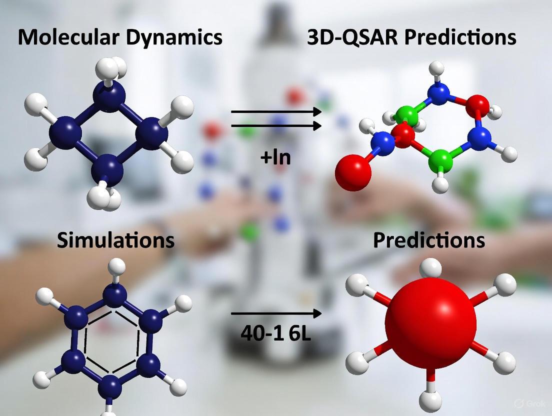
Integrating Molecular Dynamics Simulations to Validate and Enhance 3D-QSAR Predictions in Drug Discovery
This article provides a comprehensive guide for researchers and drug development professionals on the integrated use of Molecular Dynamics (MD) simulations to validate and refine 3D-Quantitative Structure-Activity Relationship (QSAR) predictions.
Integrating Molecular Dynamics Simulations to Validate and Enhance 3D-QSAR Predictions in Drug Discovery
Abstract
This article provides a comprehensive guide for researchers and drug development professionals on the integrated use of Molecular Dynamics (MD) simulations to validate and refine 3D-Quantitative Structure-Activity Relationship (QSAR) predictions. It covers the foundational principles of both methodologies, outlines a practical workflow for their synergistic application, and addresses key challenges in model interpretation and optimization. Through illustrative case studies and a discussion of validation protocols, the content demonstrates how this combined computational approach provides dynamic, mechanistic insights that surpass the static predictions of 3D-QSAR alone, ultimately leading to more reliable and efficacious drug candidate design.
Understanding the Synergy: 3D-QSAR and Molecular Dynamics in Modern Drug Design
Quantitative Structure-Activity Relationship (QSAR) modeling represents a cornerstone of modern computational drug discovery. The evolution from classical QSAR to three-dimensional (3D-QSAR) methods marks a significant paradigm shift from treating molecules as simple collections of descriptors to analyzing them as complex three-dimensional entities with specific shapes and interaction fields. 3D-QSAR has emerged as a natural extension to classical Hansch and Free-Wilson approaches, which exploits the three-dimensional properties of the ligands to predict their biological activities using robust chemometric techniques [1]. This approach considers the molecule as a 3D object with a specific shape and interaction fields (regions of positive/negative charge, steric bulk, etc.) around it, providing a more realistic representation of molecular interactions in biological systems [2].
The integration of 3D-QSAR within broader drug discovery pipelines, particularly when combined with molecular dynamics simulations, creates a powerful framework for validating predictions and understanding the dynamic behavior of protein-ligand complexes. This combination allows researchers to move beyond static snapshots of binding interactions toward a more comprehensive understanding of conformational flexibility and binding stability under physiologically relevant conditions [3] [4].
Fundamental Principles of 3D-QSAR
Core Theoretical Concepts
The fundamental principle underlying all QSAR formalism is that differences in structural properties are responsible for variations in biological activities of compounds [1]. While classical 2D-QSAR methods use numeric descriptors of molecules that are invariant to conformation, 3D-QSAR derives descriptors directly from the spatial structure of the molecule [2]. This fundamental distinction enables 3D-QSAR to capture essential steric and electronic features that govern molecular recognition and binding.
The theoretical foundation rests on several key principles:
- Molecular Similarity: Structurally similar molecules are likely to exhibit similar biological activities
- Field Analysis: Molecular interactions can be quantified through steric, electrostatic, hydrophobic, and hydrogen-bonding fields
- Alignment Dependency: Accurate spatial alignment of molecules is crucial for meaningful comparison of their interaction fields
- Conformational Dependency: Biological activity is influenced by the three-dimensional conformation that molecules adopt when binding to their targets
Comparison of 2D vs. 3D-QSAR Approaches
Table 1: Fundamental Differences Between 2D and 3D-QSAR Approaches
| Feature | 2D-QSAR | 3D-QSAR |
|---|---|---|
| Molecular Representation | Flat structures, topological descriptors | 3D structures, spatial descriptors |
| Descriptors Used | logP, molar refractivity, topological indices | Steric, electrostatic, hydrophobic fields |
| Conformational Sensitivity | Insensitive to conformation | Highly dependent on bioactive conformation |
| Alignment Requirement | Not required | Critical step in model development |
| Information Content | Global molecular properties | Local interaction properties |
| Visualization Capability | Limited to structure-activity trends | 3D contour maps guiding structural modifications |
Methodological Framework and Protocols
Standard 3D-QSAR Workflow
The development of a robust 3D-QSAR model follows a systematic workflow encompassing multiple critical stages. The diagram below illustrates this comprehensive process from data collection to model application.
Data Collection and Preparation Protocol
Protocol 3.2.1: Compound Selection and Activity Data Curation
Compound Selection Criteria:
- Select 20-50 compounds with structurally related scaffolds but varying substituents
- Ensure sufficient diversity in substituents to cover a range of electronic, steric, and hydrophobic properties
- Include compounds with a wide range of biological activities (ideally spanning 2-3 orders of magnitude in IC₅₀, Kᵢ, or equivalent metrics)
Activity Data Requirements:
Dataset Division:
- Divide compounds into training set (80-85%) and test set (15-20%)
- Ensure test set compounds represent the structural and activity space of the training set
- Use rational selection methods (e.g., Kennard-Stone, sphere exclusion) rather than random selection
Molecular Modeling and Alignment Protocol
Protocol 3.3.1: 3D Structure Generation and Optimization
Structure Generation:
- Convert 2D representations to 3D coordinates using tools like RDKit or Sybyl [2]
- Generate multiple conformers for each compound using systematic search or stochastic methods
- Select lowest energy conformer or putative bioactive conformation for alignment
Geometry Optimization:
- Perform molecular mechanics optimization using universal force field (UFF) or similar [2]
- For higher accuracy, apply quantum mechanical methods (semi-empirical or DFT)
- Ensure convergence criteria are properly set (energy gradient < 0.05 kcal/mol·Å)
Molecular Alignment:
- Identify common substructure or pharmacophore using maximum common substructure (MCS) or Bemis-Murcko scaffolds [2]
- Superimpose molecules to a reference compound based on atomic correspondence
- Validate alignment quality through visual inspection and RMSD calculations
Descriptor Calculation and Model Building
Protocol 3.4.1: Field-Based Descriptor Calculation
Grid Generation:
- Create a 3D grid that encompasses all aligned molecules with 1-2 Å extension in all directions
- Set grid spacing to 1-2 Å for optimal resolution and computational efficiency
Interaction Field Calculation:
- CoMFA (Comparative Molecular Field Analysis): Calculate steric (Lennard-Jones) and electrostatic (Coulombic) fields using a probe atom at each grid point [2]
- CoMSIA (Comparative Molecular Similarity Indices Analysis): Compute similarity indices for steric, electrostatic, hydrophobic, and hydrogen bond donor/acceptor fields using Gaussian-type functions [2]
Table 2: Comparison of CoMFA and CoMSIA Descriptor Methods
| Characteristic | CoMFA | CoMSIA |
|---|---|---|
| Field Types | Steric and electrostatic | Steric, electrostatic, hydrophobic, hydrogen bond donor/acceptor |
| Probe Atoms | sp³ carbon with +1 charge | Various probe atoms for different fields |
| Distance Dependence | Lennard-Jones and Coulomb potentials | Gaussian-type distance dependence |
| Alignment Sensitivity | Highly sensitive to molecular alignment | More robust to small alignment variations |
| Contour Maps | Steric (green/yellow) and electrostatic (blue/red) | Multiple contour types for different interactions |
| Applicability | Best for congeneric series with reliable alignment | Suitable for structurally diverse datasets |
- Model Building Algorithms:
- Apply Partial Least Squares (PLS) regression to correlate field descriptors with biological activity [2]
- Use cross-validation (leave-one-out or leave-group-out) to determine optimal number of components
- Validate model performance using external test set predictions
Advanced AI-Enhanced 3D-QSAR Protocols
Protocol 3.5.1: Machine Learning Integration in 3D-QSAR
Descriptor Enhancement:
Machine Learning Algorithms:
- Implement random forest (RF) for robust, non-linear relationship modeling [6]
- Apply support vector machines (SVM) for high-dimensional descriptor spaces [6]
- Utilize multilayer perceptron (MLP) neural networks for complex, hierarchical patterns [6]
- Employ graph neural networks (GNNs) for direct learning from molecular graphs [5] [7]
Consensus Modeling:
Research Reagents and Computational Tools
Table 3: Essential Research Reagent Solutions for 3D-QSAR Studies
| Tool Category | Specific Tools/Software | Primary Function | Key Features |
|---|---|---|---|
| Molecular Modeling | Sybyl-X, RDKit, ChemDraw | 3D structure generation and optimization | Force field optimization, conformer generation, geometry minimization [3] [2] |
| 3D-QSAR Analysis | COMSIA, CoMFA, OpenEye's 3D-QSAR | Field calculation and model development | Steric/electrostatic field computation, PLS regression, contour map generation [4] [8] |
| Molecular Docking | AutoDock, GOLD, Glide | Binding mode prediction and alignment guidance | Protein-ligand docking, binding pose prediction, scoring functions [3] [4] |
| Molecular Dynamics | GROMACS, AMBER, NAMD | Validation of 3D-QSAR predictions and stability assessment | Simulation of temporal evolution, binding free energy calculations, stability analysis [3] [4] |
| ADMET Prediction | SwissADME, pkCSM, PreADMET | Pharmacokinetic and toxicity profiling | Absorption, distribution, metabolism, excretion, and toxicity prediction [5] [4] |
| Quantum Chemistry | Gaussian, ORCA, GAMESS | Electronic property calculation and descriptor generation | DFT calculations, molecular orbital analysis, electrostatic potential maps [4] |
Integration with Molecular Dynamics for Validation
MD Simulation Protocol for 3D-QSAR Validation
Protocol 5.1.1: Molecular Dynamics Validation of 3D-QSAR Predictions
System Preparation:
- Build protein-ligand complexes for top predicted compounds and reference molecules
- Solvate systems in appropriate water model (TIP3P, SPC) with counterions for neutrality
- Apply force field parameters (GAFF, CGenFF) for small molecules compatible with protein force field
Simulation Parameters:
- Perform energy minimization using steepest descent and conjugate gradient algorithms
- Equilibrate system with positional restraints on protein and ligands (NVT and NPT ensembles)
- Run production MD simulations for 100-200 ns with 2 fs time step at 300K [3] [4]
- Employ periodic boundary conditions and particle mesh Ewald for long-range electrostatics
Trajectory Analysis:
- Calculate RMSD (Root Mean Square Deviation) to assess complex stability (fluctuations between 1.0-2.0 Å indicate stable binding) [3]
- Compute RMSF (Root Mean Square Fluctuation) to identify flexible regions
- Analyze hydrogen bonding patterns and interaction persistence throughout simulation
- Perform principal component analysis to identify essential dynamics
Binding Free Energy Calculations:
- Apply MM-PBSA/GBSA (Molecular Mechanics Poisson-Boltzmann/Generalized Born Surface Area) methods
- Conduct energy decomposition to identify key residue contributions [3]
- Calculate per-residue decomposition to validate 3D-QSAR contour map interpretations
Case Study: Integrated 3D-QSAR/MD Workflow for MAO-B Inhibitors
A recent study on novel 6-hydroxybenzothiazole-2-carboxamides as monoamine oxidase B (MAO-B) inhibitors demonstrates the powerful integration of 3D-QSAR with molecular dynamics validation [3]. The researchers developed a 3D-QSAR model with excellent predictive ability (q² = 0.569, r² = 0.915) and identified compound 31.j3 as the most promising candidate. Subsequent MD simulations confirmed the stability of the MAO-B-31.j3 complex, with RMSD values fluctuating between 1.0 and 2.0 Å, indicating stable binding. Energy decomposition analysis further revealed the contribution of key amino acid residues to binding energy, validating the steric and electrostatic features identified in the original 3D-QSAR model [3].
Applications and Advanced Implementation
Contemporary Applications in Drug Discovery
Modern 3D-QSAR applications span diverse therapeutic areas and target classes:
- Neurodegenerative Disease Drug Discovery: Development of MAO-B inhibitors for Parkinson's disease treatment using COMSIA-based 3D-QSAR models [3]
- Antidiabetic Agent Development: Design of novel α-glucosidase inhibitors through CoMFA and CoMSIA modeling with MD validation [4]
- Cancer Therapeutics: Identification of aromatase inhibitors for breast cancer treatment using 3D-QSAR combined with molecular docking and ADMET prediction [9]
- Endocrine Disruptor Screening: Prediction of estrogen receptor-binding activity of small molecules using machine learning-based 3D-QSAR models [6]
Best Practices and Implementation Guidelines
Model Quality Assessment:
- Ensure cross-validated correlation coefficient (q²) > 0.5 for predictive models
- Maintain conventional correlation coefficient (r²) > 0.8 for model fit
- Achieve predictive r² for test set (r²ₜₑₛₜ) > 0.6 for external validation [4]
- Report standard error of estimate and F-value for model significance
Regulatory Compliance:
- Adhere to OECD principles for QSAR validation: defined endpoint, unambiguous algorithm, defined domain of applicability, appropriate measures of goodness-of-fit, robustness, and predictivity
- Document all parameters and procedures for reproducibility
- Provide applicability domain characterization for reliable predictions
Troubleshooting Common Issues:
- Poor Predictive Ability: Revisit molecular alignment, consider alternative bioactive conformations, or apply different descriptor sets
- Overfitting: Reduce descriptor dimensionality, increase training set size, or apply more stringent cross-validation
- Inconsistent Contour Maps: Verify alignment consistency, check for outliers, or explore alternative field calculation methods
The integration of 3D-QSAR with molecular dynamics simulations represents a powerful paradigm in modern drug discovery, enabling both predictive modeling and dynamic validation of compound interactions. This synergistic approach facilitates the rational design of novel therapeutic agents with optimized binding characteristics and improved pharmacological profiles.
The Role of Molecular Dynamics in Capturing Dynamic Biomolecular Processes
Molecular Dynamics (MD) simulations have become an indispensable tool in structural biology and computational drug discovery, providing atomic-level insight into the dynamic biomolecular processes that static structures cannot capture. By predicting the motion of every atom in a molecular system over time, MD simulations reveal how proteins and other biomolecules function, interact with ligands, and undergo conformational changes [10]. The integration of MD with other computational methods, particularly three-dimensional quantitative structure-activity relationship (3D-QSAR) modeling, creates a powerful framework for validating and refining predictive models of biological activity [11] [12] [13]. This integration is crucial for translating static structural predictions into dynamically validated drug candidates, significantly enhancing the reliability of computational screening in the drug development pipeline.
Integrating MD Simulations with 3D-QSAR Workflows
The Complementary Computational Workflow
Quantitative Structure-Activity Relationship (QSAR) modeling, particularly 3D-QSAR approaches like Comparative Molecular Field Analysis (CoMFA) and Comparative Molecular Similarity Indices Analysis (CoMSIA), establishes a correlation between the three-dimensional structural features of compounds and their biological activity [11] [12]. However, these models typically rely on a single, static conformation of each molecule. MD simulations address this limitation by providing a dynamic validation of the binding modes and conformational stability predicted by QSAR and molecular docking.
The standard integrated workflow proceeds through several key stages:
- 3D-QSAR Model Development: A predictive model is built using a set of compounds with known activities.
- Activity Prediction & Compound Design: The model predicts activities of novel compounds and guides the design of new candidates.
- Binding Pose Assessment: Molecular docking predicts how these new compounds interact with the target protein.
- Dynamic Validation: MD simulations validate the stability of the docked complexes under physiologically relevant conditions [14] [11] [12].
This workflow ensures that compounds identified as promising by static models are rigorously tested for the stability of their protein-ligand interactions through dynamic simulation.
Protocol for MD Validation of 3D-QSAR Predictions
The following protocol outlines the key steps for using MD simulations to validate potential inhibitors identified through a 3D-QSAR screening campaign.
Step 1: System Preparation
- Obtain the initial coordinates for the protein-ligand complex from molecular docking studies. The ligand is typically the candidate compound identified via 3D-QSAR [14] [11].
- Place the solvated complex in a simulation box with explicit water molecules (e.g., TIP3P water model) and add ions (e.g., Na⁺, Cl⁻) to neutralize the system and achieve a physiological salt concentration (e.g., 0.15 M NaCl).
- Assign a molecular mechanics force field (e.g., CHARMM, AMBER, OPLS-AA) to the protein, ligand, and ions [15].
Step 2: Energy Minimization and Equilibration
- Perform energy minimization (typically 5,000-50,000 steps) using a steepest descent algorithm to remove any steric clashes introduced during system setup.
- Conduct a two-phase equilibration:
- NVT Ensemble: Heat the system to the target temperature (e.g., 310 K) using a thermostat (e.g., Berendsen, Nosé-Hoover) and run for 50-100 ps while restraining the heavy atoms of the protein-ligand complex.
- NPT Ensemble: Release the restraints and equilibrate the system for 100-500 ps using a barostat (e.g., Parrinello-Rahman) to achieve the correct density (1 atm pressure) [15].
Step 3: Production MD Simulation
- Run an unrestrained production simulation for a duration sufficient to capture relevant biological processes. For validating a binding pose, simulations of 100 ns to 300 ns are commonly used [14] [12].
- Use a time step of 2 fs, enabled by constraining bonds involving hydrogen atoms.
- Save the atomic coordinates to a trajectory file at regular intervals (e.g., every 10-100 ps) for subsequent analysis.
Step 4: Trajectory Analysis
- Root Mean Square Deviation (RMSD): Calculate the backbone RMSD of the protein and the heavy-atom RMSD of the ligand to assess the overall stability of the complex and the ligand's binding pose.
- Root Mean Square Fluctuation (RMSF): Analyze per-residue fluctuations to identify flexible regions of the protein and check if key binding residues remain stable.
- Hydrogen Bonds and Interactions: Monitor the number and stability of specific hydrogen bonds and hydrophobic contacts between the ligand and the protein throughout the simulation [14] [12].
- Binding Free Energy Calculations: Employ methods like Molecular Mechanics/Poisson-Boltzmann Surface Area (MM/PBSA) to compute the binding free energy, providing a quantitative measure of ligand affinity that validates the 3D-QSAR prediction [12].
Case Studies in Drug Discovery
Identification of MCF-7 Breast Cancer Inhibitors
A seminal study demonstrated this integrated approach by identifying naphthoquinone derivatives as inhibitors of topoisomerase IIα for breast cancer therapy [14]. Six QSAR models were developed using Monte Carlo optimization, which screened 2,300 naphthoquinones. After ADMET filtering, 16 promising compounds were selected for docking. The top candidate, compound A14, was subjected to a 300 ns MD simulation. The simulation confirmed the complex's stability, showing that compound A14 maintained key interactions with the target, thus validating the QSAR predictions and underscoring its potential as a lead compound [14].
Design of MAO-B Inhibitors for Neurodegenerative Diseases
In neurodegenerative disease research, 3D-QSAR (CoMSIA), molecular docking, and MD were used to design new 6-hydroxybenzothiazole-2-carboxamide derivatives as monoamine oxidase B (MAO-B) inhibitors [11]. The most promising compound, 31.j3, demonstrated a stable binding mode in MD simulations, with RMSD values fluctuating between 1.0 and 2.0 Å, indicating high conformational stability. Energy decomposition analysis further revealed the key residues contributing to binding through van der Waals and electrostatic interactions, dynamically validating the structural features highlighted by the 3D-QSAR model [11].
Development of Anti-Breast Cancer Aromatase Inhibitors
Another study on anti-breast cancer agents designed 1,4-quinone and quinoline derivatives using 3D-QSAR (CoMFA and CoMSIA) and molecular docking [12]. The binding stability of the designed ligands to the aromatase enzyme (3S7S) was confirmed through 100 ns MD simulations. The analysis of RMSD, RMSF, and H-bond parameters, complemented by MM-PBSA calculations, identified ligand 5 as the most promising candidate due to its stable binding profile, leading to its recommendation for experimental testing [12].
Table 1: Key Metrics from MD Simulations in Case Studies
| Case Study | Target Protein | Simulation Duration | Key Stability Metrics | Outcome |
|---|---|---|---|---|
| MCF-7 Inhibitors [14] | Topoisomerase IIα | 300 ns | Stable RMSD; maintained key interactions | Compound A14 validated as lead |
| MAO-B Inhibitors [11] | Monoamine Oxidase B | Not Specified | RMSD: 1.0-2.0 Å | Compound 31.j3 confirmed stable |
| Aromatase Inhibitors [12] | Aromatase (3S7S) | 100 ns | Stable RMSD/RMSF; favorable MM-PBSA | Ligand 5 selected for testing |
Visualization of Workflows and Processes
Integrated 3D-QSAR and MD Validation Workflow
The following diagram illustrates the sequential, iterative process of integrating 3D-QSAR modeling with MD simulations for dynamic validation in drug discovery.
Key Biomolecular Processes Captured by MD
MD simulations provide critical insights into several dynamic processes that are fundamental to understanding protein-ligand interactions at an atomic level.
The Scientist's Toolkit: Essential Research Reagents and Software
Successful execution of integrated 3D-QSAR and MD studies relies on a suite of specialized software tools and computational resources.
Table 2: Essential Research Reagent Solutions for 3D-QSAR and MD Simulations
| Category | Tool/Reagent | Primary Function | Application Note |
|---|---|---|---|
| QSAR Modeling | Sybyl-X [11] | Builds 3D-QSAR models (CoMFA/CoMSIA). | Used to correlate 3D molecular fields with biological activity for predictive design. |
| CORAL [14] | Develops QSAR models using Monte Carlo optimization. | Employs SMILES-based descriptors to predict activity endpoints. | |
| Molecular Docking | AutoDock, GOLD, Surflex | Predicts the binding pose and affinity of ligands within a protein's active site. | Generates initial protein-ligand complex structures for MD simulation. |
| MD Simulation & Analysis | GROMACS, AMBER, NAMD | Performs molecular dynamics simulations. | Open-source and commercial software for running production MD; includes energy minimization, equilibration, and trajectory analysis tools. |
| VMD, PyMOL | Visualizes and analyzes MD trajectories. | Critical for inspecting structural stability, interactions, and creating publication-quality figures. | |
| Force Fields | CHARMM, AMBER, OPLS-AA | Provides empirical parameters for calculating potential energy in MD. | The physical model defining atomic interactions; choice depends on the system and software [15]. |
| Hardware | GPUs (Graphics Processing Units) [10] | Accelerates MD calculations. | Makes simulations on biologically relevant timescales (100s of ns) accessible. |
Molecular Dynamics simulations provide the critical link between static predictions and dynamic reality in computational drug discovery. By validating 3D-QSAR models, MD simulations confirm the stability of predicted binding modes, quantify interaction strengths, and ultimately prioritize the most promising lead compounds with a higher probability of experimental success. The continued development of more accurate force fields, faster hardware, and robust automated protocols will further solidify the role of MD as an indispensable component of the modern drug design and validation toolkit.
Quantitative Structure-Activity Relationship (QSAR) modeling, particularly three-dimensional QSAR (3D-QSAR), represents one of the most important computational tools in pharmaceutical discovery pipelines. These methods analyze the quantitative relationship between chemical structures and their biological activities using physicochemical or structural parameters [16]. The 3D-QSAR approaches, including Comparative Molecular Field Analysis (CoMFA) and Comparative Molecular Similarity Indices Analysis (CoMSIA), introduce three-dimensional structural information to study structure-activity relationships, indirectly characterizing non-bonded interactions between molecules and biomacromolecules [17]. However, these methods provide only a static snapshot of molecular interactions, failing to capture the dynamic nature of biological systems that is crucial for accurate drug design.
While 3D-QSAR models demonstrate impressive statistical parameters in development (typically requiring R² > 0.9 and Q² > 0.5 for internal validation [18]), these metrics alone cannot guarantee predictive reliability for novel compounds. The inherent limitation of these static approaches lies in their treatment of proteins as rigid entities and their inability to account for the temporal evolution of ligand-receptor complexes. This application note establishes the critical scientific rationale for supplementing static 3D-QSAR predictions with Molecular Dynamics (MD) validation, providing detailed protocols for implementation within drug discovery workflows.
The Critical Gap: When 3D-QSAR Predictions Require Experimental Validation
Fundamental Limitations of 3D-QSAR Methodology
3D-QSAR techniques operate on the principle that biological activity correlates with molecular interaction fields surrounding compounds. These methods consider ligand properties calculated in their bioactive conformation, making them theoretically more suitable than classic 2D approaches for studying ligand-receptor interactions [19]. However, several fundamental assumptions limit their predictive accuracy:
- Static Binding Mode Assumption: 3D-QSAR assumes a single, dominant binding conformation without accounting for protein flexibility or alternative binding modes [19] [16].
- Limited Solvation Effects: Implicit solvation models fail to capture specific water-mediated interactions critical to binding.
- Entropic Considerations: These methods largely ignore entropic contributions to binding free energy.
- Time-Independent Properties: 3D-QSAR cannot observe the time evolution of complex stability or interaction persistence.
Statistical Validation Isn't Enough: The Deception of Good Numbers
Robust 3D-QSAR models must meet stringent statistical criteria including:
- Internal validation: Leave-One-Out (LOO) cross-validation with q² > 0.5 [18]
- External validation: R²pred > 0.5 for test set predictions [18]
- Golbraikh and Tropsha criteria: R² > 0.6, 0.85 < k < 1.15, and [(R² - R₀²)/R²] < 0.1 [20] [18]
However, these statistical parameters alone cannot confirm model validity [20]. A comprehensive analysis of 44 published QSAR models revealed that employing the coefficient of determination (r²) alone could not indicate the true validity of a QSAR model, and established validation criteria have specific advantages and disadvantages that must be considered [20]. This underscores that statistical validation represents a necessary but insufficient condition for establishing predictive reliability.
Table 1: Case Studies Demonstrating 3D-QSAR Prediction Gaps Resolved by MD Validation
| Study Focus | 3D-QSAR Results | MD Validation Insights | Impact |
|---|---|---|---|
| MAO-B Inhibitors [11] | CoMSIA model with q²=0.569, r²=0.915 predicted IC₅₀ values | MD simulations (100 ns) revealed binding stability, key residue contributions through energy decomposition | Identified stable binding conformation not apparent in static analysis |
| Anti-Breast Cancer Agents [12] | CoMSIA/SEA model indicated electrostatic, steric, H-bond fields significant | 100 ns MD simulations with RMSD, RMSF, SASA, MM-PBSA showed only one designed compound maintained stable binding | Prevented false positive from static prediction alone |
| Ovarian Cancer (AKT1) [21] | 3D-QSAR model with R²=0.822, Q²=0.613 identified potential inhibitors | MD simulations revealed stability issues in physiological conditions, identified taxifolin as truly promising | Uncovered dynamic instability not detected in static approach |
| mIDH1 Inhibitors [17] | CoMFA (R²=0.980, Q²=0.765) and CoMSIA (R²=0.997, Q²=0.770) models built | MD simulations with FEL, Rg, SASA, PSA identified C2 with highest binding free energy (-93.25 ± 5.20 kcal/mol) | Revealed compounds with superior binding properties beyond QSAR predictions |
Molecular Dynamics Simulations: Capturing Biological Reality
The Dynamic View of Molecular Interactions
Molecular Dynamics simulations address the fundamental limitations of static approaches by modeling the temporal evolution of molecular systems. MD incorporates:
- Protein flexibility and side-chain rearrangements
- Explicit solvation with water molecules and ions
- Temperature and pressure controls mimicking physiological conditions
- Time-dependent phenomena such as hydrogen bond formation/breakage
MD simulations provide atomic-level insights into biological processes by solving Newton's equations of motion for all atoms in the system, typically at femtosecond resolution, over simulation timescales ranging from nanoseconds to microseconds [11] [12].
Complementary Strengths: 3D-QSAR and MD Integration
The integration of 3D-QSAR and MD creates a powerful synergistic workflow:
- 3D-QSAR: Rapidly screens large chemical spaces, identifies critical molecular features, and predicts activity trends
- MD Simulations: Validate binding stability, identify specific atomic interactions, and calculate binding free energies
This combination is particularly valuable for studying mutant protein targets where structural alterations significantly impact dynamics and binding. For example, in ovarian cancer research, MD simulations revealed how a W80R point mutation in AKT1 affected flavonoid binding stability, information inaccessible to static 3D-QSAR methods [21].
Experimental Protocols and Methodologies
Protocol 1: Standard 3D-QSAR Model Development with CoMFA/CoMSIA
Purpose: To develop predictive 3D-QSAR models for initial activity prediction and pharmacophore mapping.
Materials and Software:
- Molecular Modeling Suite (Sybyl-X, Schrodinger Maestro): For compound building, optimization, and alignment
- Quantum Chemical Software (Gaussian 09): For geometry optimization at DFT level (e.g., M06-2X/6-31G(d,p)) [19]
- Descriptor Calculation Tools (Dragon Software): Generate structural and physicochemical descriptors [19]
- Statistical Analysis Package (QSARINS): For genetic algorithm variable selection and model validation [19]
Methodology:
- Dataset Curation: Collect 30-50 compounds with consistent experimental activity measurements (e.g., IC₅₀, Ki) [17]. Ensure structural diversity while maintaining a common scaffold.
- Molecular Alignment: Perform substructure or pharmacophore-based alignment using the most active compound as template [17].
- Descriptor Calculation and Selection:
- Calculate steric, electrostatic, hydrophobic, and hydrogen-bonding field descriptors
- Apply genetic algorithm (population size: 300, mutation rate: 65) for descriptor selection [19]
- Remove cross-correlated descriptors (r ≥ 0.85)
- Model Development:
- Model Application: Predict activities of novel designed compounds and generate contour maps for structure optimization.
Protocol 2: Molecular Dynamics Validation of 3D-QSAR Predictions
Purpose: To validate the stability and binding mechanisms of 3D-QSAR-predicted active compounds through nanosecond-scale simulations.
Materials and Software:
- MD Simulation Engine (GROMACS, AMBER, NAMD): For running dynamics simulations
- Force Field Parameters (CHARMM36, AMBER ff14SB, OPLS-AA): Determine atom interactions and energetics
- Visualization Software (VMD, PyMol): For trajectory analysis and figure generation
- Free Energy Calculation Tools (MM-PBSA, MM-GBSA): For binding affinity quantification
Methodology:
- System Preparation:
- Obtain protein structure from PDB or homology modeling
- Parameterize ligands using antechamber/GAFF
- Solvate in explicit water (TIP3P) with neutralizing ions (0.15 M NaCl)
- Energy Minimization:
- Perform steepest descent minimization (5,000 steps) until Fmax < 1000 kJ/mol/nm
- Equilibration Phases:
- NVT equilibration (100 ps, 300 K, Berendsen thermostat)
- NPT equilibration (100 ps, 1 bar, Parrinello-Rahman barostat)
- Production MD:
- Run unrestrained simulation (50-100 ns) with 2 fs time step
- Apply periodic boundary conditions and PME for electrostatics
- Trajectory Analysis:
- Calculate RMSD (backbone stability), RMSF (residue flexibility), Rg (compactness)
- Monitor hydrogen bonds and interaction persistence
- Perform energy decomposition to identify key residue contributions [11]
- Binding Free Energy Calculations:
- Apply MM-PBSA/GBSA on 100-500 evenly spaced frames from stable trajectory region
- Calculate van der Waals, electrostatic, polar, and non-polar solvation components
Table 2: Key Parameters for MD Validation of 3D-QSAR Predictions
| Validation Metric | Target Range | Interpretation | Computational Tool |
|---|---|---|---|
| RMSD (Protein Backbone) | < 2.0-3.0 Å | System stability and equilibration | GROMACS, CPPTRAJ |
| RMSF (Residue) | < 1.5 Å (core), < 3.0 Å (loops) | Regional flexibility and binding effects | VMD, Bio3D |
| Hydrogen Bonds | Consistent count (±1-2) | Specific interaction maintenance | VMD, MDAnalysis |
| Radius of Gyration (Rg) | Stable fluctuations < 0.5 Å | Global structural compactness | GROMACS |
| MM-PBSA Binding Energy | Negative value (more negative = better) | Quantitative binding affinity | MMPBSA.py, g_mmpbsa |
| Solvent Accessible Surface Area (SASA) | Stable with minor fluctuations | Burial of hydrophobic surfaces | GROMACS, VMD |
The Research Toolkit: Essential Reagents and Computational Solutions
Table 3: Research Reagent Solutions for 3D-QSAR and MD Workflows
| Reagent/Software Category | Specific Examples | Function in Workflow |
|---|---|---|
| Molecular Modeling Suites | Sybyl-X, Schrodinger Maestro, OpenBabel | Compound building, optimization, conformational analysis, and alignment for 3D-QSAR |
| Quantum Chemistry Packages | Gaussian 09, ORCA, GAMESS | Geometry optimization and electronic property calculation at DFT/ab initio levels |
| Descriptor Calculation Tools | Dragon Software, PaDEL-Descriptor | Generation of structural, topological, and physicochemical descriptors for QSAR |
| Statistical Analysis Software | QSARINS, SIMCA, R | Model development, variable selection, and statistical validation |
| MD Simulation Engines | GROMACS, AMBER, NAMD, OpenMM | Running molecular dynamics simulations with various force fields |
| Force Field Parameters | CHARMM36, AMBER ff14SB, OPLS-AA, GAFF | Defining atomistic interactions, bonding, and non-bonded parameters |
| Trajectory Analysis Tools | VMD, PyMol, MDAnalysis, CPPTRAJ | Visualization, measurement, and analysis of MD trajectories |
| Free Energy Calculators | MMPBSA.py, g_mmpbsa, WHAM | Binding free energy calculations from MD trajectories |
Workflow Integration: From Static Prediction to Dynamic Validation
The sequential application of 3D-QSAR and MD simulations creates a powerful workflow for rational drug design. The process begins with 3D-QSAR to rapidly explore chemical space and identify promising candidates, followed by MD validation to confirm binding stability and mechanism.
Integrated 3D-QSAR and MD Validation Workflow
This integrated approach efficiently leverages the strengths of both methodologies: the high-throughput screening capability of 3D-QSAR and the biological fidelity of MD simulations. The workflow ensures that only compounds demonstrating both favorable predicted activity and stable binding dynamics advance to resource-intensive experimental stages.
The integration of Molecular Dynamics simulations with 3D-QSAR predictions represents a paradigm shift in computational drug design, moving beyond static structural approximations to embrace the dynamic reality of biological systems. This approach addresses fundamental limitations of standalone 3D-QSAR methods, providing critical insights into:
- Temporal stability of ligand-receptor complexes
- Specific interaction mechanisms at atomic resolution
- Energetic contributions of individual residues to binding
- Structural adaptations and conformational changes upon binding
As MD methodologies continue to advance with specialized hardware, enhanced sampling algorithms, and machine learning integration, the accessibility and timescales of simulations will further improve. The scientific community increasingly recognizes that comprehensive validation protocols combining multiple computational approaches provide the most reliable path to efficient therapeutic development. For research teams investing in computational drug discovery, the 3D-QSAR/MD validation framework represents not merely an enhancement, but an essential component of robust, predictive molecular design.
This application note details the critical components for constructing reliable computational models in drug design, framed within a research context that utilizes molecular dynamics (MD) simulations to validate 3D-QSAR predictions. We provide a structured overview of key molecular descriptors and force fields, followed by a standardized protocol for their application.
Table of Key Molecular Descriptor Classes
Table 1: Categories of molecular descriptors critical for building accurate machine learning and QSAR models.
| Descriptor Class | Key Examples | Description | Primary Application |
|---|---|---|---|
| Global Interatomic Descriptors | Coulomb Matrix, Inverse Interatomic Distances [22] | Encodes all pairwise atomic interactions within a molecule, providing a complete global representation. | Machine Learning Force Fields (MLFFs) for molecular dynamics [22] |
| Reduced/Feature-Selected Descriptors | Automatized Reduced Descriptors [22] | A minimized set of features selected automatically from a global descriptor to maintain accuracy while improving computational efficiency. | Efficient MLFFs for large, flexible molecules like peptides and DNA [22] |
| 3D-QSAR Field Descriptors | Comparative Molecular Similarity Indices Analysis (CoMSIA) Fields [11] | Describes 3D molecular fields (steric, electrostatic, etc.) surrounding aligned molecules in a dataset. | 3D-QSAR models for predicting biological activity (e.g., IC50) [11] |
Force fields are computational models that describe the potential energy of a system of atoms and molecules, enabling the calculation of forces for molecular dynamics simulations [23]. The basic functional form for an additive force field is:
( E{\text{total}} = E{\text{bonded}} + E_{\text{nonbonded}} )
Where:
- ( E{\text{bonded}} = E{\text{bond}} + E{\text{angle}} + E{\text{dihedral}} ) [23]
- ( E{\text{nonbonded}} = E{\text{electrostatic}} + E_{\text{van der Waals}} ) [23]
Experimental Protocol: Integrating 3D-QSAR and MD Simulations for Model Validation
The following protocol describes a workflow for developing a predictive model and validating its binding modes using molecular dynamics simulations, as applied in studies of Monoamine Oxidase-B (MAO-B) and Thyroid Peroxidase (TPO) inhibitors [11] [24].
Workflow Diagram: 3D-QSAR & MD Validation Pipeline
Phase 1: 3D-QSAR Model Development & Validation
Compound Dataset Curation
Structure Optimization and Conformer Generation
- Use software like ChemDraw and Sybyl-X to build and optimize 3D molecular structures [11].
- Generate low-energy conformers for each compound.
Molecular Docking and Alignment
3D-QSAR Model Construction
Model Validation
- Internal Validation: Use the Leave-One-Out (LOO) method to obtain cross-validated correlation coefficient ((q^2)). A robust model typically has (q^2 > 0.5) [18].
- External Validation: Predict the activity of the test set compounds. A model with high predictive power should have a predictive (R^2) ((R^2_{\text{pred}})) > 0.5 and a low mean absolute error (MAE) [18].
Phase 2: Model Exploitation and MD Validation
Design and Virtual Screening
- Design novel derivatives based on the 3D-QSAR contour maps.
- Use the validated model to predict the activities of the newly designed compounds [11].
Molecular Dynamics Simulation Setup
- Select the top-scoring compound(s) from docking and QSAR prediction.
- Solvate the protein-ligand complex in an explicit water box (e.g., TIP3P water model) and add ions to neutralize the system.
- Use a force field such as CHARMM36, GAFF, or a Neural Network Potential (NNP) to describe atomic interactions [25] [26].
- Employ software like NAMD (integrated in BIOVIA Discovery Studio) or GROMACS for simulations [26].
MD Simulation Execution
- Energy-minimize the system to remove steric clashes.
- Gradually heat the system to the physiological temperature (e.g., 310 K) under an NVT ensemble.
- Equilibrate the system with pressure coupling (NPT ensemble) to achieve correct density.
- Run a production MD simulation for a sufficient duration (e.g., 100 ns to 1 µs) to assess stability [11] [27] [24].
Trajectory Analysis and Binding Free Energy Calculation
- Stability Analysis: Calculate the Root Mean Square Deviation (RMSD) of the protein backbone and ligand. Fluctuations between 1.0-2.0 Å indicate a stable complex [11].
- Interaction Analysis: Identify key residue interactions (hydrogen bonds, hydrophobic contacts) that persist during the simulation.
- Energy Decomposition: Perform energy decomposition analysis (e.g., using MM/GBSA) to reveal the contribution of specific residues to binding, highlighting the role of van der Waals and electrostatic interactions [11].
The Scientist's Toolkit: Essential Research Reagents & Software
Table 2: Key software, force fields, and computational tools required for the described protocols.
| Tool/Reagent | Function/Description | Example Use in Protocol |
|---|---|---|
| Sybyl-X | Molecular modeling and 3D-QSAR software suite. | Used for structure optimization, molecular alignment, and CoMSIA model construction [11]. |
| BIOVIA Discovery Studio | Software providing simulation and analysis tools. | Used for MD simulations leveraging integrated CHARMm and NAMD engines [26]. |
| CHARMM Force Field | A family of all-atom force fields for proteins, nucleic acids, and lipids. | Used to parametrize the protein and standard organic molecules during MD setup [26]. |
| CGenFF | CHARMM General Force Field for drug-like molecules. | Used to generate parameters for novel ligands [26]. |
| Neural Network Potential (NNP) | Machine-learning-based force field for high accuracy. | Can be used in MD protocols for more precise prediction of material properties (e.g., thermal stability) [25]. |
| GAFF (General AMBER Force Field) | A force field for organic molecules, parameters often derived via DFT. | Used to describe radiometal-chelator complexes in MD simulations [27]. |
| Water Models (e.g., TIP3P) | Explicit solvent models representing water molecules. | Used to solvate the protein-ligand complex in a biologically relevant environment [26]. |
Statistical Validation Criteria for 3D-QSAR Models
For a 3D-QSAR model to be considered reliable and predictive, it must meet several statistical criteria as shown in Table 3.
Table 3: Key statistical parameters for evaluating the quality of 3D-QSAR models [18].
| Validation Type | Parameter | Threshold for a Predictive Model |
|---|---|---|
| Internal Validation | LOO cross-validated (q^2) | > 0.5 |
| Internal Validation | Non-cross-validated (r^2) | > 0.9 |
| External Validation | Predictive (R^2) ((R^2_{\text{pred}})) | > 0.5 |
| External Validation | Mean Absolute Error (MAE) | ≤ 0.1 × training set activity range |
A Practical Workflow: Integrating MD Simulations into Your 3D-QSAR Pipeline
Building and Validating a Predictive 3D-QSAR Model (e.g., CoMSIA)
This application note provides a detailed protocol for building and validating a predictive Three-Dimensional Quantitative Structure-Activity Relationship (3D-QSAR) model, specifically using the Comparative Molecular Similarity Indices Analysis (CoMSIA) method. Within the broader context of a research thesis integrating molecular dynamics simulations, a robust 3D-QSAR model serves as the critical initial step for identifying and optimizing potential lead compounds in drug discovery [28] [12]. The model establishes a quantitative relationship between the three-dimensional molecular interaction fields of a compound set and their corresponding biological activities, enabling the prediction of activities for novel compounds and providing visual guidance for rational molecular design [2]. The subsequent validation of these predictions through molecular dynamics simulations ensures their reliability and biological relevance, creating a powerful combined computational approach.
The entire process, from data preparation to model application, follows a structured workflow to ensure the generation of a statistically robust and predictive model. The following diagram outlines the key stages of this protocol:
Detailed Experimental Protocols
Data Collection and Curation
The foundation of a reliable 3D-QSAR model is a high-quality, consistent dataset.
- Activity Data: Assemble a congeneric series of compounds with experimentally determined biological activities (e.g., IC₅₀, EC₅₀, pIC₅₀). A minimum of 20-30 compounds is typically required for meaningful statistical analysis [2]. The pIC₅₀ value (-logIC₅₀) is often used as the dependent variable to linearize the relationship with free energy changes [18].
- Data Integrity: All biological activity data must be acquired under uniform experimental conditions to minimize noise and systematic bias [2]. The dataset should be divided into a training set (typically ~80% of compounds) for model construction and a test set (the remaining ~20%) for external validation of predictive ability [18] [12].
Molecular Modeling and Alignment
This step transforms 2D molecular structures into their bioactive 3D conformations and aligns them for comparative analysis.
- 3D Structure Generation: Convert 2D structures into 3D coordinates using molecular modeling software (e.g., Sybyl, MOE, RDKit) [11] [2].
- Geometry Optimization: Minimize the energy of the initial 3D structures using molecular mechanics force fields (e.g., Tripos, OPLS_2005, UFF) or quantum mechanical methods to achieve realistic, low-energy conformations [28] [2].
- Molecular Alignment: This is a critical step that significantly impacts model quality. Align all molecules onto a common 3D reference frame based on a presumed bioactive conformation. Common strategies include:
- Pharmacophore-Based Alignment: Superimpose molecules based on key pharmacophoric features.
- Database Alignment: Use the structure of a potent compound or a common scaffold (e.g., Bemis-Murcko scaffold) as a template for alignment [2].
- Maximum Common Substructure (MCS): Identify and align the largest common substructure shared across the dataset [2].
CoMSIA Descriptor Calculation
The CoMSIA method characterizes molecules based on their similarity to a probe atom at regularly spaced grid points surrounding the aligned molecules. Unlike CoMFA, CoMSIA uses Gaussian-type functions to avoid singularities and offers additional field types [2].
- Protocol: Using CoMSIA module in software like Sybyl, calculate five distinct molecular interaction fields:
- Steric (S): Represents van der Waals interactions and molecular bulk.
- Electrostatic (E): Represents Coulombic potential.
- Hydrophobic (H): Characterizes lipophilicity.
- Hydrogen Bond Donor (D): Represents the ability to donate a hydrogen bond.
- Hydrogen Bond Acceptor (A): Represents the ability to accept a hydrogen bond.
- A probe atom (typically a sp³ hybridized carbon with a +1 charge) is placed at each grid point, and the similarity indices are computed using a Gaussian function, which makes the calculation less sensitive to minor alignment deviations compared to CoMFA [2].
Model Building with Partial Least Squares (PLS) Regression
The high number of CoMSIA descriptors (independent variables) relative to the number of compounds requires a dimensionality reduction technique.
- Protocol: Employ the Partial Least Squares (PLS) algorithm to correlate the CoMSIA descriptors (X-matrix) with the biological activity data (Y-matrix, e.g., pIC₅₀) [18] [2].
- Cross-Validation: Perform Leave-One-Out (LOO) cross-validation with the training set to determine the optimal number of components (ONC) that maximizes predictive ability while minimizing the risk of overfitting. The ONC is the number of latent variables that yields the highest cross-validated correlation coefficient, q² [18].
- Final Model: A final model is then generated using the optimal number of components on the entire training set, yielding the conventional correlation coefficient, r² [18].
Model Validation and Interpretation
A model must be rigorously validated before it can be used for prediction.
- Internal Validation: Assess the model's self-consistency and internal predictive ability.
- External Validation: This is the gold standard for evaluating predictive power. Use the model to predict the activity of the external test set compounds that were not used in model building [18] [12].
- Contour Map Analysis: Visualize the CoMSIA results by generating 3D contour maps. These maps highlight regions in space where specific molecular properties favor or disfavor biological activity, providing a powerful tool for medicinal chemists [2]. For example:
- Green contours: Regions where bulky groups increase activity.
- Yellow contours: Regions where bulky groups decrease activity.
- Blue contours: Regions where electropositive groups increase activity.
- Red contours: Regions where electronegative groups increase activity.
The following diagram summarizes the key stages and decision points in the model validation process:
Research Reagent Solutions and Essential Materials
Table 1: Essential Software and Tools for 3D-QSAR Modeling
| Tool/Software | Type | Primary Function in Protocol |
|---|---|---|
| Sybyl-X [11] | Commercial Software Suite | Integrated environment for molecular modeling, CoMFA/CoMSIA analysis, PLS regression, and contour map visualization. |
| Open3DALIGN [2] | Open-Source Tool | Automated molecular alignment, a critical and often challenging step in the 3D-QSAR workflow. |
| RDKit [28] [2] | Open-Source Cheminformatics | Python library used for 2D to 3D structure conversion, geometry optimization, and maximum common substructure (MCS) alignment. |
| Python/Scikit-learn [28] | Programming Environment | Custom scripting for data preprocessing, advanced machine learning model building, and hyperparameter tuning beyond traditional PLS. |
| GROMACS/AMBER | Molecular Dynamics Software | Used in the broader thesis context for validating 3D-QSAR predictions via molecular dynamics simulations and free energy calculations. |
Application in a Thesis Integrating Molecular Dynamics Simulations
In a thesis focused on molecular dynamics (MD) to validate 3D-QSAR predictions, the CoMSIA model serves as the starting point for a multi-stage computational investigation.
- Prediction and Design: The validated CoMSIA model is used to predict the activity of virtual compounds and guide the design of new derivatives with improved predicted potency [12] [29]. The contour maps provide specific structural guidance, such as suggesting the introduction of a bulky hydrophobic group in a green-contoured region.
- Molecular Docking: The highest-ranked newly designed compounds are then subjected to molecular docking studies to predict their binding mode and key interactions with the target protein's active site [11] [12].
- Molecular Dynamics Validation: This is the core validation step in the thesis context. The top ligand-receptor complexes from docking are simulated using MD (e.g., for 100 ns) to assess the stability of the predicted binding poses under dynamic, physiological-like conditions [11] [12]. Key analyses include:
- Root Mean Square Deviation (RMSD): Confirms the overall stability of the protein-ligand complex.
- Root Mean Square Fluctuation (RMSF): Identifies flexible regions in the protein upon ligand binding.
- Hydrogen Bond Analysis: Quantifies persistent key interactions.
- MM-PBSA/GBSA Calculations: Estimates the binding free energy, providing a quantitative measure of binding affinity that can be correlated with the 3D-QSAR predictions [12]. A stable MD trajectory with a favorable computed binding energy provides strong corroborative evidence for the 3D-QSAR model's prediction.
Case Studies and Performance Metrics
The following table summarizes the performance of CoMSIA models from recent literature, demonstrating the typical outputs and benchmarks of a successful study.
Table 2: Exemplary Performance Metrics from Published 3D-QSAR CoMSIA Studies
| Study Focus | Statistical Parameters | Outcome and Validation |
|---|---|---|
| MAO-B Inhibitors [11] | q² = 0.569, r² = 0.915, SEE = 0.109, F = 52.714 | The model guided the design of new derivatives, with the top compound showing stable binding in MD simulations (RMSD 1.0-2.0 Å). |
| LSD1 Inhibitors [29] | q² = 0.728, r² = 0.982, Rₚᵣₑ𝒹² = 0.814 | The model demonstrated high predictive power for an external test set, leading to the design of novel inhibitors with predicted nanomolar activity. |
| Anti-Breast Cancer Agents [12] | CoMSIA/SEA model identified key electrostatic, steric, and hydrogen bond acceptor fields. | The model was used to design new compounds, with the top candidate subjected to 100 ns MD simulations confirming binding stability via RMSD, RMSF, and MM-PBSA. |
| Antioxidant Peptides (ML-enhanced) [28] | Traditional PLS: R² = 0.755, R²test = 0.575GB-RFE/GBR: R² = 0.872, R²test = 0.759 | Highlights the potential of machine learning (Gradient Boosting) to build more predictive CoMSIA models compared to traditional PLS, mitigating overfitting. |
The transition from a static, predictive Quantitative Structure-Activity Relationship (QSAR) model to a dynamic, physiologically relevant molecular simulation is a critical step in modern computational drug discovery. While 3D-QSAR models provide invaluable insights into the structural and electrostatic features governing biological activity, they operate on a static representation of reality. Molecular Dynamics (MD) simulations bridge this gap, offering a temporal dimension that can validate QSAR predictions by revealing the stability, conformational flexibility, and fundamental binding mechanics of a ligand-receptor complex. This protocol details the systematic preparation of molecular systems for MD, ensuring that the dynamic validation of QSAR hypotheses is built upon a robust and reliable foundation. The process is framed within a broader research thesis aiming to correlate 3D-QSAR contour maps with dynamic binding interactions observed over multi-nanosecond timescales.
Preparing the Ligand-Receptor Complex for Dynamics
The initial setup of the ligand-receptor complex is paramount, as errors introduced at this stage can propagate and compromise the entire simulation. This phase involves translating the most promising QSAR predictions into a three-dimensional, fully parameterized system solvated in a biologically relevant environment.
Initial System Construction and Optimization
The journey from QSAR to MD begins with the construction and energetic refinement of the molecular components.
- Ligand Preparation: The most active compounds identified from the QSAR model, typically in 2D format, must be converted into accurate 3D structures. This can be achieved using chemical drawing software like ChemDraw Ultra 8.0, after which the 3D geometries are energetically minimized. Common protocols use the MM2 and MOPAC algorithms with a root-mean-square (rms) gradient of 0.001 to ensure stable starting conformations [30].
- Receptor Preparation: The 3D structure of the target protein is obtained from sources like the Protein Data Bank (PDB). This structure must be prepared by adding hydrogen atoms, assigning protonation states at physiological pH, and fixing any missing residues. Software suites like Sybyl-X and BIOVIA Discovery Studio offer specialized tools for this purpose [3] [26].
- Complex Assembly: The optimized ligand is docked into the protein's active site to form the initial complex. Docking engines such as CDOCKER can be used for flexible ligand docking and pose refinement [26]. The resulting docked pose, which should be consistent with the steric and electrostatic fields of the 3D-QSAR model, serves as the starting point for MD system preparation.
Force Field Parameterization and Solvation
A physically accurate simulation requires that all system components be described by a suitable force field and placed in an explicit solvent environment.
- Force Field Assignment: A molecular mechanics force field defines the potential energy functions and parameters for the system. The protein and standard residues are typically assigned parameters from well-established force fields like CHARMm36 or CGenFF available within Discovery Studio [26]. For novel ligand molecules, tools like the MATCH method are used to assign atom types and partial charges that are consistent with the chosen force field.
- Solvation and Ionization: The ligand-receptor complex is then placed in an explicit solvent box, such as the TIP3P water model. The size of the box should ensure a sufficient buffer (e.g., 10 Å) between the solute and the box edges to prevent artificial self-interaction. To mimic physiological ionic strength, ions (e.g., Na⁺ and Cl⁻) are added to the system, often replacing solvent molecules to achieve a neutral net charge. Discovery Studio provides fast explicit aqueous solvation methods, including the option to add counter-ions, suitable for very large systems [26].
Table 1: Key Research Reagent Solutions for System Preparation and Simulation
| Reagent/Material | Function/Description | Example Software/Tool |
|---|---|---|
| Molecular Modeling Software | Constructs, visualizes, and optimizes 2D/3D molecular structures. | ChemDraw Ultra 8.0 [30], Sybyl-X [3] |
| Force Field | Defines potential energy functions for molecular interactions. | CHARMm, CHARMm36, CGenFF [26] |
| Parameterization Tool | Assigns force field atom types and charges for novel ligand molecules. | MATCH [26] |
| Solvation Tool | Embeds the solute in an explicit solvent box and adds ions for physiological realism. | BIOVIA Discovery Studio [26] |
| Simulation Engine | Performs the energy minimization, equilibration, and production MD simulations. | NAMD, OpenMM (via drMD) [31] [26] |
| Analysis Suite | Analyzes MD trajectories to compute properties like RMSD, RMSF, and binding free energy. | BIOVIA Discovery Studio [26] |
Workflow for System Preparation and Simulation
The following diagram illustrates the integrated workflow from the initial QSAR model to the final production MD simulation, highlighting the critical preparation steps.
A Practical Protocol: From a QSAR Prediction to an MD-Ready System
This section provides a detailed, step-by-step application note for preparing a molecular system, using a hypothetical PfDHODH inhibitor identified from a QSAR study as an example [30].
System Setup and Minimization
Objective: To construct, parameterize, and energetically minimize a stable complex of a 3,4-Dihydro-2H,6H-pyrimido[1,2-c][1,3]benzothiazin-6-imine derivative bound to PfDHODH, ready for equilibration.
Materials:
- Software: BIOVIA Discovery Studio [26] or the drMD pipeline [31].
- Force Field: CHARMm36 [26].
- Ligand: The 2D structure of the QSAR-identified lead compound (e.g., Compound 31 from [30]).
- Receptor: The crystal structure of PfDHODH (e.g., PDB ID 6EFD).
Methodology:
- Ligand Preparation: Convert the 2D ligand structure into a 3D model. Subject it to energy minimization using the MM2/MOPAC algorithms until an rms gradient of 0.001 is reached, generating a stable low-energy conformation [30].
- Receptor Preparation: Load the PfDHODH structure. Use the protein preparation tools to add hydrogen atoms, predict residue pKa values for correct protonation states, and remove crystallographic water molecules unless they are part of a known catalytic mechanism.
- Complex Formation: Dock the minimized ligand into the PfDHODH active site using the CDOCKER protocol [26]. Select the top-ranked pose that best aligns with the steric and electrostatic requirements of the founding QSAR model for subsequent steps.
- System Building: Use the "Solvate" module to embed the protein-ligand complex in a cubic box of TIP3P water molecules, maintaining a minimum distance of 10 Å from the box edges. Add Na⁺ and Cl⁻ ions to a concentration of 0.15 M and neutralize the system.
- Energy Minimization: Run a two-stage minimization protocol to relieve any atomic clashes introduced during solvation and system building:
- Stage 1: Minimize only the positions of the hydrogen atoms (500 steps Steepest Descent).
- Stage 2: Minimize the entire system (1000 steps Steepest Descent followed by 1000 steps Conjugate Gradient).
Equilibration and Production
Objective: To gradually bring the minimized system to the target temperature and pressure, and then run a production simulation for data collection.
Methodology:
- System Heating: Under NVT conditions (constant Number of particles, Volume, and Temperature), gradually heat the system from 0 K to 310 K over 100 ps, using a Langevin thermostat. Restrain the heavy atoms of the protein and ligand to allow the solvent to equilibrate around the solute.
- Density Equilibration: Switch to NPT conditions (constant Number of particles, Pressure, and Temperature) and run a 200 ps simulation at 310 K and 1 atm, using a Langevin barostat. Keep the restraints on the protein and ligand heavy atoms.
- Unrestrained Equilibration: Perform a final 500 ps NPT simulation with all restraints removed, allowing the entire system to relax. Monitor the potential energy, temperature, and density of the system until they stabilize, indicating equilibrium has been reached.
- Production MD: Launch an unconstrained production simulation in the NPT ensemble at 310 K and 1 atm for a duration sufficient to capture the biological phenomenon of interest (e.g., 100 ns to 1 µs). Configure the simulation to save trajectory frames every 10-100 ps for subsequent analysis.
Table 2: Key Parameters for MD Simulation Stages
| Simulation Stage | Ensemble | Duration | Temperature Control | Pressure Control | Restraints on Protein/Ligand |
|---|---|---|---|---|---|
| Energy Minimization | N/A | ~2500 steps | N/A | N/A | None |
| System Heating | NVT | 100 ps | Langevin (0 K → 310 K) | N/A | Heavy atoms |
| Density Equilibration | NPT | 200 ps | Langevin (310 K) | Langevin (1 atm) | Heavy atoms |
| Unrestrained Equilibration | NPT | 500 ps | Langevin (310 K) | Langevin (1 atm) | None |
| Production MD | NPT | 100+ ns | Langevin (310 K) | Langevin (1 atm) | None |
Expected Outcomes and Analysis for QSAR Validation
A successfully prepared and simulated system will yield a stable trajectory that can be analyzed to validate QSAR predictions.
- Stability Metrics: The root-mean-square deviation (RMSD) of the protein backbone and the ligand should converge and fluctuate within a stable range (e.g., 1-3 Å), indicating the complex has reached a stable conformational state. For instance, a well-prepared PfDHODH-inhibitor complex should show RMSD values remaining within an acceptable range, indicating stable interactions [30].
- Interaction Analysis: The simulation trajectory should be analyzed for the formation and persistence of key hydrogen bonds, hydrophobic contacts, and salt bridges. The specific amino acids involved in these interactions, as identified in the molecular docking study, should remain engaged throughout the simulation, providing dynamic evidence for the binding mode predicted by the static models.
- Free Energy Calculations: Advanced techniques like Molecular Mechanics Poisson-Boltzmann Surface Area (MM-PBSA) or free energy perturbation (FEP), available in packages like Discovery Studio [26], can be used to calculate the binding free energy. A strongly negative ΔGbind provides quantitative validation of the ligand's potency, which was initially predicted qualitatively by the QSAR model.
Within the framework of a thesis focused on validating 3D-QSAR predictions, molecular dynamics (MD) simulations serve as a critical bridge between static structural models and biological reality. While 3D-QSAR can predict biological activity from ligand structure, and molecular docking can generate putative binding poses, MD simulations provide the means to rigorously test the stability and physical plausibility of these predicted complexes over time in a near-physiological environment [24] [32]. Running and monitoring these simulations effectively is therefore essential for assessing whether a computationally designed or identified ligand remains bound to its target, a key determinant of its potential efficacy as a drug candidate. This protocol details the practical steps for executing and monitoring MD simulations to evaluate binding stability, a cornerstone for establishing a credible link between QSAR predictions and experimental outcomes.
Key Concepts and Relevance to 3D-QSAR Validation
The integration of MD simulations into a 3D-QSAR workflow addresses a fundamental limitation of static modeling: the lack of dynamic and entropic considerations. The primary objectives of this step are:
- Pose Stability Validation: To determine if the docking pose predicted for a potent ligand (as suggested by 3D-QSAR) is stable over the simulation time or undergoes significant conformational changes that would invalidate the initial model.
- Binding Affinity Corroboration: To provide qualitative and quantitative insights into binding strength through the analysis of interaction persistence, energy calculations, and the compactness of the complex.
- Identification of Critical Interactions: To uncover key residue-specific interactions, such as hydrogen bonds, hydrophobic contacts, and salt bridges, that sustain the binding and may not be fully apparent in the static docking pose [32] [33].
As demonstrated in a study on flavonoids as Bcl-2 family protein inhibitors, a reliable 3D-QSAR model (with R² and Q² of 0.91 and 0.82, respectively) was followed by MD simulations. These simulations confirmed that residues like Phe97, Tyr101, Arg102, and Phe105 formed stable hydrophobic interactions with the most active ligands, thereby validating and providing a structural basis for the QSAR predictions [32].
Experimental Protocol
System Setup and Minimization
A properly prepared system is a prerequisite for a stable and meaningful MD simulation.
- Solvation: Place the protein-ligand complex (from docking) in a simulation box (e.g., a cubic or rectangular box) and solvate it with explicit water molecules, such as the TIP3P water model. The box size should ensure a minimum distance of, for example, 10-12 Å between the solute and the box edges.
- Neutralization: Add ions (e.g., Na⁺ or Cl⁻) to neutralize the system's net charge. Additional ions can be added to mimic physiological salt concentration (e.g., 150 mM NaCl).
- Energy Minimization: Perform energy minimization to remove any steric clashes or unfavorable contacts introduced during the solvation and ionization process. This is typically done in two steps:
- Minimize the positions of the solvent molecules and ions while restraining the heavy atoms of the protein-ligand complex.
- Perform a full minimization of the entire system without restraints.
- Tools like CHARMM-GUI [34] and BIOVIA Discovery Studio [26] can automate much of this setup process, ensuring best practices are followed.
Equilibration
Gradually relax the minimized system and bring it to the desired temperature and density.
- Heating: Heat the system from a low initial temperature (e.g., 0 K or 100 K) to the target temperature (e.g., 310 K for biological systems) over 50-100 ps while applying positional restraints to the protein and ligand heavy atoms. This allows the solvent to equilibrate around the solute.
- Density Equilibration: Run a short simulation (e.g., 100-500 ps) at constant temperature and pressure (NPT ensemble) to adjust the density of the system to the correct value, still with restraints on the solute.
Production MD Simulation
This is the main simulation phase where data for analysis is collected.
- Run Unrestrained Simulation: Initiate a production run with all restraints removed. The length of this simulation depends on the system and scientific question, but for initial binding stability assessment, a range of 100 ns to 1000 ns is common in contemporary studies [24] [33] [35].
- Configure Parameters: Use an integration time step of 2 fs. Maintain temperature and pressure using algorithms like Langevin dynamics or Nosé-Hoover, and Parrinello-Rahman barostat, respectively. Trajectory coordinates should be saved at regular intervals (e.g., every 10-100 ps) for subsequent analysis.
Simulation Monitoring in Real-Time
It is crucial to monitor simulations as they run to detect instabilities or errors early. Key metrics to track in real-time include:
- Potential Energy: Should be stable and negative, indicating a properly functioning simulation.
- Temperature and Pressure: Should fluctuate around their set-point averages.
- Root Mean Square Deviation (RMSD): The RMSD of the protein backbone and the ligand heavy atoms relative to the starting structure can be calculated on-the-fly. A plateau in RMSD suggests the system has reached a stable state, whereas continuous large drifts may indicate unfolding or unstable binding.
- Root Mean Square Fluctuation (RMSF): Per-residue RMSF can be monitored to identify regions of high flexibility.
Table 1: Essential Real-Time Monitoring Metrics and Their Interpretation
| Metric | Description | Target/Interpretation |
|---|---|---|
| Potential Energy | Total potential energy of the system. | Should be stable and negative. |
| Temperature | Average kinetic energy of the system. | Should fluctuate around the set value (e.g., 310 K). |
| Pressure | Internal pressure of the system. | Should fluctuate around the set value (e.g., 1 bar). |
| Protein Backbone RMSD | Measures structural drift of the protein. | Should plateau, indicating convergence. |
| Ligand Heavy Atoms RMSD | Measures stability of the ligand in the binding pocket. | A low, stable value indicates stable binding. |
Post-Simulation Analysis for Binding Stability
After completing the production run, a comprehensive analysis of the trajectory is performed to assess binding stability.
Trajectory Analysis
- Overall Stability: Calculate the RMSD of the protein's Cα atoms and the ligand's heavy atoms over the entire trajectory relative to the starting structure. A stable complex will show a plateau in both RMSDs after an initial equilibration period.
- Local Flexibility: Calculate the RMSF for each residue. This helps identify flexible loops and regions. Importantly, it can show if residues in the binding site become more or less flexible upon ligand binding.
- Binding Site Compactness: Monitor the Radius of Gyration (Rg) of the protein, which measures its overall compactness. A stable fold will have a relatively constant Rg. The Solvent Accessible Surface Area (SASA) of the binding pocket can also indicate stability, with a stable complex showing minimal fluctuation in SASA.
- Specific Interactions:
- Hydrogen Bonds: Count the number of hydrogen bonds between the protein and ligand over time. Persistent H-bonds are often critical for stable binding.
- Hydrophobic Contacts & Salt Bridges: Analyze the persistence of non-polar contacts and ionic interactions, which are key drivers of binding affinity.
Energetic and Advanced Analysis
- Binding Free Energy Calculation: Use methods like Molecular Mechanics/Generalized Born Surface Area (MM/GBSA) or MM/PBSA to estimate the binding free energy of the complex. Although absolute values can have significant error, the relative ranking of ligands often correlates well with experimental data [33]. As demonstrated in a study on DNA topoisomerase-IA, MM/GBSA revealed a significantly stronger binding free energy for a complex stabilized by Mg²⁺ compared to Na⁺, with a net difference of -404.2 kcal/mol [33].
- Principal Component Analysis (PCA): PCA can be used to identify the large-scale, collective motions dominating the simulation. A study on topoisomerase showed that the presence of the correct metal ion (Mg²⁺) led to a 37% reduction in these conformational motions, indicating enhanced stability [33].
- Umbrella Sampling: For a more rigorous quantification, umbrella sampling can be used to calculate the potential of mean force (PMF) and obtain the absolute binding free energy. This was used in a study of the antibiotic oritavancin to characterize its binding to peptidoglycan, showing the enhanced binding was due to increased hydrophobic and electrostatic interactions [35].
Table 2: Key Quantitative Metrics for Post-Simulation Analysis of Binding Stability
| Analysis Type | Metric | Indication of Stable Binding |
|---|---|---|
| Structural | Protein/Ligand RMSD | Low value that plateaus over time (e.g., ~2.5-3.2 Å for protein [33]). |
| Ligand RMSF | Low fluctuations for ligand atoms, especially those making key interactions. | |
| Energetic | MM/GBSA Binding Energy | A large, negative value (e.g., -404.2 kcal/mol for a stable system vs. a control [33]). |
| Interactions | Hydrogen Bond Count | Consistent number of specific H-bonds maintained throughout the simulation (e.g., >20 vs. ~15 in a less stable system [33]). |
| System Properties | Radius of Gyration (Rg) | Stable value, indicating no major unfolding or compaction. |
| SASA (Binding Pocket) | Low fluctuation, indicating a consistently buried binding interface. |
Workflow Visualization
The following diagram illustrates the comprehensive workflow for running and analyzing MD simulations to assess binding stability, integrating the setup, production, and analysis phases detailed in this protocol.
The Scientist's Toolkit: Research Reagent Solutions
Table 3: Essential Software and Tools for MD Simulations
| Tool / Resource | Type | Primary Function | Relevance to Protocol |
|---|---|---|---|
| CHARMM-GUI [34] | Web-based platform | Interactive input generator for MD simulations. | Streamlines system setup (solvation, ionization) for various simulation packages (CHARMM, NAMD, GROMACS, AMBER, etc.). |
| BIOVIA Discovery Studio [26] | Commercial Software Suite | Integrated environment for simulation and analysis. | Provides access to simulation engines (CHARMm, NAMD), GaMD for enhanced sampling, and tools for binding energy calculations (MM/GBSA). |
| AMBER, GROMACS, NAMD [34] [26] | MD Simulation Engines | Core programs to run energy minimization and MD simulations. | High-performance engines that execute the calculations defined in the input files prepared during setup. |
| PyMOL [36] [37] | Molecular Visualization System | 3D structure visualization and image/movie creation. | Crucial for visual inspection of the starting structure, intermediate states, and final trajectory, and for creating publication-quality figures and movies. |
| MDWeb [38] | Web-based platform | Standard protocols for preparing and running MD simulations. | Provides a workspace with automated workflows for preparation, simulation (using AMBER, NAMD, GROMACS), and analysis, useful for non-experts. |
This case study details the integrated application of computational drug design methodologies to develop novel 6-hydroxybenzothiazole-2-carboxamide derivatives as potent and selective monoamine oxidase B (MAO-B) inhibitors for neurodegenerative disease treatment [3] [11]. The workflow exemplifies how 3D-QSAR predictions, molecular docking, and molecular dynamics (MD) simulations can be systematically combined to accelerate the discovery of neuroprotective agents, validating each step through successive computational layers to prioritize the most promising candidate, compound 31.j3, for experimental investigation.
Computational Workflow and Methodologies
The drug design process follows a sequential, validated workflow where the output of each stage informs the next, creating an iterative design-validate cycle.
Compound Preparation and 3D-QSAR Modeling
Objective: To develop predictive 3D-QSAR models that elucidate the structural determinants of MAO-B inhibitory activity [3] [11].
Protocol:
- Compound Construction: A training set of known 6-hydroxybenzothiazole-2-carboxamide derivatives was constructed and their energy-minimized 3D structures were generated using ChemDraw and Sybyl-X software [3] [11].
- Molecular Alignment: The compounds were aligned within a defined molecular lattice using the database alignment method, ensuring consistent orientation for comparative analysis.
- QSAR Model Development: Comparative Molecular Similarity Indices Analysis (COMSIA) was performed using the Sybyl-X software package to correlate molecular field properties with biological activity (pIC₅₀) [3] [11].
- Model Validation: The statistical robustness and predictive ability of the model were assessed through internal cross-validation (q²) and external validation using a test set of compounds not included in model building [3].
Key Reagents & Software:
- Sybyl-X Software: Primary platform for compound optimization, molecular alignment, and COMSIA model development [3] [11].
- ChemDraw: Used for initial 2D compound construction and structure editing [3] [11].
Table 1: Statistical Parameters of the Developed 3D-QSAR Model
| Parameter | Value | Interpretation |
|---|---|---|
| q² (Cross-validated correlation coefficient) | 0.569 | Indicates a model with good predictive reliability [3] [11]. |
| r² (Non-cross-validated correlation coefficient) | 0.915 | Demonstrates a strong correlation between actual and predicted activities [3] [11]. |
| SEE (Standard Error of Estimate) | 0.109 | Reflects a high degree of model accuracy [3] [11]. |
| F Value | 52.714 | Suggests a high level of statistical significance [3] [11]. |
Compound Design and Molecular Docking
Objective: To design novel compounds with predicted high activity and validate their binding mode and affinity within the MAO-B active site [3] [39].
Protocol:
- Virtual Compound Design: Leveraging the 3D-QSAR contour maps, a series of novel 6-hydroxybenzothiazole-2-carboxamide derivatives were designed by introducing strategic substituents predicted to enhance inhibitory activity [3].
- Activity Prediction: The pIC₅₀ values of the newly designed compounds were predicted using the validated COMSIA model [3].
- Protein Preparation: The 3D structure of MAO-B (PDB ID: 2V5Z) was prepared for docking. This involved adding hydrogen atoms, assigning correct protonation states, and optimizing the structure using a protein preparation workflow [39].
- Consensus Docking: The top-ranked designed compounds were subjected to molecular docking. A consensus approach using both Glide (Schrödinger) and GOLD software was employed to enhance the reliability of pose prediction and binding affinity estimation [39].
Key Reagents & Software:
- MAO-B Crystal Structure (PDB: 2V5Z): The preferred structural template for docking studies, co-crystallized with safinamide [39].
- Glide (Schrödinger) & GOLD (CCDC): Docking software used for consensus scoring and binding pose prediction [39].
- MM/GBSA (Molecular Mechanics/Generalized Born Surface Area): A more robust rescoring method applied to docking hits to improve the accuracy of binding free energy calculations [39].
Table 2: Top Designed Compounds and Their Predicted and Docking Scores
| Compound ID | Predicted pIC₅₀ | Docking Score | Key Structural Features |
|---|---|---|---|
| 31.j3 | Highest | Highest Score | Optimal substituents favoring steric, electrostatic, and H-bond donor fields [3]. |
| EM-DC-19 | 6.52 (IC₅₀ = 0.299 µM) | N/A | Pyrrole-based molecule identified from virtual screening [39]. |
| EM-DC-27 | 6.46 (IC₅₀ = 0.344 µM) | N/A | [4-(2,5-dimethyl-1H-pyrrol-1-yl)phenyl]acetic acid [39]. |
Molecular Dynamics Simulation for Binding Validation
Objective: To assess the binding stability and conformational dynamics of the protein-ligand complex under simulated physiological conditions and to calculate binding free energies [3] [10].
Protocol:
- System Setup: The top-ranked complex from molecular docking (e.g., MAO-B bound to compound 31.j3) was solvated in a TIP3P water box with a 10 Å buffer region. Counterions were added to neutralize the system [3] [40].
- Simulation Parameters: MD simulations were performed using the AMBER force field for a duration of 100-120 ns. The temperature was maintained at 300 K, and data were collected every 100 ps for analysis [3] [40].
- Trajectory Analysis: The stability of the complex was evaluated by calculating the Root Mean Square Deviation (RMSD) of the protein backbone and the ligand. Root Mean Square Fluctuation (RMSF) was analyzed to determine residue-wise flexibility [3].
- Energy Decomposition: Binding free energy was calculated using the Molecular Mechanics/Generalized Born Surface Area (MM/GBSA) method. This analysis decomposes the energy to identify key amino acid residues contributing to the binding stability through van der Waals and electrostatic interactions [3] [39].
Key Reagents & Software:
- AMBER Force Field: Used to define interatomic interactions and potential energies during the MD simulation [40].
- TIP3P Water Model: The solvent model used to create an aqueous environment around the protein-ligand complex [40].
- Cresset Flare Pro Plus / GROMACS: Software packages capable of running and analyzing MD simulations [40].
Case Study Outcome: Compound 31.j3
The application of this workflow successfully identified compound 31.j3 as a promising candidate [3].
- 3D-QSAR & Docking: This compound exhibited the highest predicted pIC₅₀ and achieved the top molecular docking score, indicating strong complementary fit within the MAO-B active site [3].
- MD Validation: MD simulations confirmed the stability of the 31.j3-MAO-B complex. The RMSD values plateaued and fluctuated between 1.0 and 2.0 Å over the simulation time, indicating a stable binding conformation without significant drift [3].
- Key Interactions: Energy decomposition analysis revealed that van der Waals interactions and electrostatic contributions were the primary drivers of binding stability, with specific amino acid residues making critical contributions to the binding free energy [3].
The Scientist's Toolkit: Essential Research Reagents and Software
Table 3: Key Research Reagent Solutions for Neuroprotective MAO-B Inhibitor Design
| Reagent/Software | Function/Application |
|---|---|
| Sybyl-X | Integrated software suite for molecular modeling, compound optimization, and 3D-QSAR (COMSIA) studies [3] [11]. |
| Glide (Schrödinger) | High-throughput molecular docking tool for predicting ligand binding modes and affinities [39]. |
| GOLD (CCDC) | Alternative docking software employing a genetic algorithm; used for consensus docking to improve reliability [39]. |
| AMBER Force Field | A collection of potential functions used in MD simulations to calculate the energy and forces between atoms in the system [40]. |
| MAO-B-pred Web App | A machine learning-based web application for predicting the bioactivity of molecules against MAO-B, useful for initial screening [41]. |
| MAO-B (PDB: 2V5Z) | The crystal structure of human MAO-B in complex with safinamide; serves as a validated template for structure-based design [39]. |
Tuberculosis (TB) remains a monumental global health challenge, causing over 1.25 million deaths annually and surpassing COVID-19 as the world's deadliest infectious disease [42]. The resilience of Mycobacterium tuberculosis (Mtb) is largely attributed to its exceptionally complex cell envelope, which provides a formidable barrier against antibiotics [42]. The rise of multidrug-resistant (MDR-TB) and extensively drug-resistant (XDR-TB) strains has dramatically intensified the need for novel therapeutic strategies that can overcome existing resistance mechanisms [43].
In this challenging landscape, multi-targeted drug discovery represents a paradigm shift in anti-tubercular drug development. This approach aims to simultaneously inhibit multiple essential bacterial pathways, potentially reducing treatment duration and curtailing the emergence of resistance [43]. Among the most promising target pairs are Enoyl acyl carrier protein reductase (InhA) and Decaprenyl phosphoryl-β-D-Ribose 20-epimerase (DprE1)—both clinically validated targets critical for Mtb survival [43] [44]. InhA is a key enzyme in the biosynthesis of mycolic acid, an essential component of the mycobacterial cell wall, while DprE1 is vital for the production of arabinogalactan and lipoarabinomannan, other critical cell wall constituents [43] [44].
The integration of advanced computational methodologies has revolutionized the identification and optimization of such multi-target agents. This case study examines how 3D Quantitative Structure-Activity Relationship (3D-QSAR) modeling and Molecular Dynamics (MD) simulations can be synergistically employed to discover novel compounds with simultaneous activity against both InhA and DprE1, framed within the context of validating 3D-QSAR predictions through molecular dynamics.
Key Targets in Mycobacterium tuberculosis
Biological Significance of Target Proteins
InhA (Enoyl acyl carrier protein reductase): This NADH-dependent enzyme catalyzes the final step in the fatty acid synthase II (FAS-II) pathway, essential for the production of mycolic acids [43]. These exceptionally long-chain (C60-C90) fatty acids form the inner leaflet of the mycobacterial outer membrane (mycomembrane) and are crucial for bacterial survival [42]. Inhibition of InhA disrupts mycolic acid biosynthesis, compromising cell wall integrity and leading to bacterial death [43].
DprE1 (Decaprenyl phosphoryl-β-D-Ribose 20-epimerase): This enzyme works in conjunction with DprE2 to catalyze the conversion of decaprenylphosphoryl-d-ribose (DPR) to decaprenylphosphoryl-d-arabinose (DPA)—the sole arabinose donor for the synthesis of arabinogalactan and lipoarabinomannan [44]. These heteropolysaccharides are essential components of the mycobacterial cell envelope. DprE1 performs the oxidation step in this epimerization, utilizing FAD as a cofactor [44]. Inhibition of DprE1 disrupts cell wall biosynthesis, making it a highly vulnerable target for TB drug discovery.
TB Drug Target Pathway
The following diagram illustrates the crucial biosynthetic pathways targeted by anti-tuberculosis drugs, highlighting the roles of InhA and DprE1 in cell wall formation:
Computational Workflow for Multi-Target Drug Discovery
The integrated computational approach for multi-target TB drug discovery encompasses multiple synergistic methodologies, as visualized in the following workflow:
Application Notes & Experimental Protocols
3D-QSAR Modeling Protocol
Dataset Preparation and Compound Alignment
- Compound Selection: Curate a dataset of compounds with confirmed anti-tubercular activity and consistent biological assay data. For example, a study on 2-nitroimidazooxazines derivatives utilized 58 compounds with IC~50~ values ranging from 0.035 to 2.8 µM [43]. Another study on quinoline-based agents used 112 molecules [45].
- Activity Conversion: Convert concentration-based values (IC~50~ or MIC) to pIC~50~ (-log~10~IC~50~) to create a linearly distributed response variable for modeling [43] [44].
- Data Splitting: Randomly divide the dataset into a training set (≈70-80%) for model development and a test set (≈20-30%) for external validation. For example, one study used 41 compounds for training and 17 for testing [43].
- Molecular Alignment: Align molecules using common structural features or a template molecule. Database alignment in SYBYL or field-fit methods can be employed to superimpose compounds in 3D space [46] [45].
Model Generation and Validation
- Descriptor Calculation: Employ methods like Comparative Molecular Field Analysis (CoMFA) to calculate steric (Lennard-Jones) and electrostatic (Coulombic) fields around each molecule [46] [45]. Comparative Molecular Similarity Indices Analysis (CoMSIA) can provide additional fields (hydrophobic, hydrogen bond donor/acceptor) [45].
- Statistical Analysis: Use Partial Least Squares (PLS) regression to correlate field descriptors with biological activity. Validate models using leave-one-out (LOO) cross-validation to determine the optimal number of components and cross-validated correlation coefficient (Q²).
- Model Validation: Assess model predictive power using the external test set. A robust model typically exhibits high R² (>0.8) and Q² (>0.5) values [43]. For instance, an atom-based 3D-QSAR model for anti-tubercular agents demonstrated a high R² value of 0.9521 and Q² value of 0.8589 [43].
Table 1: Representative Statistical Parameters from 3D-QSAR Studies on Anti-TB Compounds
| Compound Class | QSAR Method | R² | Q² | Reference |
|---|---|---|---|---|
| 2-Nitroimidazooxazines | Atom-based 3D-QSAR | 0.9521 | 0.8589 | [43] |
| Azaindoles (DprE1 inhibitors) | 3D-QSAR | 0.9608 | 0.7313 | [44] |
| Nitroimidazoles (Ddn inhibitors) | MLR-based QSAR | 0.8313 | 0.7426 | [47] |
| Quinolines | CoMFA | Not specified | Good predictive power | [45] |
Molecular Docking Protocol for Multi-Target Screening
Protein and Ligand Preparation
- Protein Preparation: Retrieve 3D crystal structures of target proteins (e.g., InhA PDB: 2NSD, DprE1 PDB: 4FDO or 4KW5) from the Protein Data Bank. Conduct protein preparation by adding hydrogen atoms, assigning bond orders, correcting missing residues, and optimizing hydrogen bonding networks using tools like the Protein Preparation Wizard in Maestro [43] [44].
- Ligand Preparation: Sketch or download ligand structures. Generate low-energy 3D conformations using LigPrep module, applying appropriate ionization states at physiological pH (7.0±0.5), and enumerating possible stereoisomers [43] [44].
Docking and Scoring
- Grid Generation: Define the active site for each target protein using receptor grid generation. For InhA, the grid is typically centered on the substrate binding pocket near the NADH cofactor, while for DprE1, it is centered near the FAD cofactor and residues like Cys387 [43] [44].
- Docking Execution: Perform flexible ligand docking using Glide with standard precision (SP) or extra precision (XP) modes. XP docking provides a more rigorous sampling and scoring, useful for virtual screening hits [44].
- Hit Identification: Select compounds based on docking scores (e.g., <-9.0 kcal/mol for both targets) and analysis of key interactions with active site residues (e.g., hydrogen bonds with Pro63, Lys79, Met87 in Ddn protein; interactions with Tyr158 and NADH in InhA) [43] [47] [44].
Molecular Dynamics Simulation Protocol
System Setup and Equilibration
- Force Field Selection: Employ specialized force fields for accurate simulation of mycobacterial systems. The recently developed BLipidFF (Bacteria Lipid Force Fields) provides parameters for key Mtb lipids like phthiocerol dimycocerosate (PDIM) and α-mycolic acid (α-MA), offering significant advantages over general force fields [48].
- System Building: Solvate the protein-ligand complex in an explicit water model (e.g., TIP3P) in an orthorhombic box with a 10-12 Å buffer distance from the protein. Add counterions to neutralize the system charge [43] [42].
- Energy Minimization and Equilibration: Minimize the system energy using steepest descent algorithm to remove steric clashes. Gradually heat the system to 310 K under NVT ensemble, followed by density equilibration under NPT ensemble (1 atm pressure) using Berendsen or Parrinello-Rahman barostat [43].
Production MD and Trajectory Analysis
- Production Simulation: Run unrestrained MD simulations for a sufficient duration (typically 100-200 ns) to observe stable binding. Use a 2-fs integration time step with bonds involving hydrogen atoms constrained using LINCS algorithm [43] [44].
- Trajectory Analysis: Analyze the root mean square deviation (RMSD) of the protein backbone and ligand to assess complex stability. Calculate the root mean square fluctuation (RMSF) to evaluate residue flexibility. Monitor hydrogen bonds and interaction fingerprints throughout the simulation to identify persistent key interactions [43] [47].
- Binding Free Energy Calculation: Employ molecular mechanics/generalized Born surface area (MM/GBSA) or molecular mechanics/Poisson-Boltzmann surface area (MM/PBSA) methods to calculate binding free energies. For instance, a promising nitroimidazole derivative (DE-5) showed a binding free energy of -34.33 kcal/mol for the Ddn protein [47].
Table 2: Key Parameters for MD Simulations of Anti-TB Target Complexes
| Parameter | Specification | Purpose |
|---|---|---|
| Force Field | BLipidFF [48], CHARMM36m [48], OPLS3e [44] | Accurate parametrization of lipids and ligands |
| Water Model | TIP3P | Solvation |
| Simulation Temperature | 310 K | Physiological relevance |
| Simulation Time | 100-200 ns [43] [44] | Assess complex stability |
| Analysis Metrics | RMSD, RMSF, H-bonds, R~g~, SASA, MM/GBSA | Evaluate stability, interactions, and binding affinity |
Case Study: Identification of a Multi-Target Inhibitor
A recent study exemplifies this integrated workflow. Researchers performed atom-based 3D-QSAR and pharmacophore modeling on a dataset of 58 anti-tubercular compounds [43]. The generated model (R² = 0.9521, Q² = 0.8589) and pharmacophore hypothesis were used for virtual screening of the PubChem database [43]. The top hit, compound MK3, showed excellent docking scores of -9.2 kcal/mol with InhA and -8.3 kcal/mol with DprE1 [43].
Molecular dynamics simulations of the MK3 complexes with both proteins over 100 ns revealed minimal deviation from the initial structures, indicating stable binding with little movement [43]. The compound maintained key interactions throughout the simulation, and its favorable ADMET properties further supported its drug-like potential [43]. This case demonstrates the successful application of 3D-QSAR predictions validated by MD simulations to identify a promising multi-target inhibitor.
Table 3: Key Research Reagents and Computational Tools for Multi-Target TB Drug Discovery
| Resource Category | Specific Tool/Resource | Application/Function |
|---|---|---|
| Software Suites | Schrodinger Suite [43] [44] | Integrated platform for molecular modeling, QSAR, docking, MD simulations |
| QSAR & Pharmacophore | SYBYL [46] [45], QSARINS [47], PHASE [44] | 3D-QSAR model generation and pharmacophore hypothesis development |
| Docking Tools | Glide [44], AutoDock [47] | Molecular docking and binding pose prediction |
| MD Simulation | GROMACS [43], Desmond [44] | Molecular dynamics simulations to study protein-ligand stability |
| Force Fields | BLipidFF [48], OPLS3e [44], CHARMM36m [48] | Specialized parameters for lipids, proteins, and ligands in simulations |
| ADMET Prediction | SwissADME [47] [44], QikProp [44], pkCSM [44] | Prediction of pharmacokinetics, drug-likeness, and toxicity profiles |
| Target Structures | InhA (PDB: 2NSD) [43], DprE1 (PDB: 4FDO, 4KW5) [43] [44] | Experimentally determined 3D protein structures for structure-based design |
| Compound Databases | PubChem [43], chEMBL [44] | Public repositories of chemical compounds for virtual screening |
The integration of 3D-QSAR and molecular dynamics represents a powerful paradigm for accelerating multi-target anti-tubercular drug discovery. This computational workflow enables the efficient identification of promising hit compounds with desired activity against multiple biological targets, such as InhA and DprE1, while also providing atomic-level insights into their binding mechanisms and stability. The validation of 3D-QSAR predictions through rigorous MD simulations adds a critical layer of confidence in compound selection before resource-intensive synthetic and biological testing. As force fields like BLipidFF continue to improve the accuracy of simulations for complex mycobacterial systems [48], and machine learning approaches further enhance virtual screening [49] [50], this integrated computational strategy promises to play an increasingly vital role in developing novel therapeutic agents to combat the global TB crisis.
Navigating Challenges: Optimization and Best Practices for Robust Models
Selecting the Right Force Field and Water Model for Your System
Molecular dynamics (MD) simulations have become an indispensable tool in computational chemistry and drug discovery, providing atomic-level insight into the behavior of biological systems over time. A critical step in setting up reliable MD simulations is the selection of appropriate force fields and water models, as this combination fundamentally determines the accuracy of the observed dynamics and resulting properties [51]. The choice is particularly crucial in research workflows that use MD simulations to validate 3D Quantitative Structure-Activity Relationship (3D-QSAR) predictions, as it ensures the structural models and binding interactions proposed by QSAR are physically realistic and stable [11] [52].
This application note provides a structured guide for researchers and drug development professionals to select and implement appropriate force field and water model combinations, with a specific focus on validating 3D-QSAR predictions of small molecule inhibitors binding to their protein targets.
Theoretical Background
Force Fields
A force field is a mathematical expression comprising a set of parameters used to calculate the potential energy of a system of atoms. The accuracy of conformational ensembles derived from MD simulations inevitably relies on the quality of the underlying force field [51]. The most common force fields for biomolecular simulations include:
- AMBER: Known for its importance in partial charge calculations and extensive use for proteins and nucleic acids [53].
- CHARMM: Widely used for biological systems such as proteins and bilayers, with the CHARMM36 version being particularly popular [53] [54].
- OPLS-AA: Parameterized for a large number of organic molecules, making it convenient for complex systems containing multi-component mixtures [54].
- GROMOS: Famous for its importance in thermodynamics [53].
Water Models
Water models are specific parameterizations for water molecules and are equally critical for simulation accuracy. Different models approximate water behavior with varying degrees of complexity:
- Rigid Models (e.g., TIP3P, SPC/E): Use fixed bond lengths and angles, offering computational efficiency [55].
- Flexible Models: Feature bond lengths and angles modeled as harmonic oscillators, allowing for vibration and more accurate dynamics [55].
- Polarizable Models (e.g., AMOEBA): Include more physically detailed representations of electronic polarization, providing higher accuracy at increased computational cost [53] [55].
Selection Guidelines
System-Dependent Recommendations
Table 1: Recommended Force Field and Water Model Combinations for Different Systems
| System Type | Recommended Force Field | Recommended Water Model | Key Considerations |
|---|---|---|---|
| Soluble Proteins | AMBER (ff14SB, ff19SB) | TIP3P (ff14SB), OPC (ff19SB) | Balance of accuracy and computational efficiency [51] |
| Membrane Proteins | CHARMM36 | TIP3P (CHARMM-modified) | Accurate lipid bilayer properties [54] |
| General Biomolecules | OPLS-AA | TIP4P variants | Good for organic molecules and mixtures [54] |
| High-Accuracy Water Properties | Specialized water models (MB-Pol, AMOEBA) | Model-specific | When water structure/spectroscopy is critical [55] |
Performance Considerations
Different force fields show varying performance in reproducing experimental properties. A comparative study on diisopropyl ether (DIPE) demonstrated that CHARMM36 and COMPASS provided quite accurate density and viscosity values, while GAFF and OPLS-AA/CM1A overestimated density by 3-5% and viscosity by 60-130% [54]. This highlights the importance of force field selection for specific thermodynamic and transport properties.
The water model can be equally important as the force field itself. Research has shown substantial differences in thermodynamics and kinetics of protein folding depending on the combination of the protein force field and water model, originating primarily from the different water models [51].
Implementation Protocols
Workflow for System Setup
The following diagram illustrates the general workflow for setting up an MD simulation system, from initial structure preparation to production simulation:
Detailed Protocol for Protein-Ligand Systems
This protocol provides specific steps for setting up MD simulations of protein-ligand complexes, particularly relevant for validating 3D-QSAR predictions of novel inhibitors.
System Preparation
- Obtain protein coordinates: Download protein structure from RCSB Protein Data Bank or use homology modeling if structure is unavailable [56].
Structure preparation:
Generate topology and coordinate files:
This command prompts selection of an appropriate force field (e.g.,
ffG53A7is recommended for proteins with explicit solvent in GROMACS v5.1) [56].
Force Field and Water Model Selection
- Select compatible force field and water model based on system type (refer to Table 1).
Define simulation box:
The
-d 1.4flag creates a cubic box with edges approximately 1.4 nm from the protein periphery [56].Solvate the system:
This adds the required number of water molecules based on box dimensions [56].
Neutralize system charge:
Add counter ions according to the total charge of the system [56].
Simulation Parameters
- Energy minimization: Use steepest descent or conjugate gradient algorithm to remove bad contacts.
- Equilibration:
- NVT equilibration (constant Number of particles, Volume, Temperature): 100-500 ps
- NPT equilibration (constant Number of particles, Pressure, Temperature): 100-500 ps
- Production simulation: Run for timescales appropriate to the biological process (typically 50-500 ns).
Application to 3D-QSAR Validation
Integration with QSAR Workflow
MD simulations play a crucial role in validating 3D-QSAR predictions by providing dynamic assessment of binding stability. The workflow integration can be visualized as follows:
Case Study: MAO-B Inhibitors
A recent study on 6-hydroxybenzothiazole-2-carboxamide derivatives as monoamine oxidase B (MAO-B) inhibitors exemplifies this approach [11]:
- QSAR modeling was performed using the COMSIA method, yielding a model with q² = 0.569 and r² = 0.915 [11]
- Novel compounds were designed based on the model, with compound 31.j3 showing the highest predicted IC₅₀ value [11]
- MD simulations were performed to validate stability, showing RMSD values fluctuating between 1.0-2.0 Å, indicating conformational stability [11]
- Energy decomposition analysis revealed contributions of key amino acid residues to binding energy, particularly van der Waals and electrostatic interactions [11]
This approach demonstrated how MD simulations with appropriate force fields can verify the stability of complexes predicted by QSAR models and molecular docking.
Research Reagent Solutions
Table 2: Essential Computational Tools for MD Simulations
| Tool Name | Type | Primary Function | Application Notes |
|---|---|---|---|
| GROMACS | MD Software Suite | High-performance MD simulations | Open source; excellent for biomolecules [56] |
| AMBER | MD Software Package | Biomolecular simulations | Comprehensive force fields for proteins/nucleic acids |
| CHARMM-GUI | Web-Based Tool | Input file generation | Simplifies setup of complex systems [57] |
| AutoDock Vina | Docking Software | Ligand pose prediction | Often used prior to MD simulations [11] [52] |
| VMD | Visualization Tool | Trajectory analysis | Essential for analyzing and visualizing MD results |
| WaterDock | Specialized Tool | Water molecule prediction | Predicts binding sites of water molecules [58] |
Selecting appropriate force field and water model combinations is fundamental for obtaining reliable MD simulation results, particularly when validating 3D-QSAR predictions in drug discovery. The recommendations provided in this application note offer a structured approach to this selection process, emphasizing the importance of matching the force field and water model to the specific system being studied. By following the protocols and guidelines outlined here, researchers can enhance the reliability of their computational predictions and accelerate the development of novel therapeutic agents.
The field of Quantitative Structure-Activity Relationship (QSAR) modeling has evolved significantly from its origins in classical statistical modeling. While R² (coefficient of determination) and related metrics like the cross-validated R² (Q²) provide valuable initial insights into model performance, they present critical limitations for modern applications, particularly in drug discovery where models are deployed for virtual screening of ultra-large chemical libraries. Traditional best practices have emphasized dataset balancing and balanced accuracy (BA) as key objectives for model development [59]. However, this paradigm fails to account for the practical realities of experimental follow-up, where only a minuscule fraction of virtually screened compounds can be tested—typically limited to batches as small as 128 molecules corresponding to standard well plate formats [59].
The fundamental shift in perspective requires moving from global performance metrics to those that prioritize early enrichment of true actives. As demonstrated in recent studies, training on imbalanced datasets to maximize Positive Predictive Value (PPV) achieves hit rates at least 30% higher than models developed using balanced datasets [59]. This article outlines advanced statistical validation protocols that extend beyond R² to ensure QSAR models deliver practical value in real-world drug discovery pipelines, with particular emphasis on integration with molecular dynamics simulations for robust prediction validation.
Critical Validation Metrics Beyond R²
Performance Metrics for Different Context of Use
Table 1: Advanced Statistical Metrics for QSAR Model Validation
| Metric Category | Specific Metric | Formula | Interpretation | Optimal Use Case |
|---|---|---|---|---|
| Early Enrichment | Positive Predictive Value (PPV/Precision) | TP / (TP + FP) | Proportion of true actives among predicted actives | Virtual screening with limited experimental capacity [59] |
| Boltzmann-Enhanced Discrimination of ROC (BEDROC) | Weighted AUROC emphasizing early enrichment | Measures early recognition with parameter α controlling emphasis | When early enrichment is critical but parameter tuning is feasible [59] | |
| Enrichment Factor (EF) | (TP{topX%} / N{topX%}) / (Total actives / Total compounds) | Fold-enrichment of actives in top fraction | Library prioritization and virtual screening [59] | |
| Classification Performance | Balanced Accuracy (BA) | (Sensitivity + Specificity) / 2 | Accuracy accounting for class imbalance | When equal importance is placed on active/inactive prediction [59] |
| Matthews Correlation Coefficient (MCC) | (TP×TN - FP×FN) / √((TP+FP)(TP+FN)(TN+FP)(TN+FN)) | Balanced measure for binary classification | Imbalanced datasets where all classes are important | |
| Predictive Ability | Predictive r² (r²pred) | (SD - PRESS) / SD | External validation predictive power | Assessment of model generalizability on unseen data [60] |
| Q²F3 | Variant of Q² for external validation | Predictive capability for external test set | Rigorous external validation [61] |
The Paradigm Shift: PPV vs. Balanced Accuracy for Virtual Screening
The conventional recommendation for QSAR modeling has emphasized balancing datasets to achieve high balanced accuracy. However, for virtual screening applications, this approach produces suboptimal results. Recent research demonstrates that models trained on imbalanced datasets with optimization for PPV identify significantly more true actives in the top scoring compounds—the critical output for experimental follow-up [59].
The superiority of PPV-driven models stems from practical constraints in drug discovery laboratories. While a model might predict thousands of compounds as putative actives, experimental validation is typically limited to small batches (e.g., 128 compounds matching well plate formats). A model with high balanced accuracy but low PPV in the top predictions would yield few true actives in these batches, rendering it ineffective despite ostensibly good global performance [59].
Integrated Protocol for Advanced QSAR Validation
Comprehensive Model Development and Validation Workflow
Figure 1: Comprehensive QSAR Model Development and Validation Workflow
Protocol 1: Development of 3D-QSAR Models with Rigorous Validation
Objective: To develop statistically robust 3D-QSAR models using CoMFA/CoMSIA approaches with validation extending beyond conventional R² metrics.
Materials and Software:
- Molecular modeling suite (Sybyl-X, Schrödinger, or Cresset Flare)
- Dataset of compounds with experimental bioactivity values (IC₅₀)
- High-performance computing resources for MD simulations
Procedure:
Dataset Preparation and Curation
Molecular Alignment
- Select the most active compound as template for alignment
- Perform ligand-based or structure-based alignment using common scaffolds [17]
- Verify alignment quality through visual inspection and statistical measures
Descriptor Calculation and Field Analysis
Partial Least Squares (PLS) Analysis
- Implement PLS regression with leave-one-out cross-validation to determine optimal number of components [60]
- Apply appropriate column filtering (typically 2.0 kcal/mol) to improve model performance
- Calculate conventional metrics (R², Q², standard error of estimate)
Advanced Statistical Validation
- Calculate predictive r² (r²pred) using a rigorously separated test set [60]
- Compute PPV for top-ranked predictions (simulating virtual screening scenario)
- Perform bootstrapping analysis (100+ runs) to assess model stability [60]
- Apply randomization tests (Y-scrambling) to verify non-chance correlations
Expected Outcomes: A validated 3D-QSAR model with demonstrated predictive capability for external compounds, particularly in the critical early enrichment region relevant for virtual screening.
Protocol 2: Integration of Molecular Dynamics for Binding Stability Validation
Objective: To validate QSAR predictions through molecular dynamics simulations assessing binding stability and interaction persistence.
Materials and Software:
- Molecular dynamics simulation package (AMBER, GROMACS, or Desmond)
- Previously developed QSAR model
- Target protein structure (experimental or homology model)
Procedure:
System Preparation
Simulation Parameters
Trajectory Analysis
- Calculate Root Mean Square Deviation (RMSD) of protein-ligand complex to assess stability [11]
- Compute Root Mean Square Fluctuation (RMSF) to identify flexible regions
- Determine Radius of Gyration (Rg) to monitor compactness
- Analyze hydrogen bond persistence and interaction fingerprints
Binding Free Energy Calculations
Expected Outcomes: Correlation between QSAR predictions and MD simulation results, with stable complexes (RMSD 1.0-2.0 Å) and favorable binding free energies for predicted actives [11].
Table 2: Key Research Reagents and Computational Tools for Advanced QSAR
| Category | Item/Software | Specific Function | Application Notes |
|---|---|---|---|
| Molecular Modeling | Sybyl-X | 3D-QSAR model development (CoMFA/CoMSIA) | Industry standard for 3D-QSAR with comprehensive field analysis [11] [60] |
| Schrödinger Suite | Molecular docking, structure preparation, MD simulations | Integrated platform for structure-based drug design [60] | |
| Cresset Flare | Molecular dynamics, protein preparation, visualization | Specialized for protein-ligand interaction analysis [40] | |
| Cheminformatics | RDKit | Molecular descriptor calculation | Open-source alternative for 1D/2D descriptor computation [5] |
| PaDEL-Descriptor | Molecular feature extraction | Generates comprehensive descriptor sets for ML-QSAR [5] | |
| DRAGON | Advanced descriptor calculation | Commercial software with extensive descriptor libraries [5] | |
| Simulation & Analysis | AMBER | Molecular dynamics simulations | Specialized for biomolecular systems with accurate force fields [40] |
| GROMACS | High-performance MD simulations | Open-source alternative suitable for large systems [11] | |
| PLS Toolbox | Partial least squares regression | MATLAB-based statistical analysis for QSAR [60] | |
| Validation Tools | Bootstrap Resampling | Model stability assessment | Statistical technique implemented in R/Python [60] [61] |
| SHAP (SHapley Additive exPlanations) | ML model interpretability | Explains feature importance in complex models [5] |
Case Study: TTK Protein Kinase Inhibitor Development
A recent integrated study on TTK protein kinase inhibitors demonstrates the practical application of these advanced validation principles. Researchers developed 3D-QSAR models using CoMFA (q² = 0.583, Pred r² = 0.751) and CoMSIA (q² = 0.690, Pred r² = 0.767) with structure-based alignment and MMFF94 charges [60].
The critical advancement in this work was the extension beyond conventional metrics to include:
- Comprehensive external validation using a rigorously separated test set
- MD simulation validation of top predictions (100ns simulations)
- Binding free energy calculations via MM/PBSA to confirm predicted activities
- PPV assessment for virtual screening prioritization
This integrated approach confirmed the structural stability of TTK complexes with newly designed compounds and demonstrated that these compounds bind with reasonably good affinity to the TTK protein, validating the QSAR predictions through complementary computational methods [60].
The evolution of QSAR modeling demands a corresponding advancement in validation methodologies. While R² and related conventional metrics provide a foundational assessment of model quality, they are insufficient for evaluating performance in practical applications like virtual screening. The integration of PPV optimization, rigorous external validation, and complementary molecular dynamics simulations creates a robust framework for developing QSAR models with demonstrated utility in real-world drug discovery.
Future directions in this field include the increased incorporation of AI and machine learning methods, with enhanced focus on model interpretability through techniques like SHAP analysis [5]. Additionally, the growing emphasis on prospective validation and experimental confirmation of computational predictions will further bridge the gap between in silico modeling and practical drug discovery applications. By adopting these advanced validation protocols, researchers can significantly increase the success rate of translating QSAR predictions into experimentally confirmed bioactive compounds.
Three-dimensional Quantitative Structure-Activity Relationship (3D-QSAR) represents a pivotal computational approach in modern drug discovery, enabling the prediction of compound activity based on three-dimensional molecular structure fields. Unlike traditional QSAR methods that rely on two-dimensional molecular descriptors, 3D-QSAR considers the specific conformations of molecules within the active site, thereby enabling more accurate predictions of biological activity [11]. However, a significant challenge persists in translating these computational models into interpretable, actionable insights for rational drug design.
This application note addresses the critical interpretation challenges associated with 3D-QSAR models and provides a structured framework for extracting meaningful structural insights. Furthermore, we establish a robust protocol for validating these predictions through molecular dynamics (MD) simulations, creating an integrated workflow that enhances confidence in computational results and accelerates lead optimization in drug development pipelines.
Interpretation Challenges in 3D-QSAR Modeling
The "black-box" nature of many 3D-QSAR models stems from their complex mathematical foundations, which can obscure the relationship between molecular structure and biological activity. The primary interpretation challenges include:
Model Complexity: Advanced machine learning approaches like Gaussian Process Regression (GPR) and kernel Partial Least Squares (kPLS), while powerful for small datasets, create complex descriptor-activity relationships that resist straightforward interpretation [8] [62].
Descriptor Abstraction: Molecular similarity descriptors derived from shape and electrostatic potential comparisons (ROCS and EON) provide excellent predictive capability but lack immediate chemical translatability [8] [62].
Spatial Visualization Difficulty: Interpreting the three-dimensional contour maps generated by methods like CoMFA and CoMSIA requires significant expertise to connect field differences with specific structural modifications [3] [11].
Confidence Assessment: Determining the reliability of predictions for novel compound structures remains challenging without clear domain of applicability definitions [8] [18].
Actionable Interpretation Framework
Quantitative Model Validation Metrics
Robust validation represents the foundational step toward reliable interpretation. The table below summarizes the critical validation parameters for 3D-QSAR models and their acceptable thresholds:
Table 1: Essential Validation Parameters for Reliable 3D-QSAR Models
| Validation Type | Parameter | Symbol | Acceptable Threshold | Interpretation |
|---|---|---|---|---|
| Internal Validation | Leave-One-Out Cross-Validated Correlation Coefficient | q² | > 0.5 [18] | Measures model robustness against data perturbation |
| Internal Validation | Non-Cross-Validated Correlation Coefficient | r² | > 0.9 [18] | Measures goodness-of-fit for training set |
| Internal Validation | Standard Error of Estimate | SEE | Lower values preferred [3] | Indicates precision of model predictions |
| Internal Validation | F-value | F | Higher values preferred [3] | Measures statistical significance of model |
| External Validation | Predictive Correlation Coefficient | R²pred | > 0.5 [18] | Measures predictive power for test set |
| External Validation | Mean Absolute Error | MAE | ≤ 0.1 × training set range [18] | Quantifies average prediction error magnitude |
| External Validation | Slope of Regression Line | k | 0.85 < k < 1.15 [18] | Ensures proportionality between predicted and actual values |
The CoMSIA model developed for 6-hydroxybenzothiazole-2-carboxamide derivatives exemplifies a successfully validated model, with demonstrated values of q² = 0.569 and r² = 0.915, exceeding the minimum thresholds for reliability [3] [11].
Visual Interpretation of Model Regions
Modern 3D-QSAR implementations address interpretation challenges through visual representations that highlight regions where specific molecular features positively or negatively influence activity [8]. These visual maps indicate favorable sites for functional groups, enabling medicinal chemists to formulate specific structural hypotheses without requiring deep knowledge of the underlying model mathematics.
For the 6-hydroxybenzothiazole-2-carboxamide derivatives, energy decomposition analysis further refined these interpretations by revealing the contribution of key amino acid residues to binding energy, highlighting the particular importance of van der Waals interactions and electrostatic interactions in stabilizing the complex [3].
Figure 1: 3D-QSAR Model Interpretation Workflow. This workflow transforms quantitative model outputs into actionable structural hypotheses through systematic validation and visualization.
Experimental Protocol: Molecular Dynamics Validation of 3D-QSAR Predictions
Research Reagent Solutions
The following table details essential materials and computational tools required for implementing the integrated 3D-QSAR/MD validation workflow:
Table 2: Essential Research Reagents and Computational Tools for 3D-QSAR and MD Simulations
| Category | Item/Software | Specification/Version | Primary Function |
|---|---|---|---|
| Molecular Modeling | Sybyl-X | Current version | 3D-QSAR model development using CoMSIA method [3] |
| MD Simulation Suite | Gromacs | 5.1 or later [56] | Molecular dynamics simulations and trajectory analysis [56] |
| Force Fields | ffG53A7 | Gromacs-compatible [56] | Describes physical systems as collections of atoms with interatomic forces [56] |
| Visualization | Rasmol | Current version | Molecular visualization and structure inspection [56] |
| Structure Repository | RCSB Protein Data Bank | Online access [56] | Source of protein structure coordinate files in PDB format [56] |
| Computing Resources | Desktop Workstation | 16 GB memory, multi-core processor [56] | Initial setup and preprocessing of simulations [56] |
| Computing Resources | Computer Cluster | High-performance computing nodes [56] | Production MD runs for longer simulations [56] |
Integrated 3D-QSAR and MD Simulation Protocol
This protocol provides a detailed methodology for validating 3D-QSAR predictions through molecular dynamics simulations, using the Gromacs MD suite [56].
Stage 1: System Preparation (Days 1-2)
Obtain Protein Structure: Download the protein structure coordinates from RCSB PDB (http://www.rcsb.org). Visually inspect the structure using Rasmol to identify any structural anomalies [56].
Prepare Structure Files:
Define Simulation Box:
- Create a cubic box with approximately 1.4 nm distance from protein periphery:
editconf -f protein.gro -o protein_editconf.gro -bt cubic -d 1.4 -cThe-cflag centers the protein in the box [56].
- Create a cubic box with approximately 1.4 nm distance from protein periphery:
Solvate the System:
- Add water molecules:
gmx solvate -cp protein_editconf.gro -p protein.top -o protein_water.groThis updates the topology file to include water molecules [56].
- Add water molecules:
Neutralize the System:
- Add counter ions to neutralize net charge:
genion -s protein_b4em.tpr -o protein_genion.gro -nn [number] -nq [charge] -n index.ndxFor example, add chloride ions for a system with +3 net charge [56].
- Add counter ions to neutralize net charge:
Stage 2: Simulation Parameters (Day 3)
Energy Minimization:
- Perform energy minimization to remove steric clashes:
grompp -f em.mdp -c protein_water -p protein.top -o protein_b4em.tprmdrun -v -deffnm protein_b4em[56]
- Perform energy minimization to remove steric clashes:
Equilibration Phases:
- Conduct NVT equilibration (constant Number of particles, Volume, and Temperature) for 100-500 ps.
- Conduct NPT equilibration (constant Number of particles, Pressure, and Temperature) for 100-500 ps [56].
Stage 3: Production Run and Analysis (Days 4-30)
Production MD Simulation:
- Execute production run for timescales relevant to biological process (typically 50-100 ns or longer):
grompp -f md.mdp -c protein_npt.gro -p protein.top -o protein_md.tprmdrun -v -deffnm protein_md[56]
- Execute production run for timescales relevant to biological process (typically 50-100 ns or longer):
Trajectory Analysis:
- Calculate Root Mean Square Deviation (RMSD):
gmx rms -s protein_md.tpr -f protein_md.xtc -o rmsd.xvgStable RMSD values (fluctuating between 1.0-2.0 Å) indicate conformational stability [3] [11]. - Perform energy decomposition analysis to identify key residue contributions [3].
- Analyze hydrogen bonding, radius of gyration, and other relevant interaction parameters.
- Calculate Root Mean Square Deviation (RMSD):
Figure 2: Molecular Dynamics Simulation Workflow for 3D-QSAR Validation. This three-stage protocol ensures proper system preparation, equilibration, and production analysis to validate computational predictions.
Case Study: 6-Hydroxybenzothiazole-2-carboxamide Derivatives as MAO-B Inhibitors
The application of this integrated approach to novel 6-hydroxybenzothiazole-2-carboxamide derivatives demonstrates the power of combining 3D-QSAR with MD validation:
3D-QSAR Modeling: A CoMSIA model was developed with excellent predictive statistics (q² = 0.569, r² = 0.915), enabling the design of novel compounds with predicted MAO-B inhibitory activity [3] [11].
Compound Design: Based on the 3D-QSAR interpretations, researchers designed compound 31.j3, which exhibited the highest predicted IC₅₀ value and achieved the highest molecular docking score [3].
MD Validation: Molecular dynamics simulations confirmed the binding stability of compound 31.j3 with MAO-B, showing RMSD values fluctuating between 1.0-2.0 Å, indicating excellent conformational stability [3] [11].
Mechanistic Insights: Energy decomposition analysis identified the specific amino acid residues contributing most significantly to binding energy, revealing that van der Waals interactions and electrostatic interactions played crucial roles in complex stabilization [3].
This case study exemplifies how the transformation from black-box model to actionable insights facilitates rational drug design, with MD simulations providing critical validation of the structural hypotheses generated from 3D-QSAR interpretations.
The integration of robust 3D-QSAR interpretation frameworks with molecular dynamics validation represents a powerful paradigm for modern drug discovery. By systematically addressing interpretation challenges through quantitative validation metrics, visual modeling, and dynamical verification, researchers can transform abstract computational predictions into concrete structural guidance for lead optimization. The provided protocols and frameworks offer researchers a clear pathway to enhance the reliability and actionability of their computational drug design efforts, ultimately accelerating the development of novel therapeutic agents for neurodegenerative diseases and other pathological conditions.
Molecular dynamics (MD) simulations have emerged as a critical bridge between theoretical predictions and experimental validation in modern computational drug discovery. This application note details protocols for optimizing key MD parameters—time scale, temperature, and solvation—within the specific context of validating 3D Quantitative Structure-Activity Relationship (QSAR) predictions. The integration of MD simulations provides a dynamic assessment of molecular behavior that surpasses the static picture offered by docking studies alone, enabling researchers to verify the stability and mechanistic insights suggested by 3D-QSAR models. For instance, in the development of monoamine oxidase B (MAO-B) inhibitors, MD simulations confirmed the stable binding and key residue interactions of novel 6-hydroxybenzothiazole-2-carboxamide derivatives, thereby validating the initial 3D-QSAR predictions [3]. The following sections provide detailed methodologies and parameter optimization strategies to enhance the reliability of such validation workflows.
Parameter Optimization Strategies
Time Scale and Integration Parameters
Table 1: Time Step and Integrator Selection Guide
| System Type | Recommended Time Step (fs) | Recommended Integrator | Key Considerations |
|---|---|---|---|
| Standard atomistic system with constraints | 1-2 | Velocity Verlet (md-vv), Leap-frog (md) [63] |
Default for most biomolecular systems; provides good balance of accuracy and efficiency. |
| System with fast vibrations (e.g., bonds with H) | 0.5 | Leap-frog (md) [63] |
Required for stability when using flexible water models or without constraint algorithms. |
| System with mass repartitioning | 4 | Velocity Verlet (md-vv-avek) [63] |
Allows longer time steps by scaling hydrogen masses; requires constraints=h-bonds. |
| Enhanced sampling (MTS) | 2 (base) | MTS integrator (mts = yes) [63] |
Evaluates slow forces (e.g., PME) less frequently; mts-level2-factor sets the interval. |
| Energy minimization | N/A | Steepest descent (steep), Conjugate gradient (cg) [63] |
Not true dynamics; emstep for max step size; emtol for tolerance. |
The choice of time step (dt) and integration algorithm is fundamental to simulation stability and efficiency. For most drug-target systems, a time step of 1-2 fs is recommended, which can be increased to 4 fs if hydrogen masses are repartitioned, a technique that enhances numerical stability without sacrificing accuracy [63]. The velocity Verlet (md-vv) and leap-frog (md) integrators are standard choices for producing reliable trajectories in the NVE ensemble [63]. For simulations aiming to validate 3D-QSAR predictions, which often require longer observation periods to confirm binding stability, multiple time-stepping (MTS) algorithms can improve computational efficiency. These algorithms evaluate computationally expensive long-range non-bonded forces less frequently (mts-level2-factor) than short-range forces [63].
Temperature and Pressure Control
Table 2: Thermostat and Barostat Parameters for Ensemble Control
| Ensemble | Thermostat Type | Recommended τ (ps) | Barostat Type | Recommended τ (ps) | Typical Use Case |
|---|---|---|---|---|---|
| NVT | Nose-Hoover (nh) [64] |
0.5 - 2.0 | N/A | N/A | Equilibration, stability testing. |
| NPT | Berendsen (berendsen), Nose-Hoover (nh) [63] |
0.5 - 2.0 | Berendsen (berendsen), MTK (mtk) [63] |
2.0 - 5.0 | Production runs under realistic conditions. |
| NVE | None (none) |
N/A | None (none) |
N/A | Microcanonical energy studies. |
| Replica Exchange | Various (per replica) | Varies by temperature | Various (per replica) | Varies by pressure | Enhanced sampling across states. |
Thermostats and barostats maintain realistic environmental conditions. Temperature control is critical, as it directly influences molecular motion, complex stability, and the rates of dynamical processes. For 3D-QSAR validation, which often involves monitoring protein-ligand complex stability over hundreds of nanoseconds, the Nose-Hoover thermostat is recommended for production runs due to its well-defined ensemble and good energy conservation properties [64]. The Berendsen thermostat, while not strictly canonical, is excellent for rapid equilibration due to its strong coupling to the heat bath [63]. The coupling constant (τ, tau-t) determines the strength of the thermostat coupling, with values of 0.5-2.0 ps being typical. For pressure control in NPT simulations, the Parrinello-Rahman barostat is recommended for production runs, while the Berendsen barostat is suitable for equilibration [63]. The time constant for pressure coupling (tau-p) is typically set longer than for temperature, around 2.0-5.0 ps.
Solvation and Environmental Models
Table 3: Solvation Model Selection and Parameters
| Model Type | Key Parameters | Optimal Use Cases | Computational Cost |
|---|---|---|---|
| Explicit Solvent (SPC, TIP3P, TIP4P) | rcoulomb = 1.0-1.2 nm, rvdw = 1.0-1.2 nm, PME for long-range electrostatics [65] |
Detailed solvation dynamics, ion transport, validation of binding poses. | High |
| Implicit Solvent (GB, PBSA) | coulombtype = Cut-off, Cut-off = 1.0-1.5 nm, alphalj = 1.0-1.5 [65] |
Rapid screening, free energy calculations, large conformational changes. | Low to Medium |
| Co-solvent Systems | Box size to prevent self-interaction, appropriate ion concentration for neutrality/physiology. | Simulating specific biological environments or excipients. | High |
| Deep Eutectic Solvents | Specific force field parameters (e.g., for reline [66]). | Studying drug solubility and aggregation in alternative solvents [66]. | Medium to High |
The solvation environment profoundly impacts a ligand's behavior, its interaction with the target, and the resulting stability of the complex—key factors in confirming 3D-QSAR predictions. Explicit solvent models, while computationally demanding, provide the most realistic representation of solute-solvent interactions, including specific hydrogen bonding and hydrophobic effects. For these models, particle mesh Ewald (PME) is the standard for handling long-range electrostatic interactions [65]. Implicit solvent models, which approximate the solvent as a dielectric continuum, offer a faster alternative suitable for high-throughput screening or initial stability checks. Recent studies have also explored complex solvent environments like deep eutectic solvents (e.g., reline), where MD simulations can elucidate drug solvation and aggregation behavior, providing a broader context for solubility predictions relevant to drug formulation [66].
Detailed Experimental Protocols
System Setup and Equilibration for a Protein-Ligand Complex
This protocol is designed to prepare a stable system for validating a 3D-QSAR-predicted protein-ligand binding mode, such as that of a MAO-B inhibitor [3].
- Initial Structure Preparation: Obtain the protein structure from a database (e.g., PDB) and the ligand topology from a force field library or generated via tools like
acpypeor theAMSMATeengine. The complex structure can be derived from molecular docking. - Solvation: Place the protein-ligand complex in the center of a cubic box (e.g., using the
editconfmodule in GROMACS). Extend the box edges at least 1.0 nm from the solute. Fill the box with explicit water molecules, such as the SPC or TIP3P model [65]. - Neutralization and Ion Concentration: Add ions (e.g., Na⁺ and Cl⁻) to neutralize the system's net charge. Subsequently, add ions to achieve a physiological salt concentration (e.g., 0.15 M).
- Energy Minimization: Run a steepest descent energy minimization (e.g.,
integrator=steep) for 1000-5000 steps to relieve any steric clashes introduced during system setup. The energy tolerance (emtol) should be set to 10.0-100.0 kJ/mol·nm. - Equilibration in NVT Ensemble: Perform a 100 ps simulation with position restraints applied to the heavy atoms of the protein and ligand (using
define = -DPOSRES). Use the Berendsen thermostat (thermostat type = berendsen) to gradually heat the system to the target temperature (e.g., 310 K). - Equilibration in NPT Ensemble: Perform a 100-500 ps simulation with the same position restraints. Use the Berendsen thermostat and the Berendsen barostat (
barostat type = berendsen) to adjust the system density to the target pressure (e.g., 1 bar). - Unrestrained Production MD: Initiate the final production simulation without restraints for the desired duration (e.g., 100 ns to 1 µs), using the Nose-Hoover thermostat and Parrinello-Rahman barostat for correct ensemble generation [63].
Thermal Stability Assessment Protocol
This protocol, adapted from studies on energetic materials [25] [67], can be used to assess the conformational stability of a protein or complex under thermal stress, providing a rigorous test for a 3D-QSAR model's predicted binding mode.
- Initial Equilibration: Follow the system setup and a standard NPT equilibration at 300 K as described in section 3.1.
- Heating Phase: Under the NVT ensemble, linearly increase the system temperature from 300 K to the target high temperature (e.g., 3500 K for extreme stability testing [67] or a more moderate 400-500 K for proteins) over a defined period (e.g., 50-150 ps). A lower heating rate (e.g., 0.001 K/ps) can provide more accurate stability rankings [25].
- Stability Phase: Maintain the system at the target high temperature for a sustained period (e.g., 150 ps) while monitoring key observables.
- Analysis: Monitor the Root Mean Square Deviation (RMSD) of the protein backbone and the ligand to evaluate structural integrity. Calculate the radius of gyration (
gyrate) to monitor compaction. Track the number of specific protein-ligand contacts (e.g., hydrogen bonds, hydrophobic contacts) over time to identify the breakdown of key interactions predicted by the 3D-QSAR model.
Visualization and Workflows
MD Validation Workflow for 3D-QSAR
The following diagram illustrates the integrated workflow for using MD simulations to validate 3D-QSAR predictions.
Temperature Control Logic
This diagram details the logical flow for configuring temperature control in a simulation, a critical aspect of the protocols in Sections 3.1 and 3.2.
The Scientist's Toolkit
Table 4: Essential Research Reagents and Software for MD Simulations
| Item Name | Function/Description | Example Use Case |
|---|---|---|
| GROMACS [63] [65] | A versatile software package for performing MD simulations; high performance and widely used. | All stages of simulation, from energy minimization to production runs and analysis. |
| AMBER | A suite of biomolecular simulation programs with powerful force fields. | Simulation of proteins, nucleic acids, and other biomolecules. |
| LAMMPS [67] | A classical molecular dynamics code with a focus on materials modeling. | Simulations of energetic materials (e.g., FOX-7 decomposition [67]) and large systems. |
| GROMOS Force Field [65] | A unified force field for biomolecular simulation; includes parameter sets like 54a7. | Modeling drug molecules in aqueous solution for solubility studies [65]. |
| COMPASS Force Field [68] | A force field enabling accurate simulation of condensed matter properties. | Studying solvation and thickening of polymers in supercritical CO₂ [68]. |
| ReaxFF Force Field [67] | A reactive force field for simulating chemical reactions. | Modeling decomposition reactions of energetic materials under extreme conditions [67]. |
| PLUMED [63] | A plugin for free-energy calculations in molecular systems; enhances sampling. | Metadynamics, umbrella sampling, and other advanced sampling techniques. |
| VMD / PyMOL | Molecular visualization and analysis programs. | System setup, trajectory analysis, and figure generation. |
| Python (MDAnalysis, MDTraj) | Libraries for analyzing MD trajectory data programmatically. | Custom analysis scripts for calculating specific interaction energies or order parameters. |
Proof of Concept: Validating Predictions and Comparing Computational Strategies
Molecular dynamics (MD) simulations provide critical insights into the stability and interactions of biological complexes. These analyses are particularly vital for validating predictions made by computational models like 3D Quantitative Structure-Activity Relationship (3D-QSAR), which are used in rational drug design [69] [17]. The integration of 3D-QSAR and MD simulations creates a powerful pipeline: 3D-QSAR models identify promising candidate inhibitors by correlating molecular structure with biological activity, and MD simulations subsequently validate the stability and binding mode of these candidates in a dynamic physiological environment [11] [24]. This protocol details the application of MD trajectory analysis—focusing on Root Mean Square Deviation (RMSD), Root Mean Square Fluctuation (RMSF), and Binding Free Energy calculations—to quantify the stability of protein-ligand complexes and thereby assess the reliability of 3D-QSAR predictions. This approach is framed within a broader thesis on using MD simulations to validate and refine computational drug discovery workflows.
Computational Methodology
System Setup and Simulation Parameters
The foundation of a reliable MD analysis is a properly prepared system. The following protocols are common across various studies investigating protein-ligand complexes [69] [70].
- Protein and Ligand Preparation: The protein structure, often obtained from the Protein Data Bank (PDB), should be prepared by adding hydrogen atoms and assigning protonation states appropriate for physiological pH. Ligand structures, typically identified from 3D-QSAR screening, require geometry optimization and assignment of partial charges using tools such as Gaussian or built-in force field parameters [11] [17].
- Solvation and Ionization: The solvated system is placed in a periodic box of water molecules, such as TIP3P, with a buffer distance (e.g., 10-12 Å) from the protein-ligand complex to its image. Ions (e.g., Na⁺ and Cl⁻) are added to neutralize the system's net charge and mimic a physiological salt concentration (e.g., 0.15 M) [70].
- Energy Minimization and Equilibration: The system undergoes energy minimization to remove steric clashes, followed by a two-step equilibration. First, a canonical (NVT) ensemble equilibration heats the system to the target temperature (e.g., 310 K). Second, an isothermal-isobaric (NPT) ensemble equilibration adjusts the system pressure to 1 bar [69] [70].
- Production MD Simulation: Finally, a production run is performed for a sufficient duration (typically 100 ns or more) to capture relevant biological dynamics. The integration time step is commonly 2 fs, with bonds involving hydrogen atoms constrained using algorithms like LINCS. Trajectory frames are saved at regular intervals (e.g., every 10-100 ps) for subsequent analysis [69] [24] [70].
Table 1: Representative MD Simulation Parameters from Literature
| Parameter | Typical Value/Range | Purpose | Example from Literature |
|---|---|---|---|
| Force Field | CHARMM36, AMBER ff14SB/ff19SB, OPLS-AA | Describes interatomic interactions | AMBER ff19SB used for Monkeypox VP39 protein [70] |
| Water Model | TIP3P, SPC/E, TIP4P | Solvates the system | TIP3P water model [70] |
| Temperature | 300 - 310 K | Maintains physiological temperature | 310 K for MPXV VP39 study [70] |
| Pressure | 1 bar | Maintains physiological pressure | 1 bar [70] |
| Simulation Time | ≥ 100 ns | Captures sufficient dynamics | 100 ns for hTPO validation [24] |
| Integration Time Step | 2 fs | Balances accuracy and computational cost | 2 fs with LINCS constraints [70] |
Trajectory Analysis Workflow
The analysis of MD trajectories involves a sequential workflow to evaluate different aspects of system stability and interactions, ultimately feeding back into the 3D-QSAR model validation.
Analysis Protocols and Data Interpretation
Root Mean Square Deviation (RMSD) Analysis
Purpose: RMSD measures the average change in displacement of atoms relative to a reference structure (often the starting structure) over the simulation time. It is a primary indicator of the overall structural stability and convergence of the protein or protein-ligand complex [69] [70].
Detailed Protocol:
- Fit the Trajectory: Superimpose each frame of the trajectory onto the backbone atoms of the reference structure to remove global translation and rotation.
- Calculate RMSD: Compute the RMSD for the desired set of atoms (e.g., protein backbone, ligand heavy atoms) using the formula: ( \text{RMSD}(t) = \sqrt{\frac{1}{N} \sum{i=1}^{N} |\vec{r}i(t) - \vec{r}i^{\text{ref}}|^2} ) where ( N ) is the number of atoms, ( \vec{r}i(t) ) is the position of atom ( i ) at time ( t ), and ( \vec{r}_i^{\text{ref}} ) is its position in the reference structure.
- Plot and Interpret: Plot RMSD as a function of time. A stable complex will typically show an initial rise as it relaxes from the starting coordinates, followed by a plateau with minimal fluctuations. Lower RMSD values (e.g., ~1-3 Å) indicate greater stability. Significant drift or large fluctuations suggest the structure has not stabilized or is undergoing a conformational change [69] [70].
Interpretation in Context: In a study on Monkeypox virus VP39 inhibitors, the protein RMSD for compounds 1 and 2 stabilized around 2 Å, indicating a stable protein conformation upon ligand binding. In contrast, another compound and the control showed higher flexibility, suggesting weaker binding and lower stability, which would align with poorer predicted activity from a 3D-QSAR model [70].
Root Mean Square Fluctuation (RMSF) Analysis
Purpose: RMSF quantifies the flexibility of individual residues or regions of the protein. It helps identify highly flexible loops, terminal, or specific binding site residues that may be critical for ligand binding and function [69] [17].
Detailed Protocol:
- Fit the Trajectory: Similar to RMSD, fit the trajectory to a reference structure to remove global motion.
- Calculate RMSF: For each residue ( \alpha ), calculate the RMSF using the formula: ( \text{RMSF}(\alpha) = \sqrt{\frac{1}{T} \sum{t=1}^{T} |\vec{r}{\alpha}(t) - \vec{r}{\alpha}^{\text{avg}}|^2} ) where ( T ) is the number of frames, ( \vec{r}{\alpha}(t) ) is the position of the residue's atoms at time ( t ), and ( \vec{r}_{\alpha}^{\text{avg}} ) is their average position.
- Plot and Interpret: Plot RMSF per residue. Peaks in the graph indicate regions of high flexibility. In the context of ligand binding, reduced flexibility (lower RMSF) in the binding site residues can indicate a stabilizing effect of the ligand [69].
Interpretation in Context: Analysis of mIDH1 inhibitors revealed distinct flexibility patterns in loop regions and binding site residues among different compounds. Correlating these flexibility profiles with inhibitory activity from 3D-QSAR helps identify which dynamic features are important for function [17].
Binding Free Energy Calculations using MM/GBSA and MM/PBSA
Purpose: These methods provide an estimate of the protein-ligand binding affinity, which is a crucial quantitative metric for validating 3D-QSAR predictions of activity [70] [17].
Detailed Protocol:
- Extract Snapshots: Extract a representative set of snapshots (e.g., every 100 ps) from the equilibrated portion of the MD trajectory.
- Calculate Components: For each snapshot, use the Molecular Mechanics/Generalized Born Surface Area (MM/GBSA) or Molecular Mechanics/Poisson-Boltzmann Surface Area (MM/PBSA) method to compute the binding free energy (( \Delta G{\text{bind}} )) as: ( \Delta G{\text{bind}} = G{\text{complex}} - (G{\text{protein}} + G{\text{ligand}}) ) ( \Delta G{\text{bind}} = \Delta E{\text{MM}} + \Delta G{\text{solv}} - T\Delta S ) where ( \Delta E{\text{MM}} ) is the gas-phase molecular mechanics energy (van der Waals + electrostatic), ( \Delta G{\text{solv}} ) is the solvation free energy (polar + nonpolar), and ( T\Delta S ) is the entropic contribution.
- Average Results: Average the ( \Delta G_{\text{bind}} ) values over all snapshots to get a final estimate. The entropy term is often omitted due to its high computational cost and is sometimes considered consistent across a series of similar ligands.
Interpretation in Context: In the study of pyridin-2-one mIDH1 inhibitors, the binding free energy calculated via MM/GBSA for a designed compound (C2) was -93.25 ± 5.20 kcal/mol, which was higher than the positive control. This strong binding energy provided post-simulation validation for the compound's design, which was initially guided by 3D-QSAR predictions [17].
Table 2: Key Parameters for Quantifying Stability and Binding from MD Trajectories
| Parameter | Interpretation of Stable/Bound Complex | Typical Values for Stable Complexes | Relation to 3D-QSAR Validation |
|---|---|---|---|
| RMSD | Low, stable plateau after equilibration. | Protein backbone: ~1-3 Å [70]. | A stable complex supports the predicted binding pose from docking of 3D-QSAR hits. |
| RMSF | Low fluctuations at binding site residues. | Varies by region; binding site often shows reduced RMSF. | Explains activity differences; e.g., stabilizing a key loop may correlate with higher QSAR-predicted IC₅₀. |
| Rg | Constant value, indicates compactness. | Stable over time, minor fluctuations. | Validates that the protein fold remains compact and stable, as assumed in QSAR modeling. |
| SASA | Stable value, indicates no major unfolding. | Stable over time, minor fluctuations. | Confirms structural integrity of the protein during simulation. |
| H-Bonds | Consistent number of specific interactions. | Persistent H-bonds with key catalytic residues. | Validates key intermolecular interactions hypothesized in the 3D-QSAR model contour maps. |
| Binding Free Energy (MM/GBSA) | Highly negative value indicates favorable binding. | More negative = stronger binder [17]. | Provides a quantitative correlate to the pIC₅₀ values predicted by the 3D-QSAR model [17]. |
Integrated Workflow for 3D-QSAR Validation
The ultimate goal is to use MD analyses to validate and provide mechanistic insights for 3D-QSAR predictions. The workflow involves specific checks and correlations.
Key Validation Steps:
- Pose Stability Validation: The docked binding pose of a high-scoring 3D-QSAR compound should remain stable throughout the simulation, as evidenced by low ligand RMSD. Significant pose deviation suggests the docked conformation was unstable, prompting a re-evaluation of the docking protocol or the QSAR model's active conformation assumption [69] [17].
- Binding Affinity Correlation: The calculated binding free energy (e.g., via MM/GBSA) should correlate with the bioactivity (pIC₅₀) predicted by the 3D-QSAR model. A compound predicted to be highly active by QSAR should also exhibit a highly negative binding free energy. Discrepancies can reveal limitations in either the QSAR model's descriptors or the energy calculation method [17].
- Mechanistic Insight from Interactions: MD simulations can verify if the key intermolecular interactions (e.g., hydrogen bonds, hydrophobic contacts) suggested by the 3D-QSAR contour maps are actually formed and maintained dynamically. For instance, if a sterically favored region in a CoMFA model maps to a specific protein subpocket, the MD trajectory should show the ligand maintaining van der Waals contact with that subpocket [11] [17].
The Scientist's Toolkit: Essential Research Reagents and Software
A successful MD analysis requires a suite of specialized software tools and libraries.
Table 3: Essential Software Tools for MD Trajectory Analysis
| Tool Name | Primary Function | Key Features | Relevance to Protocol |
|---|---|---|---|
| GROMACS | MD Simulation Engine | High-performance, free, open-source. Performs simulation runs and basic analysis. | Core engine for running production MD simulations [71]. |
| AMBER | MD Simulation Suite | Comprehensive suite for simulation and analysis, includes MMPBSA.py. | Alternative MD engine; widely used for binding free energy calculations. |
| MDAnalysis | Trajectory Analysis | Python library for analyzing trajectories from various formats. Flexible and scriptable. | Core tool for calculating RMSD, RMSF, Rg, SASA, etc. [72] [73]. |
| VMD | Visualization & Analysis | Molecular visualization with built-in analysis scripts and scripting (Tcl/Python). | Visualization of trajectories and initial analysis; prepares publication-quality images [71]. |
| PyMOL | Molecular Visualization | High-quality rendering and animations. | Creating static images of structures and binding poses [70] [71]. |
| MDTraj | Trajectory Analysis | Lightweight, fast Python library for MD data analysis. | Efficiently handles large trajectories for standard analyses. |
| ProLIF | Interaction Analysis | MDAKit for generating interaction fingerprints (IFPs) from MD trajectories. | Analyzes and visualizes specific protein-ligand interactions over time [72]. |
| Grace/Matplotlib | Plotting & Graphing | 2D plotting tools (Grace for xvg files, Matplotlib for Python). | Used for generating all RMSD, RMSF, and energy plots [71]. |
The integration of RMSD, RMSF, and binding free energy analysis from MD simulations provides a robust framework for quantifying the stability of protein-ligand complexes. This protocol outlines a standardized approach to validate the predictions of 3D-QSAR models, moving beyond static snapshots to dynamic and energetic assessments. By applying these methods, researchers can critically evaluate computational hits, gain deeper mechanistic insights into binding, and prioritize the most promising candidates for further experimental testing, thereby strengthening the drug discovery pipeline.
Energy Decomposition Analysis (EDA) represents a critical computational approach in structure-based drug design, enabling researchers to quantify the contribution of individual amino acid residues to the overall binding free energy of a protein-ligand complex. Within the broader context of molecular dynamics simulations to validate 3D-QSAR predictions, EDA provides a mechanistic bridge between quantitative structural models and dynamic binding processes. By decomposing interaction energies, researchers can identify crucial binding hotspots, validate QSAR contour maps, and guide rational drug optimization with atomic-level precision.
The fundamental principle underlying EDA is the partitioning of the total binding free energy into contributions from specific residues or residue pairs. This approach has demonstrated significant value across multiple drug targets, consistently revealing that a small cluster of residues often accounts for the majority of the binding energy, providing critical insights for inhibitor design [74] [75]. When integrated with 3D-QSAR studies, EDA transforms statistical correlations into testable hypotheses about specific molecular interactions, creating a powerful feedback loop for model validation and refinement.
Key Applications in Drug Discovery
Identification of Critical Binding Residues
Energy Decomposition Analysis has been successfully applied to identify clusters of amino acids that disproportionately contribute to ligand binding across multiple protein targets. These key residues often serve as anchors for inhibitor design and provide structural explanations for selectivity and potency.
Table 1: Key Residue Clusters Identified via Energy Decomposition Analysis
| Protein Target | Key Residues Identified | Functional Role | Correlation with Activity |
|---|---|---|---|
| Cyclooxygenase-2 (COX-2) | Gln178, Ser339, Tyr341, Arg499, Phe504, Val509, Ala513 | Catalytic site residues forming critical interactions with diarylheterocyclic inhibitors | -0.60 correlation coefficient with inhibitory activity [74] |
| Janus Kinase 3 (JAK3) | Lys830, Val836, Ala853, Leu905, Cys909, Asn954, Leu956, Ala966 | Glycine-rich loop, hinge region, and catalytic residues | Hydrogen bonds with Leu905 critical for binding [76] |
| Phosphoglycerate Mutase 1 (PGAM1) | F22, K100, V112, W115, R116 | Active site residues stabilizing anthraquinone derivatives | R90, W115, R116 form stable hydrogen bonds [77] |
| DNA Gyrase B (GyrB) | Ile54, Glu55, Arg83, Ala85, Val86, Thr128 | ATP-binding pocket residues essential for inhibitor binding | Key for high selectivity of antibacterial agents [78] |
Validation of 3D-QSAR Predictions
The integration of EDA with 3D-QSAR models creates a powerful framework for validating and interpreting contour maps. The steric, electrostatic, and hydrophobic fields generated in CoMFA and CoMSIA studies can be directly correlated with specific residue contributions quantified through EDA:
- Steric Field Validation: Bulky substituents indicated by steric-favorable contours in 3D-QSAR often align with van der Waals interactions identified through decomposition analysis [79] [77].
- Electrostatic Complementarity: Electrostatic contours corresponding to electron-rich or electron-deficient regions frequently match charged or polar residues with significant electrostatic energy contributions [79].
- Hydrophobic Interactions: Favorable hydrophobic contours in 3D-QSAR models consistently correlate with nonpolar residue contributions in EDA, particularly for aryl-aryl interactions [74] [77].
This complementary approach was demonstrated in HDAC1 inhibitors, where EDA confirmed that benzamide derivatives with increased electron density showed enhanced inhibitory activity due to strengthened interactions with catalytic residues [79].
Enhancing Pharmacophore Modeling
Per-residue energy decomposition has revolutionized structure-based pharmacophore development by enabling the creation of energy-optimized feature maps. Conventional pharmacophore models utilize arbitrary chemical features, but energy-based pharmacophores incorporate only features derived from high-contributing residues:
- Energy-Based Feature Selection: In HIV-1 reverse transcriptase inhibitors, pharmacophore features were exclusively derived from residues contributing significantly to binding free energy (e.g., Lys101, Lys103, Pro236) [80].
- Improved Screening Efficiency: This approach generates more concise compound libraries with enhanced hit rates compared to conventional methods [80].
- Dynamic Pharmacophores: MD simulations capture multiple binding conformations, creating ensemble-based pharmacophores that account for protein flexibility [80].
Computational Protocols
Molecular Dynamics Simulation Setup
The foundation for reliable energy decomposition analysis depends on thoroughly equilibrated molecular dynamics simulations. The following protocol establishes appropriate conditions for simulating protein-ligand complexes:
Table 2: Standard MD Simulation Parameters for EDA
| Parameter | Specification | Purpose |
|---|---|---|
| Force Field | GROMOS96 53a6 [74], ff99SB [80], Amber FF [77] | Consistent treatment of bonded and non-bonded interactions |
| Solvation Model | SPC216 [74], TIP3P [80] | Explicit water representation with periodic boundary conditions |
| Neutralization | Counter-ions (Na+, Cl-) | System charge neutralization |
| Long-range Electrostatics | Particle Mesh Ewald (PME) [74] [80] | Accurate treatment of electrostatic interactions |
| Ensemble | NPT (constant Number, Pressure, Temperature) | Physiological conditions (300K, 1 atm) |
| Simulation Time | 25-100 ns [74] [76] | Sufficient sampling of conformational space |
| Energy Minimization | Steepest descent + Conjugate gradient [74] | Initial structure relaxation before dynamics |
The simulation protocol begins with system preparation, including hydrogen addition, solvation in a cubic water box with a 10Å buffer, and ion addition for neutralization. After energy minimization, the system undergoes stepwise equilibration with position restraints on heavy atoms, first in the NVT ensemble (100ps) followed by NPT ensemble (100ps-1ns). Production simulations typically run for 25-100ns with 2fs integration steps using the leap-frog algorithm [74] [76].
Binding Free Energy Calculation
The Molecular Mechanics-Poisson Boltzmann Surface Area (MM-PBSA) and Molecular Mechanics-Generalized Born Surface Area (MM-GBSA) methods provide the foundation for energy decomposition:
- Trajectory Sampling: Extract evenly spaced snapshots (e.g., every 100ps) from the equilibrated portion of the MD trajectory [74].
- Free Energy Calculation: For each snapshot, calculate the binding free energy using: ΔGbind = Gcomplex - (Gprotein + Gligand) Where G = EMM + Gsolv - TS EMM represents molecular mechanics energy (bonded + van der Waals + electrostatic), Gsolv represents solvation energy (polar + nonpolar), and TS represents the entropy term [74] [77].
- Entropy Estimation: Although often omitted due to computational expense, normal mode analysis or quasi-harmonic approximation can estimate conformational entropy contributions [77].
Per-Residue Energy Decomposition
The critical step involves decomposing the binding free energy into individual residue contributions:
- Energy Partitioning: Apply the MM-PBSA/GBSA methodology to calculate energy contributions for each residue using the same trajectory snapshots [74].
- Interaction Analysis: For each residue, decompose energies into van der Waals, electrostatic, and solvation components [77].
- Cluster Identification: Identify residues with consistently high energy contributions across the simulation trajectory [74].
- Correlation with Bioactivity: Calculate correlation coefficients between residue energy sums and experimental inhibitory activities (e.g., pIC50) to validate biological relevance [74].
EDA Workflow Integration
The Scientist's Toolkit
Table 3: Essential Research Reagents and Computational Tools
| Tool/Resource | Function in EDA | Examples/Alternatives |
|---|---|---|
| Molecular Dynamics Software | Simulation engine for sampling conformational space | GROMACS [74], AMBER [80], NAMD |
| MM-PBSA/GBSA Tools | Binding free energy and decomposition calculations | g_mmpbsa (GROMACS), MMPBSA.py (AMBER) |
| Visualization Software | Analysis of interactions and energy landscapes | UCSF Chimera [80], VMD, PyMOL |
| 3D-QSAR Software | Development of comparative molecular field models | SYBYL [77] [78], Open3DQSAR |
| Docking Programs | Initial pose generation for MD simulations | AutoDock [74], Glide, GOLD |
| Force Fields | Parameterization of atoms and molecules | GROMOS96 [74], ff99SB [80], CHARMM |
| Quantum Chemistry Software | Electronic structure calculations for ligand parameters | Gaussian, ORCA, SwissParam [74] |
Case Study: COX-2 Inhibitor Development
A comprehensive study demonstrates the integrated application of EDA with 3D-QSAR validation. Researchers investigated 26 COX-2 inhibitors belonging to different structural classes, initially developing 3D-QSAR models that suggested important steric and electrostatic features around the diarylheterocyclic core [74].
Molecular dynamics simulations (25ns) were performed on protein-ligand complexes, followed by MM-PBSA calculations and per-residue energy decomposition. This analysis identified a cluster of seven catalytic site residues (Gln178, Ser339, Tyr341, Arg499, Phe504, Val509, Ala513) whose cumulative energy contribution showed a correlation of -0.60 with experimental COX-2 inhibitory activity [74].
The EDA results provided mechanistic validation for the 3D-QSAR contours: steric favorable regions corresponded with van der Waals interactions with Val509 and Phe504, while electrostatic contours aligned with polar interactions with Arg499 and Tyr341. This integrated approach successfully explained the enhanced activity of sulfonamide-containing inhibitors through strengthened interactions with Arg499 and Gln178 [74].
Energy Decomposition Analysis represents a pivotal methodology in modern computational drug discovery, particularly when integrated with 3D-QSAR predictions and molecular dynamics simulations. By quantifying the contribution of individual residues to binding energetics, EDA provides a mechanistic basis for structural optimization that extends beyond statistical correlations. The protocols outlined herein establish a robust framework for implementing this approach across diverse drug targets, enabling researchers to identify critical binding hotspots, validate QSAR models, and accelerate the development of potent therapeutic agents. As demonstrated in multiple case studies, this integrated strategy offers profound insights into structure-activity relationships that directly inform rational drug design.
In modern computational drug discovery, Quantitative Structure-Activity Relationship (QSAR) modeling and molecular docking serve as fundamental tools for predicting compound activity and binding modes. However, these static approaches provide an incomplete picture of ligand-receptor interactions. The integration of molecular dynamics (MD) simulations creates a powerful validation framework that addresses critical limitations of standalone QSAR and docking, significantly enhancing the reliability and predictive power of computational models [81] [5]. This protocol examines the quantitative advantages of MD validation through specific case studies and provides detailed methodologies for implementation.
MD simulations extend beyond static snapshots to model system behavior under physiological conditions, capturing essential dynamics that significantly impact binding stability and affinity predictions. As noted in research on serine/threonine kinases, "MD simulations move beyond static docking models and consider the time-resolved flexibility of kinases and their complexes" [81]. This dynamic perspective is crucial for evaluating the stability of QSAR-predicted compounds and refining their binding characteristics.
Comparative Advantages of Integrated MD Validation
Table 1: Quantitative Comparison of Standalone Docking vs. MD-Validated Approaches
| Evaluation Metric | Docking Alone | MD-Validated Approach | Significance |
|---|---|---|---|
| Binding Stability Assessment | Single-point energy estimate | Time-evolved stability via RMSD (e.g., 1.0-2.0 Å fluctuations) [3] | Confirms pose maintenance under simulated physiological conditions |
| Binding Affinity Prediction | Docking score (e.g., -9.2 kcal/mol) [82] | MM-GBSA/PBSA calculations (e.g., -117.85 kcal/mol) [83] | More accurate by accounting for solvation and entropy |
| Key Residue Identification | Static H-bond/contact analysis | Energy decomposition & interaction persistence (%) [3] | Identifies true functional residues beyond proximity |
| Selectivity Determination | Comparative docking to related targets | Dynamic behavior across multiple protein structures | Reveals conformational determinants of specificity |
| Conformational Sampling | Single rigid/flexible docking pose | Multiple states sampled (e.g., DFG-in/out, αC-helix positions) [81] | Captures induced-fit and allosteric mechanisms |
The integration of MD validation provides critical insights that dramatically enhance the interpretation of QSAR predictions and docking results. For instance, in a study on MAO-B inhibitors, the designed compound 31.j3 demonstrated excellent docking scores, but MD validation provided crucial evidence of its binding stability through RMSD values fluctuating between 1.0 and 2.0 Å, indicating conformational stability throughout the simulation [3]. Furthermore, energy decomposition analysis during MD simulations revealed the specific contributions of key amino acid residues to binding energy, particularly highlighting the importance of van der Waals interactions and electrostatic interactions in stabilizing the complex [3].
For challenging targets like serine/threonine kinases, MD simulations help address the "inherent conformational flexibility" that complicates inhibitor development, as these enzymes can exist in multiple distinct states (active/inactive, DFG-in/out) [81]. The ability to sample these various conformations dynamically provides a significant advantage over static docking for predicting true binding behavior and designing selective inhibitors.
Integrated Workflow Protocol
Experimental Design and Workflow
Diagram 1: Integrated QSAR-Docking-MD Workflow
Step-by-Step Protocol
Phase 1: QSAR Modeling and Compound Design
Dataset Curation and Preparation
- Collect a congeneric series of compounds with experimentally determined biological activities (IC₅₀, Ki, or % inhibition).
- Transform biological activity values to logarithmic scale (pIC₅₀ = -logIC₅₀) to normalize the data distribution [84].
- Divide the dataset into training and test sets using appropriate methods (e.g., 70:30 or 80:20 ratio) to ensure representative chemical space coverage [84].
Molecular Descriptor Calculation and Feature Selection
- Calculate molecular descriptors using software such as PaDEL, DRAGON, or RDKit [5].
- Apply feature selection techniques including Genetic Function Approximation (GFA), LASSO, or recursive feature elimination to identify the most relevant descriptors [84] [5].
- Remove non-informative and highly correlated descriptors to reduce dimensionality and prevent overfitting.
QSAR Model Development and Validation
- Construct QSAR models using methods such as Multiple Linear Regression (MLR), Partial Least Squares (PLS), or machine learning algorithms (Random Forest, Support Vector Machines) [5].
- Validate models using both internal (cross-validation, Q²) and external validation (test set prediction, R²test) techniques [85] [86].
- For the COMSIA 3D-QSAR approach, ensure statistical robustness with q² > 0.5 and r² > 0.8 as quality thresholds [3].
Compound Design and Activity Prediction
- Use the validated QSAR model to predict activities of novel designed analogs.
- Apply structure-based design principles to modify existing scaffolds for improved potency and properties.
- Select top candidates based on predicted activity for further computational evaluation.
Phase 2: Molecular Docking and Prioritization
Protein and Ligand Preparation
- Retrieve the 3D structure of the target protein from the Protein Data Bank (PDB).
- Prepare the protein structure by removing water molecules, adding hydrogen atoms, and assigning appropriate charges using tools like AutoDock Tools or Schrödinger's Protein Preparation Wizard [84] [87].
- Optimize ligand geometries using molecular mechanics (MMFF) followed by quantum chemical methods (DFT at B3LYP/6-31G* level) [84].
Molecular Docking Execution
- Define the binding site using grid parameters centered on known active sites or co-crystallized ligands.
- Perform docking simulations using software such as AutoDock Vina, Glide, or GOLD with appropriate sampling parameters [87].
- Account for ligand flexibility and, if feasible, limited receptor flexibility using induced-fit docking approaches.
Pose Analysis and Compound Prioritization
- Analyze binding poses based on docking scores, hydrogen bonding patterns, hydrophobic interactions, and structural complementarity.
- Compare binding modes with known active compounds to verify mechanistic relevance.
- Select the top 5-10 compounds based on docking scores and interaction profiles for MD validation.
Phase 3: Molecular Dynamics Validation
System Setup and Equilibration
- Place the protein-ligand complex in a simulation box with appropriate dimensions (e.g., 10 Å buffer from complex).
- Solvate the system using explicit water models (TIP3P) and add ions to neutralize charge and achieve physiological concentration.
- Apply force field parameters (AMBER, CHARMM, or GROMOS) compatible with both protein and ligands.
- Energy minimize the system followed by stepwise equilibration under NVT and NPT ensembles.
Production MD Simulation
- Run production simulations for 50-100 ns using software such as GROMACS, AMBER, or NAMD [3] [86] [83].
- Maintain constant temperature (300 K) and pressure (1 bar) using appropriate thermostats and barostats.
- Apply periodic boundary conditions and calculate long-range electrostatics using Particle Mesh Ewald (PME) method.
- Save trajectory frames at regular intervals (e.g., every 10-100 ps) for subsequent analysis.
Trajectory Analysis and Binding Evaluation
- Calculate Root Mean Square Deviation (RMSD) of protein backbone and ligand heavy atoms to assess system stability.
- Compute Root Mean Square Fluctuation (RMSF) of protein residues to identify flexible regions.
- Analyze hydrogen bonding occupancy and interaction fingerprints throughout the simulation.
- Perform binding free energy calculations using MM-GBSA or MM-PBSA methods on evenly spaced trajectory frames [83] [84].
- Conduct energy decomposition analysis to identify key residue contributions to binding.
Diagram 2: MD Trajectory Analysis Framework
Research Reagent Solutions
Table 2: Essential Computational Tools for Integrated QSAR-Docking-MD Workflow
| Category | Tool/Software | Specific Function | Application Note |
|---|---|---|---|
| QSAR Modeling | PaDEL-Descriptor [84] | Molecular descriptor calculation | Calculates 1D, 2D descriptors for QSAR input |
| Sybyl-X [3] | 3D-QSAR (COMSIA, CoMFA) | Creates 3D-field based models for activity prediction | |
| Material Studio (GFA) [84] | Model building with feature selection | Uses genetic function approximation for robust models | |
| Molecular Docking | AutoDock Vina [87] | Protein-ligand docking | Fast, accurate binding pose prediction |
| Glide (Schrödinger) [82] | High-throughput virtual screening | Induced-fit docking for flexible binding sites | |
| PyRx [84] | GUI-based docking workflow | Accessible interface for rapid screening | |
| MD Simulations | GROMACS [88] | Molecular dynamics simulation | High-performance MD with extensive analysis tools |
| AMBER [83] [82] | Biomolecular simulation suite | Specialized force fields for protein-ligand systems | |
| Desmond (Schrödinger) [86] | MD with user-friendly interface | Streamlined setup and analysis for drug discovery | |
| Analysis & Visualization | VMD [88] | Trajectory visualization and analysis | Interactive analysis of MD simulation results |
| MDTraj [88] | Python-based trajectory analysis | Programmatic analysis of simulation data | |
| Discovery Studio [84] | Comprehensive visualization | Interaction diagrams and binding site mapping |
The integration of MD simulations as a validation step for QSAR and docking predictions represents a critical advancement in computational drug discovery. This multi-step approach addresses fundamental limitations of static structural models by providing dynamic assessment of binding stability, quantitative binding affinity estimates, and mechanistic insights into protein-ligand interactions. The quantitative data from recent studies demonstrates that MD validation significantly enhances the reliability of computational predictions, leading to more efficient identification of promising drug candidates and reducing attrition rates in later stages of development.
As computational methodologies continue to evolve, emerging trends including machine learning-enhanced simulations, AI-augmented QSAR, and hybrid quantum-mechanical/molecular-mechanical (QM/MM) approaches promise to further strengthen this integrated framework [81] [5]. These advancements will provide researchers with increasingly powerful tools to navigate complex chemical spaces and accelerate the discovery of novel therapeutics for challenging disease targets.
Molecular dynamics (MD) simulations have become a cornerstone of computational chemistry, biophysics, and materials science, enabling researchers to study the physical movements of atoms and molecules over time with Newtonian equations of motion [89]. For researchers employing MD simulations to validate 3D-QSAR predictions in drug development, the selection and benchmarking of appropriate force fields represents a critical step that directly impacts the reliability and predictive power of their computational models. Force fields, which mathematically describe the potential energy surface of atomic interactions, have evolved significantly from traditional classical potentials to modern machine learning approaches that can achieve quantum-chemical accuracy [90]. This application note provides a structured framework for assessing different force fields and simulation setups, with particular emphasis on protocols relevant to validating 3D-QSAR predictions of ligand-receptor interactions. We synthesize current benchmarking methodologies, quantitative performance data, and practical implementation protocols to equip drug development professionals with robust tools for ensuring the accuracy of their molecular simulations.
Force Field Landscape: From Classical to Machine Learning Potentials
The force field landscape encompasses traditional classical potentials, empirically optimized variants for specific biomolecular applications, and emerging machine learning force fields (MLFFs) that offer quantum-mechanical accuracy with significantly reduced computational cost.
Table 1: Classification of Major Force Field Types with Key Characteristics
| Force Field Type | Representative Examples | Key Characteristics | Optimal Use Cases |
|---|---|---|---|
| Classical Biomolecular | AMBER (ff99sb-ildn, ff99sb-ildn-nmr), CHARMM27, OPLS-AA [91] | Parameterized to reproduce experimental data and quantum calculations on model systems; continuously refined | Protein-ligand dynamics, folding studies, routine biomolecular simulations |
| Machine Learning (MLFF) | sGDML, MACE, SO3krates, eSEN, UMA [92] [93] | Trained on high-level quantum chemical data; can achieve CCSD(T) accuracy; computationally efficient after training | Systems requiring quantum accuracy, reactive intermediates, non-covalent interactions |
| Specialized MLFF | AIMNet, Nutmeg [94] | Domain-specific training (small molecules, materials) | Targeted applications within specific chemical domains |
Recent systematic evaluations have identified specific force field variants that deliver superior performance for biomolecular systems. For protein simulations, the AMBER ff99sb-ildn-phi and ff99sb-ildn-nmr force fields have demonstrated remarkable accuracy in reproducing NMR observables, including J-couplings and chemical shifts across dipeptides, tripeptides, and full protein systems like ubiquitin [91]. The incorporation of side chain and backbone torsion modifications in these force fields enables them to achieve accuracy接近 the uncertainty inherent in current scalar coupling and chemical shift models.
Machine learning force fields represent a paradigm shift, with models like the Universal Model for Atoms (UMA) and eSEN trained on massive datasets such as Meta's Open Molecules 2025 (OMol25), which contains over 100 million quantum chemical calculations at the ωB97M-V/def2-TZVPD level of theory [93]. These models demonstrate performance matching high-accuracy DFT on molecular energy benchmarks and are particularly valuable for simulating systems where traditional force fields struggle with quantitative accuracy.
Quantitative Benchmarking Data: Performance Metrics Across Force Fields
Rigorous benchmarking requires quantitative assessment against experimental and high-level computational references. The following tables summarize key performance metrics for various force fields across different benchmark categories.
Table 2: Force Field Performance on NMR Observables (Weighted χ² Values from 524 Measurements) [91]
| Force Field | Solvent Model | Dipeptides | Tripeptides | Ubiquitin | Overall χ² |
|---|---|---|---|---|---|
| ff99sb-ildn-nmr | TIP4P-EW | 1.14 | 0.92 | 1.08 | 1.05 |
| ff99sb-ildn-phi | TIP4P-EW | 1.21 | 0.89 | 1.12 | 1.07 |
| ff99sb-ildn | TIP4P-EW | 1.38 | 1.24 | 1.31 | 1.31 |
| ff03 | TIP4P-EW | 1.65 | 1.42 | 1.53 | 1.53 |
| CHARMM27 | TIP4P-EW | 1.72 | 1.51 | 1.61 | 1.61 |
| ff96 | TIP4P-EW | 2.45 | 2.21 | 2.18 | 2.28 |
Table 3: Performance of Machine Learning Force Fields on Molecular Energy Benchmarks (WTMAD-2, kcal/mol) [93]
| Model | Training Data | Architecture | WTMAD-2 | Inference Speed (ms/call) |
|---|---|---|---|---|
| UMA-Large | OMol25 + Multi-dataset | MoLE Transformer | 0.12 | ~850 |
| eSEN-Medium | OMol25 | Equivariant Transformer | 0.15 | ~420 |
| eSEN-Small (Conserving) | OMol25 | Equivariant Transformer | 0.18 | ~180 |
| MACE-MP-0 | Materials Project | Message Passing | 0.42 | ~310 |
| ANI-2x | ANI-2x | Ensemble Network | 0.87 | ~95 |
Benchmarking studies consistently highlight that conservative-force MLFF models generally outperform their direct-force counterparts, and larger model sizes correlate with improved accuracy, though with increased computational overhead [93]. For traditional force fields, the combination with specific water models significantly influences performance, with TIP4P-EW frequently delivering optimal results across multiple force field variants [91].
Experimental Protocols for Force Field Benchmarking
Protocol 1: Benchmarking Against NMR Observables for Protein Systems
This protocol outlines the procedure for validating force fields against experimental NMR data, particularly relevant for ensuring accurate conformational sampling in 3D-QSAR validation.
Step 1: System Preparation
- Obtain or generate initial coordinates for benchmark systems (dipeptides, tripeptides, tetra-alanine, ubiquitin) [91]
- Generate topology files using the target force field and solvent model
- Solvate the system in an appropriate periodic box with minimum 1.0 nm distance between protein and box edge
- Add ions to neutralize system charge using genion
Step 2: Energy Minimization and Equilibration
- Perform energy minimization using steepest descent algorithm until maximum force < 1000 kJ/mol/nm
- Equilibrate system in NVT ensemble for 100 ps using velocity rescaling thermostat at target temperature (298-303K)
- Equilibrate system in NPT ensemble for 100 ps using Parrinello-Rahman barostat at 1 atm
- Apply position restraints to protein heavy atoms during equilibration
Step 3: Production MD Simulation
- Run production simulation for minimum 25 ns (20 ns for dipeptides) without restraints
- For statistical reliability, repeat simulation with different initial velocities [91]
- Employ particle mesh Ewald for electrostatics with 1.0 nm real-space cutoff
- Use LINCS algorithm to constrain bond lengths involving hydrogen atoms
- Set trajectory saving frequency to capture relevant dynamics (every 10 ps)
Step 4: Analysis and Comparison
- Calculate J-couplings using empirical Karplus relations [91]
- Compute chemical shifts using programs like Sparta+ [91]
- Compare simulated and experimental values using weighted χ² metric
- Analyze conformational populations for dipeptides and compare to vibrational spectroscopy data
Protocol 2: Validation of Machine Learning Force Fields
This protocol describes the procedure for validating MLFFs, which is essential when quantum-chemical accuracy is required for validating 3D-QSAR predictions.
Step 1: Dataset Curation and Partitioning
- Curate diverse set of molecular structures representing application domain
- Implement strict out-of-distribution splitting to assess generalizability [94]
- Include various chemical environments (biomolecules, electrolytes, metal complexes)
- Ensure adequate coverage of relevant conformational space
Step 2: Model Training and Fine-tuning
- For conservative-force models, employ two-phase training: direct-force prediction followed by conservative fine-tuning [93]
- Utilize multi-task learning strategies for heterogeneous data sources
- Implement early stopping based on validation loss to prevent overfitting
- For UMA-style models, employ Mixture of Linear Experts (MoLE) architecture for multi-fidelity data [93]
Step 3: Molecular Dynamics Simulations
- Initialize simulations from diverse starting configurations
- Run multiple independent trajectories to assess statistical significance
- Monitor energy conservation to validate physical correctness of forces
- Compare vibrational spectra, radial distribution functions, and other observables to reference data
Step 4: Assessment of Long-Range Interactions
- Specifically evaluate performance on non-covalent interactions (van der Waals, electrostatics)
- Test molecule-surface interfaces where long-range interactions are critical [92]
- Compare to reference DFT or experimental data where available
Workflow Visualization: Force Field Benchmarking Pipeline
The following diagram illustrates the comprehensive workflow for benchmarking force fields in the context of validating 3D-QSAR predictions:
Force Field Benchmarking Workflow for 3D-QSAR Validation
Table 4: Essential Software Tools for Force Field Benchmarking
| Tool Category | Specific Software | Primary Function | Application in Benchmarking |
|---|---|---|---|
| MD Engines | GROMACS [91], AMBER, NAMD [95] | Molecular dynamics simulation | Production MD trajectories using different force fields |
| Quantum Chemistry | ORCA, Gaussian, PSI4 | Reference calculations | Generation of training data for MLFFs and high-accuracy benchmarks |
| Analysis Suites | MDAnalysis, VMD, CPPTRAJ | Trajectory analysis | Calculation of observables from MD trajectories |
| Specialized MLFF | Rowan Platform [93], LAMBench [94] | MLFF training and deployment | Access to pre-trained models and benchmarking frameworks |
| Property Prediction | Sparta+ [91], PPM | NMR chemical shift prediction | Calculation of experimental observables from structures |
Table 5: Key Datasets for Force Field Training and Validation
| Dataset | Size | Content | Application |
|---|---|---|---|
| OMol25 [93] | 100M+ calculations | Biomolecules, electrolytes, metal complexes at ωB97M-V/def2-TZVPD | Training and validation of generalizable MLFFs |
| BioMagRes Database [91] | 524 NMR measurements | J-couplings and chemical shifts for peptides and ubiquitin | Validation of biomolecular force fields |
| LAMBench [94] | Multiple systems | Standardized benchmark tasks | Assessment of generalizability, adaptability, applicability |
The rigorous benchmarking of force fields is an essential prerequisite for generating reliable MD simulations to validate 3D-QSAR predictions in drug development. Our analysis indicates that while classically optimized force fields like ff99sb-ildn-nmr continue to offer excellent performance for biomolecular systems, machine learning force fields trained on comprehensive datasets such as OMol25 represent a transformative advancement in achieving quantum-chemical accuracy for molecular simulations. The benchmarking protocols and quantitative data presented in this application note provide researchers with a structured framework for assessing force field performance, with particular relevance to validating 3D-QSAR models of ligand-receptor interactions. As the field evolves, we anticipate increased integration of MLFFs into drug discovery pipelines, enabled by platforms like Rowan and benchmarked through systems like LAMBench, ultimately enhancing the predictive power of computational approaches in pharmaceutical development.
Conclusion
The integration of Molecular Dynamics simulations with 3D-QSAR modeling represents a powerful paradigm shift in computational drug discovery. This synergistic approach moves beyond static structural predictions to provide a dynamic, mechanistic understanding of ligand-target interactions, significantly de-risking the lead optimization process. As demonstrated in case studies from neurodegenerative diseases to tuberculosis, this combined pipeline not only validates the predictive power of QSAR models but also offers unparalleled insights into binding stability and key molecular forces. Future directions will be shaped by the increasing incorporation of AI and machine learning for descriptor generation, the move towards high-throughput MD simulations, and the growing focus on validating these computational predictions with experimental results to accelerate the development of novel therapeutics for complex diseases.
References
- 1. 3D-QSAR in drug design--a review [https://pubmed.ncbi.nlm.nih.gov/19929826/]
- 2. Beginner's Guide to 3D-QSAR in Drug Design [https://neovarsity.org/blogs/beginners-guide-3d-qsar-drug-design]
- 3. 3D-QSAR, design, molecular docking and dynamics ... [https://www.frontiersin.org/journals/pharmacology/articles/10.3389/fphar.2025.1545791/full]
- 4. Design, 3D-QSAR, molecular docking, MD simulations ... [https://www.sciencedirect.com/science/article/abs/pii/S0022286025000407]
- 5. AI-Integrated QSAR Modeling for Enhanced Drug Discovery [https://pmc.ncbi.nlm.nih.gov/articles/PMC12525248/]
- 6. Machine learning-based 3D-QSAR models for predicting ... [https://www.sciencedirect.com/science/article/pii/S3050787125000137]
- 7. 3D-QSAR.com | Online 3D QSAR Platform [https://www.3d-qsar.com/]
- 8. 3D-QSAR - Lead Optimization in Orion [https://www.eyesopen.com/3d-qsar]
- 9. QSAR-ANN modeling, molecular docking, ADMET ... [https://pubs.rsc.org/en/content/articlelanding/2025/nj/d5nj02417j]
- 10. Molecular dynamics simulation for all - PMC - PubMed Central [https://pmc.ncbi.nlm.nih.gov/articles/PMC6209097/]
- 11. 3D-QSAR, design, molecular docking and dynamics ... [https://pmc.ncbi.nlm.nih.gov/articles/PMC11841475/]
- 12. Design, 3D-QSAR, molecular docking, ADMET ... [https://www.sciencedirect.com/science/article/pii/S2667022423002943]
- 13. Robust and predictive 3D-QSAR models for ... [https://pubs.rsc.org/en/content/articlelanding/2024/ra/d4ra05342g]
- 14. Integrating QSAR modeling, ADMET screening, molecular ... [https://www.sciencedirect.com/science/article/abs/pii/S0010482525013241]
- 15. Best Practices for Foundations in Molecular Simulations ... [https://pmc.ncbi.nlm.nih.gov/articles/PMC6884151/]
- 16. A (Comprehensive) Review of the Application ... [https://www.mdpi.com/2076-3417/15/3/1206]
- 17. 3D-QSAR, Scaffold Hopping, Virtual Screening, and ... [https://www.mdpi.com/1422-0067/25/13/7434]
- 18. Validation of 3D-QSAR models [https://bio-protocol.org/exchange/minidetail?id=3616172&type=30]
- 19. Advantages and limitations of classic and 3D QSAR ... [https://pmc.ncbi.nlm.nih.gov/articles/PMC5003910/]
- 20. Comparison of various methods for validity evaluation of ... [https://bmcchem.biomedcentral.com/articles/10.1186/s13065-022-00856-4]
- 21. Simulation studies, 3D QSAR and molecular docking on a ... [https://bmcpharmacoltoxicol.biomedcentral.com/articles/10.1186/s40360-021-00512-y]
- 22. Efficient interatomic descriptors for accurate machine ... [https://www.nature.com/articles/s41467-023-39214-w]
- 23. Force field (chemistry) [https://en.wikipedia.org/wiki/Force_field_(chemistry)]
- 24. Development and experimental validation of 3D QSAR ... [https://www.sciencedirect.com/science/article/pii/S2405844024057876]
- 25. Thermal Stability Ranking of Energetic Crystals via a ... [https://www.sciencedirect.com/science/article/pii/S2667134425000525]
- 26. Molecular Dynamics Simulations | BIOVIA [https://www.3ds.com/products/biovia/discovery-studio/simulations]
- 27. and Molecular-Dynamics-Based Descriptors of Radiometal ... [https://pubmed.ncbi.nlm.nih.gov/39339411/]
- 28. Integration of machine learning in 3D-QSAR CoMSIA models ... [https://pmc.ncbi.nlm.nih.gov/articles/PMC10654693/]
- 29. 3D-QSAR, molecular docking, and molecular dynamics ... [https://pubs.rsc.org/en/content/articlelanding/2020/ra/c9ra10085g]
- 30. Exploring QSAR, molecular docking, molecular dynamics ... [https://link.springer.com/article/10.1007/s44371-025-00170-7]
- 31. drMD: Molecular Dynamics for Experimentalists [https://www.sciencedirect.com/science/article/pii/S0022283624005485]
- 32. 3D-QSAR, molecular docking, molecular dynamics, and ... [https://pubmed.ncbi.nlm.nih.gov/34791695/]
- 33. Molecular dynamics simulations and binding energy ... [https://www.sciencedirect.com/science/article/abs/pii/S2210271X25003901]
- 34. CHARMM-GUI [https://www.charmm-gui.org/]
- 35. Molecular dynamics simulations of the secondary-binding ... [https://www.nature.com/articles/s41598-022-10735-6]
- 36. Practical Pymol for Beginners [https://pymolwiki.org/index.php/Practical_Pymol_for_Beginners]
- 37. Visualizing science with PyMOL 3 - Schrödinger [https://www.schrodinger.com/life-science/learn/education/courses/pymol/]
- 38. MDWeb. Molecular Dynamics on Web [https://mmb.irbbarcelona.org/MDWeb2/]
- 39. Design, Synthesis, and Evaluation of Pyrrole-Based Selective ... [https://pmc.ncbi.nlm.nih.gov/articles/PMC12655551/]
- 40. Development and experimental validation of 3D QSAR ... [https://pmc.ncbi.nlm.nih.gov/articles/PMC11040118/]
- 41. Machine learning driven web-based app platform for the ... [https://www.nature.com/articles/s41598-024-55628-y]
- 42. Dynamic Architecture of Mycobacterial Outer Membranes ... [https://elifesciences.org/reviewed-preprints/108644]
- 43. atom-based 3D-QSAR, molecular docking, ADME-Tox, MD ... [https://pmc.ncbi.nlm.nih.gov/articles/PMC11827359/]
- 44. Identification of DprE1 inhibitors for tuberculosis through ... [https://www.nature.com/articles/s41598-024-61901-x]
- 45. 3D-QSAR study of ring-substituted quinoline class of anti-tuberculosis agents - ScienceDirect [https://www.sciencedirect.com/science/article/abs/pii/S0968089605008618]
- 46. 3D-QSAR study of ring-substituted quinoline class of anti-tuberculosis agents - PubMed [https://pubmed.ncbi.nlm.nih.gov/16214351/]
- 47. insights from QSAR modeling, ADMET, molecular docking ... [https://www.sciencedirect.com/science/article/pii/S2590093525000578]
- 48. Development of force field parameters for the atomic ... [https://pmc.ncbi.nlm.nih.gov/articles/PMC12603092/]
- 49. The impact of machine learning on future tuberculosis drug discovery - PubMed [https://pubmed.ncbi.nlm.nih.gov/35912878/]
- 50. Design principles to assemble drug combinations for ... [https://www.sciencedirect.com/science/article/pii/S2666379122002865]
- 51. The Role of Force Fields and Water Models in Protein ... [https://pubmed.ncbi.nlm.nih.gov/38373307/]
- 52. 3D-QSAR Studies, Molecular Docking, Molecular Dynamic ... [https://pmc.ncbi.nlm.nih.gov/articles/PMC9865323/]
- 53. introducing the ASTA strategy for a water model - PMC [https://pmc.ncbi.nlm.nih.gov/articles/PMC11325342/]
- 54. Force field comparison for molecular dynamics simulations ... [https://www.sciencedirect.com/science/article/abs/pii/S0167732224024061]
- 55. What is the state-of-the-art in current flexible water models? [https://mattermodeling.stackexchange.com/questions/2392/what-is-the-state-of-the-art-in-current-flexible-water-models]
- 56. Protocol for Molecular Dynamics Simulations of Proteins [https://en.bio-protocol.org/en/bpdetail?id=2051&type=0]
- 57. 3D-QSAR, Molecular Docking, Molecular Dynamics ... [https://jaoc.samipubco.com/article_178305.html]
- 58. Rapid and Accurate Prediction and Scoring of Water ... [https://journals.plos.org/plosone/article?id=10.1371/journal.pone.0032036]
- 59. One size does not fit all: revising traditional paradigms for ... [https://jcheminf.biomedcentral.com/articles/10.1186/s13321-025-00948-y]
- 60. Combined 3D-QSAR, molecular docking and dynamics ... [https://www.frontiersin.org/journals/chemistry/articles/10.3389/fchem.2022.1003816/full]
- 61. New QSAR Models to Predict Human Transthyretin ... [https://www.mdpi.com/2305-6304/13/7/590]
- 62. 3D QSAR Model: Builder [https://docs.eyesopen.com/floe/modules/models_3dqsar/docs/source/floes/3D_QSAR_Model%3A_Builder.html]
- 63. Molecular dynamics parameters (.mdp options) [https://manual.gromacs.org/2025.0-beta/user-guide/mdp-options.html]
- 64. Molecular dynamics simulations illuminate the role of ... [https://elifesciences.org/reviewed-preprints/102410]
- 65. Machine learning analysis of molecular dynamics ... [https://www.nature.com/articles/s41598-025-11392-1]
- 66. a combined molecular dynamics simulation and DFT study [https://pubs.rsc.org/en/content/articlelanding/2025/cp/d5cp01312g]
- 67. Molecular dynamics simulation of FOX-7 decomposition ... [https://pubmed.ncbi.nlm.nih.gov/41045313/]
- 68. Molecular dynamics simulation of solvation behavior and ... [https://www.sciencedirect.com/science/article/abs/pii/S016773222402141X]
- 69. Molecular dynamics simulation approach for discovering potential inhibitors against SARS-CoV-2: A structural review - PMC [https://pmc.ncbi.nlm.nih.gov/articles/PMC8916543/]
- 70. Evaluating the Binding Potential and Stability of Drug-like Compounds with the Monkeypox Virus VP39 Protein Using Molecular Dynamics Simulations and Free Energy Analysis [https://www.mdpi.com/1424-8247/17/12/1617]
- 71. Visualization Software - GROMACS 2025.1 documentation [https://manual.gromacs.org/2025.1/how-to/visualize.html]
- 72. MDAKits and MDA-based tools [https://www.mdanalysis.org/pages/mdakits/]
- 73. MDAnalysis · MDAnalysis [https://www.mdanalysis.org/]
- 74. Application of per-residue energy decomposition to identify ... [https://pmc.ncbi.nlm.nih.gov/articles/PMC7550918/]
- 75. Large-scale energy decomposition for the analysis of ... [https://www.sciencedirect.com/science/article/pii/S135581452500001X]
- 76. 3D-QSAR, docking, molecular dynamics simulation and ... [https://www.sciencedirect.com/science/article/pii/S1878535217301788]
- 77. 3D-QSAR, Molecular Docking, and MD Simulations of ... [https://pmc.ncbi.nlm.nih.gov/articles/PMC8656170/]
- 78. Studies on the antibacterial activities and molecular ... [https://arabjchem.org/studies-on-the-antibacterial-activities-and-molecular-mechanism-of-gyrb-inhibitors-by-3d-qsar-molecular-docking-and-molecular-dynamics-simulation/]
- 79. Comprehensive Structure-Activity Relationship Analysis of ... [https://www.mdpi.com/1422-0067/26/20/9970]
- 80. Per-residue energy decomposition pharmacophore model to ... [https://pmc.ncbi.nlm.nih.gov/articles/PMC4833373/]
- 81. Molecular docking and dynamics in protein serine/threonine ... [https://pmc.ncbi.nlm.nih.gov/articles/PMC12598042/]
- 82. Computational Chemistry Advances in the Development of ... [https://pmc.ncbi.nlm.nih.gov/articles/PMC12655458/]
- 83. Molecular Dynamics Simulation, Molecular Docking ... [https://www.sciencedirect.com/science/article/pii/S3050787125001039]
- 84. QSAR modeling, DFT, molecular docking, molecular dynamic ... [https://bjbas.springeropen.com/articles/10.1186/s43088-025-00669-z]
- 85. QSAR model validation [https://bio-protocol.org/exchange/minidetail?id=15285774&type=30]
- 86. QSAR, molecular docking, molecular dynamics and DFT ... [https://www.sciencedirect.com/science/article/pii/S3050620425000326]
- 87. Assessing molecular docking tools: understanding drug ... [https://fjps.springeropen.com/articles/10.1186/s43094-025-00862-y]
- 88. Machine learning-driven QSAR modeling combined with ... [https://www.sciencedirect.com/science/article/abs/pii/S0968089625002731]
- 89. molecular dynamics simulation suite [https://manual.gromacs.org/documentation/2025.4/onlinehelp/gmx.html]
- 90. Towards exact molecular dynamics simulations with ... [https://www.nature.com/articles/s41467-018-06169-2]
- 91. Are Protein Force Fields Getting Better? A Systematic ... [https://pmc.ncbi.nlm.nih.gov/articles/PMC3383641/]
- 92. Crash testing machine learning force fields for molecules ... [https://pubs.rsc.org/en/content/articlehtml/2025/sc/d4sc06530a]
- 93. Exploring Meta's Open Molecules 2025 (OMol25) & Universal ... [https://www.rowansci.com/blog/exploring-open-molecules-2025]
- 94. LAMBench: A Benchmark for Large Atomistic Models [https://arxiv.org/html/2504.19578v2]
- 95. Best CPU, GPU, RAM for Molecular Dynamics in 2024 ... [https://bizon-tech.com/blog/best-cpu-gpu-ram-for-molecular-dynamics?srsltid=AfmBOorW9uahV3nV4eCqefKqWsXG5aAGV0cKC2TaFl5XQIzgHHlgS1_g]