Integrating 3D-QSAR and Molecular Docking for Novel Tubulin Inhibitors: A Computational Framework for Anticancer Drug Design
This article provides a comprehensive guide for researchers and drug development professionals on the integrated application of 3D-QSAR and molecular docking for designing potent tubulin inhibitors.
Integrating 3D-QSAR and Molecular Docking for Novel Tubulin Inhibitors: A Computational Framework for Anticancer Drug Design
Abstract
This article provides a comprehensive guide for researchers and drug development professionals on the integrated application of 3D-QSAR and molecular docking for designing potent tubulin inhibitors. It covers the foundational principles of tubulin as a anticancer target and 3D-QSAR methodology, detailed protocols for model development and virtual screening, strategies for troubleshooting common computational challenges, and robust validation techniques using molecular dynamics and binding free energy calculations. By synthesizing recent advances and practical applications, this work serves as a strategic framework for accelerating the discovery of novel tubulin-targeting anticancer agents through computational approaches.
Tubulin as a Therapeutic Target and 3D-QSAR Fundamentals
The Critical Role of Tubulin in Cancer Cell Division and Mitosis
Microtubules, hollow cylindrical filaments of the cytoskeleton, are dynamic structures composed of α/β-tubulin heterodimers and are fundamental to multiple cellular processes, most notably cell division or mitosis [1]. During mitosis, the rapid assembly and disassembly of microtubules facilitate the formation of the mitotic spindle, a complex apparatus essential for the faithful segregation of chromosomes into two daughter cells [1]. The critical role of microtubules in mitosis has established the tubulin protein as a major target for anticancer drug discovery [2]. Disrupting the delicate dynamics of tubulin polymerization and depolymerization leads to mitotic arrest and, ultimately, the death of rapidly dividing cancer cells [3]. Furthermore, emerging research on the "tubulin code"—a mechanism combining diverse tubulin isotypes with post-translational modifications (PTMs)—has revealed its significant implications for chromosomal instability, a hallmark of human cancers implicated in tumor evolution and metastasis [4] [1].
The Tubulin Code in Mitotic Fidelity and Cancer
The "tubulin code" is a conceptual framework that describes how the generation of microtubule diversity in cells is regulated by the expression of different α/β-tubulin isotypes and a range of post-translational modifications (PTMs) [4] [1]. This code plays a crucial functional role in mitosis, particularly in ensuring accurate chromosome segregation.
- Generation of Microtubule Diversity: The tubulin code involves several key PTMs, including:
- Detyrosination/Tyrosination: The catalytic removal and re-addition of the C-terminal tyrosine on α-tubulin, a cycle critical for guiding chromosome movement [1].
- Acetylation: Modification of α-tubulin at lysine 40, enriched on stable spindle microtubules [1].
- Polyglutamylation: The addition of glutamate chains to the C-terminal tails of both α- and β-tubulin [1].
- A Navigation System for Chromosomes: The tubulin code directly regulates the activity of motor proteins during mitosis. Specifically, detyrosinated α-tubulin serves as a recognition signal for the kinesin-7 motor protein CENP-E, guiding peripheral chromosomes along stable spindle microtubules toward the metaphase plate [1]. Conversely, the microtubule minus-end-directed motor dynein shows a preference for tyrosinated microtubules, which are more dynamic [1]. This opposing guidance system is essential for proper chromosome congression.
- Implications for Cancer: Alterations in the expression of tubulin isotypes and their associated PTMs have been documented in human cancers [4] [1]. These alterations can lead to errors in chromosome segregation, promoting chromosomal instability, which fuels tumor evolution and metastasis. The concept of an emerging "cancer tubulin code" suggests that specific tubulin modification patterns could have diagnostic, prognostic, and therapeutic relevance [4] [1].
The following diagram illustrates the key PTMs of the tubulin code and their role in the mitotic spindle during cell division.
Integrating Molecular Docking and 3D-QSAR in Tubulin Inhibitor Research
The integration of computational methodologies is a powerful strategy for streamlining the discovery and optimization of novel tubulin inhibitors. Combining 3D Quantitative Structure-Activity Relationship (3D-QSAR) modeling with molecular docking provides a comprehensive workflow that links ligand-based and structure-based drug design.
3D-QSAR Modeling: Pharmacophore Generation and Validation
3D-QSAR models correlate the biological activity of compounds with their three-dimensional structural and physicochemical properties. The following protocol outlines the key steps for developing a robust 3D-QSAR model for tubulin inhibitors, based on studies of quinoline and combretastatin analogues [3] [2].
- Data Set Curation and Ligand Preparation
- Objective: Select a set of known tubulin inhibitors with measured inhibitory activity (e.g., IC₅₀ against a cancer cell line like MCF-7 or A2780) [3] [5].
- Procedure:
- Calculate the pIC₅₀ value (pIC₅₀ = -logIC₅₀) for each compound to be used as the dependent variable in the model [3] [5].
- Divide the data set randomly into a training set (typically ~80% of compounds) for model generation and a test set (the remaining ~20%) for external validation [3] [5].
- Sketch the 3D structures of all compounds and subject them to energy minimization using a molecular mechanics force field (e.g., MMFF94 or OPLS_2005) [3] [2].
- Pharmacophore Hypothesis Generation
- Objective: Identify the common three-dimensional arrangement of chemical features responsible for biological activity.
- Procedure:
- Categorize training set ligands as "active" or "inactive" based on an activity threshold (e.g., pIC₅₀ > 5.5 for active, pIC₅₀ < 4.7 for inactive) [3].
- Generate multiple conformations for each ligand.
- Use software (e.g., Schrodinger's Phase module) to develop pharmacophore hypotheses based on chemical features like hydrogen bond acceptors (A), donors (D), and aromatic rings (R) [3].
- Score and rank hypotheses using a survival score. A high-quality hypothesis, such as the AAARRR model identified for quinolines, should exhibit strong statistical correlation [3].
- Model Validation
- Objective: Ensure the model is statistically robust and has predictive power for new compounds.
- Procedure:
- Internal Validation: Perform Leave-One-Out (LOO) cross-validation on the training set to calculate the cross-validated correlation coefficient (Q²). A Q² > 0.5 is generally considered indicative of a model with good predictive ability [6] [2].
- External Validation: Use the test set, which was not used in model building, to calculate the predictive R² (R²Pred) [6] [7].
- Statistical Checks: Assess the model's non-cross-validated correlation coefficient (R²), Fisher value (F), and standard error of estimate (SEE) [3] [2].
Table 1: Representative Statistical Parameters from Published 3D-QSAR Models on Tubulin Inhibitors
| Compound Class | Model Type | N | R² | Q² | R²Pred | Reference |
|---|---|---|---|---|---|---|
| Phenylindole derivatives | CoMSIA/SEHDA | 33 | 0.967 | 0.814 | 0.722 | [6] [7] |
| Combretastatin A-4 analogues | CoMFA | 23 | 0.974 | 0.724 | - | [2] |
| Cytotoxic Quinolines | Pharmacophore (AAARRR.1061) | 62 | 0.865 | 0.718 | - | [3] |
| 1,2,4-triazine-3(2H)-one derivatives | MLR-QSAR | 32 | 0.849 | - | - | [5] |
N: Number of compounds in the dataset. R²: Non-cross-validated correlation coefficient. Q²: Leave-One-Out cross-validation coefficient. R²Pred: Predictive R² for an external test set.
Molecular Docking: Predicting Binding Poses and Affinities
Molecular docking predicts the preferred orientation and binding affinity of a small molecule (ligand) within a protein's binding site.
- Protein and Ligand Preparation
- Objective: Obtain optimized structures for the target protein and ligands.
- Procedure:
- Obtain the 3D structure of tubulin from a protein data bank (e.g., PDB ID: 1SA0, 5JVD). The colchicine binding site is a common target for novel inhibitors [2] [8].
- Prepare the protein structure by adding hydrogen atoms, assigning partial charges, and removing water molecules, except those critical for binding.
- Prepare the ligand structures by energy minimization and generating possible tautomers and protonation states at physiological pH.
- Docking Simulation and Analysis
- Objective: Identify the ligand's binding mode and estimate its binding energy.
- Procedure:
- Define the docking grid around the binding site of interest (e.g., the colchicine binding site at the α/β-tubulin interface) [8].
- Run the docking algorithm (e.g., Glide, AutoDock Vina) to generate multiple pose predictions for each ligand.
- Analyze the top-ranked poses based on the docking score (reported in kcal/mol) and examine key interactions, such as hydrogen bonds and hydrophobic contacts, with tubulin residues (e.g., Asn101, Thr145, Leu248, Ala316, Lys352) [3] [2] [5].
Table 2: Example Docking Scores of Novel Inhibitors from Recent Studies
| Compound / Study | Target | Docking Score (kcal/mol) | Key Interacting Residues |
|---|---|---|---|
| Pred28 (1,2,4-triazine derivative) | Tubulin (Colchicine site) | -9.6 | Not Specified [5] |
| STOCK2S-23597 (Quinoline derivative) | Tubulin (Colchicine site) | -10.95 | Forms 4 hydrogen bonds [3] |
| New Phenylindole derivatives | CDK2 / EGFR / Tubulin | -7.2 to -9.8 | Multiple hydrophobic and H-bond interactions [6] [7] |
| Combretastatin A-4 (Reference) | Tubulin (Colchicine site) | ~ -8.0 | Asn101, Leu248, Ala316 [2] |
Advanced Simulation and Experimental Validation Protocols
Molecular Dynamics (MD) Simulations for Stability Assessment
MD simulations assess the stability and dynamics of the tubulin-inhibitor complex under physiologically relevant conditions, providing insights beyond static docking poses.
- System Setup and Equilibrium
- Objective: Create a solvated and neutralized system for simulation.
- Procedure:
- Use the best-docked pose from the previous step as the starting structure.
- Solvate the protein-ligand complex in a water box (e.g., TIP3P water model) and add ions (e.g., Na⁺, Cl⁻) to neutralize the system's charge and emulate physiological ion concentration [9].
- Apply periodic boundary conditions.
- Energy-minimize the system and gradually heat it to the target temperature (e.g., 300 K) under constant volume (NVT) and then constant pressure (NPT) ensembles to equilibrate the system's density [10] [8].
- Production Run and Trajectory Analysis
- Objective: Run the simulation and analyze the stability of the complex.
- Procedure:
- Run a production MD simulation for a sufficient duration (typically ≥100 ns) [6] [5].
- Analyze the trajectory by calculating:
- Root Mean Square Deviation (RMSD): Measures the stability of the protein and ligand backbone. A stable complex will reach a plateau [5] [8].
- Root Mean Square Fluctuation (RMSF): Identifies flexible regions of the protein [8].
- Ligand-protein interactions: Monitors the persistence of hydrogen bonds and hydrophobic contacts over the simulation time [10] [8].
- Calculate the binding free energy using methods like MM/GBSA or MM/PBSA to obtain a more accurate estimate of the binding affinity [2] [10].
The workflow below summarizes the integrated computational and experimental validation process for tubulin inhibitor development.
Experimental Validation of Tubulin Inhibition
Computational predictions require experimental validation to confirm biological activity.
- In Vitro Tubulin Polymerization Assay
- Objective: Determine the direct effect of the candidate compound on tubulin assembly into microtubules.
- Procedure:
- Prepare a solution of purified tubulin protein in a suitable buffer.
- Add the test compound (at various concentrations), a vehicle control, and reference agents (e.g., colchicine as an inhibitor, paclitaxel as a promoter).
- Initiate polymerization by increasing the temperature and monitor the increase in absorbance at 340 nm over time due to light scattering from the forming microtubules [2].
- Calculate the IC₅₀ value, the concentration of the compound that inhibits 50% of tubulin polymerization.
- Cell-Based Cytotoxicity Assays
- Objective: Evaluate the antiproliferative effect of the compound on cancer cell lines.
- Procedure:
- Culture relevant cancer cell lines (e.g., MCF-7 breast cancer, A2780 ovarian carcinoma).
- Treat cells with a range of concentrations of the test compound for a defined period (e.g., 48-72 hours).
- Assess cell viability using a standard assay like the MTT assay, which measures mitochondrial activity in living cells.
- Calculate the IC₅₀ value for cell growth inhibition [5].
The Scientist's Toolkit: Essential Research Reagents and Materials
Table 3: Key Reagents and Materials for Tubulin Research and Inhibitor Development
| Reagent / Material | Function / Application | Examples / Specifications |
|---|---|---|
| Purified Tubulin Protein | In vitro biochemical assays, including tubulin polymerization kinetics. | Porcine brain, bovine brain, or recombinant human tubulin. |
| Cancer Cell Lines | Evaluation of compound cytotoxicity and antiproliferative activity. | MCF-7 (breast cancer), A2780 (ovarian cancer), HeLa (cervical cancer). |
| Reference Tubulin Inhibitors | Positive controls for biochemical and cellular assays. | Colchicine (destabilizer), Paclitaxel (stabilizer), Combretastatin A-4 (destabilizer). |
| Crystallized Tubulin Structures | Structural templates for molecular docking and MD simulations. | PDB IDs: 1SA0 (with DAMA-colchicine), 5JVD (with TUB092), 3J6G (with paclitaxel). |
| Molecular Modeling Software | 3D-QSAR, molecular docking, dynamics simulations, and visualization. | SYBYL (CoMFA, CoMSIA), Schrodinger Suite, AutoDock Vina, GROMACS. |
| Antibodies for PTMs | Detection and localization of tubulin code components in cells. | Anti-detyrosinated tubulin, Anti-acetylated tubulin (K40), Anti-tyrosinated tubulin. |
Tubulin's critical function in mitosis makes it a perennially validated target for cancer chemotherapy. The integration of modern computational approaches—specifically, 3D-QSAR, molecular docking, and molecular dynamics simulations—provides a powerful, rational framework for accelerating the discovery and optimization of novel tubulin inhibitors. This integrated protocol enables researchers to move efficiently from initial compound screening and activity prediction to detailed mechanistic studies of binding and stability. Furthermore, the growing understanding of the tubulin code opens new avenues for developing more precise cancer therapeutics and diagnostics. The experimental validation of computationally designed compounds remains the crucial step in translating these in-silico insights into tangible advances in cancer treatment.
Tubulin, a heterodimeric protein composed of α- and β-subunits, serves as the fundamental building block of microtubules—dynamic cytoskeletal filaments essential for numerous cellular processes including cell division, intracellular transport, and maintenance of cell shape [11]. The polymerization of αβ-tubulin heterodimers into protofilaments and their subsequent lateral association forms hollow, cylindrical microtubules [12]. This dynamic assembly and disassembly process, known as dynamic instability, is precisely regulated in normal cells but becomes a critical vulnerability in rapidly dividing cancer cells [11]. Consequently, therapeutic agents that disrupt microtubule dynamics have emerged as cornerstone treatments in medical oncology, inducing mitotic arrest and ultimately triggering apoptosis in malignant cells [13] [12].
Tubulin possesses several distinct binding sites for small molecules, with the colchicine, vinca alkaloid, and taxane sites representing the most extensively characterized and therapeutically exploited [13] [11]. Microtubule-targeting agents are broadly classified into two categories based on their mechanisms of action: microtubule-stabilizing agents (MSAs), such as taxanes, which promote tubulin polymerization, and microtubule-destabilizing agents (MDAs), including vinca alkaloids and colchicine-site inhibitors, which prevent microtubule assembly [12]. The following sections provide a detailed examination of these three key binding sites, their characteristic inhibitors, and the integration of advanced computational methods in modern drug discovery efforts.
Comprehensive Analysis of Tubulin Binding Sites
Table 1: Comparative Overview of Major Tubulin Binding Sites
| Binding Site | Ligand Examples | Cellular Effect | Therapeutic Applications | Resistance Considerations |
|---|---|---|---|---|
| Taxane Site | Paclitaxel, Docetaxel, Cabazitaxel | Microtubule Stabilization, Mitotic Arrest | Breast, Ovarian, Lung, Prostate Cancers | P-gp Overexpression, βIII-tubulin Isoform |
| Vinca Alkaloid Site | Vinblastine, Vincristine, Vinorelbine | Microtubule Depolymerization, Mitotic Spindle Disruption | Hematological Malignancies, Solid Tumors | P-gp Overexpression, Altered Tubulin Isoforms |
| Colchicine Site | Colchicine, Combretastatin A-4, Verubulin | Microtubule Destabilization, Vascular Disruption | Investigational (Gout, FMF for Colchicine) | Potentially Overcomes P-gp Resistance |
Taxane Binding Site
The taxane-binding site is located on the inner surface of β-tubulin within the microtubule polymer [13]. Agents binding to this site, including paclitaxel, docetaxel, and cabazitaxel, function as microtubule-stabilizing agents (MSAs) [12] [11]. They promote tubulin polymerization and stabilize the resulting microtubules against depolymerization, thereby suppressing dynamic instability [13] [12]. This stabilization interferes with the normal reorganization of microtubules required for chromosome segregation during mitosis, leading to cell cycle arrest at the metaphase/anaphase transition and ultimately inducing apoptosis in rapidly dividing cancer cells [11].
Taxanes have demonstrated significant clinical efficacy across various malignancies. Paclitaxel and docetaxel are widely used in the treatment of breast, ovarian, and lung cancers, while cabazitaxel is employed in prostate cancer therapy [12]. Despite their clinical success, taxanes face limitations including poor water solubility, dose-limiting toxicities (particularly neurotoxicity), and susceptibility to multidrug resistance (MDR) mechanisms [12]. The most common form of clinical resistance involves overexpression of the P-glycoprotein (P-gp) drug efflux pump, which decreases intracellular drug concentrations [13] [11]. Additionally, resistance can arise from β-tubulin mutations and overexpression of specific β-tubulin isotypes, particularly class III β-tubulin [13].
Vinca Alkaloid Binding Site
The vinca-binding domain is situated at the interface between two tubulin heterodimers, distinct from the intra-dimer taxane site [13] [12]. Vinca alkaloids, including vinblastine, vincristine, and vinorelbine, bind to this site with high affinity and function as microtubule-destabilizing agents (MDAs) [13] [11]. These inhibitors prevent tubulin assembly into microtubules by interacting at the growing tip of microtubules and suppressing microtubule dynamics [13]. This disruption leads to the formation of abnormal mitotic spindles unable to properly segregate chromosomes, resulting in mitotic arrest and apoptosis [11].
Vinca alkaloids were among the first tubulin-targeting agents to be used clinically and have significantly improved outcomes in hematological malignancies and certain solid tumors [11]. Vincristine exhibits particular potency against hematological cancers, while vinblastine and vinorelbine are used against various solid tumors [12] [11]. Similar to taxanes, their clinical utility is limited by neurotoxicity and the development of resistance primarily mediated by P-gp overexpression [12]. Additionally, alterations in tubulin isotype expression and mutations in tubulin itself contribute to resistance against this class of agents [11].
Colchicine Binding Site
The colchicine-binding site is located at the intradimer interface between α- and β-tubulin subunits [13] [12]. Unlike the taxane and vinca sites, this site remains clinically underexploited for cancer therapy despite extensive research [12]. Colchicine, a natural product from Colchicum autumnale, was the first identified tubulin destabilizing agent that binds to this site [13]. It exerts its effects by inducing conformational changes in tubulin dimers that prevent their assembly into microtubules [13]. Although colchicine itself is not used as an anticancer agent due to its narrow therapeutic window and significant toxicity (including neutropenia and gastrointestinal upset), it has FDA approval for treating gout and familial Mediterranean fever [13] [12].
Numerous colchicine binding site inhibitors (CBSIs) have been investigated as potential anticancer agents, including combretastatin A-4 (CA-4), verubulin, and various synthetic derivatives [13] [12] [2]. These compounds offer several potential advantages: they often circumvent P-gp-mediated multidrug resistance, retain efficacy against tumors overexpressing class III β-tubulin, and exhibit potent vascular disrupting activity (VDA) by targeting tumor vasculature [13] [12]. The structural motif common to many CBSIs is an "aromatic ring – bridge – aromatic ring" configuration exemplified by colchicine itself [12].
Table 2: Selected Colchicine Binding Site Inhibitors in Development
| Compound Name | Structural Class | Development Status | Key Characteristics |
|---|---|---|---|
| CA-4P (Fosbretabulin) | Combretastatin Analog | Phase II/III Clinical Trials | Vascular Disrupting Agent, Orphan Drug Status for Anaplastic Thyroid Cancer |
| Verubulin (MPC-6827) | Quinazoline Derivative | Phase II (Discontinued) | High Blood-Brain Barrier Penetration, Neurotoxicity Concerns |
| AVE8062 (Ombrabulin) | Combretastatin Analog | Phase III Clinical Trials | Improved Water Solubility, Efficacy Against Taxane-Resistant Cells |
| OXi4503 | Combretastatin A-1 Diphosphate | Phase I Clinical Trials | Dual Tubulin and Vascular Targeting |
Structural analyses reveal that the colchicine site can be divided into three zones: Zone 1 (mainly formed by α-tubulin residues) interacts with the tropone ring of colchicine; Zone 2 (a hydrophobic pocket in β-tubulin) accommodates the A ring of colchicine; and Zone 3 (buried deeper in the β subunit) provides additional binding interactions [12]. CBSIs like verubulin share similar binding modes, with their aromatic systems occupying analogous positions and forming extensive hydrophobic interactions with residues including βCys239, βLeu248, βAla250, and βLys352 [12].
Integration of Molecular Docking and 3D-QSAR in Tubulin Inhibitor Research
The integration of computational methodologies has revolutionized modern tubulin inhibitor discovery, enabling more efficient and targeted drug development. The synergy between molecular docking and three-dimensional quantitative structure-activity relationship (3D-QSAR) modeling has proven particularly valuable in optimizing compound design and understanding binding interactions at tubulin's various sites.
Molecular Docking for Binding Mode Analysis
Molecular docking simulations predict how small molecules orient themselves within binding pockets of target proteins, providing atomic-level insights into binding conformations, interaction types, and binding affinities [5]. In tubulin research, docking studies have elucidated key interactions between inhibitors and specific residues in the colchicine, vinca, and taxane sites [12] [2]. For example, docking analyses revealed that verubulin's quinazoline ring occupies space corresponding to colchicine's A and B rings, while its methoxybenzene moiety overlaps with colchicine's C ring, forming hydrophobic interactions with β-tubulin residues without direct contact with α-tubulin [12]. Similarly, docking studies of novel 1,2,4-triazine-3(2H)-one derivatives identified compound Pred28 with a high docking score of -9.6 kcal/mol, suggesting strong binding affinity to the colchicine site [5].
3D-QSAR for Activity Prediction
3D-QSAR techniques, including Comparative Molecular Field Analysis (CoMFA) and Comparative Molecular Similarity Indices Analysis (CoMSIA), correlate biological activity with three-dimensional structural and electronic features of compounds [3] [2]. These methods generate contour maps that highlight regions where specific chemical modifications would enhance or diminish biological activity [3] [7]. For instance, in a 3D-QSAR study of combretastatin A-4 analogues, both CoMFA and CoMSIA models demonstrated high predictive ability with correlation coefficients (r²) of 0.974 and 0.976, respectively, and cross-validated coefficients (q²) of 0.724 and 0.710 [2]. Another study on cytotoxic quinolines identified an optimal six-point pharmacophore model (AAARRR.1061) comprising three hydrogen bond acceptors and three aromatic rings, showing high correlation (R² = 0.865) and predictive power (Q² = 0.718) [3].
Integrated Workflow and Validation
The combined application of docking and 3D-QSAR creates a powerful iterative workflow for tubulin inhibitor optimization [3] [2]. Docking provides reliable binding conformations for 3D-QSAR alignment, while 3D-QSAR results guide structural modifications that can be validated through subsequent docking studies [2]. This integrated approach is further strengthened with molecular dynamics (MD) simulations, which assess the stability of protein-ligand complexes over time and calculate binding free energies [5] [2]. For example, in the development of phenylindole derivatives as multitarget inhibitors, 100ns MD simulations confirmed complex stability with low root mean square deviation (RMSD) values, validating docking predictions [7].
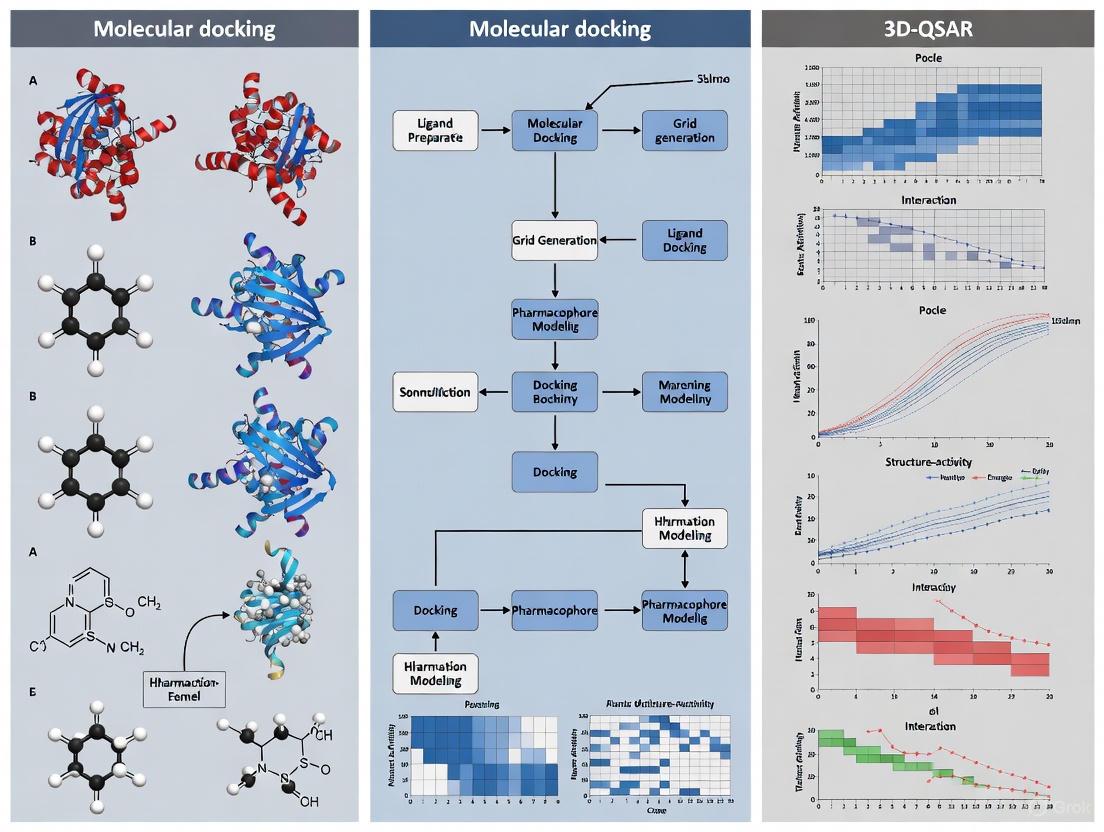
Diagram 1: Integrated Computational Workflow for Tubulin Inhibitor Development. This workflow illustrates the iterative cycle of computational methods and experimental validation in modern tubulin-targeted drug discovery.
Experimental Protocols and Methodologies
Molecular Docking Protocol for Tubulin-Colchicine Site
Objective: To predict binding modes and affinities of novel compounds at the tubulin-colchicine binding site.
Workflow:
Protein Preparation:
- Obtain tubulin structure (e.g., PDB ID: 1SA0) from Protein Data Bank.
- Remove crystallographic water molecules and co-crystallized ligands.
- Add hydrogen atoms and optimize protonation states using MolProbity or similar tools.
- Perform energy minimization using AMBER or OPLS forcefields.
Ligand Preparation:
- Sketch 2D structures of compounds using ChemDraw or similar software.
- Generate 3D conformations and minimize energy using MMFF94 or GAFF forcefields.
- Assign Gasteiger-Hückel or AM1-BCC partial charges.
Grid Generation:
- Define binding site using colchicine as reference (center coordinates: x=35.5, y=9.5, z=70.2).
- Set grid box dimensions (e.g., 60×60×60 points with 0.375Å spacing).
Docking Execution:
- Utilize AutoDock Vina, Glide, or GOLD software.
- Set exhaustiveness parameter to 20-50 for thorough sampling.
- Generate 20-50 poses per compound.
Analysis:
- Cluster poses by root-mean-square deviation (RMSD < 2.0Å).
- Identify key interactions with residues: βCys241, βLeu255, βLys352, βAsn258, βVal318.
- Select top-ranked pose based on scoring function and interaction consistency.
Key Considerations: Validate protocol by re-docking native ligand (RMSD < 2.0Å from crystal structure). Include known inhibitors (e.g., colchicine, CA-4) as positive controls.
3D-QSAR Modeling Protocol (CoMFA/CoMSIA)
Objective: To develop predictive 3D-QSAR models for tubulin inhibitor activity.
Workflow:
Dataset Curation:
- Collect 20-50 compounds with known tubulin polymerization inhibition (IC50) or cytotoxicity (IC50).
- Convert IC50 to pIC50 (-logIC50) for linear regression.
- Divide dataset: 70-80% training set, 20-30% test set.
Molecular Alignment:
- Identify common core structure (e.g., cis-stilbene for CA-4 analogs).
- Perform database alignment using RMSD atom fitting or pharmacophore-based alignment.
Field Calculation:
- CoMFA: Calculate steric (Lennard-Jones) and electrostatic (Coulombic) fields using sp³ carbon probe with +1 charge.
- CoMSIA: Calculate steric, electrostatic, hydrophobic, hydrogen-bond donor, and hydrogen-bond acceptor fields using common probe.
Partial Least Squares (PLS) Analysis:
- Apply leave-one-out (LOO) cross-validation to determine optimal components.
- Build final model with non-cross-validated PLS regression.
- Evaluate model quality: q² > 0.5, r² > 0.8, low standard error.
Contour Map Analysis:
- Visualize sterically favored/disallowed regions (green/yellow).
- Identify electropositive/electronegative favored regions (blue/red).
- Correlate contour regions with structural features for design.
Validation: Use external test set (R²pred > 0.6), y-randomization, and domain applicability analysis.
Tubulin Polymerization Inhibition Assay
Objective: To experimentally evaluate compounds for inhibition of tubulin polymerization in vitro.
Materials:
- Purified tubulin (>97% pure, cytosolic)
- GPEM buffer: 1 mM GTP, 1 mM MgCl₂, 1 mM EGTA, 0.1 M PIPES, pH 6.8
- Test compounds dissolved in DMSO (<1% final concentration)
- 96-well plates, UV-Vis spectrophotometer or fluorescence plate reader
- Temperature-controlled spectrophotometer
Procedure:
- Prepare reaction mixture containing 2 mg/mL tubulin, 1 mM GTP in GPEM buffer.
- Add test compounds (0.1-100 µM range) or controls (colchicine as positive control, DMSO as negative control).
- Transfer to pre-warmed (37°C) 96-well plates.
- Immediately monitor turbidity at 350 nm every 10-30 seconds for 30-40 minutes.
- Calculate percentage inhibition relative to DMSO control.
- Determine IC50 values using non-linear regression of concentration-response data.
Data Analysis: Compare initial rates, maximum absorbance, and area under the polymerization curve. Perform statistical analysis with n≥3 replicates.
The Scientist's Toolkit: Essential Research Reagents and Materials
Table 3: Key Research Reagents for Tubulin Binding Studies
| Reagent/Material | Specifications | Application/Function | Example Sources |
|---|---|---|---|
| Purified Tubulin | >97% purity, cytosolic, lyophilized | In vitro polymerization assays, binding studies | Cytoskeleton Inc., Sigma-Aldrich |
| Colchicine | ≥95% purity, reference standard | Positive control for binding site competition | Sigma-Aldrich, Tocris |
| Combretastatin A-4 | ≥98% purity, crystalline | Reference CBSI for biochemical assays | Abcam, Cayman Chemical |
| Paclitaxel (Taxol) | ≥97% purity, suitable for cell culture | Microtubule stabilization control | Sigma-Aldrich, MedChemExpress |
| Vinblastine Sulfate | ≥95% purity, cell culture tested | Vinca site reference inhibitor | Tocris, Selleck Chemicals |
| GTP | ≥95% purity, sodium salt | Tubulin polymerization cofactor | Sigma-Aldrich, Roche |
| PIPES Buffer | ≥99% purity, molecular biology grade | Tubulin polymerization assay buffer | Thermo Fisher, Sigma-Aldrich |
| Pre-coated ELISA Plates | High-binding, clear flat-bottom | Tubulin binding ELISAs | Corning, Thermo Fisher |
| Anti-β-Tubulin Antibody | Monoclonal, validated for WB/IF | Detection of tubulin in cellular assays | Cell Signaling, Abcam |
Emerging Directions and Novel Binding Sites
Beyond the classical binding sites, recent research has identified novel pharmacological sites on tubulin, expanding opportunities for therapeutic intervention. Gatorbulin-1 (GB1), a marine-derived cyclodepsipeptide, targets a distinct seventh binding site at the tubulin intradimer interface [14]. This site is structurally different from the colchicine, vinca, and taxane sites, with GB1 making extensive contacts and hydrogen bonds with both α- and β-chains of tubulin [14]. Structure-activity relationship studies of gatorbulin analogs revealed that the hydroxamate moiety in the N-methyl-alanine residue is critical for activity, while other structural features including C-hydroxylation of asparagine and methylation at C-4 of proline are functionally relevant [14].
The maytansine site, which partially overlaps with the vinca site, has gained prominence through the development of antibody-drug conjugates (ADCs) such as trastuzumab emtansine (Kadcyla) and polatuzumab vedotin (Polivy) [12] [11]. Additionally, the ninth binding site—the tumabulin site—was recently discovered at the interface between α1-tubulin, β1-tubulin, and RB3 [12]. These novel sites offer opportunities to overcome resistance mechanisms that limit current tubulin-targeting therapies and may provide improved therapeutic windows through unique binding modes and mechanisms of action.
The colchicine, vinca alkaloid, and taxane binding sites on tubulin represent clinically validated targets for cancer therapy, each with distinct mechanisms of action and therapeutic profiles. While taxanes and vinca alkaloids have established roles in clinical oncology, colchicine-site inhibitors offer promising avenues for overcoming multidrug resistance and targeting tumor vasculature. The integration of molecular docking and 3D-QSAR modeling has significantly advanced tubulin inhibitor development, enabling rational design of compounds with optimized binding interactions and pharmacological properties. Emerging discoveries of novel binding sites further expand the therapeutic potential of tubulin-targeting strategies. As computational methods continue to evolve alongside structural biology, the integration of these approaches will undoubtedly yield next-generation tubulin inhibitors with enhanced efficacy and reduced toxicity profiles for cancer treatment.
Application Notes
Three-dimensional Quantitative Structure-Activity Relationship (3D-QSAR) represents a pivotal advancement in computational drug design, moving beyond traditional 2D descriptors to incorporate the spatial and electrostatic properties of molecules. These techniques are particularly valuable in the rational design of tubulin inhibitors, a prominent class of anticancer agents. By correlating the three-dimensional molecular field characteristics of compounds with their biological activity, 3D-QSAR models provide visual contour maps that guide medicinal chemists in optimizing structural features to enhance potency.
The integration of 3D-QSAR with molecular docking creates a powerful synergistic workflow in tubulin research. While docking offers a detailed, atomic-level view of ligand-protein interactions within defined binding sites like colchicine or taxane sites, 3D-QSAR delivers a quantitative and predictive model of how structural modifications influence activity across a congeneric series. This combination is exceptionally suited for addressing challenges such as drug resistance mediated by βIII-tubulin isotype overexpression, enabling the design of next-generation inhibitors with improved binding affinity and selectivity [15] [16].
Core 3D-QSAR Methodologies and Their Application to Tubulin
Comparative Molecular Field Analysis (CoMFA)
CoMFA is a seminal 3D-QSAR technique that models biological activity based on steric and electrostatic molecular interaction fields. A probe atom is used to sample the space around a set of aligned molecules, and the resulting field energies are correlated with activity using Partial Least Squares (PLS) regression.
In practice, for a series of tubulin inhibitors such as colchicine analogues or phenylindole derivatives, CoMFA contour maps visually highlight regions where:
- Increased steric bulk (green contours) may enhance activity.
- Decreased steric bulk (yellow contours) may be favorable.
- Positive electrostatic potential (blue contours) is associated with higher activity.
- Negative electrostatic potential (red contours) is associated with higher activity [17] [18].
Table 1: Key Statistical Parameters for Validating 3D-QSAR Models
| Parameter | Description | Threshold for a Valid Model |
|---|---|---|
| R² | Non-cross-validated correlation coefficient | > 0.6 [17] |
| Q² (LOO) | Leave-One-Out cross-validated correlation coefficient | > 0.5 [17] [7] |
| SEE | Standard Error of Estimate | Should be low |
| F-value | Fisher F-test value for statistical significance | Should be high [3] |
| N | Optimal Number of Components from PLS | - |
| R²ₚᵣₑ𝒹 | Predictive R² for an external test set | > 0.5 [17] |
Comparative Molecular Similarity Indices Analysis (CoMSIA)
CoMSIA extends beyond CoMFA by evaluating additional physicochemical properties, offering a more nuanced view of ligand-receptor interactions. In addition to steric and electrostatic fields, CoMSIA typically includes:
- Hydrophobic fields: Identify regions where hydrophobic interactions boost activity.
- Hydrogen Bond Donor/Acceptor fields: Pinpoint optimal locations for H-bond interactions.
A robust CoMSIA model for 2-phenylindole derivatives as tubulin inhibitors demonstrated high reliability, with a non-cross-validated R² of 0.967 and a cross-validated Q² of 0.814 [7]. The inclusion of hydrophobic and hydrogen-bonding fields often makes CoMSIA models more interpretable for optimizing tubulin inhibitors, which frequently engage in complex hydrophobic and polar interactions within the binding pocket.
Pharmacophore Modeling
A pharmacophore is an abstract model that defines the essential steric and electronic features necessary for a molecule to interact with a biological target. It represents the supramolecular interaction pattern rather than a specific chemical structure. For instance, a study on cytotoxic quinolines identified a six-point pharmacophore hypothesis, AAARRR.1061, consisting of three hydrogen bond acceptors (A) and three aromatic rings (R). This model exhibited a high correlation (R² = 0.865) and was successfully used for virtual screening to identify novel tubulin inhibitor candidates [3].
Table 2: Representative 3D-QSAR and Pharmacophore Models in Tubulin Inhibitor Research
| Compound Class | Target / Activity | Model Type | Key Features / Descriptors | Statistical Performance |
|---|---|---|---|---|
| Cytotoxic Quinolines [3] | Tubulin Inhibitors | Pharmacophore (AAARRR.1061) | 3 H-bond Acceptors, 3 Aromatic Rings | R² = 0.865, Q² = 0.718 |
| 2-Phenylindole Derivatives [7] | MCF-7 Breast Cancer | CoMSIA | Steric, Electrostatic, Hydrophobic, H-Bond Donor/Acceptor | R² = 0.967, Q² = 0.814 |
| Colchicine Analogues [19] | Tubulin-Colchicine Site | 3D-QSAR | Steric and Electrostatic Fields | R² = 0.9438, Q² = 0.8915 |
| 1,2,4-Triazine-3(2H)-one [5] | Tubulin-Colchicine Site | QSAR (MLR) | Absolute Electronegativity (χ), Water Solubility (LogS) | R² = 0.849 |
Experimental Protocols
Protocol 1: Developing a CoMFA/CoMSIA Model for Tubulin Inhibitors
This protocol outlines the key steps for constructing a robust 3D-QSAR model using CoMFA and CoMSIA methodologies, tailored for a series of tubulin inhibitors.
Step 1: Data Set Curation and Preparation
- Compound Selection: Compile a dataset of compounds with known experimental biological activity (e.g., IC₅₀ for tubulin polymerization inhibition or cytotoxicity against a relevant cancer cell line). A typical dataset should include 20-50 molecules with a wide and continuous range of activity values [7] [5].
- Activity Conversion: Convert IC₅₀ values to pIC₅₀ (pIC₅₀ = -logIC₅₀) for a more linear correlation with free energy changes.
- Training and Test Sets: Randomly divide the dataset into a training set (≈80%) for model development and a test set (≈20%) for external validation [5]. Ensure both sets are representative of the structural and activity range.
Step 2: Molecular Modeling and Alignment
- Geometry Optimization: Sketch 3D structures and perform energy minimization using a molecular mechanics force field (e.g., Tripos or OPLS) with assigned atomic charges (e.g., Gasteiger-Hückel) [7].
- Molecular Alignment: This is the most critical step. Align all molecules based on a common scaffold or a putative pharmacophore. For tubulin inhibitors binding to a specific site like colchicine, the most active compound is often used as a template for aligning the dataset using the "distill" method in SYBYL or similar software [7].
Step 3: Field Calculation and PLS Analysis
- Grid Generation: Create a 3D cubic grid with a spacing of 2.0 Å that encompasses all aligned molecules [17] [7].
- Field Calculation:
- CoMFA: Calculate steric (Lennard-Jones) and electrostatic (Coulombic) fields using an sp³ carbon probe with a +1 charge.
- CoMSIA: Calculate steric, electrostatic, hydrophobic, and hydrogen-bond donor/acceptor fields using the respective probes.
- PLS Regression: Use the Partial Least Squares (PLS) algorithm to correlate the field descriptors with the biological activity (pIC₅₀). First, perform Leave-One-Out (LOO) cross-validation to determine the optimal number of components (ONC) that gives the highest Q². Then, perform a non-cross-validated regression with this ONC to generate the final model with R² and standard error of estimate (SEE) [17] [7].
Step 4: Model Validation
Robust validation is essential to ensure the model's predictive power.
- Internal Validation: Q² > 0.5 is generally considered statistically significant [17].
- External Validation: Predict the activity of the test set compounds and calculate the predictive R² (R²ₚᵣₑ𝒹). R²ₚᵣₑ𝒹 > 0.5 indicates a good predictive model [17] [20].
- Additional Checks: Perform Y-randomization to rule out chance correlation and define the Applicability Domain (APD) to identify the model's scope [20].
Protocol 2: Generating a Common Pharmacophore Hypothesis
This protocol describes the process of identifying a common set of chemical features shared by active tubulin inhibitors.
Step 1: Conformational Analysis and Feature Mapping
- Ligand Preparation: Generate a diverse set of low-energy conformers for each molecule in the dataset (e.g., a maximum of 100 conformers per molecule) [3].
- Activity Classification: Categorize compounds as "active" or "inactive" based on a predefined activity threshold (e.g., pIC₅₀ > 5.5 for active, pIC₅₀ < 4.7 for inactive) [3].
- Feature Identification: Define potential pharmacophore features such as Hydrogen Bond Acceptor (A), Hydrogen Bond Donor (D), Hydrophobic (H), Positively Charged (P), Negatively Charged (N), and Aromatic Ring (R) [3] [20].
Step 2: Hypothesis Generation and Scoring
- Hypothesis Generation: The software (e.g., Phase in Schrödinger) develops common pharmacophore hypotheses that are present in the active compounds but absent in the inactive ones.
- Hypothesis Scoring: Rank the generated hypotheses using a scoring function (e.g., Survival Score) that considers the alignment of vectors, volumes, and how well the hypothesis reflects the activity data. Select the top-ranked hypothesis (e.g., AAARRR.1061 for quinolines) for further studies [3].
Step 3: Pharmacophore-Based 3D-QSAR and Virtual Screening
- 3D-QSAR Model Building: Use the selected pharmacophore to align the molecules and build a 3D-QSAR model using PLS, similar to the CoMFA/CoMSIA process.
- Database Screening: Use the validated pharmacophore model as a 3D query to screen large chemical databases (e.g., ZINC, IBScreen) to identify novel hit compounds that match the essential feature arrangement [3] [15].
Workflow for Integrated 3D-QSAR and Docking
Research Reagent Solutions
Table 3: Essential Software and Computational Tools for 3D-QSAR in Tubulin Research
| Tool / Resource | Category | Primary Function in Workflow | Example Use Case |
|---|---|---|---|
| Maestro (Schrödinger) [3] | Integrated Suite | Ligand preparation (LigPrep), conformational analysis, and pharmacophore modeling (Phase). | Preparing a set of quinoline derivatives for 3D-QSAR [3]. |
| SYBYL [7] | Molecular Modeling | Structure building, energy minimization, CoMFA/CoMSIA analysis, and molecular alignment. | Developing a CoMSIA model for 2-phenylindole derivatives [7]. |
| Gaussian 09W [5] | Quantum Chemistry | Calculating electronic descriptors (e.g., HOMO/LUMO energies) for QSAR using DFT methods. | Computing absolute electronegativity for 1,2,4-triazine-3(2H)-one derivatives [5]. |
| AutoDock Vina/InstaDock [15] | Molecular Docking | Predicting binding poses and affinities of hits within the tubulin binding site (e.g., colchicine site). | Screening natural compounds against the βIII-tubulin isotype [15]. |
| Open Babel [15] | Cheminformatics | File format conversion for large compound libraries during virtual screening. | Converting SDF files from ZINC database to PDBQT format for docking [15]. |
| PaDEL-Descriptor [15] | Descriptor Calculation | Generating molecular descriptors and fingerprints for machine learning-based QSAR. | Creating features for ML classifiers to identify active tubulin inhibitors [15]. |
| GROMACS/AMBER [15] [5] | Molecular Dynamics | Simulating the stability of tubulin-ligand complexes over time using RMSD, RMSF, Rg, SASA. | Confirming the stable binding of a hit compound over 100 ns simulation [5]. |
Key Molecular Descriptors for Tubulin Inhibitor Activity Prediction
The rational design of tubulin inhibitors represents a crucial strategy in anticancer drug development, particularly for overcoming limitations of conventional therapies such as drug resistance and systemic toxicity [21]. The integration of molecular docking with three-dimensional quantitative structure-activity relationship (3D-QSAR) modeling has emerged as a powerful computational framework for identifying and optimizing novel tubulin-targeting agents [22]. This protocol details the key molecular descriptors and experimental methodologies essential for predicting tubulin inhibitor activity within this integrated framework, providing researchers with a structured approach to accelerate anticancer drug discovery.
Molecular descriptors quantitatively characterize structural and physicochemical properties that govern biological activity. For tubulin inhibitors, these descriptors help elucidate critical binding interactions with various tubulin sites, including the colchicine, vinca alkaloid, and taxane binding domains [21] [22]. The accurate prediction of inhibitor potency relies on identifying optimal combinations of steric, electrostatic, hydrophobic, and hydrogen-bonding features that complement these binding pockets.
Key Molecular Descriptors for Tubulin Inhibition
Experimental Data Compilation
Table 1 summarizes the primary molecular descriptors identified as critical predictors of tubulin inhibition activity across multiple compound classes, as derived from comprehensive QSAR studies.
Table 1: Key Molecular Descriptors for Tubulin Inhibitor Activity Prediction
| Descriptor Category | Specific Descriptors | Structural Interpretation | Impact on Tubulin Inhibition | Representative Compound Class |
|---|---|---|---|---|
| Pharmacophore Features | Three hydrogen bond acceptors (A), Three aromatic rings (R) [3] | Defines spatial arrangement for complementary binding | High correlation with activity (R² = 0.865) [3] | Cytotoxic quinolines [3] |
| Electronic Properties | Absolute electronegativity (χ) [5] | Overall electron-attracting ability | Higher electronegativity increases activity | 1,2,4-triazine-3(2H)-one derivatives [5] |
| Topological Descriptors | Chi1n, HeavyAtomCount, HeavyAtomMolWt [23] [24] | Molecular branching, size, and complexity | Optimal values enhance binding affinity | Phenanthrene analogs [23] [24] |
| Solubility & Hydrophobicity | Water solubility (LogS), EState_VSA8 [23] [5] | Polar surface area, hydrogen bonding capacity | Moderate hydrophobicity improves cellular uptake | 1,2,4-triazine-3(2H)-one derivatives [5] |
| Surface Property Descriptors | VSAdon, SMRVSA5 [25] | Polarizability, hydrogen bond donor capacity | Enhances tubulin polymerization inhibition | Diarylsulphonylurea derivatives [25] |
Structural Interpretation of Key Descriptors
The AAARRR.1061 pharmacophore model identified for cytotoxic quinolines exemplifies optimal spatial arrangement for tubulin binding, consisting of three hydrogen bond acceptors and three aromatic rings with specific distance and angular relationships [3]. This model demonstrated high statistical significance with a correlation coefficient (R²) of 0.865 and cross-validation coefficient (Q²) of 0.718, indicating robust predictive capability for tubulin inhibitory activity [3].
Electronic descriptors such as absolute electronegativity (χ) directly influence binding affinity through charge-transfer interactions with tubulin binding site residues [5]. For 1,2,4-triazine-3(2H)-one derivatives, higher electronegativity correlates strongly with enhanced inhibitory activity against breast cancer cell lines [5].
Topological descriptors including Chi1n and HeavyAtomCount capture aspects of molecular shape and complexity that complement the structural dimensions of tubulin binding pockets [23] [24]. In phenanthrene analogs, these descriptors showed significant correlation with anti-proliferative activity in HepG2 liver cancer cells, facilitating virtual screening of potent candidates [23].
Integrated QSAR and Molecular Docking Protocol
The following diagram illustrates the integrated computational pipeline for tubulin inhibitor activity prediction:
Dataset Preparation and Molecular Optimization
Procedure:
- Compound Selection: Compile a structurally diverse dataset of known tubulin inhibitors with corresponding experimental bioactivity values (IC₅₀) [3] [5]. For the 1,2,4-triazine-3(2H)-one derivatives study, 32 compounds with pIC₅₀ values ranging from 3.460 to 4.963 were selected [5].
- Training-Test Set Division: Implement an 80:20 ratio for splitting compounds into training and test sets using randomized selection to ensure representative chemical space coverage [5].
- Structure Optimization: Generate 3D molecular structures using builder modules in molecular modeling software (e.g., Maestro, SYBYL) [3] [7]. Perform energy minimization using appropriate force fields (e.g., OPLS_2005, Tripos molecular mechanics) with implicit solvation models [3] [7].
- Conformer Generation: Generate multiple low-energy conformers for each compound (maximum 100 conformers) to account for flexible binding orientations [3].
Molecular Descriptor Calculation and QSAR Modeling
Procedure:
- Descriptor Calculation: Compute 2D and 3D molecular descriptors using computational packages such as Gaussian (for electronic descriptors), ChemOffice (for topological descriptors), and MOE QuaSAR module (for surface area descriptors) [5] [25].
- Descriptor Selection: Apply statistical methods including principal component analysis (PCA) and stepwise variable selection to identify the most relevant, non-collinear descriptors [5].
- Model Development: Employ multiple linear regression (MLR) or partial least squares (PLS) analysis to construct QSAR models correlating descriptors with biological activity [3] [5].
- Model Validation: Validate models using leave-one-out cross-validation (Q²), external test set prediction (R²Pred), and Y-randomization testing to ensure robustness and avoid overfitting [3] [20].
Pharmacophore Modeling and Virtual Screening
Procedure:
- Pharmacophore Generation: Develop pharmacophore hypotheses using active compound alignments, identifying critical features including hydrogen bond acceptors (A), donors (D), hydrophobic groups (H), and aromatic rings (R) [3] [20].
- Hypothesis Validation: Evaluate hypotheses based on survival scores, vector alignment, and statistical correlation with activity values [3].
- Virtual Screening: Screen compound databases (e.g., IBScreen, Aldrich Market Select) against validated pharmacophore models to identify novel potential inhibitors [3] [23].
- Activity Prediction: Apply developed QSAR models to predict activity of screened compounds and prioritize candidates for further analysis [23].
Molecular Docking and Binding Mode Analysis
Procedure:
- Protein Preparation: Obtain tubulin structure from Protein Data Bank (e.g., 1SA0). Remove native ligands, add hydrogen atoms, and optimize side-chain orientations using protein preparation workflows [3] [22].
- Binding Site Definition: Define specific binding sites (colchicine, vinca alkaloid, or taxane sites) based on crystallographic data [21] [22].
- Docking Protocol: Perform molecular docking using software such as Glide, GOLD, or AutoDock with appropriate scoring functions [3] [23]. Validate docking accuracy by re-docking native ligands and calculating RMSD values (target < 2.0 Å) [20].
- Interaction Analysis: Identify key binding interactions (hydrogen bonds, hydrophobic contacts, π-π stacking) between compounds and tubulin residues [3] [26].
ADMET Prediction and Molecular Dynamics
Procedure:
- ADMET Profiling: Predict absorption, distribution, metabolism, excretion, and toxicity properties using in silico tools such as QikProp or SwissADME [23] [5]. Evaluate key parameters including LogP, LogS, polar surface area, and blood-brain barrier permeability [23].
- Molecular Dynamics Simulations: Conduct MD simulations (100-200 ns) using GROMACS or AMBER to evaluate complex stability under physiological conditions [23] [5]. Analyze root mean square deviation (RMSD), root mean square fluctuation (RMSF), and binding free energies [5].
- Candidate Prioritization: Integrate all computational analyses to select promising tubulin inhibitors with optimal activity predictions, favorable binding modes, and suitable ADMET profiles for experimental validation [23] [5].
Research Reagent Solutions
Table 2: Essential Computational Tools and Resources for Tubulin Inhibitor Research
| Resource Category | Specific Tools/Software | Application in Protocol | Key Features |
|---|---|---|---|
| Molecular Modeling Suites | Maestro (Schrödinger) [3], SYBYL [7], MOE [25] | Structure preparation, pharmacophore modeling, QSAR analysis | Integrated environments for drug discovery, force field-based minimization, conformational sampling |
| Descriptor Calculation | Gaussian [5], ChemOffice [5] | Electronic and topological descriptor computation | DFT calculations, topological index computation, property prediction |
| Docking & Simulation | Glide [3], GROMACS [23], AMBER | Molecular docking, dynamics simulations | High-throughput virtual screening, explicit solvation models, binding free energy calculations |
| Statistical Analysis | XLSTAT [5], SYSTAT [25] | QSAR model development, statistical validation | Multiple linear regression, principal component analysis, cross-validation methods |
| Compound Databases | IBScreen [3], Aldrich Market Select [23] | Virtual screening of novel compounds | Commercially available compounds, diverse chemical libraries, purchasable molecules |
The integration of molecular docking with 3D-QSAR modeling provides a powerful strategy for identifying key molecular descriptors that predict tubulin inhibitor activity. The experimental protocol outlined herein enables systematic characterization of critical structural and physicochemical properties governing tubulin binding, facilitating the rational design of novel anticancer agents with optimized potency and selectivity. By implementing this comprehensive computational pipeline, researchers can efficiently prioritize promising tubulin inhibitors for synthetic efforts and experimental validation, accelerating the discovery of next-generation microtubule-targeting therapeutics.
Data Set Curation and Preparation for Reliable Model Development
In the context of a broader thesis on integrating molecular docking with three-dimensional quantitative structure-activity relationship (3D-QSAR) modeling for tubulin inhibitor research, the critical foundation lies in the meticulous curation and preparation of high-quality data sets. Tubulin remains a validated target for cancer therapy, with its inhibitors playing a crucial role in disrupting microtubule dynamics and cancer cell proliferation [5] [22]. The reliability of any subsequent computational model—whether molecular docking, 3D-QSAR, or molecular dynamics simulations—is fundamentally dependent on the initial data set quality. This protocol outlines standardized procedures for assembling, curating, and preparing tubulin inhibitor data sets to ensure robust and predictive model development.
Data Collection and Sourcing
The initial phase involves systematic gathering of tubulin inhibitor data from diverse sources to ensure comprehensive coverage and structural diversity. Researchers should prioritize experimentally validated tubulin inhibitors with published biological activity measurements.
Table 1: Exemplar Data Sets in Tubulin Inhibitor Research
| Data Set Description | Compound Count | Biological Activity | Source/Reference |
|---|---|---|---|
| 1,2,4-triazine-3(2H)-one derivatives | 32 | pIC50 (3.460–4.963) against MCF-7 breast cancer cell line | [5] |
| Styrylquinoline derivatives | 43 | IC50 (4.12-5.95 µM) converted to pIC50 | [27] |
| Assembled tubulin-microtubule system inhibitors | 851 | pIC50 values across multiple cancer cell lines | [28] |
Activity Data Standardization
Consistent activity data representation is essential for reliable model development. Convert all half-maximal inhibitory concentration (IC50) values to pIC50 using the standard formula:
This transformation linearizes the relationship between concentration and binding affinity, improving model performance in subsequent QSAR analyses.
Compound Curation Protocols
Structure Standardization
- Representation: Draw all compound structures using standardized chemical drawing software such as Sybyl [27].
- Optimization: Perform geometry optimization using appropriate force fields (e.g., Tripos force field with convergence threshold of 0.01 kcal/mol) [27].
- Validation: Visually inspect all structures to ensure correct stereochemistry and tautomeric forms.
Data Set Division
Implement rigorous data set splitting to enable model validation:
- Training Set: Allocate 80% of compounds for model development [5].
- Test Set: Reserve 20% of compounds for external validation [5].
- Randomization: Apply randomized splitting to avoid potential biases related to data entry order [5].
Molecular Descriptor Computation
Electronic Descriptors
Compute quantum chemical descriptors using Density Functional Theory (DFT):
- Software: Employ Gaussian 09W program with DFT/B3LYP functional and 6-31G (p, d) basis set [5].
- Key Descriptors: Calculate highest occupied molecular orbital energy (EHOMO), lowest unoccupied molecular orbital energy (ELUMO), dipole moment (μm), total energy (TE) [5].
Derived Properties: Determine absolute hardness (η), absolute electronegativity (χ), and reactivity index (ω) using established equations [5]:
η = (ELUMO - EHOMO)/2 χ = (ELUMO + EHOMO)/2 ω = μ²/2η
Topological Descriptors
Calculate physiochemical and topological descriptors using specialized software:
- Software: Utilize ChemOffice software 16.0 or equivalent [5].
- Key Descriptors: Include molecular weight (MW), hydrogen bond acceptors/donors (NHA, NHD), octanol-water partition coefficient (LogP), water solubility (LogS), polar surface area (PSA), and number of rotatable bonds (NROT) [5].
3D-QSAR Specific Preparation
Molecular Alignment
Proper molecular alignment is critical for 3D-QSAR model development:
- Template Selection: Identify a reference compound with high activity as an alignment template (e.g., compound 22 in styrylquinoline studies) [27].
- Alignment Method: Use common substructure or atom-based fitting methods to superimpose all compounds [27].
- Validation: Visually inspect aligned structures to ensure pharmacophore-relevant orientation.
Conformational Analysis
- Energy Minimization: Optimize all structures using appropriate force fields prior to alignment [27].
- Active Conformation: Select the biologically relevant conformation based on experimental evidence or docking poses.
Research Reagent Solutions
Table 2: Essential Research Reagents and Computational Tools
| Reagent/Software | Function | Application in Workflow |
|---|---|---|
| Gaussian 09W | Quantum chemical calculations | Electronic descriptor computation [5] |
| ChemOffice | Topological descriptor calculation | 2D molecular property calculation [5] |
| Sybyl | Molecular modeling and alignment | 3D-QSAR model development [27] |
| Protein Data Bank (PDB) | Tubulin structure source | Molecular docking target (e.g., PDB ID: 4O2B) [27] |
| XLSTAT | Statistical analysis | QSAR model development and validation [5] |
Quality Control and Validation
Data Set Validation
- Structural Diversity: Assess chemical space coverage using principal component analysis (PCA) or t-distributed stochastic neighbor embedding (t-SNE) [5] [28].
- Activity Range: Verify that both training and test sets cover the entire activity range present in the full data set.
- Outlier Detection: Identify and investigate compounds with unusual structural features or extreme activity values.
Model Validation Techniques
- Y-randomization: Perform randomization tests to confirm model robustness by scrambling activity values and demonstrating model failure under these conditions [27].
- Applicability Domain: Define the chemical space boundaries where the model provides reliable predictions.
Workflow Visualization
Diagram 1: Data Set Curation Workflow for Tubulin Inhibitor Modeling
Diagram 2: Computational Preparation Pipeline
Robust data set curation and preparation form the foundation for developing reliable computational models in tubulin inhibitor research. By adhering to these standardized protocols—encompassing systematic data collection, structural standardization, comprehensive descriptor computation, rigorous validation, and appropriate data set division—researchers can establish high-quality data sets suitable for integrated molecular docking and 3D-QSAR studies. This meticulous approach to data preparation ensures that subsequent models will provide meaningful insights into tubulin-inhibitor interactions and facilitate the rational design of novel therapeutic agents with optimized pharmacological profiles.
Integrated Computational Workflow: From Model Building to Virtual Screening
Developing Robust 3D-QSAR Models with High Predictive Power (q² > 0.7)
The integration of computational methodologies has become a cornerstone in modern drug discovery, significantly accelerating the identification and optimization of lead compounds. Within the specific context of tubulin inhibitor research—a critical area in developing anticancer agents—Three-Dimensional Quantitative Structure-Activity Relationship (3D-QSAR) modeling serves as a powerful ligand-based drug design approach. When robustly constructed and validated, these models can predict the biological activity of novel molecules with high accuracy, guiding synthetic efforts and reducing experimental costs. This protocol details the establishment of a 3D-QSAR model with a cross-validated coefficient (q²) exceeding 0.7, a benchmark indicating high predictive power, and frames the process within a broader strategy that integrates molecular docking for tubulin inhibitor development [15] [29].
The fundamental principle of 3D-QSAR is to correlate the three-dimensional molecular fields of a set of compounds with their measured biological activities. Unlike traditional QSAR, which uses global molecular descriptors, 3D-QSAR considers the spatial and electrostatic characteristics of molecules, providing a more nuanced understanding of steric and electronic requirements for binding to a biological target like tubulin [30]. For tubulin inhibitors, which often bind to specific sites such as the colchicine or taxol site, understanding these requirements is paramount for designing potent and selective agents [15]. Recent studies on natural inhibitors of the human αβIII tubulin isotype have successfully leveraged structure-based drug design alongside machine learning, underscoring the value of advanced computational pipelines in this field [15].
Application Notes: Core Principles and Workflow
The 3D-QSAR Concept and Its Relevance to Tubulin Research
3D-QSAR models quantitatively describe how modifications to a molecule's structure, particularly in three-dimensional space, affect its biological activity. The model's output is often visualized as contour maps that highlight regions where specific molecular properties (e.g., steric bulk, electropositive character) would favorably or unfavorably influence activity. This is exceptionally valuable for optimizing tubulin inhibitors, as it provides visual, atom-level guidance for medicinal chemists [30].
For instance, a steric contour map might show a green favored region near a specific substituent of a tubulin inhibitor, suggesting that enlarging a group in that area could enhance binding affinity by filling a hydrophobic pocket in the tubulin protein. Conversely, a yellow disfavored region would warn against introducing bulky groups that might cause steric clashes [30]. This direct, interpretable feedback is crucial for the rational design of next-generation inhibitors.
The Integrated Workflow: From Data Collection to Model Application
A robust 3D-QSAR study is not an isolated event but part of a larger, iterative drug discovery cycle. For tubulin inhibitors, this often begins with the cloning, expression, and purification of the tubulin protein, followed by high-throughput screening to identify initial hit compounds with anti-tubulin activity. The subsequent workflow for a 3D-QSAR study is methodical and consists of several critical, interconnected stages, as outlined below [30] [29]:
Protocol: A Step-by-Step Methodology
This protocol provides a detailed guide for each stage of the 3D-QSAR workflow, with a focus on achieving a predictive model (q² > 0.7) for tubulin inhibitors.
Data Collection and Curation
Objective: To assemble a high-quality, congeneric dataset of tubulin inhibitors with reliably measured biological activities.
- Compound Selection: Compile a series of 30-50 compounds that are structurally related but incorporate sufficient diversity in their substituents. This ensures a meaningful structure-activity relationship can be derived. The compounds should be confirmed tubulin inhibitors, targeting a specific binding site (e.g., colchicine site) to ensure a consistent mechanism of action [30] [29].
- Biological Activity Data: Collect half-maximal inhibitory concentration (IC₅₀) values from uniform, cell-based or biochemical assays. All data should be obtained under consistent experimental conditions to minimize noise and systemic bias. Convert IC₅₀ values to logarithmic scale (pIC₅₀ = -logIC₅₀) for modeling [30] [31].
- Dataset Division: Randomly split the dataset into a training set (≈80%) for model construction and a test set (≈20%) for external validation. The test set should never be used in any part of the model building process [32].
Molecular Modeling and Conformational Analysis
Objective: To generate representative, low-energy 3D structures for each molecule in the dataset.
- Structure Construction: Draw 2D structures of all compounds using software like ChemDraw or BIOVIA Draw [31] [32].
- 3D Geometry Optimization: Convert 2D structures into 3D coordinates using tools like RDKit or Sybyl. Subject the initial 3D structures to geometry optimization to adopt realistic, low-energy conformations. This can be achieved using molecular mechanics force fields (e.g., Universal Force Field - UFF) or higher-level quantum mechanical methods for greater accuracy [30].
Molecular Alignment
Objective: To superimpose all molecules in a common 3D frame that reflects their bioactive conformation at the tubulin binding site.
- Alignment Strategy: This is the most critical and challenging step. The assumption is that all compounds share a similar binding mode.
- Atom-Based Fit: Align molecules based on a common scaffold or a known pharmacophore, often derived from a high-resolution co-crystal structure of a lead compound with tubulin [30].
- Maximum Common Substructure (MCS): Use algorithms to identify the largest substructure shared among the dataset molecules and align based on this MCS [30].
- Software Tools: Utilize functions like
AllChem.ConstrainedEmbed()in RDKit or the alignment modules in commercial software like Sybyl to achieve precise spatial congruence [30].
Descriptor Calculation using CoMFA and CoMSIA
Objective: To compute 3D molecular field descriptors that numerically represent the steric and electrostatic environments of the aligned molecules.
- Comparative Molecular Field Analysis (CoMFA): Place the aligned molecules within a 3D grid. A probe atom (typically an sp³ carbon with a +1 charge) is used to calculate steric (Lennard-Jones potential) and electrostatic (Coulombic potential) interaction energies at each grid point. This results in a large table of descriptor values for each molecule [30] [31].
- Comparative Molecular Similarity Indices Analysis (CoMSIA): As an alternative or complementary approach, CoMSIA calculates similarity indices using Gaussian-type functions. It typically evaluates five fields: steric, electrostatic, hydrophobic, and hydrogen bond donor and acceptor. CoMSIA is often more robust to minor alignment errors and can provide more interpretable insights [30] [31].
Table 1: Key 3D-QSAR Techniques and Descriptors
| Technique | Descriptor Fields | Key Characteristics | Sensitivity to Alignment |
|---|---|---|---|
| CoMFA | Steric, Electrostatic | Calculates interaction energies on a 3D grid; classic, widely used method. | Highly sensitive |
| CoMSIA | Steric, Electrostatic, Hydrophobic, H-Bond Donor, H-Bond Acceptor | Uses Gaussian functions; avoids singularities; provides additional interaction insights. | Moderately sensitive |
Model Building and Validation
Objective: To derive a statistically significant mathematical model and rigorously validate its predictive power.
- Statistical Method: Use Partial Least Squares (PLS) regression to correlate the thousands of 3D descriptors with the pIC₅₀ values. PLS reduces the dimensionality of the descriptor data by projecting them onto a smaller set of latent variables that have the highest covariance with the activity [30].
- Internal Validation: Perform leave-one-out (LOO) cross-validation to determine the optimal number of components and calculate the cross-validated correlation coefficient, q². A q² > 0.7 is considered a threshold for a model with high predictive power [30] [31].
q² = 1 - PRESS/SSYwhere PRESS is the sum of squared prediction errors and SSY is the sum of squared deviations of the observed activities from their mean. - External Validation: Use the withheld test set to calculate the external predictive metrics, notably R²ₜₑₛₜ and RMSEₜₑₛₜ, which assess the model's ability to predict truly novel compounds [32].
- Statistical Checks: The model should also be evaluated for goodness-of-fit (R² > 0.8) and low standard error of estimate (SEE) [31].
Table 2: Key Statistical Metrics for 3D-QSAR Model Validation
| Metric | Formula/Description | Target Value | Purpose |
|---|---|---|---|
| q² | q² = 1 - PRESS/SSY | > 0.7 | Indicates high internal predictive ability via cross-validation. |
| R² | R² = 1 - RSS/TSS | > 0.8 | Measures goodness-of-fit of the model to the training data. |
| SEE | Standard Error of Estimate | As low as possible | Measures the accuracy of the model's predictions for the training set. |
| R²ₜₑₛₜ | R² for the test set predictions | > 0.6 | Measures the model's predictive power for external compounds. |
| RMSEₜₑₛₜ | Root Mean Square Error for the test set | As low as possible | Measures the average prediction error for external compounds. |
Model Interpretation and Compound Design
Objective: To translate the statistical model into visual, chemically intelligible guidance for designing new tubulin inhibitors.
- Contour Map Analysis: Visualize the CoMFA or CoMSIA results as 3D contour maps. These maps are overlaid on a reference molecule.
- Steric Fields: Green contours indicate regions where increased bulk enhances activity; yellow contours indicate regions where bulk is detrimental [30].
- Electrostatic Fields: Blue contours indicate regions where positive charge is favored; red contours indicate regions where negative charge is favored [30].
- Rational Design: Based on the contour maps, propose new derivatives of your lead tubulin inhibitor. For example, if a green steric contour is observed near a phenyl ring, consider synthesizing analogs with larger hydrophobic substituents at that position [30].
The following diagram illustrates the logical process of interpreting contour maps to design new compounds, which are then synthesized and tested, creating a feedback loop for model refinement.
The Scientist's Toolkit: Essential Research Reagent Solutions
Table 3: Key Software and Resources for 3D-QSAR Modeling
| Category | Tool/Resource | Specific Function in Protocol |
|---|---|---|
| Cheminformatics & Modeling | RDKit, Open Babel | File format conversion; basic molecular descriptor calculation [15] [30]. |
| 3D-QSAR & Molecular Alignment | Sybyl/X | Comprehensive suite for molecular modeling, conformational analysis, alignment, and running CoMFA/CoMSIA studies [31]. |
| Molecular Docking | AutoDock Vina | To perform molecular docking studies for integrating structure-based insights or guiding alignment [15] [33]. |
| Database | ZINC Database | Source of purchasable compound structures for virtual screening [15]. |
| Statistical Analysis & Scripting | R, Python (scikit-learn) | For data preprocessing, statistical analysis, and custom machine learning scripts [15]. |
Molecular Docking Protocols for Tubulin Binding Site Characterization
Molecular docking has become an indispensable tool in structural biology and computer-aided drug design, providing critical insights into ligand-receptor interactions. For tubulin research, docking protocols enable the characterization of small molecule binding to distinct sites on the tubulin heterodimer, facilitating the development of novel anticancer and antiparasitic agents [34]. This protocol details the integration of molecular docking with three-dimensional quantitative structure-activity relationship (3D-QSAR) studies, creating a powerful framework for rational drug design targeting tubulin. The synergy between these methods allows researchers to not only predict binding poses but also understand the structural and electrostatic features governing biological activity, thereby accelerating the identification and optimization of tubulin inhibitors [3] [2].
Tubulin possesses several well-characterized binding sites, including the taxane, vinca alkaloid, and colchicine sites, each with distinct structural properties and therapeutic implications [35] [36]. Colchicine-binding site inhibitors (CBSIs) have recently gained significant attention due to their potential to overcome multidrug resistance and their antiangiogenic properties [37]. The protocol presented herein emphasizes characterization of this pharmacologically relevant site while providing principles adaptable to other tubulin binding pockets.
Theoretical Background and Significance
Microtubules, composed of α- and β-tubulin heterodimers, are dynamic cytoskeletal components essential for vital cellular processes including mitosis, intracellular transport, cell signaling, and maintenance of cell shape [37] [2]. Their critical role in cell division makes them prominent targets for anticancer therapy, with microtubule-targeting agents broadly classified into microtubule-stabilizing agents (e.g., taxanes) and microtubule-destabilizing agents (e.g., vinca alkaloids, colchicine site binders) [37].
The βIII-tubulin isotype has emerged as a particularly important target, as its overexpression in various carcinomas (e.g., ovarian, breast, lung cancers) is closely associated with resistance to taxane-based chemotherapy [15]. This relationship underscores the necessity of isotype-specific targeting strategies in overcoming treatment resistance. Molecular docking approaches enable the characterization of compound interactions with specific tubulin isotypes, guiding the development of agents capable of circumventing resistance mechanisms.
The integration of molecular docking with 3D-QSAR creates a comprehensive framework for tubulin inhibitor development. While docking elucidates atomic-level interactions between ligands and the tubulin binding pocket, 3D-QSAR correlates structural features of ligands with their biological activity, generating predictive models that guide structural optimization [3] [2]. This protocol details the implementation of this integrated approach for advanced tubulin drug discovery.
Computational Requirements and Reagent Solutions
Table 1: Essential Computational Tools for Tubulin Docking Studies
| Category | Specific Tools | Application in Tubulin Research |
|---|---|---|
| Molecular Docking Software | AutoDock Vina [35], Glide (Schrödinger) [37] [38], MOE [36] | Predicting ligand binding poses and affinity to tubulin binding sites |
| Molecular Dynamics | GROMACS, AMBER | Assessing binding stability and conformational changes |
| Structure Preparation | Protein Preparation Wizard [38], LigPrep [38], Open-Babel [15] | Preparing protein and ligand structures for docking |
| QSAR Modeling | Phase [3], SYBYL-X [2] | Developing 3D-QSAR models based on pharmacophore alignment |
| Visualization & Analysis | PyMOL [15], UCSF Chimera | Analyzing docking results and interaction patterns |
| Specialized Databases | ZINC [15], Specs [37] | Sources of compound libraries for virtual screening |
Table 2: Key Structural Resources for Tubulin Studies
| Resource Type | Identifier/Name | Significance in Tubulin Research |
|---|---|---|
| Tubulin Structures | PDB: 4O2B [38], 1JFF [35] [15] | High-resolution structures for docking simulations |
| Natural Compound Libraries | ZINC Natural Compounds [15] | Source of potential novel tubulin inhibitors |
| Commercial Compound Libraries | Specs Library [37] | Large collection of synthetic compounds for screening |
| Binding Site Validation Tools | Site Map [38] | Identification and characterization of binding pockets |
Integrated Docking and 3D-QSAR Protocol
Protein Structure Preparation
The initial step involves acquiring and preparing the tubulin structure for docking simulations. The following protocol ensures proper protein preparation:
Retrieve tubulin structure from the Protein Data Bank (e.g., 4O2B, 1JFF) [38]. The 1JFF structure, resolved at 3.50 Å, contains taxol co-crystallized with tubulin and serves as an excellent template for homology modeling of various tubulin isotypes [15].
Preprocess the protein using tools like Protein Preparation Wizard (Schrödinger Suite) [38]:
- Add missing hydrogen atoms
- Assign appropriate protonation states at physiological pH
- Fill in missing side chains using rotamer libraries
- Remove water molecules beyond 5Å from binding sites
Generate tubulin isotypes through homology modeling when specific isotype structures are unavailable. For βIII-tubulin, use Modeller with 1JFF as template, then select models based on Discrete Optimized Protein Energy (DOPE) scores and validate using Ramachandran plots [15].
Define binding sites using Site Map module or based on known ligand binding locations (colchicine, taxane, or vinca sites) [38]. For colchicine site characterization, focus on the interface between α- and β-tubulin subunits [36].
Ligand Library Preparation
Proper ligand preparation is essential for accurate docking results:
Obtain compound structures from databases like ZINC (natural compounds) or Specs (synthetic compounds) [15] [37].
Prepare ligands using LigPrep or similar tools:
Generate conformers for 3D-QSAR studies using a stochastic global conformational search strategy, retaining a maximum of 100 conformers per ligand [3] [35].
Molecular Docking Execution
The core docking procedure follows these steps:
Set up grid boxes for docking using the Receptor Grid Generation module:
- Center the grid on the binding site of interest
- Set appropriate grid dimensions (e.g., 20×20×20 Å) to encompass the entire binding pocket
- For blind docking, expand grid dimensions to cover the entire protein surface [38]
Perform docking using Glide SP (Standard Precision) followed by XP (Extra Precision) modes [38] or AutoDock Vina [15]:
- Retain top poses based on docking scores for further analysis
- For virtual screening, use high-throughput protocols to process thousands of compounds [37]
Validate docking protocol by redocking native ligands and calculating Root Mean Square Deviation (RMSD) between predicted and crystallographic poses. RMSD values <2.0 Å indicate appropriate optimization [35].
Post-Docking Analysis and Binding Free Energy Calculations
Analyze protein-ligand interactions to identify key residues involved in binding:
- Hydrogen bonds with residues such as Thr276, Glu27, and Pro274 [35]
- Hydrophobic interactions with aromatic residues
- π-π stacking and cation-π interactions
Calculate binding free energies using molecular mechanics/generalized Born surface area (MM-GBSA) methods [38]:
- Use implicit solvent models to compute solvation energy (ΔGsolv)
- Calculate binding free energy (ΔGbind) for each ligand-protein complex
- Correlate computed binding energies with experimental activities
Generate 3D contour maps from docking results to visualize regions where specific chemical features enhance or diminish biological activity [3].
3D-QSAR Model Development
Integration with 3D-QSAR provides complementary structure-activity insights:
Select and prepare training set compounds with known tubulin inhibitory activities (pIC50 values) [3]. Typically, 40-50 compounds provide sufficient structural diversity for model development.
Align molecules based on pharmacophore hypotheses or docking poses using database alignment methods in SYBYL-X [2].
Generate pharmacophore hypotheses using algorithms like HypoGen [36]:
- Define chemical features (hydrogen bond acceptors/donors, hydrophobic groups, aromatic rings)
- Identify excluded volumes to represent protein steric constraints
- Evaluate hypotheses using survival scores [3]
Develop 3D-QSAR models using Comparative Molecular Field Analysis (CoMFA) and Comparative Molecular Similarity Indices Analysis (CoMSIA) [2]:
- Compute steric, electrostatic, hydrophobic, and hydrogen-bonding fields
- Apply Partial Least Squares (PLS) regression to correlate fields with biological activity
- Validate models using leave-one-out cross-validation and external test sets
Table 3: Statistical Parameters for Validated 3D-QSAR Models
| Model Type | Correlation Coefficient (R²) | Cross-Validation Coefficient (Q²) | RMSD | F Value | Reference |
|---|---|---|---|---|---|
| Pharmacophore (AAARRR.1061) | 0.865 | 0.718 | - | 72.3 | [3] |
| CoMFA | 0.974 | 0.724 | 0.6977 | - | [2] |
| CoMSIA | 0.976 | 0.710 | - | - | [2] |
| Hypo1 Pharmacophore | 0.9582 | - | 0.6977 | - | [36] |
Model Validation and Application
Validate 3D-QSAR models using:
Apply validated models to screen virtual compound libraries:
- Use pharmacophore models as 3D search queries
- Apply QSAR models to predict activity of hit compounds
- Select candidates with predicted high activity for further study [3]
Integrate results from docking and 3D-QSAR to design novel tubulin inhibitors with optimized interactions and improved predicted activity.
Experimental Workflow and Signaling Pathways
The following diagram illustrates the integrated workflow for combining molecular docking with 3D-QSAR in tubulin inhibitor development:
Tubulin Binding Site Characterization Pathway
The molecular docking process elucidates the binding interactions of tubulin inhibitors, which typically modulate microtubule dynamics through several interconnected signaling pathways:
Cellular Signaling Pathways of Tubulin Inhibitors
Applications and Case Studies
Discovery of Novel Colchicine-Site Inhibitors
Virtual screening of the SPECS library (200,340 compounds) against the colchicine binding site identified compound 89, a nicotinic acid derivative that demonstrated potent tubulin polymerization inhibition [37]. The characterization protocol included:
Molecular docking confirmed binding at the colchicine site with critical interactions involving residues at the α-β tubulin interface.
Experimental validation showed antiproliferative activity against multiple cancer cell lines (Hela, HCT116) with IC50 values in the micromolar range.
Mechanistic studies revealed G2/M phase cell cycle arrest, apoptosis induction, and inhibition of tumor cell migration and invasion.
In vivo studies demonstrated significant antitumor efficacy in mouse models with no observable toxicity at therapeutic doses.
Natural Product Screening for βIII-Tubulin Targeting
Screening of 89,399 natural compounds from the ZINC database against the taxane site of αβIII-tubulin identified four promising inhibitors (ZINC12889138, ZINC08952577, ZINC08952607, ZINC03847075) through an integrated protocol [15]:
Structure-based virtual screening using AutoDock Vina narrowed candidates to 1,000 hits based on binding energy.
Machine learning classifiers further refined selections to 20 active natural compounds.
ADMET property prediction identified four candidates with favorable drug-like properties.
Molecular dynamics simulations (RMSD, RMSF, Rg, SASA analysis) confirmed structural stability of tubulin-ligand complexes.
3D-QSAR Guided Optimization of Combretastatin Analogues
Integration of 3D-QSAR with docking studies facilitated the optimization of combretastatin A-4 (CA-4) analogues as tubulin polymerization inhibitors [2]:
Development of CoMFA and CoMSIA models with excellent statistical quality (r2 = 0.974 and 0.976, respectively) and predictive ability (q2 = 0.724 and 0.710, respectively).
Contour map analysis identified structural modifications to enhance inhibitory activity.
Molecular docking elucidated binding conformations and key amino acid interactions at the colchicine site.
Molecular dynamics simulations (30 ns) confirmed binding modes and calculated binding free energies using MM/GBSA and MM/PBSA methods.
Troubleshooting and Technical Considerations
Addressing Common Docking Challenges
Inaccurate binding pose prediction:
- Verify binding site definition through site map analysis or literature review
- Use constrained docking with known interaction constraints when available
- Compare multiple docking algorithms and select consensus poses
Poor correlation between docking scores and experimental activities:
- Apply post-docking MM-GBSA calculations to improve binding affinity estimates [38]
- Consider protein flexibility through ensemble docking or molecular dynamics
- Account for solvation effects and entropic contributions
Handling tubulin structural heterogeneity:
- Consider multiple tubulin structures to account for conformational diversity
- Use molecular dynamics to sample flexible binding site residues
- For homology models, validate with known binders before screening
3D-QSAR Model Optimization
Improving model predictive power:
- Ensure training set covers sufficient chemical space and activity range
- Apply appropriate alignment rules based on pharmacophore or docking poses
- Validate models extensively using external test sets and randomization tests
Interpreting contour maps:
- Correlate steric/electrostatic features with specific protein-ligand interactions
- Combine with docking results to understand structural basis for activity
- Use maps to guide rational molecular design rather than as absolute rules
The integrated molecular docking and 3D-QSAR protocol presented herein provides a robust framework for characterizing tubulin binding sites and accelerating the discovery of novel tubulin-targeting agents. The synergy between these computational approaches enables researchers to bridge the gap between structural insights and quantitative structure-activity relationships, facilitating rational drug design.
Key advantages of this integrated approach include the ability to:
- Identify novel binding motifs through virtual screening of large compound libraries
- Optimize lead compounds using quantitative activity predictions from 3D-QSAR models
- Understand structural basis of activity through atomic-level binding pose analysis
- Prioritize compounds for synthesis and biological evaluation, reducing experimental costs
As computational methods continue to advance, the integration of molecular docking with machine learning approaches and enhanced molecular dynamics simulations will further strengthen tubulin binding site characterization, ultimately contributing to the development of more effective therapeutics for cancer and parasitic diseases.
Virtual Screening of Chemical Databases Using Combined Pharmacophore and Docking Approaches
Within the context of a broader thesis on integrating molecular docking with 3D-QSAR for tubulin inhibitor research, this application note details a practical protocol for performing virtual screening of chemical databases. The approach synergistically combines pharmacophore modeling and molecular docking to efficiently identify novel, potent tubulin inhibitors. Tubulin remains a critical anticancer target, and the discovery of new inhibitory scaffolds is of paramount importance in overcoming drug resistance and side effects associated with current antimitotic agents [3] [36]. Computational methods like those described herein are essential for reducing the time and cost of drug discovery by prioritizing candidate compounds for synthesis and experimental evaluation in vitro [3] [39].
This protocol is designed for researchers, scientists, and drug development professionals. It provides a step-by-step methodology, from initial data preparation through to the final selection of hits, complete with the rationale for each step to ensure robust and reproducible results.
Theoretical Background and Significance
The Pharmacophore Concept and Tubulin Inhibition
A pharmacophore is defined as "the ensemble of steric and electronic features that is necessary to ensure the optimal supramolecular interactions with a specific biological target structure and to trigger (or to block) its biological response" [40]. It is an abstract concept that distills the essential functional characteristics—such as hydrogen bond acceptors (A), donors (D), hydrophobic groups (H), and aromatic rings (R)—required for biological activity, rather than representing specific chemical groups [39] [40].
In tubulin research, pharmacophore models provide critical insights into the structural requirements for binding at sites like the colchicine-binding site. For instance, a study on cytotoxic quinolines identified AAARRR.1061 as an optimal pharmacophore hypothesis, consisting of three hydrogen bond acceptors and three aromatic rings [3]. This model demonstrated high predictive power with a correlation coefficient (R²) of 0.865 and a cross-validation coefficient (Q²) of 0.718 [3]. Such models encapsulate the key interactions a ligand must form with the tubulin binding pocket.
The Rationale for a Combined Screening Approach
Virtual screening of large chemical databases poses a significant computational challenge. A combined pharmacophore and docking strategy addresses this by deploying a efficient, hierarchical filtering process [40].
- Pharmacophore Screening as a Filter: Pharmacophore models rapidly filter out molecules that lack the essential chemical features to be active, typically reducing the database size by 90-95% [40]. This step is computationally inexpensive and effective for "scaffold hopping" to identify novel chemotypes that maintain critical interactions [39] [40].
- Molecular Docking for Refinement: The much smaller subset of compounds that pass the pharmacophore filter is then subjected to more computationally intensive molecular docking. Docking evaluates the quality of the fit within the binding site, estimates binding affinity, and identifies specific atomic-level interactions with key residues [3] [41].
This two-tiered approach ensures that only the most promising candidates, which possess both the necessary pharmacophoric features and a favorable binding geometry, are advanced for further analysis.
Tubulin Signaling and Inhibition Pathway
The following diagram illustrates the role of tubulin in cell division and how its inhibition leads to anticancer effects.
Diagram 1: Tubulin Function and Inhibitor Mechanism of Action. Tubulin inhibitors disrupt the dynamic equilibrium of microtubules, leading to mitotic arrest and apoptosis in cancer cells [3] [36].
Experimental Protocol
The following diagram outlines the complete virtual screening workflow, from data preparation to final hit selection.
Diagram 2: Virtual Screening Workflow. The protocol involves sequential steps of model generation, database filtering, and multi-stage screening to identify high-probability hits.
Step-by-Step Procedure
Step 1: Data Set Preparation and Ligand Preparation
- Data Curation: For a ligand-based model, assemble a set of known active compounds against your target (e.g., tubulin). For instance, a study on quinolines used 62 compounds with cytotoxic activity against the A2780 cell line [3]. Include their biological activity values (e.g., IC₅₀ or Ki) and convert them to pIC₅₀ (-logIC₅₀) for analysis [3]. Also, include a set of known inactive compounds if available for validation [41].
- Data Splitting: Randomly divide the data set into a training set (≈80%) for model generation and a test set (≈20%) for validation [3] [42].
- Ligand Preparation: Generate 3D structures of all ligands using a tool like Maestro's LigPrep [3] or MOE [41]. Conduct a conformational analysis to generate a representative set of low-energy conformers for each molecule (e.g., a maximum of 100 conformers per ligand) [3].
Step 2: Pharmacophore Model Generation
Two primary approaches can be used:
- Ligand-Based: Use the training set of active compounds to identify common pharmacophore features. For example, the Phase module in Schrödinger can be used to generate hypotheses [3]. The model with the best survival score and statistical parameters (like R² and Q²) should be selected [3].
- Structure-Based: If a crystal structure of the target (e.g., tubulin in complex with an inhibitor) is available, a pharmacophore can be derived directly from the protein-ligand interactions. Use the "Receptor-Ligand Pharmacophore Generation" protocol in Discovery Studio [41] or a similar tool. The model should include key features (e.g., hydrogen bond acceptors/donors, hydrophobic regions, aromatic rings) and can incorporate exclusion volumes to represent the shape of the binding pocket [39].
Example: A robust tubulin inhibitor pharmacophore (Hypo1) was generated, containing one hydrogen-bond acceptor, one donor, one hydrophobic feature, one ring aromatic feature, and three excluded volumes [36].
Step 3: Pharmacophore Model Validation
A model must be validated before use in screening.
- Internal Validation: Assess the model's predictive ability by using it to predict the activity of the test set compounds that were excluded from model generation. A high correlation between experimental and predicted activity indicates a good model [3].
- Statistical Validation: Perform Y-randomization tests to ensure the model is not based on chance correlations [3].
- Enrichment Assessment: Use a decoy set containing known active and inactive compounds. Screen this set with your model and calculate the Enrichment Factor (EF) and the area under the Receiver Operating Characteristic curve (AUC-ROC). A model is generally considered reliable if AUC > 0.7 and EF > 2 [41]. The EF is calculated as: (EF = \frac{(Ha \times D)}{(Ht \times A)}) where (Ha) is the number of active compounds found, (D) is the total number of compounds in the decoy set, (Ht) is the total number of actives in the decoy set, and (A) is the total number of hits from the screening [41].
Step 4: Database Pre-Filtering
- Select a Screening Database: Choose a large, commercially available database such as ZINC [42], ChemDiv [43] [41], NCI, or Specs [36].
- Apply Drug-Likeness Rules: Filter the entire database using rules such as Lipinski's Rule of Five and Veber's rules to remove compounds with poor pharmacokinetic potential [41] [44].
- Predict ADMET Properties: Further filter compounds based on predicted Absorption, Distribution, Metabolism, Excretion, and Toxicity (ADMET) properties. This includes assessing aqueous solubility, blood-brain barrier penetration, cytochrome P450 inhibition, and hepatotoxicity [41] [45]. This step ensures that identified hits have a higher probability of becoming successful drugs.
Step 5: Pharmacophore-Based Virtual Screening
- Use the validated pharmacophore model as a query to screen the pre-filtered database. This can be done using the "Pharmacophore Search" protocol in software like MOE [46], Discovery Studio [41], or Phase [3].
- The search will identify molecules that have conformations matching the spatial arrangement of the pharmacophore features within defined tolerance limits.
- The output is a significantly smaller subset of compounds (pharmacophore hits) that theoretically possess the structural requirements for biological activity.
Step 6: Molecular Docking of Pharmacophore Hits
- Protein Preparation: Obtain the 3D structure of the target protein (e.g., tubulin) from the Protein Data Bank (PDB). Prepare the protein by removing water molecules, adding hydrogen atoms, correcting missing residues, and optimizing the structure using a force field like CHARMM [41] [42].
- Ligand Preparation: Prepare the pharmacophore hits for docking, ensuring correct protonation states and tautomers.
- Docking Simulation: Perform molecular docking of the pharmacophore hits into the defined binding site of the target protein (e.g., the colchicine-binding site of tubulin) using software such as MOE [36], Smina [42], or Glide. Use a robust docking algorithm that accounts for ligand flexibility.
- Analysis of Docking Poses: Analyze the top-ranking compounds (based on docking scores) for specific favorable interactions with key amino acid residues in the binding pocket. For example, a promising tubulin inhibitor candidate (STOCK2S-23597) showed a high docking score of -10.948 kcal/mol and formed four hydrogen bonds with active site residues [3].
Step 7: Hit Selection and Experimental Validation
- Select a final, manageable number of compounds (e.g., 10-20) that have passed all filters and exhibit the best docking scores and interaction profiles.
- These compounds are considered in silico hits and should be procured or synthesized for in vitro and in vivo experimental validation to confirm their biological activity [36] [42].
Key Research Reagent Solutions
Table 1: Essential Computational Tools and Databases for Virtual Screening.
| Category | Item/Software | Function in Protocol | Example |
|---|---|---|---|
| Software & Tools | Schrödinger Suite (Phase, LigPrep, Glide) | Integrated environment for ligand preparation, 3D-QSAR pharmacophore generation, and molecular docking [3]. | Used to generate the AAARRR.1061 quinoline pharmacophore model [3]. |
| MOE (Molecular Operating Environment) | Platform for pharmacophore modeling, virtual screening, and molecular docking [36] [46]. | Used for pharmacophore screening and docking studies to identify tubulin inhibitors [36]. | |
| Discovery Studio | Software for structure-based pharmacophore generation and validation [41]. | Used to build and validate pharmacophore models for VEGFR-2 and c-Met targets [41]. | |
| Smina | Docking software specifically designed for virtual screening, used to predict binding poses and scores [42]. | Used in a machine-learning accelerated virtual screening for MAO inhibitors [42]. | |
| Databases | RCSB Protein Data Bank (PDB) | Repository for 3D structural data of proteins and nucleic acids, essential for structure-based methods [41] [42]. | Source of tubulin (e.g., 1SA0), VEGFR-2, and c-Met crystal structures. |
| ZINC Database | Free database of commercially available compounds for virtual screening [42]. | Screened to find novel MAO inhibitors [42]. | |
| ChemDiv Database | Commercial database of diverse chemical compounds used in drug discovery [43] [41]. | Screened to identify potential VEGFR-2/c-Met dual inhibitors [41]. | |
| Specs Database | Commercial compound library for high-throughput and virtual screening [36]. | Screened to discover new tubulin inhibitor leads [36]. |
Anticipated Results and Data Interpretation
Expected Outcomes
Successful execution of this protocol should yield a shortlist of potential hit compounds. The process should demonstrate a significant enrichment of active compounds compared to a random selection. For example:
- The pharmacophore model AAARRR.1061 for quinolines showed a high correlation (R² = 0.865) for the training set and successfully predicted the activity of test set compounds [3].
- Virtual screening of the Specs database using a validated tubulin pharmacophore (Hypo1), followed by docking, led to the identification of compounds with confirmed inhibitory activity against MCF-7 breast cancer cells in vitro [36].
Statistical Validation of Models
It is crucial to report key statistical metrics for the generated pharmacophore models to demonstrate their quality and predictive power.
Table 2: Key Statistical Metrics for Validating a 3D-QSAR Pharmacophore Model.
| Metric | Description | Ideal Value/Interpretation | Example from Literature |
|---|---|---|---|
| R² | Coefficient of determination for the training set. | > 0.7 indicates a good fit of the model to the training data [3]. | 0.865 for model AAARRR.1061 [3]. |
| Q² | Cross-validation coefficient (predictive power). | > 0.5 is generally acceptable; higher is better [3]. | 0.718 for model AAARRR.1061 [3]. |
| RMSE | Root Mean Square Error. | Closer to 0 indicates higher prediction accuracy. | 0.3198 for a 2-pls factor model [3]. |
| Pearson-R | Correlation between predicted and actual activity of the test set. | Close to 1.0 indicates excellent predictive ability [3]. | 0.8756 for model AAARRR.1061 [3]. |
| Enrichment Factor (EF) | Measures the enrichment of active compounds in the hit list. | > 2.0 is considered a reliable model [41]. | Used to select the best pharmacophore for VEGFR-2/c-Met screening [41]. |
Troubleshooting and Technical Notes
- Low Enrichment in Virtual Screening: If the pharmacophore model retrieves few or no active compounds during screening, the model might be overly specific. Revisit the model generation step, consider using a more diverse training set, or slightly increase the tolerance radii of the pharmacophore features.
- Poor Correlation between Docking Score and Experimental Activity: This is a common issue. Ensure the protein structure is properly prepared, the binding site is correctly defined, and the correct protonation states of key residues are assigned. Consider using multiple scoring functions or performing binding free energy calculations (MM/PBSA) on the docking poses for a more reliable affinity estimate [41].
- Handling Computational Load: Screening millions of compounds can be computationally demanding. Utilize the pre-filtering steps (drug-likeness, pharmacophore) rigorously to reduce the number of compounds for docking. For extremely large libraries, consider using machine learning models that have been trained to predict docking scores as an ultra-fast pre-filter [42].
The discovery of novel tubulin inhibitors represents a promising frontier in anticancer drug development, particularly for challenging pathologies like breast cancer and osteosarcoma [47] [48]. This case study details a successful structure-based drug discovery campaign that led to the identification of potent quinoline and triazine derivatives as effective tubulin polymerization inhibitors. The research was framed within a broader thesis investigating the integration of computational methodologies—specifically the synergy between molecular docking and three-dimensional quantitative structure-activity relationship (3D-QSAR) modeling. This integrated approach enables a more rational design of small-molecule therapeutics that target the colchicine binding site of tubulin, a key regulatory site for microtubule dynamics [47] [49]. By systematically applying and cross-validating these techniques, the study achieved significant breakthroughs in compound potency and predictive model accuracy, offering a validated protocol for future anticancer agent development.
Background and Significance
Tubulin as a Therapeutic Target
Tubulin, a protein that forms cellular microtubules, is a validated target for cancer chemotherapy due to its crucial role in cell division and proliferation [49]. Inhibiting tubulin polymerization disrupts microtubule dynamics, which prevents mitosis and leads to apoptosis in tumor cells [49]. The colchicine binding site is of particular interest for drug design because it offers a strategic pocket for developing inhibitors that can effectively halt cancer cell division [47] [50]. The clinical relevance of this target is especially pronounced in breast cancer, which remains a leading cause of cancer-related deaths among women globally [47], and osteosarcoma, a highly malignant bone tumor with poor prognosis and limited treatment options [48].
Chemical Scaffolds: Quinoline and Triazine
Quinoline and triazine derivatives have emerged as privileged scaffolds in anticancer drug discovery due to their versatile pharmacological profiles and synthetic adaptability [51]. Quinoline, a heterocyclic aromatic compound consisting of a benzene ring fused to a pyridine ring, provides an excellent framework for designing targeted therapies [51]. Similarly, the triazine nucleus—a six-membered ring with three nitrogen atoms—offers multiple sites for molecular modification, enabling fine-tuning of biological activity and pharmacokinetic properties [47] [52]. The strategic hybridization of these pharmacophores has yielded compounds with enhanced tubulin binding affinity and improved therapeutic indices [53] [47].
Integrated Computational Methodologies
Molecular Docking
Molecular docking serves as a pivotal technique for predicting how small molecules bind to their protein targets. This methodology elucidates key interactions at the atomic level, providing insights into binding affinity and orientation within the target site [47] [49].
- Purpose: To predict the optimal binding conformation and orientation of ligands within the target protein's binding pocket, and to estimate binding affinity through scoring functions [54].
- Implementation: Docking studies were performed using AutoDock Vina or similar software [54]. The crystal structure of tubulin (e.g., PDB: 1SA0) was prepared by removing water molecules and adding hydrogen atoms. Ligand structures were optimized using density functional theory (DFT) at the B3LYP/6-31G(d,p) level for accurate geometry and charge distribution [47].
- Key Interactions: For triazine derivatives, critical binding residues include Ser140, Glu183, Ile171, Thr179, and Ala180 at the tubulin colchicine binding site [49]. These interactions often involve hydrogen bonding, π-π stacking, and hydrophobic contacts that stabilize the ligand-protein complex.
3D-QSAR Modeling
3D-QSAR models establish a quantitative correlation between the spatial molecular fields of compounds and their biological activities, providing guidance for rational molecular design [49] [48].
- Purpose: To identify stereoelectronic features required for biological activity and generate predictive models for novel compound design [48] [54].
- Implementation: Using software such as SYBYL-X, researchers developed Comparative Molecular Field Analysis (CoMFA) and Comparative Molecular Similarity Indices Analysis (CoMSIA) models [49] [54]. The dataset of compounds was aligned based on a common scaffold, and steric, electrostatic, hydrophobic, and hydrogen-bond donor/acceptor fields were calculated [54].
- Statistical Validation: Models require both internal ((Q^2 > 0.5)) and external validation ((R^2_{pred} > 0.6)) to be considered predictive [54]. For styrylquinoline derivatives, reported CoMFA models achieved (q^2 = 0.86) and (r^2 = 0.934), while CoMSIA models reached (q^2 = 0.846) and (r^2 = 0.938) [49].
Molecular Dynamics Simulations
Molecular dynamics simulations provide insights into the stability and temporal evolution of protein-ligand interactions under physiologically relevant conditions [47] [54].
- Purpose: To assess the stability of docked complexes and examine conformational changes over time [47].
- Implementation: Simulations were typically run for 50-100 ns using GROMACS with the GROMOS96 force field [47] [54]. Systems were solvated in explicit water molecules, neutralized with counterions, and equilibrated before production runs [54].
- Analysis Key: Root mean square deviation, root mean square fluctuation, and radius of gyration were analyzed to evaluate complex stability [47]. For promising triazine derivative Pred28, simulations demonstrated stable binding with low RMSD (0.29 nm) over 100 ns [47].
ADMET Profiling
ADMET properties were predicted computationally to assess drug-likeness and pharmacokinetic profiles early in the development process [47].
- Purpose: To evaluate potential absorption, distribution, metabolism, excretion, and toxicity characteristics of candidate compounds [47].
- Implementation: Tools like SwissADME and admetSAR were used to predict key properties including water solubility (LogS), octanol-water partition coefficient (LogP), and metabolic stability [47].
Table 1: Key Statistical Parameters for Validated 3D-QSAR Models
| Model Type | Dataset | (Q^2) | (R^2) | (R^2_{pred}) | SEE | Reference |
|---|---|---|---|---|---|---|
| CoMFA | Styrylquinolines | 0.67 | 0.992 | 0.683 | 0.05 | [54] |
| CoMSIA/SHE | Styrylquinolines | 0.69 | 0.974 | 0.758 | 0.05 | [54] |
| CoMSIA/SEAH | Styrylquinolines | 0.66 | 0.975 | 0.767 | 0.05 | [54] |
| CoMFA | Styrylquinolines | 0.86 | 0.934 | - | - | [49] |
| CoMSIA | Styrylquinolines | 0.846 | 0.938 | - | - | [49] |
| MLR 2D-QSAR | Triazinones | - | 0.849 | - | - | [47] |
Case Study: Quinoline-Triazine Hybrid Derivatives
Compound Design and Optimization
The strategic molecular hybridization of quinoline and triazine pharmacophores yielded a series of novel derivatives with enhanced antitubulin activity [53]. The design process was guided by a deconstruction approach of previously synthesized colchicine binding site inhibitors, which led to the identification of key structural motifs responsible for biological activity [50]. Through systematic structural optimization informed by computational predictions, researchers developed compounds 7c, 7d, 7e, and 7j as the most promising candidates, with compound 7e emerging as the lead for further development [53].
Experimental Validation
Table 2: Biological Activity Profile of Lead Compounds
| Compound | Target | IC₅₀ / Binding Affinity | Cellular Activity | In Vivo Model | Reference |
|---|---|---|---|---|---|
| 7e (Quinazoline-triazine) | EGFR | - | Anticancer activity against multiple cancer cell lines | DMBA-induced tumors in female Sprague-Dawley rats | [53] |
| Pred28 (Triazinone) | Tubulin | Docking: -9.6 kcal/mol | Potent against MCF-7 breast cancer cells | - | [47] |
| Styrylquinoline derivatives | Tubulin | - | Antiproliferative activity | - | [49] |
| MT189 | Tubulin (colchicine site) | - | In vivo anticancer activity | In vivo anticancer activity demonstrated | [50] |
The lead compounds demonstrated selective cytotoxicity against cancer cell lines with minimal toxicity to normal cells [53]. Compound 7e showed significant in vivo anticancer efficacy in a DMBA-induced tumor model in Sprague-Dawley rats, affecting plasma antioxidant status, biotransformation enzymes, and lipid profile [53]. Additionally, selected derivatives exhibited potent angiogenesis inhibition in chicken egg models, further supporting their multifactorial anticancer mechanisms [53].
Integrated Workflow and Visualization
The successful application of integrated computational approaches follows a logical, sequential workflow from initial compound screening to final candidate selection.
Diagram 1: Integrated computational workflow for tubulin inhibitor development. The process involves sequential application of molecular docking, 3D-QSAR, molecular dynamics, and ADMET profiling, with iterative feedback loops for compound optimization.
The synergy between molecular docking and 3D-QSAR creates a powerful framework for tubulin inhibitor development, where each method provides complementary insights.
Diagram 2: Complementary roles of molecular docking and 3D-QSAR in tubulin inhibitor research. Docking provides atomic-level interaction details, while 3D-QSAR quantifies structural requirements for activity, together enabling more rational drug design.
The Scientist's Toolkit: Essential Research Reagents and Materials
Table 3: Essential Research Reagents and Computational Tools for Tubulin Inhibitor Development
| Category | Specific Tool/Reagent | Function/Application | Example/Supplier |
|---|---|---|---|
| Computational Software | SYBYL-X | 3D-QSAR model generation (CoMFA/CoMSIA) | Tripos/Certara [54] |
| AutoDock Vina | Molecular docking and binding affinity prediction | Scripps Research [54] | |
| GROMACS | Molecular dynamics simulations | Groningen University [54] | |
| Gaussian 09W | Quantum chemical calculations and descriptor generation | Gaussian, Inc. [47] | |
| Chemical Reagents | Quinoline intermediates | Core scaffold for derivative synthesis | Commercial suppliers (e.g., Sigma-Aldrich) [51] |
| Triazine derivatives | Central pharmacophore for molecular hybridization | Commercial suppliers (e.g., Sigma-Aldrich) [47] | |
| Aniline derivatives | Starting materials for quinoline synthesis (Skraup synthesis) [51] | Commercial suppliers | |
| Biological Materials | Tubulin protein | Target protein for binding assays | Cytoskeleton, Inc. |
| MCF-7 cell line | Human breast adenocarcinoma cells for cytotoxicity testing | ATCC [47] | |
| Chicken egg model | Angiogenesis inhibition studies | Commercial suppliers [53] |
This case study demonstrates the successful application of an integrated computational strategy for identifying novel quinoline and triazine derivatives as potent tubulin inhibitors. The synergy between molecular docking and 3D-QSAR modeling provided a powerful framework for rational drug design, enabling the prediction of compound activity and binding modes before synthetic efforts. The lead compounds identified through this approach showed promising antiproliferative activity in cellular models and in vivo efficacy, highlighting the clinical potential of this class of molecules [53] [47].
Future directions in this field should explore the subtype-specific efficacy of these compounds, particularly in aggressive cancer forms like triple-negative breast cancer [47]. Additionally, emerging technologies such as multiscale simulations that characterize photochemical structure-activity relationships offer exciting avenues for developing photoswitchable anti-cancer therapeutics with spatiotemporal precision [55]. The continued refinement of these computational protocols will undoubtedly accelerate the discovery of next-generation tubulin inhibitors with improved efficacy and safety profiles.
ADMET Property Prediction for Early-Stage Toxicity and Pharmacokinetic Assessment
Within modern drug discovery, the early assessment of Absorption, Distribution, Metabolism, Excretion, and Toxicity (ADMET) properties is a critical determinant of clinical success. This is particularly true for targeted therapies such as tubulin inhibitors, where promising in vitro efficacy must be coupled with a favorable pharmacokinetic and safety profile to avoid late-stage attrition [56] [57]. Historically, poor ADMET properties have been a major cause of failure in clinical trials [58]. The integration of in silico ADMET prediction tools at the earliest stages of research provides a powerful, cost-effective strategy to prioritize lead compounds, guide structural optimization, and increase the likelihood of developing viable therapeutics [58] [59].
This application note details protocols for integrating ADMET prediction within a broader research framework focused on developing tubulin inhibitors, specifically aligning with a thesis that incorporates molecular docking and 3D-QSAR modeling. We provide a structured guide to employing state-of-the-art computational platforms and analytical methods to comprehensively evaluate the drug-likeness and safety of novel chemical entities.
Key ADMET Properties and Predictive Platforms
Core ADMET Endpoints for Tubulin Inhibitors
For tubulin-targeting agents, a specific set of ADMET properties is crucial for ensuring efficacy while minimizing adverse effects. Key endpoints include properties related to oral bioavailability, metabolic stability, and cardiotoxicity [56] [5].
Table 1: Essential ADMET Endpoints for Early-Stage Profiling of Tubulin Inhibitors
| Category | Specific Endpoint | Significance for Tubulin Inhibitors | Target/Desired Profile |
|---|---|---|---|
| Absorption | Human Intestinal Absorption (HIA) | Predicts oral bioavailability [56]. | High absorption |
| Caco-2 Permeability | Models intestinal epithelial permeability [56]. | High permeability | |
| Distribution | P-glycoprotein Substrate (P-gp) | Impacts brain penetration and multi-drug resistance [56]. | Non-substrate preferred |
| Plasma Protein Binding (PPB) | Influences free drug concentration at target site [57]. | Moderate to low binding | |
| Metabolism | Cytochrome P450 Inhibition (e.g., CYP2C9, CYP2D6, CYP3A4) | Predicts potential for drug-drug interactions [56] [58]. | Non-inhibitor preferred |
| CYP Promiscuity | Assesses likelihood of interaction with multiple CYP enzymes [56]. | Low promiscuity | |
| Toxicity | hERG Inhibition | Flags potential for cardiotoxicity (QT prolongation) [56] [5]. | Non-inhibitor critical |
| Ames Test | Assesses mutagenic/genotoxic potential [56]. | Non-mutagenic | |
| Hepatotoxicity | Predicts drug-induced liver injury [57]. | Non-hepatotoxic |
Several robust computational platforms have been developed to facilitate high-throughput ADMET screening.
- admetSAR3.0: A comprehensive web server that allows for the prediction of 119 ADMET endpoints, more than double its previous version. It is supported by a multi-task graph neural network framework, providing robust predictions for properties like Ames mutagenicity, hERG inhibition, and CYP interactions [59].
- PharmaBench: A recently developed, large-scale benchmark dataset for ADMET properties, created using a Large Language Model (LLM)-based data mining system to integrate and standardize experimental data from public sources like ChEMBL. It addresses limitations of previous benchmarks by including more data points and compounds more representative of those in actual drug discovery pipelines [60].
- SwissADME & ProTox-II: Other widely used free web servers that provide predictions for fundamental physicochemical, pharmacokinetic, and toxicity properties [59].
Integrated Protocol for ADMET Prediction in Tubulin Inhibitor Development
This protocol outlines a systematic workflow for evaluating the ADMET properties of novel tubulin inhibitors, dovetailing with concurrent 3D-QSAR and molecular docking studies.
Protocol: Multi-Step ADMET Evaluation
Objective: To computationally predict and optimize the ADMET profile of novel tubulin inhibitors prior to synthesis and experimental testing.
Materials & Software:
- Chemical Structures: 2D or 3D structures of candidate compounds (e.g., SDF, MOL2 files).
- QSAR Modeling Software: Schrodinger Phase, or equivalent, for 3D-QSAR model development [3].
- Docking Software: AutoDock Vina, GOLD, or Schrodinger Glide for molecular docking simulations [7] [5].
- ADMET Prediction Platform: Access to admetSAR3.0 or similar web server/software [59].
- Descriptor Calculation Tools: Gaussian (for quantum chemical descriptors), RDKit, or ChemOffice (for topological descriptors) [5].
Procedure:
Compound Preparation and 3D-QSAR Modeling
- Prepare the 3D structures of your candidate compounds and a set of known active/inactive analogs for which biological data (e.g., IC50 against MCF-7 cells) is available.
- Procedure: Sketch 2D structures and convert them to 3D. Perform energy minimization using a molecular mechanics force field (e.g., OPLS_2005, Tripos) [3] [7]. Generate multiple low-energy conformations.
- 3D-QSAR: Align the molecules based on a common pharmacophore or a template structure. Develop a 3D-QSAR model (e.g., using CoMSIA) using the aligned set. Validate the model using Leave-One-Out (LOO) cross-validation (Q²) and an external test set (R²Pred) [3] [7]. Use the model to predict the activity (pIC50) of your new candidates and suggest structural modifications.
Molecular Docking for Target Engagement
- Objective: To confirm that the candidate compounds can favorably bind to the target tubulin colchicine binding site.
- Procedure: Prepare the protein structure (e.g., PDB ID for tubulin). Prepare the ligand files, generating possible tautomers and protonation states. Run the docking simulation. Analyze the binding poses, binding affinity (docking score in kcal/mol), and key interactions (e.g., hydrogen bonds, hydrophobic contacts) with active site residues [7] [5].
In silico ADMET Screening
- Objective: To obtain a comprehensive pharmacokinetic and toxicity profile for the top-scoring candidates from docking.
- Procedure:
- Input the SMILES strings or structure files of your candidates into the ADMET prediction platform (e.g., admetSAR3.0).
- Select a panel of relevant endpoints (Refer to Table 1).
- Run the predictions and compile the results.
- Analysis: Compare the predicted profiles against desired thresholds. For instance, a high-scoring compound from docking should also be a non-inhibitor of hERG and have a high probability of human intestinal absorption [56] [5].
Iterative Optimization and Candidate Selection
- Objective: To integrate all computational data for lead selection and optimization.
- Procedure: Use the contour maps from 3D-QSAR and the interaction patterns from docking to guide structural changes that may enhance potency. Re-run the ADMET prediction for the newly designed virtual compounds to check if the modifications have improved or degraded the ADMET profile [59] [5].
- Final Selection: Prioritize compounds that show a strong balance of predicted activity (high pIC50 from QSAR), strong and sensible binding to the target (docking), and a clean, drug-like ADMET profile.
The Scientist's Toolkit: Research Reagent Solutions
Table 2: Essential Computational Tools for Integrated Tubulin Inhibitor Research
| Tool/Reagent | Type | Primary Function in Workflow |
|---|---|---|
| admetSAR3.0 [59] | Web Server | Comprehensive prediction of 119 ADMET properties; includes optimization suggestions. |
| Schrodinger Suite [3] | Commercial Software | Integrated environment for 3D-QSAR (Phase), molecular docking (Glide), and MD simulations (Desmond). |
| RDKit [59] | Open-Source Cheminformatics | Calculates molecular descriptors and fingerprints; handles chemical data processing. |
| PharmaBench [60] | Benchmark Dataset | Provides large, curated datasets for training and validating custom ADMET machine learning models. |
| Gaussian 09W [5] | Quantum Chemistry Software | Calculates electronic structure descriptors (e.g., HOMO/LUMO energies) for QSAR models. |
| PyTorch/DGL [59] | Deep Learning Library | Backend for building advanced graph neural network models for ADMET prediction (e.g., in admetSAR3.0). |
Data Interpretation and Integration with Experimental Workflows
The quantitative output from ADMET prediction platforms requires careful interpretation. For example, the ADMET-score is a composite metric that integrates 18 key properties into a single value, facilitating the direct comparison of multiple compounds [56]. A case study on phenylindole derivatives demonstrated how favorable ADMET predictions, combined with strong docking scores and stable molecular dynamics simulations, can identify promising candidates for synthesis [7]. Similarly, studies on triazine-one derivatives used molecular dynamics simulations (e.g., 100 ns) to validate the stability of the docked complexes, calculating Root Mean Square Deviation (RMSD) and Root Mean Square Fluctuation (RMSF) to confirm that the ligand-protein complex remains stable over time, a good indicator of sustained efficacy [5].
Ultimately, in silico ADMET predictions are a prioritization tool. They should be used to narrow down a pool of virtual compounds to a manageable number for experimental validation using in vitro assays such as Caco-2 for permeability, human liver microsomes for metabolic stability, and hERG channel binding assays for cardiotoxicity [57]. This integrated computational and experimental approach creates a powerful, efficient pipeline for advancing the best tubulin inhibitor candidates toward preclinical development.
Addressing Computational Challenges and Enhancing Model Performance
Optimizing Molecular Alignment for Improved 3D-QSAR Accuracy
In the context of developing novel tubulin inhibitors for breast cancer therapy, the integration of computational techniques has become indispensable. Among these, three-dimensional quantitative structure-activity relationship (3D-QSAR) modeling serves as a powerful tool for elucidating the complex interactions between chemical structure and biological activity. However, the predictive accuracy and interpretative value of 3D-QSAR models are profoundly influenced by one critical methodological step: molecular alignment [61]. Proper alignment ensures that molecules are superimposed in a biologically relevant manner that reflects their actual binding orientations within the target protein's active site, thereby generating meaningful field descriptors for QSAR analysis [30].
The critical importance of alignment is encapsulated in the maxim "the three secrets to great 3D-QSAR: alignment, alignment and alignment" [61]. For tubulin inhibitors targeting the colchicine binding site, as investigated in recent studies of 1,2,4-triazine-3(2H)-one derivatives, precise molecular alignment establishes the foundation for all subsequent analyses, including descriptor calculation, model building, and ultimately, the design of novel compounds with enhanced therapeutic potential [5] [61]. This protocol outlines comprehensive strategies for optimizing molecular alignment to maximize 3D-QSAR model performance in tubulin inhibitor research.
Key Concepts and Background
The Fundamental Role of Alignment in 3D-QSAR
Molecular alignment constitutes the "engine room" of 3D-QSAR, providing the majority of the analytical signal [61]. Unlike 2D-QSAR methods that utilize fixed molecular descriptors derived from molecular graphs, 3D-QSAR descriptors are computed from spatially aligned molecular conformations, making them highly sensitive to alignment quality [30] [61]. Inaccurate alignments introduce noise that can obscure true structure-activity relationships and generate models with limited predictive power.
For tubulin inhibitors, the alignment process must reflect the binding mode at the colchicine site, which is the postulated mechanism of action for 1,2,4-triazine-3(2H)-one derivatives under investigation for breast cancer therapy [5]. Proper alignment ensures that steric and electrostatic field descriptors calculated for each molecule accurately represent their interaction potentials with the tubulin protein, enabling the development of robust QSAR models that can guide rational drug design [5] [30].
Alignment Techniques in Modern Research
Current literature describes several alignment strategies employed in 3D-QSAR studies, each with distinct advantages and limitations. Common approaches include ligand-based alignment using a common scaffold or maximum common substructure (MCS), pharmacophore-based alignment, and docking-based alignment that utilizes predicted binding orientations from molecular docking studies [62] [30]. The choice of alignment method significantly impacts model statistics and contour map interpretation, as demonstrated in studies of various drug targets including SIRT2 inhibitors and kinase inhibitors [62] [63].
Table 1: Comparison of Molecular Alignment Techniques in 3D-QSAR Studies
| Alignment Method | Key Features | Advantages | Limitations |
|---|---|---|---|
| Ligand-Based | Uses a common scaffold or maximum common substructure (MCS) for superposition [62] [30] | Intuitive; works well for congeneric series; reproducible | Requires structural similarity; may not reflect true bioactive conformations |
| Pharmacophore-Based | Aligns molecules based on key pharmacophoric features [62] | Incorporates chemical functionality beyond structure; suitable for diverse chemotypes | Dependent on accurate pharmacophore perception |
| Docking-Based | Utilizes binding orientations predicted by molecular docking [62] | Structurally informed; target-specific; accounts for protein environment | Limited by docking accuracy and scoring function reliability |
| Field-Based | Employs molecular field similarity for alignment [61] | Captures steric and electrostatic complementarity; can handle structural diversity | Computationally intensive; requires careful parameter optimization |
Experimental Protocols for Alignment Optimization
Multi-Reference Alignment Strategy
A robust alignment protocol for 3D-QSAR studies should incorporate multiple reference molecules to adequately constrain the conformational space and ensure biologically relevant superposition. The following workflow, adapted from Cresset's best practices, provides a systematic approach to alignment optimization [61]:
Step 1: Initial Reference Selection and Preparation
- Select a representative, high-activity compound as the initial reference molecule (e.g., Pred28 from triazine-based tubulin inhibitors with a docking score of -9.6 kcal/mol) [5]
- Establish a bioactively relevant conformation through reference to experimental structures (X-ray crystallography) or through computational methods like molecular docking at the colchicine binding site of tubulin
- Perform geometry optimization using appropriate force fields (e.g., Tripos force field) and charge calculation methods (e.g., Gasteiger-Hückel) with convergence criteria of 0.005 kcal mol⁻¹ Å⁻¹ [62]
Step 2: Initial Alignment and Evaluation
- Align all dataset compounds to the initial reference using substructure alignment algorithms to ensure common cores are properly superimposed
- Employ field and shape similarity scoring to optimize the alignment of variable substituents
- Visually inspect initial alignments to identify poorly aligned molecules or regions of structural diversity not adequately constrained by the single reference
Step 3: Iterative Reference Expansion
- Select additional reference molecules that represent distinct structural features or substitution patterns not well-constrained by the initial reference
- Manually adjust alignments of these candidate references to establish biologically plausible orientations
- Incorporate these as additional references and realign the entire dataset using multi-reference alignment with substructure constraints
- Repeat this process until all major structural motifs in the dataset are adequately represented in the reference set (typically 3-4 references for most datasets)
Step 4: Final Alignment Validation
- Conduct thorough visual inspection of all alignments, paying particular attention to regions with established SAR
- Verify that alignment decisions are made independently of activity data to avoid introducing bias
- Ensure the final alignment set is fixed before proceeding to QSAR model building
Integration with Molecular Docking for Tubulin Inhibitors
For tubulin inhibitors targeting the colchicine binding site, integration of molecular docking can provide structurally informed alignment templates that enhance biological relevance [5] [19]. The following protocol outlines this integrated approach:
Molecular Docking Protocol:
- Protein Preparation: Obtain the tubulin-colchicine complex structure (e.g., PDB ID 1SA0) and prepare by removing native ligands, adding hydrogen atoms, and assigning appropriate charges
- Ligand Preparation: Generate 3D structures for all compounds and optimize using molecular mechanics (e.g., Tripos force field) or semi-empirical methods
- Docking Grid Definition: Define the binding site around the colchicine binding pocket, ensuring sufficient space to accommodate diverse inhibitor scaffolds
- Molecular Docking: Perform docking simulations using validated protocols (e.g., in AutoDock Vina or Surflex-Dock) to generate putative binding poses
- Pose Selection and Analysis: Cluster docking poses and select representative binding modes based on consensus scoring and structural consistency with known SAR
Alignment Generation:
- Extract the highest-ranked docking poses for all compounds and use these as the basis for molecular alignment
- Alternatively, use the docking poses to inform manual alignment decisions within a multi-reference framework
- Validate the consistency of the docking-based alignment with known biochemical data and structure-activity relationships
This integrated approach has demonstrated success in recent studies of colchicine-based tubulin inhibitors, where docking-informed alignments facilitated the development of predictive 3D-QSAR models with conventional r² values of 0.9438 and cross-validated q² values of 0.8915 [19].
Alignment-Independent Methods
For datasets with high structural diversity or significant conformational flexibility, alignment-independent 3D-QSAR methods may offer advantages. Techniques such as 3D-SDAR (Spectral Data-Activity Relationship) utilize molecular descriptors based on interatomic distances and chemical shifts that do not require molecular superposition [64]. Studies comparing alignment-dependent and alignment-independent approaches have shown that properly implemented alignment-independent methods can achieve predictive performance comparable to traditional CoMFA, with the significant advantage of avoiding alignment-related artifacts and subjectivity [64].
Data Presentation and Analysis
Quantitative Assessment of Alignment Quality
The impact of alignment strategy on 3D-QSAR model performance can be quantitatively assessed through key statistical metrics. The following table summarizes results from recent studies comparing different alignment approaches:
Table 2: Statistical Comparison of 3D-QSAR Models with Different Alignment Methods
| Study Context | Alignment Method | q² (Cross-validated) | r² (Conventional) | SEE (Standard Error of Estimate) | F Value |
|---|---|---|---|---|---|
| SIRT2 Inhibitors [63] | Docking-based | 0.54 | 0.93 | N/R | N/R |
| CDK9 Inhibitors [65] | Not specified | 0.53 (CoMFA) 0.54 (CoMSIA) | 0.96 (CoMFA) 0.93 (CoMSIA) | 0.08 | N/R |
| MAO-B Inhibitors [66] | Pharmacophore-based | 0.569 | 0.915 | 0.109 | 52.714 |
| Pim-1 Inhibitors [67] | Docking-based | 0.524 (CoMFA) 0.586 (CoMSIA) | 0.982 (CoMFA) 0.974 (CoMSIA) | N/R | N/R |
Note: N/R = Not reported in the source material
Workflow Visualization
The following diagram illustrates the comprehensive workflow for molecular alignment optimization in 3D-QSAR studies of tubulin inhibitors:
Diagram Title: Molecular Alignment Optimization Workflow for 3D-QSAR
Successful implementation of optimized molecular alignment protocols requires access to appropriate software tools and computational resources. The following table details key solutions utilized in contemporary 3D-QSAR studies:
Table 3: Essential Computational Tools for Molecular Alignment and 3D-QSAR
| Tool/Software | Primary Function | Application in Alignment | Representative Use Cases |
|---|---|---|---|
| SYBYL-X [62] [66] | Comprehensive molecular modeling | Structure optimization, molecular alignment, CoMFA/CoMSIA | Used in studies of MAO-B inhibitors and molecular optimization protocols |
| Gaussian 09W [5] | Quantum chemical calculations | Electronic descriptor calculation, geometry optimization | Employed for DFT calculations of triazine-based tubulin inhibitors |
| Cresset Forge/Torch [61] | Field-based molecular design | Field-based alignment, 3D-QSAR model building | Recommended for field-guided multi-reference alignment strategies |
| ChemOffice Suite [5] | Cheminformatics and descriptor calculation | Topological descriptor calculation, logP, logS | Utilized for calculating physicochemical properties in QSAR studies |
| RDKit [30] | Open-source cheminformatics | Maximum common substructure (MCS) identification, conformation generation | Applied for scaffold-based alignment and 3D conformation generation |
| AutoDock Vina [5] [19] | Molecular docking | Binding pose prediction for docking-based alignment | Used for tubulin inhibitors targeting colchicine binding site |
Optimizing molecular alignment represents a critical success factor in 3D-QSAR studies aimed at developing novel tubulin inhibitors for breast cancer therapy. Through the implementation of systematic multi-reference alignment strategies, integration with molecular docking, and rigorous validation protocols, researchers can significantly enhance the predictive accuracy and interpretive value of their QSAR models. The methodologies outlined in this application note provide a structured framework for addressing the central challenge of molecular alignment, thereby supporting more efficient and effective drug discovery efforts in the ongoing development of tubulin-targeted anticancer agents.
As demonstrated in recent studies of 1,2,4-triazine-3(2H)-one derivatives, robust alignment protocols enable the identification of key structural determinants of tubulin inhibition, facilitating the rational design of compounds with improved binding affinity and therapeutic potential [5]. By adhering to these best practices for alignment optimization, researchers can maximize the return on investment from their 3D-QSAR initiatives and accelerate the development of novel chemotherapeutic agents.
Managing Overfitting Through Proper Training-Test Set Splitting Strategies
In the context of developing 3D-QSAR models for tubulin inhibitor research, managing overfitting is paramount to creating predictive computational models that generalize successfully to novel chemical compounds. Overfitting occurs when a model learns not only the underlying structure-activity relationships but also the noise and specific characteristics of the training data, resulting in poor performance on unseen data [68] [69]. This phenomenon is particularly problematic in drug discovery, where the synthesis and experimental validation of proposed compounds are both time-consuming and expensive. Proper dataset splitting into training, validation, and test sets provides a robust methodological framework to detect and prevent overfitting, thereby increasing the likelihood that computational predictions will translate to experimentally confirmed biological activity [70] [71].
The integration of molecular docking with 3D-QSAR modeling creates a powerful pipeline for tubulin inhibitor identification and optimization. However, this approach's success fundamentally depends on the statistical robustness and predictive power of the QSAR models, which is directly governed by proper validation protocols [72] [20]. Within this framework, dataset splitting strategies serve as the first line of defense against over-optimistic predictions and model overfitting.
Theoretical Foundation: Data Subsets and Their Roles
A comprehensive understanding of the distinct roles of data subsets is essential for implementing effective splitting strategies in computational drug discovery.
Training Set
The training set constitutes the portion of the dataset used directly to fit the model parameters [70]. In 3D-QSAR modeling for tubulin inhibitors, this involves determining the weights of molecular descriptors—such as steric, electrostatic, and hydrophobic fields—that best explain the variance in biological activity across the training compounds [72] [71]. The model learns the structure-activity relationship from this data through algorithms like Partial Least Squares (PLS) regression.
Validation Set
The validation set is employed for unbiased model evaluation during hyperparameter tuning and model selection [70] [69]. For 3D-QSAR models, this may involve determining the optimal number of principal components in PLS analysis or selecting the best charge model for molecular field calculations [72]. Crucially, the model does not learn directly from the validation set; instead, performance on this set guides model refinement decisions. Increasing validation error often signals overfitting, even as training error continues to decrease [68].
Test Set
The test set (or external validation set) provides a completely unbiased evaluation of the final model's predictive performance on truly unseen data [70] [69]. This set must never be used during any phase of model building or parameter tuning. In tubulin inhibitor research, the test set represents the ultimate benchmark for predicting the activity of novel compounds before synthesizing them for experimental validation [5] [7].
Table 1: Distinct Functions of Data Subsets in QSAR Modeling
| Data Subset | Primary Function | Role in Model Development | Common Size Ratio |
|---|---|---|---|
| Training Set | Model parameter fitting | Determines descriptor weights in 3D-QSAR models | ~60-80% |
| Validation Set | Hyperparameter tuning and model selection | Optimizes PLS components and selects best alignment method | ~10-20% |
| Test Set | Final performance evaluation | Provides unbiased estimate of predictive accuracy on new tubulin inhibitors | ~10-20% |
Dataset Splitting Methodologies for 3D-QSAR
Several splitting strategies are available, each with specific advantages for different scenarios in tubulin inhibitor research.
Random Sampling
Random sampling involves shuffling the dataset and randomly assigning compounds to training, validation, and test sets according to predetermined ratios [69]. This approach works well with balanced datasets where active and inactive compounds are approximately equally represented. For example, in a study on 1,2,4-triazine-3(2H)-one derivatives as tubulin inhibitors, researchers employed an 80:20 random split for training and test sets [5].
Stratified Splitting
With imbalanced datasets containing unequal proportions of active and inactive compounds, stratified splitting maintains the original distribution of activity classes across all subsets [69]. This ensures that rare but highly active tubulin inhibitors are represented in all subsets, preventing scenarios where critical activity information is missing from the training set.
Scaffold-Based Splitting
In scaffold-based splitting, compounds are divided based on their molecular frameworks to assess the model's ability to generalize to novel chemotypes [72]. This approach provides a more challenging and realistic validation of model utility in prospective drug discovery, where researchers often seek compounds with structural novelty.
Table 2: Dataset Splitting Methodologies in Tubulin Inhibitor Studies
| Splitting Method | Key Principle | Advantages | Application Example |
|---|---|---|---|
| Random Sampling | Random assignment to subsets | Simple to implement; works with balanced datasets | 1,2,4-triazine-3(2H)-one derivatives (80:20 split) [5] |
| Stratified Splitting | Preserves activity class distribution | Handles imbalanced datasets effectively | Suitable for datasets with few highly active tubulin inhibitors |
| Scaffold-Based Splitting | Segregates compounds by molecular framework | Tests model transferability to novel chemotypes | Pyrrolo pyridine derivatives separated by core substitutions [72] |
Quantitative Analysis of Splitting Ratios in Published Studies
Analysis of recent tubulin inhibitor publications reveals common practices in dataset splitting ratios and their impact on model performance.
Table 3: Dataset Splitting Ratios in Recent Tubulin Inhibitor QSAR Studies
| Compound Class | Total Compounds | Training Set | Test Set | Validation Performance (Q²) | Test Performance (R²pred) | Reference |
|---|---|---|---|---|---|---|
| Pyrrolo pyridine derivatives | 39 | 28 (72%) | 11 (28%) | 0.583 (CoMFA), 0.690 (CoMSIA) | 0.751 (CoMFA), 0.767 (CoMSIA) | [72] |
| 1,2,4-triazine-3(2H)-one derivatives | 32 | ~80% | ~20% | - | 0.849 | [5] |
| 2-Phenylindole derivatives | 33 | 28 (85%) | 5 (15%) | 0.814 | 0.722 | [7] |
| Cytotoxic quinolines | 62 | 50 (81%) | 12 (19%) | 0.718 | - | [3] |
The data indicates a consistent approach across studies, with training sets typically comprising 70-85% of total compounds and test sets containing the remaining 15-30%. The similarity in ratios across diverse compound classes suggests an emerging consensus in the field, balancing the need for sufficient training data with rigorous external validation.
Experimental Protocols for Proper Dataset Splitting
Protocol 1: Standard Training-Validation-Test Split for 3D-QSAR
This protocol outlines the procedure for implementing a three-way split in 3D-QSAR studies on tubulin inhibitors.
Dataset Curation: Compile a dataset of tubulin inhibitors with experimentally determined biological activities (e.g., IC₅₀ values). Ensure structural diversity and convert activity values to pIC₅₀ (-logIC₅₀) for modeling [5].
Initial Shuffling: Randomize the order of compounds in the dataset to remove any systematic ordering bias.
Stratification Check: Analyze the distribution of activity values. If significant imbalance exists (e.g., few highly active compounds), implement stratified splitting.
Subset Allocation:
- Allocate 60-70% of compounds to the training set
- Allocate 15-20% to the validation set
- Allocate 15-20% to the test set
Representativeness Verification: Use principal component analysis (PCA) to visualize chemical space coverage and ensure all subsets represent similar regions of chemical space [5].
Model Building and Validation:
- Develop 3D-QSAR models using only training set data
- Use validation set for hyperparameter optimization
- Evaluate final model once on test set for unbiased performance estimation
Protocol 2: Cross-Validation with Holdout Test Set
For smaller datasets common in early-stage tubulin inhibitor projects, this protocol combines internal cross-validation with an external test set.
Initial Split: Reserve 20-25% of the total dataset as an external test set, ensuring it represents the structural diversity of the complete collection.
Cross-Validation: Perform k-fold cross-validation (typically 5-fold or 10-fold) or leave-one-out (LOO) cross-validation on the remaining 75-80% of data [72] [71].
Model Training: For each cross-validation fold, train the model on the training portion and validate on the internal validation portion.
Performance Estimation: Calculate the cross-validated correlation coefficient (Q²) as an internal performance measure [72].
Final Model Training: Train the final model on the entire internal dataset (75-80% initially reserved).
External Validation: Evaluate the final model on the held-out test set to obtain the predictive R² (R²pred) [72] [20].
Common Pitfalls and Best Practices
Mistakes to Avoid
Inadequate Sample Size: Insufficient compounds in either training or test sets compromise model reliability. Training sets that are too small fail to capture structure-activity relationships, while small test sets yield unreliable performance estimates [69].
Data Leakage: Using information from the test set during model training or parameter tuning invalidates the unbiased performance assessment. This can occur when preprocessing steps (e.g., descriptor scaling) are applied to the entire dataset before splitting [69].
Improper Shuffling: Failure to properly randomize the dataset before splitting can introduce bias, particularly when compounds are ordered by structural similarity or activity [69].
Single Split Evaluation: Relying on a single random split may give misleading performance estimates due to the specific compounds selected for each subset [70].
Recommended Best Practices
Applicability Domain Definition: Clearly define the chemical space (applicability domain) where the model can make reliable predictions, based on the training set compounds [20].
Multiple Splits: Implement multiple different splits of the data to assess the stability of model performance across different training and test set compositions.
Stratification for Imbalanced Data: When working with datasets containing few active compounds, use stratified splitting to ensure all subsets contain representative examples of each activity class [69].
Complete Separation: Maintain strict separation between training, validation, and test sets throughout the model development process, using the test set only for the final evaluation [70] [69].
Visualization of Workflows
Diagram 1: Comprehensive Dataset Splitting and Modeling Workflow for 3D-QSAR
Diagram 2: Data Subset Relationships and Functions in QSAR Modeling
Table 4: Essential Software and Tools for Dataset Splitting in QSAR Studies
| Tool/Software | Primary Function | Application in Tubulin Inhibitor Research |
|---|---|---|
| Scikit-learn | Machine learning utilities | Implementing traintestsplit() for dataset division [73] |
| SYBYL | Molecular modeling and QSAR | 3D-QSAR model development with proper validation [72] |
| Schrödinger Suite | Computational drug discovery | Ligand preparation and conformational analysis [3] |
| Encord Active | Dataset management and splitting | Curating balanced training, validation, and test sets [69] |
| RDKit | Cheminformatics | Molecular descriptor calculation and chemical space analysis [71] |
| XLSTAT | Statistical analysis | PCA for representativeness assessment of data splits [5] |
Proper training-test set splitting strategies form the foundation of robust, predictive 3D-QSAR models in tubulin inhibitor research. By implementing rigorous dataset splitting protocols, researchers can effectively manage overfitting, develop models that generalize to novel chemical entities, and prioritize the most promising tubulin inhibitors for experimental validation. The integration of these strategies with molecular docking and dynamics simulations creates a powerful computational framework for accelerating the discovery of novel anticancer agents targeting tubulin.
Interpreting 3D Contour Maps to Guide Rational Molecular Design
The rational design of novel tubulin inhibitors represents a critical frontier in anticancer drug development, aiming to overcome the limitations of drug resistance and associated side effects observed with existing antimitotic agents [3]. Within this field, Three-Dimensional Quantitative Structure-Activity Relationship (3D-QSAR) modeling has emerged as an indispensable computational methodology that quantitatively correlates the three-dimensional structural and physicochemical properties of molecules with their biological activity against specific targets, such as the tubulin protein [74]. Unlike classical QSAR, which relies on predefined molecular descriptors, 3D-QSAR exploits the three-dimensional molecular fields surrounding a set of aligned molecules, providing visual insights into the steric, electrostatic, and hydrophobic requirements for optimal binding and activity [74].
When integrated with molecular docking, 3D-QSAR transforms the drug discovery pipeline, creating a powerful synergy that enables researchers to move beyond simple activity prediction toward a comprehensive understanding of ligand-target interactions at the molecular level [3] [7]. Molecular docking provides detailed atomic-level interaction patterns between ligands and the tubulin binding site, while 3D-QSAR contour maps reveal the broader physicochemical landscape that influences inhibitory potency. This integrated approach is particularly valuable in tubulin inhibitor research, where the precise interpretation of 3D contour maps can directly guide the rational design of novel compounds with enhanced binding affinity, selectivity, and reduced susceptibility to resistance mechanisms [3].
Theoretical Foundations of 3D Contour Maps
Molecular Field Analysis Techniques
3D-QSAR methodologies primarily operate through the analysis of molecular interaction fields generated by probe atoms placed at regular intervals within a three-dimensional lattice encompassing aligned molecules [74]. The two predominant techniques in this domain are Comparative Molecular Field Analysis (CoMFA) and Comparative Molecular Similarity Indices Analysis (CoMSIA), each with distinct approaches to quantifying molecular properties:
CoMFA (Comparative Molecular Field Analysis) calculates steric (Lennard-Jones potential) and electrostatic (Coulombic potential) interaction energies between a molecular ensemble and a probe atom at each grid point [74]. While highly effective, CoMFA suffers from several limitations, including sensitivity to molecular orientation and alignment within the grid, the need for arbitrary energy cut-offs, and an inability to adequately represent hydrophobic and hydrogen bonding interactions [74].
CoMSIA (Comparative Molecular Similarity Indices Analysis) was developed to overcome these limitations by employing Gaussian-type functions rather than the steep potentials used in CoMFA [74]. This approach eliminates the need for cut-off values and provides smoother sampling of molecular properties. Crucially, CoMSIA extends beyond steric and electrostatic fields to include hydrophobic, hydrogen bond donor, and hydrogen bond acceptor fields, offering a more comprehensive representation of the interaction landscape [7]. The use of similarity indices rather than interaction energies makes CoMSIA less sensitive to molecular alignment and provides more interpretable contour maps [74].
Statistical Correlation and Validation
The molecular field data generated by both CoMFA and CoMSIA are correlated with biological activity using the Partial Least Squares (PLS) regression method [3] [7]. This technique is particularly suited for 3D-QSAR as it handles the large number of collinear descriptor variables (grid points) efficiently. The robustness and predictive capability of generated models are assessed through multiple statistical parameters:
Table: Key Statistical Parameters for 3D-QSAR Model Validation
| Parameter | Symbol | Acceptable Range | Interpretation |
|---|---|---|---|
| Cross-Validation Correlation Coefficient | Q² | >0.5 | Internal predictive ability |
| Non-Cross-Validation Correlation Coefficient | R² | >0.8 | Goodness of fit |
| Standard Error of Estimate | SEE | Lower values preferred | Model precision |
| Fisher Test Value | F | Higher values preferred | Statistical significance |
| Root Mean Square Error | RMSE | Lower values preferred | Prediction error |
| Pearson-R | R | Close to 1 | Correlation between predicted vs. actual activities |
The model development process typically involves dividing compounds into training and test sets, with the former used for model building and the latter for external validation [3] [7]. Additional validation techniques such as Y-Randomization and ROC-AUC analysis are employed to ensure model robustness and prevent overfitting [3].
Interpretation of 3D-QSAR Contour Maps
Deciphering Contour Map Significations
3D-QSAR contour maps visually represent regions in space where specific molecular properties either enhance or diminish biological activity. These isosurfaces are typically color-coded for intuitive interpretation:
Table: Standard Color Conventions for 3D-QSAR Contour Maps
| Field Type | Favorable Color | Unfavorable Color | Structural Implication |
|---|---|---|---|
| Steric | Green | Yellow | Bulk favorable/unfavorable regions |
| Electrostatic | Blue | Red | Negative/positive charge favored |
| Hydrophobic | Yellow | White | Hydrophobic/hydrophilic regions favored |
| Hydrogen Bond Donor | Cyan | Purple | H-bond donor favorable/unfavorable |
| Hydrogen Bond Acceptor | Magenta | Red | H-bond acceptor favorable/unfavorable |
In a recent study on cytotoxic quinolines as tubulin inhibitors, the optimal pharmacophore model (AAARRR.1061) consisted of three hydrogen bond acceptors (A) and three aromatic rings (R), with the resulting 3D-QSAR model demonstrating excellent statistical parameters (R² = 0.865, Q² = 0.718) [3]. The interpretation of contour maps from this study would reveal specific spatial regions where introducing hydrogen bond acceptors or aromatic systems would enhance tubulin inhibitory activity.
For CoMFA steric fields, green contours indicate regions where increased bulk (e.g., adding methyl, ethyl, or phenyl groups) enhances activity, while yellow contours signify regions where steric bulk decreases activity [74]. In the case of CoMFA electrostatic fields, blue contours represent areas where positive charge enhances activity, whereas red contours indicate regions where negative charge is favorable [74].
In CoMSIA maps, additional fields provide more nuanced design guidance. Yellow contours in hydrophobic fields highlight regions where hydrophobic substituents (e.g., alkyl chains, aromatic rings) would improve activity, while white contours suggest hydrophilic groups are preferred [74]. The hydrogen bond donor and acceptor fields (cyan and magenta for favorable; purple and red for unfavorable) provide critical insights for optimizing specific polar interactions with the target protein [7].
Practical Application in Molecular Design
The practical application of contour map interpretation is exemplified in a study on phenylindole derivatives as multitarget inhibitors, where CoMSIA models demonstrated high reliability (R² = 0.967) and predictive power (Q² = 0.814) [7]. The contour maps revealed that:
- Steric bulks at specific positions of the phenylindole core significantly enhanced inhibition potency against CDK2, EGFR, and tubulin.
- Electropositive groups near certain regions of the molecular scaffold improved binding affinity through complementary interactions with negatively charged residues in the binding pockets.
- Hydrophobic substituents at defined positions formed favorable interactions with hydrophobic subpockets in all three target proteins.
- Hydrogen bond acceptor and donor groups at specific spatial locations enabled critical hydrogen bonding networks with key amino acid residues.
These insights directly guided the design of six novel compounds with predicted enhanced inhibitory activities against all three targets, confirmed through subsequent molecular docking studies [7].
Experimental Protocols for 3D-QSAR Workflow
Molecular Alignment and Field Calculation
The generation of meaningful 3D-QSAR models requires meticulous execution of several sequential steps, with molecular alignment representing perhaps the most critical phase:
Protocol 1: Data Set Preparation and Molecular Alignment
Data Set Curation
- Collect a structurally diverse set of compounds (typically 30-100 molecules) with consistent biological activity data (e.g., IC50 values against tubulin polymerization or cancer cell lines) [3] [7].
- Convert activity values to pIC50 (-logIC50) to ensure linear correlation with free energy changes [3].
- Divide the data set into training (≈80%) and test (≈20%) sets using random selection or sphere exclusion methods to ensure representative structural and activity diversity [7].
Molecular Modeling and Conformation Generation
- Sketch 2D structures using molecular modeling software (e.g., Maestro Builder, SYBYL Sketch module) [3] [7].
- Generate energy-minimized 3D structures using appropriate force fields (e.g., OPLS_2005, Tripos Force Field) and charge calculation methods (e.g., Gasteiger-Hückel) [3] [7].
- For each compound, generate a representative set of low-energy conformations (typically up to 100 conformers) using conformational search algorithms [3].
Molecular Alignment
- Select the most active compound as a template for alignment [7].
- Align all molecules using the distill alignment technique or other robust methods (e.g., database alignment, pharmacophore-based alignment) based on common structural features or supposed binding mode [7].
- Visually inspect the alignment to ensure meaningful superposition of pharmacophoric elements.
Protocol 2: 3D-QSAR Model Development and Validation
Field Calculation and PLS Analysis
- Create a 3D cubic grid with 2Å spacing that extends 4Å beyond the molecular dimensions in all directions [7].
- Calculate steric and electrostatic fields (CoMFA) or similarity indices for steric, electrostatic, hydrophobic, hydrogen bond donor, and hydrogen bond acceptor fields (CoMSIA) using appropriate probe atoms [74] [7].
- Perform Partial Least Squares (PLS) regression with Leave-One-Out (LOO) cross-validation to determine the optimal number of components and assess predictive ability (Q²) [3] [7].
Model Validation
- Validate the model using external test set predictions (R²Pred) [7].
- Perform Y-Randomization tests to ensure model robustness (typically with 100 randomizations) [3].
- Conduct ROC-AUC analysis to evaluate the model's classification capability for active vs. inactive compounds [3].
- Calculate additional statistical parameters including standard error of estimation (SEE), F-value, and Pearson-R [3].
Integration with Molecular Docking
Synergistic Workflow for Tubulin Inhibitor Design
The true power of 3D-QSAR emerges when integrated with molecular docking, creating a complementary workflow that bridges ligand-based and structure-based drug design approaches:
Protocol 3: Integrated 3D-QSAR and Molecular Docking Analysis
Structure-Based Binding Mode Analysis
- Perform molecular docking of representative active compounds into the target protein binding site (e.g., colchicine binding site of tubulin) using software such as Glide or AutoDock [3].
- Analyze key protein-ligand interactions including hydrogen bonds, hydrophobic contacts, π-π stacking, and salt bridges [3] [7].
- Identify specific amino acid residues critical for ligand binding and stabilization.
Cross-Validation of 3D-QSAR and Docking Results
- Map the co-crystallized or docked ligand pose onto the 3D-QSAR contour maps to validate consistency between the field distributions and actual binding interactions [7].
- Verify that sterically favorable (green) regions in CoMFA maps correspond to empty hydrophobic pockets or spaces in the protein binding site.
- Confirm that electrostatically favorable (blue/red) regions align with complementary charged or polar residues in the protein [74].
- Reconcile any discrepancies between 3D-QSAR predictions and docking poses by re-evaluating alignment rules or exploring alternative binding modes.
Knowledge-Based Molecular Design
- Design novel molecular structures that simultaneously satisfy the steric, electrostatic, and hydrophobic requirements indicated by 3D-QSAR contour maps while maintaining key interactions identified through docking studies [7].
- Prioritize structural modifications that address both ligand-based (3D-QSAR) and structure-based (docking) criteria for enhanced potency.
- Evaluate designed compounds using the validated 3D-QSAR model for activity prediction and re-dock to confirm maintained or improved binding interactions.
In the phenylindole derivatives study, this integrated approach enabled the design of compounds with significantly improved binding affinities (-7.2 to -9.8 kcal/mol) compared to reference drugs [7]. Similarly, in the quinoline-based tubulin inhibitor study, virtual screening using the optimal pharmacophore model followed by docking analysis identified compound STOCK2S-23597 with a high docking score (-10.948 kcal/mol) and four hydrogen bonds with active site residues [3].
Successful implementation of 3D-QSAR studies requires access to specialized software tools, computational resources, and chemical databases:
Table: Essential Resources for 3D-QSAR Studies in Tubulin Research
| Resource Category | Specific Tools/Resources | Primary Function | Application in Workflow |
|---|---|---|---|
| Molecular Modeling Software | Schrodinger Suite (Maestro, LigPrep) [3], SYBYL [7], Open3DALIGN | Structure building, optimization, and alignment | Data preparation, conformational analysis, molecular alignment |
| 3D-QSAR Specific Platforms | Phase [3], Open3DQSAR | Pharmacophore modeling, 3D-QSAR model development | Hypothesis generation, field calculation, contour map visualization |
| Molecular Docking Tools | Glide [3], AutoDock Vina, GOLD | Protein-ligand docking studies | Binding mode analysis, interaction pattern determination |
| Dynamics Simulation Software | GROMACS, AMBER, Desmond | Molecular dynamics simulations | Binding stability assessment, conformational sampling |
| Chemical Databases | IBScreen Database [3], ZINC, PubChem | Source of compounds for virtual screening | Lead identification, scaffold hopping |
| Statistical Analysis Tools | R packages (pls, caret), Python (scikit-learn) | Statistical model development and validation | PLS analysis, model validation, performance metrics calculation |
The selection of appropriate tools should consider compatibility, computational requirements, and specific research objectives. For integrated studies, software suites with modular architectures (e.g., Schrodinger) offer streamlined workflows, while standalone tools may provide greater flexibility for method customization [3].
The interpretation of 3D contour maps represents a cornerstone of modern rational molecular design, particularly in the challenging field of tubulin inhibitor development. When properly generated and validated, these maps provide invaluable visual guidance for structural optimization, highlighting specific spatial regions where molecular modifications can enhance biological activity. The integration of this ligand-based approach with structure-based molecular docking creates a powerful synergistic workflow that leverages the complementary strengths of both methodologies.
As demonstrated in recent studies on quinoline and phenylindole derivatives, this integrated approach can successfully identify novel chemical entities with improved binding affinities and predicted biological activities against key cancer targets [3] [7]. The continued refinement of 3D-QSAR methodologies, coupled with advances in computational power and algorithmic sophistication, promises to further enhance the accuracy and applicability of this approach in tubulin inhibitor research and beyond.
By adhering to the detailed protocols outlined in this application note and leveraging the essential research tools described, scientists can systematically harness the power of 3D contour map interpretation to accelerate the discovery and optimization of novel therapeutic agents in the ongoing battle against cancer and other diseases.
Balancing Binding Affinity with Drug-like Properties in Lead Optimization
The optimization of tubulin inhibitors presents a significant challenge in anticancer drug discovery: maintaining high binding affinity to the tubulin colchicine site while ensuring favorable drug-like properties for pharmacokinetics and safety. Traditional lead optimization often treats these objectives as sequential steps, potentially leading to advanced compounds failing due to poor absorption, distribution, metabolism, excretion, or toxicity (ADMET) profiles. This application note details integrated protocols combining molecular docking with 3D quantitative structure-activity relationship (3D-QSAR) studies, creating a synergistic framework for simultaneous optimization of potency and drug-likeness within tubulin inhibitor research programs. By employing these computational approaches prior to synthesis, researchers can systematically explore chemical space, predict critical properties, and prioritize compounds with the highest probability of success.
Computational Methodologies and Protocols
Molecular Docking for Binding Affinity Prediction
Objective: To predict the binding mode and affinity of novel tubulin inhibitors at the colchicine binding site, identifying key molecular interactions that drive potency.
Protocol Steps:
Protein Preparation:
- Obtain the 3D structure of tubulin in complex with a known colchicine-site inhibitor (e.g., PDB ID 1SA0). Remove the native ligand and all water molecules.
- Add hydrogen atoms and assign partial charges using a molecular mechanics force field (e.g., AMBER99SB or CHARMM27).
- Define the binding site using a 3D grid box centered on the original colchicine molecule, ensuring all key residues (e.g., Cysβ241, Valβ318, Asnβ349) are encompassed [75].
Ligand Preparation:
- Draw or import the 2D structure of the candidate compound. Generate plausible 3D conformations using tools like RDKit or CORINA.
- Minimize the ligand geometry using molecular mechanics (e.g., with the UFF or MMFF94s force field) to achieve a low-energy conformation [30].
Docking Execution:
- Perform flexible-ligand docking into the rigid receptor grid using software such as Glide-SP, AutoDock Vina, or the Molecular Operating Environment (MOE) Dock module.
- Generate a specified number of poses (e.g., 10-20) per ligand to sample different binding orientations.
Pose Analysis and Scoring:
- Cluster the resulting poses based on root-mean-square deviation (RMSD) and select the most favorable pose according to the docking scoring function.
- Visually inspect the top pose to confirm formation of critical interactions, such as hydrogen bonds with Asnβ349 and hydrophobic contacts with the trimethoxyphenyl ring pocket [36] [75].
3D-QSAR for Property Optimization
Objective: To build predictive models that correlate the 3D molecular fields of compounds with their biological activity and ADMET properties, guiding structural modifications.
Protocol Steps:
Data Set Curation:
- Assemble a training set of 20-50 compounds with known tubulin polymerization inhibition activity (e.g., IC50 values) measured under consistent assay conditions [28] [30].
- Ensure the set encompasses a wide potency range and structural diversity to build a robust model. Divide the data set into training (≈80%) and test (≈20%) sets.
Molecular Alignment (Most Critical Step):
- Identify a common scaffold or the maximum common substructure (MCS) shared among all compounds, often the trimethoxyphenyl ring system in colchicine-site inhibitors.
- Superimpose all molecules onto a reference high-affinity ligand based on this common scaffold, ensuring consistent putative bioactive conformations [30] [76]. Algorithms like Bemis-Murcko or MCS can automate this.
Descriptor Calculation (Molecular Field Generation):
- Embed the aligned molecules in a 3D grid lattice. Calculate steric (Lennard-Jones) and electrostatic (Coulombic) interaction energies at each grid point using a probe atom [76].
- For a more comprehensive model, additional fields can be calculated using the Comparative Molecular Similarity Indices Analysis (CoMSIA) method, which includes hydrophobic, and hydrogen-bond donor and acceptor fields [30].
Model Building and Validation:
- Correlate the field descriptors with biological activity using Partial Least Squares (PLS) regression.
- Validate the model internally using Leave-One-Out (LOO) cross-validation, reporting the cross-validated correlation coefficient (Q² > 0.5 is generally acceptable) [30] [77].
- Externally validate the model by predicting the activity of the withheld test set and report the predictive correlation coefficient (R²pred).
Table 1: Key 3D-QSAR Techniques and Their Primary Applications in Tubulin Inhibitor Optimization
| Technique | Descriptors Used | Primary Application | Key Advantage |
|---|---|---|---|
| CoMFA [30] [76] | Steric and Electrostatic Fields | Potency & Selectivity Optimization | High interpretability via contour maps |
| CoMSIA [30] | Steric, Electrostatic, Hydrophobic, H-bond Donor/Acceptor | Optimizing ADMET & Potency | Smoother fields, more tolerant to alignment |
| HQSAR [77] | Molecular Hologram (2D Fragments) | Rapid SAR Analysis & Scaffold Hoping | No need for 3D structure or alignment |
| Pharmacophore Modeling [36] [75] | Hydrogen Bond Donor/Acceptor, Hydrophobic Regions, Aromatic Rings | Virtual Screening & Identifying Novel Scaffolds | Directly encodes essential interaction features |
Integrated Workflow for Balanced Design
The true power of these methods is realized not in isolation, but through their integration into a coherent design cycle. The following workflow diagram illustrates this iterative process for optimizing tubulin inhibitors.
Diagram 1: Integrated lead optimization workflow for tubulin inhibitors.
Experimental Validation and Case Studies
Validation Protocol for Computational Predictions
Objective: To experimentally verify the predictions from integrated docking and 3D-QSAR models, closing the design-make-test-analyze cycle.
Protocol Steps:
- Compound Synthesis: Synthesize the top-ranked 5-10 compounds prioritized by the integrated computational protocol, ensuring chemical purity (>95% by HPLC) and correct structural characterization (e.g., NMR, mass spectrometry).
- In Vitro Tubulin Polymerization Assay:
- Prepare a solution of purified tubulin (2 mg/mL) in a PEM buffer (80 mM PIPES, 2 mM MgCl2, 0.5 mM EGTA, pH 6.9).
- Incubate the tubulin solution with varying concentrations of the test compound (e.g., 1-100 µM) and a positive control (e.g., colchicine) in a 96-well plate.
- Initiate polymerization by raising the temperature to 37°C and monitor the increase in turbidity (absorbance at 340 nm) over 60 minutes using a plate reader.
- Calculate the percentage inhibition of polymerization and the half-maximal inhibitory concentration (IC50) from the dose-response curve [28] [75].
- Cell-Based Cytotoxicity Assay:
- Culture human cancer cell lines (e.g., MCF-7 breast cancer or HeLa cervical cancer cells) in appropriate media.
- Seed cells into 96-well plates and treat with a concentration range of the test compound for 48-72 hours.
- Assess cell viability using a colorimetric assay like MTT, which measures mitochondrial activity. Calculate the half-maximal growth inhibitory concentration (GI50) [28] [75].
- Data Correlation: Compare the experimental IC50/GI50 values with the computationally predicted binding affinities and 3D-QSAR activity estimates to refine the models for the next design iteration.
Application to Tubulin Inhibitor Discovery
The efficacy of this integrated approach is demonstrated by its successful application in prior tubulin research. A quantitative pharmacophore model (Hypo1) for tubulin inhibitors was developed, featuring one hydrogen-bond acceptor, one hydrogen-bond donor, and three hydrophobic/ring aromatic features [36]. This model was used as a 3D query to screen the Specs database, identifying novel hit compounds. Subsequent molecular docking into the colchicine-binding site filtered these hits based on binding free energies and interactions with key residues like Cysβ241. Several identified leads, such as specific benzo[b]furan and benzo[b]thiophene derivatives, exhibited sub-micromolar inhibitory activity against the MCF-7 cancer cell line, validating the protocol's predictive power [36] [75].
Advanced machine learning models are now pushing the boundaries of this integration. Foundation models like LigUnity learn a shared representation space for protein pockets and small molecules, enabling efficient exploration of chemical space. It has demonstrated a greater than 50% improvement in virtual screening performance over traditional docking methods like Glide-SP, while approaching the accuracy of Free Energy Perturbation (FEP+) calculations for affinity prediction at a fraction of the computational cost [78]. Multitask frameworks like DeepDTAGen further unify the prediction of drug-target binding affinity with the generation of novel, target-aware drug molecules, directly addressing the challenge of balancing multiple objectives [79].
Table 2: Performance Comparison of Computational Methods in Tubulin Inhibitor Discovery
| Method | Primary Role | Reported Performance Metric | Key Advantage for Balancing Properties |
|---|---|---|---|
| Pharmacophore Model (Hypo1) [36] | Virtual Screening | Goodness-of-hit score: 0.81 | Identifies essential interactions for activity, filtering out poor binders early. |
| Molecular Docking (Glide-SP) | Binding Mode Prediction | N/A (Standard Tool) | Provides atomic-level insight for structure-based design to enhance affinity. |
| LigUnity Foundation Model [78] | Affinity Prediction | >50% improvement in VS; 10^6x speedup vs. docking | Unifies screening and optimization; highly efficient and accurate. |
| DeepDTAGen (Multitask) [79] | Affinity Prediction & Drug Generation | MSE: 0.146 (KIBA), CI: 0.897 | Simultaneously optimizes for binding affinity and molecular properties in a shared feature space. |
Table 3: Key Research Reagent Solutions for Tubulin Inhibitor Studies
| Reagent / Resource | Function / Application | Example / Specification |
|---|---|---|
| Purified Tubulin Protein | In vitro tubulin polymerization assay to measure direct target engagement. | Porcine brain, >99% purity, commercially available from Cytoskeleton Inc. |
| Cancer Cell Lines | Cell-based cytotoxicity assays to evaluate antiproliferative effects and cellular activity. | MCF-7 (breast cancer), HeLa (cervical cancer), A-549 (lung cancer) [28]. |
| Colchicine | Reference standard and positive control for biochemical and cellular assays. | Binds to the target site; provides a benchmark for inhibitor potency. |
| Molecular Docking Suite | Predicting ligand binding modes and affinities at the colchicine site. | Software: Glide (Schrödinger), AutoDock Vina, MOE (Chemical Computing Group). |
| 3D-QSAR Software | Building correlative models to guide chemical optimization for potency and properties. | Software: SYBYL (Tripos), Open3DQSAR, or equivalent platforms supporting CoMFA/CoMSIA [30] [76]. |
| Chemical Database | Source of compounds for virtual screening to identify novel chemical starting points. | Specs, ZINC, or in-house corporate libraries [36] [75]. |
Strategies for Multi-Target Inhibitor Design Against Tubulin and Complementary Pathways
Cancer remains a formidable global health challenge due to the inherent limitations of single-target therapies, which often fail to provide long-term efficacy because cancer cells activate compensatory pathways to bypass drug effects. Multi-target inhibition has emerged as a promising strategy to enhance therapeutic outcomes and overcome resistance mechanisms by simultaneously targeting key proteins involved in tumor progression [6] [80]. Among the most critical molecular targets in cancer therapy are tubulin, CDK2, and EGFR, each playing pivotal yet complementary roles in cancer cell survival, proliferation, and metastasis [6] [7] [80].
Tubulin, as a structural component of microtubules, is essential for cell division and mitosis. Disruptions in tubulin dynamics cause chromosomal instability and contribute to resistance against microtubule-targeting agents [6] [80]. Simultaneously targeting tubulin along with cell cycle regulators (CDK2) and signaling receptors (EGFR) addresses multiple pathways involved in cancer progression, potentially preventing or overcoming resistance mechanisms that develop with single-target therapies [81] [82]. This multi-target approach enhances treatment efficacy by more effectively controlling tumor growth, reducing recurrence risk, and improving patient outcomes, particularly for resistant cancers [6].
The integration of computational methods with experimental validation provides a powerful framework for designing these sophisticated therapeutic agents. This protocol details the application of 3D-QSAR, molecular docking, and dynamics simulations for the rational design of multi-target inhibitors, with particular emphasis on tubulin and complementary pathways.
Computational Design Protocols
3D-QSAR Modeling for Compound Optimization
Purpose: To establish a quantitative relationship between molecular structural features and biological activity for designing novel inhibitors with enhanced potency.
Dataset Preparation
- Compile a dataset of known inhibitors with reported biological activities (IC₅₀ values) against all target proteins [6] [5].
- Convert IC₅₀ values to pIC₅₀ (pIC₅₀ = -log IC₅₀) for linear regression analysis [5].
- Divide compounds into training (80-85%) and test sets (15-20%) using randomized selection to ensure representative structural diversity [5].
Molecular Alignment and Field Calculation
- Sketch molecular structures using molecular modeling software such as SYBYL 2.0 [6] [80].
- Optimize geometries using molecular mechanics force fields (e.g., Tripos force field) with Gasteiger-Hückel charges [6].
- Perform molecular alignment using the distill alignment technique with the most active compound as template [6].
- Compute CoMSIA descriptor fields (steric, electrostatic, hydrophobic, hydrogen-bond donor, and acceptor) within a 3D grid with 2Å spacing [6].
Statistical Analysis and Model Validation
- Employ Partial Least Squares (PLS) regression to correlate descriptor fields with biological activity [6].
- Determine optimal number of components using Leave-One-Out (LOO) cross-validation based on the highest Q² value [6].
- Validate model robustness using external test sets and statistical parameters (R², F-value, standard error of estimation) [6] [5].
- For reliable models, target Q² > 0.5 and R² > 0.8 [6].
Table 1: Representative QSAR Model Validation Metrics from Recent Studies
| Study Compound | Model Type | R² | Q² | R²Pred | Reference |
|---|---|---|---|---|---|
| Phenylindole derivatives | CoMSIA/SEHDA | 0.967 | 0.814 | 0.722 | [6] |
| 1,2,4-triazine-3(2H)-one derivatives | MLR | 0.849 | N/A | N/A | [5] |
Molecular Docking for Binding Affinity Assessment
Purpose: To predict binding orientations and affinities of designed compounds against multiple targets and identify key molecular interactions.
Protein Preparation
- Retrieve crystal structures of target proteins (CDK2, EGFR, Tubulin) from Protein Data Bank [6] [83].
- Remove co-crystallized water molecules and ligands, add hydrogen atoms, and assign partial charges using appropriate force fields [83].
- For tubulin, specifically prepare both taxane and colchicine binding sites for virtual screening [83].
Ligand Preparation and Virtual Screening
- Prepare ligand structures using sketch modules and optimize geometries with molecular mechanics force fields [6].
- Assign Gasteiger-Hückel charges and minimize energy until reaching a gradient convergence of 0.01 kcal/mol [6].
- Perform high-throughput virtual screening of compound libraries (e.g., Specs library with >200,000 compounds) using docking programs such as Glide [83].
- Select top candidates based on docking scores and visual inspection of binding modes [83].
Binding Affinity Analysis
- Conduct molecular docking studies using optimized compounds from QSAR models [6].
- Evaluate binding affinities (docking scores) and compare to reference drugs [6].
- Identify specific binding interactions (hydrogen bonds, hydrophobic interactions, π-π stacking) with key residues in active sites [6] [83].
Table 2: Sample Docking Results for Multi-Target Inhibitors
| Compound Type | CDK2 Affinity (kcal/mol) | EGFR Affinity (kcal/mol) | Tubulin Affinity (kcal/mol) | Reference |
|---|---|---|---|---|
| Phenylindole derivatives | -7.2 to -9.8 | -7.2 to -9.8 | -7.2 to -9.8 | [6] |
| Compound 89 (Nicotinic acid derivative) | N/A | N/A | -9.6 (colchicine site) | [83] |
| Pred28 (1,2,4-triazine-3(2H)-one) | N/A | N/A | -9.6 | [5] |
Diagram 1: Integrated Workflow for Multi-Target Inhibitor Design
Molecular Dynamics for Binding Stability Evaluation
Purpose: To validate the stability of protein-ligand complexes and binding mechanisms under simulated physiological conditions.
System Preparation
Simulation Parameters
Trajectory Analysis
- Calculate Root Mean Square Deviation (RMSD) to assess system stability and convergence [5].
- Analyze Root Mean Square Fluctuation (RMSF) to evaluate residue flexibility and stability [5].
- Monitor hydrogen bonding patterns and interaction distances throughout simulation trajectories [6] [5].
- For stable complexes, target RMSD < 0.3 nm and low RMSF values at binding sites [5].
Experimental Validation Protocols
In Vitro Antiproliferative Activity Assessment
Purpose: To evaluate the cytotoxicity and potency of designed compounds against cancer cell lines.
Cell Culture and Treatment
- Maintain human cancer cell lines (e.g., MCF-7, Hela, HCT116) in appropriate media with 10% FBS and antibiotics [83].
- Plate cells in 96-well plates at density of 5×10³ cells/well and allow to adhere for 24 hours [83].
- Treat cells with serially diluted compounds (typically 0.1-100 µM range) for 48-72 hours [83] [84].
Viability Assessment
Colony Formation Assay
Tubulin Polymerization Inhibition Assay
Purpose: To confirm direct targeting of tubulin and evaluate effects on microtubule dynamics.
Tubulin Preparation
Polymerization Kinetics
- Add test compounds to tubulin solution (2 mg/mL final concentration) in 96-well plates [84].
- Monitor tubulin polymerization kinetics by increase in absorbance at 340nm every minute for 60 minutes at 37°C [84].
- Compare polymerization rates and extent to vehicle control and reference compounds (colchicine, paclitaxel) [84].
Competitive Binding Assays
Cell Cycle and Apoptosis Analysis
Purpose: To determine mechanistic consequences of multi-target inhibition on cell division and survival.
Cell Cycle Distribution
- Treat cells with IC₅₀ concentrations of compounds for 24 hours [83].
- Fix cells in 70% ethanol at -20°C for 2 hours [83].
- Stain with propidium iodide (50 µg/mL) containing RNase A (100 µg/mL) for 30 minutes [83].
- Analyze DNA content by flow cytometry, quantifying G1, S, and G2/M populations [83] [84].
Apoptosis Detection
- Stain cells with Annexin V-FITC and propidium iodide using commercial apoptosis detection kits [84].
- Analyze by flow cytometry within 1 hour of staining [84].
- Distinguish viable (Annexin V⁻/PI⁻), early apoptotic (Annexin V⁺/PI⁻), late apoptotic (Annexin V⁺/PI⁺), and necrotic (Annexin V⁻/PI⁺) populations [84].
Mitochondrial Membrane Potential
Diagram 2: Multi-Target Inhibitor Mechanisms and Signaling Pathways
Research Reagent Solutions
Table 3: Essential Research Reagents for Multi-Target Inhibitor Development
| Reagent/Category | Specific Examples | Application Purpose | Experimental Context |
|---|---|---|---|
| Chemical Libraries | Specs library (200,340 compounds) [83] | Virtual screening source for novel tubulin inhibitors | Initial hit identification |
| Computational Software | SYBYL 2.0 [6], Glide [83], Gaussian 09W [5] | 3D-QSAR modeling, molecular docking, descriptor calculation | Computational design and optimization |
| Cell Lines | MCF-7 (breast), Hela (cervical), HCT116 (colonic) [6] [83] | In vitro antiproliferative activity assessment | Biological potency evaluation |
| Tubulin Proteins | Purified tubulin from bovine brain [84] | Tubulin polymerization assays | Target engagement validation |
| Antibodies | Anti-PCNA, Anti-CDK1, Anti-Cyclin B1, Anti-Cdc25c [83] | Western blot analysis of cell cycle proteins | Mechanistic studies |
| Apoptosis Kits | Annexin V-FITC/PI apoptosis detection kits [84] | Flow cytometry analysis of programmed cell death | Mechanism of action studies |
| Fluorescent Dyes | JC-1 (mitochondrial membrane potential) [84], Hoechst 33342 [83] | Cellular staining for functional assays | Apoptosis and nuclear morphology analysis |
This integrated protocol for multi-target inhibitor design against tubulin and complementary pathways demonstrates the power of combining computational prediction with experimental validation. The structured approach encompassing 3D-QSAR modeling, molecular docking, dynamics simulations, and mechanistic assays provides a robust framework for developing novel therapeutic agents capable of overcoming the limitations of single-target therapies. The highlighted methodologies have proven successful in designing promising candidates such as phenylindole derivatives and deoxypodophyllotoxin analogs with potent multi-target activity against CDK2, EGFR, and tubulin [6] [84]. As the field advances, this integrated strategy offers researchers a comprehensive pathway for addressing the complex challenges of cancer drug resistance and improving therapeutic outcomes.
Advanced Validation Techniques and Multi-Method Comparative Analysis
Molecular Dynamics Simulations for Binding Stability Assessment (100 ns MD)
Molecular Dynamics (MD) simulations have become an indispensable tool in structural biology and computer-aided drug design, providing atomic-level insights into the dynamic behavior of biomolecular complexes. In the context of tubulin inhibitor research, MD simulations bridge the critical gap between static structural models obtained from docking experiments and the dynamic reality of ligand-receptor interactions in physiological-like environments. This protocol details the application of 100 ns MD simulations specifically for assessing the binding stability of small molecule inhibitors to tubulin, serving as an essential component of an integrated workflow that combines molecular docking with 3D Quantitative Structure-Activity Relationship (3D-QSAR) studies [22].
The tubulin-microtubule system represents a particularly challenging yet valuable target for anticancer drug development. Tubulin heterodimers exhibit intrinsic structural flexibility, adopting multiple conformational states (curved versus straight) and containing numerous binding sites for chemically diverse ligands [85]. The taxane site on β-tubulin, which binds paclitaxel and related microtubule-stabilizing agents, has been extensively studied using MD simulations to understand binding modes and resistance mechanisms [10]. Recent research has identified at least 27 distinct binding sites on tubulin, with 11 previously undescribed pockets revealed through combined computational and crystallographic fragment screening approaches [85]. This structural complexity underscores the necessity of dynamic rather than static structural analysis for effective inhibitor design.
Table 1: Key Tubulin Binding Sites Relevant to Inhibitor Design
| Site Identifier | Known Ligands | Location | Functional Role |
|---|---|---|---|
| Taxane site (pID βV/βXI) | Paclitaxel, Docetaxel | β-tubulin, facing lumen | Microtubule stabilization |
| Colchicine site (pID βIII/βIV) | Colchicine, Combretastatins | Interface of α/β-tubulin | Microtubule destabilization |
| Vinca site (pID βI) | Vinblastine, Vincristine | β-tubulin at interdimer interface | Microtubule destabilization |
| Laulimalide/peloruside site (pID βX) | Laulimalide, Peloruside | β-tubulin exterior | Microtubule stabilization |
| Novel pocket (pID βVI) | Fragment compounds | Between taxane site and nucleotide site | Potential allosteric modulation |
Integrating MD simulations with 3D-QSAR and docking creates a powerful pipeline for rational drug design. While docking provides initial binding pose predictions and 3D-QSAR correlates structural features with biological activity, MD simulations validate binding mode stability, capture induced-fit phenomena, and provide quantitative metrics for binding affinity estimation [22]. This multi-technique approach is particularly valuable for optimizing tubulin inhibitors where subtle changes in molecular structure can significantly impact potency and selectivity through dynamic effects on protein-ligand interactions [10] [22].
Theoretical Background
Molecular Basis of Tubulin-Ligand Interactions
Tubulin's remarkable ability to bind diverse chemical scaffolds stems from its structural plasticity and abundance of transient pockets that emerge during dynamics simulations. The protein undergoes significant conformational fluctuations during microtubule assembly and disassembly, with key flexible regions including the M-loop (S7-H9), H1-S2 loop, and S9-S10 loop [85]. These mobile structural elements create a dynamic binding landscape that can accommodate ligands of varying sizes and chemotypes. Long-timescale MD simulations (exceeding 20 μs cumulative sampling) have revealed that paclitaxel samples multiple binding poses within its pocket on β-tubulin, with its core baccatin ring system maintaining relatively stable interactions while the side chain explores alternative conformations [10].
The communication between distinct binding sites on tubulin represents another crucial aspect revealed by MD simulations. Analysis of pocket dynamics during a 1.1 μs simulation revealed an intricate network of interconnected sites, with particularly notable communication between the taxane site (pID βV) and the nucleotide-binding site via a novel bridging pocket (pID βVI) [85]. This allosteric network provides a structural basis for understanding how taxane-site ligands influence GTP/GDP binding and vice versa, with implications for designing allosteric modulators that exploit these native communication pathways.
Role of MD in Binding Stability Assessment
MD simulations contribute unique information to the drug design pipeline by capturing the temporal evolution of protein-ligand complexes. While molecular docking generates static snapshots of potential binding modes, MD simulations assess the stability of these poses under realistic conditions, filtering out false positives that may score well in docking but fail to maintain stable interactions when solvent, ions, and full protein flexibility are accounted for [10]. For tubulin-specific applications, MD simulations have demonstrated particular utility in identifying the role of specific residues in drug resistance mechanisms, mapping allosteric networks, and characterizing novel binding pockets that emerge only in certain conformational states [85].
Simulations of tubulin-ligand complexes have revealed that effective inhibitors often share common dynamic signatures beyond mere structural complementarity. These include: (1) formation of persistent hydrogen bonds with key residues (e.g., Glu22, Arg278, Asp226 in the taxane site), (2) stabilization of flexible loops (particularly the M-loop which mediates lateral contacts in microtubules), and (3) reduction of overall fluctuation in binding site residues [10]. The 100 ns simulation timeframe specified in this protocol represents a practical balance between computational feasibility and biological relevance, allowing sufficient time for local conformational rearrangements and ligand repositioning while remaining accessible to most research groups.
Experimental Setup and System Preparation
Initial Structure Preparation
The foundation of a reliable MD simulation lies in careful system preparation. For tubulin-ligand complexes, researchers can utilize several high-resolution structures available in the Protein Data Bank. Recommended starting structures include the cryo-EM models of sus scrofa tubulin in complex with paclitaxel (PDB codes: 3J6G, 5SYF, 6EW0) which provide relevant conformational variants [10]. Alternatively, for novel inhibitors, researchers typically generate initial complexes through molecular docking against a representative tubulin structure.
The preparation workflow involves the following critical steps:
Structure Cleaning and Completion: Remove non-essential molecules (waters, ions, additives) except those directly involved in binding. Add missing residues and loops using comparative modeling approaches, with particular attention to the flexible M-loop (residues 275-287) and H1-S2 loop (residues 15-25) which are often disordered in experimental structures.
Protonation State Assignment: Using tools like PROPKA or H++, assign physiologically appropriate protonation states to all ionizable residues. Pay special attention to residues with atypical pKa values near the binding site, such as the catalytic glutamate residues in the nucleotide-binding site.
Ligand Parameterization: Generate accurate force field parameters for inhibitor molecules using the General Amber Force Field (GAFF) with AM1-BCC partial charges or the CGenFF framework for CHARMM-based simulations. For complex natural product-derived tubulin inhibitors, consider employing quantum mechanical calculations (HF/6-31G*) to refine torsional parameters.
Solvation and Ion Placement
Proper solvation creates a physiologically relevant environment and screens electrostatic interactions. For tubulin simulations, we recommend:
- Water Model: Use the TIP3P or SPC/E water models for compatibility with most biomolecular force fields.
- System Size: Create a rectangular or octahedral box extending at least 10 Å beyond the protein surface in all directions to prevent artificial periodicity effects.
- Ion Concentration: Add NaCl to a physiological concentration of 150 mM, ensuring charge neutralization of the system.
Table 2: System Setup Parameters for Tubulin-Ligand MD Simulations
| Parameter | Recommendation | Rationale |
|---|---|---|
| Force Field | CHARMM36m or AMBER ff19SB | Optimized for proteins, excellent with nucleic acids |
| Water Model | TIP3P | Balanced accuracy and computational efficiency |
| Box Type | Orthorhombic | Simplifies pressure coupling |
| Minimum Padding | 10 Å | Eliminates artificial periodic interactions |
| Ionic Strength | 150 mM NaCl | Physiological relevance |
| Neutralization | Counterions (Na+/Cl-) | Ensures charge neutrality |
Energy Minimization and Equilibration
Before initiating production MD, the system must be carefully relaxed to remove steric clashes and equilibrate the solvent around the protein-ligand complex. The recommended equilibration protocol consists of:
- Solvent Relaxation: 5,000 steps of steepest descent minimization while restraining protein and ligand heavy atoms (force constant 1000 kJ/mol/nm²).
- Ion Equilibration: 100 ps of MD in the NVT ensemble with continued positional restraints on protein and ligand.
- System-Wide Equilibration: 100 ps of MD in the NPT ensemble with semi-isotropic pressure coupling, gradually reducing positional restraints from 100 to 0 kJ/mol/nm².
This stepwise approach prevents dramatic conformational changes while allowing the solvent and ions to adopt realistic configurations around the protein-ligand complex.
Production MD Simulation Protocol
Simulation Parameters
The production phase follows equilibration and employs parameters optimized for stability and accurate sampling:
- Integration Algorithm: Use the leap-frog integrator with a 2-fs time step, employing LINCS constraints for all bonds involving hydrogen atoms.
- Temperature Coupling: Maintain system temperature at 310 K using the Nosé-Hoover thermostat with separate coupling groups for protein, ligand, and solvent/ions.
- Pressure Coupling: Use the Parrinello-Rahman barostat with semi-isotropic pressure coupling at 1 bar, compensating for membrane-less systems while maintaining appropriate box dimensions.
- Electrostatics: Employ the Particle Mesh Ewald method with a 10 Å real-space cutoff for accurate treatment of long-range electrostatic interactions.
- Simulation Duration: Conduct a minimum of 100 ns per replicate, with triplicate simulations recommended for robust statistical analysis (total 300 ns sampling per system).
Performance Considerations
The computational demands of tubulin MD simulations require careful planning. A typical tubulin dimer-ligand system contains approximately 150,000 atoms. Benchmarking studies indicate that such systems achieve approximately 20-50 ns/day performance on modern GPU workstations (e.g., NVIDIA A100 or V100), making 100 ns simulations feasible within several days [86]. For larger systems incorporating tubulin protofilaments or explicit microtubule segments, high-performance computing clusters become necessary. Researchers should conduct shorter test simulations (1-5 ns) to establish performance benchmarks and optimize resource allocation before initiating full-length production runs.
Analysis Methods and Metrics
Trajectory Processing and Stability Assessment
Proper trajectory analysis begins with preprocessing to remove global translation and rotation, allowing meaningful analysis of internal conformational changes. Recommended preprocessing includes:
- Alignment: Fit each trajectory frame to a reference structure (initial simulation frame) using the protein backbone atoms.
- Core Stability Assessment: Calculate the root-mean-square deviation (RMSD) of protein Cα atoms and ligand heavy atoms to evaluate overall complex stability. Systems typically equilibrate within 20-50 ns, with RMSD fluctuations below 2 Å indicating stable binding.
Interaction Analysis
Comprehensive characterization of protein-ligand interactions provides mechanistic insights into binding stability:
- Interaction Fingerprints: Document hydrogen bonds, hydrophobic contacts, ionic interactions, and π-effects throughout the trajectory, calculating persistence percentages (fraction of simulation time interaction is maintained).
- Binding Free Energy Estimation: Employ molecular mechanics/Poisson-Boltzmann surface area (MM/PBSA) or molecular mechanics/generalized Born surface area (MM/GBSA) methods to estimate binding affinities. For tubulin inhibitors, these methods have successfully identified key residues contributing to binding, including Glu22, Arg278, Asp26, Asp226, and His229 [10].
- Per-Residue Energy Decomposition: Identify individual residue contributions to binding using MM/PBSA decomposition, highlighting hotspot residues for inhibitor optimization.
Dynamics and Allostery
Advanced analyses capture the dynamic consequences of ligand binding:
- Root-Mean-Square Fluctuation: Calculate per-residue fluctuations to identify ligand-induced stabilization effects, particularly in flexible regions like the M-loop.
- Cross-Correlation Analysis: Map changes in correlated motions between binding sites to identify allosteric pathways [10].
- Principal Component Analysis: Extract large-scale collective motions that distinguish bound and unbound tubulin states.
Diagram 1: MD Analysis Workflow for Tubulin-Inhibitor Complexes. The sequential process for comprehensive analysis of molecular dynamics trajectories, from basic preprocessing to advanced dynamics characterization.
Integration with 3D-QSAR and Docking
The true power of MD simulations emerges when integrated with complementary computational approaches. In the context of tubulin inhibitor development, MD serves as a validation bridge between docking-predicted poses and 3D-QSAR biological activity predictions [22]. The integration strategy involves:
Docking Pose Refinement: Use stable binding modes identified through MD (after initial pose randomization) as input for 3D-QSAR model development, replacing static docking poses that may not represent physiological binding configurations.
Dynamic Pharmacophore Generation: Extract time-averaged interaction patterns from MD trajectories to create dynamic pharmacophore models that account for binding site flexibility, enhancing virtual screening accuracy [22].
MD-Informed 3D-QSAR: Incorporate MD-derived descriptors (e.g., interaction persistence, binding energy components) alongside traditional structural descriptors to enhance 3D-QSAR predictive capability and mechanistic interpretability.
This integrated approach has proven successful in recent tubulin inhibitor campaigns, including the development of novel taxane-site binders where MD simulations revealed alternative side chain conformations that enhanced hydrophobic contacts with the β-tubulin subpocket formed by helices H1, H7, and loop B9-B10 [10].
Research Reagent Solutions
Table 3: Essential Research Reagents and Computational Tools for Tubulin MD Simulations
| Resource Category | Specific Tools/Reagents | Primary Function | Application Notes |
|---|---|---|---|
| Simulation Software | GROMACS, AMBER, NAMD | MD simulation engines | GROMACS recommended for CPU/GPU hybrid systems |
| Force Fields | CHARMM36m, AMBER ff19SB | Molecular mechanics parameters | CHARMM36m optimized for intrinsically disordered regions |
| Visualization Tools | VMD, PyMOL | Trajectory analysis and rendering | VMD preferred for large trajectory manipulation |
| Analysis Packages | MDTraj, CPPTRAJ | Automated trajectory metrics | MDTraj for Python workflows, CPPTRAJ for AMBER |
| Tubulin Structures | PDB: 3J6G, 5SYF, 6EW0 | Experimental starting models | Cryo-EM structures with paclitaxel bound |
| Enhanced Sampling | PLUMED, COLVAR | Free energy calculations | Essential for binding/unbinding studies |
Diagram 2: Integrated Computational Workflow. The cyclic process of tubulin inhibitor optimization combining docking, MD simulations, and 3D-QSAR modeling to inform rational design decisions.
Anticipated Results and Interpretation
Stable versus Unstable Binding
A successfully bound inhibitor will demonstrate:
- Ligand RMSD stabilization below 2 Å after an initial equilibration period
- Persistent protein-ligand interactions with key binding site residues
- Limited departure from the initial binding pose, though conformational sampling of side chains is expected
Conversely, unstable binding manifests as:
- Progressive increase in ligand RMSD, often exceeding 3-4 Å
- Intermittent or loss of critical interactions with binding site residues
- Complete dissociation from the binding pocket in severe cases
Correlation with Experimental Data
Validation of simulation predictions against experimental data strengthens confidence in the approach. For tubulin inhibitors, key correlations include:
- Binding Affinity: MM/PBSA-calculated binding energies should correlate with experimentally measured IC₅₀ values for inhibition of microtubule formation or cell proliferation.
- Residue Importance: Identification of critical binding residues through energy decomposition should align with mutagenesis studies showing reduced affinity upon mutation.
- Structural Data: Stable binding poses should remain consistent with available crystallographic or cryo-EM data, though MD may reveal side chain rearrangements not visible in medium-resolution structures.
Troubleshooting Guide
Table 4: Common MD Simulation Issues and Solutions
| Problem | Possible Causes | Solutions |
|---|---|---|
| Ligand rapidly dissociates | Incorrect initial pose, inadequate parameterization | Verify docking pose, check ligand charges and dihedrals |
| System instability | Steric clashes, improper solvation | Extend minimization, review ion placement |
| High protein RMSD | Incomplete equilibration, unfolding | Check temperature stability, extend restrained equilibration |
| Abrupt energy changes | Numerical instability, constrained bonds | Reduce time step, check constraint algorithms |
| Poor performance | Suboptimal hardware utilization | Adjust domain decomposition, optimize GPU usage |
The 100 ns MD simulation protocol described herein provides a robust framework for assessing binding stability of tubulin inhibitors, serving as a critical validation step in an integrated computational workflow that combines molecular docking with 3D-QSAR. When properly executed and analyzed, these simulations yield unique insights into dynamic protein-ligand interactions, residue-specific energy contributions, and allosteric effects that static approaches cannot capture. As computational power continues to increase and force fields improve, the role of MD simulations in rational tubulin inhibitor design will expand, potentially enabling direct prediction of binding affinities and resistance mechanisms for novel chemical entities before synthesis.
Binding Free Energy Calculations Using MM/GBSA and MM/PBSA Methods
Molecular Mechanics/Generalized Born Surface Area (MM/GBSA) and Molecular Mechanics/Poisson-Boltzmann Surface Area (MM/PBSA) are advanced computational methods that estimate the free energy of binding for small ligands to biological macromolecules. These techniques occupy a crucial middle ground in structure-based drug design, offering a balance between the high speed of empirical docking scores and the high accuracy—but extreme computational cost—of rigorous alchemical free energy methods like free energy perturbation [87]. Their application is particularly valuable in rational drug design campaigns, such as the development of novel tubulin inhibitors for cancer therapy, where they can prioritize compounds from virtual screens or explain the structural basis of observed binding affinities [15] [88].
The core principle of these methods is to decompose the binding free energy into contributions from molecular mechanics energy terms and implicit solvation models. MM/GBSA uses the Generalized Born model to approximate the polar solvation energy, while MM/PBSA typically employs a numerical solver for the Poisson-Boltzmann equation [87] [89]. Both methods supplement this with a non-polar solvation term estimated from the solvent-accessible surface area. A key advantage of their modular nature is that they do not require calculations on a training set, making them broadly applicable to novel target systems [87].
Theoretical Foundations
The binding free energy (( \Delta G_{bind} )) for a receptor (R) and ligand (L) forming a complex (PL) is calculated as shown in Equation (1) [87] [89]:
[ \Delta G{bind} = G{PL} - (G{P} + G{L}) ]
The free energy of each state (complex, receptor, ligand) is estimated using Equation (2) [87]:
[ G = E{MM} + G{solv} - TS ]
- ( E{MM} ) represents the molecular mechanics energy in vacuum, which includes:
- ( E{internal} ): Bond, angle, and dihedral energies (( E{bond} ), ( E{angle} ), ( E{torsion} )) [89].
- ( E{electrostatic} ): Electrostatic interactions calculated by Coulomb's law.
- ( E_{vdW} ): Van der Waals interactions, typically modeled with a Lennard-Jones potential.
- ( G{solv} ) is the solvation free energy, decomposed into:
- ( -TS ) represents the entropic contribution at absolute temperature ( T ), which is often omitted in relative binding affinity calculations due to its high computational cost and potential to introduce error [88].
Table 1: Key Energy Components in MM/GBSA and MM/PBSA Calculations.
| Energy Component | Description | Typical Calculation Method |
|---|---|---|
| ( E_{electrostatic} ) | Coulombic interactions between atomic partial charges | Coulomb's law |
| ( E_{vdW} ) | Van der Waals interactions | Lennard-Jones potential |
| ( G_{polar} ) | Polar contribution to solvation | Poisson-Boltzmann (PB) or Generalized Born (GB) equation |
| ( G_{non-polar} ) | Non-polar contribution to solvation | Linear function of SASA |
| ( -TS ) | Conformational entropy | Normal mode analysis (often omitted) |
The polar solvation term (( G_{polar} )) is a major differentiator between the two methods. The PB equation provides a more rigorous solution by numerically solving for the electrostatic potential in a heterogeneous dielectric medium, whereas the GB model uses a pairwise approximation for greater computational speed [89]. The choice between them involves a trade-off between theoretical rigor and computational efficiency, heavily dependent on the specific system under study [87].
Experimental Protocols and Workflows
System Preparation
- Initial Coordinates: Obtain 3D structures of the protein-ligand complex. These can come from:
- Protein Preparation: Using tools like the Protein Preparation Wizard (Schrödinger) or
pdb4amber:- Add missing hydrogen atoms.
- Assign protonation states for histidine and other ionizable residues relevant to the binding site.
- Add missing side chains or loops, if necessary.
- Remove crystallographic water molecules, except those critically involved in binding [91].
- Ligand Preparation: Using tools like LigPrep (Schrödinger) or the Antechamber suite:
Molecular Dynamics (MD) Simulation for Ensemble Generation
While single minimized structures can be used, ensemble averaging from MD simulations generally provides more reliable results [87] [88].
- Solvation and Ionization: Place the solvated complex in an explicit solvent box (e.g., TIP3P water) and add ions to neutralize the system and achieve physiological concentration.
- Energy Minimization: Perform steepest descent and conjugate gradient minimizations to remove steric clashes.
- System Equilibration:
- Heat the system from 0 K to the target temperature (e.g., 300 K) over 50-100 ps in the NVT ensemble.
- Further equilibrate for 100-500 ps in the NPT ensemble to stabilize pressure and density [88].
- Production MD: Run a production simulation (typically 10 ns to 100+ ns) with a 2 fs time step while saving snapshots at regular intervals (e.g., every 10-100 ps) for subsequent MM/GBSA analysis [88].
MM/GBSA and MM/PBSA Calculation
The binding free energy is calculated by post-processing the snapshots from the MD trajectory. Two primary approaches exist:
- One-Average (1A) Approach: The most common and computationally efficient method. Only the complex (PL) is simulated. Snapshots of the free receptor (P) and ligand (L) are generated by simply deleting the other component from each complex snapshot. This approach assumes the bound conformation is representative of the unbound state, but it improves precision and enables cancellation of internal strain energy [87].
- Three-Average (3A) Approach: Three separate simulations are run for the complex (PL), receptor (P), and ligand (L). This accounts for conformational changes upon binding but is computationally expensive and can lead to larger statistical uncertainties [87].
The following workflow diagram illustrates the standard one-average MM/GBSA/PBSA protocol:
Diagram 1: MM/GBSA/PBSA Binding Free Energy Workflow.
For each snapshot, the energy components in Table 1 are calculated. The final reported binding free energy is the average over all analyzed snapshots, and the standard error can be calculated by block averaging or bootstrapping to estimate uncertainty [87] [89].
Application in Tubulin Inhibitor Research
The MM/GBSA method has been successfully integrated into the structure-based design pipeline for discovering novel tubulin inhibitors. A recent study aimed at identifying natural inhibitors of the human αβIII tubulin isotype exemplifies this application [15]. The overall drug design workflow, highlighting the role of MM/GBSA, is shown below:
Diagram 2: Drug Design Workflow for Tubulin Inhibitors.
In this study, after initial virtual screening of 89,399 compounds and machine learning-based classification, molecular docking shortlisted potential hits. Subsequently, MD simulations were performed for the top candidates complexed with αβIII tubulin. MM/GBSA rescoring of the MD trajectories provided a more reliable estimate of binding affinity, which successfully ranked the compounds in the order ZINC12889138 > ZINC08952577 > ZINC08952607 > ZINC03847075 [15]. This final ranking, based on calculated binding energy, helps prioritize the most promising candidates for synthesis and experimental testing.
The value of MM/GBSA in improving initial docking results was also demonstrated in a study on antithrombin ligands. The correlation between calculated and experimental binding free energies improved significantly from R² = 0.36 (with single-structure MM/GBSA) to R² = 0.69 when using ensemble-average MM/GBSA over MD trajectories [88]. This underscores the importance of conformational sampling for accuracy.
Table 2: Performance of MM/GBSA in Selected Studies.
| Target System | Key Finding | Reference |
|---|---|---|
| αβIII Tubulin Isotype | MM/GBSA provided a reliable ranking of natural product inhibitors after MD simulation. | [15] |
| Antithrombin | Ensemble-average MM/GBSA rescoring improved correlation with experiment (R²=0.69) over single-structure rescoring (R²=0.36). | [88] |
| General Review | MM/GBSA is widely used to reproduce experimental results and improve the outcomes of virtual screening. | [87] |
The Scientist's Toolkit: Essential Reagents and Software
Table 3: Key Software and Resources for MM/GBSA/PBSA Calculations.
| Tool Name | Type/Function | Application in Workflow |
|---|---|---|
| Amber | Molecular Dynamics Suite | A primary software for running MD simulations and performing MM/GBSA/PBSA calculations, incorporating force fields like ff19SB for proteins and GAFF for ligands. [88] |
| Schrödinger (Flare) | Commercial Drug Discovery Suite | Provides an integrated platform for protein preparation (Protein Prep Wizard), docking (Glide), MD, and MM/GBSA calculations, facilitating a seamless workflow. [92] [90] |
| Discovery Studio | Commercial Life Science Suite | Includes protocols for molecular docking (CDOCKER) and subsequent MM/GBSA calculation to verify the free energy of ligand-receptor complexes. [91] |
| AutoDock Vina | Docking Program | Used for high-throughput virtual screening to generate initial protein-ligand complex poses for subsequent MD and MM/GBSA rescoring. [15] [88] |
| Open Babel | Chemical File Tool | Converts ligand file formats (e.g., SDF to PDBQT) for preparation in virtual screening pipelines. [15] |
| Modeller | Homology Modeling | Used to build 3D atomic coordinates of protein targets when an experimental structure is unavailable (e.g., human βIII tubulin isotype). [15] |
| PaDEL-Descriptor | Molecular Descriptor Calculator | Generates molecular descriptors and fingerprints from compound structures for machine learning-based activity prediction in virtual screening. [15] |
Critical Considerations and Best Practices
While powerful, MM/GBSA and MM/PBSA methods involve approximations that must be considered for proper interpretation of results.
- Sampling and Convergence: The accuracy of the method depends on adequate conformational sampling from the MD simulation. Results from a single minimized structure can be highly dependent on the starting geometry, while ensemble averaging provides a more statistically robust estimate [87]. It is crucial to ensure that the energy averages have converged.
- Entropy Calculation: The conformational entropy term (-TΔS) is often omitted because it is computationally expensive to calculate (e.g., via normal mode analysis) and can be a major source of error, potentially reducing the overall accuracy [87] [88]. For ranking congeneric series, the entropy contribution is often similar and cancels out, justifying its neglect.
- Solvation Model: The choice between PB and GB, and the specific parameters within each model (e.g.,
igb=5in Amber), can significantly impact results, particularly for highly charged systems [88]. The performance is system-dependent. - The One-Average Assumption: The standard 1A-MM/PBSA approach ignores conformational changes in the receptor and ligand upon binding. If such changes are significant, the 3A approach should be considered, despite its higher cost and noise [87].
- Limitations: These methods contain several crude approximations, such as the general lack of conformational entropy and the treatment of water molecules as a continuum, which can be problematic when specific water molecules are critical for binding [87]. They are best used for relative, not absolute, binding affinity predictions.
The discovery and optimization of tubulin inhibitors represent a cornerstone of anticancer drug development. This application note provides a comparative analysis of three prominent chemotypes—Quinolines, Triazines, and Combretastatins—within the research framework of integrating molecular docking with three-dimensional quantitative structure-activity relationship (3D-QSAR) studies. Such integration is crucial for elucidating the binding modes and structural determinants of biological activity, thereby accelerating the rational design of novel tubulin-targeting therapeutics [93] [94]. Microtubules, dynamic polymers of α/β-tubulin heterodimers, are validated targets for cancer chemotherapy, and inhibitors binding to the colchicine site, such as Combretastatin A-4 (CA-4), are of particular interest due to their ability to disrupt tumor vasculature [95] [96]. The overarching goal of this integrated approach is to overcome common challenges in the field, including drug resistance and the isomerization instability inherent in the lead compound CA-4, by guiding the design of potent and stable analogs [96] [97].
Chemotype Profiles and Quantitative Activity Comparison
The three chemotypes share the overarching mechanism of tubulin polymerization inhibition but possess distinct structural and pharmacological characteristics. Table 1 summarizes the representative quantitative data and key features for each chemotype.
Table 1: Comparative Profile of Quinolines, Triazines, and Combretastatins as Tubulin Inhibitors
| Chemotype | Representative Compound & Activity (IC50) | Key Structural Features | Reported 3D-QSAR Model Performance (q²/r²) | Primary Experimental Evidence |
|---|---|---|---|---|
| Quinolines | 12c: 0.010 - 0.042 µM (various cancer cell lines) [95] | Bicyclic nitrogen-containing aromatic system; often used as a bioisostere for the B-ring of CA-4 [97]. | CoMFA (q²=0.786, r²=0.988) for CA-4 analogs including quinoline-like structures [93]. | Tubulin polymerization inhibition; G2/M phase arrest; apoptosis induction; competitive binding with [³H]colchicine [95]. |
| 19h: 0.02 - 0.04 µM (various cancer cell lines) [96] | ||||
| Combretastatins | CA-4: Potent sub-micromolar activity [93] | cis-stilbene core; 3,4,5-trimethoxyphenyl (Ring A); 4-methoxyphenyl (Ring B) [95]. | CoMFA (q²=0.786, r²=0.988) [93]. | Strong inhibition of tubulin polymerization; binding to the colchicine site [93] [97]. |
| Triazoles (Note: Triazines were not found in search results; related Triazoles are presented here) | Tetrazole 5: Nano-molar anti-proliferative activity (HeLa cells) [15] | Five-membered ring containing three nitrogen atoms; can serve as a rigid linker to prevent cis-trans isomerization [98]. | Information specific to triazole/triazine tubulin inhibitors not available in search results. | Inhibition of tubulin polymerization; cell cycle arrest at G2/M phase; induction of apoptosis [98]. |
Integrated Computational-Experimental Workflow
The synergy between computational predictions and experimental validation is paramount for efficient drug design. The following protocol outlines a standardized workflow for the identification and optimization of novel tubulin inhibitors, applicable across diverse chemotypes.
Protocol 1: Integrated Molecular Docking and 3D-QSAR Analysis
Objective: To predict the binding mode and identify critical structural features influencing the tubulin inhibition activity of a compound set.
Materials and Software:
- Protein Preparation: Crystal structure of tubulin (e.g., PDB ID: 1SA0, 4O2B) [93] [94].
- Ligand Preparation: A dataset of compounds with known anti-tubulin activity (e.g., IC50 values).
- Docking Software: Molegro Virtual Docker (MVD) [93] or AutoDock Vina [15].
- 3D-QSAR Software: SYBYL molecular modeling package with CoMFA and CoMSIA modules [93] [94].
Procedure:
- Data Set Curation and Preparation: Collect a series of compounds (e.g., 43 styrylquinolines) with consistent biological activity data (e.g., IC50). Convert IC50 values to pIC50 (-logIC50) for QSAR analysis. Divide the dataset randomly into a training set (~80%) for model building and a test set (~20%) for external validation [94].
- Molecular Docking:
- Prepare the tubulin protein by removing water molecules and adding hydrogen atoms.
- Define the binding site (e.g., the colchicine binding site) using a grid box.
- Dock all compounds into the binding site. Validate the docking protocol by re-docking a native ligand (e.g., colchicine from PDB 1SA0) and ensuring the reproduced pose has a low root-mean-square deviation (RMSD < 1.0 Å) [93].
- Select the consensus docking pose for each compound that represents the predicted binding mode.
- Molecular Alignment and 3D-QSAR Model Building:
- Use the docking-oriented alignment method, where the lowest-energy docked conformations of all compounds are superimposed based on their positions in the binding site [93].
- Construct CoMFA and CoMSIA models using the aligned molecules. For CoMSIA, standard fields include Steric (S), Electrostatic (E), Hydrophobic (H), and Hydrogen-Bond Donor and Acceptor (D, A).
- Use the Partial Least Squares (PLS) algorithm to correlate the CoMFA/CoMSIA fields with the biological activity. Perform leave-one-out (LOO) cross-validation to determine the optimal number of components and the cross-validated correlation coefficient (q²). A q² > 0.5 is generally considered statistically significant [93] [94].
- Model Validation and Interpretation:
- Validate the model externally by predicting the activity of the test set compounds and calculating the predictive correlation coefficient (r²pred).
- Perform a Y-randomization test to rule out chance correlation [94].
- Interpret the contour maps generated by the model (e.g., regions where bulky groups increase or decrease activity) to derive structure-activity relationship insights.
Protocol 2: Experimental Validation of Tubulin Inhibitors
Objective: To experimentally confirm the antiproliferative activity and mechanism of action of designed compounds.
Materials:
- Cell Lines: Human cancer cell lines (e.g., MCF-7 breast cancer, HeLa cervical cancer, HCT-116 colon cancer) and a non-cancerous cell line (e.g., MCF-10A) for selectivity assessment [95] [96].
- Key Reagents: Tubulin protein kit (e.g., Bovine brain tubulin), colchicine, anti-β-tubulin antibody, flow cytometry reagents for cell cycle (Propidium Iodide) and apoptosis (Annexin V-FITC) [95] [37].
Procedure:
- In Vitro Antiproliferative Assay (MTS/MTT):
- Seed cells in 96-well plates and allow to adhere overnight.
- Treat cells with a range of concentrations of the test compounds for 48-72 hours.
- Add MTS or MTT reagent and incubate for 1-4 hours. Measure the absorbance at 490 nm (MTS) or 570 nm (MTT).
- Calculate the percentage of cell viability and determine the half-maximal inhibitory concentration (IC50) using non-linear regression analysis [95] [37].
- Tubulin Polymerization Inhibition Assay:
- Perform the assay using a commercial tubulin polymerization kit according to the manufacturer's instructions.
- Monitor the increase in fluorescence (due to the incorporation of a fluorescent reporter into microtubules) over time at 37°C in the presence of the test compound, a vehicle control (DMSO), and a positive control (e.g., CA-4).
- A compound that inhibits polymerization will show a lag and a reduced rate of fluorescence increase compared to the control [95] [96].
- Cell Cycle Analysis by Flow Cytometry:
- Treat cells with the test compound at its IC50 concentration for 24 hours.
- Harvest cells, fix in cold ethanol, and treat with RNase A.
- Stain cellular DNA with Propidium Iodide (PI) and analyze the DNA content using a flow cytometer.
- An increase in the percentage of cells in the G2/M phase indicates cell cycle arrest at the mitotic phase, a hallmark of tubulin-targeting agents [95] [37].
- Apoptosis Assay (Annexin V/PI Staining):
- Treat cells with the test compound, harvest, and resuspend in binding buffer.
- Stain cells with Annexin V-FITC and Propidium Iodide (PI) for 15-20 minutes in the dark.
- Analyze by flow cytometry. Early apoptotic cells are Annexin V+/PI-, while late apoptotic/necrotic cells are Annexin V+/PI+ [95].
Pathway Analysis and Mechanistic Insights
Tubulin inhibitors exert their anticancer effects primarily by disrupting microtubule dynamics, which triggers a cascade of cellular events leading to cell death. The following diagram illustrates the key mechanistic pathway.
The mechanistic pathway, as elucidated through the cited experimental protocols, shows a consistent mode of action across effective chemotypes. For instance, quinoline derivative 12c was confirmed to inhibit tubulin polymerization, compete with [³H]colchicine in binding assays, induce G2/M phase arrest, and activate the mitochondrial apoptosis pathway, evidenced by caspase activation and reactive oxygen species generation [95]. Similarly, compound 89, a novel nicotinic acid derivative identified through virtual screening, was shown to bind the colchicine site, inhibit proliferation, migration, and invasion, and induce G2/M arrest and apoptosis, partly through modulation of the PI3K/Akt signaling pathway [37].
The Scientist's Toolkit: Essential Research Reagents and Materials
Table 2: Key Reagent Solutions for Tubulin Inhibitor Research
| Reagent / Material | Function / Application | Example Usage in Protocols |
|---|---|---|
| Recombinant Tubulin Protein | In vitro assessment of direct tubulin polymerization inhibition. | Used in Protocol 3.2, Step 2 to measure the rate of polymerization in the presence of inhibitors [95]. |
| ³H-labeled Colchicine | Radioligand for competitive binding assays to confirm binding at the colchicine site. | Determining if a novel compound (e.g., 12c) competes with colchicine for tubulin binding [95]. |
| Cancer Cell Line Panel | In vitro evaluation of antiproliferative activity and selectivity. | Used in Protocol 3.2, Step 1 (e.g., MCF-7, HeLa, HCT-116) [95] [96]. |
| Non-Cancerous Cell Line (e.g., MCF-10A) | Assessment of compound selectivity and potential toxicity to normal cells. | Used to demonstrate a favorable therapeutic window (e.g., compound 19h) [96]. |
| Flow Cytometry Assay Kits (Annexin V, PI) | Quantification of apoptosis and necrosis in cell populations. | Used in Protocol 3.2, Step 4 to confirm programmed cell death induction [95] [37]. |
| Molecular Docking Software (e.g., MVD, AutoDock Vina) | Prediction of ligand binding mode and affinity within the tubulin binding pocket. | Used in Protocol 3.1, Step 2 to generate conformations for 3D-QSAR alignment and virtual screening [93] [15]. |
| 3D-QSAR Software (e.g., SYBYL with CoMFA/CoMSIA) | Quantification of steric, electrostatic, and other fields to build predictive activity models. | Used in Protocol 3.1, Step 3 to correlate molecular fields with biological activity and guide design [93] [94]. |
This application note demonstrates the powerful synergy between computational and experimental methods in the rational design of tubulin inhibitors. The comparative analysis highlights that while the Combretastatin pharmacophore remains a highly potent scaffold, quinoline-based analogs have emerged as successful bioisosteric replacements, offering enhanced stability and potent activity, as evidenced by their low nanomolar IC50 values. The integration of molecular docking with 3D-QSAR provides a robust framework for understanding binding interactions and quantitatively predicting activity, thereby guiding the optimization of all featured chemotypes. The standardized protocols and toolkit provided herein offer a reproducible roadmap for researchers to identify and characterize novel tubulin inhibitors, ultimately contributing to the development of next-generation anticancer therapeutics.
External Validation and Y-Randomization for Model Robustness Verification
In the context of computer-aided drug design, the predictive power of a model is as crucial as its statistical fit. For research focused on integrating molecular docking with 3D-QSAR for the discovery of novel tubulin inhibitors, establishing model robustness is a critical step in the workflow [28] [3]. A model that performs well on its training data may fail when presented with new, unseen chemical structures if it has learned noise rather than the underlying structure-activity relationship. This Application Note details the protocols for two essential verification techniques: Y-randomization and External Validation. Y-randomization tests the model's statistical foundation, while external validation assesses its predictive power on an independent compound set, together ensuring the developed QSAR model is reliable and fit for its purpose in tubulin inhibitor research.
Theoretical Background and Definitions
The Role of Validation in Tubulin Inhibitor Research
The development of tubulin inhibitors is a mature field, making the discovery of novel chemotypes particularly challenging [28] [34]. Activity landscape modeling of a large dataset of 851 tubulin inhibitors has revealed the presence of activity cliffs—pairs of structurally similar compounds with large potency differences—highlighting the complexity of the structure-activity relationships (SAR) in this system [28]. This complexity underscores the necessity for robust QSAR models that can reliably navigate the chemical space and identify promising new inhibitors. Validation protocols ensure that models capture the true SAR of tubulin inhibition rather than overfitting to the specific training data, which is a prerequisite for their successful application in virtual screening or lead optimization campaigns.
Key Concepts
- Y-Randomization: Also known as permutation testing, this procedure assesses the risk of a QSAR model being based on chance correlation. The biological activity values (the Y-vector) of the training set are randomly shuffled, and a new model is built using the original descriptor matrix and the scrambled activities [99]. A robust original model should perform significantly better than these randomized models.
- External Validation: This is considered the gold standard for evaluating a model's predictive ability. It involves testing the model on a fully independent set of compounds that were not used in any phase of model building or internal validation. This simulates a real-world scenario where the model predicts the activity of truly novel compounds [3].
- Applicability Domain (AD): A critical concept for reliable QSAR predictions, the AD defines the chemical space region in which the model is trained and can make reliable predictions. Compounds falling outside this domain may have unreliable predictions [99] [28].
Experimental Protocols
Protocol 1: Y-Randomization Test
This protocol verifies that a QSAR model captures a real structure-activity relationship and is not the product of chance correlation.
Procedure
- Randomization: Randomly shuffle the experimental activity values (e.g., pIC50) among the compounds in the training set. The molecular structures and their associated descriptors remain unchanged.
- Model Development: Using the same QSAR modeling method and descriptor set as the original model, develop a new model using the scrambled activity data.
- Performance Calculation: Calculate the standard statistical metrics (e.g., R², Q²) for this randomized model.
- Iteration: Repeat steps 1-3 a sufficient number of times (typically 100-1000 iterations) to build a distribution of randomized model performances.
- Evaluation: Compare the performance metrics (R² and Q²) of the original model with the average R² and Q² from the randomized models.
Interpretation and Acceptance Criteria
A model is considered robust and not due to chance correlation if it meets the following criteria:
- The average R² of the randomized models is low (typically < 0.3-0.4) [99].
- The average Q² of the randomized models is low (often negative).
- The calculated cRp² value is greater than 0.5. This composite metric is derived using the formula:
Table 1: Exemplary Y-Randomization Results for a Hypothetical Tubulin Inhibitor QSAR Model
| Model Type | Iterations | Average R² | Average Q² | cRp² | Conclusion |
|---|---|---|---|---|---|
| Original Model | - | 0.865 | 0.718 | - | - |
| Randomized Models | 100 | 0.12 | -0.35 | 0.72 | Robust |
Protocol 2: External Validation
This protocol provides the most stringent test of a model's predictive power using a pre-defined external test set.
Procedure
- Data Set Division: Before model development, randomly divide the full dataset into a training set (typically 70-80%) for model building and a test set (20-30%) for external validation. Ensure both sets are representative of the overall chemical space and activity range.
- Model Training: Develop the final QSAR model using only the training set data.
- Prediction: Use the finalized model to predict the activities of the compounds in the external test set.
- Performance Calculation: Calculate external validation metrics by comparing the predicted activities against the experimental values. Do not use the test set compounds for any model calibration or refinement.
Interpretation and Acceptance Criteria
A model is considered predictive if it satisfies the following standard criteria for external validation:
- R²ₑₓₜ > 0.6: The coefficient of determination for the external test set predictions.
- Q²₍F₁, F₂, F₃₎ > 0.6: Various variants of the predictive squared correlation coefficient should exceed the threshold.
- (R² - R₀²)/R² < 0.1 and 0.85 ≤ k ≤ 1.15: These metrics, proposed by Golbraikh and Tropsha, ensure the model is free of systematic error [3].
Table 2: Key Metrics for External Validation of a QSAR Model
| Metric | Formula / Description | Threshold |
|---|---|---|
| R²ₑₓₜ | Coefficient of determination for the test set | > 0.6 |
| Q²₍F₁₎ | Predictive squared correlation coefficient | > 0.6 |
| Concordance Correlation Coefficient (CCC) | Measures both precision and accuracy | > 0.85 |
| Root Mean Square Error (RMSE) | Measure of prediction error | As low as possible |
Integrated Workflow for Model Validation
The following diagram illustrates the logical sequence of model development and validation, integrating both Y-randomization and external validation within the broader context of a QSAR study for tubulin inhibitors.
The Scientist's Toolkit: Research Reagent Solutions
The following table details key computational tools and materials essential for performing rigorous model validation in QSAR studies.
Table 3: Essential Research Reagents and Tools for QSAR Validation
| Item Name | Function / Description | Example Use in Validation |
|---|---|---|
| Chemical Dataset | A curated set of compounds with experimentally determined biological activities (e.g., IC₅₀ against tubulin). | Serves as the foundation for splitting into training and external test sets. Example: A dataset of 851 tubulin inhibitors [28]. |
| Molecular Descriptors | Quantitative representations of molecular structure (e.g., ECFP4 fingerprints, physicochemical properties). | The independent variables (X-block) in the model. Used in both original and Y-randomized models. |
| Activity Data (pIC₅₀) | The negative logarithm of the half-maximal inhibitory concentration; the dependent variable (Y-vector). | The values that are shuffled during Y-randomization and predicted during external validation. |
| QSAR Modeling Software | Software capable of building models and performing Y-randomization (e.g., Schrodinger Phase, KNIME, R/Python scripts). | Used to automate the iterative process of model building and randomization. |
| Validation Scripts/Tools | Custom or commercial scripts for calculating external validation metrics (R²ₑₓₜ, Q²_F1, CCC, etc.). | Ensures consistent and correct calculation of all mandatory validation metrics post-prediction. |
| Applicability Domain Tool | A method to define the model's domain, such as leverage calculation. | Identifies compounds in the test set for which predictions may be unreliable [99]. |
Troubleshooting and Best Practices
- Low cRp² Value in Y-Randomization: This indicates a model based on chance correlation. Revise the model by checking for overfitting, reducing the number of descriptors, or using a different variable selection method.
- Poor External Predictive Performance: If R²ₑₓₜ is low, the model may not have learned generalizable rules. Ensure the training and test sets come from the same chemical space. The model may be overfitted, or the external set may contain structural features not represented in the training set. Revisit the data division strategy and the model's applicability domain.
- Defining the Applicability Domain: Use the leverage approach. Calculate the leverage (hᵢ) for each test compound and compare it to the warning leverage (h* = 3p'/n, where p' is the number of model parameters + 1, and n is the number of training compounds). A test compound with hᵢ > h* is an influential outlier, and its prediction should be considered unreliable [99].
- Integration with Docking: In a combined 3D-QSAR/molecular docking workflow for tubulin inhibitors, use the validation protocols described here for the QSAR component. The docking poses and scores can provide a structural rationale for the QSAR model, while the validated model offers a predictive, quantitative filter for virtual screening hits [3].
Benchmarking Against Reference Compounds and Clinical Tubulin Inhibitors
Within the broader thesis on integrating molecular docking with 3D-QSAR for tubulin inhibitor research, benchmarking against established reference compounds is a critical step for validating new computational models and experimental hits. This protocol provides a standardized framework for the comparative analysis of novel tubulin inhibitors against clinically relevant agents and well-characterized reference compounds. The integration of computational predictions with experimental validation enables a rigorous assessment of inhibitory potency, binding mode, and anticancer efficacy, thereby accelerating the identification of promising candidates for further development [83] [15].
Reference Compounds and Clinical Tubulin Inhibitors
Table 1: Benchmark Tubulin-Targeting Agents for Comparative Studies
| Compound Name | Classification | Primary Binding Site | Clinical Status | Key Experimental IC₅₀ / Binding Affinity |
|---|---|---|---|---|
| Paclitaxel (Taxol) | Microtubule Stabilizer | Taxane Site | FDA-Approved | Reference stabilizer; IC₅₀ values in nM range for various cancer cells [82] |
| Vinblastine | Microtubule Destabilizer | Vinca Site | FDA-Approved | Reference destabilizer; inhibits tubulin polymerization [82] |
| Colchicine | Microtubule Destabilizer | Colchicine Site | Investigational | Reference CBS inhibitor; binding affinity well-characterized [100] |
| Verubulin (MPC-6827) | Microtubule Destabilizer | Colchicine Site | Clinical Trials | Potent CBS inhibitor; retains efficacy in multidrug-resistant cells [101] |
| Compound 89 [83] | Microtubule Destabilizer | Colchicine Site | Preclinical | Novel nicotinic acid derivative; IC₅₀ ~low μM range (Hela, HCT116); docks to CBS |
| CA-4P (Combretastatin A-4 Phosphate) | Microtubule Destabilizer | Colchicine Site | Clinical Trials | Vascular disrupting agent; potent inhibitor of tubulin polymerization [82] |
Table 2: Select Dual-Target Tubulin Inhibitors (2021-Present)
| Dual-Target Compound | Secondary Target | Design Strategy | Reported Advantage |
|---|---|---|---|
| Tubulin-EGFR inhibitors [82] | Epidermal Growth Factor Receptor (EGFR) | Hybrid pharmacophore or fused scaffold | Overcomes resistance, enhances efficacy in EGFR-driven cancers |
| Tubulin-HDAC inhibitors [82] | Histone Deacetylase (HDAC) | Hybrid pharmacophore | Synergistic apoptosis induction, improved antiproliferative activity |
| Tubulin-CDK2 inhibitors [7] | Cyclin-Dependent Kinase 2 (CDK2) | Multitarget pharmacophore modeling | Simultaneously disrupts cell cycle progression and mitotic spindle formation |
Experimental Protocols for Benchmarking
Protocol 1: Tubulin Polymerization Inhibition Assay
Objective: To quantitatively measure the ability of a test compound to inhibit the polymerization of tubulin in a cell-free system, a hallmark of colchicine-site inhibitors [83].
Materials:
- Purified tubulin proteins (e.g., from bovine brain)
- GTP in buffer (e.g., PEM buffer: 80 mM PIPES, 2 mM MgCl₂, 0.5 mM EGTA, pH 6.9)
- Reference inhibitor (e.g., Colchicine or Combretastatin A-4)
- Test compounds
- Plate reader capable of measuring absorbance at 340-350 nm and temperature control
Procedure:
- Solution Preparation: Prepare a working solution of tubulin in cold GTP-supplemented polymerization buffer.
- Compound Addition: Pre-incubate the test compound and reference inhibitor at various concentrations with the tubulin solution on ice for 15-30 minutes.
- Polymerization Initiation: Transfer the reaction mixture to a pre-warmed microplate in a spectrophotometer maintained at 37°C.
- Kinetic Measurement: Immediately monitor the increase in absorbance at 340 nm every minute for 60-90 minutes. Microtubule formation increases turbidity, leading to higher absorbance.
- Data Analysis: Plot the absorbance versus time for each concentration. Calculate the rate of polymerization and the percentage of inhibition compared to a vehicle control (DMSO). The IC₅₀ value can be determined from dose-response curves.
Protocol 2: Cell-Based Antiproliferative and Cytotoxicity Assay
Objective: To determine the half-maximal inhibitory concentration (IC₅₀) of test compounds against a panel of human cancer cell lines.
Materials:
- Cancer cell lines (e.g., HeLa cervical cancer, HCT116 colon cancer, MCF-7 breast cancer) [83] [28]
- Cell culture media and reagents
- MTS or MTT cell viability assay kit
- Microplate reader
Procedure:
- Cell Seeding: Seed cells in a 96-well plate at a density of 3-5 x 10³ cells/well and incubate for 24 hours.
- Compound Treatment: Treat cells with a serial dilution of the test compound, reference drug, and vehicle control. Include a blank (media only). Incubate for 48-72 hours.
- Viability Measurement: Add MTS reagent to each well and incubate for 1-4 hours. Measure the absorbance at 490 nm.
- Data Analysis: Calculate the percentage of cell viability for each concentration. Plot the dose-response curve and calculate the IC₅₀ value using non-linear regression analysis.
Protocol 3: Cell Cycle Analysis by Flow Cytometry
Objective: To assess the effect of tubulin inhibitors on cell cycle progression, specifically the induction of G2/M phase arrest.
Materials:
- Treated cancer cells
- Phosphate Buffered Saline (PBS)
- 70% ethanol (in PBS, cold)
- Propidium Iodide (PI) staining solution with RNase A
- Flow cytometer
Procedure:
- Cell Fixation: After compound treatment, harvest cells, wash with PBS, and fix in cold 70% ethanol at -20°C for several hours or overnight.
- Staining: Wash fixed cells with PBS and resuspend in PI/RNase A staining solution. Incubate in the dark for 30-60 minutes at room temperature.
- Acquisition and Analysis: Analyze the DNA content of the stained cells using a flow cytometer. Use software to determine the percentage of cells in each phase of the cell cycle (G0/G1, S, G2/M). A significant increase in the G2/M population indicates mitotic arrest [83].
Integrated Computational Workflow for Benchmarking
The following workflow integrates molecular docking and 3D-QSAR to provide a rationale for a compound's activity and guide the optimization of novel tubulin inhibitors prior to experimental benchmarking [3] [15] [7].
Diagram 1: Integrated computational and experimental workflow for tubulin inhibitor discovery and benchmarking.
Computational Protocols
Protocol 4: Structure-Based Virtual Screening and Molecular Docking
- Objective: To predict the binding affinity and mode of novel compounds within the target binding site (e.g., colchicine site of tubulin) [83] [15].
- Procedure:
- Protein Preparation: Retrieve the tubulin structure (e.g., PDB ID 1SA0). Remove water molecules, add hydrogens, and assign partial charges using a molecular modeling suite.
- Ligand Preparation: Obtain 3D structures of test and reference compounds. Optimize geometries and generate probable tautomers and protonation states at physiological pH.
- Grid Generation: Define the docking grid box centered on the centroid of the co-crystallized reference ligand (e.g., colchicine).
- Docking Execution: Perform molecular docking using software like Glide or AutoDock Vina. Set the number of poses to generate per ligand.
- Pose Analysis & Ranking: Analyze the docking poses. Rank compounds based on docking scores and examine key interactions (hydrogen bonds, hydrophobic contacts) with binding site residues.
Protocol 5: 3D-QSAR Model Development and Activity Prediction
- Objective: To build a predictive model that correlates the 3D molecular fields of compounds with their tubulin inhibitory activity [3] [7] [5].
- Procedure:
- Dataset Curation: Collect a set of compounds with known tubulin inhibitory activity (pIC₅₀). Divide into training and test sets.
- Molecular Alignment: Superimpose all molecules using a common scaffold or a distill alignment technique based on the most active compound.
- Descriptor Calculation: Calculate 3D field descriptors (e.g., steric, electrostatic, hydrophobic) using CoMSIA or CoMFA methods.
- Model Building: Use Partial Least Squares (PLS) regression to build the QSAR model. Validate with Leave-One-Out (LOO) cross-validation (Q²) and external test set prediction (R²ₚᵣₑd).
- Activity Prediction: Apply the validated model to predict the activity of newly designed or virtual hit compounds.
The Scientist's Toolkit: Research Reagent Solutions
Table 3: Essential Reagents and Tools for Tubulin Inhibitor Research
| Category | Specific Item / Assay | Function in Research |
|---|---|---|
| Biological Reagents | Purified Tubulin Protein (e.g., from Cytoskeleton) | In vitro tubulin polymerization assays [83] |
| Cancer Cell Line Panel (HeLa, HCT116, MCF-7, A549) | Cell-based antiproliferative and mechanism studies [83] [28] | |
| Tubulin Polymerization Assay Kit (e.g., BK006P) | Standardized kit for measuring polymerization kinetics | |
| Reference Compounds | Colchicine, Paclitaxel, Vinblastine, Combretastatin A-4 | Benchmark controls for experimental validation [82] [100] |
| Software & Databases | Molecular Docking Suite (Schrodinger, AutoDock Vina) | Structure-based virtual screening and pose prediction [83] [15] |
| 3D-QSAR Software (Sybyl, Open3DQSAR) | Building predictive pharmacophore and QSAR models [3] [7] | |
| Chemical Databases (ZINC, ChEMBL, Specs) | Source of compounds for virtual screening [83] [15] [102] | |
| Assay Kits | MTS/MTT Cell Viability Assay | Quantification of cell proliferation and cytotoxicity |
| Apoptosis Detection Kit (Annexin V/PI) | Differentiating modes of cell death (apoptosis/necrosis) | |
| Cell Cycle Flow Kit (Propidium Iodide) | Analysis of cell cycle distribution and G2/M arrest [83] |
Conclusion
The integration of 3D-QSAR and molecular docking provides a powerful computational framework for the rational design of novel tubulin inhibitors, significantly accelerating the early stages of anticancer drug discovery. This synergistic approach enables researchers to identify critical structural features governing tubulin inhibition, predict binding affinities, and optimize lead compounds with improved potency and selectivity. Future directions should focus on developing multi-target inhibitors to overcome drug resistance, incorporating artificial intelligence for enhanced predictive modeling, and advancing the translation of computational hits into preclinical candidates through experimental validation. The continued refinement of these integrated computational strategies holds tremendous potential for delivering next-generation tubulin-targeted therapies with improved efficacy and safety profiles.
References
- 1. The Tubulin Code in Mitosis and Cancer - PMC [https://pmc.ncbi.nlm.nih.gov/articles/PMC7692294/]
- 2. Identification of potential tubulin polymerization inhibitors by ... [https://pubs.rsc.org/en/content/articlehtml/2017/ra/c7ra04314g]
- 3. 3D-QSAR-Based Pharmacophore Modeling, Virtual ... [https://pmc.ncbi.nlm.nih.gov/articles/PMC8410400/]
- 4. The Tubulin Code in Mitosis and Cancer - PubMed [https://pubmed.ncbi.nlm.nih.gov/33114575/]
- 5. one derivatives as tubulin inhibitors for breast cancer therapy [https://www.nature.com/articles/s41598-024-66877-2]
- 6. Multitarget inhibition of CDK2, EGFR, and tubulin by ... [https://pubmed.ncbi.nlm.nih.gov/40526618/]
- 7. Insights from 3D-QSAR, molecular docking, and dynamics for ... [https://journals.plos.org/plosone/article?id=10.1371/journal.pone.0326245]
- 8. Molecular Dynamics and Machine Learning reveal ... [https://www.sciencedirect.com/science/article/abs/pii/S1476927123001950]
- 9. Atomistic molecular dynamics simulations of tubulin ... [https://pmc.ncbi.nlm.nih.gov/articles/PMC8448678/]
- 10. Exploring tubulin-paclitaxel binding modes through ... [https://www.nature.com/articles/s41598-025-92805-z]
- 11. What are the therapeutic candidates targeting Tubulin? [https://synapse.patsnap.com/article/what-are-the-therapeutic-candidates-targeting-tubulin]
- 12. Progress and promise in tubulin-targeted cancer therapy [https://www.sciencedirect.com/science/article/abs/pii/S0223523425008773]
- 13. An Overview of Tubulin Inhibitors That Interact with the ... [https://pmc.ncbi.nlm.nih.gov/articles/PMC3667160/]
- 14. Probing a distinct druggable tubulin binding site with ... [https://www.sciencedirect.com/science/article/pii/S0968089623003541]
- 15. Structure based drug design and machine learning ... [https://www.nature.com/articles/s41598-025-17708-5]
- 16. Structure based drug design and machine learning ... [https://pubmed.ncbi.nlm.nih.gov/40993165/]
- 17. 3.2. CoMFA and CoMSIA Models [https://bio-protocol.org/exchange/minidetail?id=6533469&type=30]
- 18. Computational Approaches to the Rational Design of ... [https://www.mdpi.com/2218-273X/13/2/285]
- 19. Design of Colchicine-Based Tubulin Inhibitors as ... [https://pubmed.ncbi.nlm.nih.gov/40726304/]
- 20. Pharmacophore modeling, 3D-QSAR, docking study and ... [https://www.sciencedirect.com/science/article/pii/S1878535216302131]
- 21. Tubulin: Structure, Functions and Roles in Disease - PMC [https://pmc.ncbi.nlm.nih.gov/articles/PMC6829893/]
- 22. Computational Approaches to the Rational Design of ... [https://pmc.ncbi.nlm.nih.gov/articles/PMC9952983/]
- 23. Phenanthrenes as anti-liver cancer agents [https://www.sciencedirect.com/science/article/abs/pii/S0039914025009944]
- 24. Phenanthrenes as anti-liver cancer agents [https://pubmed.ncbi.nlm.nih.gov/40602021/]
- 25. Quantitative Structure Activity Relationship Analysis of ... [https://scialert.net/fulltext/?doi=ijcr.2008.1.11]
- 26. Structure-Activity Relationship Studies of β-Lactam-azide ... [https://www.nature.com/articles/s41598-017-12912-4]
- 27. 3D-QSAR, molecular docking, ADMET, simulation dynamic ... [https://www.degruyterbrill.com/document/doi/10.1515/chem-2024-0041/html?lang=en&srsltid=AfmBOorcCLFYISdoR2qr4xOxxqgPqLaXcfublBgGEOOR5trEb1Kaq_uZ]
- 28. Tubulin Inhibitors: A Chemoinformatic Analysis Using Cell- ... [https://pmc.ncbi.nlm.nih.gov/articles/PMC8123128/]
- 29. A (Comprehensive) Review of the Application ... [https://www.mdpi.com/2076-3417/15/3/1206]
- 30. Beginner's Guide to 3D-QSAR in Drug Design [https://neovarsity.org/blogs/beginners-guide-3d-qsar-drug-design]
- 31. 3D-QSAR, design, molecular docking and dynamics ... [https://pmc.ncbi.nlm.nih.gov/articles/PMC11841475/]
- 32. Construction of reliable QSPR models for predicting the ... [https://www.nature.com/articles/s41598-025-95129-0]
- 33. An Integrated Network Pharmacology, Molecular Docking ... [https://pmc.ncbi.nlm.nih.gov/articles/PMC12384097/]
- 34. Tubulin inhibitors. Selected scaffolds and main trends in ... [https://www.sciencedirect.com/science/article/pii/S004520682300737X]
- 35. Molecular Docking and Dynamics Simulation of Protein β- ... [https://pmc.ncbi.nlm.nih.gov/articles/PMC6767525/]
- 36. Tubulin inhibitors: pharmacophore modeling, virtual ... [https://www.nature.com/articles/aps201434]
- 37. Discovery of a novel potent tubulin inhibitor through virtual ... [https://pmc.ncbi.nlm.nih.gov/articles/PMC12365163/]
- 38. 4.9. Molecular Docking [https://bio-protocol.org/exchange/minidetail?id=10895191&type=30]
- 39. Drug Design by Pharmacophore and Virtual Screening ... [https://pmc.ncbi.nlm.nih.gov/articles/PMC9145410/]
- 40. Strategies for 3D pharmacophore-based virtual screening [https://www.sciencedirect.com/science/article/abs/pii/S1740674910000375]
- 41. Pharmacophore screening, molecular docking, and MD ... [https://www.frontiersin.org/journals/pharmacology/articles/10.3389/fphar.2025.1534707/full]
- 42. Machine learning accelerates pharmacophore-based ... [https://www.nature.com/articles/s41598-024-58122-7]
- 43. Structure-based pharmacophore modeling, molecular ... [https://www.sciencedirect.com/science/article/pii/S3050787125000307]
- 44. Pharmacophore-based virtual screening and in silico ... [https://pmc.ncbi.nlm.nih.gov/articles/PMC12009819/]
- 45. Pharmacophore modeling in drug design [https://pubmed.ncbi.nlm.nih.gov/40175047/]
- 46. 3.2. Pharmacophore-Based Virtual Screening [https://bio-protocol.org/exchange/minidetail?id=7580127&type=30]
- 47. one derivatives as tubulin inhibitors for breast cancer therapy [https://pmc.ncbi.nlm.nih.gov/articles/PMC11252338/]
- 48. 3D,2D-QSAR study and docking of novel quinazolines as ... [https://pmc.ncbi.nlm.nih.gov/articles/PMC9990820/]
- 49. Discovery of novel tubulin polymerization inhibitors by ... [https://pubs.rsc.org/en/content/articlelanding/2022/nj/d2nj02773a]
- 50. - Structure and Antiproliferative... | ScienceGate Activity Relationships [https://www.sciencegate.app/document/10.1016/j.bioorg.2021.104793]
- 51. Quinoline derivatives ' biological interest for anti-malarial and... [https://pubs.rsc.org/en/content/articlehtml/2025/ra/d5ra00534e]
- 52. Revealing the Bacterial HslV Protease Activation Potential with... [https://link.springer.com/article/10.1007/s10930-025-10286-5]
- 53. Hybrid Quinazoline 1,3,5- Triazines as Epidermal Growth Factor... [https://pubmed.ncbi.nlm.nih.gov/33155373/]
- 54. 3D-QSAR Studies, Molecular Docking, Molecular Dynamic ... [https://www.mdpi.com/2075-1729/13/1/127]
- 55. Elucidating Photochemical Structure - Activity of... Relationships [https://chemrxiv.org/engage/chemrxiv/article-details/68584f8e3ba0887c33ff6ef0]
- 56. ADMET-score – a comprehensive scoring function for ... [https://pmc.ncbi.nlm.nih.gov/articles/PMC6350845/]
- 57. ADMET prediction | Medicinal Chemistry Class Notes [https://fiveable.me/medicinal-chemistry/unit-11/admet-prediction/study-guide/Nxye5LI32z7cqP8k]
- 58. Leveraging machine learning models in evaluating ADMET ... properties [https://hrcak.srce.hr/332827]
- 59. admetSAR3.0: a comprehensive platform for exploration ... [https://pmc.ncbi.nlm.nih.gov/articles/PMC11223829/]
- 60. PharmaBench: Enhancing ADMET benchmarks with large ... [https://www.nature.com/articles/s41597-024-03793-0]
- 61. The three secrets to great 3D-QSAR: Alignment, alignment ... [https://cresset-group.com/about/news/the-three-secrets-to-great-3d-qsar-alignment-align/]
- 62. Molecular optimization and alignment [https://bio-protocol.org/exchange/minidetail?id=3616161&type=30]
- 63. Integration of 3D-QSAR, molecular docking, and machine ... [https://www.sciencedirect.com/science/article/pii/S1476927124002305]
- 64. Alignment-independent technique for 3D QSAR analysis - PMC [https://pmc.ncbi.nlm.nih.gov/articles/PMC4833814/]
- 65. 3D-QSAR, docking, and molecular dynamics simulations [https://www.sciencedirect.com/science/article/abs/pii/S1476927123001949]
- 66. 3D-QSAR, design, molecular docking and dynamics ... [https://www.frontiersin.org/journals/pharmacology/articles/10.3389/fphar.2025.1545791/full]
- 67. Integrated molecular docking, 3D QSAR and ... [https://pubmed.ncbi.nlm.nih.gov/31931654/]
- 68. Why the train/validate/test split helps to avoid overfitting — 04 [https://medium.com/analytics-vidhya/training-neural-networks-for-dummies-04-pt-1-train-validate-test-split-9b1141496c63]
- 69. Training, Validation, Test Split for Machine Learning Datasets [https://encord.com/blog/train-val-test-split/]
- 70. Training, validation, and test data sets [https://en.wikipedia.org/wiki/Training,_validation,_and_test_data_sets]
- 71. A Beginner's Guide to QSAR Modeling in Cheminformatics ... [https://neovarsity.org/blogs/cheminformatics-qsar-beginner-guides]
- 72. Combined 3D-QSAR, molecular docking and dynamics ... [https://pmc.ncbi.nlm.nih.gov/articles/PMC9666879/]
- 73. Split Your Dataset With scikit-learn's train_test_split() [https://realpython.com/train-test-split-python-data/]
- 74. QSAR Approaches and 3 Analysis | PPTX D Contour Map [https://www.slideshare.net/slideshow/3-d-qsar-approaches-and-contour-map-analysis/248451245]
- 75. The discovery of potential tubulin inhibitors: A combination ... [https://www.sciencedirect.com/science/article/abs/pii/S1876107014002156]
- 76. 3D-QSAR : Principles and Methods [https://drugdesign.org/chapters/3d-qsar/]
- 77. Newer QSAR Techniques [https://basicmedicalkey.com/newer-qsar-techniques/]
- 78. Hierarchical affinity landscape navigation through learning ... [https://www.sciencedirect.com/science/article/pii/S2666389925002193]
- 79. DeepDTAGen: a multitask deep learning framework for ... [https://www.nature.com/articles/s41467-025-59917-6]
- 80. Multitarget inhibition of CDK2, EGFR, and tubulin by ... [https://pmc.ncbi.nlm.nih.gov/articles/PMC12233951/]
- 81. Dual-targeting inhibitors involving tubulin for the treatment ... [https://www.sciencedirect.com/science/article/abs/pii/S0045206824010216]
- 82. Tubulin-based Dual-target Compounds and Protein ... [https://www.sciencedirect.com/science/article/abs/pii/S0223523425009560]
- 83. Discovery of a novel potent tubulin inhibitor through virtual ... [https://www.nature.com/articles/s41420-025-02679-3]
- 84. Design, synthesis and biological activity of tubulin ... [https://www.sciencedirect.com/science/article/abs/pii/S0045206825006637]
- 85. Comprehensive Analysis of Binding Sites in Tubulin - PMC [https://pmc.ncbi.nlm.nih.gov/articles/PMC8251789/]
- 86. Running 100 ns GROMACS MD of a protein-ligand complex ... [https://gromacs.bioexcel.eu/t/running-100-ns-gromacs-md-of-a-protein-ligand-complex-locally-on-mac-is-it-feasible-or-worth-it/12283]
- 87. The MM/PBSA and MM/GBSA methods to estimate ligand- ... [https://pmc.ncbi.nlm.nih.gov/articles/PMC4487606/]
- 88. A Comprehensive Docking and MM/GBSA Rescoring ... [https://pmc.ncbi.nlm.nih.gov/articles/PMC5403970/]
- 89. Recent Developments and Applications of the MMPBSA ... [https://www.frontiersin.org/journals/molecular-biosciences/articles/10.3389/fmolb.2017.00087/full]
- 90. Docking and scoring - Schrödinger [https://www.schrodinger.com/life-science/learn/white-papers/docking-and-scoring/]
- 91. Molecular docking and MM/GBSA calculation [https://bio-protocol.org/exchange/minidetail?id=4747717&type=30]
- 92. Flare MM/GBSA - Cresset Group [https://cresset-group.com/software/flare-mmgbsa/]
- 93. 3D-QSAR Study of Combretastatin A-4 Analogs Based on ... [https://pmc.ncbi.nlm.nih.gov/articles/PMC6264539/]
- 94. 3D-QSAR, molecular docking, ADMET, simulation dynamic ... [https://www.degruyterbrill.com/document/doi/10.1515/chem-2024-0041/html?lang=en&srsltid=AfmBOoqxd-8DQ-9auqYsIhAqNtceErn3NkZzNKZEwceh2X7vrJJMfcY8]
- 95. Discovery of novel quinoline-based analogues of ... [https://pmc.ncbi.nlm.nih.gov/articles/PMC7993375/]
- 96. Potent Quinoline-Containing Combretastatin A-4 Analogues [https://www.mdpi.com/1424-8247/13/11/393]
- 97. Advances in antitumor research of CA-4 analogs carrying ... [https://www.frontiersin.org/journals/chemistry/articles/10.3389/fchem.2022.1040333/full]
- 98. Pharmacological potential and in silico studies of triazole ... [https://www.sciencedirect.com/science/article/pii/S3050787125000939]
- 99. 2.9. Y – randomization test [https://bio-protocol.org/exchange/minidetail?id=6598023&type=30]
- 100. Quantitative analysis of the effect of tubulin isotype expression ... [https://pmc.ncbi.nlm.nih.gov/articles/PMC2890610/]
- 101. Progress and promise in tubulin-targeted cancer therapy [https://pubmed.ncbi.nlm.nih.gov/40939498/]
- 102. Machine Learning-Driven Drug Repurposing Investigation ... [https://www.preprints.org/manuscript/202508.1142/v1]