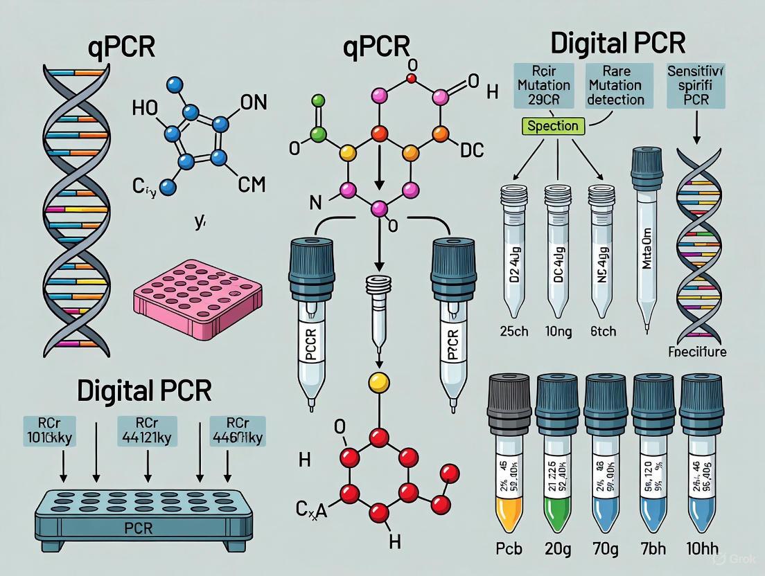
dPCR vs qPCR: Choosing the Optimal Tool for Rare Mutation Detection in Cancer
This article provides a comprehensive comparison of Quantitative Real-Time PCR (qPCR) and Digital PCR (dPCR) for detecting rare mutations in cancer research and diagnostics.
dPCR vs qPCR: Choosing the Optimal Tool for Rare Mutation Detection in Cancer
Abstract
This article provides a comprehensive comparison of Quantitative Real-Time PCR (qPCR) and Digital PCR (dPCR) for detecting rare mutations in cancer research and diagnostics. Tailored for researchers and drug development professionals, it explores the foundational principles of both technologies, delves into specific methodological applications like liquid biopsy analysis, offers troubleshooting and optimization strategies, and presents a direct performance comparison. The synthesis of current data underscores dPCR's superior sensitivity for detecting variants at frequencies as low as 0.1%, while also acknowledging the enduring role of qPCR in high-throughput, cost-effective workflows, ultimately guiding informed platform selection for precision oncology.
Understanding the Core Technologies: The Principles of qPCR and dPCR
Real-time quantitative PCR (qPCR) is a fundamental technique in molecular biology that enables researchers to monitor the amplification of DNA as it occurs during the polymerase chain reaction. Unlike conventional PCR that provides endpoint detection, qPCR allows for the quantification of specific DNA sequences throughout the amplification process through the detection of fluorescent signals [1] [2]. This real-time monitoring capability has revolutionized approaches to DNA and RNA quantitation, providing both quantitative and qualitative data in applications ranging from gene expression analysis to pathogen detection [3] [1].
In the context of cancer research, particularly for detecting rare mutations such as KRASG12C in non-small cell lung cancer (NSCLC), the precision and accuracy of qPCR becomes critically important [4]. While digital PCR (dPCR) has emerged as a more sensitive alternative for detecting low-abundance targets, understanding qPCR principles remains essential for researchers selecting appropriate methodologies for their specific experimental needs [3] [5]. This guide explores the fundamental principles of real-time fluorescence detection in qPCR and the application of standard curves for quantification, with particular emphasis on performance comparisons relevant to rare mutation detection in cancer research.
Principles of Real-Time Fluorescence Detection
The core principle of qPCR revolves around detecting and quantifying fluorescent signals that increase proportionally with the amount of amplified DNA product. Two primary chemistries enable this detection: TaqMan probe-based chemistry and SYBR Green I dye chemistry [1] [2].
TaqMan Chemistry
The TaqMan system utilizes a fluorogenic probe that specifically hybridizes to the target DNA sequence between the forward and reverse primers [1] [2]. The mechanism operates as follows:
Probe Design: An oligonucleotide probe is constructed with a reporter fluorescent dye at the 5' end and a quencher dye at the 3' end. While the probe remains intact, the proximity of the quencher dye suppresses reporter fluorescence through fluorescence resonance energy transfer (FRET) [1] [2].
Probe Hydrolysis: During PCR amplification, when the target sequence is present, the TaqMan probe anneals to the specific complementary sequence. The 5' nuclease activity of Taq DNA polymerase cleaves the probe as the primer is extended, separating the reporter dye from the quencher dye [1] [2].
Signal Detection: This separation results in a measurable increase in fluorescence intensity from the reporter dye. With each subsequent amplification cycle, additional reporter dye molecules are released, generating a fluorescent signal proportional to the amount of amplified product [1] [2].
The TaqMan MGB (Minor Groove Binder) probe represents an advanced version that incorporates a non-fluorescent quencher and a minor groove binder at the 3' end, which increases the melting temperature (Tm) of shorter probes and improves discrimination between matched and mismatched probes—a particularly valuable feature for detecting single-nucleotide mutations in cancer research [1].
SYBR Green I Dye Chemistry
As an alternative to probe-based methods, SYBR Green I dye chemistry employs a different detection mechanism [1] [2]:
DNA Binding: The SYBR Green I dye binds to the minor groove of all double-stranded DNA molecules in the reaction.
Fluorescence Enhancement: When bound to double-stranded DNA, the dye exhibits significantly enhanced fluorescence compared to its unbound state.
Signal Accumulation: As PCR amplification progresses, more double-stranded DNA amplicons are generated, allowing more dye molecules to bind and resulting in increased fluorescence proportional to the total DNA concentration.
A critical limitation of SYBR Green chemistry is its non-specific binding to any double-stranded DNA, including non-specific PCR products, which may lead to false positive signals without careful reaction optimization [1].
The Standard Curve Method for Absolute Quantification
Fundamentals of Standard Curve Quantification
The standard curve method represents one approach for absolute quantification in qPCR, allowing researchers to determine the exact quantity of target nucleic acid in unknown samples [6] [7]. This method relies on preparing a dilution series of standards with known concentrations to establish a reference curve that correlates cycle threshold (Ct) values with initial template quantities [8] [6].
The quantification process involves several key steps. First, a standard curve is prepared using serial dilutions (typically 5-fold or 10-fold) of known template concentrations [8]. Each dilution is then amplified via qPCR, during which the fluorescence is monitored in real-time to determine the Ct value for each standard [6]. Next, a semi-logarithmic graph is plotted with the Ct values on the Y-axis and the logarithm of the initial template quantities on the X-axis, generating a standard curve with data fitted to a straight line [8]. The correlation coefficient (R²) for this line should be 0.99 or greater to ensure assay precision [8]. Finally, unknown samples are amplified under identical conditions, and their quantities are determined by interpolating their Ct values from the standard curve [6] [7].
Experimental Protocol for Standard Curve Setup
Implementing the standard curve method requires careful experimental setup and execution [8] [7]:
Standard Preparation: Create five or more serial dilutions (2-fold, 5-fold, or 10-fold) of cDNA or DNA template with known high abundance of the target gene. The absolute quantities of these standards must first be determined by independent methods, such as spectrophotometric measurement (A260) followed by conversion to copy number using molecular weight [7].
qPCR Amplification: Perform real-time PCR reactions using each serial dilution in separate reactions. The threshold cycle (Ct) values are determined for each dilution, representing the cycle number at which fluorescence crosses a predetermined threshold [8] [1].
Standard Curve Generation: Plot the Ct values against the logarithm of the initial template concentration. The resulting standard curve should display a strong linear relationship (R² ≥ 0.99) [8].
Sample Quantification: Amplify unknown samples using the same reaction conditions and determine their concentrations by comparing their Ct values to the standard curve [6] [7].
For gene expression studies, this process must be performed separately for both the target gene and an appropriate endogenous control (housekeeping gene). The relative expression levels are then determined by normalizing the target gene quantity to the endogenous reference in each sample [8].
qPCR Performance in Rare Mutation Detection
Sensitivity Limitations for Low-Abundance Targets
In the context of cancer research, detecting rare mutations presents significant challenges for qPCR technology. The sensitivity of qPCR is limited by its reliance on standard curves and its inability to detect targets present at very low frequencies in complex mixtures [3]. While qPCR remains the gold standard for many applications including gene expression analysis and pathogen detection when targets are present in sufficient quantities, its effectiveness diminishes when targeting rare mutations such as cancer-associated variants in circulating tumor DNA (ctDNA) [3].
The table below summarizes key performance characteristics of qPCR compared to digital PCR for mutation detection:
Table 1: Performance Comparison Between qPCR and dPCR for Rare Mutation Detection
| Parameter | qPCR | Digital PCR |
|---|---|---|
| Quantification Method | Relative or absolute using standard curves [3] [7] | Absolute without standard curves [3] [7] |
| Sensitivity for Rare Mutations | Moderate [3] | High (0.10% mutation fraction or better) [9] |
| Precision | Dependent on standard curve quality [8] | High precision determined by number of partitions [3] [7] |
| Dynamic Range | 5-6 logs [6] | 5 logs with linear response [7] |
| Ability to Detect Without Preamplification | Limited for low-abundance targets [9] | Direct detection of 3 mutant molecules in 1 ng DNA [9] |
| Tolerance to PCR Inhibitors | Moderate | High [7] |
Experimental Data on Detection Limits
Meta-analyses comparing detection platforms have demonstrated the sensitivity limitations of qPCR for liquid biopsy applications. One comprehensive analysis of circulating tumor HPV DNA (ctHPVDNA) detection across multiple cancer types found that qPCR showed significantly lower sensitivity compared to both droplet digital PCR (ddPCR) and next-generation sequencing (NGS) when detecting viral DNA in blood samples [5]. This performance gap is particularly pronounced in applications requiring detection of rare mutations in ctDNA, where the mutant DNA molecules represent only a small fraction (<1%) of the total cell-free DNA [9].
In a study focused on detecting cancer mutations directly from circulating DNA, researchers developed a single-color digital PCR approach that could detect as few as three mutation-bearing DNA molecules in a single reaction without preamplification, achieving a sensitivity of 0.10% for BRAF V600E and KRAS G12D mutations [9]. This level of sensitivity is challenging to achieve with standard qPCR due to its reliance on standard curves and limited ability to detect minute differences in initial template concentration [3].
The Scientist's Toolkit: Essential Research Reagents
Successful implementation of qPCR assays, particularly for challenging applications like rare mutation detection, requires careful selection of reagents and optimization. The following table outlines essential research reagent solutions for qPCR-based mutation detection:
Table 2: Essential Research Reagents for qPCR Mutation Detection Assays
| Reagent/Category | Specific Examples | Function in qPCR Assay |
|---|---|---|
| Polymerase Enzyme | AmpliTaq Gold DNA Polymerase [1] [2] | Engineered for hot-start activation to reduce non-specific amplification |
| Detection Chemistry | TaqMan MGB Probes [1] | Enhanced allele discrimination for single-nucleotide variants |
| Reference Dyes | ROX, PASSIVE REFERENCE dye [1] [2] | Internal reference for signal normalization across wells |
| Standard Materials | Plasmid DNA, in vitro transcribed RNA [7] | Known concentration standards for curve generation |
| Sample Preservation | EDTA blood collection tubes, LoBind tubes [9] | Prevent degradation of labile circulating tumor DNA |
| Nucleic Acid Isolation | Maxwell 16 Circulating DNA Plasma Kit [9] | Optimized recovery of fragmented cell-free DNA |
qPCR technology, with its real-time fluorescence detection capabilities and standardized quantification methods, remains a powerful tool in molecular biology research. The standard curve method provides a reliable approach for absolute quantification when properly implemented with appropriate controls and validation. However, for emerging applications in cancer research—particularly detection of rare mutations in circulating tumor DNA—the sensitivity limitations of qPCR have become increasingly apparent [3] [5].
While qPCR offers advantages in throughput, established workflows, and cost-effectiveness for many applications, researchers targeting low-abundance mutations (<1% allele frequency) should consider more sensitive technologies like digital PCR [3] [9]. The selection between these platforms ultimately depends on the specific research requirements, with qPCR maintaining its position as a versatile workhorse for routine quantification, and digital PCR emerging as the preferred technology for challenging detection scenarios where maximum sensitivity and precision are paramount.
The detection of rare mutations, such as those found in circulating tumor DNA (ctDNA), represents one of the most significant challenges in modern cancer research and liquid biopsy development. For years, quantitative PCR (qPCR) has served as the cornerstone for nucleic acid analysis, providing reliable relative quantification for a wide range of applications. However, the emergence of digital PCR (dPCR) has revolutionized the field by enabling absolute quantification through sample partitioning—a methodological shift that is particularly transformative for detecting minute genetic alterations in complex biological samples [3] [10].
This paradigm shift from relative to absolute quantification is redefining the possibilities in cancer research, especially for monitoring minimal residual disease, assessing treatment response, and detecting emerging resistance mutations. While qPCR relies on standard curves and reference genes, which introduce variability and limit precision for low-abundance targets, dPCR's partitioning approach allows direct molecular counting without calibration curves [3]. This technical comparison examines how dPCR's fundamental architecture—splitting samples into thousands of individual partitions—confers critical advantages for rare mutation detection in oncology applications, supported by experimental data and detailed methodological insights.
Core Technological Differences: Partitioning as a Game Changer
Fundamental Principles and Mechanisms
The primary distinction between these PCR technologies lies in their approach to quantification. Quantitative PCR (qPCR) operates through amplification monitoring in real-time using fluorescent probes or DNA-binding dyes, with quantification based on the cycle threshold (Ct) at which fluorescence crosses a detection threshold. This measurement is always relative to standard curves or reference genes, making it susceptible to amplification efficiency variations and inhibitor effects [3] [10].
Digital PCR (dPCR) employs a fundamentally different strategy based on sample partitioning. The reaction mixture is divided into thousands (or millions) of individual partitions, each containing zero, one, or a few target DNA molecules. Following end-point amplification, each partition is analyzed as positive or negative for the target sequence [3]. The absolute concentration of the target molecule is then calculated directly using Poisson statistics, eliminating the need for standard curves and providing unprecedented precision for rare allele detection [11] [10].
Comparative Performance Analysis
Experimental data from recent studies demonstrates clear performance differences between these technologies, particularly for challenging applications like rare mutation detection. The table below summarizes key comparative characteristics:
Table 1: Performance Comparison Between qPCR and dPCR for Rare Mutation Detection
| Performance Characteristic | Quantitative PCR (qPCR) | Digital PCR (dPCR) |
|---|---|---|
| Quantification Approach | Relative (requires standard curve) | Absolute (direct molecular counting) [3] [10] |
| Sensitivity for Rare Targets | Limited, typically >1% variant allele frequency (VAF) | Exceptional, can detect <0.1% VAF [10] |
| Precision at Low Concentrations | Moderate to poor | Excellent (Poisson distribution-based) [11] |
| Impact of PCR Inhibitors | Significant reduction in efficiency | Reduced due to partitioning effect [10] |
| Dynamic Range | Wide (6-7 orders of magnitude) | Narrower but optimal for low abundance targets [10] |
| Throughput | High (96- or 384-well plates) | Lower but improving with newer systems [3] [10] |
| Cost Per Reaction | Lower | Higher [10] |
A 2025 study comparing dPCR and Real-Time RT-PCR for respiratory virus detection provided compelling evidence of dPCR's superior accuracy, particularly for medium and high viral loads [12]. The research found that dPCR demonstrated greater consistency and precision than Real-Time RT-PCR, especially in quantifying intermediate viral levels—a performance advantage that directly translates to rare mutation detection in oncology contexts [12].
Experimental Evidence: dPCR in Action for Rare Mutation Detection
Methodological Framework and Protocols
The superior performance of dPCR for rare mutation detection stems from its unique workflow, which incorporates partitioning at the core of its design:
Diagram 1: dPCR Workflow for Rare Mutation Detection
The critical partitioning step varies by platform technology. Droplet Digital PCR (ddPCR) generates thousands of nanoliter-sized droplets in a water-in-oil emulsion [12], while Crystal Digital PCR employs 2D arrays of monodisperse droplets that self-assemble into periodic arrangements [13]. Nanowell-based systems (e.g., QIAcuity) use fixed microfluidic chambers for partitioning [12]. Despite implementation differences, all platforms share the common principle of limiting-dilution partitioning followed by end-point amplification and binary detection.
Key Research Reagent Solutions
Successful implementation of dPCR for rare mutation detection requires carefully selected reagents and systems. The following table outlines essential components:
Table 2: Essential Research Reagents and Platforms for dPCR-Based Rare Mutation Detection
| Reagent/Platform | Function | Application Notes |
|---|---|---|
| Partitioning Oil/Stabilizers | Forms discrete reaction compartments | Platform-specific formulations critical for partition integrity [13] |
| Mutation-Specific Probes | Detects wild-type vs. mutant sequences | FAM/HEX/VIC labeling common; competing probes for allele discrimination |
| Multiplex PCR Master Mix | Amplification in partitions | Optimized for endpoint PCR; resistant to inhibition |
| Reference Assays | Quality control and normalization | Copy number variation reference or sample quality metrics |
| QIAcuity System | Nanowell-based dPCR platform | Integrated partitioning and thermal cycling [12] |
| Naica System | Crystal digital PCR platform | Three-color multiplexing capability [13] |
| QuantStudio Absolute Q | Chip-based dPCR system | Fully integrated workflow [3] |
Multiplexing Strategies for Complex Mutation Panels
Advanced dPCR systems enable sophisticated multiplexing approaches that are particularly valuable for cancer panels targeting multiple mutations simultaneously. The Naica System, for example, employs three-color detection, allowing each target to be identified by unique fluorescence signatures [13]. Even more impressively, the Nio Digital PCR system can detect up to 21 targets in a single reaction through its 7-color channels and color combination strategies, where a single target is identified using two separate fluorophores detected in different channels [14].
This multiplexing capability is exemplified by the development of an 18-plex ESR1 (17 mutations) Crystal Digital PCR Assay, which enables comprehensive profiling of endocrine resistance mutations in breast cancer from limited liquid biopsy samples [14]. Such multiplexed approaches provide unprecedented comprehensive mutation screening from minimal input material, a crucial advantage for clinical trial monitoring and translational research.
Comparative Experimental Data: qPCR vs. dPCR Performance
Sensitivity and Precision Metrics
Direct comparative studies demonstrate dPCR's superior performance for low-abundance targets. In a methodological study focusing on Lacticaseibacillus casei detection, ddPCR showed lower detection limits in both pure culture and milk samples compared to real-time PCR, with researchers noting this advantage would be particularly valuable for samples containing low concentrations of target DNA or PCR inhibitors [11].
A comprehensive 2025 respiratory virus study providing direct comparative data found that dPCR "demonstrated superior accuracy, particularly for high viral loads of influenza A, influenza B, and SARS-CoV-2, and for medium loads of RSV" [12]. The researchers further reported that dPCR "showed greater consistency and precision than Real-Time RT-PCR, especially in quantifying intermediate viral levels" [12]. While this study focused on virology, the technical principles directly translate to rare mutation detection in oncology, where precise quantification at intermediate and low concentrations is critical for clinical decision-making.
Tolerance to PCR Inhibitors
The partitioning approach of dPCR provides inherent resistance to PCR inhibitors present in complex biological samples—a significant advantage for liquid biopsy applications where sample quality varies considerably. As noted in comparative assessments, "qPCR can be affected by PCR inhibitors, such as contaminants in environmental, forensic, or clinical samples," while "dPCR, by partitioning samples into individual micro-reactions, reduces the impact of inhibitors, improving robustness for complex or partially purified samples" [10].
This tolerance stems from the "dilution" of inhibitors across thousands of partitions, with many partitions containing no inhibitors and thus amplifying efficiently even when other partitions show suppression. This compartmentalization makes dPCR particularly valuable for challenging clinical samples such as formalin-fixed paraffin-embedded (FFPE) tissues, plasma ctDNA, and fine-needle aspirates where conventional qPCR might fail or provide unreliable quantification.
Implementation Considerations for Cancer Research
Strategic Technology Selection
Choosing between qPCR and dPCR requires careful consideration of research objectives, sample characteristics, and resource constraints. The following decision framework illustrates the optimal application domains for each technology:
Diagram 2: Technology Selection Framework for Cancer Research Applications
For large-scale screening studies where relative quantification suffices and target abundance is moderate, qPCR remains the preferred option due to its lower cost and higher throughput [3] [10]. However, for rare mutation detection, absolute quantification, and challenging samples, dPCR's partitioning advantage proves decisive. Many research groups adopt a hybrid approach, using qPCR for initial screening followed by dPCR confirmation for borderline or critical samples [10].
Economic and Workflow Considerations
While dPCR provides superior performance for specific applications, implementation barriers exist. Digital PCR instruments typically range from $70,000 to $150,000 for entry-level platforms, with advanced systems exceeding $200,000—significantly higher than qPCR systems at $15,000 to $50,000 [10]. Per-reaction costs are also higher for dPCR ($5-10) compared to qPCR ($1-3) [10]. Additionally, dPCR systems generally offer lower throughput, processing fewer samples per run [3] [10].
These economic factors must be balanced against the scientific value of obtaining reliable absolute quantification for rare mutations. For drug development programs where decisions hinge on detecting minimal residual disease or emerging resistance mutations, the additional cost and reduced throughput may be justified by the quality and reliability of the resulting data.
Digital PCR's partitioning approach represents a fundamental advancement in nucleic acid quantification, particularly for the challenging domain of rare mutation detection in cancer research. By enabling absolute quantification through sample division into thousands of individual reactions, dPCR overcomes critical limitations of qPCR related to standard curve dependence, sensitivity constraints, and inhibitor susceptibility. While qPCR remains the more practical choice for high-throughput applications where extreme sensitivity is not required, dPCR's revolutionary partitioning methodology provides cancer researchers with an indispensable tool for liquid biopsy analysis, treatment response monitoring, and rare mutation detection—ultimately accelerating precision oncology and therapeutic development.
As dPCR technology continues to evolve with improved multiplexing capabilities, reduced costs, and enhanced workflows, its adoption in cancer research laboratories will likely expand, further solidifying the partitioning revolution as a transformative paradigm in molecular quantification.
In the field of cancer research, the detection of rare mutations is critical for advancing our understanding of tumor heterogeneity, monitoring minimal residual disease (MRD), and developing targeted therapeutic strategies. The choice of molecular tool for this task profoundly impacts the reliability, accuracy, and clinical relevance of the findings. This guide provides an objective, data-driven comparison between quantitative PCR (qPCR) and digital PCR (dPCR), focusing on the key performance metrics of sensitivity, specificity, and limit of detection (LoD) within the context of rare mutation detection in oncology.
Table 1: Key Performance Metrics for qPCR vs. dPCR in Rare Mutation Detection
| Metric | qPCR (Real-time PCR) | Digital PCR (dPCR) |
|---|---|---|
| Quantification Method | Relative (requires standard curve) [15] | Absolute (no standard curve required) [15] [16] |
| Theoretical Sensitivity for Mutation Detection | >1% mutant allele frequency [15] | ≥ 0.1% mutant allele frequency [15], down to 0.001% in optimized applications [16] |
| Specificity | High, but can be affected by primer/probe design and reaction efficiency | Enhanced specificity, particularly with advanced chemistries (e.g., LNA probes) [17] |
| Limit of Detection (LoD) | Higher LoD, constrained by the dynamic range of the standard curve [18] | Lower LoD, capable of detecting rare targets in a wild-type background [16] [18] |
| Precision & Reproducibility | Well-established protocols; precision can be influenced by standard curve quality [15] | Higher precision and reproducibility across different laboratories [15] [12] |
| Tolerance to PCR Inhibitors | Prone to inhibition, which affects amplification efficiency [15] [19] | High tolerance due to sample partitioning [15] [19] |
| Ideal Application in Cancer Research | Gene expression analysis, initial pathogen detection, microarray validation [15] | Copy number variation, rare mutation detection (e.g., in ctDNA), MRD monitoring [15] [16] [17] |
Experimental Protocols for Performance Validation
The superior sensitivity and specificity of dPCR are demonstrated through specific experimental designs. The following protocols outline the methods used to generate the comparative data.
Protocol for Rare Mutation Detection using dPCR
This protocol is adapted from studies on detecting somatic mutations in hematologic malignancies, such as the JAK2V617F mutation [16].
- Sample Preparation: Extract genomic DNA from patient peripheral blood or biopsy samples. For circulating tumor DNA (ctDNA) analysis, isolate cell-free DNA from blood plasma [17].
- Assay Design: Utilize hydrolysis probe-based assays (e.g., TaqMan). For superior specificity, employ Locked Nucleic Acid (LNA) probes, which increase the binding affinity and specificity for discriminating single-nucleotide variants [17].
- Partitioning and PCR Amplification:
- For Droplet Digital PCR (ddPCR): Mix the DNA sample with the PCR master mix and probes. Generate thousands of nanoliter-sized water-in-oil emulsion droplets, effectively partitioning the sample so that each droplet contains zero, one, or a few target molecules [16].
- For Nanoplated-based dPCR: Load the reaction mixture into a nanoplate containing fixed partitions [15].
- Perform endpoint PCR amplification on the partitioned samples.
- Data Analysis: After amplification, analyze each partition for fluorescence. Partitions are scored as positive (mutant), positive (wild-type), or negative. The absolute concentration of the mutant allele (in copies/μL) and the mutant allele frequency are calculated directly using Poisson statistics, without the need for a standard curve [16].
Protocol for Determining Limit of Detection (LoD) and Limit of Blank (LoB)
This methodology is crucial for validating any dPCR assay for clinical research applications [16].
- Limit of Blank (LoB): Prepare multiple replicates of a blank sample that is known to contain no target mutant sequences. Analyze these replicates via dPCR. The LoB is defined as the highest apparent target concentration measured in these blank samples [16].
- Limit of Detection (LoD): Prepare samples with known, low concentrations of the mutant allele in a background of wild-type DNA. The LoD is the lowest target concentration at which the mutation can be reliably distinguished from the LoB. For a well-optimized dPCR JAK2V617F assay, sensitivities of 0.01% have been achieved, which is half a log higher than qPCR [16].
- Limit of Quantification (LoQ): This is the lowest concentration of the mutant allele at which the assay can not only detect but also provide a precise and accurate quantitative measurement [16].
Technology Workflow Comparison
The core difference between qPCR and dPCR lies in the workflow, which directly impacts their performance metrics.
Research Reagent Solutions for dPCR in Cancer Research
Transitioning to a dPCR workflow, especially for sensitive applications like liquid biopsy, requires specific reagents and tools optimized for the technology.
Table 2: Essential Reagents and Materials for dPCR Mutation Detection
| Item | Function | Example in Cancer Research |
|---|---|---|
| dPCR LNA Mutation Assays | Probe-based assays enhanced with Locked Nucleic Acid (LNA) technology to improve specificity and sensitivity for discriminating single-nucleotide variants. | Over 200 predesigned assays are available for detecting oncogenic mutations in genes like KRAS, NRAS, and BRAF with sensitivity as fine as 0.1% [17]. |
| dPCR Instrument/System | The platform for performing partitioning, thermocycling, and imaging. | Nanoplate-based systems (e.g., QIAcuity) or droplet-based systems (e.g., QX200 ddPCR System). The choice depends on required throughput, multiplexing capability, and workflow preferences [15] [16]. |
| Nanoplates or Cartridges | Disposable consumables that hold the sample and enable partitioning. | 96- or 24-well nanoplates with fixed partitions prevent coalescence and variation, contributing to more reproducible results [15] [17]. |
| dPCR Master Mix | A optimized chemical mixture containing DNA polymerase, dNTPs, and buffers tailored for efficient amplification in partitioned reactions. | Commercial master mixes are formulated to work with specific dPCR platforms and assay chemistries (e.g., probe-based vs. EVAGreen) [17]. |
| Reference Standards | Samples with known mutation concentrations. | Used for initial assay validation, determining LoD, LoQ, and for periodic quality control to ensure assay performance remains consistent over time [16]. |
The selection between qPCR and dPCR for detecting rare mutations in cancer research is application-dependent. qPCR remains a powerful, cost-effective tool for high-throughput applications where extreme sensitivity is not the primary requirement. In contrast, dPCR excels in scenarios demanding the highest levels of precision, absolute quantification, and sensitivity, such as monitoring minimal residual disease (MRD) and analyzing circulating tumor DNA (ctDNA) for low-frequency mutations [15] [16] [17]. The experimental data and workflows presented herein provide a framework for researchers to make an informed choice based on the specific demands of their oncological research.
The Critical Role of Rare Mutation Detection in Oncology
The precise detection of rare mutations is a cornerstone of modern oncology, directly influencing capabilities in early cancer diagnosis, minimal residual disease monitoring, and tracking therapy-resistant clones. The emergence of liquid biopsies—analyzing circulating tumor DNA (ctDNA) shed into the bloodstream—has intensified the need for technologies capable of finding needle-in-a-haystack genetic alterations amidst an abundant background of wild-type DNA [20] [21]. Among molecular tools, Quantitative PCR (qPCR) and Digital PCR (dPCR) have emerged as pivotal technologies for these applications. While qPCR has served as the long-standing gold standard, dPCR represents a more recent innovation that partitions samples into thousands of individual reactions to enable absolute target quantification without standard curves [3]. This guide provides an objective comparison of their performance characteristics, supported by experimental data, to inform researchers and drug development professionals in selecting the optimal methodology for their specific applications in cancer research.
Technology Comparison: qPCR vs. dPCR
Fundamental Principles and Workflows
Quantitative PCR (qPCR), also known as real-time PCR, is a high-throughput technique that measures DNA amplification as it occurs during the exponential phase of the reaction. It relies on fluorescent dyes or probes to detect amplification, providing either relative or absolute quantification contingent upon the use of standard curves prepared from known DNA concentrations [3]. For rare mutation detection, specialized qPCR chemistries like allele-specific primers coupled with wild-type blocking oligonucleotides are often employed to enhance specificity and suppress amplification of the dominant wild-type sequence [22].
Digital PCR (dPCR) takes a fundamentally different approach by partitioning a single PCR reaction into thousands (or millions) of nanoliter-scale reactions, such that each partition contains either zero, one, or a few target DNA molecules. Following end-point amplification, the fraction of positive partitions is counted, and the absolute concentration of the target sequence is calculated using Poisson statistics, eliminating the need for standard curves [3] [23]. This partitioning effectively enriches the target of interest, making dPCR particularly suited for detecting rare mutations in a high-background of wild-type DNA [21].
The workflow differences between these technologies for rare mutation detection are illustrated below:
Performance Comparison and Experimental Data
Direct comparative studies reveal distinct performance characteristics between qPCR and dPCR, particularly for challenging samples with low target abundance or the presence of inhibitors.
Table 1: Quantitative Performance Comparison for Rare Mutation Detection
| Parameter | qPCR | Digital PCR | Experimental Context |
|---|---|---|---|
| Detection Sensitivity | ~5-10% mutant allele fraction [22] | As low as 0.1% mutant allele frequency [21] | PIK3CA mutation detection in genomic DNA [22] |
| Absolute Quantification | Requires standard curves [3] | Yes, without standard curves [3] [23] | Fundamental methodological difference |
| Reproducibility (CV for low templates) | Mean CV ~126% [24] | Mean CV ~40% (p=0.01) [24] | HPV mRNA in serial dilutions of SiHa cells [24] |
| Tolerance to Inhibitors | Moderate - Cq values shift with inhibitors [25] | High - concentration results stable with inhibitors [25] | Synthetic DNA with RT reaction contaminants [25] |
| Theoretical LOD | Varies with assay design | 0.2 copies/µL (Naica System example) [26] | Instrument-specific partitioning efficiency |
| Sample Throughput | High [3] | Lower [27] | Operational characteristic |
A 2017 comparative study analyzing HPV mRNA in sentinel lymph nodes highlighted a significant advantage for dPCR in reproducibility, especially for low-template samples. The researchers found that dPCR demonstrated "a substantially reduced subsampling error," which they attributed to the ability to analyze larger cDNA amounts without inhibition from background nucleic acids [24]. This enhanced reproducibility is crucial for clinical applications where consistent results across samples and laboratories are paramount.
Another critical consideration is the effect of sample contaminants common in extracted nucleic acids. A 2017 study systematically evaluated this by spiking synthetic DNA with varying amounts of reverse transcription (RT) mix, a common source of PCR inhibitors. The research found that while qPCR reaction efficiency dropped from approximately 90% to 67% with increasing RT mix contamination (causing significant Cq shifts), ddPCR maintained consistent quantification despite the inhibitors [25]. This robustness to variable contamination makes dPCR particularly valuable for analyzing samples that cannot be purified to the same extent as standard preparations.
Experimental Protocols for Rare Mutation Detection
qPCR Protocol for PIK3CA Mutations
The following detailed protocol is adapted from a 2018 Scientific Reports publication that developed a robust real-time qPCR method for detecting the clinically relevant PIK3CA H1047R and E545K mutations in breast cancer [22].
Assay Design Principle: This method utilizes a mutation-specific primer with the variant base at its 3' end to preferentially amplify the mutant allele. Amplification of the wild-type sequence is suppressed by a non-productive, phosphate-modified oligonucleotide blocker that partially overlaps with the mutant-specific primer and has the variant base located approximately in its middle [22].
Step-by-Step Protocol:
- DNA Extraction: Extract genomic DNA from frozen tissue, FFPE samples, or cell lines using a standard silica-membrane or magnetic bead-based method. Preferentially use samples with DNA integrity numbers (DIN) >7 for optimal amplification.
- Reaction Setup: Prepare a master mix for each sample (or standard curve point) as follows. Reactions are typically performed in triplicate.
- 1X SYBR Green PCR Master Mix
- Mutation-specific forward primer: 400 nM
- Common reverse primer: 400 nM
- Wild-type blocking oligonucleotide: 800 nM
- Genomic DNA template: 2000 copies (recommended for 5% sensitivity)
- Nuclease-free water to a final volume of 20 µL
- qPCR Run Parameters:
- Initial Denaturation: 95°C for 10 minutes
- 45 Cycles of:
- Denaturation: 95°C for 15 seconds
- Annealing/Extension: 62°C for 1 minute (optimum temperature must be empirically determined)
- Melt Curve Analysis: 60°C to 95°C with continuous fluorescence reading.
- Data Analysis: The ∆Cq method is used. A separate, parallel reaction amplifying a non-mutated internal control sequence (e.g., a reference gene) from the same sample is required for normalization. The relative amplification of the mutant allele is calculated using the formula 2^(-∆Cq), where ∆Cq = Cqinternalcontrol - Cqmutantassay. A sample is considered positive if the relative amplification differs significantly from a wild-type genomic DNA control [22].
dPCR Protocol for EGFR T790M Mutation
This protocol, informed by a detailed tutorial from Stilla Technologies, outlines the detection of the EGFR T790M mutation, a key marker of resistance in non-small cell lung cancer (NSCLC) [26].
Assay Design Principle: A single set of primers amplifies the region encompassing the EGFR T790 locus. Two sequence-specific hydrolysis probes (TaqMan) are used for detection: one labeled with FAM targets the wild-type sequence, and another labeled with a different fluorophore (e.g., Cy3) targets the T790M mutant allele [26].
Step-by-Step Protocol:
- DNA Input Calculation: Critical for sensitivity. For human genomic DNA, use the formula: Number of copies = mass of DNA (in ng) / 0.003. This converts nanograms to haploid genome equivalents. For example, 10 ng of input DNA equals ~3,333 copies of the EGFR gene. The theoretical limit of detection is determined by the system's sensitivity and total target input [26].
- PCR Mix Preparation: Prepare a master mix on ice in a clean area to prevent contamination.
- 1X Digital PCR Master Mix
- Reference Dye (if required by the system)
- EGFR T790 Forward and Reverse Primers: 500 nM each
- EGFR T790 WT Probe (FAM-labeled): 250 nM
- EGFR T790M Probe (Cy3-labeled): 250 nM
- Human Genomic DNA: Calculated amount (e.g., 10 ng/µL)
- Nuclease-free water to the manufacturer's recommended volume (e.g., 25 µL)
- Partitioning and Amplification:
- Load the PCR mix into the designated dPCR consumable (chip, cartridge, etc.).
- Perform partitioning according to the instrument's protocol (e.g., droplet generation or microfluidic chamber loading).
- Seal the plate or cartridge and place it in the thermal cycler.
- Run the following cycling program (optimized for the Naica System):
- Initial Denaturation: 95°C for 10 minutes
- 45 Cycles of:
- Denaturation: 95°C for 30 seconds
- Annealing/Extension: 62°C for 15 seconds
- Final Hold: 12°C indefinitely [26]
- Data Acquisition and Analysis:
- After cycling, transfer partitions to a reader for fluorescence imaging (chip-based systems) or read droplets sequentially (flow-based systems).
- Apply fluorescence compensation to correct for spectral spillover between channels.
- The software automatically generates 1D or 2D amplitude plots, identifies positive/negative partitions for each target, and uses Poisson statistics to provide the absolute concentration (copies/µL) and variant allele frequency [26].
The Scientist's Toolkit: Essential Research Reagents
Successful rare mutation detection requires carefully selected reagents and controls. The following table details key solutions for setting up these critical experiments.
Table 2: Essential Research Reagent Solutions for Rare Mutation Detection
| Reagent / Solution | Function | Example Use Case |
|---|---|---|
| Mutation-Specific Primers & Probes | Specifically anneal to and detect the mutant allele sequence; critical for assay specificity. | Hydrolysis probes for EGFR T790M [26]; allele-specific primers with blocking oligos for PIK3CA [22]. |
| Wild-Type Blocking Oligonucleotide | Suppresses amplification of the wild-type allele by binding to it and preventing primer extension, enriching for mutant signal. | Phosphate-modified blocker in the PIK3CA qPCR assay [22]. |
| Digital PCR Master Mix | Contains DNA polymerase, dNTPs, buffer, and MgCl₂ optimized for efficient amplification within partitions. | Used with the QuantStudio Absolute Q system or Naica System [21] [26]. |
| Fluorescence Compensation Matrix | Corrects for spectral spillover between fluorophores in multiplexed dPCR experiments; essential for accurate cluster calling. | Required for duplex assays (e.g., FAM and Cy3) to prevent aberrant results [26]. |
| Reference Gene Assay | Amplifies a conserved, non-mutated genomic region; serves as an internal control for DNA quality and quantity. | Amplification of GUSB or ATP2B4 for copy number normalization in dPCR and qPCR [28]. |
| Partitioning Oil/Stabilizer | Creates stable, monodisperse water-in-oil emulsions for droplet-based dPCR; prevents droplet coalescence during thermal cycling. | Critical consumable for ddPCR systems like Bio-Rad's QX200 [23]. |
Application in Liquid Biopsies and Clinical Translation
The ability to detect rare mutations finds its most impactful application in the analysis of circulating tumor DNA (ctDNA) from liquid biopsies. ctDNA fragments are typically short and exist in very low concentrations in plasma, making the high sensitivity of dPCR an ideal tool for their study [21]. Characterizing ctDNA helps researchers detect cancer early, measure therapeutic response, quantify residual tumor burden, and monitor emerging resistance mutations [21]. Clinical validation studies have demonstrated high concordance (93-96%) between liquid biopsy results using technologies like dPCR or specialized qPCR and traditional tissue biopsies for key oncogenic drivers in EGFR, BRAF, and KRAS [20].
The relationship between mutation detection technology and clinical decision-making is summarized below:
Furthermore, dPCR has proven invaluable in emerging therapeutic fields like monitoring Chimeric Antigen Receptor (CAR) T-cell therapy. A 2023 study validated a high-sensitivity dPCR assay for tracking CAR T-cell constructs post-infusion, achieving a consistent detection limit of 0.001%. This enabled monitoring of early expansion and long-term persistence, with the highest CAR levels strongly correlating with the diagnosis of severe cytokine release syndrome (CRS) [28].
Both qPCR and dPCR are powerful technologies for rare mutation detection in oncology, each with distinct strengths. The choice between them should be guided by the specific requirements of the research or clinical application.
Choose qPCR when:
- Your project demands high-throughput processing of many samples quickly and cost-effectively [3].
- The expected mutant allele frequency is above 5% and ultra-high sensitivity is not critical [22].
- The research question involves relative quantification (e.g., comparing gene expression levels) rather than absolute copy number [3].
- The laboratory infrastructure and budget favor a established, widely available technology.
Choose dPCR when:
- The highest possible sensitivity and precision are required for detecting mutations below 1% allele frequency [21] [24].
- Absolute quantification without standard curves is necessary for rigorous biomarker studies [3] [23].
- The sample type is challenging (e.g., liquid biopsy ctDNA) or may contain variable inhibitors that could compromise qPCR efficiency [25].
- The application involves monitoring low-abundance targets over time, such as MRD or therapy resistance [21] [28].
The ongoing evolution of both technologies continues to push the boundaries of detection, enabling researchers to unravel tumor heterogeneity and improve cancer patient management through increasingly precise molecular diagnostics.
From Theory to Practice: Deploying qPCR and dPCR in Cancer Research
The analysis of circulating tumor DNA (ctDNA) from liquid biopsies represents a transformative approach for non-invasive cancer diagnosis, prognosis, and therapy monitoring. However, a significant challenge limits its widespread application: ctDNA fragments often constitute only a tiny fraction (as low as 0.01%) of the total cell-free DNA (cfDNA) in a patient's blood, against a high background of wild-type DNA [29] [30]. This necessitates detection technologies with exceptional sensitivity and precision. Within this context, the comparison between quantitative real-time PCR (qPCR) and digital PCR (dPCR) becomes critical for molecular researchers. This guide objectively compares the performance of these two technologies, demonstrating why dPCR has become the preferred method for rare mutation detection in ctDNA analysis.
Technology Face-Off: qPCR vs. dPCR
Fundamental Principles and Workflows
qPCR (Quantitative Real-Time PCR) is a well-established relative quantification method. It monitors PCR amplification in real-time using fluorescent reporters, with quantification based on the cycle threshold (Cq) at which the fluorescence signal crosses a predefined threshold. Its quantification is relative, requiring standard curves or reference samples for calibration [15].
dPCR (Digital PCR) is a third-generation PCR technology that enables absolute nucleic acid quantification without standard curves. It works by partitioning a PCR reaction into thousands to millions of individual nanoliter-scale reactions, so that each partition contains either 0, 1, or a few target molecules. Following end-point amplification, the fraction of positive partitions is counted, and the absolute target concentration is calculated using Poisson statistics [15] [23].
The workflow differences are visualized in the diagram below:
Performance Comparison for ctDNA Analysis
The table below summarizes the key performance characteristics of qPCR and dPCR relevant to ctDNA detection:
| Parameter | qPCR / Real-Time PCR | Digital PCR (dPCR) |
|---|---|---|
| Quantification Type | Relative (requires standard curve) [15] | Absolute (no standard curve needed) [15] [23] |
| Sensitivity (Mutation Detection) | Detects mutation rates >1% [15] | Detects mutation rates ≥ 0.1% [15] [21] |
| Tolerance to PCR Inhibitors | Lower (impacted by sample impurities) [15] | Higher (robust due to sample partitioning) [15] |
| Impact of PCR Efficiency Variations | Affected (data collected at exponential phase) [15] | Less affected (end-point detection) [15] |
| Precision & Reproducibility | Well-established protocols [15] | Higher precision for better cross-lab reproducibility [15] |
| Ideal Application in Liquid Biopsy | Broad dynamic range applications [15] | Rare mutation detection, low abundance targets [15] [21] |
Experimental Data: dPCR in Action
Case Study 1: Monitoring ctDNA Kinetics During Tumor Resection
A 2017 study established a highly sensitive nested qPCR method (PNB-qPCR) to quantify KRAS mutations in ctDNA from limited plasma samples during colorectal cancer surgery [31].
Experimental Protocol:
- Method: Pooled, Nested, WT-Blocking qPCR (PNB-qPCR).
- Target: Seven most frequent point mutations in KRAS exon 2.
- Sample: Blood plasma from patients undergoing tumor resection.
- Key Techniques: First-round PCR with wild-type blocking primers to enrich mutant fragments, followed by a second-round qPCR using mutation-specific ARMS primers and short LNA probes on pooled first-round products [31].
Results and dPCR Advantage: The study successfully tracked ctDNA kinetics, noting a surge post-resection. The method achieved a limit of detection (LOD) as low as 0.003% mutant allele frequency, requiring complex optimization to approach dPCR-level sensitivity [31]. This demonstrates that while advanced qPCR can detect rare mutations, dPCR offers a more straightforward and robust path to this ultra-high sensitivity.
Case Study 2: Multiplex Detection of Eight Cancers via Methylation Signatures
A 2024 study developed a multiplex droplet digital PCR (ddPCR) assay for multi-cancer detection using DNA methylation biomarkers [32].
Experimental Protocol:
- Method: Multiplex ddPCR.
- Targets: Three differentially methylated regions identified via bioinformatics analysis of public datasets (TCGA).
- Sample Type: 103 tumor and 109 normal adjacent fresh frozen tissues from eight cancer types (lung, breast, colorectal, prostate, pancreatic, head and neck, liver, and esophageal) [32].
- Workflow: DNA extraction → bisulfite conversion → multiplex ddPCR with two distinct assays → combined data analysis [32].
Results and dPCR Advantage: The combined ddPCR assay achieved a cross-validated area under the curve (cvAUC) of 0.948, with sensitivities of 53.8-100% and specificities of 80-100% across cancer types. The authors highlighted that combining targets in ddPCR drastically increased sensitivity and specificity compared to single-target approaches while lowering DNA input requirements [32]. This showcases dPCR's power for precise, multi-target quantification in complex biomarker panels.
The Scientist's Toolkit: Essential Reagents for dPCR-based ctDNA Analysis
| Item | Function | Example Application |
|---|---|---|
| dPCR Instrument | Partitions sample, performs thermocycling, and reads fluorescence. | Nanoplate-based systems (e.g., QIAcuity) or droplet-based systems (ddPCR) [15] [23]. |
| TaqMan Assays | Target-specific probes and primers for selective amplification. | Pre-designed assays for known somatic mutations (e.g., Absolute Q Liquid Biopsy dPCR assays) [21]. |
| DNA Bisulfite Conversion Kit | Converts unmethylated cytosines to uracils, allowing methylation detection. | Essential for preparing DNA for methylation-based cancer detection assays [32]. |
| Cell-Free DNA Extraction Kit | Isolves short-fragment cfDNA from blood plasma with high yield and purity. | Magnetic bead-based methods are efficient for recovering short cfDNA fragments [30]. |
| Microfluidic Array Plates/Consumables | Creates the nanoliter-scale partitions for the dPCR reaction. | Specific to the dPCR platform (e.g., nanoplates for QIAcuity) [15]. |
For the detection of rare mutations in ctDNA—a cornerstone of effective liquid biopsy applications—digital PCR presents a compelling advantage over traditional qPCR. Its ability to provide absolute quantification without standard curves, coupled with superior sensitivity (detecting down to 0.1% mutant allele frequency), robustness, and precision, makes it uniquely suited for this challenge [15] [21]. While qPCR remains a valuable tool for many applications, the experimental data confirms that dPCR is the more powerful technology for profiling cancer genomes non-invasively through liquid biopsies, enabling earlier cancer detection, improved therapy monitoring, and better patient outcomes.
The success of targeted cancer therapies is intrinsically linked to the accurate identification of specific driver mutations in genes such as KRAS, PIK3CA, and EGFR. These genetic alterations play pivotal roles in oncogenic signaling pathways and determine patient eligibility for precisely matched treatments. For researchers and drug development professionals, selecting the optimal molecular detection method is paramount, as it directly impacts the sensitivity, specificity, and ultimate success of both diagnostic protocols and therapeutic outcomes. This guide provides a objective comparison of two cornerstone technologies—quantitative PCR (qPCR) and digital PCR (dPCR)—framed within the broader thesis of their application in detecting rare mutations in cancer research. The ability to reliably identify low-frequency mutations is particularly critical for assessing tumor heterogeneity, monitoring minimal residual disease, and detecting the emergence of therapy-resistant clones before clinical progression becomes evident.
Performance Comparison of Detection Technologies
The choice between qPCR and dPCR involves balancing sensitivity, throughput, cost, and multiplexing capabilities. The following sections and tables summarize the key performance metrics and technical characteristics of each method.
Table 1: Analytical Performance Metrics for Mutation Detection
| Technology | Theoretical Sensitivity | Practical Sensitivity (Typical) | PIK3CA H1047R Detection | KRAS G12D Detection | EGFR T790M Detection |
|---|---|---|---|---|---|
| qPCR (ARMS) | ~1% [33] | 1-5% [33] [22] | ~5% mutant allele fraction [22] | Information Missing | 1% mutation rate (6,000 copy background) [33] |
| Digital PCR (ddPCR) | <0.1% [34] [26] | 0.1-1% [33] [9] [34] | Information Missing | 0.10% [9] | 0.1% mutation rate (avg. 6 mutant copies) [33] |
| NGS | Varies (1-5%) | ~2-6% [35] | Information Missing | Information Missing | Information Missing |
Table 2: Technical and Practical Characteristics
| Characteristic | qPCR (ARMS) | Digital PCR (ddPCR) | NGS |
|---|---|---|---|
| Quantification | Relative (Ct-based) | Absolute (molecule counting) | Relative (read count-based) |
| Multiplexing Capacity | Moderate | Moderate | High |
| Throughput | High | Medium | Varies (often lower) |
| Cost per Sample | Low | Medium | High |
| Primary Advantage | Cost-effective, widely available, fast | High sensitivity and precision for rare alleles | Comprehensive, untargeted mutation discovery |
| Key Disadvantage | Limited sensitivity for rare alleles | Higher cost, limited multiplexing | Cost, complexity, data analysis overhead |
A 2021 meta-analysis of KRAS mutation detection in colorectal cancer patients' cell-free DNA substantiates these comparisons, reporting a pooled sensitivity of 0.77 and specificity of 0.87 for the combined use of dPCR, ARMS, and NGS techniques [35]. This confirms the overall high accuracy of these methods in a clinically relevant context. Furthermore, a meta-analysis on circulating tumor HPV DNA found that the sensitivity of detection was significantly greater with NGS, followed by ddPCR, and then qPCR, while specificity was similarly high across platforms [5].
Experimental Data and Case Studies
Direct Comparison: EGFR T790M Detection
A seminal 2015 study directly compared Amplification Refractory Mutation System-based qPCR (ARMS-qPCR) and droplet digital PCR (ddPCR) for detecting the EGFR T790M resistance mutation. The ARMS-qPCR method stably detected mutations in plasmid samples with 5% and 1% mutation rates. In contrast, ddPCR reliably detected mutations at rates of 5%, 1%, 0.5%, and 0.1% (an average of just 6 mutant copies in a background of 6,000 wild-type copies) [33]. This demonstrates ddPCR's exceptional capability for rare allele detection. In clinical validation using 10 non-small cell lung cancer (NSCLC) samples, both methods yielded consistent results for 9 samples. Crucially, in one sample (N006) that was indicated as wild-type by ARMS-qPCR, ddPCR identified a clear T790M mutation with 7 copies of mutant alleles amid 6,000 wild-type copies [33]. This case highlights ddPCR's potential for early diagnosis of acquired resistance before it becomes clinically evident.
KRAS Mutation Detection in Colorectal Cancer
The high sensitivity of dPCR is also evident in KRAS mutation profiling. Studies have demonstrated the ability to detect and quantify the KRAS G12V mutation at a prevalence as low as 0.1% [34]. Furthermore, a single-color dPCR assay developed to detect KRAS G12D in cell-free DNA achieved a sensitivity of 0.10% using just 1 ng of non-amplified DNA input (approximately 300 genome equivalents) [9]. This highlights dPCR's utility in liquid biopsy applications where the target is scarce and of low abundance.
PIK3CA Mutation Analysis in Breast Cancer
For PIK3CA, a common oncogene in breast cancer, qPCR-based methods have been successfully developed and deployed. One study described a real-time qPCR assay for the H1047R mutation that demonstrated a detection limit of approximately 5% mutant allele fraction in genomic DNA from frozen biopsies, FFPE material, and cell lines [22]. Another study focusing on the E545K mutation reported a slightly lower sensitivity of around 10% [22]. This illustrates that while standard qPCR is capable of detecting PIK3CA mutations, its sensitivity is substantially lower than that of dPCR, a critical factor when analyzing heterogeneous tumor samples or liquid biopsies.
Detailed Experimental Protocols
Principle: The sample is partitioned into thousands of nanoliter-sized droplets, and end-point PCR is performed in each droplet. Fluorescence analysis of the droplets allows for absolute quantification of both mutant and wild-type DNA molecules.
Protocol:
- Reaction Mix Preparation: Assemble a 25 µL reaction containing:
- 12.5 µL of 2X ddPCR Master Mix
- 1.25 µL of 20X primer and TaqMan probe mix (e.g., FAM-labeled for wild-type, HEX/VIC-labeled for T790M mutant)
- 2.5 µL of DNA template (optimize input amount, e.g., 10-100 ng)
- Nuclease-free water to 25 µL
- Droplet Generation: Load the reaction mix into a droplet generator cartridge along with droplet generation oil. The instrument partitions the sample into ~20,000 droplets.
- PCR Amplification: Transfer the emulsified sample to a 96-well PCR plate, seal, and run on a thermal cycler using the following program:
- Hold: 95°C for 10 minutes (enzyme activation)
- Cycle (40x): 94°C for 30 seconds (denaturation) and 58°C for 1 minute (annealing/extension)
- Hold: 98°C for 10 minutes (enzyme deactivation) and 4°C hold.
- Droplet Reading and Analysis: Place the plate in a droplet reader, which counts the fluorescent-positive and negative droplets for each channel. The software applies Poisson statistics to calculate the absolute concentration (copies/µL) of mutant and wild-type DNA in the original sample.
Principle: This method uses a mutation-specific primer with the variant base at its 3' end to preferentially amplify the mutant allele. A phosphate-modified oligonucleotide (blocker) complementary to the wild-type sequence is included to further suppress wild-type amplification.
Protocol:
- Reaction Mix Preparation: For a 20 µL reaction, combine:
- 1X PCR buffer (e.g., 65 mM Tris-HCl, pH 8.9, 3 mM MgSO4)
- 0.2 mM dNTPs
- 600 nM of forward and H1047R mutant-specific reverse primers
- A wild-type blocking oligonucleotide (e.g., 100-200 nM)
- 100 nM fluorescent hydrolysis probe (e.g., TaqMan)
- 1 U of Taq polymerase
- DNA template (e.g., 50-100 ng)
- PCR Amplification: Run the reaction on a real-time PCR instrument with the following cycling conditions:
- Hold: 95°C for 5 minutes
- Cycle (40-45x): 95°C for 15 seconds and 60°C for 1 minute.
- Data Analysis: The cycle threshold (Ct) for the mutant-specific amplification is determined. A sample is considered positive if its amplification signal is significantly earlier (lower Ct) than that of a wild-type control sample. The ΔCt value between the mutant reaction and an internal control reaction is used for interpretation.
Technology Selection Workflow
The following diagram illustrates the decision-making process for selecting the appropriate mutation detection technology based on key experimental requirements.
The Scientist's Toolkit: Essential Research Reagent Solutions
Successful mutation detection requires careful selection of reagents and tools. The following table lists key components and their functions.
Table 3: Essential Reagents and Materials for PCR-Based Mutation Detection
| Item | Function/Description | Example Use Case |
|---|---|---|
| TaqMan SNP Genotyping Assays | Wet lab-validated assays for specific somatic mutations (e.g., in EGFR, BRAF, KRAS). Include primers and probes for both wild-type and mutant alleles [34]. | Detecting a panel of known hotspot mutations with high specificity on dPCR or qPCR platforms. |
| Digital PCR System | Instrumentation for partitioning samples and reading droplets/chips (e.g., Bio-Rad QX200, Thermo Fisher QuantStudio 3D, Stilla Technologies Naica) [33] [34] [26]. | Absolute quantification of rare mutant alleles in FFPE DNA or cell-free DNA from plasma. |
| cfDNA Extraction Kit | Optimized kits for purifying short-fragment, low-concentration cell-free DNA from blood plasma (e.g., QIAamp Circulating Nucleic Acid Kit) [36]. | Preparing analyte for liquid biopsy-based mutation detection and monitoring. |
| Phosphoryl Guanidine (PG)-Modified Primers | Novel modified primers that increase discrimination between wild-type and mutated DNA in allele-specific PCR, improving specificity with unfavorable mismatches [37]. | Enhancing the performance of qPCR assays for challenging point mutations like PIK3CA E542K and E545K. |
| NGS Solid Cancer Panels | Targeted sequencing panels covering hundreds of mutations in cancer-related genes (e.g., Sysmex Plasma-SeqSensei panel) [36]. | Comprehensive, tumor-agnostic profiling of ctDNA for mutation discovery and monitoring. |
The selection between qPCR and dPCR for detecting KRAS, PIK3CA, and EGFR mutations is not a matter of one technology being universally superior, but rather of matching the tool to the specific research question. qPCR remains a powerful, cost-effective workhorse for detecting mutations present at moderate-to-high allele frequencies (≥1-5%), especially in settings with limited resources. In contrast, dPCR provides an unparalleled level of sensitivity and precision for detecting rare mutations (down to <0.1%), making it the preferred choice for applications like monitoring residual disease, studying tumor heterogeneity, and early detection of resistance mutations in liquid biopsies. The emergence of modified primers and optimized assay chemistries continues to push the performance boundaries of both platforms. Researchers must weigh the critical factors of required sensitivity, need for absolute quantification, budget, and sample type to make an informed decision that ensures the reliability and clinical relevance of their findings in the era of precision oncology.
Monitoring Minimal Residual Disease (MRD) and Treatment Response
Minimal Residual Disease (MRD) refers to the small number of cancer cells that persist in patients after treatment who have achieved clinical remission [38]. These residual cells are a latent reservoir of disease that can lead to relapse if not properly addressed. Accurate MRD detection provides critical prognostic information, guides treatment decisions, and enables risk stratification, ultimately improving long-term survival outcomes [38]. The emergence of liquid biopsy, which analyzes circulating tumor DNA (ctDNA) from blood samples, has revolutionized MRD monitoring by offering a minimally invasive alternative to tissue biopsies [39].
The challenge of MRD detection lies in the extremely low abundance of ctDNA in blood, which can constitute as little as 0.01% of total cell-free DNA, especially in early-stage cancers or during post-treatment monitoring [39]. This technical challenge has driven the development of increasingly sensitive detection technologies, with digital PCR (dPCR) emerging as a powerful tool that complements and in some cases surpasses the capabilities of traditional quantitative PCR (qPCR) [23] [15].
Technology Comparison: dPCR vs. qPCR
Fundamental Principles and Workflows
Quantitative PCR (qPCR) operates as a bulk reaction that monitors amplification fluorescence at each cycle during the exponential phase. It requires standard curves or reference samples for quantification and is susceptible to inhibition and efficiency variations [15]. In contrast, digital PCR (dPCR) employs a sample partitioning strategy, dividing the reaction into thousands of individual compartments, performing end-point amplification, and applying Poisson statistics to calculate absolute target concentration without needing standard curves [23] [15].
The table below summarizes the core technological differences:
Table 1: Fundamental Differences Between qPCR and dPCR
| Parameter | Quantitative PCR (qPCR) | Digital PCR (dPCR) |
|---|---|---|
| Quantification Method | Relative (requires standard curves) | Absolute (no standards needed) |
| Reaction Format | Bulk reaction | Partitioned sample |
| Data Collection | Real-time during exponential phase | End-point measurement |
| Impact of PCR Inhibitors | More susceptible | Higher tolerance |
| Sensitivity for Rare Mutations | >1% mutant allele frequency [15] | ≥0.1% mutant allele frequency [15] |
| Detection Limit | Varies by application | Can detect single molecules |
Partitioning Methods and Instrumentation
Digital PCR platforms primarily utilize two partitioning approaches: droplet-based systems (ddPCR) that create water-in-oil emulsions, and nanoplate-based systems that use arrays of microscopic wells [23] [17]. Droplet systems offer high scalability but require careful stabilization to prevent coalescence, while nanoplate systems provide fixed partitions that prevent size variation and enable simultaneous imaging [23] [17].
Commercial dPCR platforms include the QIAcuity (Qiagen) nanoplate system, QuantStudio 3D (Applied Biosystems), and droplet-based systems from Bio-Rad [23] [17]. These platforms vary in throughput, partitioning capacity, and degree of automation, with modern systems integrating partitioning, thermocycling, and imaging into fully automated instruments [17].
Figure 1: Digital PCR Workflow. The process involves sample partitioning, endpoint amplification, fluorescence imaging, and absolute quantification using Poisson statistics.
Performance Comparison in Clinical Applications
Sensitivity and Detection Limits
Multiple clinical studies have demonstrated the superior sensitivity of dPCR compared to qPCR for MRD detection. A 2024 meta-analysis focusing on circulating tumor HPV DNA (ctHPVDNA) detection across HPV-associated cancers found significant differences in sensitivity between platforms: NGS (94%), ddPCR (81%), and qPCR (51%) [40]. While NGS showed the highest sensitivity, dPCR provided substantially better detection than conventional qPCR.
In hematological malignancies, dPCR has shown remarkable performance for monitoring treatment response. A study on Chronic Myeloid Leukemia (CML) patients found that dPCR either anticipated or coincided with Deep Molecular Response (DMR) achievement in 69 of 79 patients (87.3%) compared to RT-qPCR, with statistical significance (p = 0.0012) [41]. This earlier detection capability could potentially improve the selection of candidates for treatment-free remission.
Table 2: Analytical Performance Comparison Across Detection Methods
| Method | Sensitivity Range | Key Advantages | Major Limitations |
|---|---|---|---|
| qPCR | 10⁻⁴ to 10⁻⁶ [38] | Widely used, standardized, lower costs [38] | Only one gene per assay, requires standards [38] [15] |
| dPCR | 10⁻⁴ to 10⁻⁶ (0.1% MAF) [38] [42] | Absolute quantification, high precision, robust to inhibitors [15] [17] | Higher initial investment, emerging standardization [17] |
| NGS | 10⁻² to 10⁻⁶ [38] | Multiple genes simultaneously, broad applicability [38] | High cost, complex data analysis, slower turnaround [38] |
| Flow Cytometry | 10⁻³ to 10⁻⁶ [38] | Wide application range, relatively fast, inexpensive [38] | Lack of standardization, fresh cells required [38] |
MAF: Mutant Allele Frequency
Quantitative Performance and Reproducibility
Digital PCR provides superior quantitative capabilities for low-abundance targets, which is critical for monitoring MRD dynamics during treatment. A blinded prospective study comparing qPCR and ddPCR for acute lymphoblastic leukemia (ALL) monitoring found that ddPCR outperformed qPCR with a significantly better quantitative limit of detection and sensitivity [43]. The number of critical MRD estimates below the quantitative limit was reduced by threefold in the prospective cohort and sixfold in a retrospective cohort of selected critical low-positive samples [43].
The reproducibility of dPCR has been validated in multiple studies. A 2025 study on Acute Myeloid Leukemia (AML) demonstrated that dPCR assays for IDH1 and IDH2 mutations showed only small variations (<0.5 log₁₀) near the limit of detection and excellent reproducibility in interrun and intrarun comparisons (variation ranges from 0 to 0.48 log₁₀) [42]. This reproducibility ensures comparable results for patient follow-ups, which is essential for longitudinal monitoring.
Experimental Protocols and Applications
Protocol for dPCR-Based MRD Detection in Hematological Malignancies
The following protocol is adapted from validated studies for MRD detection in AML and CML using dPCR [42] [41]:
Sample Collection and RNA Extraction: Collect peripheral blood samples in EDTA tubes. Isopeerform RNA extraction within 24 hours using approved clinical methods (e.g., NucleoSpin RNA plus kit). Quantify RNA using fluorescence-based methods (e.g., Qubit RNA High Sensitivity kit).
Reverse Transcription: Use 1 μg of total RNA for cDNA synthesis with reverse transcriptase (100U), dNTPs (1 mM), DTT (10 mM), and random hexamers (25 μM) in a 20 μL reaction. Incubate at room temperature for 10 minutes, 42°C for 45 minutes, and 99°C for 3 minutes.
dPCR Reaction Setup: Prepare reaction mix containing cDNA template, dPCR supermix, and target-specific primers/probes. For mutation detection, use assays with enhanced specificity features such as Locked Nucleic Acid (LNA) technology.
Partitioning and Amplification: Load samples into dPCR plates or cartridges. For nanoplate-based systems (e.g., QIAcuity), the instrument automatically performs partitioning, thermocycling, and imaging. Standard thermocycling conditions typically include: 95°C for 10 minutes (enzyme activation), followed by 40 cycles of 95°C for 30 seconds (denaturation) and 55-60°C for 1 minute (annealing/extension).
Data Analysis: Analyze fluorescence amplitude plots to distinguish positive and negative partitions. Calculate absolute target concentration (copies/μL) using Poisson statistics. For BCR::ABL1 monitoring in CML, a value of <0.468 BCR::ABL1 copies/μL may identify patients with better probability of maintaining treatment-free remission [41].
The Researcher's Toolkit: Essential Reagents and Materials
Table 3: Essential Research Reagents for dPCR-Based MRD Detection
| Reagent/Material | Function | Examples/Specifications |
|---|---|---|
| Nucleic Acid Extraction Kits | Isolation of high-quality RNA/DNA from patient samples | NucleoSpin RNA plus (Macherey Nagel) [41] |
| Reverse Transcription Kits | cDNA synthesis from RNA templates | Superscript I or II with random hexamers [41] |
| dPCR Master Mix | Provides enzymes, dNTPs, buffer for amplification | dPCR supermix with optimized formulation |
| Target-Specific Assays | Selective amplification of targets of interest | LNA-enhanced mutation assays [17] |
| Partitioning Plates/Cartridges | Create nanoscale reaction chambers | 96-well or 24-well nanoplates [17] |
| Quantification Standards | Verify RNA/DNA concentration and quality | Qubit RNA HS and ssDNA kits [41] |
| Reference Gene Assays | Normalization for sample input quality | ABL1, GUSB, or other stable reference genes |
Clinical Validation and Case Studies
Hematological Malignancies
In Chronic Myeloid Leukemia, dPCR has demonstrated significant advantages over RT-qPCR for monitoring treatment response. A study of 79 CML patients undergoing tyrosine kinase inhibitor therapy found that dPCR not only anticipated DMR achievement but also showed that transcript type (e13a2 vs. e14a2) and TKI choice did not influence DMR achievement or stability when assessed by dPCR, suggesting it may overcome technical limitations of qPCR [41].
For Acute Myeloid Leukemia, a validated dPCR assay for IDH1 and IDH2 mutations achieved a limit of detection of 0.1% for all mutations except IDH2 R140Q (0.5%), with high concordance to NGS results [42]. This sensitivity level is sufficient for MRD monitoring and offers a rapid, cost-effective alternative to NGS.
Solid Tumors
Digital PCR platforms are also being incorporated into comprehensive liquid biopsy assays for solid tumors. The RaDaR assay, for instance, demonstrates high concordance (97%) between its different versions for detecting molecular residual disease across 15 solid tumor types [44]. Such tumor-informed approaches use patient-specific mutations identified through sequencing of tumor tissue to create customized dPCR assays for tracking MRD in plasma [39].
Figure 2: Clinical Decision Pathway for MRD Detection. dPCR results guide risk-adapted treatment strategies, enabling early intervention for MRD-positive patients and reduced monitoring intensity for MRD-negative patients.
Digital PCR represents a significant advancement in MRD detection technology, offering absolute quantification, enhanced sensitivity for rare mutations, and superior reproducibility compared to traditional qPCR. The capabilities of dPCR are particularly valuable in clinical scenarios requiring precise monitoring of low-level disease, such as assessing treatment response in leukemia patients or detecting molecular recurrence in solid tumors.
While NGS offers broader genomic coverage and remains the most sensitive technology for some applications, dPCR provides an optimal balance of sensitivity, throughput, and cost for targeted MRD monitoring [40] [39]. The choice between dPCR and qPCR ultimately depends on the specific application requirements: qPCR remains suitable for applications needing broad dynamic range where extreme sensitivity is not critical, while dPCR excels when detecting rare mutations, quantifying small fold-changes, or when absolute quantification without standard curves is needed [15].
As MRD detection continues to transform cancer management, dPCR technologies are poised to play an increasingly important role in enabling personalized treatment approaches and improving patient outcomes through more sensitive disease monitoring.
In the field of molecular biology, the choice between quantitative PCR (qPCR) and digital PCR (dPCR) is application-dependent. While dPCR offers superior sensitivity and absolute quantification for specific applications like rare mutation detection, qPCR remains the gold standard for high-throughput screening due to its speed, scalability, and cost-effectiveness [15] [3]. This guide objectively compares the performance of qPCR and dPCR, focusing on scenarios where qPCR's throughput and flexibility make it the preferred technology for researchers and drug development professionals.
Technology Comparison: qPCR vs. dPCR
The table below summarizes the core technical and operational differences between qPCR and dPCR, highlighting their respective strengths.
Table 1: Key Characteristics of qPCR and dPCR
| Characteristic | Quantitative PCR (qPCR) | Digital PCR (dPCR) |
|---|---|---|
| Quantification Type | Relative or absolute (requires standard curve) [15] | Absolute, without standard curves [15] [3] |
| Reaction Format | Bulk reaction in a single tube [15] | Sample partitioned into thousands of nanoreactions [15] [33] |
| Data Collection | Measures fluorescence at each cycle (real-time) [15] | Measures fluorescence at endpoint [15] |
| Throughput | High (96, 384, or 1536 wells per run) [45] | Lower (nanoplate-based systems aim to increase throughput) [15] |
| Sensitivity for Rare Mutations | Detects mutation rates >1% [15] | Detects mutation rates as low as 0.1% [15] [33] |
| Tolerance to PCR Inhibitors | Moderate, can be impacted [15] | High, due to sample partitioning [15] |
| Key Strengths | Speed, broad dynamic range, high-throughput, established protocols [15] [3] | Absolute quantification, high precision and sensitivity, robust [15] [3] |
Experimental Data: Performance in Mutation Detection
A direct comparison of qPCR and dPCR performance in detecting epidermal growth factor receptor (EGFR) mutations, relevant to non-small cell lung cancer, illustrates their capabilities. The following table summarizes data from a study that tested both methods on plasmid samples with varying mutation rates [33].
Table 2: Experimental Detection of EGFR T790M Mutation Using ARMS-qPCR and ddPCR
| Mutation Rate | Mutant Copies (in 6,000 wild-type) | ARMS-qPCR Result | Droplet Digital PCR Result |
|---|---|---|---|
| 5% | 300 | Reliably detected | Reliably detected |
| 1% | 60 | Reliably detected | Reliably detected |
| 0.5% | 30 | Not specified | Reliably detected (24 copies) |
| 0.1% | 6 | Not specified | Reliably detected (~6 copies) |
This data shows that while qPCR (specifically the ARMS-qPCR method) is reliable for detecting mutations at 1% abundance and higher, dPCR extends reliable detection to much lower mutation rates (0.1%), demonstrating its superior sensitivity [33]. In a clinical sample, dPCR was able to detect a T790M mutation that was missed by ARMS-qPCR, identifying seven mutant copies in a background of 6,000 wild-type copies [33].
High-Throughput qPCR Workflow for Functional Screening
qPCR is not only for nucleic acid quantification but can also be used as a high-throughput functional readout. One powerful application is the rapid cloning of rare, tumor-reactive T cells from patient samples for adoptive immunotherapy. The workflow below details this methodology, which leverages qPCR's throughput to screen thousands of microcultures [46].
High-Throughput T-Cell Cloning Workflow
Detailed Experimental Protocol
This protocol, adapted from a study on cloning gp100-specific T cells from melanoma patients, outlines the key steps for using qPCR as a high-throughput functional screen [46]:
- In Vitro Sensitization (6-10 days): Isolated peripheral blood mononuclear cells (PBMCs) from HLA-A2+ patients are cultured in 96-well plates (3 × 10^5 cells/well). Cells are sensitized with a peptide antigen (e.g., 1 μg/ml gp100154–162) on day 1. Recombinant interleukin-2 (IL-2, 90 IU/ml) is added on day 2 to support T-cell growth [46].
- High-Throughput Functional Assay: On day 6 or 10, T2 cells (antigen-presenting cells) are pulsed with the relevant peptide and an irrelevant control peptide. Aliquots of the sensitized PBMC cultures are co-cultured with these peptide-pulsed T2 cells for 3 hours [46].
- RNA Extraction and cDNA Synthesis: After co-culture, cell pellets are lysed, and total RNA is isolated in a 96-well format using a robotic system (e.g., RNeasy 96 BioRobot 8000 kit). The RNA is then transcribed into complementary DNA (cDNA) [46].
- Quantitative RT-PCR: The cDNA is subjected to real-time PCR on a system like the ABI 7500 Fast to determine the copy number of interferon-γ (IFN-γ) mRNA, a marker of T-cell activation. A stimulation index (SI) is calculated for each PBMC culture as: SI = IFN-γ (relevant peptide) / IFN-γ (irrelevant peptide). Cultures with an SI > 2 are considered positive for specific reactivity [46].
- Cloning and Expansion: The microcultures exhibiting the highest reactivity are selected for limiting dilution cloning (plating at 1-5 cells/well) to isolate individual T-cell clones. These clones are then expanded to large numbers for further functional validation and potential therapeutic use [46].
This method demonstrates the exceptional utility of qPCR in high-throughput screening, where the study reported a detection sensitivity of 1 antigen-specific T cell in 100,000 background cells [46].
Essential Research Reagent Solutions
The following table lists key reagents and materials critical for successfully implementing high-throughput qPCR screening experiments.
Table 3: Key Reagents for High-Throughput qPCR Screening
| Reagent / Material | Function in the Workflow |
|---|---|
| qPCR Master Mix | Provides optimized buffer, enzymes, and dNTPs for efficient and specific amplification. Products like the Luna qPCR series are designed for robust performance [45]. |
| Reverse Transcription Kit | Essential for RT-qPCR workflows; converts extracted RNA into cDNA for subsequent amplification [46]. |
| Gene-Specific Primers & Probes | Enables specific detection of the target sequence. Hydrolysis probes (e.g., TaqMan) or intercalating dye chemistries (e.g., SYBR Green I) can be used [45]. |
| RNA Extraction Kit (96-well) | Allows for the parallel purification of high-quality RNA from multiple samples in a microplate format, which is crucial for throughput and reproducibility [46]. |
| Cell Culture Plates (96-well) | The standard vessel for high-throughput cell culture and in vitro sensitization steps [46]. |
| Cytokines (e.g., IL-2) | Used during in vitro sensitization to support the survival and proliferation of antigen-reactive T cells [46]. |
| Synthetic Peptides | Defined antigens used to sensitize PBMCs and screen for the presence of antigen-reactive T cells [46]. |
In the context of rare mutation detection in cancer research, dPCR is unparalleled for its ability to absolutely quantify very low-abundance targets (<0.1%) [15] [33]. However, for high-throughput screening scenarios where the goal is to process hundreds or thousands of samples rapidly and cost-effectively, qPCR remains an indispensable tool. Its strengths in speed, dynamic range, and well-established, scalable protocols make it the preferred technology for applications like gene expression analysis, pathogen detection, and high-throughput functional screens where extreme sensitivity is not the primary requirement [15] [3]. Researchers should select the technology that best aligns with their specific needs for throughput, precision, and detection sensitivity.
Maximizing Assay Performance: Critical Factors and Pitfalls
In molecular biology, particularly in cancer research, the accurate detection of rare mutations is often hampered by the presence of polymerase chain reaction (PCR) inhibitors in complex sample matrices. These inhibitors, which can include substances like humic acids in environmental samples, heparin in blood, or melanin in tissue biopsies, negatively impact amplification efficiency and can lead to false negatives or inaccurate quantification [47]. While quantitative PCR (qPCR) has been the gold standard for years, its susceptibility to inhibitors presents a significant challenge for applications requiring high precision, such as liquid biopsy analysis for circulating tumor DNA (ctDNA) [48]. Digital PCR (dPCR), with its unique partitioning-based methodology, demonstrates superior resilience to these interfering substances, offering enhanced robustness for analyzing challenging samples [47]. This guide objectively compares the performance of dPCR and qPCR in the presence of inhibitors, providing supporting experimental data relevant to researchers and drug development professionals focused on rare mutation detection in cancer.
Fundamental Technology Comparison: qPCR vs. dPCR
The core difference between Quantitative PCR (qPCR) and Digital PCR (dPCR) lies in their methods of quantification and data analysis. Understanding this distinction is key to appreciating their differing responses to PCR inhibitors.
qPCR (Quantitative PCR): Also known as real-time PCR, this technique monitors the amplification of target DNA in real-time using fluorescent dyes or probes. The cycle at which the fluorescence crosses a predefined threshold (Ct value) is used for quantification, relative to a standard curve [3] [49]. This reliance on amplification efficiency and external calibration makes the technique susceptible to inaccuracies if inhibitors are present, as they can delay the Ct value [47] [50].
dPCR (Digital PCR): This technique partitions a PCR reaction into thousands to millions of individual nanoliter-scale reactions. Following end-point amplification, each partition is analyzed as positive or negative for the target. The absolute concentration of the target is then calculated directly using Poisson statistics, without the need for a standard curve [23]. This binary readout and the partitioning of inhibitors make dPCR more tolerant to substances that would otherwise affect amplification efficiency in a bulk reaction [47].
Table 1: Core Principles of qPCR and dPCR
| Feature | Quantitative PCR (qPCR) | Digital PCR (dPCR) |
|---|---|---|
| Quantification Principle | Relative quantification via Cycle Threshold (Ct) compared to a standard curve. | Absolute quantification by counting positive and negative partitions using Poisson statistics. |
| Readout | Real-time fluorescence during amplification. | End-point fluorescence after amplification. |
| Signal Dependency | Relies on amplification efficiency; highly sensitive to changes. | Binary (yes/no) outcome per partition; robust to efficiency variations. |
| Effect of Inhibitors | Delayed Ct value leads to underestimation of target concentration. | Partitions reduce local inhibitor concentration; minimal impact on final count. |
Experimental Workflow Comparison
The diagram below illustrates the key procedural differences between the two technologies, highlighting the steps where inhibitor resistance is conferred in dPCR.
Experimental Data: Direct Performance Comparison with Inhibitors
Evidence from Environmental and Clinical Studies
Independent studies across diverse fields consistently demonstrate dPCR's enhanced performance with complex samples. Research on ammonia-oxidizing bacteria (AOB) in environmental samples with high inhibitor content found that dPCR "produced precise, reproducible, and statistically significant results in all samples," even when DNA extracts showed very low 260/230 ratios, indicating the presence of contaminants. The study concluded that partitioning minimizes the impact of inhibitors, transforming the signal into robust binary scores [47].
In clinical microbiology, a 2025 study comparing multiplex dPCR and qPCR for detecting periodontal pathobionts found that dPCR showed lower intra-assay variability (median CV%: 4.5%) than qPCR and demonstrated superior sensitivity, detecting lower bacterial loads. The Bland-Altman analysis confirmed good agreement at medium/high loads, but significant discrepancies occurred at low concentrations, where qPCR produced false negatives. This led to a 5-fold underestimation of the prevalence of A. actinomycetemcomitans in periodontitis patients by qPCR [51]. This superior sensitivity is critical for detecting rare mutations in a high background of wild-type DNA, a common scenario in liquid biopsy applications.
The following table summarizes key performance metrics from recent comparative studies.
Table 2: Experimental Performance Comparison of dPCR vs. qPCR in Challenging Samples
| Study & Sample Type | Key Quantitative Metric | qPCR Performance | dPCR Performance | Implication for Rare Mutation Detection |
|---|---|---|---|---|
| Periodontal Pathobionts [51] | Intra-assay Precision (Median CV%) | > 4.5% | 4.5% | Superior precision of dPCR ensures more reliable measurement of low-frequency mutations. |
| Periodontal Pathobionts [51] | Detection of A. actinomycetemcomitans in Periodontitis | Underestimated prevalence | 5x higher prevalence detected | dPCR eliminates false negatives common with qPCR at low target concentrations. |
| Respiratory Viruses [12] | Quantification Accuracy (vs. Ct value strata) | Variability in medium/low viral loads | Superior accuracy for influenza A/B, RSV, SARS-CoV-2 | More consistent quantification across different target concentrations, vital for monitoring disease burden. |
| Environmental AOB [47] | Sensitivity in inhibitor-rich samples (Low 260/230 ratios) | Limited reliability, requires dilution | Precise and significant results | dPCR is a viable tool for complex samples with low target/background ratios and PCR inhibitors. |
Detailed Experimental Protocol: dPCR Assay for Low-Abundance Targets
The protocol below is adapted from a published study on periodontal pathobionts, which demonstrated dPCR's superiority in a multiplex format [51]. This serves as a template for developing assays for rare mutation detection.
Methodology: Multiplex dPCR Assay
- Sample Preparation: Subgingival plaque samples were collected with absorbent paper points and pooled into sample tubes containing 1 mL of reduced transport fluid (RTF) with 10% glycerol. Samples were stored at -20°C prior to DNA extraction [51].
- DNA Extraction: DNA was extracted using the QIAamp DNA Mini kit (Qiagen) following the manufacturer's instructions. DNA concentration and quality were measured using a spectrophotometer [51].
- dPCR Reaction Setup: Nanoplate-based microfluidic multiplex dPCR assays were performed using the QIAcuity Probe PCR Kit (Qiagen). The 40 μL reaction mixtures contained:
- 10 µL of sample DNA.
- 10 µL of 4× Probe PCR Master Mix.
- 0.4 µM of each specific primer.
- 0.2 µM of each specific, double-quenched hydrolysis probe (e.g., targeting genes like 16S rRNA of specific pathogens).
- 0.025 U/µL of the restriction enzyme Anza 52 PvuII (Thermo Scientific).
- Nuclease-free water to volume.
- Partitioning and Thermocycling: Reaction mixtures were transferred to a QIAcuity Nanoplate 26k 24-well plate, sealed, and loaded into the dPCR instrument (QIAcuity Four). The workflow involved:
- Partitioning: The mixture was partitioned into ~26,000 partitions.
- Amplification: Thermocycling conditions: initial denaturation/enzyme activation at 95°C for 2 min; 45 cycles of 15 s at 95°C and 1 min at 58°C.
- Data Analysis: Fluorescence imaging was performed on multiple channels (e.g., green, yellow, crimson) with defined thresholds. Data were analyzed using the QIAcuity Software Suite, which automatically calculates DNA concentrations based on Poisson statistics. A reaction was considered positive if at least three partitions were positive. For samples with high target concentration (>10⁵ copies/reaction), dilutions were tested to avoid saturation [51].
The Scientist's Toolkit: Key Research Reagent Solutions
Table 3: Essential Materials and Reagents for dPCR Experiments
| Item | Function/Benefit | Example Product(s) |
|---|---|---|
| DNA Extraction Kit | Purifies high-quality DNA from complex samples while removing inhibitors. | QIAamp DNA Mini Kit [51] |
| dPCR Master Mix | Optimized buffer, enzymes, and dNTPs for efficient amplification in partitioned reactions. | QIAcuity Probe PCR Kit [51] |
| Assay Plates/Chips | Microfluidic devices that partition the sample into thousands of nanoreactions. | QIAcuity Nanoplate 26k [51] |
| Hydrolysis Probes | Target-specific, fluorescently-labeled probes for multiplexed detection and high specificity. | Double-quenched TaqMan probes [51] |
| Restriction Enzyme | Can be added to improve the accessibility of the DNA template, especially for tandemly repeated genes or complex genomes. | Anza 52 PvuII [51], HaeIII, EcoRI [52] |
The collective experimental evidence firmly establishes that digital PCR outperforms qPCR in applications involving complex, inhibitor-rich samples. Its partitioning technology confers inherent robustness, leading to superior precision, accuracy, and sensitivity—particularly for low-abundance targets. For researchers and drug development professionals in cancer research, where the reliable detection of rare mutations in liquid biopsies or heterogeneous tumors is paramount, dPCR represents a more reliable tool for precise molecular diagnostics and monitoring.
Navigating Low DNA Input and the Challenge of Molecular Dropout
In the field of molecular oncology, the accurate detection of rare mutations is paramount for early cancer diagnosis, monitoring treatment response, and understanding tumor heterogeneity. However, this critical task is often challenged by two significant technical hurdles: low DNA input and molecular dropout (also known as allelic dropout). Low DNA input is a common scenario in liquid biopsy applications, where circulating tumor DNA (ctDNA) fragments represent only a minute fraction of the total cell-free DNA. Molecular dropout occurs when stochastic amplification failures or sequence-specific factors prevent the detection of an allele that is genuinely present in a sample, leading to false-negative results and inaccurate genetic profiles [53] [54].
The selection of an appropriate PCR technology is crucial for overcoming these challenges. Quantitative PCR (qPCR) and Digital PCR (dPCR) represent two evolutionary stages of polymerase chain reaction technology, each with distinct advantages and limitations for sensitive detection applications [3] [10]. While qPCR has been the workhorse of molecular diagnostics for decades, offering high throughput and established methodologies, dPCR has emerged as a powerful alternative that provides absolute quantification without standard curves and enhanced sensitivity for rare variant detection [23] [55]. This comparison guide objectively evaluates the performance of both technologies in the context of low DNA input and molecular dropout, providing researchers with evidence-based insights to inform their experimental designs in cancer research and drug development.
Technology Fundamentals: Principles and Mechanisms
Quantitative PCR (qPCR) Workflow and Quantification Approach
Real-Time Quantitative PCR (qPCR) operates on the principle of monitoring DNA amplification in real-time using fluorescent reporter systems. The process begins with sample preparation and DNA extraction, followed by the setup of a bulk PCR reaction typically in microtiter plates with 96-384 wells. During thermal cycling, fluorescence accumulation is measured cycle-by-cycle, with the signal intensity being directly proportional to the amount of amplified product. The critical measurement point is the cycle threshold (Ct), which represents the cycle number at which the fluorescence crosses a predetermined threshold value [3] [10].
qPCR relies on relative quantification, where the target DNA concentration is determined by comparing its Ct value to a standard curve of known concentrations. This method requires reference genes or external standards for normalization, introducing potential variables that can affect quantification accuracy. The technique employs two primary detection chemistries: DNA-binding dyes (e.g., SYBR Green) that fluoresce when intercalated with double-stranded DNA, and sequence-specific probes (e.g., TaqMan probes) that utilize fluorescence resonance energy transfer (FRET) for target-specific detection with higher specificity [3] [10].
Digital PCR (dPCR) Partitioning and Absolute Quantification
Digital PCR (dPCR) employs a fundamentally different approach based on sample partitioning and end-point detection. The workflow initiates with the division of a PCR reaction into thousands to millions of discrete partitions, either through water-in-oil emulsion droplets (droplet digital PCR or ddPCR) or microfluidic chambers (chip-based dPCR). This partitioning process follows a Poisson distribution, statistically ensuring that each compartment contains zero, one, or a few target DNA molecules. Following conventional PCR amplification, each partition is analyzed for fluorescence to determine whether amplification occurred (positive) or not (negative) [23] [10].
The quantification in dPCR is absolute, calculated directly from the ratio of positive to total partitions using Poisson statistics, without the need for standard curves. This approach significantly enhances precision, particularly for low-abundance targets, by effectively reducing background noise and competitive inhibition. The binary nature of detection (positive/negative) in dPCR provides superior sensitivity and resistance to amplification efficiency variations, making it particularly suited for applications requiring precise quantification of small fold-changes or rare alleles in complex biological samples [23] [10].
Figure 1: Comparative workflows of qPCR and dPCR technologies highlighting fundamental differences in sample processing and quantification approaches.
Performance Comparison: Experimental Data and Metrics
Quantitative Comparison of Sensitivity and Accuracy
The performance differential between qPCR and dPCR becomes particularly evident when analyzing samples with limited DNA input or low-abundance targets. A 2025 study published in Scientific Reports directly compared ddPCR with qPCR for copy number variation (CNV) analysis using pulsed field gel electrophoresis (PFGE) as a reference standard. The results demonstrated striking differences in accuracy and precision between the two methodologies [56].
Table 1: Concordance Analysis of dPCR and qPCR for Copy Number Variation Measurement Against PFGE Gold Standard
| Performance Metric | Digital PCR (dPCR) | Quantitative PCR (qPCR) |
|---|---|---|
| Concordance with PFGE | 95% (38/40 samples) | 60% (24/40 samples) |
| Statistical Correlation | Spearman r = 0.90 (p < 0.0001) | Spearman r = 0.57 (p < 0.0001) |
| Median Difference from PFGE | 0 (IQR [0,0]) | -1.0 (IQR [-2,1]) |
| Average Deviation from PFGE | 5% | 22% |
| Regression Slope vs. PFGE | 0.9953 (95% CI [0.9607,1.030]) | 0.8889 (95% CI [0.8114,0.9664]) |
The data reveals that dPCR achieved significantly higher concordance with the gold standard PFGE method (95% vs. 60%), with nearly perfect agreement demonstrated by the regression slope approaching 1.0. In contrast, qPCR consistently underestimated copy numbers, particularly at higher values, as indicated by the regression slope of 0.8889. The median difference of zero for dPCR further confirms its superior accuracy for CNV quantification, a critical parameter in cancer research for detecting gene amplifications or deletions [56].
Comprehensive Technology Comparison Table
When selecting between qPCR and dPCR platforms, researchers must consider multiple performance and practical factors beyond raw sensitivity. The following comprehensive comparison summarizes the key characteristics of each technology across critical parameters relevant to cancer research applications.
Table 2: Comprehensive Comparison of qPCR and dPCR Technologies for Molecular Detection
| Parameter | Real-Time PCR (qPCR) | Digital PCR (dPCR) |
|---|---|---|
| Quantification Method | Relative (requires standard curve) | Absolute (direct counting) |
| Sensitivity | Moderate (detection limit ~0.1-1%) | High (detection limit ~0.001-0.01%) |
| Precision | Good for moderate to high abundance targets | Excellent for rare targets and small fold changes |
| Dynamic Range | Wide (6-7 orders of magnitude) | Narrower (dependent on partition count) |
| Throughput | High (96-384 well formats) | Moderate (lower sample processing capacity) |
| Cost Considerations | Instrument: $15,000-$100,000Consumables: $1-$3/reaction | Instrument: $70,000-$200,000Consumables: $5-$10/reaction |
| Resistance to Inhibitors | Sensitive to PCR inhibitors | High (partitioning dilutes inhibitors) |
| Data Analysis Complexity | Moderate (requires reference genes/curves) | Straightforward (binary positive/negative counts) |
| Best Applications | Gene expression, pathogen detection, high-throughput screening | Rare mutation detection, liquid biopsy, copy number variation, low viral load |
The comparison highlights that while qPCR offers advantages in throughput and cost-efficiency for routine applications, dPCR provides superior performance for challenging scenarios requiring detection of rare mutations or precise absolute quantification. The partitioning approach of dPCR not only enhances sensitivity but also confers greater resistance to PCR inhibitors commonly encountered in clinical samples, an important consideration for analyzing low-quality specimens [3] [10].
Molecular Dropout: Mechanisms and Experimental Evidence
Understanding Molecular Dropout Mechanisms
Molecular dropout, also referred to as allelic dropout, represents a significant challenge in molecular diagnostics, particularly when working with low-quality or limited-quantity DNA samples. This phenomenon occurs when one allele of a heterozygous locus fails to amplify to detectable levels, resulting in a false homozygous profile. The primary mechanism underlying molecular dropout is the stochastic amplification of limited template DNA, where, by chance, one allele is not represented in the amplification reaction or is amplified with significantly lower efficiency [54].
Several factors contribute to molecular dropout, including DNA degradation (fragmentation), very low template concentrations (typically <100 pg), and sequence-specific amplification biases. In forensic science, where low-copy number DNA analysis is common, allelic dropout has been extensively studied. Research demonstrates that reducing PCR volume while maintaining the same biochemical ratios can help mitigate dropout by increasing the effective template concentration, though this approach has limitations with severely degraded samples [54]. Recent investigations into multi-template PCR have revealed that sequence-specific amplification efficiency varies significantly between templates, with certain motifs adjacent to priming sites particularly prone to poor amplification, leading to non-homogeneous representation of targets in the final product [53].
dPCR's Advantage in Minimizing Molecular Dropout
Digital PCR's partitioned architecture provides a distinct advantage in managing molecular dropout through statistical resolution at the single-molecule level. By dividing the reaction into thousands of nanoliter-scale partitions, dPCR ensures that most partitions contain either zero or one target molecule, effectively eliminating competition between alleles during amplification. This approach transforms the challenge of detecting a rare mutation in a background of wild-type sequences from a quantitative problem to a qualitative detection problem across multiple partitions [23] [55].
The resistance of dPCR to molecular dropout is particularly valuable in liquid biopsy applications for cancer, where circulating tumor DNA fragments often represent less than 0.1% of total cell-free DNA and may be highly fragmented. Studies applying dPCR to non-invasive prenatal testing (NIPT), which faces similar challenges with low fractional concentrations of cell-free fetal DNA, have demonstrated robust detection performance even with fetal DNA fractions as low as 1-2%. This sensitivity translates directly to oncology applications, where dPCR has become the preferred method for monitoring minimal residual disease and tracking resistance mutations during targeted therapy [55].
Figure 2: Molecular dropout mechanisms and dPCR mitigation strategies. dPCR's partitioning approach addresses multiple pathways that contribute to allelic dropout in conventional PCR.
Essential Research Reagent Solutions
The successful implementation of PCR-based detection assays, particularly for challenging applications with low DNA input, requires careful selection of specialized reagents and components. The following table outlines essential research reagent solutions and their specific functions in optimizing assay performance.
Table 3: Essential Research Reagent Solutions for Low Input DNA PCR Applications
| Reagent/Category | Function/Purpose | Application Notes |
|---|---|---|
| Cell-Stabilizing Blood Collection Tubes | Prevents leukocyte lysis and preserves fetal/cell-free DNA fraction | Critical for liquid biopsy; maintains sample integrity during transport [55] |
| Methylation-Specific Primers/Probes | Targets epigenetic signatures to distinguish tumor DNA | Enables selective amplification of hypomethylated fetal/tumor DNA [55] |
| High-Efficiency DNA Polymerases | Enhances amplification efficiency from fragmented templates | Particularly important for degraded clinical samples [53] |
| Droplet Stabilizing Surfactants | Prevents coalescence of water-in-oil emulsions | Essential for ddPCR workflow integrity during thermal cycling [23] |
| Inhibitor-Resistant Polymerase Formulations | Maintains activity in presence of sample contaminants | Reduces false negatives in challenging samples (e.g., blood, tissue) [10] |
| Nuclease-Free Water & Buffers | Elimulates enzymatic degradation of nucleic acids | Fundamental for reagent preparation and sample dilution [54] |
| Fluorescent Hydrolysis Probes | Enables target-specific detection (TaqMan chemistry) | Provides superior specificity compared to intercalating dyes [3] [10] |
| Digital PCR Partitioning Oil & Reagents | Creates stable emulsion for droplet generation | Specific to ddPCR platform requirements [23] |
The selection and optimization of these reagent solutions significantly impact assay sensitivity, specificity, and reproducibility. For instance, the use of cell-stabilizing blood collection tubes is crucial for liquid biopsy applications, as it prevents the release of background genomic DNA that would dilute the already scarce tumor-derived DNA. Similarly, inhibitor-resistant polymerase formulations enhance robustness when analyzing complex clinical samples that may contain heme, heparin, or other PCR inhibitors [10] [55].
The comparative analysis of qPCR and dPCR technologies reveals a clear distinction in their capabilities for addressing the challenges of low DNA input and molecular dropout in cancer research. qPCR remains the preferred technology for high-throughput applications where abundant, high-quality template DNA is available, offering cost-effectiveness, established protocols, and wide dynamic range. Its position in the molecular biology toolkit remains secure for routine gene expression analysis, pathogen detection, and quality control applications [3] [10].
However, for the critical emerging applications in cancer research and diagnostics – particularly liquid biopsy, rare mutation detection, minimal residual disease monitoring, and copy number variation analysis in heterogeneous samples – dPCR demonstrates superior performance. Its partitioning approach directly addresses the fundamental challenges of molecular dropout by transforming quantitative detection problems into qualitative presence/absence determinations across thousands of partitions. The absolute quantification capability eliminates uncertainties associated with standard curve preparation and reference gene selection, while the enhanced resistance to inhibitors provides greater robustness with challenging clinical samples [56] [23] [55].
The experimental data presented in this guide provides compelling evidence that dPCR offers significantly higher accuracy (95% vs. 60% concordance with gold standard methods) and precision, particularly for low-abundance targets. As the field of oncology continues to advance toward earlier detection and personalized treatment approaches, the technical advantages of dPCR position it as an increasingly essential technology in the research and drug development pipeline. Researchers should consider implementing a complementary strategy, utilizing qPCR for initial screening applications while reserving dPCR for validation studies and challenging detection scenarios where its superior sensitivity and precision are required [56] [10].
Assay Design and Validation for Rare Allele Detection
The detection of rare alleles, such as somatic mutations present in a small fraction of cells within a tumor or in circulating tumor DNA (ctDNA), is critical for cancer research, patient stratification, and treatment monitoring. The choice of polymerase chain reaction (PCR) technology profoundly impacts the sensitivity, accuracy, and reliability of these detections. This guide provides an objective comparison between quantitative real-time PCR (qPCR) and digital PCR (dPCR) methodologies, focusing on their performance in identifying rare mutations amid a background of wild-type DNA.
Technology Comparison: qPCR vs. dPCR
Fundamental Principles
The core difference between these technologies lies in how they handle the amplification and quantification of nucleic acids.
Quantitative PCR (qPCR): This method relies on the real-time monitoring of PCR amplification fluorescence. The cycle at which the fluorescence crosses a predetermined threshold (Ct value) is proportional to the initial amount of target DNA. Quantification is achieved by comparing the Ct value of an unknown sample to a standard curve generated from samples of known concentration [23] [27]. For rare allele detection, allele-specific primers or probes are often used to enrich for the mutant sequence [22].
Digital PCR (dPCR): This technique partitions a single PCR reaction into thousands to millions of individual nanoliter-scale reactions. This dilution effect means that most partitions contain either zero or one target molecule. Following end-point PCR amplification, the fraction of positive partitions is counted, and the absolute concentration of the target sequence is calculated using Poisson statistics, without the need for a standard curve [23] [27].
Performance Comparison
The following table summarizes key performance metrics for rare allele detection based on recent experimental data.
Table 1: Performance Comparison of qPCR and dPCR for Rare Allele Detection
| Performance Metric | qPCR | Digital PCR | Experimental Context & Citation |
|---|---|---|---|
| Limit of Detection (LoD) | ~5-10% mutant allele fraction [22] | <0.1% to 0.15% mutant allele fraction [34] [26] | Detection of PIK3CA mutations (qPCR) [22] vs. KRAS/EGFR mutations (dPCR) [34] [26]. |
| Sensitivity | Lower; struggles with very low abundance targets [51] [22] | Higher; can detect 1 mutant in 13,000 - 180,000 wild-type genes [57] [58] | EGFR T790M detection; dPCR showed superior sensitivity [57]. |
| Precision (Variability) | Higher intra-assay variability (Median CV% cited as higher than 4.5%) [51] | Lower intra-assay variability (Median CV%: 4.5%) [51] | Comparison in quantification of periodontal pathogens [51]. |
| Quantification Method | Relative (requires standard curve) | Absolute (Poisson statistics) | Fundamental technological difference [23] [27]. |
| Accuracy at High Copy Number | Decreases due to compressed Ct fold-relationship; average 22% difference from PFGE gold standard [56] | High; 95% concordance with PFGE, 5% average difference [56] | Copy number variation (CNV) analysis of DEFA1A3 gene [56]. |
| Tolerance to PCR Inhibitors | Moderate | Higher [51] [27] | Analysis in complex clinical samples like subgingival plaque [51]. |
Experimental Protocols for Rare Mutation Detection
Digital PCR Protocol for EGFR T790M Detection
The following protocol, adapted from published methodologies, outlines the steps for detecting the EGFR T790M mutation using a probe-based dPCR assay [57] [26].
The Scientist's Toolkit: Key Reagents for dPCR
- dPCR System and Consumables: Instrument and specific chips/plates for partitioning (e.g., Naica System, QIAcuity, QX200) [51] [26].
- PCR Mastermix: A commercial mix containing DNA polymerase, dNTPs, buffer, and MgCl₂ [26].
- Primers and Probes: One set of primers to amplify the EGFR T790 locus. Two hydrolysis probes: one labeled with FAM to detect the T790M mutant sequence, and one labeled with a different fluorophore (e.g., VIC, Cy3) to detect the wild-type sequence [57] [26].
- Reference Dye: Required by some instruments for normalization [26].
- Blocking Oligonucleotide (Optional): A phosphate-modified oligonucleotide complementary to the wild-type sequence can be used to further suppress its amplification and enhance specificity [22].
Step-by-Step Workflow:
PCR Mix Preparation: Assemble a reaction mix containing:
- 1X PCR Mastermix
- Reference dye (as per manufacturer's instructions)
- Forward and Reverse Primers (500 nM each, final concentration)
- EGFR T790M Mutant Probe (250 nM, final concentration)
- EGFR T790 Wild-Type Probe (250 nM, final concentration)
- Template DNA (see DNA input calculation below)
- Nuclease-free water to the final volume (e.g., 25 µL) [26].
- Include monocolor controls for each probe and a non-template control (NTC) for fluorescence compensation and contamination checks [26].
Partitioning: Load the PCR mix into the dPCR instrument's consumable (chip or cartridge) to generate thousands of partitions [23].
Thermal Cycling: Perform PCR amplification with a protocol such as:
- Initial Denaturation: 95°C for 10 minutes.
- 45 Cycles of:
- Denaturation: 95°C for 30 seconds.
- Annealing/Extension: 62°C for 15 seconds [26].
Data Acquisition: After cycling, the instrument reads the fluorescence in each partition [26].
Data Analysis: Software identifies positive (mutant, wild-type, or both) and negative partitions. The concentration of the mutant allele is calculated absolutely, often reported as a variant allele frequency (VAF) [34] [26].
dPCR Workflow for Rare Mutation Detection
Key Experimental Design Considerations
DNA Input and Sensitivity: The amount of input DNA directly determines the theoretical limit of detection. For example, with 10 ng of human genomic DNA, a dPCR system can theoretically detect a mutant allelic fraction down to 0.15% with 95% confidence. Lower inputs raise this detection limit [26]. The required DNA mass is calculated based on the number of genome copies needed to ensure statistical significance for rare events.
Assay Validation: A robust validation includes a titration series of mutant DNA diluted into wild-type DNA to empirically determine the assay's LoD and linearity. Furthermore, multiple wild-type-only samples should be run to establish the false-positive rate and the limit of blank (LoB), which is critical for defining the threshold for a positive call [57].
Application in Cancer Research
The superior sensitivity and precision of dPCR make it particularly suited for specific oncology research applications where qPCR has limitations.
Table 2: Key Research Applications and Supporting Data
| Application | qPCR Performance | dPCR Performance & Data |
|---|---|---|
| Minimal Residual Disease (MRD) / Relapse Monitoring | Can miss early molecular relapse due to lower sensitivity. | Detects molecular recurrence months before clinical relapse. In CML, dPCR can track BCR-ABL1 transcripts with high precision at ultra-low levels, informing treatment decisions [58]. |
| Liquid Biopsy / ctDNA Analysis | Limited by the low abundance of ctDNA in plasma. | Enables highly sensitive genotyping of ctDNA. One study detected KRAS mutations in pancreatic cancer with a LoD below 0.2% VAF and identified mutations in 82.3% of patients with metastases [58]. |
| Tumor Heterogeneity & Rare Mutation Detection | Standard assays have a detection limit of ~20% mutant allele fraction [34]. | Reliably identifies mutations present at <1% VAF, and as low as 0.1%. This allows for the study of subclonal populations within a tumor [34]. |
| Copy Number Variation (CNV) Analysis | Accuracy decreases at higher copy numbers; showed only 60% concordance with PFGE and underestimated copies [56]. | High accuracy and precision across a wide range; demonstrated 95% concordance with the PFGE gold standard method [56]. |
Decision Guide: Selecting a PCR Technology
The choice between qPCR and dPCR for rare allele detection is dictated by the specific requirements of the research question. qPCR remains a powerful, high-throughput, and cost-effective tool for applications where the target is not extremely rare and absolute quantification is not critical. However, for the demanding frontier of cancer research—including liquid biopsy analysis, minimal residual disease monitoring, and dissecting tumor heterogeneity—dPCR offers a demonstrable advantage. Its superior sensitivity, precision, and ability to provide absolute quantification without a standard curve make it the preferred technology for detecting and validating rare genomic events, ultimately providing researchers with more reliable data for translational studies.
Selecting the appropriate PCR technology is a critical decision in molecular diagnostics and cancer research, directly impacting data quality, operational efficiency, and laboratory resource allocation. When comparing quantitative PCR (qPCR) and digital PCR (dPCR), particularly for applications like rare mutation detection in oncology, understanding their practical workflow differences is essential. This guide objectively compares the throughput, cost, and hands-on time of these technologies, providing researchers and drug development professionals with experimental data to inform their platform selection.
Throughput and Turnaround Time
Throughput refers to the number of samples or analyses a system can process within a given time frame, while turnaround time measures the total time from sample preparation to result availability. These factors determine how quickly a laboratory can generate data and scale its operations.
Experimental Data and Comparisons
Commercial qPCR systems typically support higher throughput formats, most commonly 96-well plates, with some systems accommodating 384-well plates. This allows for parallel processing of dozens of samples in a single run. A study comparing EGFR mutation detection methods reported that pyrosequencing (a qPCR-based method) required approximately 12 hours from sample to result, including DNA extraction and sequencing analysis [59].
In contrast, dPCR systems generally process fewer samples per run. For instance, the Bio-Rad's QX200 ddPCR system utilizes 96-well plates but requires additional droplet generation and reading steps. However, the Idylla system, an automated cartridge-based dPCR platform, significantly reduces hands-on time and completes analysis in approximately 3 hours for EGFR mutation testing, representing a substantial reduction in turnaround time compared to conventional qPCR workflows [59].
A novel dPCR assay for MET amplification detection in lung cancer demonstrated a remarkable turnaround time of just 3 hours compared to 2 days for FISH analysis, while maintaining high sensitivity and specificity [60]. This demonstrates dPCR's potential for rapid clinical reporting.
Table 1: Throughput and Turnaround Time Comparison
| Parameter | qPCR | dPCR | Experimental Context |
|---|---|---|---|
| Typical Sample Processing Format | 96-well or 384-well plates | 96-well plates (with droplet generation) | Platform specifications [59] |
| Turnaround Time (Extraction to Results) | ~12 hours | ~3 hours (Idylla system) | EGFR mutation detection in NSCLC [59] |
| Turnaround Time (for specific assays) | N/A | 3 hours (vs. 2 days for FISH) | MET amplification detection in lung cancer [60] |
| Key Throughput Limitation | Preparation of standard curves for absolute quantification | Partition generation and reading steps | Technical workflows [27] |
Cost Analysis
The cost structure of qPCR versus dPCR differs significantly, encompassing reagent expenses, instrumentation, consumables, and personnel time. Understanding these differences is crucial for budget planning and cost-effective laboratory management.
Experimental Cost Comparisons
A comprehensive cost analysis of EGFR mutation testing revealed that for pyrosequencing (qPCR), close to 57% of the total cost was related to hands-on time and 39% to reagent kits [59]. The remaining 4.5% was allocated to extraction and consumables. For the Idylla dPCR system, the cost structure was fundamentally different: 99.4% of the total cost was related to the cartridge price, which includes all reagents for liquefaction, DNA extraction, amplification, and fluorescence measurement [59]. Hands-on time accounted for only 0.5% of the total test cost. This study found a cost difference of 38 EUR in favor of Idylla versus pyrosequencing for EGFR mutation analysis [59].
In circulating tumor DNA (ctDNA) applications, research has shown that the operational costs of ctDNA detection with ddPCR are 5–8.5-fold lower than next-generation sequencing (NGS) [61]. While ddPCR is generally a low-cost assay, its overall expenses depend on the frequency of the targeted mutation, as custom probes for rare mutations may not be practical due to high development costs [61].
For standard qPCR assays, the detection chemistry significantly impacts cost. While SYBR Green assays appear cheaper initially, probe-based qPCR becomes more cost-effective when analyzing multiple targets due to multiplexing capabilities. The average cost per reaction has been calculated at $0.56 for SYBR Green and $0.82 for probe-based detection. However, when adding a second target, SYBR Green costs double ($1.13) as it requires separate reactions, while probe-based assays only marginally increase to $0.89 [62].
Table 2: Cost Structure Comparison
| Cost Component | qPCR | dPCR | Notes |
|---|---|---|---|
| Reagent Cost Percentage | ~39% of total cost [59] | >99% in cartridge systems [59] | Varies by specific platform |
| Hands-On Time Percentage | ~57% of total cost [59] | ~0.5% of total cost [59] | For automated systems |
| Multiplexing Cost Impact | Cost doubles per additional target with SYBR Green [62] | Marginal cost increase for additional targets [62] | Probe-based systems |
| Overall Cost Comparison | Higher hands-on time costs | Lower operational costs for specific applications [59] [61] | Platform-dependent |
Hands-On Time and Workflow Efficiency
Hands-on time represents the active labor required to perform experiments, directly impacting personnel costs and workflow efficiency. Technologies that minimize hands-on time increase laboratory capacity and reduce operational expenses.
Workflow Complexity Comparisons
Traditional qPCR workflows require multiple manual steps including reaction setup, plate sealing, and instrument operation. The previously mentioned EGFR mutation detection study documented that 57% of the total cost for pyrosequencing was attributed to hands-on time [59], indicating substantial manual involvement.
dPCR systems, particularly automated platforms, dramatically reduce hands-on time. The Idylla system reduces hands-on time to just 5 minutes for EGFR mutation testing, representing less than 0.5% of the total cost [59]. This reduction is achieved through integrated DNA extraction, amplification, and analysis in a single cartridge system.
Droplet-based dPCR systems like Bio-Rad's QX200 require additional steps for droplet generation compared to qPCR, but their sample-independent analysis provides significant workflow advantages. Unlike qPCR, which requires standard curves and interplate calibration, dPCR eliminates the need for standard curves and allows immediate processing of individual samples as they become available without compromising data quality [63]. This flexibility is particularly valuable for longitudinal studies where samples are collected over extended periods.
The Scientist's Toolkit: Essential Research Reagent Solutions
Table 3: Key Reagents and Materials for PCR-Based Mutation Detection
| Reagent/Material | Function | Application Notes |
|---|---|---|
| Nucleic Acid Extraction Kits (e.g., QIAamp, EZ2 AllPrep) | Isolation of high-quality DNA from various sample types including FFPE tissue and plasma | Critical for both qPCR and dPCR; sample quality directly impacts assay sensitivity [64] [60] |
| Hydrolysis Probes (TaqMan-style) | Sequence-specific detection with fluorophore and quencher | Essential for probe-based qPCR and dPCR; provide superior specificity compared to intercalating dyes [64] [62] |
| dPCR Cartridges/Chips | Sample partitioning into thousands of nanoliter reactions | Platform-specific consumables (e.g., Bio-Rad ddPCR, Idylla cartridges); major cost component [59] [27] |
| Master Mixes | Contains DNA polymerase, dNTPs, buffers for amplification | Available with different formulations for qPCR vs dPCR; optimal magnesium concentration is critical [64] |
| Reference Assay Reagents | Detection of reference genes for normalization | Essential for data normalization in both technologies; particularly important in copy number variation studies [60] |
Experimental Protocols for Key Applications
Protocol 1: JAK2 V617F Mutation Burden Quantification Using ddPCR
This protocol is adapted from a study comparing qPCR and ddPCR for JAK2 V617F allele burden quantification in myeloproliferative neoplasms [64]:
- DNA Extraction: Extract genomic DNA from peripheral blood leukocytes using silica-based columns (e.g., NucleoSpin Tissue). Quantify using spectrophotometry (NanoDrop).
- Reaction Setup: Prepare 20μL reactions containing:
- 10μL of 2× ddPCR Supermix for Probes
- 2μL of 10× primer/probe mix (300nM primers, 200nM probes)
- 3μL nuclease-free water
- 5μL genomic DNA (approximately 25ng)
- Droplet Generation: Generate approximately 20,000 droplets using a QX200 Droplet Generator.
- PCR Amplification: Perform on a thermal cycler with conditions:
- 95°C for 10min (enzyme activation)
- 40 cycles of: 94°C for 30s, 57°C for 60s
- 98°C for 10min (enzyme deactivation)
- Droplet Reading: Read plates using QX200 Droplet Reader.
- Data Analysis: Calculate mutant allele frequency using QuantaSoft software: %JAK2V617F = [copiesJAK2V617F/(copiesJAK2V617F + copiesJAK2wild-type)] × 100
This protocol demonstrated a limit of detection of 0.01% for ddPCR compared to 0.12% for qPCR, highlighting ddPCR's superior sensitivity for low-abundance mutation detection [64].
Protocol 2: EGFR Mutation Detection in NSCLC Using Integrated dPCR System
This protocol utilizes the fully automated Idylla system for EGFR mutation detection [59]:
- Sample Preparation: Cut formalin-fixed paraffin-embedded (FFPE) tissue sections (1 section of ~1mm thickness).
- Cartridge Loading: Place single FFPE section directly into the sample chamber of the Idylla EGFR Mutation Test Cartridge.
- Instrument Operation: Insert cartridge into Idylla instrument and initiate run.
- Automated Processing: The system automatically performs:
- Tissue liquefaction and DNA extraction
- PCR amplification with mutation-specific probes
- Fluorescence measurement and data analysis
- Result Interpretation: Review automated mutation report generated by the system.
This streamlined protocol requires less than 5 minutes of hands-on time and completes analysis in approximately 3 hours, compared to 12 hours for conventional pyrosequencing methods [59].
The choice between qPCR and dPCR involves careful consideration of throughput, cost, and workflow requirements. qPCR maintains advantages in throughput for high-volume screening applications, while dPCR offers significant benefits in sensitivity for rare mutation detection, cost-effectiveness for specific applications, and workflow efficiency particularly in automated systems.
For clinical laboratories processing large sample batches with known targets, qPCR remains a robust and efficient platform. For research and diagnostic applications requiring detection of low-frequency mutations, minimal hands-on time, and absolute quantification without standard curves, dPCR presents a compelling alternative. The optimal technology choice ultimately depends on the specific application requirements, sample availability, and operational constraints of each laboratory.
Head-to-Head: A Data-Driven Comparison of qPCR and dPCR Performance
The detection of rare genetic mutations is a cornerstone of modern cancer research, driving advancements in early diagnosis, minimal residual disease (MRD) monitoring, and personalized treatment strategies. The limit of detection (LOD)—the lowest variant allele frequency (VAF) reliably detectable—often determines the technological utility in these applications. This comparison guide objectively evaluates the performance of two pivotal technologies: quantitative PCR (qPCR) and digital PCR (dPCR). While established qPCR methods typically offer sensitivity in the 1-5% VAF range, third-generation dPCR technology consistently achieves sensitivities down to 0.1% and beyond, enabling researchers to detect rare mutant alleles in a vast background of wild-type DNA [21] [16]. This order-of-magnitude improvement is transforming non-invasive liquid biopsy approaches and MRD monitoring by allowing scientists to track oncogenic mutations with unprecedented precision [58] [21].
Quantitative PCR (qPCR)
qPCR operates by monitoring PCR amplification in real-time using fluorescent reporters. The cycle threshold (Ct), at which fluorescence crosses a predetermined threshold, is proportional to the starting quantity of the target nucleic acid. Quantification requires comparison to a standard curve run in parallel [23]. This "bulk" reaction approach means that rare mutant sequences are amplified alongside a vast excess of wild-type templates, fundamentally limiting sensitivity. Inhibitors present in the sample can also skew amplification efficiency and reduce quantitative accuracy.
Digital PCR (dPCR)
dPCR represents the third generation of PCR technology [23]. It operates on a simple but powerful principle: partitioning a single PCR reaction into thousands to millions of parallel nanoliter reactions so that each contains zero, one, or a few nucleic acid targets [23] [16]. Following end-point PCR amplification, each partition is analyzed as positive or negative for the target. The absolute concentration of the target molecule is then calculated directly from the fraction of positive partitions using Poisson statistics [23] [65]. This compartmentalization effectively enriches the target, eliminates template competition, and reduces the impact of inhibitors, enabling the detection of rare mutations at frequencies as low as 0.1% and, with high-partitioning systems, down to 0.001% [28] [21] [16].
Visualizing the dPCR Workflow
The following diagram illustrates the core workflow of dPCR that enables its high sensitivity:
Performance Comparison: Quantitative Data Analysis
The superior sensitivity of dPCR is consistently demonstrated across multiple cancer research applications, from detecting somatic mutations in hematologic malignancies to monitoring ctDNA. The table below summarizes key performance metrics derived from published studies.
Table 1: Performance Comparison of qPCR and dPCR in Mutation Detection
| Parameter | qPCR | dPCR | Experimental Context |
|---|---|---|---|
| Typical Sensitivity (VAF) | 1-5% [66] | 0.1% (standard) to 0.001% (high-partitioning) [28] [21] [16] | Detection of known somatic mutations (e.g., in liquid biopsies) [21] |
| Quantification Method | Relative (requires standard curve) | Absolute (Poisson statistics, no standard curve) [23] [65] | Target nucleic acid concentration measurement |
| Precision (CV) | Varies with target concentration & standard curve | <10% for concentrations above LOQ [65] | Inter-assay reproducibility for gene copy number quantification |
| Impact of Inhibitors | Moderate to High | Low (due to compartmentalization) [16] | Analysis of samples with potential PCR inhibitors |
| Key Application Example | Viral load monitoring, high VAF mutation screening | MRD, liquid biopsy, rare mutation detection [58] [16] | Monitoring ctDNA or low-abundance mutant clones |
Experimental Protocols and Supporting Data
Protocol: dPCR for CAR T-Cell Monitoring
This protocol, adapted from a clinical validation study, highlights the steps to achieve ultra-sensitive detection below 0.01% [28].
- Step 1: DNA Extraction. Extract genomic DNA from patient peripheral blood samples using magnetic bead-based technology (e.g., Chemagen on a Hamilton ChemagicSTAR instrument). Quantify DNA using a fluorometer (e.g., Qubit dsDNA HS Assay) [28].
- Step 2: Assay Design. Use TaqMan-based assays with primers and probes specifically targeting the unique CAR transgene sequence. A reference gene (e.g., ATP2B4 or GUSB) is co-amplified in a multiplex reaction for relative quantification [28].
- Step 3: Reaction Partitioning. For a low-partitioning system (e.g., Bio-Rad QX200), set up duplicate reactions with different DNA inputs (e.g., 20 ng and 500 ng) to maximize sensitivity and dynamic range. The reaction mix is partitioned into ~20,000 nanodroplets [28].
- Step 4: PCR Amplification. Run end-point PCR on the partitioned sample with optimized thermocycling conditions specific to the CAR construct and reference gene assays [28] [66].
- Step 5: Analysis and Quantification. Read the droplets on a droplet reader (fluorescence detection for FAM and HEX/VIC channels). Use manufacturer's software to count positive and negative droplets for both target and reference. The CAR transgene concentration is calculated using Poisson correction and expressed as a percentage of the reference gene copies [28].
Supporting Data: This method demonstrated consistent detection of CAR constructs at a level of approximately 1×10⁻⁵ (0.001%) with excellent specificity and reproducibility, showing 100% accuracy compared to a high-partitioning reference method [28].
Protocol: dPCR for Somatic Mutation Detection (e.g., JAK2V617F)
This protocol is foundational for detecting low-abundance somatic mutations in myeloid neoplasms [16].
- Step 1: Sample Preparation. Isolate DNA from peripheral blood or bone marrow. Serum can also be used as a source of cell-free DNA for mutation detection [16].
- Step 2: Assay Selection. Use a validated TaqMan SNP genotyping assay or a custom LNA-enhanced probe assay to discriminate between wild-type and mutant (JAK2V617F) alleles [17].
- Step 3: dPCR Setup. Partition the PCR reaction mix containing the sample DNA, assay primers/probes, and dPCR master mix on a nanoplate-based (e.g., QIAcuity) or droplet-based system [65] [17].
- Step 4: Fluorescence Detection and Enumeration. After PCR, detect fluorescence in each partition. Partitions are classified as mutant-positive, wild-type-positive, or double-positive (for mixed templates). The ratio of mutant-positive partitions to total partitions (positive for either allele) yields the VAF [16].
Supporting Data: In a direct comparison, dPCR for JAK2V617F detection showed a sensitivity of 0.01%, which was half a log higher than conventional qPCR. A strong correlation was observed between mutation loads in peripheral blood and serum samples [16].
The Scientist's Toolkit: Essential Reagents and Materials
Successful implementation of sensitive dPCR assays requires specific, high-quality reagents. The following table details key solutions for rare mutation detection workflows.
Table 2: Essential Research Reagent Solutions for dPCR-based Rare Mutation Detection
| Item | Function | Example Products & Notes |
|---|---|---|
| dPCR Master Mix | Provides optimized buffer, dNTPs, and hot-start polymerase for robust partitioning and amplification. | Must be compatible with the partitioning method (droplet or nanoplate). Often includes a restrictive enzyme to digest double-stranded DNA and prevent false positives in probe-based assays. |
| LNA-enhanced Probes | Hydrolysis probes (e.g., TaqMan) with Locked Nucleic Acids to increase binding affinity and specificity, crucial for discriminating single-base mutations at low frequencies. | dPCR LNA Mutation Assays [17]. LNA technology allows for sensitivity as fine as 0.1% in a single well [17]. |
| Primer/Probe Assays | Target-specific oligonucleotides for mutant and wild-type sequences. | Pre-designed, wet-lab tested assays (e.g., Absolute Q Liquid Biopsy dPCR Assays [21] or custom designs). |
| Digital PCR Plates/Consumables | The physical substrate for partitioning. | Nanoplates (with fixed partitions) or Droplet Generation Cartridges. Nanoplates prevent partition variation and coalescence [17]. |
| Nucleic Acid Extraction Kits | To obtain high-quality, inhibitor-free DNA from various sample types (blood, tissue, plasma). | Kits based on magnetic bead technology (e.g., MagNA Pure system) are commonly used for automated, high-yield extraction [67] [28]. |
| Reference Gene Assay | For quality control and relative quantification (e.g., copy number variation analysis). | Assays for housekeeping genes like RNase P (RPP30) [67] or ATP2B4 [28], labeled with a different fluorophore. |
The data and protocols presented confirm a definitive sensitivity showdown: dPCR provides a clear advantage over qPCR for applications requiring the detection of mutations below 1% VAF. The fundamental principle of sample partitioning enables dPCR to deliver absolute quantification with sensitivity reaching 0.1% on standard platforms and 0.001% on high-partitioning systems, outperforming the 1-5% typical sensitivity of qPCR [21] [16] [66]. This makes dPCR the technology of choice for critical research areas such as liquid biopsy analysis, ultra-sensitive MRD monitoring, and tracking CAR T-cell persistence [28] [58] [66]. While qPCR remains a robust and cost-effective tool for higher-abundance targets, the transition to dPCR is justified when the research question demands unparalleled precision and sensitivity for rare mutation detection.
Colorectal cancer (CRC) is the third most common malignant tumor globally and the second leading cause of cancer-related death, with steadily increasing incidence rates in many regions, including China [68]. Advancements in medical research have highlighted the critical role of genetic mutations in CRC development and progression, with mutations in genes such as APC, TP53, and KRAS widely acknowledged as the main drivers of the disease [68]. Among these, KRAS mutations are particularly critical for both the onset and prognosis of CRC, occurring in approximately 27% to 43% of patients [68]. Common mutation hotspots are located in codons 12 and 13 of exon 2, including notable mutations such as G13D, G12D, G12V, G12S, G12R, and G12C [68].
The detection of KRAS mutations is essential not only for CRC diagnosis but also for genotyping and therapy selection. For instance, the effectiveness of epidermal growth factor receptor (EGFR) inhibitors like cetuximab is limited to patients with wild-type KRAS genes, making KRAS genotyping crucial for individualized treatment planning [68]. This case study examines the performance of various PCR-based detection methods within the broader context of identifying rare mutations in cancer research, providing researchers with comparative data to inform their methodological selections.
Technology Comparison: qPCR vs. Digital PCR for Rare Mutation Detection
The accurate detection of KRAS mutations is technically challenging due to the need to identify low-frequency mutations within a background of wild-type DNA. This section compares the principles and performance characteristics of quantitative PCR (qPCR) and digital PCR (dPCR) platforms for this application.
Technology Principles and Workflows
Quantitative PCR (qPCR) monitors the amplification of DNA in real-time using fluorescent reporters, providing relative quantification of the target sequence when compared to standards of known concentration [23]. While standard qPCR forms the basis for many clinical assays, advanced variants have been developed to enhance mutation detection capabilities. The Long Blocker Displacement Amplification (LBDA) system represents one such innovation, employing a wild-type-specific nucleic acid blocker that binds WT templates with higher affinity, thereby suppressing their amplification while allowing mutant-type templates to be amplified [68].
Digital PCR (dPCR), the third generation of PCR technology, takes a fundamentally different approach by partitioning a PCR mixture into thousands to millions of individual reactions so that each partition contains either 0, 1, or a few nucleic acid targets [23]. Following PCR amplification, the fraction of positive partitions is counted via endpoint measurement, enabling absolute quantification of the target concentration without the need for a standard curve [23] [58]. Two major partitioning methods have emerged: water-in-oil droplet emulsification (ddPCR) and microchambers embedded in a solid chip [23].
Table 1: Core Principles of qPCR and dPCR for Mutation Detection
| Feature | Quantitative PCR (qPCR) | Digital PCR (dPCR) |
|---|---|---|
| Principle | Real-time monitoring of amplification | Endpoint detection after partitioning |
| Quantification | Relative (requires standard curve) | Absolute (Poisson statistics) |
| Partitioning | None (bulk reaction) | Thousands to millions of partitions |
| Sensitivity | Moderate | High for rare alleles |
| Variant Allele Frequency Detection | ~1% with specialized methods | <0.1% to 0.01% |
| Key Innovation for Mutation Detection | Blocker probes (e.g., LBDA) | Drop-off assays, duplex detection |
Performance Metrics and Detection Limits
Multiple studies have directly compared the analytical performance of qPCR and dPCR platforms for KRAS mutation detection in colorectal cancer. The LBDA method, a specialized qPCR approach, demonstrated a detection limit of 0.08% variant allele frequency with 20 ng of synthetic DNA input [68]. When applied to 59 CRC tumor samples, this method identified KRAS mutations in 37.29% of cases, achieving 88% sensitivity and 100% specificity compared to next-generation sequencing results [68].
In contrast, droplet digital PCR (ddPCR) platforms have demonstrated even lower detection limits. A novel KRAS exon 2 drop-off ddPCR assay achieved a limit of detection of 0.57 copies/μL with a limit of blank of 0.13 copies/μL [69]. This assay accurately identified single nucleotide variants in 97.2% of circulating tumor DNA-positive samples from a patient validation cohort [69]. Another study utilizing ddPCR for KRAS mutation detection in extracellular vesicle DNA (evDNA) defined positive results at a remarkably low variant allele frequency ≥ 0.1% [70].
A meta-analysis of 12 studies evaluating dPCR for KRAS mutation detection in plasma samples of CRC patients reported pooled sensitivity of 0.83 and specificity of 0.91, with an area under the curve of the summarized ROC curve of 0.9322 [71]. This comprehensive analysis confirms the high reliability of dPCR for plasma-based mutation detection.
Table 2: Performance Comparison of KRAS Mutation Detection Methods
| Method | Sensitivity | Specificity | Detection Limit | Sample Type | Reference |
|---|---|---|---|---|---|
| LBDA qPCR | 88% | 100% | 0.08% VAF | Tissue | [68] |
| ddPCR Drop-off | 97.2% | High (specific value not reported) | 0.57 copies/μL | Cell-free DNA | [69] |
| BEAMing dPCR | 69.7%-96.4% | 83.1%-94% | <0.1% VAF | Plasma | [71] |
| dPCR (QS3D) | 68.8%-85.1% | 90.9%-100% | ~0.1% VAF | Plasma | [71] |
| Exosomal DNA ddPCR | 37.8% (Stage II) 43.1% (Stage III) | Not specified | ≥0.1% VAF | Plasma EV DNA | [70] |
Emerging Approaches and Sample Type Considerations
Beyond traditional PCR methodologies, several innovative approaches are enhancing KRAS mutation detection capabilities. Machine learning (ML) applications, particularly deep learning (DL) models based on pathological images, have demonstrated remarkable diagnostic accuracy with a c-index of 0.96, sensitivity of 0.83, and specificity of 0.87 [72]. Similarly, DL models based on MRI images showed a c-index of 0.93, sensitivity of 0.85, and specificity of 0.83 [72].
Sample type selection significantly impacts detection sensitivity. While tissue biopsies remain the gold standard, liquid biopsy approaches analyzing cell-free DNA (cfDNA) and extracellular vesicle DNA (evDNA) offer non-invasive alternatives. Recent research indicates that evDNA provides superior sensitivity compared to conventional cfDNA, particularly in early-stage disease. One study reported KRAS mutation detection sensitivities of 37.8% in Stage II and 43.1% in Stage III CRC using evDNA, significantly higher than the 13.3% and 13.6% achieved with cfDNA in the same cohort [70]. This enhanced performance is attributed to evDNA comprising very long, undamaged DNA strands compared to the fragmented nature of conventional cfDNA [70].
For non-invasive screening, fecal DNA testing represents another promising approach. One study found that combining M3 gene expression, methylation burden, and KRAS mutation detection achieved excellent diagnostic performance for all patients (AUC: 0.920) and particularly for early CRC patients (AUC: 0.931) [73].
Experimental Protocols for KRAS Mutation Detection
LBDA qPCR Protocol for KRAS Mutation Detection
The Long Blocker Displacement Amplification method provides a highly sensitive qPCR-based approach for KRAS genotyping [68]:
Sample Preparation:
- DNA is extracted from tissue or plasma samples using standard kits (e.g., QIAamp DNA Stool Mini Kit for fecal samples)
- DNA concentration is measured with a spectrophotometer (NanoDrop), with samples below 10 ng/μL or with OD260/OD280 ratios outside 1.8-2.2 re-extracted
Reaction Setup:
- Each 10 μL qPCR reaction with blocker contains:
- 3.6 μL double-distilled water
- 2 μL Q5 Reaction Buffer
- 1 μL each of upstream and downstream primers (4 μmol/L)
- 1 μL of 20 μM blocker
- 0.2 μL of 10 mM dNTP
- 0.1 μL Q5 high-fidelity DNA polymerase
- 0.1 μL SYBR Green
- 1 μL template DNA
- Primers and blockers are designed according to LBDA principles, with final concentrations between 400 nM and 2 μM
Thermal Cycling Conditions:
- Initial denaturation: 98°C for 30 seconds
- 50 cycles of:
- 98°C for 10 seconds
- 70°C for 30 seconds
- 72°C for 3 minutes
Signal Detection:
- SYBR Green dye binds to amplified double-stranded DNA, generating fluorescence signals
- Fluorescence is monitored in real-time, with mutant templates generating signals earlier due to preferential amplification
ddPCR Drop-Off Assay Protocol for KRAS Codon 12/13
The ddPCR drop-off assay provides a comprehensive approach for detecting multiple KRAS mutations within a hotspot region [69]:
Sample Preparation:
- Cell-free DNA is extracted from 2-4 mL plasma using commercial kits (e.g., PME-free circulating DNA extraction kit)
- DNA concentration is quantified using fluorometry (Qubit 4 Fluorometer)
- A maximum of 60 ng DNA per well is recommended to prevent droplet damage
Probe and Primer Design:
- A 17-bp HEX-labeled LNA-based drop-off probe spans the mutation hotspot (codons 12/13) and is complementary to the wild-type sequence
- A 19-bp FAM-labeled LNA-based reference probe binds upstream of the drop-off probe without overlapping the target region
- Locked Nucleic Acid (LNA) bases enhance specificity at G12/G13 loci
Droplet Digital PCR Setup:
- A standardized DNA volume of 10 μL per well is used
- The PCR mixture is partitioned into approximately 20,000 droplets using a droplet generator
- Thermal cycling is performed with appropriate annealing/extension temperatures for the LNA probes
Signal Interpretation:
- Wild-type molecules: Double-positive (HEX+FAM) signal
- Mutant molecules: FAM-positive only (HEX signal "drops off" due to mismatch)
- The percentage of mutant molecules is calculated based on the ratio of FAM-only to total positive droplets
Validation Parameters:
- Limit of detection: 0.57 copies/μL
- Limit of blank: 0.13 copies/μL
- Inter-assay precision (r²): 0.9096
The Scientist's Toolkit: Essential Reagents and Materials
Table 3: Key Research Reagent Solutions for KRAS Mutation Detection
| Reagent/Material | Function | Example Products | Application Notes |
|---|---|---|---|
| High-Fidelity DNA Polymerase | PCR amplification with low error rate | Q5 High-Fidelity DNA Polymerase | Essential for accurate mutation detection [68] |
| LNA-Based Probes | Enhanced specificity for mutation detection | Custom LNA TaqMan Probes | Increased binding specificity for G12/G13 loci [69] |
| DNA Extraction Kits | Nucleic acid isolation from various samples | QIAamp DNA Stool Mini Kit, NucleoSpin Soil Kit | Critical for sample preparation [73] |
| Droplet Generation Oil | Creation of water-in-oil emulsions for ddPCR | ddPCR Droplet Generation Oil | Must include appropriate surfactants for droplet stability [23] |
| EV Isolation Reagents | Extraction of extracellular vesicles from plasma | Total Exosome Isolation Kit | Enables evDNA analysis [70] |
| Quantitative Standards | Assay validation and standardization | Synthetic KRAS WT and MT plasmids | Used for standard curve establishment [68] |
| DNA Binding Dyes | Detection of amplified products | SYBR Green | Intercalates with double-stranded DNA in qPCR [68] |
This comprehensive comparison of KRAS mutation detection methodologies demonstrates that both advanced qPCR techniques like LBDA and dPCR platforms offer viable options for researchers, with selection dependent on specific application requirements. The LBDA qPCR method provides an excellent balance of sensitivity (88%), specificity (100%), and cost-effectiveness for tissue-based genotyping, while various dPCR platforms offer superior detection limits (<0.1% VAF) essential for liquid biopsy applications and minimal residual disease monitoring.
Emerging approaches including evDNA analysis, machine learning integration, and multi-marker panels continue to push the boundaries of detection sensitivity and clinical utility. The experimental protocols and reagent toolkit provided herein offer researchers a practical foundation for implementing these technologies in colorectal cancer research programs, ultimately supporting the advancement of personalized treatment approaches for CRC patients based on accurate KRAS mutation profiling.
PIK3CA gene mutations represent one of the most frequent genomic alterations in breast cancer, occurring in approximately 30-40% of cases, with particularly high prevalence in hormone receptor-positive (HR+), HER2-negative subtypes [74] [75] [76]. These mutations activate the PI3K/AKT/mTOR signaling pathway, driving uncontrolled cell growth, survival, and metabolic reprogramming that fuels tumor progression [74] [76]. The H1047R mutation in exon 20 and E545K in exon 9 account for nearly 60% of all PIK3CA mutations in breast cancer, making them primary targets for molecular analysis [77].
The clinical significance of PIK3CA profiling has substantially increased with the development of PI3K inhibitors such as alpelisib, which specifically target the mutant pathway [74] [75]. Additionally, emerging research indicates that PIK3CA mutations may serve as prognostic markers, with database analyses revealing significantly shorter progression-free survival (median 12.98 months) in mutation-positive patients compared to those with wild-type PIK3CA (median 22.93 months) [76]. This evolving clinical landscape has intensified the need for reliable, sensitive, and accessible detection methods that can guide targeted therapeutic decisions.
PIK3CA Signaling Pathway and Mutational Landscape
The PI3K pathway functions as a crucial intracellular signaling hub that regulates multiple essential cellular processes. Understanding this pathway is fundamental to appreciating the clinical impact of PIK3CA mutations.
This visualization illustrates how PIK3CA mutations, particularly in the helical (exon 9) and kinase (exon 20) domains, result in constitutive pathway activation independent of upstream signals, effectively "sticking the switch in the 'on' position" that drives uncontrolled cancer growth [74].
Methodologies for PIK3CA Mutation Detection
Real-Time Quantitative PCR (qPCR) Approaches
qPCR-based methods provide cost-effective solutions for detecting specific PIK3CA hotspot mutations in clinical specimens. These approaches typically offer faster turnaround times and lower technical barriers compared to more complex technologies.
3.1.1 Allele-Specific qPCR with Blocking Oligonucleotides
A sophisticated real-time qPCR approach developed for detecting H1047R and E545K mutations employs mutation-specific primers combined with wild-type blocking oligonucleotides to enhance specificity [77]. The method positions the variant base at the 3' end of the mutant-specific antisense primer to minimize wild-type amplification, while a phosphate-modified non-productive oligonucleotide complementary to the wild-type sequence further suppresses wild-type amplification.
Table 1: Performance Characteristics of qPCR Detection Methods for PIK3CA Mutations
| Method | Mutations Detected | Sensitivity | Specificity Features | Clinical Applications |
|---|---|---|---|---|
| Allele-Specific qPCR [77] | H1047R, E545K | 5% (H1047R), 10% (E545K) mutant allele fraction | 8-fold (H1047R) and 5-fold (E545K) preferential mutant amplification | Frozen biopsies, FFPE, cell lines; low-resource settings |
| ARMS-PCR [78] [76] | H1047R, H1047L, E542K, E545K, E545D | Varies by platform | Allele-specific primer design | Clinical FFPE samples, routine diagnostics |
| HRM PCR [78] | Multiple exon 9/20 mutations | ~41% detection rate in clinical samples | Melting curve differentiation | Mutation screening with confirmation required |
| Cobas PIK3CA Test [78] | Defined mutation panel | 37.8% detection rate in clinical samples | FDA-approved standardized platform | Companion diagnostic for PI3K inhibitors |
3.1.2 Comparative Performance of qPCR Platforms
A 2019 study directly compared three real-time PCR assays for PIK3CA mutation detection in 46 FFPE breast carcinoma samples [78]. The Cobas PIK3CA Mutation Test identified mutations in 17 samples (37.8%), HRM PCR in 19 samples (41.3%), and ARMS PCR in 13 samples (28.36%). While high concordance was observed between Cobas and HRM (kappa = 0.95), the variability in detection rates highlights the importance of platform selection based on specific clinical requirements and sample characteristics.
Digital PCR (dPCR) Technologies
Digital PCR represents a technological evolution that enables absolute quantification of mutant DNA molecules by partitioning samples into thousands of individual reactions, allowing detection of rare mutations with high precision.
3.2.1 ddPCR for Liquid Biopsy Applications
Droplet digital PCR (ddPCR) has emerged as a powerful technique for detecting and monitoring PIK3CA mutations in circulating tumor DNA (ctDNA) from blood plasma [79]. This approach leverages the natural fragmentation of cell-free DNA (cfDNA), which typically consists of fragments smaller than 200 base pairs, with ctDNA representing the tumor-derived fraction that may be present at very low levels (0.1% to 50%) in the circulation [79].
The fundamental principle of dPCR involves partitioning a single PCR reaction into thousands of nanoliter-sized droplets or microchambers, effectively creating a digital array where each compartment contains 0, 1, or a few target molecules [23]. Following endpoint amplification, the fraction of positive partitions is counted and absolute target concentration is calculated using Poisson statistics, eliminating the need for standard curves and enabling exceptional sensitivity down to 0.1% mutant allele frequency [23].
3.2.2 Multiplex dPCR Assays
Recent advancements have led to the development of multiplex digital PCR assays capable of simultaneously detecting multiple ESR1 and PIK3CA mutations in a single reaction [80]. These assays incorporate multiple probe sets with different fluorescent labels, allowing comprehensive mutation profiling from limited liquid biopsy samples. The technology has demonstrated robust performance in metastatic breast cancer patient plasma, enabling monitoring of treatment response and emerging resistance mechanisms.
Table 2: Performance Comparison of PIK3CA Mutation Detection Technologies
| Parameter | Sanger Sequencing | Real-Time qPCR | Digital PCR | Next-Generation Sequencing |
|---|---|---|---|---|
| Sensitivity | ~20% mutant alleles [77] | 5-10% mutant alleles [77] | 0.1-1% mutant alleles [79] [23] | 1-5% mutant alleles [81] |
| Turnaround Time | 1-3 days | 2-4 hours | 4-6 hours | 3-10 days |
| Cost | Low | Low-medium | Medium | High |
| Multiplexing Capability | Limited | Moderate | High with multiplex dPCR [80] | Very high |
| Quantification | Semi-quantitative | Relative quantification | Absolute quantification | Semi-quantitative |
| Sample Requirements | High DNA quality & quantity | Moderate | Low (works with fragmented DNA) | High |
| Best Applications | Initial screening when mutation burden high | Targeted hotspot detection | Liquid biopsy, minimal residual disease | Comprehensive genomic profiling |
Next-Generation Sequencing and Emerging Approaches
3.3.1 Targeted Next-Generation Sequencing
A 2025 comparative study evaluated the performance of targeted next-generation sequencing against multiplex digital PCR assays for detecting ERBB2, ESR1, and PIK3CA mutations in plasma ctDNA [81]. The study demonstrated an overall concordance of 95% and a high degree of correlation (R² = 0.9786) between the platforms. Notably, NGS enabled detection of additional PIK3CA mutations (p.P539R) that were subsequently confirmed with newly designed dPCR assays, highlighting the complementary nature of these technologies.
3.3.2 Machine Learning-Based Radiomics
Emerging non-invasive approaches are exploring the potential of multimodal image radiomics combined with machine learning to predict PIK3CA mutation status [75]. Using mammography and ultrasound images from 186 breast cancer patients, researchers developed predictive models with area under the curve (AUC) values of 0.899 for hybrid clinicopathological + US + MMG + logistic regression models, demonstrating the potential of artificial intelligence to extract genomic information from conventional imaging.
Experimental Protocols for PIK3CA Mutation Detection
Allele-Specific qPCR Protocol for PIK3CA H1047R Detection
This protocol adapts the methodology described by [77] for reliable detection of the H1047R mutation in clinical samples.
4.1.1 Sample Preparation and DNA Extraction
- Extract genomic DNA from frozen breast cancer biopsies, FFPE sections, or cancer cell lines using commercial kits
- Quantify DNA concentration using UV spectrophotometry and adjust to 3.0 ng/μL for consistent results
- For FFPE samples, ensure tumor cell content exceeds 20% through pathological evaluation
4.1.2 Primer and Probe Design
- Design mutation-specific antisense primer with H1047R variant base at 3' end
- Incorporate wild-type blocking oligonucleotide with phosphate modification
- Position blocking oligonucleotide to partially overlap with mutant-specific primer
- Include internal control reaction with primers targeting non-mutated sequence
4.1.3 qPCR Reaction Setup
- Prepare reaction mixture containing:
- 1X SYBR Green PCR master mix
- 200 nM mutation-specific primer
- 200 nM wild-type blocking oligonucleotide
- 50-100 ng template DNA
- Run amplification with thermal cycling conditions:
- Initial denaturation: 95°C for 5 minutes
- 25 cycles of: 95°C for 25s, 64°C for 20s, 72°C for 20s
- 31 cycles of: 93°C for 25s, 60°C for 35s, 72°C for 20s (with signal acquisition)
- Analyze results based on cycle threshold (CT) values compared to wild-type controls
Droplet Digital PCR Protocol for Liquid Biopsy Analysis
This protocol follows established dPCR methodologies for detecting PIK3CA mutations in plasma ctDNA [79] [80].
4.2.1 Plasma Collection and cfDNA Extraction
- Collect peripheral blood in EDTA-containing tubes and process within 2 hours
- Isolate plasma through double centrifugation (1600×g for 10 minutes, 16,000×g for 10 minutes)
- Extract cfDNA using specialized circulating nucleic acid kits
- Quantify cfDNA using fluorometric methods sensitive to low concentrations
4.2.2 Droplet Generation and PCR Amplification
- Prepare 20μL reaction mixture containing:
- 1X ddPCR supermix for probes
- 900 nM forward and reverse primers
- 250 nM FAM and HEX-labeled probes
- 5-20 ng cfDNA template
- Generate droplets using automated droplet generator
- Transfer droplets to 96-well PCR plate and seal
- Perform amplification with thermal cycling:
- 95°C for 10 minutes
- 40 cycles of: 94°C for 30s, 55-60°C (assay-specific) for 60s
- 98°C for 10 minutes (enzyme deactivation)
4.2.3 Droplet Reading and Data Analysis
- Place plate in droplet reader for sequential analysis
- Count positive and negative droplets for each fluorescence channel
- Apply Poisson statistics to calculate absolute concentration of mutant and wild-type alleles
- Determine mutant allele frequency as: [mutant copies/(mutant + wild-type copies)] × 100%
Comparative Experimental Data and Clinical Validation
Analytical Performance Across Platforms
Recent studies have provided direct comparisons of PIK3CA mutation detection capabilities across different technological platforms:
5.1.1 Concordance Between dPCR and Targeted NGS
The 2025 comparative study reported 95% overall concordance between multiplex dPCR and targeted NGS for detecting ERBB2, ESR1, and PIK3CA mutations in plasma samples from metastatic breast cancer patients [81]. The high correlation (R² = 0.9786) between mutant allele frequencies measured by both platforms validates dPCR as a highly accurate method for liquid biopsy applications. Interestingly, the study identified two ESR1 mutations by drop-off dPCR that were also detected by targeted NGS with comparable frequencies, demonstrating complementary detection capabilities.
5.1.2 Clinical Correlations and Prognostic Significance
Analysis of 40 breast cancer patients using ARMS-PCR identified 20 PIK3CA mutations, with 12 in exon 20 and 8 in exon 9 [76]. The H1047R mutation was most frequent, present in 11 patients (14.8%). Mutations were significantly associated with HR+ status and shorter progression-free survival (median 13.00 months for mutant vs. 25.00 months for wild-type, p=0.004), confirming the clinical relevance of PIK3CA testing.
The Scientist's Toolkit: Essential Research Reagents and Platforms
Table 3: Key Research Reagents and Platforms for PIK3CA Mutation Detection
| Category | Specific Products/Platforms | Application Notes |
|---|---|---|
| Commercial dPCR Systems | QIAcuity (Qiagen), QuantStudio (Thermo Fisher), Bio-Rad ddPCR | Partitioning technologies: microchambers vs. droplets [23] |
| qPCR Platforms | Cobas PIK3CA Test (Roche), CFX96 (Bio-Rad) | FDA-approved companion diagnostics vs. research use [78] |
| DNA Extraction Kits | Circulating nucleic acid kits (plasma), FFPE DNA extraction kits | Specialized protocols for different sample types [79] [76] |
| Mutation Detection Reagents | Human PIK3CA Gene Mutation Detection Kit (AID) | ARMS-PCR based kits for clinical specimens [76] |
| Reference Materials | PIK3CA mutant cell lines (BT-20, T47D, MCF-7) | Validation controls with known mutation status [77] |
| Analysis Software | Bio-Rad QuantaSoft, Qiagen QIAcuity Software, PyRadiomics | Specialized for partition analysis or radiomics feature extraction [75] [23] |
The optimal choice for PIK3CA mutation detection depends on specific clinical or research requirements, sample availability, and resource constraints. The following workflow visualization illustrates a strategic approach to methodology selection:
For routine clinical diagnostics with sufficient sample material, qPCR-based methods like the Cobas test provide standardized, cost-effective solutions. When monitoring treatment response or minimal residual disease through liquid biopsies, digital PCR offers the sensitivity and quantitative precision required for detecting rare mutant molecules. For comprehensive genomic profiling in research settings or when investigating resistance mechanisms, targeted NGS enables broader mutation detection across multiple genes simultaneously.
The integration of these technologies creates a powerful ecosystem for precision oncology, where each method contributes unique strengths to the overarching goal of guiding personalized treatment strategies for breast cancer patients based on their specific PIK3CA mutation profile.
In oncology research, particularly for the detection of rare mutations in cancer genes, the precision and reproducibility of molecular diagnostics are paramount. The choice between quantitative PCR (qPCR) and digital PCR (dPCR) can significantly influence data confidence, experimental outcomes, and ultimately, clinical decisions. Quantitative PCR has long been the workhorse for nucleic acid quantification, valued for its speed, broad dynamic range, and ease of use [15]. However, for applications requiring superior accuracy and sensitivity—such as detecting low-abundance mutations, copy number variations, or minimal residual disease—digital PCR often demonstrates superior performance by providing absolute quantification without the need for standard curves [15] [82]. This guide objectively compares the performance of these two technologies through the lens of inter-laboratory studies and experimental data, providing researchers with a evidence-based framework for selecting the appropriate platform for rare mutation detection in cancer research.
Technology Comparison: qPCR versus Digital PCR
Core Principles and Workflows
Quantitative PCR (qPCR) monitors the amplification of DNA in real-time as the reaction occurs. The cycle threshold (Ct), the point at which fluorescence crosses a predetermined threshold, is used for quantification relative to a standard curve [15] [23]. Its functionality relies on bulk reaction analysis, where the entire sample is amplified in a single tube.
Digital PCR (dPCR) takes a different approach by partitioning a PCR reaction into thousands to millions of individual nanoliter-scale reactions. After end-point PCR amplification, the platform counts each partition as positive or negative for the target. The absolute quantification of the target molecule is then calculated using Poisson statistics, eliminating the need for a standard curve [23].
The workflow diagrams below illustrate the fundamental differences between these two technologies.
Performance Metrics: Comparative Data from Inter-laboratory Studies
Robust inter-laboratory studies provide the most reliable data for comparing technology performance. The following tables summarize key findings on precision, sensitivity, and reproducibility.
Table 1: Comparative Performance of qPCR and dPCR for Detection of Somatic Mutations
| Performance Metric | qPCR (ARMS-based) | Digital PCR (ddPCR) | Experimental Context |
|---|---|---|---|
| Limit of Detection | ~1% mutation rate [33] | ≥0.1% mutation rate [15] [33] | EGFR T790M mutation in plasmid DNA and NSCLC patient samples [33] |
| Precision/Reproducibility | Higher inter-lab variability [83] [84] | Lower inter-lab variability; higher concordance [83] [84] | Inter-laboratory comparison of BLV detection [83] and norovirus quantification [84] |
| Quantification Basis | Relative (requires standard curve) [15] [82] | Absolute (no standard curve) [15] [82] [23] | Fundamental principle of operation |
| Tolerance to Inhibitors | Prone to inhibition affecting efficiency [15] [82] | Robust / higher tolerance [15] [82] | Analysis of complex matrices (e.g., respiratory samples, FFPE tissue) [15] [12] |
Table 2: Key Advantages and Limitations for Platform Selection [15] [82]
| Feature | qPCR | Digital PCR |
|---|---|---|
| Throughput | High (384-well formats) | Medium (typically 96-well or plate-based) |
| Dynamic Range | Broad (6-8 orders of magnitude) | Limited (≈4 orders of magnitude) |
| Cost per Sample | Lower | Higher |
| Ease of Use/Protocols | Well-established, standardized | Evolving, though newer systems offer qPCR-like workflows |
| Data Analysis | Requires reference genes/standard curves | Direct absolute quantification |
| Ideal Application | Gene expression, pathogen detection where extreme sensitivity is not critical | Rare mutation detection, copy number variation, liquid biopsy, viral load monitoring |
A 2025 study on respiratory viruses during the 2023-2024 "tripledemic" provided further evidence of dPCR's performance in a clinical context. The research found that dPCR "demonstrated superior accuracy, particularly for high viral loads of influenza A, influenza B, and SARS-CoV-2," and showed "greater consistency and precision than Real-Time RT-PCR, especially in quantifying intermediate viral levels" [12]. This enhanced performance is attributed to the partitioning step, which effectively dilutes inhibitors and reduces their impact on amplification efficiency [15] [12].
Experimental Protocols for Inter-laboratory Comparison
To ensure the validity and reproducibility of performance comparisons, rigorous and standardized experimental protocols are essential. The following section details key methodologies cited in the literature.
Protocol 1: Detection of EGFR T790M Mutation in NSCLC
This protocol, adapted from a study comparing ARMS-qPCR and ddPCR, highlights the workflow for detecting a key resistance mutation in non-small cell lung cancer [33].
A. Sample Preparation:
- Source Material: Formalin-Fixed Paraffin-Embedded (FFPE) tumor tissue from NSCLC patients.
- DNA Extraction: Use a dedicated FFPE DNA extraction kit (e.g., QIAamp DNA FFPE Tissue Kit).
- Quality Control: Assess DNA concentration and purity via spectrophotometry (e.g., Nanodrop). A 260/280 ratio of ~1.8-2.0 is acceptable. Visually assess tumor content via H&E staining; samples should have ≥50% tumor content.
B. Assay Setup:
- qPCR (ARMS-based):
- Kit: Use a commercially available EGFR mutations detection kit.
- Platform: 7500 Real Time PCR System (Applied Biosystems).
- Reaction: 5-10 µL of template DNA (e.g., 10 ng/µL).
- Cycling Conditions: 95°C for 5 min; 40 cycles of 95°C for 15 sec and 60°C for 1 min.
- ddPCR:
- Platform: QX200 Droplet Digital PCR System (Bio-Rad).
- Reaction Mix: 12.5 µL of 2X ddPCR Mastermix, 1.25 µL of 20X primer-probe mix, and 2.5 µL of template DNA, made up to 25 µL with nuclease-free water.
- Droplet Generation: Load 20 µL of reaction mix into a droplet generator cartridge with 70 µl of droplet generation oil.
- Cycling Conditions: 95°C for 10 min; 40 cycles of 94°C for 30 sec and 58°C for 1 min; 98°C for 10 min (ramp rate: 2°C/sec).
C. Data Analysis:
- qPCR: Analyze Ct values relative to internal controls and a standard curve to determine mutation status.
- ddPCR: Use QuantaSoft software to analyze droplets. The concentration (copies/µL) of mutant and wild-type alleles is calculated directly, allowing for the determination of the mutant allele frequency.
Protocol 2: Inter-laboratory Comparison of PCR Assays
This protocol outlines the methodology for a multi-center study comparing different PCR assays, as used in a bovine leukemia virus (BLV) study, which is directly relevant to assessing reproducibility [83].
A. Sample Panel Creation:
- Sample Selection: Create a panel of DNA samples from characterized positive and negative sources (e.g., seropositive cattle for BLV, cell lines with known mutations for cancer).
- Blinding and Distribution: Aliquot and lyophilize identical sample sets. Code all samples to blind the testing. Distribute identical sets to all participating laboratories.
B. Standardized and Laboratory-Defined Protocols:
- Centralized Component: Some core reagents (e.g., a shared plasmid DNA standard) can be provided to all participants to reduce variability.
- Laboratory-Defined Methods: Each participating laboratory performs its own validated qPCR or dPCR assay, using their established protocols, reagents, and instruments. This tests real-world reproducibility.
C. Data Collection and Statistical Analysis:
- Data Reporting: Labs return raw data (Ct values, copy numbers) and a final interpretation (positive/negative, concentration) for each sample.
- Statistical Measures: Calculate key metrics including:
- Diagnostic Sensitivity/Specificity: Percentage of true positives/negatives correctly identified.
- Concordance: Rate of agreement between laboratories.
- Coefficient of Variation (CV%): Measures the precision of quantitative results across labs.
The logical flow of an inter-laboratory comparison study is summarized below.
The Scientist's Toolkit: Essential Research Reagent Solutions
Successful and reproducible PCR analysis, whether qPCR or dPCR, relies on a suite of essential reagents and tools. The following table details key components for setting up and validating these assays.
Table 3: Essential Reagents and Kits for PCR-based Rare Mutation Detection
| Reagent / Kit | Function | Example Use Case |
|---|---|---|
| Nucleic Acid Extraction Kits | Purify DNA/RNA from complex matrices (blood, FFPE, tissue). Specialized kits for FFPE tissue are critical for recovering fragmented, cross-linked DNA. | Isolating genomic DNA from NSCLC FFPE sections for EGFR T790M detection [33]. |
| dPCR Mastermix | Proprietary enzyme and buffer formulations optimized for specific dPCR platforms and partitioning technologies. | QIAcuity Mastermix (Qiagen) for nanoplate-based dPCR; ddPCR Supermix (Bio-Rad) for droplet-based dPCR [15] [33]. |
| Mutation Detection Assays | Target-specific primer and probe sets. TaqMan hydrolysis probes are the gold standard for both qPCR and dPCR. | Commercially available or custom-designed assays targeting oncogenic mutations (e.g., KRAS G12D, EGFR T790M) [33]. |
| Reference Gene Assays | Primers/probes for constitutively expressed genes used for normalization in qPCR to control for input variation. | Amplification of GAPDH or β-actin in qPCR assays for gene expression or copy number analysis [85]. |
| Internal Positive Controls | Non-interfering synthetic templates or control genes spiked into reactions to confirm PCR is functioning correctly. | Distinguishing true negatives from PCR inhibition in diagnostic samples [12]. |
| Quantified Standard Curves | Serially diluted DNA/RNA standards of known concentration essential for relative quantification in qPCR. | Creating a standard curve for a qPCR assay to quantify proviral load [83]. |
The body of evidence from inter-laboratory comparisons consistently indicates that digital PCR platforms offer enhanced precision, reproducibility, and sensitivity for the detection of rare mutations compared to qPCR. This makes dPCR particularly suited for applications in cancer research such as liquid biopsy, monitoring minimal residual disease, and detecting emerging resistance mutations like EGFR T790M, where detecting a signal in a high background of wild-type DNA is required [33] [23]. The absolute quantification provided by dPCR, which eliminates variability introduced by standard curves, is a key factor in its superior inter-laboratory reproducibility [84].
However, qPCR remains a powerful and cost-effective tool for applications where its dynamic range and high throughput are defining factors, and where extreme sensitivity is not the primary requirement [15] [82]. The choice between the two technologies should therefore be guided by the specific requirements of the research question, balancing the need for utmost sensitivity and reproducibility against considerations of throughput, cost, and existing laboratory infrastructure. As dPCR technology continues to evolve with increased automation and higher throughput systems, its adoption in routine clinical and research settings is expected to grow, further solidifying its role in the precise molecular characterization of cancer.
Conclusion
The choice between qPCR and dPCR is not a matter of one being universally superior, but rather of selecting the right tool for the specific research question and context. qPCR remains a powerful, cost-effective workhorse for high-throughput applications where extreme sensitivity is not critical. In contrast, dPCR has firmly established itself as the gold standard for applications demanding the utmost precision and sensitivity, such as detecting rare mutations in liquid biopsies, monitoring minimal residual disease, and tracking therapy resistance. The future of molecular diagnostics in oncology lies in leveraging the strengths of both technologies, potentially in complementary workflows, to accelerate the development of personalized cancer therapies and improve patient outcomes through unparalleled molecular monitoring.
References
- 1. 实时荧光定量PCR原理及基础知识 - 赛默飞 [https://www.thermofisher.cn/cn/zh/home/life-science/pcr/real-time-pcr/real-time-pcr-learning-center/real-time-pcr-basics/essentials-real-time-pcr.html]
- 2. Essentials of Real-Time PCR [https://www.thermofisher.com/us/en/home/life-science/pcr/real-time-pcr/real-time-pcr-learning-center/real-time-pcr-basics/essentials-real-time-pcr.html]
- 3. Digital PCR (dPCR) vs Real-Time PCR (qPCR) [https://firalismolecularprecision.com/2025/04/09/dpcr-qpcr-differences/]
- 4. Advances in Diagnosis and Targeted Therapy of ... [https://pmc.ncbi.nlm.nih.gov/articles/PMC12580393/]
- 5. Comparing the Diagnostic Performance of Quantitative ... [https://www.sciencedirect.com/science/article/pii/S1525157823002921]
- 6. 实时荧光定量PCR系列4-PCR技术的定量方法(绝对定量法) [https://shiyanjia.com/knowledge-articleinfo-3309.html]
- 7. Absolute vs. Relative Quantification for qPCR [https://www.thermofisher.com/us/en/home/life-science/pcr/real-time-pcr/real-time-pcr-learning-center/real-time-pcr-basics/absolute-vs-relative-quantification-real-time-pcr.html]
- 8. What is the standard curve method for qPCR assay data ... [https://www.qiagen.com/us/resources/faq/2691]
- 9. Single-Color Digital PCR Provides High-Performance ... [https://pmc.ncbi.nlm.nih.gov/articles/PMC6593258/]
- 10. Digital PCR vs. Real-Time PCR: Understanding the Key ... [https://www.labmanager.com/digital-pcr-vs-real-time-pcr-understanding-the-key-differences-33665]
- 11. Droplet digital PCR method for the absolute quantitative ... [https://www.sciencedirect.com/science/article/pii/S0740002023000527]
- 12. Comparative Performance of Digital PCR and Real-Time RT ... [https://pmc.ncbi.nlm.nih.gov/articles/PMC12474457/]
- 13. Three-color crystal digital PCR - PMC [https://pmc.ncbi.nlm.nih.gov/articles/PMC5154636/]
- 14. How to multiplex with color combination in Crystal Digital ... [https://www.stillatechnologies.com/digital-pcr/naica-system-analysis/how-to-multiplex-with-color-combination-in-crystal-digital-pcr/]
- 15. dPCR vs qPCR [https://www.qiagen.com/us/applications/digital-pcr/beginners/dpcr-vs-qpcr]
- 16. Digital PCR: A Reliable Tool for Analyzing and Monitoring ... [https://pmc.ncbi.nlm.nih.gov/articles/PMC7247671/]
- 17. Cancer Research - Digital PCR Assays - GeneGlobe [https://geneglobe.qiagen.com/us/applications/digital-pcr/cancer-research]
- 18. Comparison of quantitative PCR and digital PCR assays ... [https://pubmed.ncbi.nlm.nih.gov/38061673/]
- 19. When to Choose Digital PCR vs. Real-Time PCR? [https://www.thermofisher.com/blog/behindthebench/when-to-choose-digital-pcr-vs-real-time-pcr/]
- 20. Clinical validation of qPCR Target Selector™ assays using ... [https://pubmed.ncbi.nlm.nih.gov/32132121/]
- 21. Digital PCR for Rare Mutation Detection [https://www.thermofisher.com/us/en/home/life-science/pcr/digital-pcr/mutation-detection.html]
- 22. A simple and robust real-time qPCR method for the ... [https://www.nature.com/articles/s41598-018-22473-9]
- 23. Digital PCR: from early developments to its future application ... [https://pubs.rsc.org/en/content/articlehtml/2025/lc/d5lc00055f]
- 24. A comparative study of digital PCR and real-time qPCR for ... [https://pmc.ncbi.nlm.nih.gov/articles/PMC5663113/]
- 25. Droplet Digital PCR versus qPCR for gene expression ... [https://www.nature.com/articles/s41598-017-02217-x]
- 26. Rare Mutation Detection Digital PCR Tutorial [https://www.stillatechnologies.com/digital-pcr/primer-design/rare-mutation-detection/]
- 27. Quantitative or digital PCR? A comparative analysis for ... [https://www.sciencedirect.com/science/article/abs/pii/S0165993624001584]
- 28. Validation of a High-Sensitivity Assay for Detection of ... [https://pmc.ncbi.nlm.nih.gov/articles/PMC10488325/]
- 29. Circulating Tumor DNA Detection by Digital-Droplet PCR in ... [https://pmc.ncbi.nlm.nih.gov/articles/PMC7956845/]
- 30. Techniques of using circulating tumor DNA as a liquid ... [https://pmc.ncbi.nlm.nih.gov/articles/PMC6197739/]
- 31. Establishing PNB-qPCR for quantifying minimal ctDNA ... [https://www.nature.com/articles/s41598-017-09137-w]
- 32. Simultaneous detection of eight cancer types using a ... [https://pmc.ncbi.nlm.nih.gov/articles/PMC11705734/]
- 33. Comparison of droplet digital PCR and conventional ... [https://pmc.ncbi.nlm.nih.gov/articles/PMC4353752/]
- 34. Go Digital PCR: Pt 5 – Rare Mutations - Behind the Bench [https://www.thermofisher.com/blog/behindthebench/go-digital-pcr-pt-5-rare-mutations/]
- 35. A systematic review and meta-analysis | PLOS One [https://journals.plos.org/plosone/article?id=10.1371/journal.pone.0248775]
- 36. In-depth assessment of BRAF, NRAS, KRAS, EGFR, and ... [https://jeccr.biomedcentral.com/articles/10.1186/s13046-025-03457-w]
- 37. Allele-Specific PCR for PIK3CA Mutation Detection Using ... [https://pmc.ncbi.nlm.nih.gov/articles/PMC9858071/]
- 38. Minimal Residual Disease Detection: Implications for ... [https://pmc.ncbi.nlm.nih.gov/articles/PMC12079024/]
- 39. A comprehensive overview of minimal residual disease in ... [https://www.nature.com/articles/s41698-025-00984-9]
- 40. Comparing the Diagnostic Performance of Quantitative ... [https://pmc.ncbi.nlm.nih.gov/articles/PMC10918646/]
- 41. Digital PCR (dPCR) is able to anticipate the achievement of ... [https://pmc.ncbi.nlm.nih.gov/articles/PMC11868186/]
- 42. The Validation of Digital PCR-Based Minimal Residual ... [https://pubmed.ncbi.nlm.nih.gov/39615653/]
- 43. A New View on Minimal Residual Disease Quantification in ... [https://www.sciencedirect.com/science/article/pii/S1525157822001453]
- 44. NeoGenomics to Present RaDaR ST Bridging Study at ... [https://ir.neogenomics.com/news-events/press-releases/detail/310/neogenomics-to-present-radar-st-bridging-study-at-islb-2025-demonstrating-reliable-mrd-detection-across-solid-tumors]
- 45. High-Throughput Data Analysis for qPCR [https://www.neb.com/en-us/tools-and-resources/feature-articles/development-of-a-high-throughput-data-analysis-method-for-quantitative-real-time-pcr?srsltid=AfmBOoryGEtqQJX023tSYOudSXrN5AhLTjF_WWu7rQkHCs9jODaEdT6Q]
- 46. Use of high to rapidly clone low... throughput qPCR screening [https://pmc.ncbi.nlm.nih.gov/articles/PMC2577625/]
- 47. Comparative Study Between Droplet Digital PCR and Real ... [https://www.mdpi.com/2076-2607/13/11/2426]
- 48. How qPCR and dPCR Technologies are Advancing ... [https://www.thermofisher.com/blog/behindthebench/how-qpcr-and-dpcr-technologies-are-advancing-cancer-biomarker-research-perspectives-from-a-cancer-biologist/]
- 49. qPCR vs. dPCR: Which Technology is Right for Your Lab? [https://avam.com/avam-articles/qpcr-vs-dpcr-which-technology-is-right-for-your-lab/]
- 50. qPCR vs Digital PCR: Understanding the Key Differences [https://synapse.patsnap.com/article/qpcr-vs-digital-pcr-understanding-the-key-differences]
- 51. Digital PCR outperforms quantitative real-time PCR for the ... [https://pmc.ncbi.nlm.nih.gov/articles/PMC12288185/]
- 52. Comparing the precision of two digital PCR applications for ... [https://www.nature.com/articles/s41598-025-13143-8]
- 53. Predicting sequence-specific amplification efficiency in ... [https://www.nature.com/articles/s41467-025-64221-4]
- 54. Preliminary Results of Reduced Polymerase Chain ... [https://www.mdpi.com/2673-8856/5/1/2]
- 55. Application of Digital Polymerase Chain Reaction (dPCR) in ... [https://pmc.ncbi.nlm.nih.gov/articles/PMC11940399/]
- 56. Digital droplet PCR is an accurate and precise method to ... [https://www.nature.com/articles/s41598-025-20944-4]
- 57. Determining lower limits of detection of digital PCR assays ... [https://pmc.ncbi.nlm.nih.gov/articles/PMC5129438/]
- 58. How Digital PCR Enables Early Molecular Relapse Detection [https://www.thermofisher.com/blog/behindthebench/early-molecular-relapse-detection-dpcr/]
- 59. Optimization of EGFR mutation detection by the fully- ... [https://pmc.ncbi.nlm.nih.gov/articles/PMC5732710/]
- 60. A Novel Digital PCR Assay for Accurate Detection and ... [https://www.mdpi.com/2072-6694/17/5/811]
- 61. Performance Comparison of Droplet Digital PCR and Next ... [https://pmc.ncbi.nlm.nih.gov/articles/PMC12062871/]
- 62. How Much is Your qPCR Assay Really Costing You? - LGC [https://blog.biosearchtech.com/how-much-is-your-qpcr-assay-really-costing-you]
- 63. ddPCR Vs . RT- qPCR - The Results Are In! | Technology Networks [https://www.technologynetworks.com/genomics/blog/ddpcr-vs-rtqpcr-the-results-are-in-228133]
- 64. A comparison of qPCR and ddPCR used for quantification of the... [https://link.springer.com/article/10.1007/s00277-018-3451-1]
- 65. Comparing the precision of two digital PCR applications for ... [https://pmc.ncbi.nlm.nih.gov/articles/PMC12297429/]
- 66. Digital PCR Improves Sensitivity and Quantification in ... [https://www.sciencedirect.com/science/article/pii/S2666636723024119]
- 67. Analytical Sensitivity and Specificity of Two RT-qPCR ... [https://pmc.ncbi.nlm.nih.gov/articles/PMC7599812/]
- 68. Analysis of Colorectal Cancer Gene Mutations and Application ... [https://pmc.ncbi.nlm.nih.gov/articles/PMC12110167/]
- 69. A novel KRAS exon 2 drop-off digital PCR assay for mutation ... [https://diagnosticpathology.biomedcentral.com/articles/10.1186/s13000-025-01637-y]
- 70. Detection of KRAS Mutations Using Extracellular Vesicle ... [https://pmc.ncbi.nlm.nih.gov/articles/PMC12127118/]
- 71. Reliability of digital PCR in detecting KRAS mutation ... [https://pmc.ncbi.nlm.nih.gov/articles/PMC7360253/]
- 72. Performance of Machine Learning in Diagnosing KRAS ... [https://www.jmir.org/2025/1/e73528]
- 73. Diagnostic accuracy evaluation of individual or ... [https://www.frontiersin.org/journals/immunology/articles/10.3389/fimmu.2025.1627130/full]
- 74. Breast Cancer Breakthroughs Episode 16: On the Horizon ... [https://www.komen.org/blog/bcbt-pi3k-pathway/]
- 75. Predicting PIK3CA mutation in breast cancer with machine ... [https://eurjmedres.biomedcentral.com/articles/10.1186/s40001-025-03435-0]
- 76. gene PIK status associated with poor prognosis of... 3 CA mutation [https://bmccancer.biomedcentral.com/articles/10.1186/s12885-025-13486-5]
- 77. A simple and robust real-time qPCR method for the detection of... [https://www.nature.com/articles/s41598-018-22473-9?error=cookies_not_supported&code=e1a11885-5583-494c-abdf-3f37d7238573]
- 78. of Comparison Real-Time Three Assays for the Detection of... PCR [https://link.springer.com/article/10.1007/s12253-018-0538-x]
- 79. The Clinical Utility of Droplet Digital PCR for Profiling ... [https://pmc.ncbi.nlm.nih.gov/articles/PMC9776872/]
- 80. Development of sensitive and robust multiplex digital PCR ... [https://www.sciencedirect.com/science/article/pii/S0009898123001687]
- 81. performance of a targeted next-generation sequencing... Comparative [https://pubmed.ncbi.nlm.nih.gov/41072730/]
- 82. When to Choose Quantitative PCR vs Digital PCR [https://kcasbio.com/blogs/quantitative-pcr-vs-digital-pcr-choosing-the-right-platform-for-bioanalysis/]
- 83. Inter-laboratory comparison of eleven quantitative or digital ... [https://bmcvetres.biomedcentral.com/articles/10.1186/s12917-024-04228-z]
- 84. An international inter-laboratory study to compare digital ... [https://www.sciencedirect.com/science/article/pii/S0740002024000169]
- 85. Analyzing qPCR data: Better practices to facilitate rigor and ... [https://www.sciencedirect.com/science/article/pii/S2405580825004431]