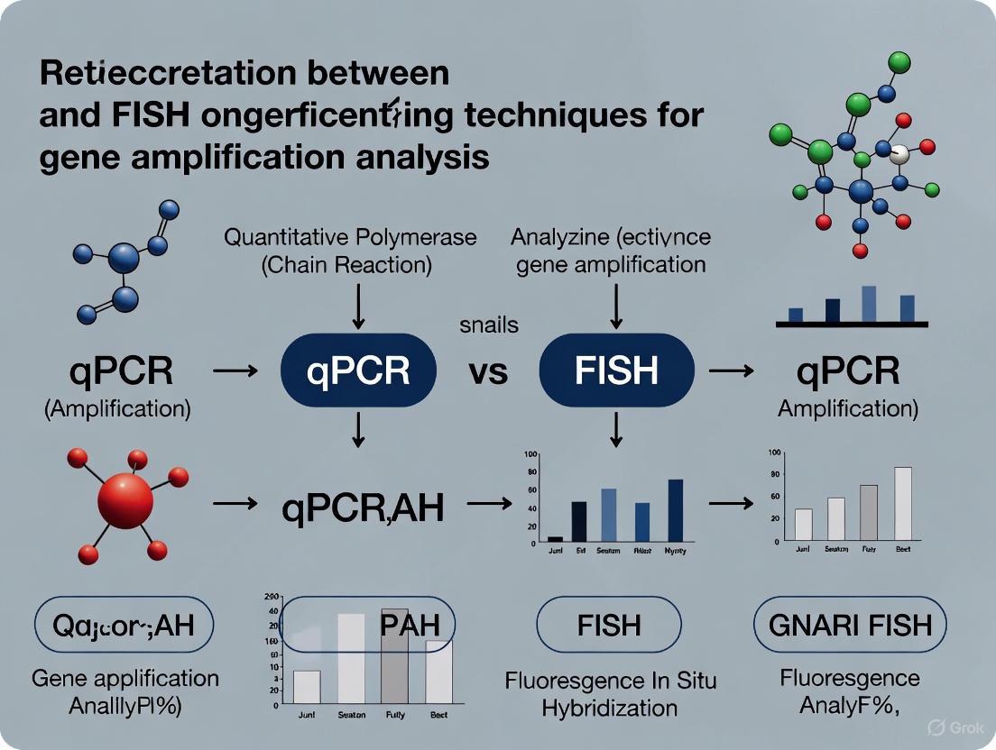
Bridging Quantification and Localization: The Powerful Correlation Between qPCR and FISH in Gene Analysis
This article provides a comprehensive overview for researchers and drug development professionals on the synergistic relationship between Quantitative PCR (qPCR) and Fluorescence In Situ Hybridization (FISH).
Bridging Quantification and Localization: The Powerful Correlation Between qPCR and FISH in Gene Analysis
Abstract
This article provides a comprehensive overview for researchers and drug development professionals on the synergistic relationship between Quantitative PCR (qPCR) and Fluorescence In Situ Hybridization (FISH). It explores the foundational principles that make these techniques complementary, detailing methodological applications from basic research to clinical diagnostics. The content offers practical troubleshooting guidance for both methods and presents rigorous validation studies comparing their performance. By synthesizing key takeaways, the article demonstrates how integrating qPCR's quantitative power with FISH's spatial resolution builds a more complete understanding of gene expression and amplification, ultimately advancing biomedical research and therapeutic development.
Understanding the Core Technologies: What qPCR and FISH Bring to Genetic Analysis
In gene amplification research, quantitative polymerase chain reaction (qPCR) and fluorescence in situ hybridization (FISH) represent two foundational technologies with distinct and complementary strengths. While both methods detect nucleic acids, their fundamental approaches yield different types of information that, when correlated, provide a comprehensive understanding of genetic events. qPCR excels at sensitive quantification of nucleic acid sequences in solution, providing precise measurements of copy numbers. In contrast, FISH offers spatial localization within intact cells or tissues, preserving morphological context while identifying specific genetic sequences. This methodological comparison explores the technical performance, experimental applications, and synergistic potential of these established techniques within modern research and diagnostic pipelines, particularly in oncology, microbiology, and pathogen detection.
The correlation between qPCR and FISH data is a critical consideration in molecular diagnostics. Discrepancies can arise from their different detection principles: qPCR identifies separated nucleic acids, while FISH detects targets within their cellular context. Understanding these methodological roles enables researchers to select the appropriate tool for their specific application and to interpret results within the correct technical framework.
qPCR: Solution-Based Quantitative Amplification
qPCR, also known as real-time PCR, is a gold standard for the quantification of specific DNA or RNA sequences. The technique involves the amplification of target nucleic acids in a solution-based reaction, with fluorescence-based monitoring of product accumulation during each PCR cycle. The core principle relies on the detection of a fluorescent signal that increases proportionally with the amount of amplified product. Two main chemistries dominate: DNA-binding dyes (e.g., SYBR Green) that bind non-specifically to double-stranded DNA, and sequence-specific probes (e.g., TaqMan probes) that provide enhanced specificity through hybridization to internal target sequences.
The TaqMan probe-based qPCR exemplifies the high-specificity approach. This mechanism utilizes a dual-labeled fluorogenic probe that hybridizes to a specific sequence between the PCR primers. The 5' exonuclease activity of the DNA polymerase cleaves the probe during amplification, separating the fluorophore from the quencher and generating a fluorescent signal. The point in the amplification process where the fluorescence crosses a defined threshold (Cycle threshold or Ct value) is inversely proportional to the log of the initial target copy number, enabling precise quantification [1] [2].
FISH: In Situ Spatial Localization
FISH is a cytogenetic technique that enables the visualization and localization of specific nucleic acid sequences within morphologically preserved cells, tissues, or chromosomes. The fundamental process involves fixing samples to maintain structural integrity, denaturing DNA duplexes to make targets accessible, and hybridizing fluorescently labeled nucleic acid probes to complementary sequences. After washing to remove non-specifically bound probes, the results are visualized via fluorescence microscopy, providing spatial information about genetic loci, gene expression, or microbial identification within their architectural context.
Single-molecule RNA-FISH (smRNA-FISH) represents an advanced application for detecting individual RNA molecules within cells. This method typically uses multiple short, fluorescently labeled oligonucleotide probes that bind to a single transcript, amplifying the signal sufficiently for detection at the single-molecule level. The design of these probe sets is critical for performance, requiring careful consideration of specificity, binding affinity, and secondary structure to minimize off-target binding and maximize signal-to-noise ratio [3]. The technique preserves the spatial organization of RNA, allowing researchers to quantify transcript abundance and monitor subcellular localization in individual cells.
Performance Comparison: Quantitative Data and Detection Limits
Sensitivity, Specificity, and Operational Characteristics
Table 1: Direct Performance Comparison of qPCR and FISH Methodologies
| Parameter | qPCR | FISH |
|---|---|---|
| Detection Limit | 2-200 copies/reaction [1] [2] | Single RNA molecules [3] |
| Quantification Capability | Excellent (Broad dynamic range: 101-1010 copies) [1] | Semi-quantitative (Based on signal counting) |
| Spatial Resolution | None (Solution-based) | Excellent (Subcellular localization) |
| Throughput | High (96/384-well formats) | Low to Moderate (Microscope slide-based) |
| Assay Time | 2-4 hours (Including preparation) [4] | 3-5 hours (Excluding analysis) [4] [5] |
| Automation Potential | High | Low to Moderate |
| Multiplexing Capacity | Moderate (4-6 targets typically) | Moderate (Limited by fluorescence spectrum) |
| Sample Requirements | Extracted nucleic acids | Intact cells/tissues |
Table 2: Experimental Detection Rates in Clinical Studies
| Study Context | qPCR Positive Rate | FISH Positive Rate | Culture Positive Rate | Reference |
|---|---|---|---|---|
| Pediatric Sepsis | 71.7% | 39.1% | 18% | [4] |
| ALK in NSCLC | 100% sensitivity vs. FISH | Gold Standard | N/A | [6] |
The data reveal fundamental performance differences. qPCR demonstrates superior analytical sensitivity for detecting low-copy targets in solution, with modern assays achieving detection limits as low as 2 copies per reaction [2]. This exceptional sensitivity makes it invaluable for applications requiring precise quantification of rare targets, such as monitoring minimal residual disease or detecting low-abundance pathogens.
FISH, while potentially less sensitive for absolute detection limit comparisons, provides unparalleled spatial context that enables unique applications. The technique can localize specific DNA loci within chromosomes for cytogenetic analysis, determine subcellular RNA distribution, and identify microorganisms within complex tissue architectures without culture. Recent advancements in smRNA-FISH have pushed its sensitivity to the single-molecule level, allowing precise counting of individual transcripts within cells [3].
The impact of antibiotic therapy on detection efficacy highlights an important practical distinction. In bacteremia detection, neither qPCR nor FISH requires viable organisms, making them less affected by prior antibiotic administration compared to culture methods. This characteristic provides a significant diagnostic advantage for patients already undergoing treatment [4].
Experimental Protocols: Detailed Methodologies
TaqMan qPCR Assay Protocol
The development of a robust qPCR assay requires careful optimization at each stage. The following protocol for detecting Carpione rhabdovirus (CAPRV2023) exemplifies a well-validated approach [1] [2]:
Primer and Probe Design:
- Target the conserved region of the N or G protein gene
- Design primers with melting temperature (Tm) of 58-60°C
- Design TaqMan probe with Tm 10°C higher than primers
- Label probe with FAM at 5' end and BHQ quencher at 3' end
- Verify specificity using BLAST against genomic databases
Reaction Setup:
- Prepare 20μL reaction mixture containing:
- 10μL of 2× TaqMan Universal PCR Master Mix
- 0.8μL of each primer (10μM)
- 0.4μL of probe (10μM)
- 2μL of template DNA/cDNA
- 6μL of nuclease-free water
- Perform amplification with the following cycling conditions:
- Initial denaturation: 95°C for 2 minutes
- 40 cycles of:
- Denaturation: 95°C for 15 seconds
- Annealing/Extension: 60°C for 1 minute
- Analyze using standard curve method with serial dilutions of plasmid containing target sequence
Validation Parameters:
- Efficiency: 90-110% (ideal: 100%)
- Linearity: R² > 0.990
- Sensitivity: Limit of detection determined by probit analysis
- Specificity: No cross-reactivity with related pathogens
RNA-FISH Protocol for Viral Detection
This optimized protocol for detecting SARS-CoV-2 RNA demonstrates contemporary FISH methodology [5]:
Sample Preparation:
- Collect buccal cells by swabbing and prepare smears on glass coverslips
- Fix cells with 4% paraformaldehyde for 10 minutes at room temperature
- Wash twice with phosphate-buffered saline (PBS)
Hybridization:
- Prepare hybridization buffer containing:
- 40% formamide
- 25% dextran sulfate
- 20μg/mL single-stranded salmon sperm DNA
- 20μg/mL yeast tRNA
- 0.4% bovine serum albumin
- 20mM ribonucleoside vanadyl complex
- 2× SSC buffer
- Pre-hybridize with 40μL buffer for 30 minutes at 37°C
- Hybridize with ATTO-labeled probes (1μg in 40μL hybridization buffer) for 1-4 hours at 37°C
Probe Design Considerations:
- Design 20nt probes targeting E, N, and ORF1a genes
- Ensure GC content ≥50% where possible
- Avoid self-complementarity and dimer formation
- Verify specificity using Primer-BLAST
Post-Hybridization Processing:
- Wash with 40% formamide/1× SSC for 20 minutes at 37°C
- Wash twice with 1× SSC for 10 minutes each at room temperature
- Counterstain with DAPI (0.5μg/mL) for 5 minutes
- Mount with antifade mounting medium
- Image using fluorescence microscopy with appropriate filter sets
Research Reagent Solutions: Essential Materials
Table 3: Key Reagents and Their Functions in qPCR and FISH
| Reagent Category | Specific Examples | Function | Application |
|---|---|---|---|
| Polymerase Enzymes | Taq DNA Polymerase, Hot Start variants | DNA amplification with 5'→3' polymerase and 5' exonuclease activity | qPCR [1] |
| Fluorescent Probes | TaqMan probes, Molecular Beacons | Sequence-specific detection through FRET | qPCR [1] [2] |
| Nucleic Acid Dyes | SYBR Green I, EVAGreen | Non-specific intercalation with dsDNA | qPCR [2] |
| Labeled Oligonucleotides | ATTO-dye conjugated probes, DIG-labeled probes | Target hybridization with fluorescent detection | FISH [5] |
| Hybridization Buffers | Formamide-based buffers with dextran sulfate | Promote specific hybridization while reducing background | FISH [5] |
| Mounting Media | Antifade mounting media with DAPI | Preserve fluorescence and counterstain nuclei | FISH [5] |
| Reverse Transcriptase | M-MLV, PrimeScript | RNA template conversion to cDNA | RT-qPCR [7] |
| Reference Genes | HPRT1, HSP90AA1, B2M, ACTB | Normalization of technical variations | qPCR [7] [8] |
Applications and Workflow Integration
Technology Selection Guide
Diagnostic and Research Implementation
In clinical diagnostics, particularly oncology, the correlation between qPCR and FISH has been extensively validated. For ALK rearrangement detection in non-small cell lung cancer (NSCLC), RT-PCR demonstrated 100% sensitivity compared to FISH, with sequencing confirming ALK fusions in most discordant cases (RT-PCR positive/FISH negative) [6]. This highlights how qPCR can detect functionally relevant fusions that may be challenging for FISH interpretation, particularly with complex rearrangement patterns or low tumor content.
In microbiology, qPCR and FISH offer complementary advantages for pathogen detection. While qPCR provides rapid, sensitive screening, FISH enables visual confirmation within morphological context. A sepsis study demonstrated significantly higher detection rates for qPCR (71.7%) compared to both FISH (39.1%) and blood culture (18%), with neither molecular method affected by antibiotic therapy [4]. This supports a diagnostic model where qPCR serves as a sensitive screening tool, with FISH providing morphological correlation.
Environmental monitoring applications further demonstrate how these technologies address different questions. For detecting invasive fish species, qPCR of environmental DNA (eDNA) provides sensitive presence/absence data and rough biomass estimation [9], while FISH could theoretically localize organisms within complex ecosystems, though this application is less developed.
Methodological Synergies and Correlation
Integrated Workflow for Comprehensive Analysis
Correlation Analysis and Data Interpretation
The correlation between qPCR and FISH data requires careful interpretation of their methodological differences. In ALK detection, the 100% sensitivity of RT-PCR compared to FISH [6] suggests qPCR may identify biologically relevant fusions that challenge FISH interpretation. However, FISH provides the advantage of visualizing genetic alterations within tissue architecture and tumor heterogeneity.
Potential sources of discrepancy include:
- Tumor heterogeneity: FISH may miss focal rearrangements in samples with low tumor content
- Variant patterns: Complex rearrangement patterns may be interpretively challenging by FISH
- Expression vs. rearrangement: qPCR detects fusion transcripts while FISH identifies genomic rearrangements
- Sensitivity differences: qPCR typically offers higher analytical sensitivity for detection
Optimal integration utilizes both technologies strategically: qPCR for sensitive screening and quantification, with FISH providing spatial confirmation and heterogeneity assessment. This approach is particularly valuable in clinical diagnostics, where both sensitivity and morphological correlation impact therapeutic decisions.
qPCR and FISH represent complementary rather than competitive technologies in molecular analysis. qPCR provides superior quantification, sensitivity, and throughput for solution-based detection, while FISH offers irreplaceable spatial context and morphological preservation. Their correlation strengthens diagnostic and research conclusions, with qPCR excelling at absolute detection sensitivity and FISH providing crucial localization information.
The evolving molecular landscape continues to leverage both technologies, with advancements in qPCR chemistries pushing detection limits lower, and smRNA-FISH methodologies enhancing spatial resolution to the single-molecule level. Understanding their distinct roles, performance characteristics, and implementation requirements enables researchers to select appropriate methodologies, interpret results within technical limitations, and design integrated approaches that leverage the unique strengths of each platform for comprehensive genetic analysis.
The Fundamental Principle of Complementarity in Gene Amplification Studies
In the field of gene amplification studies, the fundamental principle of complementarity establishes that quantitative PCR (qPCR) and fluorescence in situ hybridization (FISH) are not competing technologies but rather orthogonal approaches that, when integrated, provide a more comprehensive biological understanding. While qPCR excels at sensitive quantification of nucleic acids, FISH provides crucial spatial context at the single-cell level. This principle asserts that the correlation between these methodologies is strongest when each is properly validated and applied to appropriate biological questions with recognition of their inherent technical limitations.
The foundation of this complementary relationship lies in their shared dependence on nucleic acid hybridization but divergent endpoints in analysis. qPCR provides temporal amplification data during PCR cycles, enabling precise quantification of transcript or gene copy numbers across bulk samples [10] [11]. In contrast, FISH captures the spatial distribution of nucleic acids within individual cells, preserving architectural context but with traditionally lower quantitative precision [3]. The integration of these approaches is particularly powerful in drug development, where understanding both the magnitude of gene expression changes and their cell-to-cell variability is critical for assessing therapeutic mechanisms.
Recent methodological advances are strengthening this complementary relationship. Enhanced probe design algorithms for FISH [3] and more stable reference genes for qPCR normalization [10] [11] are reducing technical variability and improving cross-method correlation. This guide systematically compares these methodologies through experimental data, technical protocols, and visualization of their integrated application in pharmaceutical research.
Quantitative PCR (qPCR): Amplification-Based Quantification
qPCR operates on the principle of monitoring DNA amplification in real-time using fluorescent reporters, enabling precise quantification of initial template concentration. Two primary chemistries dominate the field: SYBR Green, which intercalates nonspecifically into double-stranded DNA, and TaqMan probes, which provide sequence-specific detection through fluorescently labeled oligonucleotides [2] [12]. The technology provides exceptional sensitivity, with detection limits frequently reaching single-digit copy numbers per microliter [2] [12].
The analytical power of qPCR depends critically on proper normalization using stable reference genes. As demonstrated in studies of fish parasites and zebrafish models, inappropriate reference genes can significantly distort expression patterns, leading to erroneous conclusions [10] [13]. The stability of these reference genes must be empirically validated for each experimental system, as commonly used genes like β-actin and GAPDH show substantial variability under different physiological conditions [13].
FISH (Fluorescence In Situ Hybridization): Spatial Context Preservation
FISH utilizes fluorescently labeled nucleic acid probes to detect specific DNA or RNA sequences within intact cells or tissues, preserving spatial information lost in bulk extraction methods. Single-molecule RNA FISH (smRNA-FISH) represents the current state-of-the-art, enabling visualization and quantification of individual RNA molecules with subcellular resolution [3].
The specificity and sensitivity of FISH depend critically on probe design, with recent computational advances significantly improving performance. The TrueProbes platform, for instance, integrates genome-wide BLAST-based binding analysis with thermodynamic modeling to generate high-specificity probe sets that minimize off-target binding [3]. This attention to probe design has elevated FISH from a qualitative morphological technique to a quantitatively robust methodology.
Performance Comparison: Quantitative Data Analysis
Table 1: Comparative Performance Metrics of qPCR and FISH Technologies
| Parameter | qPCR | FISH | Experimental Context |
|---|---|---|---|
| Sensitivity | 1-10 copies/μL [2] [12] | Single RNA molecules [3] | Limit of detection in optimal conditions |
| Dynamic Range | 7-8 orders of magnitude [2] | ~3 orders of magnitude [3] | Linear quantification range |
| Throughput | High (96-384 well plates) | Medium (multi-well imaging) | Samples processed per experiment |
| Spatial Resolution | None (bulk analysis) | Subcellular (single-molecule) [3] | Localization information |
| Sample Requirements | Homogenized tissue/cells | Intact cells/tissue sections | Preservation state needed |
| Quantitative Precision | CV: 0.23-1.95% [2] | Variable (probe-dependent) [3] | Technical reproducibility |
| Temporal Resolution | Snapshot of expression | Snapshot of expression | Time-course capability |
| Multiplexing Capacity | Moderate (4-6 plex) | High (dozens with spectral imaging) [3] | Simultaneous targets |
Table 2: Method-Specific Technical Considerations and Limitations
| Aspect | qPCR | FISH |
|---|---|---|
| Key Advantages | Excellent quantification precision, high throughput, established validation protocols [10] [2] | Spatial context, single-cell resolution, no amplification bias [3] |
| Primary Limitations | No spatial information, requires RNA destruction, population averaging [10] [11] | Lower throughput, complex image analysis, quantification challenges [3] |
| Critical Validation Requirements | Reference gene stability [10] [11] [13], primer efficiency [2] | Probe specificity [3], hybridization efficiency |
| Sample Compatibility | Homogenates, extracted nucleic acids, body fluids | Intact cells, tissue sections, whole mounts |
| Data Output | Cycle threshold (Ct), relative quantification, absolute copy number [10] [2] | Molecule counts per cell, subcellular localization patterns [3] |
Experimental Protocols: Method-Specific Workflows
qPCR Reference Gene Validation Protocol
The accuracy of qPCR quantification depends critically on using properly validated reference genes. The following protocol, adapted from studies on Argulus siamensis and black rockfish, provides a robust framework for reference gene validation [10] [11]:
Candidate Gene Selection: Select 5-8 candidate reference genes representing different functional classes (e.g., EF-1α, β-actin, GAPDH, 18S rRNA, ribosomal proteins) to minimize co-regulation [10] [13].
RNA Extraction and cDNA Synthesis: Extract RNA using standardized methods (e.g., Trizol protocol) from samples representing all experimental conditions. Treat with DNase I to remove genomic DNA contamination. Synthesize cDNA using reverse transcriptase with oligo(dT) and/or random hexamer primers [11].
Primer Validation: Design primers with the following characteristics:
- Amplicon size: 80-200 bp
- Primer length: 18-22 nucleotides
- Tm: 58-62°C
- GC content: 40-60% Validate primer efficiency using a 5-10 point standard curve from serial dilutions. Acceptable efficiency ranges from 90-110% with R² > 0.985 [10].
Stability Analysis: Run qPCR reactions on all candidate genes across all experimental samples. Analyze expression stability using at least three algorithms:
Comprehensive Ranking: Use the RefFinder tool or similar approach to integrate results from all algorithms into a comprehensive stability ranking [10].
Experimental Validation: Test the selected reference genes by measuring expression of known target genes under conditions where they are expected to change significantly [13].
Advanced FISH Probe Design and Validation
Modern FISH methodology relies on computationally optimized probe design to maximize specificity and signal-to-noise ratio. The TrueProbes workflow represents the current state-of-the-art approach [3]:
Target Sequence Identification: Retrieve full transcript sequence from reference databases. For isoform-specific detection, identify unique exonic or untranslated regions.
Genome-Wide Off-Target Analysis:
- Use BLAST to identify potential off-target binding sites across the entire transcriptome
- Calculate binding energies for both on-target and off-target interactions
- Incorporate expression data if available to weight off-targets by abundance
Probe Selection and Ranking:
- Screen all possible oligonucleotides (typically 17-22 nt) tiling the target
- Rank probes by specificity score considering:
- Number of off-targets (expression-weighted)
- Difference between on-target and off-target binding energies
- Self-hybridization potential
- Cross-dimerization with other probes in the set
- Select top-ranked non-overlapping probes (typically 20-48 per target)
Experimental Validation:
- Test probe sets in target-deficient systems (knockout cells) to measure background
- Compare signal intensity in high-expressing and low-expressing cell lines
- Perform competition experiments with unlabeled oligonucleotides
- Validate spatial patterns against known marker genes
Quantification Setup:
- Establish automated image analysis pipelines for spot counting
- Develop thresholds for signal versus background discrimination
- Create controls for hybridization efficiency and photobleaching correction
Integrated Workflow: Visualizing Complementary Application
The following diagram illustrates how qPCR and FISH can be integrated in a drug discovery pipeline to provide complementary insights:
Integrated Drug Discovery Workflow Using qPCR and FISH
Research Reagent Solutions: Essential Materials and Tools
Table 3: Key Research Reagents and Their Applications in Gene Amplification Studies
| Reagent Category | Specific Examples | Function & Importance | Technical Notes |
|---|---|---|---|
| Reference Genes | EF-1α, RPL17, 18S rRNA [10] [11] | qPCR normalization controls | Must be validated for each experimental system [13] |
| Probe Design Tools | TrueProbes, Stellaris, MERFISH [3] | FISH probe selection & optimization | Computational specificity assessment critical [3] |
| Fluorescent Reporters | SYBR Green, TaqMan probes [2] [12] | qPCR detection | TaqMan offers superior specificity [2] |
| Hybridization Buffers | Formamide-based systems [3] | FISH stringency control | Concentration affects specificity & background |
| Polymerase Systems | FastStart Essential DNA Green Master [10] | qPCR amplification | Hot-start reduces primer-dimer formation |
| Image Analysis Software | Custom spot counting algorithms [3] | FISH quantification | Enables single-molecule resolution |
The fundamental principle of complementarity between qPCR and FISH technologies represents a powerful paradigm for comprehensive gene amplification analysis in pharmaceutical research and development. Rather than viewing these methods as alternatives, the most insightful studies strategically employ both to answer complementary biological questions. qPCR provides the quantitative foundation for assessing expression changes across treatment groups with high precision and statistical power, while FISH delivers the spatial context essential for understanding heterogeneity, cellular localization, and tissue-level distribution patterns.
The correlation between qPCR and FISH findings is maximized when each technology is implemented with appropriate validation controls—reference gene stability assessment for qPCR and probe specificity validation for FISH. Technical advances in both methodologies continue to strengthen this correlation, with improved probe design algorithms enhancing FISH quantification reliability and better reference gene panels increasing qPCR accuracy across diverse experimental conditions.
For drug development professionals, this complementary approach offers a more complete picture of drug mechanisms, pharmacokinetic-pharmacodynamic relationships, and therapeutic effects at both population and single-cell levels. By integrating these orthogonal technologies through the workflows and protocols outlined in this guide, researchers can achieve unprecedented insights into gene expression regulation and its modulation by therapeutic interventions.
In biomedical research and clinical diagnostics, the accurate detection of gene amplification is paramount for disease stratification, prognosis, and treatment selection. For decades, fluorescence in situ hybridization (FISH) has been considered the "gold standard" technique for visualizing gene amplification within morphological context. Meanwhile, quantitative polymerase chain reaction (qPCR) has emerged as a powerful, high-throughput molecular technique. Research and clinical practice increasingly require a deep understanding of the correlation and comparative performance between these two fundamental methods. This guide provides an objective, data-driven comparison of FISH and qPCR, drawing from multicenter clinical studies and empirical research to inform scientists and drug development professionals in their methodological selections.
Fluorescence In Situ Hybridization (FISH)
FISH is a cytogenetic technique that uses fluorescently labeled DNA probes to bind specific parts of chromosomal regions with high sequence complementarity. It allows for the visualization of gene amplification status within the context of cell morphology and tissue architecture. The key strength of FISH lies in its ability to provide spatial information and detect heterogeneity within a sample, as it is performed on intact cells or tissue sections. However, the method requires specialized fluorescent microscopy, is time-consuming, and demands significant technical expertise for interpretation [14].
Quantitative PCR (qPCR)
qPCR is a molecular technique that amplifies and simultaneously quantifies a targeted DNA molecule. It enables the determination of gene copy number by comparing the amplification of the target gene to a reference gene. The method is characterized by its high sensitivity, rapid turnaround, potential for high-throughput analysis, and ability to work with limited or degraded DNA samples. Its main limitation is the lack of spatial context, as DNA is extracted from homogenized samples [14] [15].
Comparative Performance Data: Concordance and Diagnostic Accuracy
Large-scale clinical studies have systematically compared the performance of FISH and qPCR for gene amplification analysis, particularly in HER2 testing for breast cancer.
Table 1: Concordance Rates Between FISH and Alternative Techniques for HER2 Detection
| Technique | Number of Cases | Concordance with FISH (Ratio-Based) | Concordance with FISH (Copy Number-Based) | Sensitivity | Specificity |
|---|---|---|---|---|---|
| SISH | 498-587 | 97% | 98% | 99%-95% | Not Reported |
| CISH | 108-204 | 98% | 75% | 100%-99% | Not Reported |
| qPCR | 699-773 | 95% | 93% | 89%-80% | Not Reported |
Data adapted from a multicenter study on 840 breast cancer cases [14]
A separate study of 131 invasive breast carcinoma cases found that "qPCR is a valuable tool for the evaluation of Her2 gene overexpression/amplification," with results that "positively correlated with the results from IHC and FISH analysis" [15]. The study further highlighted that, in contrast to IHC or SISH/FISH, "the results obtained by qPCR were not encumbered with any subjective error on the part of the evaluator," indicating an advantage in objectivity [15].
Table 2: Comparison of Technical Characteristics Between FISH and qPCR
| Parameter | FISH | qPCR |
|---|---|---|
| Spatial Context | Preserved (tissue architecture) | Lost (homogenized sample) |
| Throughput | Low to moderate | High |
| Turnaround Time | Longer (including hybridization) | Shorter (few hours) |
| Automation Potential | Limited | High |
| DNA Quantity Required | Larger | Small (can work with limited DNA) |
| Subjectivity | Higher (requires interpretation) | Lower (based on quantitative metrics) |
| Cost | Higher (specialized reagents, imaging) | Lower |
Experimental Protocols: Detailed Methodologies
Standard FISH Protocol for HER2 Detection
The following protocol is adapted from multicenter studies comparing HER2 amplification techniques [14]:
- Sample Preparation: Cut 4-5 μm sections from formalin-fixed, paraffin-embedded (FFPE) tissue blocks. Use core biopsies or representative tumor sections.
- Deparaffinization and Pretreatment: Bake slides at 60°C for 1 hour. Deparaffinize in xylene and ethanol series. Pretreat with a pretreatment solution (e.g., citrate buffer) at 95-99°C for 15-30 minutes to expose target DNA.
- Enzymatic Digestion: Apply pepsin or other protease solution (0.5-2 mg/mL) at 37°C for 10-30 minutes to digest proteins and permit probe access.
- Probe Hybridization: Apply commercially available HER2/CEP17 dual-color FISH probes. Co-denature specimen and probe at 82°C for 5-10 minutes. Hybridize at 45°C overnight (16-18 hours) in a humidified chamber.
- Post-Hybridization Wash: Wash slides in stringency buffer (2X SSC/0.3% NP-40) at 46°C for 10 minutes to remove unbound probe.
- Counterstaining and Mounting: Apply DAPI counterstain and mount with antifade mounting medium.
- Analysis: Visualize using a fluorescence microscope with appropriate filter sets. Count HER2 (orange) and CEP17 (green) signals in 60-100 non-overlapping interphase nuclei. Calculate HER2/CEP17 ratio; a ratio >2.2 is considered amplified.
Standard qPCR Protocol for HER2 Copy Number Determination
This protocol is based on studies that designed qPCR assays specifically for HER2 amplification detection from FFPE samples [14] [16]:
- DNA Extraction: Extract genomic DNA from FFPE tissue sections or core biopsies. Macro-dissect or micro-dissect to enrich tumor cell percentage. Use commercial kits designed for FFPE DNA extraction.
- DNA Quantification and Quality Control: Precisely quantify DNA using a spectrophotometer or fluorometer. Assess DNA quality (e.g., A260/A280 ratio ~1.8-2.0).
- Primer and Probe Design: Design TaqMan assays targeting:
- Target Gene: HER2 (commonly exons 8 and 26).
- Reference Genes: A stable single-copy gene on chromosome 17 (e.g., TAOK1, UTP6) to control for polysomy, and diploidy control genes (e.g., TSN, LAP3) on other chromosomes.
- qPCR Reaction Setup: Prepare reactions in 20 μL volumes containing: 10-20 ng genomic DNA, 1X TaqMan Universal PCR Master Mix, 900 nM of each primer, and 250 nM of probe.
- Thermal Cycling: Run on a real-time PCR instrument with the following conditions: 95°C for 10 min (initial denaturation/activation), followed by 40-45 cycles of 95°C for 15 sec and 60°C for 1 min.
- Data Analysis: Determine cycle threshold (Ct) values for target and reference genes. Calculate the ΔΔCt or use a standard curve method to determine the relative copy number. An amplification ratio >2.7 is often used as a cut-off for HER2 amplification in well-calibrated assays [16].
Workflow and Logical Pathway Diagrams
Diagram 1: Comparative Workflows of FISH and qPCR
Diagram 2: Decision Pathway for Method Selection
Research Reagent Solutions and Essential Materials
Table 3: Essential Research Reagents and Materials for FISH and qPCR
| Category | Specific Item | Function/Application |
|---|---|---|
| Sample Preparation | Formalin-Fixed Paraffin-Embedded (FFPE) Tissue Blocks | Preserves tissue architecture for morphological analysis |
| Microtome | Sectioning FFPE blocks into thin slices for slide mounting | |
| Xylene and Ethanol Series | Deparaffinization of tissue sections | |
| Protease Solution (e.g., Pepsin) | Enzymatic digestion to expose target DNA | |
| FISH-Specific Reagents | HER2/CEP17 Dual-Color FISH Probes | Commercially available probes for specific gene detection |
| DAPI (4',6-diamidino-2-phenylindole) | Counterstain for nuclear visualization | |
| Antifade Mounting Medium | Preserves fluorescence during microscopy | |
| qPCR-Specific Reagents | DNA Extraction Kits (for FFPE) | Isolates high-quality genomic DNA from challenging samples |
| TaqMan Universal PCR Master Mix | Contains enzymes, dNTPs, and buffer for efficient amplification | |
| HER2-specific Primers and Probes | Targets specific exons (e.g., 8, 26) for amplification | |
| Reference Gene Assays (e.g., TAOK1, TSN) | Controls for DNA input and chromosome number | |
| General Consumables | Nuclease-Free Water | Prevents enzymatic degradation of reactions |
| PCR Tubes/Plates | Reaction vessels compatible with thermal cyclers | |
| Microscope Slides and Coverslips | Sample mounting for FISH analysis |
The correlation between FISH and qPCR for gene amplification analysis is well-established, with large multicenter studies demonstrating >90% concordance in optimal conditions. The choice between these techniques is not a matter of superiority but of strategic application aligned with research or diagnostic objectives. FISH remains indispensable when spatial context, tumor heterogeneity, and morphological correlation are required. In contrast, qPCR offers significant advantages in throughput, cost-effectiveness, objectivity, and application to limited or challenging samples. Future directions point toward the continued refinement of qPCR assays, the emergence of digital PCR for even greater precision in quantification, and the development of integrated diagnostic pathways that strategically employ both techniques to leverage their complementary strengths. For researchers and drug development professionals, this evidence-based comparison provides a foundation for informed methodological selection in both basic science and clinical translation.
In gene amplification research, the choice of analytical technique fundamentally shapes the type of questions a scientist can answer. Quantitative Polymerase Chain Reaction (qPCR) and Fluorescence In Situ Hybridization (FISH) represent two cornerstone methodologies with complementary strengths. While qPCR excels at providing sensitive, quantitative data on gene abundance in bulk samples, FISH offers spatial context at the cellular or subcellular level, preserving morphological information. This guide provides an objective comparison of their performance, supported by experimental data and detailed protocols, to inform researchers and drug development professionals on selecting the appropriate tool for their specific biological questions.
Fundamental Principles and Technical Comparison
qPCR is a bulk assay that quantifies nucleic acid sequences through amplification and fluorescent detection in a homogeneous solution. It involves DNA extraction from a lysed sample, followed by amplification with sequence-specific primers and a fluorescent probe. The cycle threshold (Ct) at which fluorescence crosses a background level is inversely proportional to the initial target concentration, enabling precise quantification [17] [18].
In contrast, FISH is a spatial imaging technique that uses fluorescently labeled nucleic acid probes to hybridize to complementary target sequences within intact cells or tissue sections. This preserves the architectural context of the sample, allowing researchers to visualize the physical location and distribution of genetic targets [14] [3]. Advanced forms like smRNA-FISH (single-molecule RNA-FISH) can detect individual RNA molecules with high spatial resolution [3].
Table 1: Core Technical Characteristics and Applications of qPCR and FISH
| Feature | qPCR | FISH |
|---|---|---|
| Primary Output | Quantitative (copy number, concentration) | Spatial (location, distribution, morphology) |
| Sample Processing | Destructive (requires sample lysis) | Non-destructive (preserves tissue/cell structure) |
| Sensitivity | High (detects low copy numbers in a sample) [19] | Variable; single-molecule sensitivity possible with optimized probes [3] |
| Throughput | High (can process many samples in parallel) | Lower (imaging and analysis are time-intensive) |
| Key Applications | Gene expression, pathogen load, gene amplification quantification [17] [20] | Tumor heterogeneity, chromosome mapping, subcellular RNA localization [14] |
Performance Comparison: Sensitivity, Specificity, and Quantitative Correlation
Multiple studies have directly compared the performance of qPCR and FISH, particularly in clinical diagnostics where detecting gene amplification is critical.
A multicenter study analyzing HER2 amplification status in breast cancer patients demonstrated a strong correlation between the techniques. The study reported an excellent 95% concordance between qPCR and FISH results when using the HER2/CEN17 ratio on core biopsies. The sensitivity of alternative techniques like SISH and CISH compared to FISH was 99% and 100%, respectively, while qPCR showed a slightly lower but still robust sensitivity of 89% [14].
In environmental DNA (eDNA) applications, a study comparing species-specific qPCR and metabarcoding (a method related to FISH in its use of hybridization principles) for detecting pelagic fish found a positive correlation between the methods. However, qPCR consistently demonstrated a higher detection rate than the sequencing-based method, partly due to less susceptibility to amplification biases in complex samples [21].
For low-abundance targets, digital PCR (a derivative of qPCR) has shown superior sensitivity and quantification precision compared to standard qPCR, particularly at concentrations below 1 copy/μL [19]. This highlights the evolving nature of quantitative PCR technologies for challenging applications.
Table 2: Experimental Comparison Data from Peer-Reviewed Studies
| Study Context | Concordance with FISH | Sensitivity vs. FISH | Key Findings |
|---|---|---|---|
| HER2 in Breast Cancer (n=840) [14] | 95% (based on HER2/CEN17 ratio) | 89% (based on HER2/CEN17 ratio) | qPCR is a reliable, less expensive alternative to FISH for core biopsies. |
| Fish eDNA Detection [21] | Positive correlation reported | qPCR detection rate higher than metabarcoding | qPCR is less prone to amplification bias in complex environmental samples. |
| Telomere Length Measurement [22] | Strong correlation between qPCR and Flow-FISH | Both methods showed comparable reduction with age | Flow-FISH provides single-cell resolution, while qPCR offers higher throughput. |
Detailed Experimental Protocols
This protocol is adapted for detecting a specific genetic target, such as an amplified gene, from sample DNA.
Assay Design and Validation:
- Generate Sequence Database: Compile mitochondrial or nuclear DNA sequences for the target gene from public databases (e.g., NCBI, BOLD) for both target and non-target species.
- Align Sequences: Use alignment software (e.g., Geneious, MUSCLE) to identify regions with high inter-species divergence and low intra-species variation.
- Design Primers/Probe: Utilize design tools (e.g., IDT's PrimerQuest) to create a qPCR assay with forward and reverse primers and a fluorescently labeled probe.
- Test Specificity: In silico testing (BLAST) followed by empirical testing against DNA from related species to ensure no cross-reactivity.
qPCR Reaction Setup:
- Prepare a reaction mix containing: 0.5 μL cDNA (or extracted DNA), 5.0 μL of 2x SYBR Green or probe master mix, 0.2 μL of each primer (10 μM), 0.2 μL of probe (10 μM), and nuclease-free water to 10.0 μL [23].
- Include negative controls (no-template) and positive controls (sample with known target DNA).
qPCR Amplification:
- Run on a real-time PCR instrument with cycling conditions typically being: 94°C for 3 minutes, followed by 40 cycles of 94°C for 15 seconds, 60°C for 15 seconds, and 72°C for 20 seconds [23].
Data Analysis:
- Determine the Ct value for each sample.
- Quantify the target concentration by comparing Ct values to a standard curve of known concentrations or use absolute quantification with digital PCR methods [19].
This protocol outlines the workflow for detecting RNA molecules within their cellular context.
Probe Design (TrueProbes Method):
- Genome-Wide Screening: Use software (e.g., TrueProbes) that performs a BLAST-based analysis against the entire transcriptome to select probe sequences.
- Rank by Specificity: Probes are ranked based on predicted binding affinity, minimal off-target interactions, and low self-hybridization potential.
Sample Preparation and Hybridization:
- Cell Culture and Fixation: Grow cells on coverslips and fix with paraformaldehyde to preserve morphology.
- Permeabilization: Treat cells with a detergent (e.g., Triton X-100) to allow probe entry.
- Hybridization: Apply fluorescently labeled oligonucleotide probes in a hybridization buffer containing formamide. Incubate overnight in the dark at a specified temperature (e.g., 37°C).
Washing and Imaging:
- Stringency Washes: Perform washes with saline-sodium citrate (SSC) buffer at a defined temperature to remove non-specifically bound probes.
- Counterstaining and Mounting: Stain nuclei with DAPI and mount coverslips on slides.
- Image Acquisition: Use a fluorescence or confocal microscope with high-resolution cameras to capture z-stacks of the cells.
Data Analysis:
- Use image analysis software to count distinct fluorescent spots, each representing an individual RNA molecule, and determine their subcellular localization.
Essential Research Reagent Solutions
The reliability of both qPCR and FISH experiments depends heavily on the quality and specificity of key reagents.
Table 3: Essential Research Reagents for qPCR and FISH
| Reagent / Solution | Function | Application Notes |
|---|---|---|
| Sequence-Specific Primers & Probes | Binds to target DNA/RNA for amplification (qPCR) or detection (FISH). | Critical for assay specificity. Probe-based qPCR and FISH both require fluorophore-labeled probes [3] [18]. |
| DNA Polymerase Master Mix | Enzymatically amplifies target DNA during PCR. | Choice of master mix can affect sensitivity and inhibitor resistance [19]. |
| Formamide-Based Hybridization Buffer | Creates stringent conditions for specific probe binding in FISH. | Reduces the melting temperature of DNA-RNA hybrids, enabling specific binding [3]. |
| Stringent Wash Buffers (e.g., SSC) | Removes non-specifically bound probes after hybridization in FISH. | Critical for reducing background fluorescence and improving signal-to-noise ratio [3]. |
| Reference Genes (for qPCR) | Used for normalization of gene expression data. | Must be stably expressed across experimental conditions (e.g., RAB10, PFDN2, NDUFS7 in fish studies) [23]. |
qPCR and FISH are not competing techniques but rather complementary tools in the molecular biologist's arsenal. The decision to use one over the other is dictated by the specific biological question. qPCR is the tool of choice when the research goal demands high-throughput, sensitive quantification of nucleic acids from processed samples, such as validating gene amplification levels or measuring pathogen load [17] [20]. FISH is indispensable when the spatial distribution of a target is the key parameter, such as mapping heterogeneity within a tumor, visualizing chromosomal translocations, or localizing RNA to specific cellular compartments [14] [3]. A thorough understanding of their respective strengths, limitations, and the experimental data they generate is fundamental for designing robust experiments and making sound conclusions in gene amplification research and drug development.
In the field of molecular biology and clinical diagnostics, accurately determining gene status is paramount for both basic research and personalized medicine. Two principal methodologies have emerged as cornerstones for this purpose: quantitative Polymerase Chain Reaction (qPCR) and Fluorescence In Situ Hybridization (FISH). While the former provides sensitive, quantitative data on gene copy number and expression from purified nucleic acids, the latter offers direct spatial visualization of genetic material within its cellular context [24] [25]. The central thesis of this guide is that these techniques are not mutually exclusive but are, in fact, powerfully complementary. A growing body of evidence demonstrates a strong correlation between their findings, validating their combined use for robust gene amplification research, particularly in clinical settings such as HER2 status determination in breast cancer [25]. This guide objectively compares the performance, protocols, and applications of qPCR and FISH, providing researchers with the experimental data necessary to inform their methodological choices.
Methodological Comparison: Principles, Strengths, and Limitations
Core Principles and Technical Comparison
Quantitative PCR (qPCR) operates as an in vitro reaction that exponentially amplifies a target DNA sequence from an extracted sample. The accumulating fluorescent signal is plotted against amplification cycles to determine the initial quantity of the target, reported as Cycles to Quantification (Cq) [24]. When applied to gene amplification studies, it can precisely quantify both gene copy number at the DNA level and expression levels at the cDNA level [25]. In contrast, FISH is a microscopy-based technique that hybridizes fluorescently labeled probes directly to specific DNA or RNA sequences within intact cells or tissues. It does not rely on signal amplification but instead uses the direct binding of multiple probes (e.g., up to 48 for Stellaris FISH) to create discreet, countable fluorescent spots at the site of each transcript or gene locus, preserving morphological information [26] [24].
Table 1: Fundamental Characteristics of qPCR and FISH
| Feature | qPCR | FISH |
|---|---|---|
| Analytical Basis | Amplification of extracted nucleic acids [24] | Direct hybridization in situ [24] |
| Quantification Method | Cq values correlated to a standard curve [24] [25] | Digital counting of fluorescent spots [24] |
| Throughput | High-throughput, suitable for screening many samples [24] | Lower throughput, more suited for detailed analysis of limited samples [26] |
| Spatial Resolution | No spatial context; destroys sample morphology [24] | High; locates transcripts to subcellular compartments [26] [24] |
| Key Application | Sensitive quantification and gene expression profiling [25] | Visualizing gene localization and heterogeneity [26] |
Performance and Concordance Data
Comparative studies consistently show a strong correlation between qPCR and established techniques like FISH and IHC. In a 2025 study on HER2 status, qPCR analysis of DNA and RNA demonstrated complete concordance with IHC in ten samples. Notably, the molecular approach (qPCR) agreed with a subsequent FISH test for an equivocal sample that was positive by IHC, potentially altering therapeutic decisions [25]. This supports earlier findings, such as a 2013 study by Wang et al., which reported a Spearman rank correlation of 0.82 between qPCR and FISH [25]. The high sensitivity of qPCR allows it to detect gene amplification even in samples with a low fraction (as low as 5%) of tumor cells, which can be a challenge for other methods [25].
Table 2: Experimental Concordance and Performance Metrics
| Study / Application | Key Performance Finding | Implication for Correlation |
|---|---|---|
| HER2 Status (2025) [25] | 100% concordance with IHC in 10 samples; resolved an equivocal case. | Validates qPCR as a reliable alternative to FISH. |
| Wang et al., 2013 [25] | Spearman correlation of 0.82 (p < 0.0001) with FISH. | Demonstrates a strong statistical relationship between the methods. |
| Sensitivity (HER2) [25] | Detects amplification with as little as 5% tumor cell fraction. | qPCR is highly sensitive, reducing false negatives. |
| StAR Transcription Analysis [26] | qPCR and FISH provide complementary quantitative data on RNA species. | Techniques together resolve temporal and spatial expression. |
Experimental Protocols: From Sample to Result
Protocol for qPCR-Based Gene Copy Number Determination
This protocol, adapted from a 2025 HER2 study, details the steps for absolute quantification of gene copy number using qPCR [25].
- Sample Preparation: Extract genomic DNA from tissue samples, such as Formalin-Fixed Paraffin-Embedded (FFPE) blocks. Treat DNA with a repair enzyme mix if derived from FFPE to fix damage and use inhibitor removal columns if necessary [25].
- Standard Curve Construction: Prepare a threefold serial dilution of a Control Genomic Human DNA (e.g., from 1 ng/µL to 50 pg/µL). This standard of known concentration is used to create a calibration curve for absolute quantification [25].
- qPCR Reaction Setup: Perform reactions in duplicates for reproducibility. Use a SYBR Green-based detection system. The reaction mix includes:
- Template DNA (from sample or standard)
- Forward and reverse primers for the target gene (e.g., HER2)
- Forward and reverse primers for a stable reference gene located in a genomically stable region (e.g., APP on chromosome 21) [25].
- Data Analysis: The qPCR software generates a standard curve for both the target and reference genes. The concentration of the target gene (in ng/µL) in the sample is determined relative to the standard curve and then normalized to the concentration of the reference gene. A positive result for gene amplification is typically defined as a copy number greater than five above the average ploidy [25].
Protocol for Quantitative RNA-FISH
This protocol outlines the process for detecting and quantifying specific RNA transcripts within cells, integrating details from studies on Stellaris FISH and StAR gene expression [26] [24].
- Cell Culture and Fixation: Culture cells (e.g., MA-10 Leydig cells) on poly-Lysine coated coverslips. Stimulate cells as required (e.g., with 8-Br-cAMP) and then fix them to preserve cellular architecture and RNA integrity [26].
- Probe Design and Hybridization: Design a set of approximately 40-48 singly labeled oligonucleotide probes, each targeting different regions of the same mRNA transcript. These probes are labeled with a fluorescent dye (e.g., Quasar dye). Hybridize the probe set to the fixed cells [26] [24].
- Microscopy and Image Acquisition: Use high-resolution fluorescence microscopy, such as Structured Illumination Microscopy (N-SIM), to image the cells. The binding of multiple probes to a single mRNA molecule results in a bright, discreet fluorescent spot that can be distinguished from background noise [26].
- Image Quantification: Employ automated image processing algorithms (e.g., "FISH quant") to recognize cellular boundaries, identify genuine transcript spots based on a set intensity threshold, and assign a digital count of mRNA molecules per cell. This allows for localization analysis to subcellular regions, such as mitochondria [26].
Diagram 1: Experimental workflow for qPCR and FISH.
The Scientist's Toolkit: Essential Research Reagents
Successful execution of these correlative studies requires a suite of reliable reagents and tools. The following table details key solutions used in the protocols cited above.
Table 3: Key Research Reagent Solutions for qPCR and FISH
| Reagent / Kit | Function | Experimental Role |
|---|---|---|
| Control Genomic Human DNA [25] | Provides a standard of known concentration for absolute quantification. | Essential for constructing the standard curve to calculate gene copy number in qPCR assays. |
| FFPE DNA/RNA Repair Mix [25] | Reverses nucleic acid damage caused by formalin fixation. | Critical for recovering amplifiable DNA from archived clinical FFPE samples for qPCR. |
| PCR Inhibitor Removal Kit [25] | Removes contaminants that can inhibit polymerase activity. | Increases qPCR assay reliability and sensitivity, especially from complex samples like FFPE tissue. |
| Stellaris FISH Probe Sets [24] | A pool of ~48 fluorescently labeled oligos targeting a single mRNA. | Enables sensitive and specific detection of individual RNA molecules without amplification for FISH. |
| Random Hexamers & Reverse Transcriptase [26] | Primers and enzyme for synthesizing complementary DNA (cDNA). | Used in reverse transcription to convert RNA into cDNA for gene expression analysis by qPCR. |
| SYBR Green qPCR Master Mix [25] | Contains dyes, enzymes, and dNTPs for real-time PCR. | The core reagent for performing quantitative PCR, allowing fluorescence-based detection of amplicons. |
Integrated Analysis: Visualizing the Correlative Workflow
The correlation between qPCR and FISH is not merely observational but is rooted in their shared biological target. The following diagram synthesizes how these methods converge to validate findings, using gene amplification and expression as a central example.
Diagram 2: The correlative relationship between qPCR and FISH data.
Integrated Workflows: Practical Applications of qPCR and FISH in Concert
In gene amplification research, the combined use of quantitative PCR (qPCR) and Fluorescence in Situ Hybridization (FISH) represents a powerful methodological synergy. qPCR serves as an exceptionally sensitive tool for initial screening and quantification of nucleic acid targets, while FISH provides spatial context and validation at the single-cell level. This sequential approach leverages the respective strengths of each technique: qPCR offers rapid, quantitative assessment of gene expression or amplification across many samples, and FISH confirms these findings with precise morphological localization, distinguishing cell-to-cell heterogeneity that bulk analysis might miss [22] [3].
The correlation between qPCR and FISH data strengthens experimental conclusions, as findings obtained through solution-based quantification are verified by direct visualization within intact cells or tissues. This guide objectively compares the performance characteristics, experimental protocols, and integrated applications of qPCR and FISH to provide researchers and drug development professionals with a framework for their sequential implementation in gene amplification studies.
Performance Comparison: qPCR vs. FISH
The table below summarizes the core performance characteristics of qPCR and FISH based on experimental data, highlighting their complementary nature.
Table 1: Performance Comparison of qPCR and FISH Techniques
| Feature | qPCR | FISH/smRNA-FISH |
|---|---|---|
| Primary Function | Quantitative nucleic acid detection and quantification [2] | Spatial localization and visualization of nucleic acids in situ [3] |
| Throughput | High (96/384-well plates) | Low to medium (individual samples/slides) |
| Sensitivity | High (detects as low as 2 copies/μL) [2] | Single-molecule resolution possible [3] |
| Quantification | Highly quantitative (absolute or relative) [2] | Semi-quantitative to quantitative (with careful calibration) [22] |
| Spatial Context | No (homogenized sample) | Yes (preserves cellular and subcellular architecture) [3] |
| Turnaround Time | Rapid (a few hours post nucleic acid extraction) [27] | Slow (can require overnight hybridization) [22] |
| Key Strength | Sensitivity, throughput, and precise quantification | Spatial resolution and morphological correlation |
| Major Limitation | Lacks spatial information and cannot detect heterogeneity in mixed cell populations | Lower throughput and more complex, specialized analysis [22] |
A direct comparison study measuring telomere content highlighted that while both methods provide robust measurement and show comparable reduction with age, Flow-FISH, a cytometric variant, measured a relative content longer than qPCR at a single-cell level [22]. This underscores how the chosen methodology can influence specific numerical outcomes, even when trends correlate.
Experimental Protocols for Sequential Analysis
qPCR Screening Protocol
The following methodology is adapted from validated assays for pathogen detection [2] [27], which can be tailored for gene amplification studies.
- Sample Lysis and Nucleic Acid Extraction: Extract total RNA or DNA from homogenized tissue or cell pellets using commercial kits (e.g., DNeasy Blood & Tissue Kit, Qiagen). For cells, use ~20 mg of tissue or 10^6 cells. Elute in nuclease-free water or TE buffer and quantify using a spectrophotometer (e.g., NanoDrop) [2] [9].
- Reverse Transcription (for RNA targets): Synthesize cDNA using a Reverse Transcription kit with random hexamers and/or gene-specific primers.
- Primer and Probe Design: Design TaqMan probes to target the gene of interest (e.g., an amplified oncogene) and a reference gene (e.g., a housekeeping gene). Probes are typically labeled with FAM at the 5' end and a quencher (e.g., BHQ1) at the 3' end. Primers should be designed to amplify a 50-150 bp product [2].
- qPCR Reaction Setup and Amplification:
- Reaction Mix: 5 μL of 2X TaqMan Probe qPCR Master Mix, 0.2-1.0 μM each of forward and reverse primer, 62.5-250 nM of probe, and 1 μL of cDNA/DNA template. Adjust with nuclease-free water to a 10 μL total volume [2].
- Thermocycling Conditions: Initial denaturation at 95°C for 60 seconds, followed by 40 cycles of 95°C for 10 seconds (denaturation) and 51-59°C for 30 seconds (annealing/extension). The optimal annealing temperature should be determined empirically [2].
- Data Analysis: Calculate the cycle threshold (Ct) values. Use a standard curve for absolute quantification or the ΔΔCt method for relative quantification of gene amplification.
FISH Validation Protocol
This protocol is based on single-molecule RNA FISH (smRNA-FISH) principles [3] and can be adapted for DNA targets, such as visualizing specific genomic loci.
- Sample Preparation and Fixation: Culture cells on glass coverslips or use tissue sections. Rinse with PBS and fix with 4% formaldehyde in PBS for 10-15 minutes at room temperature. Permeabilize cells with 0.1-0.5% Triton X-100 in PBS for 10 minutes [3].
- Probe Design and Labeling: For high-specificity detection, use a probe design tool like TrueProbes, which uses genome-wide BLAST and thermodynamic modeling to generate probe sets with high specificity and minimal off-target binding [3]. Typically, 20-48 oligonucleotide probes (each 17-22 nucleotides long) are designed against the target sequence and labeled with fluorophores (e.g., Cy3, Cy5, FAM).
- Hybridization:
- Post-Hybridization Washes: After hybridization, wash the samples to remove unbound probe. A typical wash series includes: 2X SSC at 37°C for 15-30 minutes, 1X SSC at room temperature for 15 minutes, and 0.1X SSC for 10 minutes. A DAPI counterstain can be included in one of the washes to label nuclei.
- Imaging and Analysis: Mount coverslips and image using a high-resolution fluorescence or confocal microscope. For smRNA-FISH, individual RNA molecules appear as distinct fluorescent spots. Quantification involves counting these spots per cell using image analysis software (e.g., ImageJ, FlowJo) [22] [3].
Workflow Visualization
The following diagram illustrates the sequential integration of qPCR and FISH in a typical experiment.
Biological Context: The Telomere Maintenance Pathway
The correlation between qPCR and FISH is particularly relevant in studying gene amplification and genomic instability. A key area is telomere biology, where changes in telomere length are linked to cancer, aging, and hematologic disorders [22]. Both qPCR and FISH (specifically Flow-FISH) are established methods for telomere length assessment, with qPCR measuring average telomere content in a sample and FISH providing length distribution at the single-cell level.
Table 2: Key Research Reagent Solutions
| Reagent / Solution | Function | Example Use Case |
|---|---|---|
| TaqMan Probe Master Mix | Contains DNA polymerase, dNTPs, and optimized buffer for probe-based qPCR detection. | Absolute quantification of a target gene's copy number in a sample [2]. |
| DNeasy PowerSoil Pro Kit | Efficiently extracts high-quality DNA from complex biological samples, including tissues. | Preparing DNA templates for qPCR from fish or mammalian tissue [28]. |
| smRNA-FISH Probe Sets | Fluorophore-labeled oligonucleotides designed for specific hybridization to target RNA/DNA in situ. | Visualizing the spatial distribution of a specific mRNA or genomic locus in fixed cells [3]. |
| GoTaq Probe qPCR Master Mix | A ready-to-use mix for probe-based qPCR applications, ensuring robust amplification. | Used in eDNA studies for sensitive detection of specific species from environmental samples [9]. |
| Formamide-based Hybridization Buffer | A key component of the hybridization buffer that helps control the stringency of probe binding. | Critical for ensuring specific binding of FISH probes to their target sequences while minimizing off-target signals [22] [3]. |
The following diagram outlines the core telomere maintenance pathway, a system frequently investigated using both qPCR and FISH techniques.
The sequential application of qPCR for screening and FISH for validation creates a powerful, correlative framework for gene amplification research. qPCR delivers the quantitative power and throughput necessary to efficiently analyze large sample sets, while FISH provides the indispensable spatial validation that confirms findings and reveals cellular heterogeneity. The experimental data and protocols detailed in this guide provide a roadmap for researchers in drug development and biomedical science to robustly integrate these techniques. This approach ensures that quantitative data is consistently grounded in morphological reality, leading to more reliable and impactful scientific conclusions.
Fluorescence in situ hybridization (FISH) is a powerful cytogenetic technique essential for chromosome identification, mapping alien introgressions, and studying plant genome evolution [29]. Despite its utility, FISH is time-consuming and labor-intensive, requiring significant resources for probe development and hybridization. This case study explores how quantitative polymerase chain reaction (qPCR) serves as a predictive tool for FISH outcomes, enabling researchers to pre-screen tandem repeats and select the most promising cytogenetic markers efficiently [29].
The correlation between qPCR and FISH extends beyond plant cytogenetics, finding application in clinical diagnostics, such as HER-2 amplification assessment in breast cancer, where qPCR demonstrates high concordance with FISH while offering advantages in processing challenging samples [30]. This guide objectively compares the performance of qPCR and FISH, providing experimental data and methodologies that underscore their complementary roles in gene amplification research.
Comparative Performance Analysis
Key Advantages and Limitations
Table 1: Method comparison between qPCR and FISH
| Parameter | qPCR | FISH |
|---|---|---|
| Primary Function | Quantifies copy number of specific DNA sequences | Provides spatial localization of sequences on chromosomes |
| Throughput | High-throughput, suitable for rapid screening | Lower throughput, more time-intensive |
| Spatial Information | No chromosomal location data | Preserves spatial context and chromosomal position |
| Sensitivity | Highly sensitive for quantifying repeat abundance [29] | Limited by resolution (~10,000 nt) [29] |
| Sample Requirements | Works with fragmented DNA (e.g., FFPE samples) [30] | Requires intact chromosomal morphology |
| Turnaround Time | Rapid (hours to 1 day) | Slow (several days) |
| Quantitative Capability | Precise copy number quantification [29] | Semi-quantitative based on signal intensity |
| Cost | Lower per sample | Higher due to specialized reagents and equipment |
Concordance Validation Studies
Table 2: Correlation evidence between qPCR and FISH
| Study Context | qPCR Findings | FISH Validation | Concordance Level |
|---|---|---|---|
| Dasypyrum species (pHv-961 repeat) | ~1000x copy number difference between D. breviaristatum and D. villosum [29] | Bright signals in D. breviaristatum only; absent in D. villosum [29] | Complete correlation |
| Thinopyrum species (19-202 repeat) | Lowest copy number in Th. ponticum; higher in other Thinopyrum species and D. breviaristatum [29] | Corresponding signal intensity and localization patterns matched qPCR quantification [29] | High correlation |
| Aegilops species (CL244 repeat) | Low copy number in Ae. tauschii; higher in Ae. crassa and Th. bessarabicum [29] | Bright signals in Ae. crassa and Th. bessarabicum; absent in Ae. tauschii [29] | Complete correlation |
| Breast cancer (HER-2 detection) | CNV calculated using comparative Ct method [30] | FISH performed with Texas-red labeled HER-2 and FITC-labeled CEP17 probes [30] | 100% concordance in 71 samples [30] |
| Lepidoptera cytogenetics | Estimated copy numbers for 5S rDNA and U1/U2 snRNA genes [31] | Explained negative FISH results for low-copy number sequences [31] | Complementary explanation |
Experimental Protocols
qPCR Workflow for Tandem Repeat Quantification
The following diagram illustrates the complete experimental workflow for using qPCR to predict FISH outcomes:
DNA Extraction and Quality Control
Extract genomic DNA from plant tissues using CTAB or commercial kit methods. For challenging samples like formalin-fixed paraffin-embedded (FFPE) tissues, use phenol-chloroform extraction with special attention to DNA fragmentation issues [30]. Assess DNA purity by measuring A260/A280 ratio (optimal range: 1.8-2.0) and quantify using spectrophotometry. Verify DNA integrity by agarose gel electrophoresis if needed [30].
Primer and Probe Design
Design primers targeting tandem repeat monomers of 50-500 bp length. For sequence-specific detection, TaqMan probes can be employed with the following considerations [1] [27]:
- Target Genes: Conserved regions within the repeat sequence
- Specificity Check: BLAST analysis against reference genomes
- Melting Temperature: 58-60°C for primers, 68-70°C for probes
- Amplicon Size: 70-150 bp for optimal efficiency
- Degenerate Primers: Necessary for genus-level detection of diverse sequences [27]
Include reference genes (e.g., Actin, TFRC, GAPDH) for normalization when calculating copy number variations [30].
qPCR Reaction Setup
Prepare reactions with the following components [30]:
- DNA Template: 20-50 ng of genomic DNA
- Master Mix: Commercial qPCR mix (e.g., SsoFast Evagreen, GoTaq Probe qPCR Master Mix)
- Primers: 0.2-0.5 μM each forward and reverse primer
- Probes: 0.1-0.3 μM when using TaqMan chemistry
- Water: Nuclease-free to volume
Thermocycling conditions typically include [30]:
- Initial denaturation: 98°C for 2 minutes
- 39-45 cycles of:
- Denaturation: 98°C for 15 seconds
- Annealing/Extension: 60°C for 15-60 seconds
Data Analysis and CNV Calculation
Calculate copy number variation using the comparative Ct (ΔΔCt) method with the formula [30]: CNV = 2^(-ΔΔCT), where ΔΔCt = (Ct,target - Ct,reference)sample - (Ct,target - Ct,reference)normal
Normalize against reference genes and compare to control samples with known copy numbers. Establish thresholds for "FISH-promising" repeats based on correlation with previous successful markers.
FISH Validation Protocol
Probe Labeling
Label tandem repeat sequences using:
- Nick Translation with fluorescently tagged nucleotides (e.g., FluorX-dCTP, Cy3-dUTP, Cy5-dUTP)
- PCR Amplification with labeled primers
- Commercial Labeling Kits for consistent results
Purify labeled probes using column purification or ethanol precipitation.
Chromosome Preparation
- Mitotic Chromosomes: Collect root tips, treat with colchicine, fix in ethanol:acetic acid (3:1)
- Slide Preparation: Digest cell walls with cellulase/pectinase, squash in 45% acetic acid
- Age Slides: Bake at 60°C overnight or use protease treatment for better hybridization
In Situ Hybridization
- Denaturation: 70-80°C for 5-10 minutes in formamide-containing buffer
- Hybridization: Add probe mixture (50% formamide, 2×SSC, 10% dextran sulfate, probe DNA)
- Incubation: 37-45°C overnight in humidified chamber
- Stringency Washes: 42-45°C in 2×SSC/50% formamide, followed by 2×SSC and 1×SSC
- Counterstaining: DAPI (0.5-2 μg/mL) for chromosome identification
Signal Detection and Imaging
Visualize using epifluorescence microscopy with appropriate filter sets. Capture images with CCD camera and process using image analysis software. Compare signal patterns with qPCR quantification data.
Research Reagent Solutions
Table 3: Essential research reagents for qPCR-FISH workflow
| Reagent/Category | Specific Examples | Function/Application |
|---|---|---|
| qPCR Master Mixes | SsoFast Evagreen [30], GoTaq Probe qPCR Master Mix [9] | Provides enzymes, buffers, and fluorescence detection for qPCR |
| DNA Extraction Kits | DNeasy Blood & Tissue Kit [9], TRIzol Reagent [32] | Isolate high-quality DNA from various sample types |
| Reverse Transcription Kits | High-Capacity cDNA Reverse Transcription Kit [32] | Convert RNA to cDNA for gene expression studies |
| Fluorescent Nucleotides | FluorX-dCTP, Cy3-dUTP, Cy5-dUTP | Label DNA probes for FISH detection |
| Hybridization Buffers | Formamide-based hybridization buffer [31] | Create optimal stringency conditions for FISH |
| Counterstains | DAPI [31] | Visualize chromosome morphology in FISH |
| Enzymes | Cellulase, Pectinase [31] | Digest cell walls for chromosome spread preparation |
| Fixatives | Ethanol:Acetic Acid (3:1), Formalin [30] | Preserve tissue and chromosomal structure |
Technical Considerations and Troubleshooting
Methodological Challenges
Both qPCR and FISH present specific technical challenges that researchers must address:
qPCR-Specific Issues:
- Reference Gene Selection: Lack of appropriate reference genes for normalization in non-model plants [29]
- Primer Efficiency: Self-complementary primers or low-efficiency amplification can skew results [29]
- Inhibition Substances: Plant secondary metabolites can inhibit PCR amplification
FISH-Specific Issues:
- Sample Quality: Poor chromosome spreads can compromise results [30]
- Fixation Artifacts: Delayed formalin fixation can degrade FISH signals while qPCR remains effective [30]
- Probe Accessibility: Chromatin condensation can limit probe binding to target sequences
- Signal Resolution: Limited to sequences >10,000 nt, restricting detection of smaller repeats [29]
Integration Strategies
The complementary nature of qPCR and FISH enables robust experimental designs:
Sequential Application: Use qPCR for high-throughput screening of multiple candidate repeats, then apply FISH only to the most promising markers with sufficient copy numbers [29].
Parallel Confirmation: Run both techniques simultaneously to confirm results, as demonstrated in HER-2 testing where qPCR and FISH showed 100% concordance [30].
Explanatory Combination: Apply qPCR to explain unexpected FISH results, such as when low-copy number sequences fail to generate detectable signals [31].
qPCR serves as a powerful predictive tool for FISH outcomes in plant cytogenetics, enabling researchers to efficiently screen tandem repeat markers based on copy number quantification before committing to labor-intensive FISH procedures. The high correlation between qPCR quantification and FISH signal intensity, demonstrated across plant species and clinical samples, validates this integrated approach.
The complementary strengths of these techniques—qPCR's quantitative precision and FISH's spatial resolution—create a robust framework for cytogenetic research. This synergistic combination optimizes resource allocation, accelerates marker development, and enhances the reliability of cytogenetic analyses in both plant and medical genetics contexts.
In studies of RNA abundance and gene expression, no single technique can comprehensively address all research questions, making it necessary to use complementary experimental methods in concert [33]. Two powerful RNA detection and measurement techniques—Reverse Transcription quantitative Polymerase Chain Reaction (RT-qPCR) and RNA Fluorescence in Situ Hybridization (RNA-FISH)—provide particularly valuable synergistic capabilities when combined [33]. While RT-qPCR excels at quantifying average expression levels across cell populations with high throughput and precision, RNA-FISH provides spatial information on RNA distribution within individual cells and tissues while preserving cellular morphology [33]. This case study examines how European researchers successfully integrated these methodologies to develop a detailed mathematical model predicting RNA production, splicing, and 3'-end maturation in budding yeast, demonstrating the powerful correlation between these techniques for gene amplification research [33] [34].
The fundamental value of combining these approaches lies in their complementary strengths and weaknesses. RT-qPCR provides quantitative results that are less prone to subjective interpretation and has exceptional sensitivity for detecting low-abundance transcripts, but it destroys cellular morphology during RNA extraction [33]. Conversely, RNA-FISH maintains spatial context and enables researchers to detect which specific cells express a gene within a tissue population, quantify RNA molecules per cell, and observe transcriptional bursting dynamics from nucleus to cytoplasm [33] [34]. When used together, these methods enable researchers to obtain both absolute measurements of transcript numbers and specific information about RNA localization within cell populations and individual cells [33].
Experimental Framework: The RiboSys Study Design
Strain Construction and Reporter Gene System
To obtain a quantitative description of RNA processing at high resolution in budding yeast, researchers constructed a sophisticated model gene expression system using tetON (for induction studies) and tetOFF (for repression, derepression, and RNA degradation studies) yeast strains [34]. These strains contained a series of reporter genes integrated into the genome under control of a tetO7 promoter, enabling precise temporal control of transcription without the pleiotropic effects associated with nutritionally modulated promoters [34]. The system utilized tetracycline repressor (tetR) and trans-activator (tTA) fusion proteins that bind promoters containing tetO DNA sequences only in the presence or absence of tetracycline or its analog doxycycline, providing tight regulation and a superior dynamic range for kinetic studies [34].
The research team developed "RiboSys" reporter genes based on hybrid ACT1/PGK1 sequences in which the ACT1 intron was modified by inserting two copies of the λ boxB sequence (57 base pairs each), allowing it to be readily distinguished by RT-qPCR from the endogenous ACT1 intron without affecting splicing efficiency [34]. Variants of the wild-type reporter included constructs with point mutations at the 5' splice site (5'SS) or 3' splice site (3'SS), or lacking the intron entirely (IL), facilitating detailed investigation of splicing mechanisms [34].
Integrated Methodological Approach
Table 1: Core Methodological Components of the RiboSys Study
| Method Component | Technical Approach | Primary Research Application |
|---|---|---|
| Strain Engineering | tetON/tetOFF system with genomic integration | Precise temporal control of reporter gene transcription |
| Reporter Design | Modified ACT1/PGK1 hybrid sequences with λ boxB inserts | Distinguishing reporter transcripts from endogenous RNAs |
| RT-qPCR Quantification | Absolute copy number determination per cell population | Measuring average transcript abundance and splicing kinetics |
| Single-molecule FISH | Multiplexed fluorescent probe hybridization | Validating single-cell transcript distribution and counting |
| Mathematical Modeling | Kinetic parameter estimation from time-series data | Predicting RNA production, splicing, and degradation rates |
Technical Protocols: Integrated qPCR and FISH Methodologies
Absolute Quantification by RT-qPCR
The RiboSys study adapted RT-qPCR methods to determine mRNA abundance as the average number of copies per cell in a population, overcoming the limitations of reference gene normalization which can be unreliable under different growth conditions [34]. The comprehensive quantification protocol addressed multiple critical stages:
Cell lysis efficiency estimation: Researchers determined the amount of genomic DNA recovered compared to the theoretical expected amount, with the difference primarily attributable to incomplete cell lysis [34].
RNA recovery assessment: The team added a known amount of non-yeast in vitro transcribed RNA and quantified recovery by RT-qPCR after RNA purification, establishing correction factors for extraction efficiency [34].
Reverse transcription efficiency: Controlled using synthetic RNA standards to measure conversion efficiency to cDNA [34].
qPCR quantification: Implemented with rigorous controls for amplification efficiency using external standards and calibration curves [34].
This multi-stage validation provided an unprecedented level of quantification accuracy, enabling measurement of transcript numbers as absolute copies per cell rather than relative values [34]. The approach allowed researchers to distinguish products of different splicing steps and map 3'-end cleavage and polyadenylation with kinetic detail suitable for mathematical modeling [34].
Single-Molecule RNA FISH Validation
To validate their RT-qPCR approach and examine cell-to-cell variability, researchers implemented single-molecule FISH measurements of transcript numbers in individual cells [34]. The protocol involved:
Sample fixation: Preserving cellular architecture while maintaining RNA accessibility.
Probe hybridization: Using short fluorescently-labeled probes complementary to target RNA sections, with multiple singly-labeled probes per transcript to enhance detection sensitivity [33].
Microscopy and quantification: Imaging fluorescent signals in individual cells and counting discrete RNA molecules [34].
The FISH measurements confirmed the broad distribution of transcript levels within cell populations while validating the RT-qPCR approach for average copy-number determination [34]. This combination proved particularly powerful for monitoring transcription bursts moving from the nucleus to the cytoplasm, as demonstrated in complementary research from the University of Geneva [33].
Splicing-Specific Quantification Methods
For precise analysis of alternative splicing patterns, the study employed specialized primer design strategies similar to those detailed in splicing quantification protocols [35]. These approaches included:
Variant-specific primers: Designing forward primers that span exon-exon junctions created when variable exons are skipped, ensuring specific amplification of particular splice isoforms [35].
Inclusion primers: Placing forward primers within variable exons and reverse primers in downstream constitutive exons to selectively amplify isoforms containing the variable exon [35].
Control amplifications: Using primers in constitutive exons immediately adjacent to one another to detect all mature mRNA isoforms from a transcript [35].
This methodological framework enabled precise quantification of splicing efficiency and identification of cotranscriptional splicing events, where researchers demonstrated that reporter transcripts are spliced prior to their 3'-end cleavage and polyadenylation [34].
Quantitative Results: Correlation Between Methodologies
Method Validation and Comparative Performance
The correlation between RT-qPCR and RNA-FISH measurements was systematically evaluated in the RiboSys study, with single-molecule FISH validating the RT-qPCR approach for average copy-number determination despite broad distributions of transcript levels within cell populations [34]. This validation was crucial for establishing the reliability of the quantitative model.
Table 2: Quantitative Performance Comparison of qPCR and FISH Techniques
| Performance Metric | RT-qPCR | RNA-FISH | Combined Advantage |
|---|---|---|---|
| Detection Sensitivity | Excellent (detects low-abundance transcripts) | Moderate (requires multiple transcripts/cell) | Comprehensive sensitivity range |
| Quantitative Precision | High (low technical variability) | Moderate (counting statistics limitations) | Statistically robust quantification |
| Spatial Resolution | None (destroys sample morphology) | Excellent (single-molecule localization) | Preserved spatial context with quantification |
| Single-Cell Resolution | Indirect (via single-cell RT-qPCR) | Direct visualization and counting | Validated single-cell distribution data |
| Temporal Resolution | Excellent (kinetic time courses) | Fixed time points | High-resolution kinetic modeling |
| Multiplexing Capacity | Moderate (4-6 targets with different dyes) | High (sequential hybridization) | Comprehensive co-expression analysis |
Research on the correlation between expression profiles measured in single cells versus traditional bulk samples has further demonstrated the consistency between these approaches [36]. Analysis of 95 genes measured in 12 bulk samples and 693 individual astrocytes revealed a sigmoidal relationship between bulk Cq values and the percentage of single cells expressing given genes, with a Cq50 of 14.85 cycles (the bulk Cq value corresponding to gene expression in 50% of cells) and a slope of -1.12 percent per cycle [36]. This mathematical relationship confirms that single-cell profiling data are fully consistent with bulk measurements when appropriate quality controls are implemented [36].
Key Experimental Findings
The combined methodological approach yielded several significant insights into RNA processing dynamics:
Cotranscriptional splicing: The high-resolution kinetic analysis provided direct evidence that reporter transcripts are spliced prior to their 3'-end cleavage and polyadenylation [34].
Transcriptional bursting: The integration of qPCR and FISH enabled researchers to follow transcription bursts moving from the nucleus to the cytoplasm, revealing dynamic spatial and temporal regulation of RNA biogenesis [33].
Splicing kinetics: The system permitted quantitative monitoring of both steps of pre-mRNA splicing with unprecedented temporal resolution, allowing detailed characterization of splicing efficiency and regulation [34].
Cell-to-cell variability: FISH measurements confirmed substantial heterogeneity in transcript levels between individual cells, highlighting the importance of single-cell validation for population-averaged quantification methods [34].
Implementation Guidelines: Research Reagent Solutions
Table 3: Essential Research Reagents and Solutions for Combined qPCR-FISH Studies
| Reagent Category | Specific Products | Research Function |
|---|---|---|
| RNA Isolation | TRIzol reagent, E.Z.N.A. Total RNA Isolation Kit | High-quality RNA extraction preserving integrity for both qPCR and FISH |
| Reverse Transcription | GoScript Reverse Transcriptase, random hexamer primers | cDNA synthesis with high efficiency for quantitative applications |
| qPCR Master Mix | SensiMix One-step Kit, GoTaq Green Master Mix, SYBR Green solutions | Sensitive detection with minimal inhibition for precise quantification |
| FISH Probes | Singly-labeled oligonucleotide probes, multiplex FISH probe sets | Specific target hybridization with minimal background for single-molecule detection |
| Reference Genes | RPS29, RPL19, TBP (TATA-binding protein) | Normalization standards for qPCR with stable expression across conditions |
| Cell Staining | DAPI, phalloidin, other counterstains | Cellular architecture visualization for spatial context in FISH |
Successful implementation of the combined qPCR-FISH approach requires careful attention to reagent quality and experimental validation. For qPCR, selection of appropriate reference genes is critical, with ribosomal protein S29 (RPS29) and ribosomal protein L19 (RPL19) demonstrating superior stability across different tissues and experimental conditions in systematic evaluations [37]. For FISH, probes must be designed to maximize specificity and signal-to-noise ratio, typically employing multiple singly-labeled probes per transcript to enable robust single-molecule detection [33].
Technical Workflow Integration
Experimental Workflow Integration
This integrated workflow highlights the parallel processing paths for qPCR and FISH methodologies, with convergence at the data integration stage where the complementary quantitative and spatial information are combined to generate comprehensive mathematical models of RNA processing dynamics.
The RiboSys case study demonstrates the powerful synergy achieved by combining RT-qPCR and RNA-FISH methodologies for studying RNA production and splicing in yeast. The correlation between these techniques provides researchers with both validated quantitative data and crucial spatial context, enabling the development of detailed mathematical models of RNA processing kinetics. This integrated approach offers a template for comprehensive gene expression analysis across diverse biological systems, highlighting how methodological complementarity can yield insights unattainable through either technique alone. As molecular analysis continues to advance, the strategic combination of established methods like qPCR and FISH remains essential for addressing complex biological questions in gene regulation and RNA dynamics.
In the fields of molecular biology and clinical diagnostics, the ability to detect multiple genetic targets simultaneously—a process known as multiplexing—has become increasingly crucial for comprehensive biological understanding and efficient resource utilization. Both quantitative polymerase chain reaction (qPCR) and fluorescence in situ hybridization (FISH) have established themselves as cornerstone technologies for gene detection and amplification research. While both techniques can be adapted for multiplexed analysis, they differ fundamentally in their approaches, capabilities, and the type of information they yield. qPCR excels at providing sensitive, quantitative data on nucleic acid concentrations in a sample, but traditionally required separate reactions for different targets. FISH provides spatial context at the single-cell level, revealing where specific RNA or DNA molecules are located within tissue architecture or cellular compartments. Understanding the multiplexing capabilities of each platform, along with strategies for coordinating their use, enables researchers to design more powerful experiments that leverage the strengths of both methodologies. This guide objectively compares the multiplexing performance of qPCR and FISH, supported by experimental data, to inform researchers and drug development professionals in designing robust multi-gene detection strategies.
Technical Foundations of Multiplexing
qPCR Multiplexing Fundamentals
Multiplex qPCR enables the simultaneous detection and quantification of multiple nucleic acid targets in a single reaction vessel. This is achieved by incorporating multiple primer and probe sets, each specifically designed for a unique target gene and labeled with distinct fluorescent dyes. The core challenge lies in optimizing reaction conditions so that all primer sets function efficiently without cross-reactivity or competition that could compromise quantification accuracy. Successful multiplex qPCR assays must overcome hurdles related to primer compatibility, reaction efficiency, and spectral overlap of fluorescent signals. Advances in probe chemistry and instrumentation have progressively increased the multiplexing capacity of qPCR platforms, with some systems capable of detecting 4-5 targets simultaneously in a single reaction [38]. The practical limit is often determined by the number of available fluorescent channels on the detection instrument and the ability to distinguish their emission spectra with minimal overlap.
FISH Multiplexing Fundamentals
Multiplexed FISH techniques enable the visualization of multiple nucleic acid targets within their native cellular or tissue context. Traditional FISH was limited to detecting a few targets simultaneously due to spectral constraints of available fluorophores. However, recent technological innovations have dramatically expanded FISH's multiplexing capabilities. Methods such as Multiplexed Error-Robust FISH (MERFISH) and sequential FISH approaches now permit the detection of hundreds to thousands of distinct RNA species in individual cells [39]. These advanced techniques employ combinatorial barcoding strategies, where each target is assigned a unique binary barcode represented by the presence or absence of signal across multiple hybridization rounds. The readout probes are hybridized, imaged, and then removed or inactivated before the next round of hybridization, enabling the detection of vastly more targets than the number of available fluorophores. This approach maintains the spatial information inherent to FISH while exponentially increasing its multiplexing capacity.
Performance Comparison and Experimental Data
Quantitative Comparison of Multiplexing Capabilities
Table 1: Comparative Multiplexing Performance of qPCR and FISH Platforms
| Parameter | Multiplex qPCR | Multiplex FISH (Basic) | Advanced Multiplex FISH (e.g., MERFISH) |
|---|---|---|---|
| Maximum Practical Targets (Single Reaction) | 4-5 targets [38] | 3-8 colors simultaneously | 10,000+ genes [39] |
| Detection Limit | 4-7 copies/μL for viral targets [38] | Single molecules [39] | Single molecules with high detection efficiency [39] |
| Sample Throughput | High (96-384 well plates) | Low to moderate (single slides) | Moderate (tissue sections) |
| Quantitative Capability | Excellent (dynamic range of 7-8 logs) | Semi-quantitative (molecule counting) | Quantitative (digital counting) |
| Spatial Information | None (sample homogenized) | Excellent (subcellular resolution) | Excellent (subcellular resolution) |
| Assay Development Time | Weeks to months | Months | Extensive (probe design and validation) |
| Hands-on Technical Expertise | Moderate | High (imaging expertise) | Very high (complex workflows) |
Experimental Evidence and Validation Data
Multiple studies have demonstrated the performance characteristics of multiplexed qPCR and FISH in various applications. In aquaculture diagnostics, a multiplex qPCR assay was developed for simultaneous detection of Largemouth Bass Virus (LMBV) and Infectious Spleen and Kidney Necrosis Virus (ISKNV). This assay demonstrated high sensitivity with detection limits of 4 copies/μL for LMBV and 7 copies/μL for ISKNV, and exhibited excellent specificity without cross-reactivity with 10 other common fish viruses [38]. The assay showed strong reproducibility with intra- and inter-assay coefficients of variation below 3%, making it suitable for routine diagnostic screening.
In a separate study focusing on sepsis diagnosis, researchers compared a nested multiplex qPCR method against FISH, commercial SeptiFast, and blood culture methods. The qPCR approach demonstrated significantly higher detection rates (71.8%) compared to FISH (29.6%), SeptiFast (25.3%), and blood culture (36.6%) [40]. This highlights the superior sensitivity of multiplex qPCR for detecting low-abundance targets in complex clinical samples, though it sacrifices the spatial information provided by FISH.
For advanced multiplex FISH, systematic optimization of MERFISH protocols has significantly improved performance. Modifications to encoding probe design, hybridization conditions, and imaging buffers have enhanced the signal-to-noise ratio and detection efficiency [39]. These improvements enable more accurate identification and quantification of numerous RNA species simultaneously in their native spatial context, providing insights into cellular organization and heterogeneity that would be impossible with bulk analysis methods like qPCR.
Table 2: Experimental Validation Data from Comparative Studies
| Study Application | Technique | Key Performance Metrics | Reference |
|---|---|---|---|
| Sepsis Diagnosis | Nested Multiplex qPCR | 71.8% detection rate; confirmed all FISH results | [40] |
| Sepsis Diagnosis | FISH | 29.6% detection rate; all results confirmed by qPCR | [40] |
| Fish Pathogen Detection | Duplex qPCR | Detection limits: 4 copies/μL (LMBV), 7 copies/μL (ISKNV); <3% CV | [38] |
| Tilapia Pathogen Detection | Multiplex PCR + Nanopore | Detection limits: 1,000 copies/reaction for most pathogens | [41] |
| Spatial Transcriptomics | MERFISH | Hundreds to thousands of genes; single-molecule sensitivity | [39] |
Experimental Protocols for Multiplex Assays
Protocol for Duplex qPCR Pathogen Detection
The development of a multiplex qPCR assay for simultaneous detection of Largemouth Bass Virus (LMBV) and Infectious Spleen and Kidney Necrosis Virus (ISKNV) illustrates key principles in multiplex assay design [38]:
Primer and Probe Design: Design species-specific primers and TaqMan probes targeting conserved regions of the major capsid protein (MCP) gene for LMBV and DNA polymerase gene for ISKNV. Select final primer-probe combinations based on fluorescence value and early detection time.
Reaction Optimization: Test various primer concentrations to achieve balanced amplification efficiency for both targets. Establish thermal cycling conditions that accommodate both primer sets: 95°C for 3 min, followed by 40 cycles of 95°C for 15 sec and 60°C for 30 sec.
Specificity Validation: Test against a panel of non-target viruses (e.g., NNV, EHN, SVCV, CEV, KHV, CyHV-2) to confirm absence of cross-reactivity.
Sensitivity Determination: Perform limit of detection studies using serial dilutions of standard plasmids containing target sequences. Determine the minimum detectable copy number for each target.
Reproducibility Assessment: Conduct intra-assay (same run) and inter-assay (different runs) replicates to calculate coefficients of variation.
Clinical Validation: Test the assay on field samples (n=229 fish) and compare results with conventional PCR methods.
Protocol for Multiplexed FISH (MERFISH)
The implementation of multiplexed error-robust FISH (MERFISH) involves these key steps [39]:
Encoding Probe Design: Design approximately 80 encoding probes per target RNA, each containing a targeting region (30-50 nt) complementary to the RNA and a barcode region with readout sequences.
Sample Preparation: Fix cells or tissues and permeabilize to allow probe access while preserving RNA integrity and spatial organization.
Hybridization of Encoding Probes: Hybridize the pool of encoding probes to the sample in optimized buffer containing formamide (concentration optimized based on target region length) at 37°C for 24-48 hours.
Sequential Readout Hybridization: Perform multiple rounds of hybridization with fluorescent readout probes:
- Hybridize fluorescent readout probes (complementary to readout sequences) for 10-30 minutes
- Image sample using epifluorescence or confocal microscopy
- Remove or quench fluorescence
- Repeat with next set of readout probes
Image Processing and Barcode Decoding: Align images from all rounds and decode the combinatorial barcode for each detected RNA molecule.
Data Analysis: Map RNA locations, quantify expression levels, and analyze spatial patterns of gene expression.
Complementary Applications and Correlation Studies
When used in concert, qPCR and FISH provide complementary data that can yield more robust biological insights than either method alone. Several studies demonstrate this powerful synergy:
In studies of cellular differentiation and development, researchers have combined these techniques to identify gene markers distinguishing cell populations at different differentiation stages. While qPCR quantified overall expression differences, RNA-FISH revealed cell-to-cell variability and identified which specific cells expressed these markers within heterogeneous populations [33].
In neuroscience research, the combination has been particularly valuable for studying region-specific gene expression. Brain regions are often defined by specific neuropeptides localized to discrete areas. While qPCR requires careful dissection of regions of interest—a process prone to variability—RNA-FISH precisely identifies expressing cells within intact tissue architecture, guiding accurate dissection or directly providing spatial validation of expression patterns [33].
In cancer diagnostics, particularly for breast cancer HER2 status determination, multiple techniques are often employed for confirmation. A multicenter study demonstrated excellent concordance (95%) between FISH and qPCR for HER2 amplification status, supporting qPCR as a reliable, cost-effective alternative to FISH in routine clinical practice [14].
Research Reagent Solutions
Table 3: Essential Research Reagents for Multiplexed Detection Platforms
| Reagent Category | Specific Examples | Function in Multiplex Assays |
|---|---|---|
| Fluorescent Dyes/Probes | TaqMan probes, Molecular Beacons, SYBR Green | Enable detection and distinction of multiple targets in qPCR via distinct emission spectra |
| Readout Probes | Fluorescently labeled oligonucleotides complementary to readout sequences | Generate detectable signals from encoding probes in multiplex FISH |
| Encoding Probes | DNA oligonucleotides with target-binding and barcode regions | Bind specifically to target RNAs and provide barcode sequences for readout in MERFISH |
| Hybridization Buffers | Formamide-based buffers with denaturants | Control stringency of hybridization in FISH to balance specificity and signal intensity |
| Polymerase Enzymes | Hot-start Taq polymerases | Catalyze DNA amplification in qPCR with reduced primer-dimer formation |
| Image Stabilization Buffers | Antioxidant imaging buffers (e.g., with Trolox) | Enhance fluorophore photostability during multi-round FISH imaging |
| Nucleic Acid Extraction Kits | PathoGen-spin DNA/RNA kits, phenol-chloroform methods | Isolve high-quality nucleic acids preserving target integrity for both platforms |
The multiplexing capabilities of qPCR and FISH platforms offer complementary strengths that, when strategically coordinated, provide a more comprehensive understanding of gene expression patterns than either approach alone. qPCR excels at sensitive, quantitative detection of multiple targets in a high-throughput format, making it ideal for screening applications and validation studies where spatial context is secondary. Advanced FISH techniques, particularly MERFISH and related methodologies, provide unprecedented spatial resolution and multiplexing capacity at the single-cell level, enabling the study of cellular heterogeneity and tissue organization at genomic scale. The experimental data presented demonstrates that the choice between platforms depends heavily on research priorities: quantitative precision favors qPCR, while spatial context demands FISH approaches. For the most comprehensive analysis, particularly in complex biological systems and clinical diagnostics, integrating both platforms provides validation through orthogonal methods and leverages their complementary strengths. As both technologies continue to evolve, with qPCR achieving greater multiplexing capacities and FISH becoming more quantitative and accessible, their coordinated application will undoubtedly yield deeper insights into gene regulation and expression in health and disease.
In gene amplification research and the study of cellular heterogeneity, the accurate detection of cell-to-cell variation is not merely a technical detail—it is a fundamental requirement for meaningful biological discovery. Such variation can signify the presence of rare cell populations, indicate early disease states, or reveal heterogeneous responses to treatment. The correlation between quantitative PCR (qPCR) and fluorescence in situ hybridization (FISH) methodologies provides a powerful framework for quantifying this variability with high precision. While FISH offers single-cell resolution and spatial context, and qPCR delivers sensitive quantification, their combined application creates a synergistic effect that significantly enhances the reliability of cell-to-cell variation detection. This guide objectively compares the performance of these techniques and their integrated application, providing researchers with the experimental data and protocols necessary to implement this robust approach in gene amplification studies.
Methodological Comparison: qPCR vs. FISH in Gene Amplification Studies
The selection of an appropriate detection methodology significantly influences the accuracy, resolution, and biological relevance of findings in gene amplification research. The table below provides a systematic comparison of qPCR and FISH techniques based on validation studies across multiple research contexts.
Table 1: Performance Comparison of qPCR and FISH for Detection of Gene Amplification
| Parameter | qPCR | FISH | Experimental Context |
|---|---|---|---|
| Concordance with Reference Method | 93-95% [42] | Gold standard [42] | HER2 amplification in breast cancer (840 cases) [42] |
| Sensitivity | 80-89% [42] | 99-100% [42] | HER2 amplification in breast cancer [42] |
| Specificity | 97% [42] | 97% [42] | HER2 amplification in breast cancer [42] |
| Throughput Capacity | High [42] | Lower [42] | Multicenter validation study [42] |
| Tissue Requirements | Small amounts of DNA [42] | Tissue sections requiring morphology [42] | Paraffin-embedded core biopsies [42] |
| Detection Resolution | Bulk population analysis [43] | Single-cell resolution [44] | HER2/neu in breast carcinomas [44] |
| Spatial Information | No preservation of spatial context [43] | Preservation of spatial and morphological context [43] | Tumor tissue analysis [43] |
| Subjectivity in Interpretation | Minimal (automated quantification) [43] | Potential for interpreter bias [43] | Comparison of IHC, FISH, SISH, and qPCR [43] |
Experimental Data: Quantitative Validation of Method Correlation
Robust validation studies across diverse biomedical applications have generated substantial quantitative data supporting the strong correlation between qPCR and FISH techniques.
Table 2: Concordance Rates Between qPCR and FISH Across Study Designs
| Study Focus | Sample Size | Concordance Rate | Key Findings |
|---|---|---|---|
| HER2 amplification in breast cancer [42] | 840 cases | 95% (based on HER2/CEN17 ratio) [42] | qPCR reliable for paraffin-embedded core biopsies [42] |
| HER2/neu in breast carcinomas [44] | 210 breast carcinomas | High correlation in 36/45 comparable cases [44] | qPCR more sensitive for detecting amplification in IHC 2+ cases [44] |
| HER2 status determination [43] | 131 patients | Positive correlation (R=0.57) with IHC [43] | qPCR results not encumbered by subjective evaluator error [43] |
Experimental Protocols for Method Comparison Studies
DNA Isolation and qPCR Analysis for Gene Amplification Detection
The following protocol, adapted from HER2 amplification studies, ensures reliable DNA extraction and qPCR analysis from clinical samples [43]:
- Sample Preparation: Cut 5-10 μm sections from formalin-fixed, paraffin-embedded (FFPE) tissue blocks. Deparaffinize slides using xylene and ethanol washes [43].
- DNA Isolation: Extract DNA using a commercial kit (e.g., QIAamp DNA Mini Kit, Qiagen). Quantify DNA concentration using a spectrophotometer and normalize samples to a consistent concentration (e.g., 10-50 ng/μL) for analysis [43].
- qPCR Setup: Prepare reactions using a commercial LightCycler 480 system (Roche) with a HER2/neu detection kit (e.g., LightMix, Tib MolBiol). Include a DNA calibrator to generate a standard curve for copy number quantification [43].
- Amplification Conditions: Program the thermocycler with: initial denaturation at 95°C for 10 minutes, followed by 50 cycles of: denaturation at 95°C for 10 seconds, annealing at 60°C for 10 seconds, and extension at 72°C for 10 seconds [43].
- Data Analysis: Normalize the HER2 DNA level to a reference gene (e.g., Ribosomal Protein L23, RPL23). Calculate the normalized HER2:RPL23 ratio, with a ratio >2.0 considered positive for gene amplification [43].
FISH Protocol for Gene Amplification Detection
The FISH protocol provides single-cell resolution for gene amplification studies and can be performed as follows [43]:
- Sample Preparation: Cut 5 μm sections from FFPE tissue blocks. Deparaffinize in xylene and rehydrate through an ethanol series [43].
- Pretreatment: Treat slides with 0.2N HCl followed by a sodium thiocyanate (NaSCN) solution at 80°C. Apply proteolytic treatment (e.g., Protease II, 25 mg in 50 mL saline, pH 2.0) for 45-60 minutes to expose target DNA [43].
- Probe Hybridization: Apply FISH probe (e.g., PathVysion HER-2 DNA Probe Kit, Abbott Vysis) to the target area. Seal with rubber cement, co-denature specimen and probe at 85°C for 1 minute, then hybridize overnight at 37°C in a humidified chamber [43].
- Post-Hybridization Wash: Remove unbound probe by washing in 0.4xSSC/0.3% NP-40 solution at 74°C. Dehydrate slides through an ethanol series and air dry [43].
- Counterstaining and Analysis: Apply counterstain (e.g., 4,6-diamidino-2-phenylindole, DAPI). Analyze signals using a fluorescence microscope (e.g., Olympus Provis AX70). For each sample, evaluate a minimum of 20 cells. A HER2:CEP17 ratio >2.0 is typically considered positive for amplification [43].
Research Reagent Solutions for Detection Studies
The table below outlines essential reagents and their specific functions in gene amplification detection protocols.
Table 3: Essential Research Reagents for Gene Amplification Studies
| Reagent/Kit | Specific Function | Application Context |
|---|---|---|
| HercepTest (DakoCytomation) [43] | Manual immunohistochemical detection of HER2 protein overexpression | Initial screening of HER2 status in breast cancer tissues [43] |
| PathVysion HER-2 DNA Probe Kit (Abbott Vysis) [43] | FISH-based detection of HER2 gene amplification using fluorescently labeled probes | Confirmatory testing for HER2 amplification in ambiguous IHC cases [43] |
| QIAamp DNA Mini Kit (Qiagen) [43] | Isolation of high-quality DNA from FFPE tissue samples | DNA extraction for downstream qPCR analysis [43] |
| LightMix Her2/neu Kit (Tib MolBiol) [43] | qPCR-based detection and quantification of HER2 gene copy number | Quantitative assessment of HER2 amplification status [43] |
| Ventana Inform Her2 Dual ISH DNA Probe Cocktail [43] | Automated brightfield in situ hybridization for HER2 gene status | Alternative to FISH that doesn't require fluorescence microscopy [43] |
Integrated Workflow for Comprehensive Variation Analysis
The complementary strengths of qPCR and FISH are maximized when implemented within an integrated workflow. This approach leverages the quantification power of qPCR with the spatial resolution of FISH to provide a comprehensive analysis of cell-to-cell variation.
The combined application of qPCR and FISH represents a significant advancement in detecting cell-to-cell variation in gene amplification research. While each method has distinct strengths—qPCR offering superior quantification and throughput, and FISH providing essential spatial context and single-cell resolution—their correlation in validation studies demonstrates remarkable concordance. This synergistic relationship enables researchers to cross-validate findings, minimize methodological biases, and obtain a more complete understanding of cellular heterogeneity. For researchers and drug development professionals, adopting this integrated approach provides a robust framework for investigating gene amplification events with the precision necessary to drive meaningful discoveries and therapeutic advancements.
Resolving Technical Challenges: A Guide to qPCR and FISH Troubleshooting
In the field of gene amplification research, quantitative polymerase chain reaction (qPCR) and fluorescence in situ hybridization (FISH) represent two pivotal technologies for nucleic acid analysis. While FISH provides crucial spatial context within cells and tissues, qPCR offers superior sensitivity for quantifying minute amounts of genetic material. Understanding how to properly interpret qPCR amplification curves is fundamental to generating reliable data, particularly when correlating findings with FISH methodologies. This guide examines common qPCR artifacts, their diagnostic features, and provides experimental protocols for ensuring data quality when comparing qPCR and FISH performance.
Fundamentals of qPCR Amplification Curve Analysis
The Anatomy of a qPCR Amplification Curve
A typical qPCR amplification curve progresses through three distinct phases: the ground phase, exponential phase, and plateau phase. During the ground phase, fluorescence remains at baseline levels as amplification products accumulate below the detection threshold. The exponential phase represents the period of optimal amplification where the amount of DNA theoretically doubles with each cycle, assuming 100% efficiency. Finally, the reaction enters the plateau phase as reagents become depleted and amplification efficiency declines [45] [46].
The Cycle threshold (Ct) value, also known as quantification cycle (Cq), is the fractional cycle number at which the fluorescence crosses a predetermined threshold. This value is inversely correlated with the starting quantity of the target nucleic acid—lower Ct values indicate higher target concentrations in the original sample [45] [47].
Establishing the Standard Curve
Quantitative interpretation of qPCR data relies heavily on a properly constructed standard curve. This curve is generated using serial dilutions (typically 10-fold or 3-fold) of a sample with known concentration. The Ct values from each dilution are plotted against the logarithm of their concentrations, producing a linear relationship within the quantifiable range of the assay [45].
The standard curve provides two critical parameters for assay validation:
- Slope: Used to calculate amplification efficiency using the formula: E = [(10^(-1/slope)) - 1] × 100
- Y-intercept: Represents the Ct value corresponding to one unit of the standard [45]
The coefficient of determination (R²) should exceed 0.99 to confirm strong linearity, while amplification efficiencies between 90-110% (corresponding to slopes of -3.6 to -3.3) are generally considered acceptable [45] [48].
Common qPCR Artifacts and Diagnostic Approaches
Non-Specific Amplification and Primer-Dimer Formation
When using intercalating dyes like SYBR Green, non-specific amplification products and primer-dimers represent frequent artifacts. SYBR Green binds indiscriminately to all double-stranded DNA, potentially generating signal from unwanted products and compromising quantification accuracy [49].
Diagnosis: Melt curve analysis performed after amplification is the primary diagnostic tool. A single sharp peak suggests specific amplification, while multiple peaks, shoulder peaks, or unusually wide peaks indicate non-specific products or primer-dimer formation [49].
Solutions:
- Redesign primers with improved specificity
- Optimize annealing temperature
- Reduce primer concentration in the reaction
- Incorporate a hot-start polymerase to minimize mispriming [49]
Inhibition and Abnormal Efficiency
PCR inhibitors present in sample preparations can dramatically impact amplification efficiency. Common inhibitors include heparin, hemoglobin, polysaccharides, ethanol, phenol, and SDS, which may be carried over from nucleic acid extraction procedures [48].
Diagnosis: Amplification efficiency exceeding 110% often indicates inhibition, particularly in concentrated samples. Inhibitors prevent normal amplification, causing Ct values to shift less than expected between dilutions, thereby flattening the standard curve slope and artificially inflating efficiency calculations [48].
Solutions:
- Dilute template DNA to reduce inhibitor concentration
- Implement additional purification steps
- Use inhibitor-tolerant master mixes
- Assess sample purity spectrophotometrically (A260/A280 ratios >1.8 for DNA, >2.0 for RNA) [48]
Baseline and Threshold Setting Artifacts
Improper baseline and threshold settings represent common technical artifacts that can skew quantification.
Diagnosis: Incorrect baseline adjustment occurs when fluorescence from early amplification cycles is included in baseline calculations, potentially resulting in amplification curves that drop below zero or show decreasing plateau values [47] [46].
Solutions:
- Set baseline from cycles where amplification is clearly not occurring (typically cycles 5-15)
- Ensure threshold is set within the exponential phase of all amplifications
- View amplification plots with logarithmic Y-axis to properly identify the exponential phase [47]
Table 1: Troubleshooting Common qPCR Artifacts
| Artifact | Diagnostic Features | Corrective Actions |
|---|---|---|
| Non-specific Amplification | Multiple peaks in melt curve; low melting temperature products | Optimize annealing temperature; redesign primers; reduce primer concentration |
| Primer-Dimer Formation | Single peak at low melting temperature (~65-75°C) | Increase annealing temperature; redesign overlapping primers; use hot-start enzymes |
| Inhibition | Efficiency >110%; poor dilution linearity; elevated Cq in concentrated samples | Dilute template; repurify sample; use inhibitor-resistant polymerase |
| Baseline Setting Error | Amplification curves descending below zero; irregular plateau phases | Adjust baseline cycles to exclude early amplification; use trendline-based correction |
Comparative Analysis: qPCR Versus FISH Methodologies
Performance Characteristics in Clinical Applications
Direct comparisons between qPCR and FISH methodologies reveal distinct performance characteristics that inform their appropriate applications. In studies measuring BCR-ABL fusion transcripts for leukemia monitoring, qPCR and FISH demonstrated good correlation (coefficient = 0.77, p<0.0001), with 84.4% overall concordance across 77 timepoints. However, qPCR showed superior sensitivity for minimal residual disease detection due to its lower detection limit [50].
For telomere length measurement, flow-FISH outperformed qPCR in several key parameters. While both methods showed 100% sensitivity and similar specificity (93% vs 89%) for distinguishing very short telomeres, qPCR demonstrated lower sensitivity (40% vs 80%) and specificity (63% vs 85%) for detecting telomeres below the tenth percentile. Flow-FISH also showed better inter-assay reproducibility (CV 9.6% vs 16%) [51].
Table 2: Method Comparison: qPCR vs. FISH for Genetic Analysis
| Parameter | qPCR | FISH |
|---|---|---|
| Sensitivity | High (detects minimal residual disease) [50] | Moderate [50] |
| Specificity | Moderate to High (primer/probe dependent) | High (visual confirmation) [51] |
| Reproducibility | Moderate (inter-assay CV ~16%) [51] | High (inter-assay CV ~9.6%) [51] |
| Throughput | High (96-384 well formats) | Low (manual microscopic evaluation) |
| Spatial Context | No | Yes (cellular and subcellular localization) [51] |
| Quantification | Absolute or relative copy numbers | Semi-quantitative (fluorescence intensity) |
| Sample Requirement | Low (nanograms of DNA/RNA) [51] | High (intact cells required) |
Correlation Data and Concordance Rates
The relationship between qPCR and FISH varies by application. In BCR-ABL monitoring, despite good overall correlation, discrepancies occurred in 15.6% of cases, highlighting the importance of understanding methodological limitations when interpreting clinical results [50].
For telomere length analysis, Bland-Altman analysis demonstrated poor agreement between qPCR and the reference method (TRF analysis), particularly for patient samples, whereas flow-FISH showed significantly better agreement with the reference standard [51].
Experimental Protocols for Method Comparison
qPCR Efficiency Determination Protocol
Materials:
- Serial dilutions of standard template (106 to 101 copies/μL)
- qPCR master mix with intercalating dye
- Primers for target sequence
- qPCR instrument with melt curve capability
Procedure:
- Prepare minimum five serial dilutions of standard template
- Run qPCR amplification with standard cycling conditions
- Generate standard curve plotting Ct against log10 template concentration
- Calculate slope and R² value from linear regression
- Determine efficiency: E = [(10(-1/slope)) - 1] × 100
- Perform melt curve analysis (60°C to 95°C with 0.5°C increments)
- Verify single amplification product by distinct melt peak [45] [47]
Acceptance Criteria:
- Efficiency: 90-110%
- R² value: >0.99
- Single peak in melt curve analysis
Side-by-Side Comparison Protocol for qPCR and FISH
Sample Preparation:
- For qPCR: Extract nucleic acids using standardized protocol
- For FISH: Prepare cell suspensions fixing with appropriate fixative
Parallel Processing:
- Split samples for simultaneous qPCR and FISH analysis
- For qPCR: Run in triplicate with no-template and positive controls
- For FISH: Hybridize with specific probes, counterstain, and image
Data Correlation:
- Quantify FISH signals per cell using image analysis software
- Compare with qPCR quantification cycle (Cq) values
- Perform statistical correlation analysis (e.g., Spearman correlation)
- Determine concordance rates between methodologies [50] [51]
Essential Research Reagent Solutions
Table 3: Key Reagents for qPCR and FISH Experiments
| Reagent/Category | Function/Purpose | Considerations |
|---|---|---|
| Intercalating Dyes (SYBR Green) | Binds dsDNA for fluorescence detection | Cost-effective; requires melt curve analysis; prone to primer-dimer detection [49] |
| Alternative DNA Dyes (SYTO-82, SYTO-13) | Binds dsDNA with less PCR inhibition | Reduced preferential binding to GC-rich sequences; less effect on melting temperature [52] |
| Reverse Transcriptase (Maxima H-, SuperScript IV) | cDNA synthesis for RNA targets | High efficiency crucial for single-cell applications; thermostable variants reduce secondary structures [53] |
| Hot-Start DNA Polymerase | Prevents non-specific amplification during reaction setup | Reduces primer-dimer formation; improves assay specificity [49] |
| PNA Probes (for FISH) | Hybridizes to telomere repeats with high specificity | Higher binding affinity than DNA probes; resistant to nucleases [51] |
| Nuclease-Free Water | Diluent for reagents and samples | Maintains pH stability; prevents nucleic acid degradation [54] |
| TE Buffer | Primer resuspension and storage | Maintains primer stability; prevents acid hydrolysis [54] |
Workflow Visualization
Proper interpretation of qPCR amplification curves is essential for generating reliable data in gene amplification studies. Understanding common artifacts—including non-specific amplification, inhibition effects, and baseline setting errors—enables researchers to implement appropriate corrective measures. When correlating qPCR with FISH methodologies, recognition of their complementary strengths and limitations ensures appropriate experimental design and data interpretation. qPCR offers superior sensitivity for low-abundance targets, while FISH provides invaluable spatial context within cellular architectures. By adhering to optimized protocols and rigorous quality control measures, researchers can confidently employ these powerful techniques in tandem to advance genetic research and diagnostic applications.
Quantitative PCR (qPCR) is a cornerstone technique in molecular diagnostics and life science research, prized for its sensitivity and specificity. In the critical field of gene amplification research, such as HER2 amplification testing in breast cancer, qPCR is often evaluated against the traditional gold standard, fluorescence in situ hybridization (FISH). Studies have demonstrated an excellent concordance, often between 93% and 95%, between qPCR and FISH for determining HER2 status, highlighting qPCR's reliability as a diagnostic tool [42] [55]. However, this high sensitivity also makes qPCR vulnerable to specific technical pitfalls—namely contamination, inhibition, and primer-dimer formation—which can compromise data integrity. This guide objectively compares qPCR's performance with alternative techniques and provides detailed protocols to mitigate these common challenges, ensuring the generation of reproducible, publication-quality data.
Performance Comparison: qPCR vs. Alternative Methodologies
The choice of molecular technique often involves trade-offs between throughput, cost, sensitivity, and technical requirements. The following table summarizes a direct comparison between qPCR and FISH, based on data from clinical studies focusing on HER2 amplification in breast cancer.
Table 1: Direct Performance Comparison of qPCR and FISH in Clinical HER2 Testing
| Feature | qPCR | FISH (Gold Standard) | Experimental Data and Context |
|---|---|---|---|
| Concordance with FISH | 93% - 95% [42] [55] | 100% (by definition) | Based on 840 breast cancer cases; concordance based on HER2 copy number [42]. |
| Sensitivity | 80% - 89% [42] | 100% (by definition) | Sensitivity is lower when FISH is expressed as HER2 copy number versus HER2/CEN17 ratio [42]. |
| Specificity | ~97% [42] | 100% (by definition) | High specificity is maintained across studies [42]. |
| Throughput & Speed | High-throughput, rapid [42] | Low-throughput, time-consuming [42] | qPCR is suitable for processing large sample batches quickly. |
| Cost | Less expensive [42] | Expensive [42] | Cost-effectiveness is a significant advantage for qPCR in routine practice. |
| Automation Potential | High | Low | qPCR is more readily automated. |
| Objectivity | High (minimal subjective error) [15] [55] | Moderate (requires evaluator interpretation) [55] | qPCR results are not encumbered by subjective error on the part of the evaluator [15]. |
Other studies have compared qPCR to different techniques for various applications. For instance, in telomere length measurement, qPCR showed poorer agreement and higher inter-assay variability compared to flow-FISH, making the latter more suitable for clinical diagnostic purposes in that specific context [51]. Furthermore, digital PCR (dPCR) has been shown to be less affected by PCR inhibitors than qPCR because dPCR relies on end-point rather than kinetic (Cq) measurements, offering more robust quantification in the presence of inhibitory substances [56].
Experimental Protocols for Robust qPCR
Protocol 1: A Standardized qPCR Workflow for Gene Amplification Detection
This protocol is adapted from methodologies used in comparative studies of HER2 amplification [42] [55].
Sample Acquisition & Nucleic Acid Extraction:
- Use fixed, paraffin-embedded (FFPE) core biopsies or tissue specimens.
- Extract genomic DNA using a commercial kit (e.g., QIAamp DNA Mini Kit).
- Assess DNA quantity and quality using a spectrophotometer (e.g., NanoDrop). The A260/A280 ratio should be ~1.8.
Assay Design:
- Targets: Design primers and probes for the target gene (e.g., HER2). Also, design assays for a reference gene located on the same chromosome (e.g., a gene on chromosome 17 for HER2 testing) and for a diploidy control gene [42].
- Specificity: BLAST all primer and probe sequences to ensure specificity.
- Amplicon Length: Keep amplicons short, ideally between 50-150 bp, for optimal efficiency from FFPE-derived DNA [57].
- Probe Design: For TaqMan assays, ensure the probe's melting temperature (Tm) is approximately 10°C higher than the primers' Tm [57].
qPCR Setup:
- Use a commercial probe-based qPCR master mix (e.g., GoTaq Probe qPCR Master Mix).
- Include essential controls in each run:
- Perform all reactions in triplicate (technical replicates) to ensure reproducibility.
Thermal Cycling:
- A typical cycling condition is:
- Initial Denaturation: 95°C for 10 minutes.
- 50 Cycles of:
- Denaturation: 95°C for 10 seconds.
- Annealing/Extension: 60°C for 10-60 seconds (data acquisition) [55].
- A typical cycling condition is:
Data Analysis:
- The target gene level is normalized to the reference gene(s).
- A normalized ratio (e.g., HER2/reference gene) is calculated. A ratio >2 is typically considered positive for gene amplification [55].
The following workflow diagram illustrates this multi-stage process and its critical control points.
Protocol 2: Identifying and Managing PCR Inhibition
Inhibition is a major source of false negatives in qPCR. The following protocol outlines how to test for it and potential solutions [56] [59].
Testing for Inhibition via Spiking Assay:
- Prepare a mastermix containing a primer/probe set for a non-genomic target (e.g., a synthetic oligo or a plant gene).
- Run two reactions:
- Reference Reaction: Mastermix + Water. Record the Cq value.
- Test Reaction: Mastermix + your sample's DNA. Record the Cq value.
- Interpretation: A significant increase in the Cq value (e.g., > 0.5 cycles) in the test reaction compared to the reference indicates the presence of inhibitors in the DNA sample [59].
Solutions to Overcome Inhibition:
- Dilute the DNA Template: This can dilute the inhibitor to a sub-critical concentration, but it also dilutes the target DNA.
- Re-purify the DNA: Use a more rigorous purification method or a different kit chemistry.
- Use Inhibitor-Tolerant Master Mixes: Specialized polymerases or enzyme blends are available that are more resistant to common inhibitors [56].
The Scientist's Toolkit: Key Reagent Solutions
Successful qPCR experiments rely on a set of key reagents, each with a specific function. The following table details these essential components.
Table 2: Essential Reagents for Robust qPCR
| Research Reagent | Critical Function | Implementation Example |
|---|---|---|
| No Template Control (NTC) | Detects contamination in reagents or environmental carryover [58]. | A well containing all qPCR components except nucleic acid template. Amplification here indicates contamination. |
| Positive Control | Verifies that the assay is functioning correctly [58]. | A well containing a known positive sample or synthetic control template. |
| Inhibition Test Assay | Identifies the presence of substances that inhibit the PCR reaction [59]. | A separate qPCR reaction spiked with a control template and the test sample DNA. A delay in Cq indicates inhibition. |
| Uracil-DNA Glycosylase (UNG) | Prevents carryover contamination from previous PCR amplicons [58]. | Added to the master mix; degrades uracil-containing DNA from prior reactions before the current PCR begins. |
| Inhibitor-Tolerant DNA Polymerase | Enhances amplification efficiency in the presence of common inhibitors [56]. | Used when analyzing complex samples (e.g., blood, soil, FFPE) without extensive re-purification. |
| Pre-designed Assays | Eliminates primer/probe design problems and minimizes optimization [57]. | Commercial TaqMan assays provide pre-optimized, highly specific primer-probe sets for known targets. |
Critical Pitfalls and Their Proven Solutions
The high sensitivity of qPCR makes it vulnerable to specific pitfalls. The table below outlines the mechanisms and solutions for the three major challenges.
Table 3: Mechanisms and Solutions for Major qPCR Pitfalls
| Pitfall | Underlying Mechanism | Consequence | Proven Solution |
|---|---|---|---|
| Contamination [58] | Introduction of template DNA into reactions from the environment, contaminated reagents, or amplicon carryover. | False Positive Results, leading to erroneous data and incorrect conclusions. | Use separate physical areas for pre- and post-PCR work. Use UNG treatment. Employ rigorous cleaning with bleach and ethanol. Use NTCs in every run [58]. |
| PCR Inhibition [56] | Sample-derived substances (e.g., humic acid, hemoglobin, heparin) interfere with DNA polymerase or quench fluorescence. | Reduced Sensitivity or False Negative Results, skewed quantification. | Use a spiking assay to detect inhibitors. Dilute or re-purify the DNA sample. Use inhibitor-tolerant DNA polymerases [56]. |
| Primer Dimers [58] [57] | Nonspecific hybridization of primers to themselves or each other, leading to amplification of a short, non-target product. | Overestimation of target concentration, reduced amplification efficiency, and unreliable data. | Optimize primer concentrations. Use a hot-start polymerase. Design primers with a Tm of 58-60°C and ensure they are within 1°C of each other. Perform melt-curve analysis if using intercalating dyes [58] [57]. |
The relationships between the sources of these pitfalls, their mechanisms, and the final experimental outcomes can be visualized as follows.
qPCR stands as a powerful and quantitatively robust method for gene amplification studies, demonstrating high concordance with established techniques like FISH. Its advantages in speed, cost, and objectivity make it a strong candidate for both research and clinical diagnostics. However, its reliability is contingent upon the rigorous management of contamination, inhibition, and primer-dimer artifacts. By implementing the detailed experimental protocols, validation controls, and mitigation strategies outlined in this guide, researchers can confidently navigate these pitfalls. A meticulous approach to qPCR experimental design and execution is the key to unlocking the technique's full potential, ensuring the generation of precise, reproducible, and biologically meaningful data.
This guide provides an objective comparison of methods for the critical sample preparation phase of Fluorescence In Situ Hybridization (FISH), contextualized within a broader research thesis investigating the correlation between qPCR and FISH data for gene amplification studies. Consistent and optimized sample preparation is a foundational prerequisite for ensuring that FISH results are quantitatively reliable and can be meaningfully correlated with quantitative data from other platforms like qPCR.
The Critical Role of Sample Preparation in FISH
The initial steps of sample preparation—fixation and permeabilization—are arguably the most crucial in determining the success of a FISH experiment. Effective fixation preserves the morphological integrity of the sample and the cellular location of the target nucleic acids, while permeabilization allows the FISH probes to access their targets. Inadequacies at this stage can lead to high background noise, weak or false-negative signals, and a loss of ultrastructural detail, ultimately compromising the accuracy and reproducibility of the data [60] [61].
For research aiming to correlate FISH findings with qPCR data, standardization of these pre-analytical steps is paramount. qPCR provides a highly sensitive, quantitative measure of gene amplification from a lysed sample but lacks spatial context. FISH, while less quantitative in its basic form, offers precise spatial localization of gene amplification within tissue architecture or individual cells. Discrepancies between the two techniques can often be traced to poor sample preparation for FISH, which can obscure the true correlation between gene copy number (measured by qPCR) and visible gene amplification events (visualized by FISH) [62]. Studies have shown that methodological limitations in FISH can lead to an underestimation of copy number variations compared to next-generation sequencing, highlighting the impact of protocol choice on data fidelity [62].
Comparing Fixation and Permeabilization Methods
The optimal protocol for fixation and permeabilization varies significantly depending on the sample type (e.g., cell culture, tissue section, whole mount) and its intrinsic properties (e.g., Gram-positive vs. Gram-negative bacteria). The table below summarizes the core components, common applications, and key performance characteristics of different strategies.
Table 1: Comparison of Fixation and Permeabilization Methods for FISH
| Method | Core Components | Common Applications | Key Performance Notes |
|---|---|---|---|
| Aldehyde Fixation [60] [61] | 4% Paraformaldehyde (PFA) or Formalin | Universal; standard for most cell cultures and tissues. | Preserves morphology and RNA integrity; requires optimization of time and temperature. |
| Alcohol Fixation [60] | Ethanol or Methanol | An alternative or complement to PFA; useful for dehydration. | Can dehydrate cells; sometimes used in combination with formaldehyde. |
| Detergent Permeabilization [60] [63] | Triton X-100, Tween-20, SDS | Standard for many eukaryotic cells; concentration is critical. | Disrupts lipid membranes; concentration must be balanced to avoid destroying tissue integrity. |
| Enzymatic Permeabilization [60] [63] | Lysozyme, Proteinase K | Gram-positive bacteria; tissues with difficult probe penetration. | Digests specific cell wall/components; over-incubation can damage sample. |
| Combined Kits [64] | Commercial buffer sets (e.g., Fixation/Permeabilization) | Standardized intracellular & intranuclear staining. | Offer optimized, reproducible protocols for specific targets like transcription factors. |
The choice of method must be empirically optimized. A systematic study on Peptide Nucleic Acid FISH (PNA-FISH) for bacteria demonstrated that the optimal permeabilization conditions for Gram-positive species were harsher than those needed for Gram-negative species. Furthermore, the combination of paraformaldehyde and ethanol provided significantly superior performance for all tested bacteria, especially for Gram-positive species [63]. This underscores that protocol optimization is not one-size-fits-all and must be tailored to the biological sample.
Experimental Data and Protocol Comparison
To illustrate the impact of protocol optimization, we can examine quantitative data from a systematic study that evaluated permeabilization agents for PNA-FISH on different bacterial types using response surface methodology [63].
Table 2: Quantitative Comparison of Permeabilization Agent Efficacy in PNA-FISH
| Bacterial Species | Cell Envelope Type | Optimal Permeabilization Agent(s) | Relative Fluorescence Outcome vs. Other Protocols |
|---|---|---|---|
| E. coli | Gram-negative | Ethanol | Significantly superior performance [63]. |
| P. fluorescens | Gram-negative | Ethanol | Significantly superior performance [63]. |
| L. innocua | Gram-positive | Ethanol | Significantly superior performance (p<0.05) [63]. |
| S. epidermidis | Gram-positive | Ethanol | Significantly superior performance (p<0.05) [63]. |
| B. cereus | Gram-positive | Ethanol | Significantly superior performance (p<0.05) [63]. |
Detailed Experimental Protocol for Sample Preparation
The following methodology is adapted from optimized protocols for cell culture and bacterial samples, which can be tailored for specific sample types [60] [63] [61].
1. Fixation
- Prepare a 4% paraformaldehyde (PFA) solution in a suitable buffer (e.g., PBS). Note: PFA solutions should be prepared fresh or aliquots from a single-use frozen stock should be used, as stored PFA can polymerize and become less effective [60].
- For cell cultures, carefully aspirate the growth medium and gently overlay the cells with the 4% PFA solution. Incubate at room temperature for a duration that requires optimization. Protocols can range from 10 minutes for bacterial cells to several hours for whole tissues [60].
- Aspirate the PFA and wash the fixed cells twice with a buffered solution like PBS.
2. Permeabilization
- Select a permeabilization agent based on your sample type (see Table 1).
- For detergent-based permeabilization: Incubate the fixed sample with a solution containing a detergent like 0.1% - 4% Triton X-100 for a set time (e.g., 10-30 minutes). The concentration must be optimized; higher concentrations are needed for bulkier samples [60].
- For enzymatic permeabilization: For tough cell walls like those of Gram-positive bacteria, incubate with an enzyme such as lysozyme (concentration and time require optimization) [63].
- After permeabilization, wash the sample twice with PBS or a specialized staining buffer to remove residual agents [64].
3. Hybridization and Washing
- Apply the specific FISH probe(s) in an appropriate hybridization buffer. Optimal buffers often include formamide to lower the hybridization temperature, dextran sulfate as a crowding agent, and blocking agents like BSA or sheared salmon sperm DNA to reduce nonspecific binding [60] [61].
- Incubate for several hours to allow for probe hybridization.
- Perform stringent post-hybridization washes to remove unbound probe. Washes typically move from higher to lower salt concentrations and can increase in temperature to dissociate weakly bound probes [60] [61].
The workflow and decision-making process for optimizing this critical stage is summarized in the diagram below.
FISH Sample Prep Workflow
The Scientist's Toolkit: Key Research Reagent Solutions
The following table details essential reagents used in FISH sample preparation, with explanations of their function in the protocol.
Table 3: Essential Reagents for FISH Sample Preparation
| Reagent Solution | Function in FISH Protocol |
|---|---|
| Paraformaldehyde (PFA) [60] | A cross-linking fixative that preserves cellular architecture and immobilizes nucleic acids by creating a mesh within the cell. |
| Ethanol [63] | A dehydrating agent that can be used as a fixative and a permeabilization agent, particularly effective for Gram-positive bacteria. |
| Triton X-100 [60] [63] | A non-ionic detergent that solubilizes lipid membranes, creating pores for probe entry. Concentration is critical (0.1%-4%). |
| Lysozyme [63] | An enzyme that digests the peptidoglycan layer in bacterial cell walls, crucial for permeabilizing many Gram-positive species. |
| Hybridization Buffer [60] [61] | A specialized solution containing formamide (lowers hybridization T°), dextran sulfate (crowding agent), and blocking agents (reduce noise). |
| Formamide [60] | A component of hybridization buffers that denatures nucleic acids and reduces the thermal stability of duplexes, allowing hybridization to occur at lower, gentler temperatures. |
| Stringent Wash Buffers [60] [61] | Buffers with controlled salt concentration and temperature used after hybridization to remove nonspecifically bound probe, reducing background. |
The selection and optimization of fixation and permeabilization protocols are not mere technical preliminaries but are decisive factors in the quality, reliability, and interpretability of FISH data. As the experimental data shows, the optimal strategy is highly dependent on the biological sample, with Gram-positive organisms, for instance, requiring harsher permeabilization conditions than Gram-negative ones. A rigorous, empirically optimized sample preparation protocol is the indispensable foundation upon which valid correlation between FISH and qPCR data is built. Without it, observed discrepancies may reflect methodological artifacts rather than true biological variation, undermining the integrated analysis essential for advanced gene amplification research.
Fluorescence in situ hybridization (FISH) has established itself as an indispensable technique in biomedical research and clinical diagnostics, providing precise molecular information at the cellular and tissue levels. In the context of gene amplification studies—particularly in oncology—FISH serves as a critical validation tool that bridges transcriptomic data from quantitative PCR (qPCR) with spatial context within tissues. The technique enables visualization of specific DNA/RNA sequences within cell nuclei, providing information on the presence, location, and structural integrity of genes on chromosomes [65]. However, researchers frequently encounter technical challenges including poor signal intensity, high background fluorescence, and compromised morphology that can obscure critical data and complicate interpretation. This guide systematically addresses these issues while contextualizing FISH performance relative to alternative methodologies within a comprehensive gene amplification research framework.
The FISH-qPCR Correlation in Gene Amplification Research The correlation between FISH and qPCR represents a fundamental relationship in molecular diagnostics and gene amplification research. While qPCR offers rapid, sensitive quantification of gene copy number variations, it lacks spatial context and may be influenced by tissue heterogeneity. FISH provides this crucial spatial dimension, allowing researchers to visualize amplification patterns within specific cell populations and tissue architectures. Studies have demonstrated excellent concordance between these techniques when properly optimized. For HER2 amplification testing in breast cancer, one multicenter analysis found 95-96% concordance between IHC and FISH, with alternative ISH techniques showing 97-98% concordance with FISH results [14]. This strong correlation underscores the value of FISH as a confirmatory technique while highlighting the importance of optimal assay performance.
Technical Comparison of Genetic Analysis Methods
Performance Characteristics of Key Techniques
Table 1: Comparative analysis of genetic detection techniques
| Technique | Key Principle | Sensitivity/Specificity | Applications in Gene Amplification | Key Limitations |
|---|---|---|---|---|
| FISH | Fluorescently labeled probes hybridize to specific DNA sequences | High specificity and sensitivity for DNA detection [66] | Detection of gene amplifications (HER2), rearrangements (ALK, ROS1), deletions (1p/19q) [67] | Fluorescence fading, specialized equipment required, background interference [67] |
| qPCR | Quantitative amplification of target sequences with fluorescent detection | High sensitivity for nucleic acid quantification [37] | Gene expression analysis, copy number variation validation [37] | No spatial information, requires nucleic acid extraction, prone to inhibitors [68] |
| CISH | Chromogenic detection of hybridized probes | 97.5% sensitivity, 94% specificity vs. FISH for HER2 [67] | Gene amplification detection (HER2), rearrangement analysis [14] | Lower sensitivity for low-level amplifications, no ratio result for amplification [67] |
| SISH | Silver-enhanced in situ hybridization | 97-98% concordance with FISH [14] | HER2 amplification status, automated analysis compatible [14] | Limited brightness compared to fluorescence, specialized development required |
| IHC | Antibody-based protein detection | Variable based on antibody quality and fixation [67] | Protein overexpression (HER2) | Semi-quantitative, false positives from polysomy, no direct gene copy information [67] |
Diagnostic Concordance Data
Table 2: Concordance rates between FISH and alternative techniques for HER2 status determination
| Technique | Number of Cases | Concordance with FISH (Ratio) | Concordance with FISH (Copy Number) | Notes |
|---|---|---|---|---|
| SISH | 498-587 | 97% | 98% | Excellent performance in core biopsies [14] |
| CISH | 108-204 | 98% | 75% | Lower performance with copy number due to polysomy 17 [14] |
| qPCR | 699-773 | 95% | 93% | Reliable but requires careful optimization [14] |
| IHC (0/1+ vs 2+/3+) | 840 | 96% | 95% | Requires FISH confirmation for 2+ cases [14] |
Experimental Workflow for Method Comparison
The following diagram illustrates a standardized experimental workflow for comparing FISH with qPCR in gene amplification studies:
Figure 1: Experimental workflow for correlating FISH and qPCR data in gene amplification studies
Troubleshooting FISH Technical Issues
Systematic Approach to Signal Quality Problems
Poor Signal Intensity Insufficient signal detection represents one of the most frequent challenges in FISH assays. This issue typically stems from inadequate probe penetration or suboptimal hybridization efficiency. Based on experimental data, several factors contribute to this problem:
- Insufficient denaturation temperature or time: For FFPE samples, which are highly cross-linked following formalin fixation, using the optimal denaturation temperature and time stated in the FISH probe protocol is critical. Too low a temperature prevents effective probe binding, while insufficient denaturation time reduces probe access to target sequences [69].
- Over-fixation of samples: Excessive cross-linking from extended formalin fixation masks target sequences, reducing accessibility to probes. This both diminishes signal intensity and elevates background through non-specific binding [69].
- Inadequate protease digestion: Insufficient digestion leaves cellular components intact that obstruct target DNA sequences, while over-digestion damages tissue morphology and target sequences [65] [69]. Optimization requires balancing these competing concerns.
- Probe degradation or insufficient volume: Using suboptimal probe volume results in weak signals, making target sequence detection difficult. Proper storage conditions and verification of probe activity are essential [69].
High Background Fluorescence Excessive background signal represents another common challenge that obscures specific FISH signals and complicates interpretation. Experimental evidence identifies several contributing factors:
- Insufficient stringency washes: The stringency of post-hybridization washes must be carefully optimized. Washes that are insufficiently stringent fail to remove non-specifically bound probes, while overly stringent washes may remove specific signals [69].
- Sample autofluorescence: Inadequately removed cellular components during pre-treatment exert natural autofluorescence. Using hypotonic solutions such as potassium chloride during blood smear fixation can reduce this background fluorescence [69].
- Probe over-concentration or non-specific binding: Excessive probe concentration contributes to non-specific binding and elevated background. Adherence to manufacturer-recommended volumes is crucial [69].
- Degraded wash buffers or optical filters: Contaminated or degraded wash buffers fail to effectively remove non-specifically bound probes. Additionally, worn or damaged optical filters in fluorescence microscopes exhibit a mottled appearance that weakens specific signals while contributing to background noise [69].
Poor Morphology Preservation Maintaining tissue architecture while ensuring adequate probe access represents a fundamental challenge in FISH assays:
- Section thickness issues: For FFPE tissue, sections of 3-4μm thick are recommended to avoid issues with probe penetration and interpretation. Thicker sections impede uniform probe penetration, while thinner sections may compromise morphological integrity [69].
- Fixation artifacts: Both under-fixation and over-fixation compromise morphology. Under-fixation produces incomplete preservation of cellular structure, while over-fixation creates excessive cross-linking that masks target sequences [69].
- Enzyme digestion problems: Over-digestion with protease damages tissue architecture and target sequences, while under-digestion leaves obstructive cellular components intact [69].
Troubleshooting Workflow Diagram
Figure 2: Systematic troubleshooting workflow for common FISH signal issues
Advanced FISH Enhancement Strategies
Signal Amplification and Multiplexing Approaches
Signal Amplification Strategies Advanced FISH methodologies have been developed to enhance signal detection, particularly for low-abundance targets:
- Branched DNA (bDNA) Systems: These systems employ primary probes that bind to the target sequence, followed by secondary probes that bind to the primary probes, and finally multiple enzyme-conjugated tertiary probes that provide substantial signal amplification through enzymatic deposition of fluorescent tyramides [66].
- Hybridization Chain Reaction (HCR): This method utilizes metastable DNA hairpin probes that undergo a cascade of hybridization events upon initiation by a target-specific probe, resulting in amplified fluorescent signal without enzymatic steps [66].
- Rolling Circle Amplification (RCA): This technique employs a circular DNA template that undergoes isothermal amplification, generating a long single-stranded DNA product that can be labeled with numerous fluorescent probes for significant signal enhancement [66].
Throughput Enhancement Methods Multiplex FISH approaches enable simultaneous detection of multiple targets:
- Barcode Strategies: Combinatorial labeling with different fluorophores creates unique spectral barcodes that identify multiple targets within a single sample. This approach has been used to visualize all 24 human chromosomes in different colors [66].
- Sequential Hybridization: Multiple rounds of hybridization, imaging, and probe removal allow detection of numerous targets beyond the number of available fluorophores through temporal rather than spectral multiplexing [66].
Specificity Enhancement Techniques Novel approaches improve signal-to-noise ratio in complex samples:
- Tissue Clearing Methods: These techniques reduce light scattering in thick tissue samples by matching refractive indices throughout the tissue, particularly beneficial for samples with high autofluorescence such as brain, kidney, lung, and tumors [66].
- Split-FISH Technology: This approach divides the binding sequence into two fragments that only produce fluorescence when both bind in close proximity, dramatically reducing background from non-specific binding [66].
Three-Dimensional FISH Analysis
Advanced imaging and analysis approaches have significantly enhanced FISH capabilities:
- Confocal Whole Slide Imaging: This technology provides superior depth resolution compared to wide-field fluorescence imaging through use of a spatial pinhole to eliminate out-of-focus light. The ability to perform serial optical sections enables 3D tissue reconstruction for volumetric spatial analysis [65].
- Automated 3D Scoring Algorithms: Tools like SHIMARIS PAFQ employ 3D calculations for segmenting individual nuclei shapes, detecting gene signals, and analyzing break-apart probe signal patterns. This automated approach enables counting of more cell nuclei and detection of additional variations in gene signal patterns compared to conventional clinical counting methods [65].
- Superquadric Modeling: Advanced computational approaches use parametric surface estimation to determine the 3D position of FISH signals within nuclei, enabling quantitative analysis of nuclear architecture changes associated with conditions such as aneuploidy [70].
Research Reagent Solutions and Experimental Protocols
Essential Research Reagents for FISH Optimization
Table 3: Key research reagents for FISH assay optimization
| Reagent Category | Specific Examples | Function | Optimization Tips |
|---|---|---|---|
| Fixatives | Freshly prepared Carnoy's solution, Formalin | Preserve cellular architecture while maintaining DNA accessibility | Use freshly prepared solutions, adhere strictly to fixation times, store Carnoy's at -20°C [69] |
| Pre-treatment Solutions | CytoCell LPS 100 Tissue Pretreatment Kit | Remove proteins, lipids that mask target DNA | Heat pretreatment solution to 98-100°C, maintain temperature for ≥30 minutes [69] |
| Enzymatic Digestion | Protease solutions | Break peptide bonds to improve probe access | Titrate concentration and time; insufficient digestion causes high background, excessive digestion damages morphology [65] [69] |
| Hybridization Buffers | Fast-hybridization buffers | Accelerate probe binding to target sequences | Reduce hybridization time from overnight to few hours while maintaining specificity [67] |
| Stringency Wash Buffers | Saline-sodium citrate (SSC) with NP-40 | Remove non-specifically bound probes | Use freshly prepared buffers, optimize stringency by adjusting pH, temperature, and incubation time [67] [69] |
| Counterstains | DAPI (4',6-diamidino-2-phenylindole) | Visualize nuclear boundaries | Use Vectashield mounting medium with DAPI for chromosome counterstaining [65] |
Standardized FISH Protocol for Gene Amplification Studies
Sample Preparation
- Tissue Sectioning: Cut formalin-fixed paraffin-embedded (FFPE) tissue blocks at 3-4μm thickness using an automated sectioning machine. For hematological samples, prepare smears and fix with freshly prepared Carnoy's solution stored at -20°C [65] [69].
- Fixation Optimization: For FFPE tissues, ensure fixation time is optimized to avoid both under-fixation (incomplete preservation leading to high background) and over-fixation (excessive cross-linking reducing signal) [69].
Pre-treatment and Digestion
- Deparaffinization and Rehydration: Heat slides at 60°C followed by immersion in xylene and graded ethanol series [67].
- Heat Pretreatment: Incubate slides in pretreatment solution at 98-100°C for 30 minutes (adjust based on tissue type and fixation) [69].
- Enzyme Digestion: Treat slides with protease solution (concentration and time optimized for specific tissue type). Terminate digestion by dehydrating in alcohol series and air-drying [65].
Hybridization and Detection
- Denaturation: Denature sample DNA and probe at 73°C for 1-5 minutes (optimize based on sample type and cross-linking) [69].
- Hybridization: Apply fluorochrome-labeled probes, coverslip, and incubate in humidified chamber at 37°C for 4-16 hours using fast-hybridization buffers to reduce incubation time [67] [69].
- Post-Hybridization Washes: Perform stringency washes in SSC solutions with non-ionic detergent (NP-40) at optimized temperature and duration to reduce background while preserving specific signals [67] [69].
- Counterstaining and Mounting: Apply DAPI-containing mounting medium (e.g., Vectashield) and coverslip. Store slides at -20°C protected from light [65].
Microscopy and Analysis
- Image Acquisition: Use confocal whole slide imaging scanners with high magnification objectives (40×) for optimal resolution. Acquire z-stack images for 3D reconstruction and analysis [65].
- Signal Quantification: Employ automated analysis software (e.g., SHIMARIS PAFQ) for 3D segmentation, signal detection, and pattern classification to ensure objective, reproducible scoring [65].
FISH maintains its critical position in the molecular diagnostic toolkit, particularly when correlated with qPCR data for comprehensive gene amplification analysis. While qPCR offers superior quantification sensitivity and throughput for nucleic acid detection, FISH provides essential spatial context that informs biological interpretation. The troubleshooting approaches outlined herein address the most prevalent technical challenges—poor signal, high background, and morphological compromise—through systematic optimization of pre-analytical, hybridization, and detection parameters.
Emerging methodologies including enhanced signal amplification, multiplexing strategies, and automated 3D analysis are expanding FISH applications while improving reproducibility. When properly optimized and correlated with complementary techniques like qPCR, FISH continues to provide indispensable insights in both research and clinical diagnostics, particularly in personalized oncology where spatial genomic information directly informs therapeutic decisions.
This guide objectively compares the performance of quantitative PCR (qPCR) and Fluorescence In Situ Hybridization (FISH) within gene amplification research, focusing on the experimental designs and control strategies that underpin reproducible and reliable results.
Methodological Foundations and Applications
qPCR is a gold-standard, solution-based method for the quantitative analysis of gene expression and amplification. It involves the amplification and detection of target DNA sequences in real-time using fluorescent reporters, providing high sensitivity and a broad dynamic range for absolute quantification. Its applications in research and diagnostics are vast, including pathogen detection, gene expression analysis, and food authenticity testing [17] [71] [2].
FISH is a spatial-context-based method that allows for the visualization and localization of specific DNA or RNA sequences within cells and tissues. By using fluorescently labeled nucleic acid probes that hybridize to complementary sequences, it preserves morphological information and enables the analysis of gene amplification, such as in the detection of HER2 in cancer cells, within its native cellular environment.
The following workflow diagrams illustrate the standard experimental protocols for each method, highlighting key stages where standardization is critical for reproducibility.
Performance Comparison: Quantitative Data and Experimental Protocols
The table below summarizes key performance metrics for qPCR and FISH, based on data from validation studies.
| Performance Metric | qPCR (TaqMan Probe-Based) | FISH |
|---|---|---|
| Sensitivity (Limit of Detection) | 2-15 copies/μL for viral detection [2]; 0.00001% fish component in food [72] | Highly variable; depends on probe penetration and signal amplification |
| Specificity | High; no cross-reactivity with other aquatic pathogens [2] or non-target species [73] | High; dependent on probe design and stringency of washes |
| Quantitative Capability | Excellent; high linearity (R²) and efficiency (102.8%-104.7%) [2] | Semi-quantitative; based on signal counting per cell/nucleus |
| Assay Reproducibility | High; intra-assay CV: 0.23%-0.95%, inter-assay CV: 0.28%-1.95% [2] | Moderate; can be impacted by sample preparation and operator interpretation |
| Key Advantage | High sensitivity, robustness, and precise quantification [17] [2] | Spatial context and preservation of tissue morphology |
Detailed Experimental Protocols
- Sample Preparation: Total RNA is extracted from approximately 20 mg of spleen tissue using commercial kits. RNA purity and concentration are assessed spectrophotometrically.
- Primer/Probe Design: Primers and TaqMan probes are designed to target a conserved region of the pathogen's G protein gene. Specificity is confirmed via BLAST analysis against public databases. Probes are labeled with FAM at the 5' end and BHQ1 at the 3' end.
- Reaction Setup: The qPCR mixture includes TaqMan Universal Master Mix, forward and reverse primers (optimized concentration 0.2–1.0 μM), probe (optimized concentration 62.5–250 nM), and template cDNA/DNA.
- Amplification Protocol: The thermocycling conditions are: UNG incubation at 50°C for 2 minutes, polymerase activation at 95°C for 10 minutes, followed by 40 cycles of denaturation at 95°C for 10 seconds, and a combined annealing/extension at 57–59°C for 30 seconds.
- Data Analysis: A standard curve is generated using serially diluted plasmids with known copy numbers. The cycle threshold (Ct) values of unknown samples are plotted against the standard curve for absolute quantification.
- Study Design: Fourteen laboratories participate in a blind-coded study, each analyzing 24 test samples of frozen fish. The performance of the qPCR method is compared against the culture-based reference method.
- Controls: Samples are artificially inoculated with Salmonella at low and high levels, alongside uninoculated negative controls. Temperature monitors are used during shipment to ensure sample integrity.
- DNA Extraction: Both manual and automated DNA extraction methods are evaluated and compared.
- Data Analysis: Key metrics include the positive detection rate, negative/positive deviation from the reference method, and relative level of detection (RLOD). The method is deemed successful if all metrics fall within acceptability limits set by FDA and ISO guidelines [17].
The Scientist's Toolkit: Essential Research Reagent Solutions
The table below lists key reagents and their critical functions for ensuring reproducibility in qPCR and FISH.
| Item | Function & Importance |
|---|---|
| TaqMan Universal Master Mix | A ready-to-use buffer containing DNA polymerase, dNTPs, and optimized salts. It ensures consistent reaction efficiency and robustness in qPCR, which is vital for inter-laboratory reproducibility [17] [2]. |
| Species-Specific Primers & Probes | oligonucleotides designed to uniquely bind a target gene sequence. Their specificity and optimization are fundamental for assay accuracy, preventing false positives from non-target species [73] [74] [2]. |
| Internal Positive Control (IPC) | A control sequence added to each qPCR reaction to identify the presence of PCR inhibitors. Normalizing results against the IPC corrects for inhibition, standardizing quantification across different sample types [74]. |
| Fluorescently Labeled Nucleic Acid Probes | The core reagent for FISH, responsible for binding and marking the target sequence. The choice of fluorophore and labeling efficiency directly impacts signal strength and specificity. |
| Stringency Wash Buffers | Buffers with controlled salt and detergent concentrations used post-hybridization in FISH. They remove weakly bound probes, which is a critical step for reducing background noise and enhancing signal-to-noise ratio. |
Standardization and Control Strategies for Reproducibility
Achieving reproducibility, especially across different laboratories, requires implementing rigorous standardization and control strategies. The following diagram maps these critical control points to the stages of the experimental workflow.
- Multi-Laboratory Validation (MLV): For qPCR, MLV studies involving multiple labs are the benchmark for establishing reproducibility. A successful MLV for a Salmonella qPCR method demonstrated that the method yielded reproducible results across 14 participating laboratories, with performance metrics (positive rate, deviation from reference method) falling within strict FDA and ISO acceptability limits [17].
- Automation and Sample Integrity: Utilizing automated DNA extraction systems improves qPCR sensitivity by providing higher quality DNA and reducing the risk of cross-contamination and manual error, thereby enhancing throughput and reproducibility [17]. Monitoring sample integrity during storage and shipment is also critical [17].
- Assay Design and Optimization: For both qPCR and FISH, careful in-silico validation of primers and probes using tools like BLAST is non-negotiable [73] [2]. Furthermore, empirical optimization of reaction conditions is required. For qPCR, this includes determining the optimal primer and probe concentrations and annealing temperatures to achieve high amplification efficiency (ideally 90-110%) and sensitivity [2].
- Rigorous In-Assay Controls: Every run of qPCR or FISH must include a full set of controls.
- qPCR: This includes a standard curve of known concentrations for absolute quantification, a no-template control (NTC) to detect contamination, and an internal positive control (IPC) to identify PCR inhibition [74] [2].
- FISH: Essential controls consist of a known positive sample, a negative control without the probe, and often a cell line with a known copy number of the target gene to validate hybridization efficiency and signal interpretation.
Weighing the Evidence: Validation Studies and Performance Comparisons
Telomere length measurement serves as a critical diagnostic tool for telomere biology disorders (TBDs) and a valuable biomarker in aging and disease research. This comprehensive comparison examines two prevalent methodologies: fluorescence in situ hybridization coupled with flow cytometry (Flow-FISH) and quantitative PCR (qPCR). Through systematic analysis of performance metrics including accuracy, reproducibility, sensitivity, and specificity, we evaluate these techniques within the broader context of molecular diagnostics for genetic and age-related conditions. Current evidence indicates that Flow-FISH demonstrates superior performance for clinical diagnostic applications, while qPCR remains suitable for large-scale epidemiological studies where sample throughput and cost are primary considerations.
Telomeres, the nucleoprotein complexes comprising tandem TTAGGG repeats at chromosome termini, play a crucial role in maintaining genomic stability by protecting against chromosomal degradation and end-to-end fusions [75] [76]. Progressive telomere shortening occurs naturally with cellular division and aging, but excessively short telomeres constitute the molecular etiology of a spectrum of disorders collectively known as telomere biology disorders, including dyskeratosis congenita, aplastic anemia, and idiopathic pulmonary fibrosis [75] [77] [78]. Accurate telomere length measurement is therefore essential both for clinical diagnosis of telomeropathies and for research investigating cellular aging and disease mechanisms.
Several methodologies have been developed to quantify telomere length, each with distinct technical approaches and applications. Terminal restriction fragment (TRF) analysis by Southern blot remains the historical gold standard, providing direct measurement of average telomere length in kilobases [75] [79]. However, its limitations including substantial DNA requirements, labor-intensive protocols, and incorporation of subtelomeric sequences in measurements have motivated development of alternative techniques [75] [80]. Flow-FISH combines fluorescent in situ hybridization with flow cytometry, using peptide nucleic acid (PNA) probes to hybridize to telomere repeats in cells in suspension [75] [22]. Quantitative PCR determines telomere length by calculating the ratio of telomere repeat copy number to a single-copy gene (T/S ratio) [75] [80]. Understanding the comparative performance characteristics of these methods is essential for appropriate methodological selection in both clinical and research contexts.
Technical Mechanisms and Workflows
Flow-FISH Methodology
The Flow-FISH technique involves quantitative fluorescence hybridization to telomeric repeats within intact cells, followed by analysis via flow cytometry. The standard protocol encompasses several critical stages [75] [22]:
- Sample Preparation: Isolated white blood cells (WBCs) or peripheral blood mononuclear cells (PBMCs) are aliquoted into replicate tubes. Fixed control cells (typically bovine thymocytes or the 1301 cell line) are added to each sample as an internal reference standard.
- Hybridization: Cells are subjected to denaturation at 82°C for 10 minutes in a hybridization solution containing a fluorescein-labeled telomere-specific PNA probe (CCCTAA)₃. Hybridization proceeds overnight in the dark at room temperature to allow probe binding to telomeric sequences.
- Post-Hybridization Processing: Excess probe is removed through stringent washing steps. Cells are subsequently stained with propidium iodide or other DNA stains to enable cell cycle analysis and DNA content quantification.
- Flow Cytometry Analysis: Samples are analyzed using a flow cytometer calibrated with fluorescent beads (Quantum FITC MESF beads) to convert fluorescence intensity into standard units. Cells are gated based on light scatter properties to exclude debris and doublets, and median fluorescence intensity (MFI) is measured for both test and internal control cells.
- Data Calculation: Telomere length is calculated based on the fluorescence ratio between sample cells and internal controls, converted to kilobases using a standard curve generated from TRF analysis [75] [22].
qPCR Methodology
The qPCR approach for telomere length measurement is a solution-based technique that determines relative telomere content through amplification efficiency [80]. The monochrome multiplex qPCR (mmqPCR) variant, which co-amplifies telomeric sequences and a single-copy reference gene in the same reaction, represents a common implementation [80] [81]:
- DNA Extraction: Genomic DNA is isolated from patient samples, typically using commercial extraction kits. DNA quality and integrity are verified by spectrophotometry and gel electrophoresis.
- Reaction Setup: Multiplex PCR reactions are prepared containing genomic DNA, SYBR Green master mix, and primer sets for both telomeric sequences and a single-copy reference gene (e.g., 36B4, encoding acidic ribosomal phosphoprotein P0).
- Amplification Protocol: Reactions undergo thermal cycling with a staged protocol: telomere amplification data are collected during early cycles, followed by a higher temperature data collection phase for the single-copy gene after telomere product melting.
- Data Analysis: The relative telomere length is expressed as the T/S ratio - the factor by which the sample differs from a reference DNA sample in its telomere to single-copy gene ratio. This value is calculated using the comparative Cq (ΔΔCq) method [80].
The following workflow diagrams illustrate the key procedural steps for each methodology:
Performance Comparison and Experimental Data
Accuracy and Correlation with Gold Standard
Multiple studies have directly compared Flow-FISH and qPCR against TRF analysis as the reference method. The consistency between techniques varies significantly:
Table 1: Method Correlation with TRF Analysis
| Sample Type | Flow-FISH vs. TRF (R²) | qPCR vs. TRF (R²) | Citation |
|---|---|---|---|
| Healthy Subjects | 0.60 | 0.35 | [75] [77] |
| Patients with Telomeropathies | 0.51 | 0.20 | [75] [77] |
Flow-FISH demonstrates substantially better correlation with TRF measurements across both healthy individuals and patient populations [75] [77]. Bland-Altman analyses further confirm superior agreement between Flow-FISH and TRF, while qPCR shows poor agreement with wide limits of agreement and significant bias [75]. The direct comparison between Flow-FISH and qPCR reveals only modest correlation in healthy samples (R²=0.33) and no significant correlation in patient samples (R²=0.1, p=0.08) [75] [77].
Precision and Reproducibility
Assay precision, measured through intra-assay and inter-assay coefficients of variation (CV), represents another critical performance differentiator:
Table 2: Assay Precision Metrics
| Parameter | Flow-FISH | qPCR | Statistical Significance |
|---|---|---|---|
| Intra-Assay CV | 10.8% ± 7.1% | 9.5% ± 7.4% | p = 0.35 (NS) |
| Inter-Assay CV | 9.6% ± 7.6% | 16.0% ± 19.5% | p = 0.02 |
| Variability Rate | ~5% | Up to 20% | [78] |
While both methods demonstrate comparable intra-assay variability, Flow-FISH exhibits significantly better inter-assay reproducibility with approximately half the coefficient of variation observed with qPCR [75] [78]. This enhanced reproducibility positions Flow-FISH more favorably for clinical applications requiring consistent measurements across multiple testing events.
Diagnostic Performance for Telomere Biology Disorders
The capacity to accurately identify patients with critically short telomeres represents the primary clinical application of these techniques:
Table 3: Diagnostic Performance for Telomere Biology Disorders
| Performance Metric | Flow-FISH | qPCR |
|---|---|---|
| Sensitivity for Very Short Telomeres (<1st percentile) | 100% | 100% |
| Specificity for Very Short Telomeres (<1st percentile) | 93% | 89% |
| Sensitivity for Short Telomeres (<10th percentile) | 80% | 40% |
| Specificity for Short Telomeres (<10th percentile) | 85% | 63% |
| False Positive Rate at 10th Percentile | 35% | 60% |
Both methods demonstrate perfect sensitivity for detecting extremely short telomeres below the 1st percentile [75] [77]. However, Flow-FISH maintains significantly better sensitivity and specificity for identifying telomeres below the 10th percentile, which represents a critical diagnostic threshold for many telomere biology disorders [75] [81]. The substantially lower false positive rate with Flow-FISH (35% vs. 60% for qPCR at the 10th percentile) reduces the risk of misdiagnosis, particularly important in older patient populations [81].
Research Reagent Solutions and Technical Requirements
Successful implementation of either methodology requires specific reagent systems and technical resources:
Table 4: Essential Research Reagents and Technical Requirements
| Component | Flow-FISH | qPCR |
|---|---|---|
| Specialized Reagents | FITC-labeled Telomere PNA Probe (e.g., Dako Telomere PNA Kit), DNA staining solution (propidium iodide), fluorescent calibration beads | Telomere and single-copy gene primers, SYBR Green master mix, reference DNA sample |
| Sample Requirements | Viable leukocytes (≥8×10⁵ cells), internal control cells (bovine thymocytes or 1301 cell line) | High-quality genomic DNA (≥50 ng), integrity verification required |
| Instrumentation | Flow cytometer with 488nm laser, temperature-controlled centrifuge, water bath | Real-time PCR instrument, spectrophotometer/fluorometer, thermal cycler |
| Technical Expertise | Advanced flow cytometry skills, cell culture experience, complex data interpretation | Molecular biology techniques, PCR optimization, quantitative analysis |
| Cost Considerations | ~$400/sample [78] | ~$100/sample [78] |
Flow-FISH necessitates more specialized instrumentation and technical expertise but provides cell subtype-specific resolution by incorporating fluorescent antibodies for different leukocyte populations [22]. qPCR requires standard molecular biology laboratory equipment but demands rigorous quality control and optimization to achieve reliable results [80].
Clinical Applications and Implications
The technical differences between Flow-FISH and qPCR translate to distinct clinical applications:
Diagnostic Precision Medicine
Accurate telomere length assessment directly influences treatment decisions for patients with bone marrow failure syndromes. At Johns Hopkins Hospital, implementation of Flow-FISH testing identified patients with very short telomeres among those with unexplained bone marrow failure, leading to modified treatment approaches in 90% of identified cases [78]. These modifications included reduced-intensity conditioning regimens for bone marrow transplantation, avoidance of specific immunosuppressants, and genetic screening of potential related donors - all critical considerations for optimizing outcomes in telomere biology disorder patients [78].
Epidemiological Research Applications
Despite its limitations in clinical diagnostics, qPCR remains valuable for large-scale epidemiological studies where sample throughput, cost considerations, and DNA source flexibility are paramount [79] [80]. The method's applicability to archived DNA samples and capacity for high-throughput automation enable population-level investigations of telomere dynamics in relation to environmental exposures, psychosocial factors, and disease risk [80]. When employing qPCR in such contexts, rigorous standardization, inclusion of inter-plate controls, and transparent reporting of precision metrics are essential methodological considerations [80].
Emerging Methodologies and Future Directions
Recent technological advances are expanding the telomere measurement toolkit. Digital telomere measurement (DTM) using nanopore sequencing offers single-molecule resolution of telomere length distributions with up to 30-40 base pair precision [76]. This approach enables detection of distinct telomere attrition patterns that differentiate healthy aging from telomere biology disorders and provides chromosome-specific length information when combined with telomere-to-telomere reference assemblies [76].
DNA methylation-based telomere length estimators (DNAmTL) represent another emerging approach, leveraging epigenomic data to predict telomere length [79]. While demonstrating moderate correlation with direct measurement techniques (r=0.56 with Flow-FISH, r=0.41 with qPCR), these computational methods may capture aspects of telomere maintenance mechanisms beyond mere length measurement [79].
Flow-FISH and qPCR represent methodologically distinct approaches to telomere length assessment with complementary strengths and applications. The accumulated experimental evidence indicates that Flow-FISH provides superior accuracy, reproducibility, and diagnostic performance for clinical identification of telomere biology disorders. Its direct correlation with the TRF gold standard, lower inter-assay variability, and better specificity at critical diagnostic thresholds support its preference in diagnostic settings where clinical decision-making depends on measurement precision.
qPCR offers practical advantages for large-scale research studies where sample availability may be limited and throughput requirements are high, though its technical limitations necessitate careful quality control and interpretive caution. Method selection should be guided by specific application requirements, with Flow-FISH recommended for clinical diagnostics and qPCR remaining appropriate for well-controlled epidemiological research. Emerging technologies including digital sequencing approaches promise enhanced resolution for investigating telomere biology in both health and disease.
In molecular pathology, the accurate assessment of gene amplification is fundamental for both diagnostic precision and therapeutic decision-making. The analysis of the human epidermal growth factor receptor 2 (HER2) status in breast cancer provides a compelling paradigm for examining the correlation between two powerful techniques: quantitative Polymerase Chain Reaction (qPCR) and Fluorescence In Situ Hybridization (FISH). HER2 amplification occurs in approximately 20-30% of breast carcinomas and is associated with more aggressive disease, while also representing a critical biomarker for trastuzumab (Herceptin) therapy [82]. The reliable determination of HER2 status is thus clinically essential, as it directly impacts patient management strategies.
FISH has long been regarded as the "gold standard" for detecting HER2 gene amplification, providing a direct visualization of gene copy number within the context of tissue morphology [83]. However, FISH methodology presents practical challenges: it requires specialized fluorescence microscopy, trained personnel, signals fade over time making archival revision difficult, and the procedure is relatively expensive and time-consuming [82] [14]. In contrast, qPCR offers a potentially attractive alternative—a rapid, sensitive, and quantitative method that can be performed with standard laboratory equipment, requires smaller tissue samples, and has higher throughput capacity [14] [84]. This comparison guide objectively examines the correlation strength between these two methodologies, synthesizing evidence from multiple studies to inform researchers and drug development professionals about the contexts in which these techniques align and diverge.
Quantitative Comparison: Concordance Rates Across Multiple Studies
Multiple studies have directly investigated the correlation between qPCR and FISH for assessing HER2 amplification status. The table below summarizes key concordance data from published research:
Table 1: Concordance rates between qPCR and FISH for HER2 detection
| Study | Sample Size | Concordance with FISH | Specific Context | Key Findings |
|---|---|---|---|---|
| Multicenter Study (2013) [14] | 699-773 cases | 93-95% | Core biopsies | Sensitivity: 80-89%; Excellent performance in routine practice |
| Isolated Study [82] | 43 cases | 94.6% (with CISH) | Invasive breast carcinomas | Higher discordance in 0/1+ IHC cases showing amplification |
| Iranian Study [84] | 120 cases | 88.1% (with CISH) | 2+ IHC cases only | Significant correlation (p=0.0001) between methods |
| French Study [83] | 75 cases | 96% (with CISH) | Multi-laboratory samples | Sensitivity: 97%; Specificity: 95% |
When comparing qPCR directly to other in situ hybridization methods, the concordance remains strong. A 2009 study analyzing 75 invasive breast carcinomas reported a remarkable 94.6% concordance rate between qPCR and chromogenic in situ hybridization (CISH), a method closely related to FISH but utilizing chromogenic detection [82]. The same study found that qPCR demonstrated superior precision and reproducibility compared to immunohistochemistry (IHC), particularly in challenging cases with 0 or 1+ immunostaining that nonetheless showed high-level amplification [82].
A larger multicenter study from 2013, encompassing 840 breast cancer cases, further validated the strong correlation between these methodologies. The research reported 95% concordance between qPCR and FISH based on HER2/CEN17 ratio analysis across 699 cases [14]. When considering HER2 copy number instead of ratio, the concordance remained high at 93% across 773 cases [14]. This comprehensive analysis concluded that qPCR represents a reliable, easily performable, and cost-effective alternative to in situ hybridization tests [14].
Experimental Protocols: Methodological Approaches for Comparison Studies
qPCR Methodology for HER2 Detection
The qPCR procedures for HER2 analysis typically involve careful DNA extraction from formalin-fixed paraffin-embedded (FFPE) tissue samples, followed by amplification using specifically designed primers and probes. In one standardized protocol, researchers extracted DNA using the QIAamp DNA tissue reagent, with DNA quality verified by NanoDrop spectrophotometry to ensure a purity ratio of approximately 1.8 [84]. The qPCR reactions were performed in duplicate wells containing a 10-μL mixture consisting of "0.5 g/L bovine serum albumin, 6 mM MgCl2, 0.5 μM of each primer, 0.2 μM of each hybridization probe, 0.2 mM of oxynucleotide triphosphate, and 0.5 U of Taq DNA polymerase in 1X PCR buffer, and 2 μL of DNA extraction at a concentration of 4 ng" [84].
The PCR program typically follows a three-step amplification: initial denaturation at 95°C for 30 seconds, followed by 50 cycles of denaturation at 95°C for 3 seconds, annealing at 55°C for 5 seconds, and extension at 72°C for 10 seconds [84]. Critical to accurate quantification is the simultaneous amplification of a reference gene (often IGF-1 or other chromosome 17 genes) to normalize for chromosome 17 polysomy and DNA quality. The calculation of HER2 amplification status is then determined by the ratio between the target gene (HER2) and the reference gene, with a ratio greater than 2.0 generally indicating amplification [84].
FISH Methodology for HER2 Detection
The FISH protocol for HER2 assessment typically utilizes commercially available kits containing HER2-specific probes and chromosome 17 centromere probes (CEP17). The standard procedure involves deparaffinizing tissue sections, applying a pretreatment solution to expose the target DNA, digesting proteins with protease, and then hybridizing with the DNA probes [83]. After an overnight hybridization period, slides are washed to remove unbound probe, and nuclei are counterstained with DAPI (4',6-diamidino-2-phenylindole) [82].
The interpretation of FISH results involves counting HER2 and CEP17 signals in at least 20-60 non-overlapping interphase nuclei from the invasive tumor component. The HER2/CEP17 ratio is then calculated, with a ratio greater than 2.2 indicating gene amplification, between 1.8-2.2 considered borderline, and below 1.8 indicating no amplification [14] [83]. Alternatively, some protocols assess HER2 copy number alone, with greater than 6.0 copies per nucleus indicating amplification [14].
Factors Influencing Correlation Strength: When Methods Align and Diverge
Technical and Biological Factors Affecting Concordance
The strong overall correlation between qPCR and FISH obscures important scenarios where these methods may diverge. Several technical and biological factors significantly influence concordance:
Tumor Cellularity and Sample Quality: qPCR requires sufficient tumor content in samples, with one study recommending sections containing >80% tumor cells for optimal DNA extraction [84]. Lower tumor cellularity can lead to false-negative qPCR results due to dilution effects from normal tissue DNA.
DNA Quality: The quality of DNA extracted from FFPE tissues varies considerably based on fixation time and methods. Prolonged fixation or suboptimal processing can fragment DNA, potentially impacting qPCR efficiency and accuracy [85].
Intratumoral Heterogeneity: FISH can identify heterogeneous HER2 amplification within different tumor regions, while qPCR provides an average amplification ratio across the entire sample [82]. This heterogeneity represents a key challenge, as one study identified intratumoral heterogeneity of HER2 status in three cases using CISH [82].
Chromosome 17 Polysomy: Cases with chromosome 17 polysomy (increased copies of the entire chromosome) without specific HER2 gene amplification represent a particular challenge. These cases may show increased HER2 signals by FISH but normal HER2 ratios by both FISH (when using ratio analysis) and qPCR [14].
Discordance in Specific Clinical Subgroups
The correlation between qPCR and FISH varies across different immunohistochemical subgroups:
Table 2: Discordance patterns across IHC subgroups
| IHC Subgroup | Discordance Pattern | Clinical Significance |
|---|---|---|
| IHC 0 or 1+ | 15.4% show high-level amplification by CISH [82] | Potential false-negative IHC with real amplification |
| IHC 2+ (ambiguous) | 50% nonamplified by CISH; 38.5% show downexpression by qPCR [82] | Confirmatory testing essential for treatment decisions |
| IHC 3+ | High concordance (95%) between IHC and FISH [14] | Minimal discordance in strongly positive cases |
The most significant discordances occur in immunohistochemically ambiguous cases. One study found that 20% of cases showing 0 or 1+ immunostaining demonstrated HER2 transcript overexpression by qPCR [82]. Similarly, in the challenging IHC 2+ category, approximately 50% of cases showed nonamplified status by CISH and 38.5% demonstrated HER2 downexpression by qPCR [82]. These findings highlight the limitations of relying solely on IHC and the value of confirmatory testing with either FISH or qPCR in diagnostically challenging cases.
The Scientist's Toolkit: Essential Research Reagents and Materials
Table 3: Key research reagents for HER2 amplification analysis
| Reagent/Material | Function | Application Notes |
|---|---|---|
| Formalin-Fixed Paraffin-Embedded (FFPE) Tissue | Preserves tissue architecture and biomolecules | Fixation time critical; neutral-buffered formalin preferred [83] |
| DNA Extraction Kits | Isolates DNA from tissue samples | Quality assessment via spectrophotometry (A260/A280 ~1.8) [84] |
| HER2 & Reference Gene Primers/Probes | Target-specific amplification | FAM/TAMRA labeled probes common; reference genes on chromosome 17 [84] |
| Taq DNA Polymerase | PCR amplification enzyme | Robust performance with FFPE-derived DNA essential |
| Fluorescent Probes | Visualizes gene copies in situ | Dual-probe systems (HER2/CEP17) enable ratio calculation [14] |
| Hybridization Buffers | Enables probe binding to target DNA | Stringency controls prevent non-specific binding [83] |
The comprehensive analysis of multiple studies reveals a strong overall correlation between qPCR and FISH for detecting HER2 gene amplification, with concordance rates generally exceeding 90% in appropriately selected samples. This robust correlation supports qPCR as a valuable methodological approach in molecular pathology research and diagnostic settings. However, understanding the specific contexts where these methods align and diverge is crucial for proper implementation and interpretation.
For researchers and drug development professionals, several key considerations emerge:
Method Selection: qPCR offers practical advantages for high-throughput screening scenarios and when tissue quantity is limited, while FISH provides superior spatial information for heterogeneous tumors.
Quality Control: Both methodologies require rigorous validation and quality control measures. For qPCR, this includes careful DNA quantification, reference gene selection, and threshold determination.
Complementary Approaches: Rather than viewing these methods as mutually exclusive, researchers should consider their complementary strengths. Initial screening with qPCR followed by FISH confirmation in borderline cases represents an efficient strategy.
Emerging Technologies: Newer approaches like digital PCR and next-generation sequencing may offer additional alternatives, but the well-established correlation between qPCR and FISH ensures these methods remain fundamental in molecular pathology research.
The strong correlation between qPCR and FISH, when properly performed and interpreted, provides confidence in research findings and supports the continued use of both methodologies in the evolving landscape of molecular diagnostics and personalized cancer therapy.
In the field of molecular diagnostics, the accurate detection of genetic alterations is foundational to personalized medicine, guiding therapeutic decisions for conditions ranging from cancer to infectious diseases. Two predominant techniques for this purpose are quantitative Polymerase Chain Reaction (qPCR) and Fluorescence In Situ Hybridization (FISH). While both methods are well-established, they operate on fundamentally different principles: qPCR detects target sequences through amplification and fluorescent probe quantification, whereas FISH utilizes fluorescently-labeled DNA probes for direct visualization of genetic loci within cells. Understanding the correlation, comparative performance, and appropriate application contexts of these methods is crucial for diagnostic accuracy. This guide provides an objective comparison of qPCR and FISH performance metrics, supported by experimental data and detailed methodologies, to inform researchers, scientists, and drug development professionals in their selection of optimal detection platforms for gene amplification research.
Technical Comparison of qPCR and FISH
The fundamental differences between qPCR and FISH stem from their underlying detection principles, which directly influence their application in research and clinical diagnostics.
qPCR is a solution-based method that amplifies a specific DNA target using sequence-specific primers and a fluorescently-labeled probe, typically a TaqMan probe. The quantification occurs in real-time by measuring the fluorescence intensity at each amplification cycle. The cycle threshold (Ct), the point at which fluorescence crosses a predetermined threshold, is inversely proportional to the starting quantity of the target nucleic acid. This method requires nucleic acid extraction and provides a quantitative measure of the average target abundance in the sample population [19] [1].
FISH, in contrast, is a morphology-preserving technique that uses fluorescent DNA probes to bind complementary sequences directly within intact cells or tissue sections on a slide. The results are visualized under a fluorescence microscope, allowing for the assessment of genetic alterations within the context of cellular and tissue architecture. A common application is the "break-apart" FISH assay, used for detecting gene rearrangements like those in the ALK gene, where separated red and green probe signals indicate a positive result [86].
Table 1: Fundamental Technical Characteristics of qPCR and FISH
| Feature | qPCR | FISH |
|---|---|---|
| Detection Principle | Amplification of target DNA sequence using fluorescence-labeled probes in real-time [19] [1] | Hybridization of fluorescence-labeled DNA probes to complementary target sequences within cells [86] |
| Sample Requirement | Extracted DNA or RNA | Intact cells or tissue sections |
| Throughput | High (can be automated for 96 or 384-well formats) | Low to medium (manual scoring limits speed) |
| Tumor Context | No (analysis of homogenized sample) | Yes (visualization within tissue morphology) |
| Quantification | Excellent (absolute or relative copy number) [19] | Semi-quantitative (based on signal counting in individual cells) |
| Turn-around Time | ~4 hours post nucleic acid extraction [17] | 1-3 days |
Comparative Performance Data
Direct comparisons in clinical studies reveal distinct performance profiles for qPCR and FISH across various diagnostic applications. The sensitivity, specificity, and operational characteristics of each method are critical for evaluating their suitability for specific testing scenarios.
Detection of ALK Rearrangements in Lung Cancer
In a study of 95 non-small cell lung cancer (NSCLC) samples, an RT-PCR assay (a form of qPCR) was compared to FISH and immunohistochemistry (IHC). The RT-PCR test demonstrated 100% sensitivity compared to FISH and IHC. Furthermore, RNA sequencing confirmed the presence of ALK fusion transcripts in several discordant cases that were positive by RT-PCR but negative by FISH and IHC, suggesting that RT-PCR may, in some instances, be more sensitive. The overall specificity of the RT-PCR test for cases without full-length ALK expression was 94% compared to FISH and sequencing [86].
Detection of HER2 Amplification in Breast Cancer
A study of 131 invasive breast carcinoma samples found that qPCR, FISH, and other methods were highly comparable for detecting HER2 overexpression/amplification. The results from qPCR analysis positively correlated with those from IHC and FISH. A notable advantage highlighted was that qPCR results were not subject to the evaluator's subjective error, unlike IHC or in situ hybridization methods [15].
Sepsis Diagnosis
A study comparing diagnostic methods for sepsis in 71 patient blood samples found stark differences in detection rates. The nested, multiplex qPCR method developed by the researchers detected microorganisms in 71.8% of samples. This was significantly higher than the detection rates of FISH (29.6%), the commercial SeptiFast kit (25.3%), and blood culture (36.6%). The qPCR method confirmed all bacterial findings from the SeptiFast test and all results obtained by FISH, demonstrating its high sensitivity in this challenging diagnostic context [87].
Telomere Length Measurement
For measuring telomere length in human leukocytes—a critical test for telomeropathies—Flow-FISH (a variant combining FISH with flow cytometry) demonstrated superior clinical performance compared to qPCR. When measured against Terminal Restriction Fragment (TRF) analysis as a standard, Flow-FISH showed better correlation and agreement. The inter-assay coefficient of variation was lower for Flow-FISH (9.6%) than for qPCR (16%). For distinguishing very short telomeres, both methods showed 100% sensitivity, but Flow-FISH showed higher specificity (93% vs 89%). Flow-FISH was deemed more accurate, reproducible, and specific for this diagnostic application [51].
Table 2: Summary of Comparative Performance Metrics from Clinical Studies
| Application / Study | Metric | qPCR | FISH |
|---|---|---|---|
| ALK in NSCLC [86] | Sensitivity | 100% | Reference |
| Specificity | 94% | Reference | |
| Sepsis Diagnosis [87] | Detection Rate | 71.8% | 29.6% |
| Telomere Length [51] | Inter-assay CV | 16.0% | 9.6% |
| Specificity (Very Short Telomeres) | 89% | 93% |
Detailed Experimental Protocols
To ensure reproducibility and provide insight into the operational requirements of each method, this section outlines standard protocols for a typical qPCR assay and a FISH assay.
TaqMan qPCR Protocol for Viral Detection
The following protocol is adapted from a study developing a qPCR assay for Carpione rhabdovirus (CAPRV2023), illustrating a robust and validated approach [1].
1. Sample Preparation and Nucleic Acid Extraction:
- Extract total RNA from patient samples (e.g., tissue homogenates or cell culture supernatants) using a commercial kit (e.g., QIAamp Viral RNA Mini Kit, Qiagen).
- Elute the RNA in nuclease-free water and quantify the concentration and purity using a spectrophotometer (e.g., Nanodrop). Store extracts at -80°C if not used immediately.
2. Reverse Transcription:
- Convert the extracted RNA into complementary DNA (cDNA) using a Reverse Transcription kit (e.g., High-Capacity cDNA Reverse Transcription Kit, Applied Biosystems).
- Use 1μg of total RNA in a 20μL reaction volume as per the manufacturer's instructions.
3. qPCR Reaction Setup:
- Primers and Probe: Design and synthesize a TaqMan probe and a pair of specific primers targeting a conserved region of the gene of interest (e.g., the N gene for CAPRV2023). The probe is typically labeled with a 5' reporter dye (e.g., FAM) and a 3' quencher (e.g., BHQ1).
- Reaction Mix: Prepare a master mix for each reaction containing:
- 10μL of 2x TaqMan Universal PCR Master Mix
- 0.5μL of 20μM forward primer
- 0.5μL of 20μM reverse primer
- 0.25μL of 10μM TaqMan probe
- 4.75μL of nuclease-free water
- Loading: Add 4μL of the cDNA template to 16μL of the master mix in each well of a 96-well qPCR plate. Include negative controls (nuclease-free water) and positive controls (plasmid DNA with the target sequence) in each run.
- Seal the plate with an optical adhesive cover.
4. qPCR Amplification and Data Analysis:
- Run the plate on a real-time PCR instrument (e.g., Applied Biosystems 7500 Fast) with the following cycling conditions:
- Hold stage: 50°C for 2 minutes (UNG incubation), 95°C for 10 minutes (polymerase activation)
- PCR stage (40 cycles): 95°C for 15 seconds (denaturation) and 60°C for 1 minute (annealing/extension)
- Analysis: Set the fluorescence threshold within the exponential phase of amplification. The instrument software will generate Ct values for each sample. Quantify the target concentration by comparing the Ct values to a standard curve generated from serially diluted standards of known concentration [1].
Break-Apart FISH Protocol for Gene Rearrangements
This protocol is based on the standard procedure for detecting ALK rearrangements in NSCLC using the Vysis ALK Break Apart FISH Probe Kit (Abbott Molecular) [86].
1. Sample Preparation and Slide Pretreatment:
- Cut 4-5 μm sections from a formalin-fixed, paraffin-embedded (FFPE) tissue block and mount them on charged glass slides.
- Bake the slides at 56°C for a minimum of 1 hour to ensure tissue adhesion.
- Deparaffinize the slides by immersing in xylene (3 x 10 minutes) and then hydrate through a graded ethanol series (100%, 85%, 70%) for 2 minutes each.
- Rinse the slides in purified water.
- Perform a pretreatment step by immersing the slides in a pretreatment solution (e.g., 1M Sodium Thiocyanate) at 80°C for 10-30 minutes to unmask the target DNA. Rinse in water.
- Digest the slides with a protease solution (e.g., Pepsin, 0.5mg/mL in 0.1N HCl) at 37°C for 10-60 minutes to permeabilize the tissue. Rinse in water and dehydrate through an ethanol series (70%, 85%, 100%).
2. Probe Hybridization:
- Apply the break-apart FISH probe mixture (e.g., labeled with SpectrumOrange 5' to the breakpoint and SpectrumGreen 3' to the breakpoint) to the target area on the slide and cover with a glass coverslip.
- Seal the coverslip with rubber cement.
- Co-denature the probe and target DNA simultaneously on a heated block or hybridizer at 73°C for 5 minutes.
- Immediately transfer the slides to a humidified chamber and hybridize at 37°C for 12-16 hours (overnight).
3. Post-Hybridization Washing and Counterstaining:
- Carefully remove the rubber cement and coverslip.
- Wash the slides in a post-hybridization buffer (e.g., 2x SSC / 0.3% NP-40) at 73°C for 2 minutes to remove unbound probe.
- Rinse the slides in room temperature 2x SSC for 2 minutes.
- Air-dry the slides in darkness.
- Apply a counterstain, DAPI (4',6-diamidino-2-phenylindole), and apply a new coverslip.
4. Signal Visualization and Scoring:
- Analyze the slides using an epifluorescence microscope equipped with appropriate filters for DAPI, SpectrumOrange, and SpectrumGreen.
- Score at least 50-100 non-overlapping, intact tumor cell nuclei.
- A positive result for a rearrangement is indicated by the separation of the red and green probe signals (split signals). The sample is considered positive if a predefined percentage of cells (e.g., >15%) show split signals [86].
Experimental Workflow and Decision Pathway
The following diagrams illustrate the generalized workflows for qPCR and FISH, highlighting their parallel but distinct steps, and a logical pathway for selecting the appropriate method based on research objectives.
Comparative Experimental Workflows
Diagram 1: qPCR and FISH Workflow Comparison. The workflows share conceptual stages (sample preparation, target binding, detection) but differ fundamentally in execution, with qPCR being solution-based and automated, while FISH is slide-based and visual.
Method Selection Pathway
Diagram 2: Method Selection Logic Pathway. The choice between qPCR and FISH is guided by the primary research goal, with qPCR excelling in throughput and quantification, and FISH in spatial context and heterogeneity analysis.
The Scientist's Toolkit: Key Research Reagent Solutions
Successful implementation of qPCR and FISH assays relies on a suite of specialized reagents and instruments. The following table details essential materials and their functions for each method.
Table 3: Essential Reagents and Tools for qPCR and FISH
| Category | Item | Function / Application |
|---|---|---|
| qPCR-Specific | TaqMan Universal PCR Master Mix | Contains DNA polymerase, dNTPs, and optimized buffer for robust, specific amplification with hydrolysis probes [1]. |
| Sequence-Specific Primers & Probes | Oligonucleotides designed to uniquely amplify and detect the target sequence. Probes are labeled with a reporter (e.g., FAM) and quencher dye [1]. | |
| Nucleic Acid Extraction Kits (e.g., DNeasy PowerWater Sterivex Kit, QIAamp Viral RNA Mini Kit) | For isolating high-quality, inhibitor-free DNA or RNA from complex sample types like water, tissue, or blood [19] [1]. | |
| Real-time PCR Instrument (e.g., ABI 7500 Fast) | Instrument that performs thermal cycling and measures fluorescence in real-time to generate Ct values [17]. | |
| FISH-Specific | Fluorescently-Labeled DNA Probes (e.g., Vysis ALK Break Apart FISH Probe) | Designed to hybridize to specific chromosomal loci. "Break-apart" probes indicate rearrangements via signal separation [86]. |
| DAPI (4',6-diamidino-2-phenylindole) | A fluorescent stain that binds to DNA, used as a counterstain to visualize cell nuclei under fluorescence microscopy [86]. | |
| Hybridization System (e.g., automated hybridizer) | Provides precise temperature control for the denaturation and hybridization steps of the FISH protocol. | |
| Epifluorescence Microscope | Microscope equipped with specific filter sets for DAPI, FITC, Texas Red, etc., to visualize and score FISH signals [86]. | |
| General | Formalin-Fixed Paraffin-Embedded (FFPE) Tissue Sections | The most common sample type for oncology FISH testing and a source for DNA in qPCR [86] [15]. |
| Sterivex Filter Units (0.45/0.22 μm) | For capturing eDNA from water samples in environmental studies prior to qPCR or metabarcoding [19] [88]. |
In gene amplification research, the accuracy of molecular techniques is foundational to biological discovery and clinical application. The correlation between quantitative PCR (qPCR) and fluorescence in situ hybridization (FISH) provides a critical framework for validating gene amplification findings. FISH has long been considered a gold standard for visualizing gene copy number alterations within morphological context, but its relatively low throughput and resolution limitations have created demand for complementary quantitative methods. qPCR offers a solution with its superior throughput and precise quantification capabilities, yet questions persist regarding its reproducibility across different experimental conditions and laboratory environments. This guide objectively compares the performance of various PCR-based methodologies against the FISH benchmark, with particular focus on inter-assay and intra-assay variability metrics that define methodological reliability in research and drug development settings.
The reproducibility of molecular diagnostics—encompassing both precision (inter-assay variability) and repeatability (intra-assay variability)—serves as a critical indicator of methodological robustness. For researchers and drug development professionals, understanding these variability components is essential for selecting appropriate platforms, interpreting borderline results, and establishing reliable diagnostic thresholds. This evaluation examines how different PCR platforms, including traditional qPCR, one-step/two-step RT-qPCR, and emerging digital PCR technologies, perform against the FISH benchmark in terms of analytical reproducibility, with direct implications for their utility in preclinical research and clinical assay development.
Comparative Performance Data of Molecular Detection Methods
The evaluation of inter-assay and intra-assay variability across different PCR methodologies and FISH reveals significant differences in analytical performance. The data in Table 1 demonstrates how these methods compare in terms of reproducibility, sensitivity, and efficiency.
Table 1: Performance Comparison of Molecular Detection Methods
| Method | Application Context | Intra-assay CV (%) | Inter-assay CV (%) | Detection Limit | Amplification Efficiency (%) |
|---|---|---|---|---|---|
| Two-step qPCR (TaqMan) | CAPRV2023 detection in fish [89] [2] | 0.23-0.95 | 0.28-1.95 | 2 copies/μL | 104.7 |
| One-step qPCR (TaqMan) | CAPRV2023 detection in fish [89] [2] | 0.81 (average) | - | 15 copies/μL | 102.8 |
| ddPCR | FRS2 copy number in bladder cancer [90] | 2.58-3.75 | 2.68-3.79 | 2 ng input DNA | Not applicable (absolute counting) |
| TaqMan qPCR | Simultaneous Brucella/Mycobacterium detection [91] | <4.10 | <4.10 | 10 copies/μL | Not specified |
| TaqMan qPCR | Diarrheagenic E. coli detection [12] | 0.12-0.88 | 0.67-1.62 | 16-160 copies/μL | 98.4-100 |
The performance data reveals that TaqMan-based qPCR methods generally demonstrate superior reproducibility with coefficients of variation (CV) frequently below 1% for intra-assay variability and below 2% for inter-assay variability [89] [2] [12]. This high precision makes these methods particularly valuable for applications requiring exact quantification, such as viral load monitoring and gene expression analysis. The two-step qPCR approach shows marginally better sensitivity and efficiency compared to the one-step method, though the one-step protocol offers advantages in workflow simplicity and reduced contamination risk [89] [2].
Droplet digital PCR (ddPCR) demonstrates slightly higher variability (CV of 2.58-3.79%) compared to the optimized TaqMan assays [90], though its absolute quantification approach without need for standard curves offers distinct advantages for copy number variation studies. Importantly, when validated against FISH for FRS2 copy number detection in bladder cancer, ddPCR showed 100% sensitivity and specificity with perfect agreement (kappa = 1) [90], demonstrating its strong correlation with the established gold standard method.
Experimental Protocols for Variability Assessment
Inter-assay and Intra-assay Validation Procedures
The precision of qPCR methods is rigorously evaluated through standardized experimental protocols that assess both intra-assay (repeatability) and inter-assay (reproducibility) variability. These protocols follow established guidelines such as the CLSI EP05-A3 guideline for precision measurement [90].
Intra-assay variability assessment involves running multiple replicates of the same sample within a single assay plate or run. Typically, each sample is tested in a minimum of three technical replicates [91] [12], though some protocols increase this to 5-6 replicates for greater statistical reliability [90]. The cycle threshold (Ct) values obtained from these replicates are used to calculate the mean and standard deviation, from which the coefficient of variation (CV = SD/mean × 100%) is derived. For the CAPRV2023 two-step qPCR assay, this approach yielded intra-assay CVs ranging from 0.23% to 0.95% across different template concentrations [89] [2].
Inter-assay variability assessment extends this evaluation across multiple independent experimental runs performed on different days, often by different technicians, and sometimes using different reagent lots. In the CAPRV2023 validation, inter-assay precision was measured with CVs ranging from 0.28% to 1.95% [89] [2]. Similarly, for the FRS2 ddPCR assay, inter-assay CV was determined to be 2.68-3.79% across 20 ng and 2 ng input DNA levels when measured in triplicate per day for five consecutive days [90]. This multi-day approach captures the realistic variability encountered in diagnostic and research settings.
Standard Curve Validation for qPCR Reproducibility
For qPCR methods, the inclusion of standard curves in each run is critical for maintaining reproducibility. A study evaluating RT-qPCR standard curves for viral detection found significant inter-assay variability despite all experiments having adequate efficiency (>90%) [92]. Notably, norovirus GII showed the highest inter-assay variability in efficiency, while SARS-CoV-2 N2 gene demonstrated the largest variability in Ct values (CV 4.38-4.99%) [92]. This variability highlights the importance of including standard curves in every experiment rather than relying on historical curves, as this practice significantly improves the accuracy and reliability of quantification results.
The establishment of standard curves follows a consistent methodology: serial dilutions of standards with known concentrations are amplified alongside test samples [92]. For the CAPRV2023 assay, a series of ten-fold serial dilutions of extracted plasmid, ranging from 10^9 to 2 copies/μL, was used to establish the standard curve [2]. The curve is generated by plotting the logarithm of the copy number against the cycle threshold (Ct), followed by linear regression analysis. The slope, y-intercept, and correlation coefficient (R²) of this curve are used to calculate amplification efficiency, with optimal efficiency falling between 90-110% [89] [92] [2].
qPCR-FISH Correlation in Gene Amplification Research
Analytical Validation Framework
The correlation between qPCR and FISH represents a critical validation pathway for molecular assays targeting gene copy number variations. In the context of FRS2 amplification detection in bladder cancer, this correlation was systematically evaluated using a dual-validation approach [90].
The FISH validation protocol employed a custom-designed dual-probe assay targeting the FRS2 gene and the centromeric region of chromosome 12 (CEP12) [90]. The FRS2 probe was labeled with a red fluorophore and the CEP12 probe with a green fluorophore. Following standard FISH procedures including deparaffinization, rehydration, heat-induced epitope retrieval, and DNA denaturation, the probe mix was hybridized overnight at 37°C. After stringent washes and DAPI counterstaining, fluorescence signals were analyzed under a fluorescence microscope. For each sample, signals from 25 randomly selected tumor nuclei were counted, with a case considered FISH-positive if the FRS2/CEP12 ratio was ≥2.0 and the average FRS2 copy number per cell was ≥4.0 [90].
In parallel, the ddPCR assay was developed using FRS2 as the target gene and RPP30 as the reference gene [90]. The ddPCR reaction mixture was partitioned into thousands of nano-sized droplets, with endpoint amplification followed by droplet classification as positive or negative based on fluorescence signal. The FRS2 amplification ratio was calculated as: Ratio = FRS2 copy number/RPP30 copy number. When compared directly, the ddPCR assay showed 100% sensitivity and 100% specificity relative to FISH, with a kappa value of 1 indicating perfect agreement between the two methods [90].
Methodological Complementarity in Diagnostic Applications
While FISH provides spatial context and visual confirmation of amplification within tissue architecture, qPCR and ddPCR offer superior throughput, quantification precision, and statistical robustness due to their ability to analyze thousands of individual DNA molecules [90]. The perfect concordance between ddPCR and FISH for FRS2 amplification detection demonstrates that under optimized conditions, PCR-based methods can achieve diagnostic accuracy equivalent to established gold standard methods while offering practical advantages for high-throughput applications.
This correlation is particularly valuable in clinical trial settings and drug development pipelines, where accurate patient stratification based on biomarker status is essential. The high reproducibility of qPCR methods (CV typically <5%) [90] [91] [12] combined with their correlation to FISH results enables more efficient screening for genetic alterations in large patient cohorts. Furthermore, the quantitative nature of qPCR allows for detection of more subtle copy number changes that might be challenging to interpret by FISH alone, potentially expanding the utility of these biomarkers in both research and clinical contexts.
Research Reagent Solutions for Molecular Detection
The implementation of reproducible molecular detection assays requires specific reagent systems optimized for each application. The key reagents and their functions are summarized in Table 2.
Table 2: Essential Research Reagents for Molecular Detection Assays
| Reagent Category | Specific Examples | Function & Importance |
|---|---|---|
| Fluorescent Probes | TaqMan probes (FAM/BHQ1) [89] [91] [12] | Sequence-specific detection with high specificity; FAM fluorophore with BHQ1 quencher provides optimal signal-to-noise ratio |
| Master Mixes | TaqMan Fast Virus 1-Step Master Mix [92], Gold Star Probe Mixture [91] | Optimized enzyme blends and buffer systems for efficient amplification; one-step formats combine reverse transcription and amplification |
| Nucleic Acid Standards | Quantitative synthetic RNAs [92], Plasmid standards (pUC57) [91] [12] | Calibrators for standard curves; essential for absolute quantification and inter-assay normalization |
| DNA Extraction Kits | Bacterial Genomic DNA Extraction Kit [91] [12], FFPE DNA Kit [90] | Standardized purification of template DNA; critical for consistency, especially with challenging samples like FFPE tissues |
| Reference Genes | RPP30 [90], HPRT1, HSP90AA1, B2M [7] | Normalization controls for sample input variation; essential for accurate copy number determination |
The selection of appropriate reagent systems directly impacts assay reproducibility. For instance, the transition from manual DNA extraction methods to automated systems has been shown to improve qPCR sensitivity by providing higher quality DNA extracts with reduced variability [17]. Similarly, the implementation of one-step master mixes that combine reverse transcription and PCR amplification in a single closed-tube reaction reduces handling variability and contamination risk, contributing to improved inter-assay reproducibility [92].
The choice between one-step and two-step qPCR approaches involves important trade-offs. One-step methods offer simplified workflow and reduced contamination risk by performing reverse transcription and amplification in a single tube [89] [2]. However, two-step methods provide greater flexibility in cDNA usage and typically achieve slightly higher sensitivity (2 copies/μL versus 15 copies/μL for one-step) [89] [2]. For applications requiring absolute quantification, digital PCR platforms eliminate the need for standard curves entirely, instead using Poisson statistics on thousands of individual reactions to calculate target concentration [90].
Methodological Workflows and Relationships
The relationship between different molecular detection methods and their application contexts can be visualized through their methodological workflows. The following diagram illustrates the comparative pathways of qPCR and FISH validation:
Molecular Detection Method Correlation
The methodological relationships between different PCR approaches and their performance characteristics can be further visualized through their reproducibility profiles:
Method Performance and Application Context
The evaluation of inter-assay and intra-assay variability across molecular detection methods reveals a consistent pattern of high reproducibility in modern PCR-based platforms, particularly TaqMan probe-based qPCR assays that routinely achieve coefficients of variation below 2%. The strong correlation (kappa = 1.0) between ddPCR and FISH for gene copy number validation [90] demonstrates that these quantitative methods can achieve diagnostic accuracy equivalent to established gold standard techniques while offering superior throughput, precision, and statistical robustness.
For researchers and drug development professionals, these findings support the integration of qPCR methodologies as primary screening tools with FISH confirmation in ambiguous cases. The exceptional reproducibility of optimized qPCR assays (CV frequently <1%) [89] [2] [12], combined with their perfect concordance with FISH in validation studies [90], provides confidence in their application for preclinical research and clinical trial biomarker assessment. As molecular diagnostics continue to evolve, the ongoing assessment of methodological reproducibility remains fundamental to ensuring the reliability of gene amplification research and its translation to therapeutic applications.
In gene amplification research, the determination of a "gold standard" method is not absolute but fundamentally context-dependent. The correlation between quantitative polymerase chain reaction (qPCR) and fluorescence in situ hybridization (FISH) exemplifies this principle, as each technique offers distinct advantages and limitations based on experimental objectives, sample types, and analytical requirements. While FISH provides unparalleled spatial context within morphological structures, qPCR delivers superior quantification capabilities and throughput for homogeneous samples. This comparison guide objectively evaluates both methodologies within the framework of gene amplification research, providing researchers, scientists, and drug development professionals with experimental data and protocols to inform method selection based on specific research contexts.
The debate over methodological superiority transcends mere technical specifications, extending to practical considerations of clinical utility, validation requirements, and analytical performance across diverse sample types. Through examination of recent studies and multi-laboratory validation data, this article establishes a framework for method selection that prioritizes fitness-for-purpose over hierarchical classification of techniques.
Technical Comparison: qPCR Versus FISH Fundamentals
Core Principles and Applications
qPCR is a nucleic acid amplification technique that enables both detection and quantification of specific DNA sequences through fluorescence monitoring during PCR amplification cycles. In gene amplification research, it operates by measuring the accumulation of fluorescent signals from probes or dyes that bind to target sequences, with quantification achieved through cycle threshold (Ct) values that correlate with initial template concentration. The method excels in quantifying copy number alterations (CNAs) with high precision across many samples, with recent studies demonstrating its robust performance in validating genomic biomarkers in oral cancer research [93].
FISH employs fluorescently labeled DNA probes that hybridize to complementary chromosomal sequences, allowing visual localization of specific genetic regions within morphologically preserved cells and tissues. This technique provides direct spatial information about genetic alterations within individual cells and their architectural context, making it particularly valuable for analyzing heterogeneous tissues and identifying subpopulations of cells with specific genetic alterations. In multiple myeloma diagnostics, FISH remains the gold-standard assay for detecting primary and secondary cytogenetic abnormalities with prognostic and therapeutic implications [94].
Comparative Performance Metrics
Table 1: Direct Performance Comparison of qPCR and FISH Techniques
| Parameter | qPCR | FISH |
|---|---|---|
| Sensitivity | Detection limit of 2-15 copies/μL for viral pathogens [89] | Detects abnormalities in 26% of cells in t(11;14)-positive myeloma cases [95] |
| Quantification Capability | Precise quantification with amplification efficiency of 102.8-104.7% [89] | Semi-quantitative; based on percentage of cells showing specific signals |
| Spatial Resolution | None (homogeneous analysis) | Subcellular localization within morphological context |
| Throughput | High (96-384 well formats) | Low to moderate (manual scoring required) |
| Turnaround Time | < 3 hours after nucleic acid extraction | 24-72 hours including hybridization |
| Multiplexing Capacity | Moderate (4-5 targets with different fluorophores) | Limited by spectral imaging capabilities |
| Sample Requirements | Extracted DNA/RNA of sufficient quality and purity | Intact cells/tissues with preserved morphology |
Table 2: Application-Based Method Selection Guidelines
| Research Context | Recommended Method | Rationale |
|---|---|---|
| High-Throughput Screening | qPCR | Superior throughput and quantitative precision for large sample sets [89] |
| Tumor Heterogeneity Analysis | FISH | Preservation of spatial context and cell-to-cell variation [95] |
| Minimal Residual Disease Detection | qPCR | Superior sensitivity for low-abundance targets [89] |
| Structural Chromosomal Abnormalities | FISH | Visual confirmation of translocations and rearrangements [94] |
| Retrospective Analysis | qPCR | Compatibility with archived nucleic acids |
| Diagnostic Prognostication | FISH | Clinical validation and established risk stratification [94] |
Experimental Correlation Studies: Direct Method Comparisons
Oral Cancer Copy Number Alteration Analysis
A comprehensive 2025 study directly compared qPCR and FISH for validating copy number alterations (CNAs) in oral squamous cell carcinoma (OSCC) samples, providing robust correlation data between these methodologies. The research analyzed 24 genes across 119 OSCC patient samples using both techniques, with results demonstrating a Spearman's rank correlation ranging from r = 0.188 to 0.517 across the evaluated genes [93].
The study revealed moderate to substantial agreement between methods for specific genes, with Cohen's kappa scores showing moderate to substantial agreement for eight genes (BIRC2, BIRC3, CCND1, FADD, FAT1, GHR, PDL1 and YAP1), while nine genes showed no agreement between the platforms [93]. This gene-specific variation in methodological concordance highlights the context-dependent nature of technique reliability.
Critically, the correlation between the two methods produced clinically significant discrepancies in prognostic interpretation. For the ISG15 gene, qPCR analysis associated its amplification with better clinical outcomes for recurrence-free survival [HR 0.40 (0.20—0.81), p = 0.009], disease-specific survival [HR 0.31 (0.13—0.74), p = 0.005] and overall survival [HR 0.30 (0.13—0.68), p = 0.002] [93]. In stark contrast, FISH analysis identified ISG15 as a marker of poor prognosis for the same endpoints [93]. This divergence underscores how methodological selection can directly impact clinical interpretation and patient management decisions.
Multiple Myeloma Translocation Detection
In multiple myeloma diagnostics, FISH maintains its position as the gold standard for detecting recurrent primary and secondary cytogenetic abnormalities, particularly for translocations such as t(11;14) that have significant prognostic and therapeutic implications [94]. A recent survey of 102 clinicians across 14 countries revealed that 74% utilize in-house FISH testing services, with 81% reporting that plasma cell enrichment was performed by their laboratory [94]. The survey indicated that 90% of clinicians desired FISH testing at diagnosis, while 72% requested it during disease progression [94].
Nevertheless, emerging technologies show promise for augmenting traditional FISH analysis. A 2025 study developed a deep-learning algorithm to predict t(11;14) status using H&E-stained bone marrow biopsies, achieving 88% sensitivity, 83.1% specificity, and 84.3% accuracy compared to FISH results [95]. This artificial intelligence approach demonstrated enhanced performance with higher plasma cell percentages in bone marrow, active versus smoldering myeloma, and the presence of lytic lesions [95], suggesting a potential future role for computational methods as screening tools prior to confirmatory FISH testing.
Methodological Protocols: Standardized Experimental Workflows
qPCR Assay Development and Validation Protocol
The development of robust qPCR assays requires meticulous optimization and validation, as demonstrated by a 2025 study establishing one-step and two-step qPCR assays for detecting Carpione rhabdovirus (CAPRV2023) in aquaculture [89]. The protocol encompasses the following critical stages:
Primer and Probe Design: Specific primers and probes should target conserved genomic regions, with sequences compared against the NCBI database using BLAST to ensure specificity. The CAPRV2023 study designed three primer sets targeting the viral G protein gene, with TaqMan probes labeled with 6-carboxyfluorescein (FAM) at the 5' end and Black Hole Quencher 1 (BHQ1) at the 3' end [89].
Assay Optimization: Thermal cycling conditions and reagent concentrations must be systematically optimized. The CAPRV2023 protocol utilized an initial denaturation at 95°C for 60 seconds, followed by 40 cycles of 95°C for 10 seconds and annealing at gradient temperatures from 51°C to 59°C for 30 seconds [89].
Validation Parameters: Comprehensive validation must include:
- Sensitivity assessment determining limit of detection (LOD), with the two-step qPCR achieving 2 copies/μL and one-step qPCR achieving 15 copies/μL [89]
- Amplification efficiency evaluation, with optimal assays demonstrating 102.8%-104.7% efficiency [89]
- Repeatability verification through intra- and inter-assay coefficients of variation (CVs), which ranged from 0.23% to 0.95% for the CAPRV2023 assays [89]
- Specificity confirmation against related pathogens to ensure no cross-reactivity [89]
Multiplex qPCR Implementation: For freshwater fish species identification, researchers have successfully developed multiplex probe-based qPCR assays targeting the cytochrome c oxidase (COI) gene, implementing a decoder algorithm based on cumulative qPCR results that enabled full automation of species identification with 100% accuracy in blinded experiments [96].
Figure 1: Comparative Workflows for qPCR and FISH Methodologies
FISH Assay Implementation Protocol
The implementation of clinically validated FISH testing requires standardized protocols and scoring criteria, particularly for complex applications such as multiple myeloma prognostication:
Sample Preparation: Proper sample processing is critical for FISH success. For multiple myeloma analysis, the CGC Plasma Cell Neoplasm workgroup emphasizes the importance of plasma cell enrichment, with 81% of surveyed laboratories reporting implementation of this step [94]. Bone marrow specimens must be processed to maintain cell viability and morphological integrity.
Probe Selection: Clinicians prioritize specific probes based on clinical utility. Survey data indicates the most requested FISH probes for multiple myeloma include TP53 (99%), t(4;14) (92%), 1q gain/amplification (91%), t(14;16) (90%), t(11;14) (85%), t(14;20) (76%), and 1p deletion (67%) [94].
Hybridization and Detection: The FISH protocol involves denaturation of chromosomal DNA and fluorescent probes, followed by hybridization typically overnight. Post-hybridization washes remove non-specifically bound probes, and counterstaining with DAPI enables chromosomal identification.
Microscopic Analysis and Scoring: For biodosimetry applications, a unified two-color FISH method has been developed to quantify balanced translocations, unbalanced translocations, dicentrics, and acentric fragments in the same metaphases [97]. Automated scanning systems such as the Axio Imager Z2 with Metafer5 software enable efficient metaphase capture and analysis [97].
Interpretation and Reporting: Significant challenges exist in FISH reporting clarity, with approximately 40% of clinicians expressing dissatisfaction with interpretation guidance. When challenged to interpret a FISH report, only 2% of responders interpreted results correctly, with the majority either unsure or misinterpreting the report [94].
Research Reagent Solutions: Essential Materials and Methods
Table 3: Key Research Reagents and Their Applications
| Reagent/Equipment | Function | Application Notes |
|---|---|---|
| TaqMan Probes | Sequence-specific fluorescence detection in qPCR | FAM-labeled with BHQ quenchers common; dual-labeled probes provide specific detection [89] |
| Chromosome Paint Probes | Whole-chromosome or locus-specific FISH | FITC- and Texas Red-labeled probes for chromosomes 1 and 2 enable dual-color detection [97] |
| Nuclease-Free Water | Dilution of nucleic acids and reagents | Essential for preventing RNA/DNA degradation in sensitive applications [98] |
| CTAB Extraction Buffer | DNA isolation from complex matrices | Used for DNA extraction from fish tissue per EN ISO 21571:2005 protocol [98] |
| DAPI Counterstain | Chromosomal DNA visualization in FISH | Included in ProLong Diamond Antifade Mountant for chromosome identification [97] |
| Automated Imaging Systems | High-throughput metaphase capture | Axio Imager Z2 with Metafer5 software enables automated scanning [97] |
Context-Dependent Method Selection: A Decision Framework
The determination of an appropriate gold standard method depends on multiple experimental factors and research objectives. The following decision framework facilitates appropriate method selection:
Sample Type Considerations: qPCR is preferred for limited or degraded samples where nucleic acid extraction is possible, while FISH is essential when morphological context and cellular heterogeneity must be preserved. For example, in fish species identification, qPCR provides rapid results for processed samples, while FISH would be impractical [96].
Quantitative Requirements: When precise gene copy number quantification is required, qPCR provides superior analytical performance, with demonstrated detection limits as low as 2 copies/μL and amplification efficiencies exceeding 100% [89]. However, for clinical diagnostics where established thresholds exist (e.g., 26% positive cells for t(11;14) in multiple myeloma [95]), FISH maintains its standardized utility.
Throughput and Efficiency Needs: qPCR offers significant advantages in processing large sample sets, with multi-laboratory validation studies demonstrating its reproducibility across 14 laboratories for Salmonella detection in frozen fish [17]. In contrast, FISH has limitations in throughput due to manual processing and scoring requirements.
Spatial Information Requirement: When subcellular localization or tissue architecture information is critical, FISH provides unique capabilities unmatched by qPCR. The unified FISH method for biodosimetry exemplifies this advantage, enabling simultaneous quantification of multiple cytogenetic markers from the same metaphases [97].
Regulatory and Validation Context: In clinically validated applications with established guidelines, such as multiple myeloma prognostication, FISH maintains its gold standard status [94]. For novel research applications or method development, qPCR often serves as the reference method for validation studies [93].
The determination of gold standard methodologies in gene amplification research remains fundamentally context-dependent, with both qPCR and FISH occupying complementary rather than competitive roles in the researcher's toolkit. While FISH provides critical spatial and morphological context for clinical diagnostics, qPCR offers superior quantification, throughput, and sensitivity for many research applications. The moderate correlation between these techniques [93] underscores the importance of method matching to specific research questions rather than seeking a universal gold standard.
Future methodological developments will likely further blur traditional hierarchical classifications, with emerging technologies such as deep learning approaches [95] and multiplexed quantification systems [97] creating new hybrid analytical paradigms. Ultimately, methodological selection should be guided by fitness-for-purpose considerations, validation requirements, and the specific biological questions under investigation, rather than predetermined notions of technical superiority.
Conclusion
The correlation between qPCR and FISH is not merely technical but fundamentally biological, linking quantitative abundance with spatial organization to provide a holistic view of gene activity. While qPCR excels at sensitive, high-throughput quantification and FISH provides unparalleled spatial context and single-cell resolution, their true power is unlocked when used in concert. Validation studies confirm that these methods often show strong correlation, with each serving as a valuable validation for the other. Future directions involve tighter integration of these platforms, increased automation for diagnostic use, and application in single-cell multi-omics to further dissect cellular heterogeneity. For biomedical researchers and drug developers, mastering both technologies and understanding their synergistic relationship is crucial for advancing from gene discovery to functional understanding and therapeutic application.
References
- 1. Development of a TaqMan Probe-Based Quantitative PCR ... [https://pubmed.ncbi.nlm.nih.gov/41258816/]
- 2. One-step and two-step qPCR assays for CAPRV2023 [https://www.frontiersin.org/journals/veterinary-science/articles/10.3389/fvets.2025.1620997/full]
- 3. TrueProbes: Quantitative Single-Molecule RNA-FISH ... [https://elifesciences.org/reviewed-preprints/108599]
- 4. Comparison of PCR, Fluorescent in Situ Hybridization and ... [https://pmc.ncbi.nlm.nih.gov/articles/PMC7256870/]
- 5. Use of high-resolution fluorescence in situ hybridization for ... [https://www.nature.com/articles/s41598-024-70980-9]
- 6. Performance of a RT-PCR Assay in Comparison to FISH ... [https://www.mdpi.com/2072-6694/9/8/99]
- 7. A comparison of normalization methods for the expression of ... [https://pmc.ncbi.nlm.nih.gov/articles/PMC12312509/]
- 8. Validation of reference genes for immune gene expression ... [https://aquaculturesciencemanagement.biomedcentral.com/articles/10.1186/s44365-025-00015-5]
- 9. The Molecular Monitoring of an Invasive Freshwater Fish ... [https://www.mdpi.com/2076-2615/15/5/659]
- 10. Selection of candidate reference genes for RT-qPCR ... [https://www.nature.com/articles/s41598-019-54881-w]
- 11. Determination of Stable Reference Genes ... - PubMed Central [https://pmc.ncbi.nlm.nih.gov/articles/PMC11764770/]
- 12. A TaqMan qPCR for precise detection and quantification of ... [https://www.nature.com/articles/s41598-025-96960-1]
- 13. Characterization of housekeeping genes in zebrafish: male ... [https://bmcmolbiol.biomedcentral.com/articles/10.1186/1471-2199-9-102]
- 14. SISH/CISH or qPCR as alternative techniques to FISH for ... [https://pmc.ncbi.nlm.nih.gov/articles/PMC3729815/]
- 15. Comparison of the IHC, FISH, SISH and qPCR methods for ... [https://pubmed.ncbi.nlm.nih.gov/22614884/]
- 16. Detection of HER-2/neu gene amplification in breast ... [https://link.springer.com/article/10.1007/BF02893412]
- 17. Multi-laboratory validation study of a real-time PCR method ... [https://www.sciencedirect.com/science/article/abs/pii/S0740002025000930]
- 18. Development and Testing of Species-specific Quantitative ... [https://www.jove.com/t/61825/development-testing-species-specific-quantitative-pcr-assays-for]
- 19. Quantifying the Detection Sensitivity and Precision of ... [https://pmc.ncbi.nlm.nih.gov/articles/PMC11634988/]
- 20. Quantitative PCR assays as a monitoring tool for bacterial ... [https://www.sciencedirect.com/science/article/pii/S2212429224013580]
- 21. Comparison of species-specific qPCR and metabarcoding ... [https://journals.plos.org/plosone/article?id=10.1371/journal.pone.0273670]
- 22. Comparative Analysis of qPCR and Flow-FISH Techniques [https://pmc.ncbi.nlm.nih.gov/articles/PMC6482817/]
- 23. Systematic selection of suitable reference genes for ... [https://www.nature.com/articles/s41598-024-63335-x]
- 24. RT-qPCR and the Stellaris FISH Method - LGC [https://blog.biosearchtech.com/thebiosearchtechblog/bid/96084/rt-qpcr-and-the-stellaris-fish-method]
- 25. Molecular Biological Determination of HER2 Status Using ... [https://www.mdpi.com/1422-0067/26/5/2148]
- 26. Analysis of specific RNA in cultured cells through quantitative ... [https://pmc.ncbi.nlm.nih.gov/articles/PMC6181224/]
- 27. A genus-based qPCR probe design for rapid and accurate ... [https://www.sciencedirect.com/science/article/abs/pii/S0044848625002017]
- 28. Development of qPCR assays for bacterial nitrification and ... [https://pmc.ncbi.nlm.nih.gov/articles/PMC12211025/]
- 29. qPCR as a Selective Tool for Cytogenetics - PMC [https://pmc.ncbi.nlm.nih.gov/articles/PMC9824742/]
- 30. Quantitative real-time polymerase chain reaction is an ... [https://pmc.ncbi.nlm.nih.gov/articles/PMC4637581/]
- 31. Large-scale comparative analysis of cytogenetic markers ... [https://www.nature.com/articles/s41598-021-91665-7]
- 32. Nested-PCR vs. RT-qPCR: A Sensitivity Comparison in the ... [https://www.mdpi.com/2673-8856/4/3/19]
- 33. How RNA-FISH Can Complement qPCR, and Vice Versa [https://bitesizebio.com/19087/how-rna-fish-can-complement-qpcr-and-vice-versa/]
- 34. RiboSys, a high-resolution, quantitative approach to ... [https://pmc.ncbi.nlm.nih.gov/articles/PMC2995417/]
- 35. Methods for Characterization of Alternative RNA Splicing [https://pmc.ncbi.nlm.nih.gov/articles/PMC4770877/]
- 36. The correlation between expression profiles measured in ... [https://www.nature.com/articles/srep37022]
- 37. Evaluation of qPCR reference genes for taimen (Hucho ... [https://www.nature.com/articles/s41598-021-03872-x]
- 38. Multiplex qPCR development for the simultaneous and ... [https://www.sciencedirect.com/science/article/abs/pii/S0166093424001368]
- 39. Protocol optimization improves the performance of ... [https://www.nature.com/articles/s41598-025-17477-1]
- 40. FISH; SeptiFast and blood culture methods in detection ... [https://pmc.ncbi.nlm.nih.gov/articles/PMC4302608/]
- 41. Multiplex polymerase chain reaction (PCR) with Nanopore ... [https://pmc.ncbi.nlm.nih.gov/articles/PMC12091030/]
- 42. SISH/CISH or qPCR as alternative techniques to FISH for... [https://bmccancer.biomedcentral.com/articles/10.1186/1471-2407-13-351]
- 43. Comparison of the IHC, FISH , SISH and qPCR methods for the... [https://www.spandidos-publications.com/10.3892/mmr.2012.919]
- 44. Detection of HER-2/neu gene in breast carcinomas... amplification [https://pubmed.ncbi.nlm.nih.gov/17189981/]
- 45. The mathematics of qRT-PCR in virology [https://virologyresearchservices.com/2025/01/13/the-mathematics-of-qrt-pcr-in-virology/]
- 46. Analysis of qPCR Data: From PCR Efficiency to Absolute ... [https://www.preprints.org/manuscript/202510.2485]
- 47. PCR/qPCR Data Analysis [https://www.sigmaaldrich.com/US/en/technical-documents/technical-article/genomics/qpcr/data-analysis?srsltid=AfmBOopcjficcvsRzpUSRHcpydWlwGdUshyIMXM3VQ8QEBPt-h4bELlC]
- 48. Understanding qPCR Efficiency and Why It Exceeds 100% [https://biosistemika.com/blog/qpcr-efficiency-over-100/]
- 49. Trust your SYBR Green qPCR Data [https://www.thermofisher.com/blog/behindthebench/trust-your-sybr-green-qpcr-data/]
- 50. Comparison of FISH and quantitative RT-PCR for the ... [https://pubmed.ncbi.nlm.nih.gov/17705578/]
- 51. Direct Comparison of Flow-FISH and qPCR as Diagnostic ... [https://journals.plos.org/plosone/article?id=10.1371/journal.pone.0113747]
- 52. Comparison of multiple DNA dyes for real-time PCR [https://pmc.ncbi.nlm.nih.gov/articles/PMC2095797/]
- 53. Tutorial: Guidelines for Single-Cell RT-qPCR - PMC [https://pmc.ncbi.nlm.nih.gov/articles/PMC8534298/]
- 54. 10 Tips for Consistent qPCR [https://bitesizebio.com/1118/10-tips-for-consistent-real-time-pcr-rtpcr/]
- 55. Comparison of the IHC, FISH, SISH and qPCR methods for ... [https://www.spandidos-publications.com/mmr/6/2/439]
- 56. PCR inhibition in qPCR, dPCR and MPS—mechanisms and ... [https://pmc.ncbi.nlm.nih.gov/articles/PMC7072044/]
- 57. Top Ten Pitfalls in Quantitative Real-time PCR Primer ... [https://www.thermofisher.com/us/en/home/references/ambion-tech-support/rtpcr-analysis/general-articles/top-ten-pitfalls-in-quantitative-real-time-pcr-primer.html]
- 58. Controlling for contamination in qPCR testing - LGC [https://blog.biosearchtech.com/how-to-control-for-contamination-in-qpcr-testing]
- 59. Pitfalls of Quantitative Real-Time Reverse-Transcription ... [https://pmc.ncbi.nlm.nih.gov/articles/PMC2291693/]
- 60. Overview of fluorescent in-situ hybridization for RNA detection [https://blog.biodock.ai/overview-of-rna-fish/]
- 61. A technical review and guide to RNA fluorescence in situ hybridization [https://peerj.com/articles/8806/]
- 62. Comparison of Fluorescence In Situ Hybridization, Next- ... [https://www.sciencedirect.com/science/article/pii/S0023683725000789]
- 63. Influence of the fixation / permeabilization step on peptide... | PLOS One [https://journals.plos.org/plosone/article?id=10.1371/journal.pone.0196522]
- 64. Flow Cytometry Sample ... | Thermo Fisher Scientific - DE Preparation [https://www.thermofisher.com/de/en/home/life-science/cell-analysis/flow-cytometry/sample-preparation-assays-for-flow-cytometry.html]
- 65. Automated 3D scoring of fluorescence in situ hybridization ( FISH ) using... [https://appmicro.springeropen.com/articles/10.1186/s42649-021-00053-y]
- 66. High-content enhancement strategies for fluorescence in ... [https://www.sciencedirect.com/science/article/abs/pii/S0165993625002225]
- 67. Use of Fluorescence In Situ Hybridization (FISH) in Diagnosis ... [https://pmc.ncbi.nlm.nih.gov/articles/PMC7221545/]
- 68. Troubleshooting qPCR: Interpreting Amplification Curves [https://blog.biosearchtech.com/thebiosearchtechblog/bid/63622/qpcr-troubleshooting]
- 69. How do I reduce high background in my FISH assay? [https://www.ogt.com/us/resources/fish-resources-and-support/fish-resources/optimize-your-fish-assay-simple-fixes-for-reducing-high-background-signal/]
- 70. Spatial Quantitation of FISH in Diploid versus Aneuploid Nuclei... signals [https://pmc.ncbi.nlm.nih.gov/articles/PMC3972278/]
- 71. Comparative study of Taqman-based qPCR assay for the ... [https://pmc.ncbi.nlm.nih.gov/articles/PMC12027023/]
- 72. Rapid and accurate detection of fish allergen within 30 min ... [https://www.sciencedirect.com/science/article/abs/pii/S0956713524003633]
- 73. Real-time qPCR for the detection of puffer fish components ... [https://pmc.ncbi.nlm.nih.gov/articles/PMC9760932/]
- 74. Development of a Quantitative PCR Assay for Four Salmon ... [https://pmc.ncbi.nlm.nih.gov/articles/PMC8471574/]
- 75. Direct Comparison of Flow-FISH and qPCR as Diagnostic ... [https://pmc.ncbi.nlm.nih.gov/articles/PMC4237503/]
- 76. Digital telomere measurement by long-read sequencing ... [https://www.nature.com/articles/s41467-024-49007-4]
- 77. Better Performance of Flow-FISH in Comparison to qPCR ... [https://www.sciencedirect.com/science/article/pii/S0006497119705369]
- 78. Accurate Telomere Length Test Influences Treatment ... [https://www.hopkinsmedicine.org/news/newsroom/news-releases/2018/02/accurate-telomere-length-test-influences-treatment-decisions-for-certain-diseases]
- 79. DNA-methylation-based telomere length estimator [https://www.aging-us.com/article/203126/text]
- 80. Telomere length measurement by qPCR [https://pmc.ncbi.nlm.nih.gov/articles/PMC6363640/]
- 81. Research Update: Flow FISH vs MM-qPCR telomere testing [https://repeatdx.com/research-flow-fish-qpcr/]
- 82. Quantitative real-time RT-PCR and chromogenic in situ ... [https://pmc.ncbi.nlm.nih.gov/articles/PMC2667535/]
- 83. Agreement between chromogenic in situ hybridisation ... [https://www.nature.com/articles/6600943]
- 84. Chromogenic in situ Hybridization Compared with Real ... [https://ijp.iranpath.org/article_24870.html]
- 85. Comparison between routine methods and RT-qPCR [https://journals.plos.org/plosone/article?id=10.1371/journal.pone.0255580]
- 86. Performance of a RT-PCR Assay in Comparison to FISH ... [https://pmc.ncbi.nlm.nih.gov/articles/PMC5575602/]
- 87. Comparison of nested, multiplex, qPCR; FISH; SeptiFast and ... [https://bmcmicrobiol.biomedcentral.com/articles/10.1186/s12866-014-0313-4]
- 88. Assessing the Performance of Environmental DNA ... [https://link.springer.com/article/10.1007/s12237-025-01613-1]
- 89. One-step and two-step qPCR assays for CAPRV2023 [https://pmc.ncbi.nlm.nih.gov/articles/PMC12351644/]
- 90. Droplet digital PCR assay for precise determination of FRS2 ... [https://bmccancer.biomedcentral.com/articles/10.1186/s12885-025-14611-0]
- 91. Establishment and application of a TaqMan-based ... [https://pmc.ncbi.nlm.nih.gov/articles/PMC12463887/]
- 92. The Impact of the Variability of RT-qPCR Standard Curves ... [https://www.mdpi.com/2076-2607/13/4/776]
- 93. Comparison of real-time PCR and nCounter NanoString ... [https://www.nature.com/articles/s41598-025-08036-9]
- 94. 9. Best practices for testing and reporting of FISH studies in ... [https://www.sciencedirect.com/science/article/abs/pii/S2210776224000498]
- 95. Deep-Learning-Based Prediction of t(11;14) in Multiple ... [https://pubmed.ncbi.nlm.nih.gov/40507215/]
- 96. Development and validation of probe-based multiplex real ... [https://journals.plos.org/plosone/article?id=10.1371/journal.pone.0210165]
- 97. FISH unveils a unified method for multi-marker biodose ... [https://www.nature.com/articles/s41598-025-87549-9]
- 98. Interlaboratory study on real-time PCR detection and ... [https://link.springer.com/article/10.1007/s00217-024-04578-w]