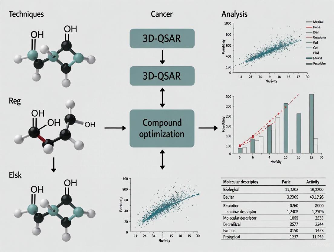
3D-QSAR in Cancer Drug Discovery: Techniques for Optimizing Anticancer Compounds
This article provides a comprehensive overview of 3D Quantitative Structure-Activity Relationship (3D-QSAR) techniques and their pivotal role in optimizing anticancer compounds.
3D-QSAR in Cancer Drug Discovery: Techniques for Optimizing Anticancer Compounds
Abstract
This article provides a comprehensive overview of 3D Quantitative Structure-Activity Relationship (3D-QSAR) techniques and their pivotal role in optimizing anticancer compounds. Aimed at researchers, scientists, and drug development professionals, it explores foundational principles, key methodological approaches including CoMFA, SOMFA, and Topomer CoMFA, and their practical applications against targets like HER2, EGFR, and aromatase. The content also addresses critical troubleshooting strategies for common challenges such as molecular alignment and model overfitting, and outlines robust validation protocols to ensure predictive reliability. By integrating 3D-QSAR with modern computational methods like machine learning and molecular docking, this guide serves as a strategic resource for accelerating the rational design of more effective and targeted cancer therapies.
Understanding 3D-QSAR: A Foundational Guide for Cancer Drug Optimization
Fundamental Concepts: From 2D-QSAR to 3D-QSAR
Quantitative Structure-Activity Relationship (QSAR) modeling represents a cornerstone of computational drug design, founded on the principle that variations in biological activity can be correlated with changes in molecular structure [1]. Classical 2D-QSAR approaches utilize physicochemical parameters—including hydrophobicity (logP), electronic properties (σ), and steric properties (Taft's Es, molar refractivity)—in a mathematical relationship, typically through multiple regression analysis [2]. The general form of a 2D-QSAR equation is Activity = A*P1 + B*P2 + C, where P1 and P2 are physicochemical properties, A and B are fitted coefficients, and C is a constant [2].
3D-QSAR extends this paradigm by incorporating the three-dimensional structural and interaction properties of molecules [1]. Instead of relying on simplistic parameters, 3D-QSAR techniques sample steric and electrostatic fields around aligned molecules within a 3D lattice, correlating these interaction fields with biological activity using robust statistical methods like Partial Least Squares (PLS) [3] [1]. This fundamental shift allows 3D-QSAR to model biomolecular recognition more directly, as it accounts for the spatial arrangement of functional groups and their complementary interactions with biological targets.
Comparative Analysis: Quantitative Performance and Strategic Advantages
Direct comparisons of 2D and 3D-QSAR methodologies consistently demonstrate the superior descriptive and predictive power of 3D approaches in most scenarios, particularly when modeling ligand-protein interactions.
Table 1: Performance Comparison of 2D-QSAR vs. 3D-QSAR Models
| Model Type | Dataset | Key Statistical Metrics | Interpretability |
|---|---|---|---|
| 2D-QSAR [3] | 36 HDAC inhibitors | R² up to 0.937 | Limited to parameter coefficients |
| 3D-QSAR (CoMFA) [3] | 36 HDAC inhibitors | 86.7% variance explained (steric), 82.3% variance explained (electrostatic) | High - 3D contour maps |
| 2D-QSAR (Machine Learning) [4] | 76 SARS-CoV-2 Mpro inhibitors | Test set R² = 0.72 (best model) | Limited - "Black box" concerns |
| 3D-QSAR (Field-based) [4] | 76 SARS-CoV-2 Mpro inhibitors | Test set R² = 0.71-0.72 | High - Visual field coefficients |
A comprehensive 2023 study directly addressing this comparison concluded that "many more significant models were obtained when combining 2D and 3D descriptors," attributing this improvement to the ability of "2D and 3D descriptors to code for different, yet complementary molecular properties" [5]. However, the unique strength of 3D-QSAR lies not only in its predictive accuracy but particularly in its interpretative capability through visual representation.
Key Strategic Advantages of 3D-QSAR
Spatial Understanding of Activity: 3D-QSAR provides visual contour maps that highlight regions where specific molecular properties enhance or diminish biological activity [1] [4]. For example, a study on SARS-CoV-2 Mpro inhibitors identified favorable steric interactions near a chlorobenzyl moiety and favorable electrostatic contributions from specific carbonyl groups [4].
Handling of Conformational Dependence: Unlike 2D methods, 3D-QSAR explicitly accounts for molecular conformation and alignment, which is critical for modeling interactions with structurally defined binding sites [5].
Guidance for Molecular Design: The visual output of 3D-QSAR directly suggests structural modifications—such as adding, removing, or repositioning functional groups—to optimize activity [4].
Diagram 1: 3D-QSAR Workflow for Cancer Compound Optimization
Core Methodologies and Protocols in 3D-QSAR
Comparative Molecular Field Analysis (CoMFA)
CoMFA, the pioneering 3D-QSAR technique, follows a standardized protocol [1]:
- Conformation Determination: Identify bioactive conformations, typically through crystallographic data or molecular docking.
- Molecular Alignment: Superimpose molecules using a common scaffold or pharmacophoric pattern.
- Grid Placement: Surround aligned molecules with a 3D lattice (typically 2.0 Ã… spacing).
- Field Calculation: Compute steric (Lennard-Jones) and electrostatic (Coulombic) interaction energies at each grid point using a probe atom.
- Statistical Analysis: Apply PLS regression to correlate field values with biological activity.
- Validation: Use leave-one-out cross-validation to determine model robustness (q²).
- Visualization: Generate 3D contour maps showing regions where specific fields enhance or reduce activity.
A CoMFA study on PDE4 inhibitors demonstrated this protocol's effectiveness, achieving a cross-validated q² of 0.565 and a conventional R² of 0.867, successfully guiding the design of more potent inhibitors [1].
Comparative Molecular Similarity Indices Analysis (CoMSIA)
CoMSIA addresses several CoMFA limitations by employing Gaussian-type distance functions and incorporating additional molecular fields [1]:
- Similarity Field Calculation: Computes steric, electrostatic, hydrophobic, and hydrogen bond donor/acceptor fields using a Gaussian function to avoid singularities.
- No Cut-off Requirements: The smooth distance dependence eliminates the need for arbitrary energy cut-offs.
- Enhanced Interpretation: The additional fields, particularly hydrophobicity and explicit hydrogen bonding, provide a more comprehensive interaction profile.
Table 2: Essential Research Reagents and Computational Tools for 3D-QSAR
| Resource Category | Specific Tools/Software | Primary Function in 3D-QSAR |
|---|---|---|
| Molecular Modeling | SYBYL [1], Chem-X [3], Flare [4] | Provides integrated environment for CoMFA/CoMSIA calculations |
| Field Calculation | Cresset FieldStere [4], OpenEye ROCS & EON [6] | Generates molecular interaction fields and shape descriptors |
| Statistical Analysis | PLS (Partial Least Squares) [3] [1] | Correlates field variables with biological activity |
| Alignment Tools | Maximum Common Substructure (MCS) [4], Database Alignment | Superimposes molecules for field comparison |
| Docking Software | Molecular docking algorithms [7] | Determines putative bioactive conformations |
Applications in Cancer Compound Optimization
3D-QSAR has demonstrated significant utility in optimizing anticancer agents, with several case studies highlighting its practical impact:
Histone Deacetylase (HDAC) Inhibitors
A seminal study on 36 indole amide hydroxamic acids as HDAC inhibitors established robust 2D and 3D-QSAR models [3]. While the 2D-QSAR achieved a high R² of 0.937, the 3D-QSAR CoMFA model provided spatial insights into steric and electrostatic requirements, explaining 86.7% and 82.3% of variance in respective fields. Docking simulations complemented these findings by highlighting critical interactions with the catalytic Zn²⺠ion. Based on these models, researchers proposed three novel compounds predicted to possess enhanced biological activity [3].
Kinase-Targeted Therapies
Receptor-based 3D-QSAR approaches have proven particularly valuable in kinase studies, combining molecular docking for pose prediction with conventional 3D-QSAR for activity correlation [8]. This hybrid methodology leverages structural information from kinase-inhibitor complexes to generate more reliable alignments and interpret results within a structural context, accelerating the optimization of selective kinase inhibitors for cancer therapy.
Adenosine A1 Receptor Antagonists
In a comprehensive drug discovery campaign for breast cancer therapeutics, researchers employed 3D-QSAR as part of an integrated computational workflow [7]. After identifying the adenosine A1 receptor as a promising target through bioinformatics analysis, the team utilized 3D-QSAR to guide the rational design of a novel compound (Molecule 10) that exhibited remarkable potency against MCF-7 breast cancer cells (IC₅₀ = 0.032 µM), significantly outperforming the positive control 5-FU [7].
Diagram 2: 3D-QSAR in Cancer Drug Discovery Pipeline
Protocol Implementation: Practical Guide for Cancer Research Applications
Standard CoMFA Protocol for Kinase Inhibitor Optimization
Based on established methodologies [1] [4], the following protocol provides a framework for implementing 3D-QSAR in cancer compound optimization:
Dataset Curation
- Collect 20-50 compounds with measured ICâ‚…â‚€ or Ki values against the cancer target
- Ensure structural diversity while maintaining a common scaffold
- Divide compounds into training (70-80%) and test sets (20-30%) using activity stratification
Molecular Alignment
- Identify maximum common substructure (MCS) using tools in SYBYL or Flare
- Align molecules based on MCS or pharmacophoric features
- Validate alignment quality through visual inspection and RMSD calculations
Field Calculation Parameters
- Grid spacing: 2.0 Ã… in x, y, z directions
- Probe atom: sp³ carbon with +1 charge
- Steric field: Lennard-Jones 6-12 potential
- Electrostatic field: Coulombic potential with distance-dependent dielectric
Statistical Analysis and Validation
- Perform PLS regression with leave-one-out cross-validation
- Require q² > 0.5 for predictive models
- Use bootstrapping or Y-scrambling to assess model robustness
- Validate with external test set (predicted R² > 0.6)
Model Interpretation and Design
- Generate steric and electrostatic contour maps at 80% and 20% contribution levels
- Identify regions where bulky substituents enhance (green) or diminish (yellow) activity
- Locate areas favoring electron-donating (blue) or electron-withdrawing (red) groups
- Propose structural modifications based on contour guidance
This protocol, when applied to a series of choline kinase inhibitors, yielded models with exceptional predictive power (q² > 0.99 for CoMFA and CoMSIA), enabling rational design of potent anticancer agents [2].
3D-QSAR represents a significant advancement over traditional 2D-QSAR methods by incorporating the critical third dimension of molecular structure and interaction fields. While 2D descriptors maintain utility for rapid screening and preliminary analysis, 3D-QSAR provides superior interpretability and direct structural guidance for molecular optimization. The technique's demonstrated success across multiple cancer drug discovery programs—from HDAC inhibitors to kinase-targeted therapies—confirms its enduring value in the medicinal chemist's toolkit. As 3D-QSAR methodologies continue to evolve through integration with machine learning and enhanced receptor-based approaches, their impact on cancer compound optimization is poised to expand further, accelerating the development of novel therapeutic agents against this complex disease.
The Critical Role of 3D-QSAR in Modern Cancer Drug Discovery
Three-dimensional Quantitative Structure-Activity Relationship (3D-QSAR) modeling has emerged as a transformative computational approach in modern anticancer drug discovery. By correlating the three-dimensional molecular properties of compounds with their biological activities, 3D-QSAR provides critical insights that guide the rational design and optimization of novel therapeutic agents. This application note explores the fundamental principles, methodological workflows, and successful implementations of 3D-QSAR techniques specifically in cancer research contexts. We present comprehensive protocols for building, validating, and applying 3D-QSAR models, along with detailed case studies demonstrating their efficacy in optimizing compounds against various cancer targets, including dihydrofolate reductase (DHFR) and breast cancer targets. The integration of 3D-QSAR with complementary computational methods such as molecular docking and ADMET profiling creates a powerful framework for accelerating anticancer drug development while reducing experimental costs.
Fundamental Principles
Traditional Two-Dimensional QSAR (2D-QSAR) methods utilize numerical descriptors derived from molecular structure to predict biological activity but lack consideration of the spatial orientation of molecules [9]. In contrast, 3D-QSAR incorporates the three-dimensional structural properties of ligands, providing a more comprehensive analysis of ligand-receptor interactions [10]. This approach is particularly valuable in cancer drug discovery, where understanding the spatial and electrostatic complementarity between potential drug candidates and their target binding sites is crucial for designing effective therapeutics.
The underlying hypothesis of 3D-QSAR is that differences in the three-dimensional structural properties of molecules are responsible for variations in their biological activities [10]. By quantifying these spatial relationships, researchers can identify key molecular features that contribute to anticancer efficacy and optimize lead compounds accordingly. 3D-QSAR has evolved into an indispensable predictive tool in the design of pharmaceuticals, significantly decreasing the number of compounds that need to be synthesized by facilitating the selection of the most promising candidates [10].
Key Methodological Approaches
Two primary computational techniques dominate the 3D-QSAR landscape: Comparative Molecular Field Analysis (CoMFA) and Comparative Molecular Similarity Indices Analysis (CoMSIA) [11]. CoMFA calculates steric (Lennard-Jones) and electrostatic (Coulomb) fields on a 3D grid surrounding aligned molecules, using a probe atom to measure interaction energies at each grid point [9]. This method provides detailed maps of regions where steric bulk or electrostatic charges influence biological activity.
CoMSIA extends this approach by employing Gaussian-type functions to evaluate steric, electrostatic, hydrophobic, and hydrogen-bonding fields, which results in smoother potential maps and reduced sensitivity to molecular alignment [9]. The enhanced descriptor set in CoMSIA provides more comprehensive insights into structure-activity relationships, particularly for structurally diverse datasets. Both methods utilize statistical techniques, primarily Partial Least Squares (PLS) regression, to correlate field descriptors with biological activity values [11].
Computational Workflows and Protocols
Molecular Modeling and Alignment
The initial phase in 3D-QSAR model development involves preparing high-quality three-dimensional molecular structures. This process begins with converting two-dimensional chemical representations into three-dimensional coordinates using cheminformatics tools such as RDKit or Sybyl [9]. The resulting 3D structures undergo geometry optimization through molecular mechanics force fields (e.g., UFF) or quantum mechanical methods to ensure they adopt realistic, low-energy conformations [9].
Molecular alignment represents the most critical step in 3D-QSAR and demands meticulous attention. As noted by Cresset, "The majority of the signal is in the alignments, so you need to get those right. If your alignments are incorrect your model will have limited or no predictive power" [12]. Alignment can be achieved through several approaches:
- Common Scaffold Alignment: Using the Bemis-Murcko method to define a core structure by removing side chains and retaining only ring systems and linkers [9]
- Maximum Common Substructure (MCS): Identifying the largest substructure shared among a set of molecules, useful for comparing diverse chemotypes [9]
- Field and Shape-Guided Alignment: Employing molecular field similarity to align compounds based on their electrostatic and shape properties [12]
A recommended protocol involves selecting a representative, highly active compound as an initial reference, aligning the dataset to this reference, identifying poorly aligned molecules, promoting well-aligned examples to additional references, and iterating until satisfactory alignment is achieved for all compounds [12].
Figure 1: Comprehensive 3D-QSAR Workflow for Cancer Drug Discovery. This flowchart illustrates the iterative process of model development, validation, and application in designing novel anticancer agents.
Descriptor Calculation and Model Building
Following molecular alignment, the next critical step involves calculating 3D molecular descriptors that numerically represent the steric and electrostatic environments of each molecule. In CoMFA, this is achieved by placing a lattice of grid points around the aligned molecules and using a probe atom (typically an sp³ carbon with a +1 charge) to measure steric (van der Waals) and electrostatic (Coulombic) interaction energies at each grid point [9]. This process effectively maps how a molecular probe "feels" the presence of the molecule at various locations, identifying regions where steric bulk or electrostatic properties influence binding.
CoMSIA extends this approach by calculating similarity indices using a Gaussian-type function for steric, electrostatic, hydrophobic, and hydrogen bond donor/acceptor fields [9]. The Gaussian function prevents singularities at atomic positions and provides smoother sampling of the molecular fields, making CoMSIA less sensitive to alignment variations than CoMFA.
With descriptors calculated, model building employs Partial Least Squares (PLS) regression to correlate the 3D field descriptors with biological activity values [11]. PLS is particularly suited for 3D-QSAR as it handles the large number of highly correlated descriptors by projecting them onto a smaller set of latent variables. The model undergoes cross-validation, typically using Leave-One-Out (LOO) methodology, to optimize the number of components and prevent overfitting [11].
Model Validation and Interpretation
Rigorous validation is essential to ensure model reliability and predictive power. Internal validation employs LOO cross-validation, where each compound is sequentially excluded from the training set and predicted by a model built from the remaining molecules [13]. The cross-validated correlation coefficient (q²) provides the first indicator of model predictivity, with q² > 0.5 generally considered acceptable [11].
External validation using a test set of compounds not included in model development offers a more robust assessment of predictive ability [11]. Additional statistical measures include the conventional correlation coefficient (r²), Fisher ratio (F), standard error of estimate, and bootstrapping analysis [11]. The model's contour maps are then interpreted to identify spatial regions where specific molecular features enhance or diminish biological activity, providing visual guidance for structural optimization [9].
Research Reagent Solutions: Essential Computational Tools
Table 1: Essential Software Tools for 3D-QSAR in Cancer Research
| Tool Category | Representative Software | Primary Function | Application in Cancer Drug Discovery |
|---|---|---|---|
| Molecular Modeling | Sybyl [11], ChemBio3D [13], RDKit [9] | 3D structure generation and optimization | Convert 2D chemical structures to 3D representations for cancer targets |
| Conformational Analysis | FieldTemplater [13], Py-ConfSearch [14] | Bioactive conformation identification | Determine likely binding conformations for anticancer compounds |
| Molecular Alignment | Py-Align [14], Forge [12] | Spatial superposition of molecules | Align compound series to common reference frame for field calculation |
| Field Calculation | Py-CoMFA [14], CoMSIA [11] | Steric/electrostatic field computation | Quantify molecular interaction fields around anticancer agents |
| Statistical Analysis | PLS algorithms [11], Py-ComBinE [14] | Model building and validation | Correlate field descriptors with anticancer activity data |
| Visualization | Py-MolEdit [14], Contour maps [9] | Interpretation of results | Visualize regions for structural modification to enhance anticancer activity |
Case Studies in Cancer Drug Discovery
DMDP Derivatives as Dihydrofolate Redase Inhibitors
A seminal application of 3D-QSAR in cancer drug discovery involved a series of 78 DMDP (2,4-diamino-5-methyl-5-deazapteridine) derivatives as potent anticancer agents targeting dihydrofolate reductase (DHFR) [11]. DHFR represents a validated anticancer target as it catalyzes the reduction of dihydrofolate to tetrahydrofolate, an essential cofactor in thymidylate and purine synthesis required for DNA replication and cell proliferation [11].
Researchers developed both CoMFA and CoMSIA models, with the CoMFA standard model demonstrating strong predictive power (q² = 0.530, r² = 0.903) and the CoMSIA model showing slightly improved statistics (q² = 0.548, r² = 0.909) [11]. The models successfully predicted the activities of a test set of ten compounds, producing predictive r² values of 0.935 and 0.842, respectively [11]. Contour map analysis revealed that highly electropositive substituents with low steric tolerance were required at the 5-position of the pteridine ring, while bulky electronegative substituents were favored at the meta-position of the phenyl ring [11].
Table 2: Statistical Parameters of 3D-QSAR Models for DMDP Derivatives as Anticancer Agents [11]
| Statistical Parameter | CoMFA Model | CoMSIA Model |
|---|---|---|
| Cross-validated q² | 0.530 | 0.548 |
| Non-cross-validated r² | 0.903 | 0.909 |
| Number of Components | 6 | 6 |
| F-value | 94.349 | Not specified |
| Standard Error of Estimate | 0.386 | Not specified |
| Predictive r² (Test Set) | 0.935 | 0.842 |
| Steric Field Contribution | 52.2% | Not specified |
| Electrostatic Field Contribution | 47.8% | Not specified |
Maslinic Acid Analogs Against Breast Cancer
Another significant application involved 3D-QSAR studies on maslinic acid analogs for anticancer activity against the breast cancer cell line MCF-7 [13]. Maslinic acid, a triterpene derived from olive oil extraction byproducts, demonstrates promising anticancer properties, though no comprehensive 3D-QSAR study had been reported previously [13].
Researchers developed a field-based 3D-QSAR model using 74 compounds with known IC₅₀ values against MCF-7 cells [13]. The derived QSAR model showed excellent statistical parameters (r² = 0.92, q² = 0.75) following leave-one-out cross-validation [13]. The model identified key structural features controlling anticancer activity and toxicity, enabling virtual screening of the ZINC database which yielded 593 initial hits [13]. Subsequent filtering through Lipinski's Rule of Five and ADMET risk assessment identified 39 promising candidates, with compound P-902 emerging as the most promising hit after docking studies against multiple breast cancer targets [13].
Benzimidazole Derivatives as Estrogen Alpha Receptor Antagonists
A recent study demonstrated the integration of 3D-QSAR with other computational methods for identifying novel benzimidazole derivatives as potential treatments for breast cancer targeting the estrogen alpha receptor (ERα) [15]. Researchers developed a pharmacophore model followed by an atom-based 3D-QSAR model with high correlation coefficients (R² = 0.9, Q² = 0.8) [15].
Virtual screening of benzimidazole scaffolds from PubChem, followed by molecular docking against ERα (PDB ID: 3ERT) and ADMET profiling, identified five promising compounds [15]. The top candidate (PubChem ID 3074802) demonstrated a binding affinity of -9.842 kcal/mol, significantly higher than the standard drug tamoxifen (-5.357 kcal/mol), along with favorable pharmacokinetic and low toxicity profiles [15]. This case study exemplifies how 3D-QSAR can be integrated into a comprehensive computational workflow for efficient anticancer lead identification and optimization.
Advanced Protocols and Implementation
Integrated Computational Workflow Protocol
The most effective implementation of 3D-QSAR in cancer drug discovery involves its integration within a broader computational framework:
Data Curation Protocol: Collect a minimum of 20-30 compounds with consistently measured biological activities (e.g., ICâ‚…â‚€ values) against a specific cancer target or cell line. Ensure structural diversity while maintaining a common scaffold for meaningful alignment [9].
Conformational Sampling Protocol: Generate low-energy conformations for each compound using systematic search or stochastic methods. Select the putative bioactive conformation using field-based similarity to known active compounds or through docking into the target protein when available [13].
Alignment Refinement Protocol: Implement the multi-reference alignment strategy described in Section 2.1, spending significant time on alignment quality before any model building activities. Critically, "Once you've hit the QSAR button, you're tainted, and are not allowed to tweak the molecules any more" to avoid statistical bias [12].
Model Optimization Protocol: Calculate both CoMFA and CoMSIA descriptors using a 2Ã… grid spacing. Optimize the region focusing and column filtering parameters to enhance signal-to-noise ratio. Build PLS models with component optimization based on LOO cross-validation [11].
Validation Protocol: Employ both internal (LOO) and external (test set) validation, with the test set comprising 15-20% of the total dataset selected to represent structural diversity and the entire activity range [11].
Figure 2: Molecular Alignment Protocol for 3D-QSAR. This specialized workflow highlights the critical alignment process that significantly influences model quality and predictive power.
Contour Map Interpretation Protocol
The practical application of 3D-QSAR models relies on accurate interpretation of contour maps:
Steric Map Interpretation: Green contours indicate regions where increased steric bulk enhances activity, while yellow contours denote regions where steric bulk decreases activity [9].
Electrostatic Map Interpretation: Blue contours represent regions where positive charge enhances activity, and red contours indicate regions where negative charge enhances activity [9].
Hydrophobicity Map Interpretation (CoMSIA): Yellow contours signify regions where hydrophobic groups favor activity, while white contours indicate regions where hydrophobic groups disfavor activity [13].
Hydrogen Bonding Map Interpretation (CoMSIA): Magenta contours (donor) and cyan contours (acceptor) identify regions where hydrogen bonding capabilities enhance activity [13].
These visual representations translate complex statistical models into intuitive guidance for medicinal chemists, directly suggesting structural modifications to enhance anticancer activity.
3D-QSAR continues to evolve as an indispensable tool in cancer drug discovery, enabling researchers to extract critical structure-activity insights and accelerate the optimization of anticancer agents. The case studies presented demonstrate how 3D-QSAR successfully identifies key structural features influencing anticancer activity across diverse chemical scaffolds and biological targets.
Future developments in 3D-QSAR methodology include the integration of advanced machine learning techniques such as Graph Convolutional Networks (GCNs), which process molecular graphs as inputs and synthesize atomic information into predictive features [14]. Additionally, network explainability methods are being developed to address the "opaqueness" of complex models, helping researchers understand which molecular regions contribute most significantly to predicted activity [14].
When properly implemented with rigorous alignment protocols and comprehensive validation, 3D-QSAR provides powerful insights that guide medicinal chemistry efforts in cancer drug discovery. By reducing the number of compounds requiring synthesis and testing through informed candidate selection, 3D-QSAR significantly enhances the efficiency of the anticancer drug development pipeline. The continued integration of 3D-QSAR with complementary computational approaches promises to further accelerate the discovery of novel, effective cancer therapeutics.
Three-dimensional Quantitative Structure-Activity Relationship (3D-QSAR) represents a fundamental advancement over classical QSAR by exploiting the three-dimensional properties of molecules to predict biological activities using robust chemometric techniques [10]. In the context of cancer therapy research, these methods have become indispensable tools for optimizing compound efficacy and overcoming drug resistance. The core principles of molecular fields, conformational analysis, and molecular alignment form the foundational triad of 3D-QSAR, enabling researchers to translate structural information into predictive models for anticancer drug design [16] [10]. The proper application of these principles allows medicinal chemists to identify critical structural features required for biological activity, thereby facilitating the rational design of novel therapeutic agents with enhanced potency and selectivity.
Molecular Fields and Descriptors
Molecular fields describe the spatial distribution of physicochemical properties around molecules, providing quantitative descriptors that correlate with biological activity [10]. These fields capture the essential interaction forces between a ligand and its biological target, including steric bulk, electrostatic potential, hydrophobic interactions, and hydrogen-bonding capabilities [16].
The primary 3D-QSAR techniques utilizing molecular fields include Comparative Molecular Field Analysis (CoMFA) and Comparative Molecular Similarity Indices Analysis (CoMSIA) [11]. CoMFA calculates steric and electrostatic fields using Lennard-Jones and Coulomb potentials, typically with a cutoff value of 30 kcal/mol to avoid energy singularities [11]. In contrast, CoMSIA employs a Gaussian-type function to eliminate singularities and incorporates additional fields including hydrophobic interactions and hydrogen bond donor/acceptor properties [16]. The selection of appropriate molecular fields depends on the specific biological target and the nature of ligand-receptor interactions being modeled.
Table 1: Key Molecular Field Types in 3D-QSAR
| Field Type | Physical Significance | Probe Atoms | Application in Cancer Research |
|---|---|---|---|
| Steric | Measures shape and bulk constraints | sp³ carbon atom | Optimizing substituents to fit binding pockets [11] |
| Electrostatic | Characterizes charge distribution | +1 charge | Enhancing selectivity through charge complementarity [16] |
| Hydrophobic | Quantifies lipophilicity | Atom-based hydrophobicity | Improving membrane permeability and bioavailability [16] |
| Hydrogen Bond Donor | Identifies H-bond donation sites | H-bond donor probe | Targeting key polar interactions in enzyme active sites [16] |
| Hydrogen Bond Acceptor | Identifies H-bond acceptance sites | H-bond acceptor probe | Exploiting complementary acceptor regions in targets [16] |
Conformational Analysis in 3D-QSAR
Conformational analysis aims to identify all possible minimum-energy structures of a molecule and establish the relationship between conformational flexibility and biological activity [17]. For flexible drug molecules, this process is critical because the bioactive conformation may not correspond to the global energy minimum, and different conformations can exhibit significantly different binding affinities to biological targets [18].
Several computational approaches exist for conformational sampling, each with distinct advantages:
- Systematic Search Method: Systematically rotates rotatable bonds through defined intervals to explore conformational space [17]
- Random Search Methods: Use stochastic algorithms to generate diverse conformational sets [17]
- Molecular Dynamics: Simulates molecular motion under physiological conditions to identify biologically relevant conformations [17]
- Neural Networks: Employ machine learning to predict stable conformations based on training data [17]
Following conformational generation, energy minimization is performed using force field methods to refine geometries. Subsequent cluster analysis groups similar conformations using root-mean-square distance (RMSD) as a similarity metric, with the lowest-energy representative from each cluster selected for further analysis [17]. In cancer drug discovery, this process is particularly important for designing compounds that target specific conformations of proteins involved in cell cycle regulation, such as CDK2 and tubulin [16].
Molecular Alignment Methodologies
Molecular alignment represents perhaps the most critical step in 3D-QSAR model development, as the results are highly sensitive to the alignment rules and overall orientation of the aligned compounds [11]. Proper alignment ensures that molecules are compared in a biologically relevant manner, mimicking their common binding mode to the target protein.
Table 2: Molecular Alignment Techniques in 3D-QSAR
| Alignment Method | Key Features | Advantages | Statistical Performance (q²/r²) |
|---|---|---|---|
| Rigid-Body Fit | Superposition based on common substructure or pharmacophore [17] | Intuitive, preserves structural similarity | Varies by dataset |
| Receptor-Based Alignment | Uses docking poses or co-crystallized conformers [17] | Biologically relevant orientation | CoMFA: q²=0.530, r²=0.903 [11] |
| Pharmacophore-Based Alignment (PBA) | Aligns molecules based on pharmacophoric features [19] | Focuses on key interaction elements | CoMSIA: q²=0.548, r²=0.909 [11] |
| Co-crystallized Conformer-Based Alignment (CCBA) | Uses experimentally determined bound conformation [19] | Highest biological relevance | Superior performance in case studies [19] |
| Distill Alignment | Template-based alignment using most active compound [16] | Optimizes for activity correlation | CoMSIA/SEHDA: Q²=0.814, R²=0.967 [16] |
Selection of the appropriate alignment method depends on available structural information. When available, co-crystallized conformer-based alignment (CCBA) generally provides the most reliable results, as demonstrated in a case study on PTP1B inhibitors where it generated CoMFA models with q²=0.694 and r²=0.992 [19]. For targets without experimental structural data, pharmacophore-based or receptor-based alignments using homology models offer viable alternatives [17].
Experimental Protocols and Application Notes
Protocol: CoMSIA Model Development for Cancer Therapeutics
This protocol outlines the development of a 3D-QSAR model using CoMSIA, based on a recent study of phenylindole derivatives as multitarget inhibitors in breast cancer therapy [16].
Step 1: Dataset Preparation and Biological Data Curation
- Compile a dataset of compounds with measured biological activities (e.g., ICâ‚…â‚€ values against cancer cell lines)
- Convert concentration values to pICâ‚…â‚€ (-logICâ‚…â‚€) to ensure linear relationship with free energy changes
- Divide dataset into training set (80-85%) for model development and test set (15-20%) for external validation
- Apply diversity analysis to ensure structural and activity ranges are represented in both sets
Step 2: Conformational Analysis and Molecular Alignment
- Generate low-energy conformations using systematic search or molecular dynamics
- Select the most active compound as template for alignment
- Align all molecules using the distill alignment method in SYBYL or similar software
- Verify alignment quality through visual inspection and RMSD calculations
Step 3: CoMSIA Field Calculations
- Create a 3D cubic grid with 2Ã… spacing extending beyond all aligned molecules
- Calculate five CoMSIA fields (steric, electrostatic, hydrophobic, H-bond donor, H-bond acceptor)
- Use a probe atom with charge +1, radius 1.0Ã…, hydrophobicity +1, and hydrogen bonding properties +1
- Set attenuation factor α to 0.3 for the Gaussian-type distance dependence
Step 4: Partial Least Squares (PLS) Analysis and Model Validation
- Perform leave-one-out (LOO) cross-validation to determine optimal number of components (N)
- Conduct non-cross-validated analysis with optimal N to generate final model
- Validate model using external test set predictions and bootstrapping analysis
- Accept models with q² > 0.5 and r² > 0.8 for reliable predictive capability
Application Note: Alignment Strategies for Flexible Molecules
For datasets containing flexible molecules with high Kier Flexibility Indices (>5.0), traditional alignment methods may prove suboptimal. Recent studies demonstrate that for certain nuclear receptors, including the androgen receptor, non-aligned 2D>3D conversion of structures can produce models with R²Test = 0.61, superior to energy-minimized and conformation-aligned approaches [18]. This alignment-independent 3D-QSDAR technique achieves this performance in only 3-7% of the computational time required for other conformational strategies, making it particularly valuable for large virtual screening campaigns in early-stage anticancer drug discovery [18].
Research Reagent Solutions
Table 3: Essential Computational Tools for 3D-QSAR in Cancer Research
| Tool Category | Specific Software/Platform | Key Functionality | Application in Protocol |
|---|---|---|---|
| Molecular Modeling | SYBYL [16] [11] | Structure building, optimization, force field calculations | Molecular sketching and Tripos force field optimization |
| 3D-QSAR Development | OpenEye Orion 3D-QSAR [6] | Consensus modeling with shape and electrostatic descriptors | Predict binding affinity using multiple similarity descriptors |
| Online QSAR Platforms | 3D-QSAR.com [20] | Web-based QSAR model development | Ligand-based and structure-based 3D-QSAR modeling |
| Visualization & Analysis | UCSF Chimera [16] | Protein-ligand interaction analysis | Visualization of docking poses and binding interactions |
| Activity Analysis | Flare QSAR [21] | Activity Atlas and Activity Miner components | SAR interpretation through Bayesian analysis of active molecules |
Visualization of Workflows
The following diagram illustrates the integrated workflow for 3D-QSAR model development in cancer therapeutic optimization:
The integration of molecular fields, conformational analysis, and alignment methodologies forms the cornerstone of successful 3D-QSAR applications in cancer drug discovery. Recent advances in these areas, particularly the development of robust CoMSIA models and alignment strategies adapted for flexible molecules, have demonstrated significant potential for optimizing anticancer agents [16] [18]. The continued refinement of these core principles, coupled with emerging technologies such as graph neural networks and machine learning approaches, promises to further enhance the predictive power of 3D-QSAR models [20]. For cancer researchers, mastering these fundamental techniques provides a powerful framework for addressing the persistent challenges of drug resistance and selectivity in oncology therapeutics, ultimately contributing to the development of more effective and targeted cancer treatments.
Three-dimensional quantitative structure-activity relationship (3D-QSAR) techniques represent a cornerstone of modern computational drug design, particularly when the three-dimensional structure of the biological target remains unknown. These methodologies establish correlations between the biological activities of structurally characterized compounds and the spatial characteristics of their molecular field properties, including steric demand, electrostatic interactions, and hydrophobicity [22]. In the specific context of cancer research, 3D-QSAR has emerged as an indispensable tool for optimizing lead compounds against various oncology targets, enabling researchers to understand the structural determinants of anticancer activity and rationally design more potent derivatives. The exponential increase in 3D-QSAR applications over recent decades underscores their value in supporting medicinal chemistry within drug discovery projects focused on oncology [22].
The fundamental premise of 3D-QSAR lies in the concept that differences in biological activity between compounds correlate with changes in their three-dimensional molecular interaction fields. Unlike traditional 2D-QSAR approaches that utilize physicochemical parameters, 3D-QSAR techniques analyze the spatial distribution of molecular properties, providing visual contour maps that directly suggest structural modifications to enhance potency [22]. This spatial understanding is particularly valuable in cancer drug discovery, where researchers can pinpoint specific structural features that influence binding to cancer-related targets such as kinase domains, hormone receptors, and apoptotic pathway components. This article provides a comprehensive overview of three pivotal 3D-QSAR techniques—CoMFA, CoMSIA, and SOMFA—with specific emphasis on their application in cancer compound optimization, complete with experimental protocols and implementation guidelines for research scientists.
Fundamental Techniques and Their Applications in Oncology
Comparative Molecular Field Analysis (CoMFA)
Comparative Molecular Field Analysis (CoMFA), pioneered by Cramer et al. in 1988, constitutes the most established 3D-QSAR technique [22]. The method operates on the principle that drug-receptor interactions are primarily governed by non-covalent forces that can be approximated by steric and electrostatic fields. In practice, CoMFA involves placing aligned molecules within a 3D grid and calculating steric (Lennard-Jones potential) and electrostatic (Coulombic potential) energies at each grid point using a probe atom [23]. Partial Least Squares (PLS) regression then correlates these field values with biological activity, generating predictive models and visual contour maps that highlight regions where specific molecular modifications would enhance activity.
In cancer drug discovery, CoMFA has demonstrated exceptional utility across multiple target classes. A recent study on quinazoline-4(3H)-one analogs as EGFR inhibitors for breast cancer treatment developed a CoMFA model with strong statistical parameters (R² = 0.872, Q² = 0.597), successfully identifying critical structural features responsible for inhibitory potency [23]. Similarly, CoMFA applications to pyrimidine-based adenosine A2A receptor antagonists for Parkinson's disease treatment (a relevant approach for managing cancer-related fatigue) yielded models with q² = 0.475 and r² = 0.977, enabling the rational design of novel antagonists with improved binding characteristics [24]. The region focusing variation of CoMFA further enhanced predictive ability (q² = 0.637), demonstrating the method's flexibility [24].
Comparative Molecular Similarity Indices Analysis (CoMSIA)
Comparative Molecular Similarity Indices Analysis (CoMSIA) extends beyond CoMFA by incorporating additional molecular fields and employing a Gaussian function type to avoid singularities at atomic positions [22]. While CoMFA considers only steric and electrostatic fields, CoMSIA typically includes hydrophobic, hydrogen bond donor, and hydrogen bond acceptor fields in addition to the fundamental steric and electrostatic components. This comprehensive approach often provides more interpretable models, particularly for cancer targets where hydrophobic interactions and hydrogen bonding play critical roles in ligand binding.
The enhanced capability of CoMSIA is evident in a recent study on triazolopyrazine derivatives as VEGFR-2 inhibitors for resistant breast cancer treatment. The CoMSIA model incorporating steric, electrostatic, and hydrophobic fields (CoMSIASEH) demonstrated excellent predictive power (Q² = 0.575, R² = 0.936, R²pred = 0.847) [25]. Similarly, research on quinazoline-based EGFR inhibitors revealed that a CoMSIA model combining steric, hydrophobic, and electrostatic fields (CoMSIASHE) achieved outstanding statistical parameters (R² = 0.982, Q² = 0.666) [23]. In studies of adenosine derivatives as antiplatelet aggregation inhibitors—particularly relevant for cancer patients at risk of thrombosis—CoMSIA yielded a significant model (q² = 0.528, r² = 0.943) that guided the design of novel therapeutic candidates [26]. The additional field types in CoMSIA provide a more nuanced understanding of structure-activity relationships, which is crucial when optimizing compounds for complex cancer targets.
Self-Organizing Molecular Field Analysis (SOMFA)
Self-Organizing Molecular Field Analysis (SOMFA) represents a simpler yet effective 3D-QSAR approach that utilizes molecular shape and electrostatic potential as primary descriptors [27]. Unlike CoMFA and CoMSIA, which rely on grid-based probe interactions, SOMFA calculates a master grid that encapsulates the average molecular properties of all compounds in the dataset. This method directly computes molecular shape and electrostatic potential without requiring probe atoms, potentially offering more intuitive interpretations, though it may capture fewer subtleties in molecular interactions.
In oncology applications, SOMFA has demonstrated robust performance in analyzing nonsteroidal anti-inflammatory drugs with cyclooxygenase-2 (COX-2) inhibitory activity—a relevant target for cancer prevention and treatment. A SOMFA study on stilbene analogs as COX-2 inhibitors produced a model with substantial statistical quality (r² = 0.806, r²cv = 0.799), which was successfully validated using an external test set (r²Test = 0.651) [28]. Research on adenosine derivatives as antiplatelet agents achieved a SOMFA model with r² = 0.615 and r²cv = 0.577, further confirming the method's utility in drug optimization workflows [26]. While SOMFA models generally exhibit lower statistical parameters than CoMFA or CoMSIA, their computational simplicity and straightforward interpretation make them valuable for initial analyses in cancer drug discovery programs.
Table 1: Comparison of Key 3D-QSAR Techniques in Cancer Research
| Technique | Molecular Fields | Statistical Performance | Advantages | Cancer Applications |
|---|---|---|---|---|
| CoMFA | Steric, Electrostatic | CoMFA_S: R²=0.872, Q²=0.597 [23] | Established method, robust performance | EGFR inhibitors, VEGFR-2 inhibitors, A2A antagonists |
| CoMSIA | Steric, Electrostatic, Hydrophobic, H-bond Donor, H-bond Acceptor | CoMSIA_SHE: R²=0.982, Q²=0.666 [23] | Multiple field types, avoids singularities | Breast cancer agents, kinase inhibitors, antiplatelet agents |
| SOMFA | Shape, Electrostatic | r²=0.806, r²cv=0.799 [28] | Simple interpretation, intuitive maps | COX-2 inhibitors, anti-inflammatory agents |
Experimental Protocols and Workflows
Molecular Alignment Protocols
Proper molecular alignment constitutes the most critical step in 3D-QSAR model development, as the resulting models are highly sensitive to the relative orientation and conformation of the molecules. Several alignment strategies have been developed, each with specific advantages for different scenarios in cancer drug discovery.
The common scaffold-based alignment approach is particularly useful when analyzing congeneric series with shared structural frameworks. In a study on quinazoline-4(3H)-one analogs as EGFR inhibitors, researchers employed the "distill rigid" module in SYBYL-X 2.1.1 software to align molecules based on their quinazolin-4-one common core, using the most active compound (compound 20) as a template [23]. Similarly, research on triazolopyrazine derivatives for breast cancer treatment utilized molecule 22 (the most active compound) as a reference structure for alignment [25]. This method ensures that the fundamental molecular skeleton is consistently positioned across all compounds, allowing the 3D-QSAR model to focus on the effects of substituent variations.
For structurally diverse compounds or when the bioactive conformation is unknown, pharmacophore-based alignment and docking-guided alignment offer viable alternatives. In a study on maslinic acid analogs for breast cancer, researchers used the FieldTemplater module in Forge software to generate a pharmacophore hypothesis from the most active compounds, then aligned all molecules to this template [29]. Docking-guided alignment leverages computational docking to generate putative binding poses, which are then used as alignment references. A study on stilbene analogs as COX-2 inhibitors employed this approach, superposing docked conformations to develop predictive SOMFA models [28]. For cancer targets with available crystal structures, this method can provide more biologically relevant alignments that approximate the true binding mode.
Model Building and Validation Procedures
Robust model building and validation are essential for developing reliable 3D-QSAR models with predictive value in cancer drug discovery. The standard workflow begins with dataset preparation, typically involving 20-50 compounds with measured biological activity (e.g., ICâ‚…â‚€, Ki) against a specific cancer target. Activity values are converted to logarithmic scales (pICâ‚…â‚€ = -logICâ‚…â‚€) to normalize the data distribution [23]. The dataset is then divided into training and test sets, typically using a 70-80%/20-30% ratio, with the test set selected randomly or through sphere exclusion methods to ensure structural and activity diversity [30].
Following molecular alignment, field calculations are performed specific to each 3D-QSAR technique. For CoMFA, steric and electrostatic fields are computed at grid points using a sp³ carbon probe with +1.0 charge and standard energy cutoff of 30 kcal/mol [25]. CoMSIA calculations incorporate additional similarity fields (hydrophobic, hydrogen bond donor, hydrogen bond acceptor) using a Gaussian function with attenuation factor α = 0.3 [23]. SOMFA computations directly calculate shape and electrostatic potential grids without probe atoms [28].
Partial Least Squares (PLS) regression serves as the core statistical method for correlating field values with biological activity. The optimal number of components is determined through leave-one-out (LOO) cross-validation, maximizing the cross-validated correlation coefficient (Q²) while minimizing overfitting [29]. The model undergoes multiple validation steps, including:
- Internal validation: Assessed by Q² > 0.5, non-cross-validated correlation coefficient (R²) > 0.6, and low standard error of estimate (SEE) [25]
- External validation: Evaluated using the test set with predicted correlation coefficient (R²pred) > 0.6 [28]
- Y-randomization: Confirms model robustness by scrambling activity values and demonstrating significantly worse performance in randomized models [25]
- Applicability domain: Assessed through William's plot (standardized residuals vs. leverage) to identify influential compounds and structural outliers [23]
Table 2: Statistical Parameters for Validated 3D-QSAR Models in Cancer Research
| Parameter | Symbol | Threshold | Example Values | Interpretation |
|---|---|---|---|---|
| Cross-validated correlation coefficient | Q² | > 0.5 | 0.560 (CoMFA) [26] | Internal predictive ability |
| Non-cross-validated correlation coefficient | R² | > 0.6 | 0.940 (CoMFA) [26] | Goodness of fit |
| Predicted correlation coefficient | R²pred | > 0.6 | 0.657 (CoMFA) [23] | External predictive ability |
| Standard error of estimate | SEE | Lower is better | 0.097 (CoMFA) [26] | Model precision |
| F-value | F | Higher is better | 71.850 (CoMFA) [26] | Statistical significance |
Model Interpretation and Compound Design
The primary value of 3D-QSAR in cancer drug discovery lies in interpreting contour maps to guide rational compound design. CoMFA steric contours indicate regions where bulky substituents enhance (green) or diminish (yellow) activity, while electrostatic contours highlight areas favoring electron-donating (blue) or electron-withdrawing (red) groups [23]. CoMSIA contours provide additional information on favorable hydrophobic (yellow/unfavorable white), hydrogen bond donor (cyan/unfavorable purple), and acceptor (magenta/unfavorable red) regions [25].
In a practical application, researchers analyzing quinazoline-4(3H)-one EGFR inhibitors used CoMSIA contour maps to identify specific structural modifications that would enhance potency [23]. Similarly, studies on triazolopyrazine VEGFR-2 inhibitors designed six novel compounds based on 3D-QSAR guidance, resulting in predicted improved binding affinities (-8.9 to -10.0 kcal/mol) compared to the reference drug Foretinib [25]. These examples demonstrate how contour map interpretation directly translates to molecular design decisions in cancer drug optimization.
Following compound design, virtual screening filters assess drug-likeness and synthetic feasibility. Standard approaches include Lipinski's Rule of Five for oral bioavailability, ADMET risk assessment for pharmacokinetic properties, and synthetic accessibility scoring [29]. Molecular docking further validates designed compounds by examining binding modes and interactions with key residues in the target protein's active site [23]. For promising candidates, molecular dynamics simulations (100 ns) with MM-PBSA calculations provide additional confirmation of binding stability and affinity [25].
Research Reagent Solutions and Technical Specifications
Successful implementation of 3D-QSAR in cancer research requires specific software tools and computational resources. The following table details essential research reagents and their applications in typical workflows.
Table 3: Essential Research Reagents and Computational Tools for 3D-QSAR Studies
| Category | Specific Tools | Function in 3D-QSAR Workflow | Application Examples |
|---|---|---|---|
| Molecular Modeling Suites | SYBYL-X, Forge, Spartan | Structure building, energy minimization, molecular alignment | SYBYL-X used for CoMFA/CoMSIA on quinazoline derivatives [23] |
| Quantum Chemical Software | Gaussian, DFT packages | Geometry optimization, charge calculation | DFT/B3LYP/6-31G* for quinazoline energy minimization [23] |
| Docking Software | Molegro Virtual Docker, AutoDock, GOLD | Binding pose generation, docking-guided alignment | MVD used for EGFR inhibitor docking studies [23] |
| ADMET Prediction | SwissADME, pkCSM | Drug-likeness screening, pharmacokinetic profiling | SwissADME used for triazolopyrazine derivative screening [25] |
| Dynamics Software | GROMACS, AMBER | Molecular dynamics simulations, binding free energy calculations | MM-PBSA calculations for VEGFR-2 inhibitors [25] |
Workflow Visualization
The following diagram illustrates the standard integrated protocol for 3D-QSAR-based cancer compound optimization, incorporating key decision points and validation steps:
Diagram 1: 3D-QSAR Cancer Compound Optimization Workflow
CoMFA, CoMSIA, and SOMFA represent powerful complementary techniques in the cancer drug discovery arsenal, each offering unique advantages for different optimization scenarios. CoMFA provides robust, interpretable models based on fundamental steric and electrostatic principles; CoMSIA delivers nuanced insights through multiple interaction fields; while SOMFA offers simplicity and directness in model interpretation. The integration of these 3D-QSAR approaches with molecular docking, ADMET prediction, and molecular dynamics simulations creates a comprehensive framework for rational drug design in oncology. As demonstrated through numerous cancer-focused applications, these methodologies successfully guide the transformation of lead compounds into optimized drug candidates with enhanced potency and improved pharmacological profiles. The continued refinement of 3D-QSAR protocols, coupled with advances in computational power and algorithmic sophistication, promises to further accelerate cancer drug discovery in the coming years.
Methodologies and Real-World Applications in Oncology
Within the framework of cancer compound optimization, Three-Dimensional Quantitative Structure-Activity Relationship (3D-QSAR) analysis serves as a pivotal computational method for correlating the three-dimensional spatial and electronic properties of molecules with their biological activity against cancer targets. This protocol details a comprehensive, step-by-step workflow for constructing robust 3D-QSAR models, from the initial critical stage of data set curation to the final model building and validation. The integration of this workflow into cancer drug discovery pipelines enables researchers to predict the activity of novel compounds, understand key interaction features, and rationally optimize lead compounds for enhanced potency, thereby accelerating the development of new oncology therapeutics [31] [32].
Protocol: Data Set Curation and Preparation
The foundation of a predictive and reliable 3D-QSAR model lies in the quality and consistency of the underlying data set. This initial phase demands meticulous attention to detail.
Data Acquisition and Initial Processing
- Source Selection: Begin by acquiring biological activity data (e.g., ICâ‚…â‚€, Ki) from publicly available databases like ChEMBL or through in-house experimental assays [33] [34]. For cancer research, ensure data is relevant to the target of interest (e.g., MCF-7 or MDA-MB-231 cell line inhibition [31]).
- Data Curation: Implement an automated curation workflow to standardize the data. This includes:
- Removing records with missing activity values.
- Handling duplicates by retaining the most reliable measurement or averaging replicates.
- Filtering out salts and standardizing tautomeric forms to ensure a consistent set of molecular structures [34].
- Modelability Assessment: Before proceeding, estimate the data set's "modelability" (MODI index). This step evaluates the feasibility of the data set to produce a predictive model, preventing unnecessary resource expenditure on non-modelable data [34].
Molecular System Preparation
- 3D Conformer Generation: Convert 2D structures (e.g., SMILES) into 3D conformations. This can be achieved using tools available in software packages like Orion, PharmQSAR, or Open Babel [35] [32] [33].
- Geometry Optimization and Charge Assignment: Minimize the energy of the generated 3D structures. Subsequently, assign partial atomic charges using appropriate methods such as AM1-BCC, Gasteiger, or semi-empirical Quantum-Mechanics (QM) approaches, which are critical for accurately describing electrostatic interactions [35] [32].
- Molecular Alignment: This is a critical step for 3D-QSAR. Align all molecules to a common reference frame based on:
- A shared, rigid scaffold (pharmacophore-based alignment).
- The predicted binding pose from molecular docking into a protein target.
- Field-based alignment using steric and electrostatic potentials [32].
Table 1: Key Parameters for Data Curation and Molecular Preparation
| Step | Key Parameter | Description/Recommended Value |
|---|---|---|
| Data Curation | Activity Data Type | ICâ‚…â‚€, Ki, log-based potency values [35] |
| Duplicate Handling | Retain consensus value or most reliable measurement [34] | |
| Conformer Generation | Method | Posit, FlexiROCS, or force-field based minimization [35] [32] |
| Charge Assignment | Charge Method | AM1-BCC, Gasteiger, or semi-empirical QM [35] [32] |
| Molecular Alignment | Alignment Rule | Pharmacophore, docking-based, or field-based [32] |
| Minimum Posit Probability (if applicable) | ≥ 0.5 for docking-derived poses [35] |
Protocol: 3D-QSAR Model Building
With a curated and aligned set of molecules, the process moves to calculating molecular descriptors and constructing the computational model.
Descriptor Calculation and Field Analysis
Calculate 3D molecular descriptors that encapsulate the steric, electrostatic, and hydrophobic properties of the aligned molecules. Modern approaches extend beyond traditional CoMFA/CoMSIA fields to include more advanced descriptors:
- Interaction Fields: Calculate steric (Lennard-Jones) and electrostatic (Coulombic) potential fields around each molecule using a probe atom [32].
- Quantum Chemical Descriptors: For enhanced accuracy, compute 3D electron density features via Density Functional Theory (DFT), which can be encoded into multi-scale descriptors like radial distribution functions and spherical harmonic expansions for a richer representation of electronic structure [33].
- Shape and Electrostatic Similarity: Utilize industry-leading tools like ROCS and EON for shape-based and electrostatic similarity descriptors, which serve as powerful inputs for machine learning models [35] [6].
Data Set Splitting and Model Training
- Training and Validation Sets: Split the curated data set into training and test sets. Common methods include:
- Model Construction: Apply machine learning algorithms to the training set to build a relationship between the 3D descriptors and biological activity. Common methods include:
- k-Partial Least Squares (k-PLS): Used for models like ROCS-kPLS and EON-kPLS, where the optimal number of features is determined via cross-validation [35].
- Gaussian Process Regression (GPR): Used for ROCS-GPR and EON-GPR models, which provides a measure of prediction confidence [35].
- Consensus (COMBO) Model: A robust approach where the final prediction is a weighted average of multiple individual models (e.g., 2D-GPR, ROCS-kPLS, ROCS-GPR, EON-kPLS, EON-GPR), with weights based on their individual prediction confidences [35].
Model Validation and Interpretation
- Statistical Validation: Evaluate model performance using multiple statistical metrics from cross-validation and external validation. Key metrics include:
- Pearson’s correlation coefficient squared (r²)
- Cross-validated correlation coefficient (q²)
- Median absolute error (MAE)
- Coefficient of Determination (COD) [35]
- Domain of Applicability: Analyze the model's applicability domain to ensure subsequent predictions are made for compounds within the chemical space of the training set [33].
- Model Interpretation: Visualize the 3D-QSAR contour maps (e.g., using PyMOL). These maps highlight regions in 3D space where specific chemical features (e.g., electron-donating groups, bulky substituents) favorably or unfavorably influence biological activity, providing direct, actionable insights for lead optimization [6] [32].
The following workflow diagram summarizes the entire process from data curation to a validated, interpretable model.
Diagram 1: A complete 3D-QSAR workflow for cancer compound optimization, from data sourcing to a validated predictive model.
Table 2: Core Statistical Metrics for 3D-QSAR Model Validation [35]
| Metric | Description | Interpretation |
|---|---|---|
| R² | Pearson’s correlation coefficient squared for training set. | Goodness-of-fit of the model. |
| q² | Cross-validated correlation coefficient (from LOO or other). | Indicator of model predictive ability. |
| COD | Coefficient of Determination from external validation. | Can be negative if worse than a baseline model predicting the average. |
| Median Absolute Error (MAE) | Median of absolute prediction errors. | Robust measure of prediction error magnitude. |
| Fraction Accurate vs. Confidence | Plot of accuracy versus model-estimated confidence. | Assesses correlation between confidence and accuracy. |
The Scientist's Toolkit: Essential Research Reagents & Software
Successful implementation of the 3D-QSAR workflow relies on a suite of specialized software tools and computational resources.
Table 3: Key Research Reagent Solutions for 3D-QSAR Analysis
| Tool/Resource Name | Type/Function | Relevance in 3D-QSAR Workflow |
|---|---|---|
| Orion 3D-QSAR Floes [35] | Automated Workflow Modules | Provides "3D QSAR Model: Builder" and "Predictor" floes for end-to-end model building and prediction using consensus models (ROCS-GPR, EON-kPLS, etc.). |
| PharmQSAR [32] | 3D QSAR Software Package | Builds statistical models (CoMFA, CoMSIA, HyPhar) using high-quality 3D molecular fields derived from semi-empirical QM calculations. |
| KNIME with Cheminformatics Extensions [36] [34] | Automated Workflow Platform | Enables the creation of fully automated, customizable workflows for data curation, descriptor calculation, machine learning, and validation. |
| 3D-QSAR.com [20] | Web Application Platform | Offers user-friendly, web-based tools for developing both ligand-based and structure-based 3D QSAR models. |
| Open Babel, RDKit [33] [34] | Cheminformatics Toolkits | Used for fundamental tasks like file format conversion, 2D to 3D structure conversion, and descriptor calculation within automated pipelines. |
| GROMACS [31] | Molecular Dynamics Simulation Software | Used for simulating the stability of protein-ligand complexes, which can inform the selection of biologically relevant conformers for 3D-QSAR. |
| Multiwfn [33] | Wavefunction Analyzer | Aids in calculating advanced quantum chemical descriptors, such as 3D electron density features, for enhanced model accuracy. |
| RN941 | RN941 (N-Phenylmaleimide) | High-purity RN941 (N-Phenylmaleimide), CAS 941-69-5. A key building block for polymer and bioconjugation research. For Research Use Only. Not for human use. |
| MMGP1 | MMGP1 Antifungal Peptide | MMGP1 is a marine metagenome-derived cell-penetrating peptide with potent activity againstCandida albicans. It is for Research Use Only. |
Application Note: A Case Study in Breast Cancer
A recent study on breast cancer treatment exemplifies this workflow. Researchers identified the adenosine A1 receptor as a key target via bioinformatics. After curating a set of active compounds, they performed molecular docking and dynamics simulations to study binding stability. A pharmacophore model was then constructed based on this binding information, which guided the virtual screening and rational design of a novel compound, Molecule 10. Subsequent synthesis and in vitro evaluation in MCF-7 breast cancer cells revealed potent antitumor activity (IC₅₀ = 0.032 µM), significantly outperforming the positive control 5-FU. This success underscores how a computational 3D-QSAR-driven workflow can efficiently deliver highly active therapeutic candidates [31].
The human epidermal growth factor receptor 2 (HER2) is a major oncogenic driver in approximately 20-30% of breast cancers and other carcinomas, where its overexpression portends poor clinical outcome [37] [38]. This receptor tyrosine kinase, a member of the ErbB family, exists as an orphan receptor with no known ligand but serves as the preferred dimerization partner for other HER family members [37] [39]. When dimerized, particularly with HER3, it activates potent downstream signaling through the MAPK and PI3K-Akt pathways, promoting uncontrolled cell growth and survival [38] [39]. Targeting the intracellular tyrosine kinase domain of HER2 has emerged as a validated therapeutic strategy, complementing antibody-based approaches that target the extracellular domain [40] [38].
Within cancer drug discovery, Three-Dimensional Quantitative Structure-Activity Relationship (3D-QSAR) techniques represent powerful computational approaches for optimizing lead compounds when the correlation between molecular structure and biological activity must be understood in three-dimensional space [41]. Unlike classical 2D-QSAR that uses molecular descriptors independent of x,y,z coordinates (e.g., logP, molar refractivity), 3D-QSAR employs a set of values measured at different locations in the space around molecules [41] [42]. Self-Organizing Molecular Field Analysis (SOMFA) is one such grid-based, alignment-dependent 3D-QSAR method that simplifies the relationship between molecular properties and biological activity by using molecular shape and electrostatic potential to construct predictive models [43] [37]. This case study details the application of SOMFA to design novel quinazoline-based HER2 kinase inhibitors, demonstrating its utility within a broader thesis on structure-based drug design for oncology targets.
HER2 Signaling Pathway and Therapeutic Significance
The HER2 signaling axis represents a critical vulnerability in HER2-positive cancers. The following diagram illustrates the key components and interactions in the HER2 signaling pathway that make it a compelling drug target.
Diagram 1: HER2-HER3 signaling pathway driving oncogenesis. The pathway initiates when neuregulin (NRG) binds HER3, promoting heterodimerization with HER2. This triggers autophosphorylation of tyrosine residues, creating docking sites for adaptor proteins that activate downstream MAPK (proliferation) and PI3K-Akt (survival) signaling cascades. HER2's role as the preferred dimerization partner, despite having no known ligand, makes it a pivotal therapeutic target in HER2-positive cancers [37] [38] [39].
While monoclonal antibodies like trastuzumab target the extracellular domain of HER2, small-molecule tyrosine kinase inhibitors (TKIs) offer a complementary approach by competing with ATP for binding at the intracellular catalytic kinase domain [38]. This blocks HER2 autophosphorylation and subsequent activation of downstream proliferative and survival signals. However, the high degree of structural conservation among kinase domains presents a challenge for achieving specificity, and drug resistance often emerges through mutations or activation of alternative signaling pathways [37] [40]. These limitations underscore the need for sophisticated structure-based design approaches like SOMFA to develop more potent and specific HER2 inhibitors.
SOMFA Methodology in 3D-QSAR
SOMFA operates on the fundamental principle that differences in biological activity between compounds can be correlated with differences in their molecular interaction fields - primarily steric (shape) and electrostatic (charge distribution) properties [43] [37]. The method is alignment-dependent, meaning that the accuracy of the model heavily relies on the correct spatial superposition of the molecules being studied, typically based on a common scaffold or pharmacophoric features [37] [41].
The following diagram illustrates the key stages of the SOMFA workflow as applied to HER2 inhibitor design.
Diagram 2: SOMFA workflow for HER2 inhibitor optimization. The process begins with curating a congeneric series of compounds with known biological activities, followed by generating their bioactive conformations through molecular docking. After spatial alignment, steric and electrostatic fields are calculated at grid points surrounding the molecules. Partial Least Squares (PLS) analysis then correlates field values with biological activity to generate a predictive model, validated through statistical measures, which guides the design of novel inhibitors with enhanced potency [43] [37].
In SOMFA, the molecular shape and electrostatic potential are calculated at numerous points within a 3D grid encompassing the aligned molecules. The steric field describes van der Waals interactions (both attractive and repulsive), while the electrostatic field captures Coulombic interactions between the molecule and a probe [41]. These fields are then correlated with biological activity using Partial Least Squares (PLS) analysis, a statistical technique particularly suited for datasets with many collinear variables [43] [37]. The resulting model generates contour maps that visually identify regions where specific molecular properties (bulk, positive/negative charge) would enhance or diminish biological activity, providing medicinal chemists with clear design guidance [43] [37] [41].
Case Study: SOMFA Application to Quinazoline-Based HER2 Inhibitors
Dataset and Molecular Alignment
The foundational study applied SOMFA to a series of 24 quinazoline derivatives reported as multi-acting inhibitors targeting histone deacetylase (HDAC), epidermal growth factor receptor (EGFR), and HER2 [43] [37]. The biological activity data consisted of ICâ‚…â‚€ values measured against the HER2 kinase domain using the HTScan HER2/ErbB2 Kinase Assay Kit, which were converted to pICâ‚…â‚€ (-logICâ‚…â‚€) for QSAR analysis [37]. This dataset provided an ideal structural diversity and activity range for robust model development.
Molecular alignment, a critical step in SOMFA, was performed using an atom-based approach with compound 1 as the reference structure. The researchers investigated three independent conformational sets generated by different docking tools: AutoDock 4.2, HyperChem 8.0, and AutoDock Vina [37]. This comparative approach ensured that the resulting models were not biased by the selection of a single conformational generation method. The alignment aimed to minimize the root-mean-square (RMS) differences in the fitting of selected atoms relative to the reference molecule, ensuring consistent spatial orientation of the common quinazoline scaffold while allowing variation in substituent positions.
SOMFA Model Development and Statistical Validation
For each conformational set (AutoDock4, HyperChem, and AutoDock Vina), independent SOMFA models were generated and evaluated using PLS analysis. The models were assessed using several statistical measures, with cross-validated correlation coefficient (q²) indicating predictive ability, non-cross-validated correlation coefficient (r²) measuring goodness-of-fit, and F-test values reflecting overall statistical significance [43] [37].
Table 1: Statistical Parameters of Generated SOMFA Models for HER2 Inhibition
| Conformation Source | Cross-validated q² | Non-cross-validated r² | F-test Value | Components |
|---|---|---|---|---|
| AutoDock Vina | 0.767 | 0.815 | 97.22 | Not specified |
| AutoDock4 | Not reported | Not reported | Not reported | Not specified |
| HyperChem | Not reported | Not reported | Not reported | Not specified |
The model derived from AutoDock Vina-generated conformations demonstrated superior statistical quality with a cross-validated q² of 0.767, non-cross-validated r² of 0.815, and F-test value of 97.22, indicating a highly predictive and statistically significant model [43] [37]. The reasonable difference between q² and r² values suggests the model was not overfitted and possessed genuine predictive capability for novel quinazoline derivatives.
Key Structural Insights from SOMFA Contour Maps
Analysis of the SOMFA contour maps provided crucial insights into the structural requirements for potent HER2 inhibition:
- Steric Field Contours: Identified specific regions where bulky substituents either enhanced or diminished activity, guiding optimal placement of aromatic rings and alkyl chains.
- Electrostatic Field Contours: Revealed areas where positive or negative charge characteristics correlated with improved potency, informing the selection of electron-donating or electron-withdrawing substituents.
These contour maps effectively visualized the architecture of the HER2 kinase active site, highlighting favorable and unfavorable interaction regions without requiring explicit protein structural information. The models suggested specific molecular modifications to the quinazoline core structure that would enhance HER2 inhibitory potency while potentially maintaining activity against other targets (HDAC, EGFR) for multi-acting therapeutic effects.
Experimental Protocols
HER2 Kinase Inhibition Assay Protocol
Purpose: To quantitatively measure the inhibitory potency (ICâ‚…â‚€) of quinazoline derivatives against the HER2 kinase domain.
Materials:
- HTScan HER2/ErbB2 Kinase Assay Kit (Cell Signaling Technology)
- Test compounds (quinazoline derivatives in DMSO stock solutions)
- Active HER2/ErbB2 kinase (GST fusion protein)
- Biotinylated peptide substrate
- Phospho-tyrosine antibody for detection
- ATP solution
- Reaction buffer
- Stop solution
- Microplate reader capable of fluorescence detection
Procedure:
- Reaction Setup: In a 96-well plate, prepare reaction mixtures containing HER2 kinase, biotinylated peptide substrate, and varying concentrations of test compounds in reaction buffer.
- Reaction Initiation: Start the kinase reaction by adding ATP to each well.
- Incubation: Incubate the reaction plate at 30°C for 1 hour.
- Reaction Termination: Add stop solution to each well to terminate the kinase reaction.
- Detection: Transfer reaction mixtures to streptavidin-coated plates and detect phosphorylated substrate using phospho-tyrosine antibody and fluorescent immuno-detection.
- Data Analysis: Calculate percentage inhibition relative to controls (no inhibitor) and determine ICâ‚…â‚€ values using non-linear regression analysis.
- Data Conversion: Convert ICâ‚…â‚€ values to pICâ‚…â‚€ (-logICâ‚…â‚€) for QSAR analysis.
Notes: Include appropriate controls (blank, vehicle, reference inhibitor). Ensure compound solubility and DMSO concentration consistency across samples (typically <1% final concentration) [37].
SOMFA Modeling Protocol
Purpose: To develop predictive 3D-QSAR models correlating molecular fields with HER2 inhibitory activity.
Materials:
- Chemical structures of quinazoline derivatives (2D)
- Biological activity data (pICâ‚…â‚€ values)
- Molecular modeling software with SOMFA capability
- Protein Data Bank structure 3PPO (HER2 kinase domain)
- Docking software (AutoDock Vina, AutoDock4, or equivalent)
- Molecular mechanics optimization software (HyperChem or equivalent)
Procedure:
- Compound Preparation:
- Generate 3D structures of all compounds using PRODRG server or equivalent.
- Optimize geometries using molecular mechanics (MM+) followed by semi-empirical AM1 method until RMS gradient <0.001 kcal molâ»Â¹.
Protein Preparation for Docking:
- Obtain HER2 kinase domain structure (PDB: 3PPO).
- Add hydrogen atoms using Hbuild command in CHARMM.
- Remove all water molecules except tightly bound active site waters.
- Perform energy minimization using Adopted Basis Newton-Raphson and steepest descent methods.
Conformation Generation:
- Dock each compound into HER2 active site using AutoDock Vina, AutoDock4, and HyperChem separately.
- Retain the highest-ranked conformation for each compound from each method.
Molecular Alignment:
- Select the most active compound as template.
- Superimpose all compounds using atom-based alignment to minimize RMSD.
SOMFA Model Development:
- Define a 3D grid large enough to encompass all aligned molecules.
- Calculate steric and electrostatic fields at each grid point.
- Perform PLS analysis to correlate field values with pICâ‚…â‚€.
- Validate models using leave-one-out cross-validation.
Model Interpretation:
Research Reagent Solutions
Table 2: Essential Research Reagents and Computational Tools for HER2 Inhibitor Development
| Category | Specific Tool/Reagent | Function/Application | Key Features |
|---|---|---|---|
| Biological Assays | HTScan HER2/ErbB2 Kinase Assay Kit | Quantitative measurement of HER2 kinase inhibition | Includes active HER2 kinase, biotinylated substrate, detection antibody; utilizes fluorescent immuno-detection |
| Structural Biology | Protein Data Bank ID 3PPO | Source of HER2 kinase domain structure for docking studies | Crystal structure of kinase domain; enables structure-based design |
| Computational Docking | AutoDock Vina | Molecular docking to generate bioactive conformations | Improved speed and accuracy over AutoDock4; used for best-performing SOMFA model |
| Computational Docking | AutoDock4 | Alternative docking tool for conformation generation | Grid-based docking with Lamarckian genetic algorithm |
| Molecular Modeling | HyperChem 8.0 | Molecular mechanics optimization and conformation generation | Uses MM+ force field and semi-empirical AM1 method for geometry optimization |
| 3D-QSAR Analysis | SOMFA Software | Self-Organizing Molecular Field Analysis | Grid-based, alignment-dependent 3D-QSAR using molecular shape and electrostatic potential |
| Statistical Analysis | Partial Least Squares (PLS) | Correlation of molecular fields with biological activity | Handles multiple collinear variables; essential for 3D-QSAR model development |
This case study demonstrates that SOMFA represents a powerful 3D-QSAR approach for rational design of HER2 kinase inhibitors, as evidenced by the development of statistically robust models (q² = 0.767, r² = 0.815) for quinazoline derivatives [43] [37]. The contour maps generated from the analysis provide visual guidance for medicinal chemists, highlighting specific structural modifications likely to enhance potency while maintaining the multi-targeting profile of these compounds.
The integration of molecular docking with SOMFA proved particularly valuable, as the best model emerged from AutoDock Vina-generated conformations rather than simple energy-minimized structures [37]. This underscores the importance of considering biologically relevant conformations in 3D-QSAR studies. Furthermore, the study validates HER2 as a druggable target for quinazoline-based small molecules, offering an alternative to antibody-based therapies like trastuzumab, which face challenges of cost, drug-induced cardiac dysfunction, and resistance mechanisms [37] [38].
From a broader perspective, this work exemplifies the strategic application of 3D-QSAR within cancer drug discovery, particularly for kinase targets where structural conservation complicates selective inhibitor design. The SOMFA methodology enabled efficient optimization of HER2 inhibitory potency while potentially maintaining activity against other targets, showcasing how computational approaches can accelerate the development of targeted cancer therapies. As HER2 continues to be a critical target in breast cancer and other malignancies, the insights and protocols described here provide a valuable framework for ongoing drug discovery efforts against this important oncogenic driver.
Aromatase, a cytochrome P-450 enzyme, catalyzes the final step in estrogen biosynthesis and is a key therapeutic target for managing estrogen-receptor-positive (ER+) breast cancer in postmenopausal women [44] [45]. Aromatase inhibitors (AIs) suppress estrogen production, offering a crucial therapeutic strategy. However, challenges such as drug resistance and side effects necessitate the development of more potent and selective inhibitors [46] [45]. This application note details how three-dimensional quantitative structure-activity relationship (3D-QSAR) modeling serves as a powerful computational technique within a drug optimization pipeline to design novel, high-efficacy aromatase inhibitors.
3D-QSAR in Aromatase Inhibitor Development: Key Case Studies
The application of 3D-QSAR techniques, specifically Comparative Molecular Field Analysis (CoMFA) and Comparative Molecular Similarity Indices Analysis (CoMSIA), has successfully guided the rational design of aromatase inhibitors across different compound classes. The following case studies and summarized data (Table 1) highlight these successes.
Table 1: Summary of 3D-QSAR Models in Aromatase Inhibitor Optimization
| Compound Class | 3D-QSAR Model Type | Statistical Results (q² / r²pred) | Key Identified Compound / Insight | Reference |
|---|---|---|---|---|
| Flavonoids | CoMFA | 0.827 / 0.710 | 7-hydroxyflavanone beta-D-glucopyranoside (Predicted IC₅₀: 1.09 μM), ~3.5x more potent than lead. | [44] |
| Steroidal AIs | CoMFA & CoMSIA | CoMFA: 0.636 / 0.658CoMSIA: 0.843 / 0.601 | Model provided steric/electrostatic guidance for novel SAI design; 6 hits from NCI database. | [45] |
| Azole-based (Imidazole/Triazole) | CoMFA/GOLPE | 0.715 / - | Properly substituted coumarin derivatives showed highest potency and selectivity. | [47] |
| 1,4-Quinone & Quinoline | CoMSIA/SEDA | - | Model highlighted Electrostatic, Steric, and H-bond Acceptor fields; one candidate (Ligand 5) identified. | [48] |
Case Study 1: Virtual Screening of Natural Flavonoids
A 3D-QSAR study on 45 flavonoids demonstrated the utility of this approach for identifying potent natural product-derived inhibitors [44]. The established CoMFA model exhibited strong predictive power, which was then used for virtual screening of a flavonoid database. This process identified 7-hydroxyflavanone beta-D-glucopyranoside as a highly promising candidate, with a predicted inhibitory concentration (IC₅₀) of 1.09 μM [44]. This represented an approximately 3.5-fold increase in potency compared to the initial lead compound, 7-hydroxyflavanone (IC₅₀: 3.8 μM). The stability of the ligand-aromatase complex was further confirmed via molecular dynamics (MD) simulation over 25 nanoseconds [44].
Case Study 2: Rational Design of Steroidal Aromatase Inhibitors
In the search for steroidal aromatase inhibitors (SAIs) with fewer side effects, 3D-QSAR was employed on a series of steroidal compounds [45]. The resulting CoMFA and CoMSIA models were statistically robust and provided 3D contour maps. These maps visualized the specific regions around the molecules where steric bulk, electrostatic charges, or hydrogen-bonding groups would enhance or diminish activity. This spatial information offers medicinal chemists a clear, visual guide for rational molecular design. The study also used a pharmacophore model for virtual screening, identifying six novel hit compounds from the NCI2000 database with predicted high activity [45].
Detailed Experimental Protocol: 3D-QSAR Workflow for Aromatase Inhibitors
The following section provides a detailed, step-by-step protocol for conducting a 3D-QSAR study on potential aromatase inhibitors, based on established methodologies [44] [45] [49].
Data Set Curation and Ligand Preparation
- Compound Selection: Assemble a data set of 30-60 compounds with known, quantitative aromatase inhibitory activity (e.g., ICâ‚…â‚€ or Ki values). Ensure structural diversity while maintaining a common core scaffold. The data set should be randomly divided into a training set (~80%) for model generation and a test set (~20%) for external validation [10] [45].
- Molecular Modeling and Optimization:
- Draw or retrieve 2D structures of all compounds from databases like PubChem.
- Convert 2D structures to 3D using software such as VLifeMDS or Maestro's LigPrep module.
- Perform geometry optimization and energy minimization using molecular mechanics force fields (e.g., MMFF94). Use a convergence criterion of 0.01 kcal/mol Ã… and an iteration limit of 100,000 [49].
Molecular Alignment
- Select a Template: Choose one of the most active compounds from the data set as a template for alignment.
- Common Substructure Alignment: Identify the common core scaffold (e.g., the flavone or steroidal nucleus) across all molecules. Superimpose all compounds onto this scaffold of the template molecule to ensure spatially consistent analysis [44] [45].
3D-QSAR Model Generation (CoMFA & CoMSIA)
- Define the Grid: Place the aligned molecules inside a 3D grid with a spacing of 2.0 Ã… in all directions. The grid should extend beyond the molecular dimensions by at least 4.0 Ã….
- Calculate Interaction Fields:
- CoMFA: Calculate steric (Lennard-Jones potential) and electrostatic (Coulombic potential) fields at each grid point using a sp³ carbon probe atom with a +1.0 charge [45].
- CoMSIA: In addition to steric and electrostatic fields, calculate hydrophobic, hydrogen bond donor, and hydrogen bond acceptor similarity indices using a Gaussian function to avoid singularities at the atomic positions [45] [48].
- Partial Least Squares (PLS) Analysis:
- Use the interaction energy values as independent variables (X) and the biological activity values (pICâ‚…â‚€ = -logICâ‚…â‚€) as the dependent variable (Y).
- Perform PLS regression to build the QSAR model. Apply leave-one-out (LOO) cross-validation to determine the optimal number of components and calculate the cross-validated correlation coefficient, q². A q² > 0.5 is generally considered statistically significant.
- Compute the conventional correlation coefficient, r², for the final non-validated model [44] [45].
Model Validation and Virtual Screening
- External Validation: Predict the activity of the external test set compounds using the generated CoMFA/CoMSIA model. Calculate the predictive correlation coefficient, r²pred, to assess the model's robustness [45].
- Contour Map Analysis: Interpret the 3D contour maps (e.g., green/yellow for favorable/unfavorable steric regions, blue/red for favorable/unfavorable electrostatic regions) to derive structural insights for molecular design [45].
- Virtual Screening: Use the validated model to predict the activity of compounds in large chemical databases (e.g., ZINC, NCI). Prioritize compounds with high predicted activity for further investigation [44] [45].
Experimental Validation
- Molecular Docking: Dock the top-scoring virtual hits into the crystal structure of aromatase (PDB ID: 3S7S) to analyze binding modes and key protein-ligand interactions (e.g., π-stacking with Phe134, Phe221, and Trp224, or H-bonding with Met374) [49] [50].
- Molecular Dynamics (MD) Simulation: Perform MD simulations (e.g., 50-100 ns) to evaluate the stability of the ligand-protein complex in a solvated environment. Monitor key parameters like root-mean-square deviation (RMSD) and root-mean-square fluctuation (RMSF) [44] [48].
- In Vitro Assays: Synthesize or procure the top candidates and evaluate their aromatase inhibitory activity and cytotoxicity in cell-based assays (e.g., using ER+ MCF-7 breast cancer cells) to confirm model predictions [48].
Overcoming Clinical Resistance: Linking Computational and Clinical Insights
A major challenge in AI therapy is the development of resistance. A key mechanistic insight involves the androgen receptor (AR). Studies comparing primary tumors with AI-resistant recurrences show significantly increased expression of the androgen receptor (AR) and its target, prostate-specific antigen (PSA), in resistant tumors [46]. This suggests a phenotypic shift from estrogen-dependent to androgen-dependent proliferation.
Table 2: Key Research Reagents and Computational Tools for AI Development
| Reagent / Tool | Function / Description | Application in AI Research |
|---|---|---|
| Aromatase (3S7S) | High-resolution X-ray crystal structure of human aromatase. | Essential for molecular docking and structure-based design. |
| VLife Molecular Design Suite | Software platform for molecular modeling, QSAR, and pharmacophore development. | Used for building 3D-QSAR models and virtual screening [49]. |
| Schrodinger Suite (Maestro) | Integrated drug discovery platform with LigPrep and Phase modules. | Ligand preparation, pharmacophore modeling, and molecular docking [51]. |
| GROMACS / AMBER | Software for molecular dynamics simulations. | Validates stability of ligand-aromatase complexes over time [44] [48]. |
| NCI Database | Public chemical database containing over 250,000 structures. | Source of compounds for virtual screening and hit identification [45]. |
| Gene Expression Signatures (e.g., E2F-GS) | Sets of genes representing biological pathways. | Identifies tumors with poor AI response and high proliferation post-therapy [52]. |
This resistance mechanism is corroborated by transcriptomic analyses from clinical trials like POETIC, which found that after just two weeks of AI therapy, tumors with a poor antiproliferative response exhibited high activity in gene signatures related to E2F transcription factors and TP53 dysfunction [52]. These pathways converge on cell cycle regulation, suggesting that resistance often involves bypassing the G1/S checkpoint. These clinical findings provide a compelling rationale for using computational models to design the next generation of AIs or combination therapies. For instance, 3D-QSAR could be employed to optimize dual-target inhibitors or compounds that simultaneously block aromatase and the AR, or to design molecules less prone to inducing these resistance pathways.
This application note demonstrates that 3D-QSAR is an indispensable tool in the modern drug developer's arsenal for optimizing aromatase inhibitors. By integrating 3D-QSAR with complementary techniques like molecular docking, dynamics, and clinical genomic data, researchers can effectively translate structural insights into potent, and potentially resistance-breaking, therapeutic candidates for ER+ breast cancer. This structured, computationally driven approach significantly accelerates the lead optimization process, paving the way for more effective and targeted cancer therapies.
Polo-like kinase 1 (PLK1) represents a critical serine/threonine protein kinase that regulates multiple aspects of the cell cycle, including centrosome maturation, kinetochore function, spindle formation, chromosome segregation, and cytokinesis [53] [54]. The significance of PLK1 as an anticancer target stems from its frequent overexpression in various human malignancies, including glioblastoma (GBM), where its elevated expression correlates with poor prognosis [55] [53]. PLK1 contains two primary domains: a conserved N-terminal catalytic kinase domain (KD) that binds ATP, and a C-terminal polo-box domain (PBD) that regulates substrate interactions and subcellular localization [56] [54]. In glioblastoma, a highly malignant and invasive brain tumor with limited treatment options, PLK1 inhibition has emerged as a promising therapeutic strategy. Current standard treatments for GBM primarily involve surgical resection, yet due to the highly infiltrative nature of GBM, complete eradication is challenging, leading to disease progression and recurrence with less than 5% of patients surviving beyond 5 years post-diagnosis [55].
Dihydropteridone derivatives represent a novel class of PLK1 inhibitors that exhibit promising anticancer activity and potential as chemotherapeutic drugs for glioblastoma [55]. These compounds exert their anticancer effects primarily by interfering with folate metabolism and inhibiting the dihydropteridone reductase pathway, thereby impeding nucleotide synthesis essential for tumor cell development and proliferation [55]. Recent structural advancements have incorporated an oxadiazole moiety into the dihydropteridone scaffold, significantly improving metabolic stability by ameliorating the inherent vulnerability of amides to hydrolysis by esterases and hepatic amidases [55]. This application case study examines the integration of computational approaches, particularly 3D-QSAR modeling, in the optimization and development of dihydropteridone-based PLK1 inhibitors for glioblastoma treatment.
Computational Methodologies for PLK1 Inhibitor Optimization
Quantitative Structure-Activity Relationship (QSAR) modeling represents a fundamental computational approach in modern drug discovery that establishes mathematical correlations between the structural attributes of compounds and their corresponding pharmacological activities [55]. These methodologies can be broadly categorized into 2D and 3D approaches, each offering distinct advantages for inhibitor optimization. 2D-QSAR focuses primarily on elucidating the impact of molecular descriptors' quantity and class on drug activity, while 3D-QSAR emphasizes the correlation between molecular spatial configuration and biological activity by analyzing steric, electrostatic, hydrophobic, and hydrogen-bonding interactions [55] [53].
The Comparative Molecular Field Analysis (CoMFA) and Comparative Molecular Similarity Indices Analysis (CoMSIA) represent the most established 3D-QSAR techniques that employ field-based descriptors to characterize molecular properties within a defined spatial grid [53] [11]. These approaches have demonstrated exceptional utility in PLK1 inhibitor development, as evidenced by multiple studies that generated models with significant statistical parameters, including high R² values (>0.90) and substantial cross-validated correlation coefficients (q² > 0.53) [53] [11]. The integration of these computational methods with experimental validation provides a powerful framework for accelerating the discovery and optimization of novel PLK1 inhibitors, including dihydropteridone derivatives for glioblastoma therapy.
Experimental Workflow for 3D-QSAR Model Development
The following diagram illustrates the comprehensive workflow for developing and validating 3D-QSAR models in PLK1 inhibitor optimization:
Figure 1: 3D-QSAR Model Development Workflow
Case Study: 3D-QSAR Analysis of Dihydropteridone Derivatives
Compound Dataset and Structural Preparation
In the seminal study by Li et al. (2023), a series of 34 dihydropteridone derivatives with incorporated oxadiazole moieties were investigated for their PLK1 inhibitory activity [55]. The experimental half-maximal inhibitory concentration (IC₅₀) values spanned from 0.18 µM to 1.07 µM, indicating a substantial range of potency suitable for robust QSAR model development. The dataset was strategically partitioned into training and test sets using a 3:1 ratio, resulting in 26 compounds allocated to the training set for model construction and 8 compounds reserved as a test set for model validation [55].
Structural optimization was performed through a multi-step computational protocol. Initial 2D structures were sketched using ChemDraw and subsequently optimized using HyperChem software. The optimization process employed molecular mechanics force field (MM+) for preliminary optimization, followed by selection of either AM1 or PM3 semi-empirical quantum mechanical methods based on the presence or absence of sulfur and phosphorus atoms. The structures were cyclically optimized using the Polak-Ribiere algorithm until the root mean square gradient reached a threshold of 0.01 [55]. This comprehensive optimization ensured accurate representation of molecular geometry and electronic distribution essential for reliable 3D-QSAR analysis.
3D-QSAR Model Development and Validation
The study implemented multiple QSAR approaches to comprehensively evaluate the structural determinants of PLK1 inhibition. The Heuristic Method (HM) was employed to construct a 2D-linear QSAR model, while the Gene Expression Programming (GEP) algorithm was utilized to develop a 2D-nonlinear QSAR model. For 3D-QSAR analysis, the CoMSIA approach was introduced to investigate the impact of drug structure on activity through field contribution analysis [55].
Table 1: Performance Metrics of QSAR Models for Dihydropteridone Derivatives
| Model Type | R² | Q² | Standard Error of Estimate (SEE) | F-value | Key Descriptors |
|---|---|---|---|---|---|
| HM Linear (2D) | 0.6682 | 0.5669 | 0.0199 | N/R | Min exchange energy for C-N bond (MECN) |
| GEP Nonlinear (2D) | 0.79 (training) 0.76 (validation) | N/R | N/R | N/R | Six molecular descriptors |
| CoMSIA (3D) | 0.928 | 0.628 | 0.160 | 12.194 | Hydrophobic field, H-bond donor/acceptor |
Abbreviation: N/R - Not reported in the source material [55]
The 3D-QSAR paradigm demonstrated superior performance, characterized by excellent fit with formidable Q² (0.628) and R² (0.928) values, complemented by an impressive F-value (12.194) and minimized standard error of estimate (SEE) at 0.160 [55]. The most significant molecular descriptor in the 2D model, which included six descriptors, was identified as "Min exchange energy for a C-N bond" (MECN). When the MECN descriptor was combined with hydrophobic field information from the 3D analysis, it generated specific structural recommendations for novel compound design, leading to the identification of compound 21E.153, a novel dihydropteridone derivative that exhibited outstanding antitumor properties and docking capabilities [55].
Contour Map Analysis and Structural Insights
The CoMSIA contour maps provided critical insights into the structural requirements for PLK1 inhibition. The steric field analysis identified regions where bulky substituents either enhanced or diminished activity, while electrostatic contours highlighted areas favoring positive or negative charges. The hydrophobic field analysis revealed molecular regions where increased hydrophobicity correlated with improved potency, and hydrogen-bonding maps identified optimal positions for hydrogen bond donors and acceptors [55].
Integration of these contour maps with molecular descriptor data enabled the researchers to formulate specific structural modifications to enhance PLK1 inhibitory activity. This integrated approach demonstrated the complementary nature of 2D and 3D-QSAR methodologies, with 2D analysis identifying critical atomic-level interactions and 3D modeling providing spatial context for optimal field interactions with the PLK1 binding pocket.
Experimental Protocols
Protocol 1: 3D-QSAR Model Construction using CoMSIA
Principle: This protocol describes the methodology for developing a 3D-QSAR model using the Comparative Molecular Similarity Indices Analysis (CoMSIA) approach, which evaluates similarity indices in steric, electrostatic, hydrophobic, and hydrogen-bonding fields between molecules to correlate with biological activity [53] [11].
Materials:
- Chemical structures of compounds with known biological activity (ICâ‚…â‚€ values)
- Computational chemistry software (SYBYL, Forge, or equivalent)
- Molecular modeling workstation
Procedure:
- Structure Preparation and Optimization:
- Sketch 2D chemical structures using ChemDraw or equivalent software
- Import structures into molecular modeling software and convert to 3D representations
- Perform geometry optimization using molecular mechanics (MM+ force field) followed by semi-empirical quantum mechanical methods (AM1 or PM3)
- Employ algorithmic optimization (Polak-Ribiere method) until RMS gradient reaches 0.01 [55]
Molecular Alignment:
- Select the most active compound as a template for alignment
- Identify common structural features across the dataset
- Align all molecules to the template using atom-based or field-based fitting methods
- Verify alignment quality through visual inspection and RMSD calculations [11]
Field Calculation and Descriptor Generation:
- Define a 3D grid that encompasses all aligned molecules with 2Ã… spacing
- Calculate steric field descriptors using Lennard-Jones potential
- Compute electrostatic fields using Coulombic potential
- Determine hydrophobic fields based on atom-based hydrophobicity parameters
- Assess hydrogen-bond donor and acceptor fields using appropriate probes [53] [11]
Partial Least Squares (PLS) Analysis:
- Implement leave-one-out (LOO) cross-validation to determine optimal number of components
- Perform non-cross-validated analysis to generate conventional R² values
- Calculate field contribution percentages for each descriptor type
- Validate model robustness through bootstrapping analysis (100 runs recommended) [11]
Model Interpretation:
- Generate 3D contour maps visualizing regions where specific molecular fields enhance or diminish activity
- Correlate contour map features with structural characteristics of high-activity compounds
- Formulate structural modification hypotheses based on contour map guidance
Notes: CoMSIA results are highly dependent on molecular alignment quality. Multiple alignment strategies should be explored, and the resulting models compared for statistical significance and predictive capability. The model should be considered reliable when Q² > 0.5 and R² > 0.8 [53] [11].
Protocol 2: Virtual Screening and Compound Design
Principle: This protocol utilizes established 3D-QSAR models for virtual screening of compound libraries and rational design of novel derivatives with predicted enhanced activity against PLK1 [56] [57].
Materials:
- Validated 3D-QSAR model
- Chemical database or virtual compound library
- Molecular docking software (AutoDock, GOLD, or equivalent)
- ADMET prediction tools
Procedure:
- Pharmacophore Model Generation:
Database Screening:
- Apply pharmacophore models to screen chemical databases (e.g., ZINC, NCI, Marine Natural Products)
- Filter results based on fit values and chemical diversity
- Select top candidates for further analysis [56]
Activity Prediction:
ADMET Profiling:
Scaffold Hopping and Molecular Hybridization:
Docking Studies and Binding Mode Analysis:
Notes: Virtual screening should balance predicted potency with structural novelty and synthetic accessibility. Consider employing multiple complementary screening approaches to reduce false positives and identify structurally diverse hit compounds [56] [59].
Research Reagent Solutions
Table 2: Essential Research Reagents and Computational Tools for PLK1 Inhibitor Development
| Reagent/Tool | Specifications | Research Application | Key Features |
|---|---|---|---|
| Chemical Modeling Software | SYBYL, Forge, HyperChem | Molecular structure optimization and conformational analysis | MM+ force field, AM1/PM3 methods, Polak-Ribiere algorithm [55] [11] |
| Descriptor Calculation Tools | CODESSA, PaDEL, Mold2 | Molecular descriptor calculation for QSAR analysis | Quantum chemical, structural, topological, geometrical descriptors [55] [54] |
| 3D-QSAR Platforms | SYBYL-X, Forge FieldQSAR | CoMFA and CoMSIA model development | Steric, electrostatic, hydrophobic, H-bond field calculation [58] [53] |
| Docking Software | AutoDock, GOLD, Molecular Operating Environment | Binding mode analysis and virtual screening | Lamarckian genetic algorithm, empirical scoring functions [53] [59] |
| Chemical Databases | ZINC, NCI, ChEMBL, Marine Natural Products Database | Compound sourcing and virtual screening | Diverse chemical libraries with drug-like properties [56] [54] [13] |
| PLK1 Protein Structures | PDB: 2RKU, 3KB7, 2YAC | Molecular docking and structure-based design | Crystal structures with resolution ≤ 2.2Å [53] [60] |
| ADMET Prediction Tools | Discovery Studio, pkCSM, admetSAR | Drug-likeness and toxicity assessment | BBB permeability, HIA, hepatotoxicity predictions [56] [13] |
The integration of 3D-QSAR modeling with complementary computational approaches has demonstrated significant utility in the optimization of dihydropteridone derivatives as PLK1 inhibitors for glioblastoma therapy. The case study presented herein illustrates how CoMSIA-based 3D-QSAR analysis successfully identified critical structural features and field interactions governing PLK1 inhibitory activity, resulting in the design of compound 21E.153 with outstanding predicted antitumor properties [55]. The exceptional statistical parameters of the developed model (R² = 0.928, Q² = 0.628) underscore the predictive capability of this approach in rational drug design [55].
The broader implication of this research lies in the validation of integrated computational strategies for accelerating anticancer drug discovery. By combining 2D molecular descriptors with 3D field analysis and molecular docking, researchers can efficiently navigate chemical space and prioritize synthetic efforts toward compounds with enhanced likelihood of success [55] [53] [57]. This methodology is particularly valuable for challenging targets like PLK1, where selectivity concerns and toxicity issues have hampered clinical development of earlier inhibitors [58] [54]. The continued refinement of these computational approaches, coupled with experimental validation, holds promise for delivering novel therapeutic options for glioblastoma patients facing limited treatment alternatives.
Integrating 3D-QSAR with Molecular Docking for Binding Mode Analysis
Within the context of cancer compound optimization research, the integration of computational methodologies has become indispensable for accelerating lead identification and development. Among these, the combination of three-dimensional Quantitative Structure-Activity Relationships (3D-QSAR) and molecular docking has emerged as a powerful synergistic strategy [61]. This protocol details their application for elucidating the binding mode of bioactive compounds, thereby enabling the rational design of novel anticancer agents with improved potency and selectivity. This approach moves beyond traditional ligand-based design by incorporating critical insights from the target protein's structure, providing a more comprehensive understanding of the molecular interactions governing biological activity.
Key Applications in Cancer Research
The integrated 3D-QSAR and molecular docking approach has been successfully applied to optimize various anticancer compound classes. The table below summarizes representative studies.
Table 1: Applications of Integrated 3D-QSAR and Docking in Cancer Compound Optimization
| Compound Class / Target | Cancer Type | Key Findings | Statistical Performance (r²/q²/pred. r²) | Citation |
|---|---|---|---|---|
| Nicotinamide-based SIRT2 Inhibitors | Various (Therapeutic potential in cancer & neurodegenerative diseases) | Developed 3D-QSAR and machine learning models to predict inhibition and selectivity for SIRT1/2/3 isoforms. | Model reliability confirmed via external validation; selectivity models showed predictive power. [62] | |
| Sipholane Inhibitors | Metastatic Breast Cancer | 3D-QSAR and pharmacophore models identified key features for Brk phosphorylation inhibition; guided design of a simplified, more synthetically accessible scaffold. | Models identified important pharmacophoric features correlating 3D structure with anti-migratory activity. [63] | |
| Maslinic Acid Analogs | Breast Cancer (MCF-7 cell line) | Field-based 3D-QSAR model identified key SAR regions; virtual screening, ADMET, and docking identified top hit compound P-902. | LOO-validated PLS model: r² = 0.92, q² = 0.75. [64] | |
| V600E B-RAF Inhibitors | Melanoma | Combined 3D-QSAR (CoMFA/CoMSIA) with docking to reveal structural features for binding affinity in the active site; new designs showed higher predicted potency. | CoMFA: q²=0.753, r²=0.962, pred. r²=0.89. CoMSIA: q²=0.807, r²=0.961, pred. r²=0.88. [65] | |
| 5-Lipoxygenase (5-LO) Inhibitors | Inflammatory/Allergic ailments (Cancer-adjacent pathways) | Molecular shape descriptors and docking yielded a predictive model, explaining variance in activity and revealing key ligand-target interactions. | Model successfully predicted inhibitory activity of an external test set. [61] |
Experimental Protocols
Protocol 1: Developing and Validating a 3D-QSAR Model
This protocol describes the creation of a field-based 3D-QSAR model, a critical step for understanding the steric and electrostatic fields influencing biological activity [64].
Data Set Curation and Preparation
- Collect a training set of compounds with reliable biological activity data (e.g., IC50, Ki) from the scientific literature. For the maslinic acid study, 74 compounds with activity against the MCF-7 breast cancer cell line were used [64].
- Convert 2D chemical structures into 3D models using software like ChemBio3D.
- Minimize the energy of all 3D structures using an appropriate force field (e.g., XED force field) with a gradient cut-off of 0.1 [64].
Pharmacophore Generation and Molecular Alignment
- Pharmacophore Generation: If the target-bound structure is unknown, use a software module like FieldTemplater. This tool uses field and shape information from highly active compounds to generate a pharmacophore hypothesis representing the putative bioactive conformation [64].
- Conformational Hunt: Use a molecular field-based similarity method (e.g., within Forge software) to find the lowest energy conformations of each training set compound that best match the generated pharmacophore template.
- Alignment: Align all training set compounds onto the identified pharmacophore template. This crucial step ensures that the molecular fields of all compounds are compared in a common 3D space.
Model Construction using Partial Least Squares (PLS) Regression
- Calculate field point-based descriptors (e.g., steric, electrostatic, hydrophobic) for the aligned molecules.
- Use the PLS regression algorithm (e.g., the SIMPLS algorithm) to build a model that correlates the molecular field descriptors with the biological activity (pIC50 = -log(IC50)) [64].
- Set parameters such as the maximum number of PLS components (e.g., 20) and the sample point distance (e.g., 1.0 Ã…) [64].
Model Validation
- Internal Validation: Perform Leave-One-Out (LOO) cross-validation to determine the cross-validated correlation coefficient (q²). A value of q² > 0.5 is generally considered acceptable [64].
- Statistical Goodness-of-Fit: Calculate the non-cross-validated correlation coefficient (r²) for the model.
- External Validation: Validate the model's predictive power using a test set of compounds that were not included in the model building process. A satisfactory predictive r² value (e.g., > 0.5) indicates a robust model [65] [64].
- Y-Randomization: Scramble the activity data and re-build the model to confirm that the original model is not the result of a chance correlation [65].
Protocol 2: Integrated Molecular Docking and Selectivity Analysis
This protocol uses docking to elucidate binding interactions and build selectivity models, which is essential for designing targeted cancer therapies [62].
Protein and Ligand Preparation
- Obtain the 3D structure of the target protein (e.g., SIRT2, B-RAF, NR3C1) from the Protein Data Bank (PDB). Prepare the protein by adding hydrogen atoms, assigning partial charges, and removing water molecules.
- Prepare the ligand structures for docking by energy minimization and generating possible tautomers and protonation states at biological pH.
Molecular Docking Simulations
- Define the binding site, often based on the location of a co-crystallized ligand (e.g., PDB ID: 1UWJ for B-RAF) [65].
- Perform docking simulations to predict the binding pose and affinity of each ligand. Multiple docking algorithms may be employed to ensure consensus.
- Analyze the predicted binding modes to identify key residues involved in hydrogen bonding, hydrophobic interactions, and π-π stacking.
Binding Mode Analysis and Selectivity Modeling
- Correlate the docking results with the 3D-QSAR contour maps. This integration helps explain the structural requirements for activity observed in the QSAR model with specific protein-ligand interactions observed in the docking pose [65] [61].
- To design selective inhibitors, dock compounds against related protein isoforms (e.g., SIRT1, SIRT2, SIRT3).
- Develop classification models using machine learning algorithms (e.g., Naive Bayes, k-nearest neighbors) on the docking results to predict and optimize isoform selectivity [62].
Workflow Visualization
The following diagram illustrates the integrated workflow of the protocols described above.
The Scientist's Toolkit: Essential Research Reagents & Materials
Table 2: Key Research Reagents and Computational Tools for 3D-QSAR and Docking
| Item/Category | Specific Examples & Details | Function/Purpose in the Workflow |
|---|---|---|
| Chemical Compounds & Biological Data | Training set compounds with measured IC50/Ki (e.g., 86 nicotinamide-based inhibitors [62]); Target cancer cell lines (e.g., MDA-MB-231 [63]). | Provides the essential experimental activity data required to build and validate the computational models. |
| Computational Software: 3D-QSAR | Forge (FieldTemplater, Field QSAR) [64]; CoMFA (Comparative Molecular Field Analysis); CoMSIA (Comparative Molecular Similarity Indices Analysis) [65]. | Used for pharmacophore generation, molecular alignment, calculation of field descriptors, and PLS regression model development. |
| Computational Software: Docking & Modeling | Molecular docking software (e.g., AutoDock, GOLD, Glide); ChemBio3D [64]; XED force field [64]. | Performs energy minimization, conformational analysis, and predicts ligand binding poses and affinities within the protein active site. |
| Protein Structure Data | RCSB Protein Data Bank (PDB) structures (e.g., PDB ID: 1UWJ for B-RAF [65]). | Provides the 3D structural coordinates of the target protein, which are essential for molecular docking simulations. |
| Validation & Analysis Tools | Leave-One-Out (LOO) and Test-set validation protocols [64]; y-randomization test [65]; Machine learning algorithms (Naive Bayes, k-NN) [62]. | Ensures the statistical robustness, predictive power, and reliability of the developed models, guarding against overfitting. |
Overcoming Common Challenges and Enhancing Model Performance
In the field of cancer compound optimization, Three-Dimensional Quantitative Structure-Activity Relationship (3D-QSAR) analysis has become an indispensable computational tool for guiding the rational design of novel therapeutic agents. Unlike traditional 2D-QSAR methods that utilize numerical descriptors derived from molecular graphs, 3D-QSAR incorporates the three-dimensional spatial orientation of molecules, providing critical insights into stereoelectronic properties that govern biological activity [9] [1]. However, the accuracy and predictive power of 3D-QSAR models are critically dependent on one fundamental prerequisite: precise molecular alignment [12].
Molecular alignment refers to the process of superimposing a set of molecules in a common 3D coordinate system based on their putative bioactive conformations. This alignment assumes that all compounds share a similar binding mode to the same biological target [9]. The profound significance of alignment stems from the fact that the majority of the predictive signal in 3D-QSAR models originates from the spatial relationships between molecules rather than just their individual properties [12]. In cancer drug discovery, where researchers often work with structurally diverse compounds targeting oncogenic proteins, proper alignment ensures that steric, electrostatic, and hydrophobic field descriptors accurately reflect the true binding interactions.
Despite its critical importance, molecular alignment constitutes one of the most technically demanding aspects of 3D-QSAR, presenting several formidable challenges [9]. These include the uncertainty in determining bioactive conformations, sensitivity to alignment rules and overall orientation, and the potential introduction of subjective bias during manual alignment procedures [12] [11]. This application note addresses these challenges by presenting best practices, detailed protocols, and advanced tools for achieving robust molecular alignment in cancer drug optimization research.
Molecular Alignment Approaches and Methodologies
Comparative Analysis of Alignment Techniques
Researchers in cancer drug discovery employ various molecular alignment strategies, each with distinct advantages and limitations. The choice of alignment method depends on factors such as structural diversity of the compound series, availability of structural biology data, and the specific research objectives.
Table 1: Comparison of Molecular Alignment Techniques in 3D-QSAR
| Alignment Method | Key Principle | Best Use Cases | Advantages | Limitations |
|---|---|---|---|---|
| Pharmacophore-Based Alignment | Aligns molecules based on common 3D pharmacophoric features [13] | Diverse chemotypes with shared functional groups | Captures essential interaction points; Intuitive | Requires knowledge of key binding elements |
| Field-Based Alignment | Maximizes similarity of molecular interaction fields (steric, electrostatic) [12] | Structurally diverse compounds without obvious common scaffold | Considers overall molecular properties; Not dependent on atom-to-atom correspondence | Computationally intensive; May require multiple reference molecules |
| Common Substructure Alignment | Superimposes largest common structural framework [9] [11] | Congeneric series with well-defined core structure | Straightforward implementation; Reproducible | Limited to compounds sharing significant structural similarity |
| Template-Based Alignment | Uses a known active compound or receptor-bound conformation as template [12] | When high-quality structural data is available for reference compound | Biologically relevant if template bioactive conformation is known | Template selection critically influences results |
| Alignment-Independent Methods | Uses internal molecular coordinates instead of spatial alignment [18] | Large diverse datasets where alignment is problematic | Bypasses alignment challenges; Faster computation | May miss critical 3D spatial relationships |
Advanced Field-Based Alignment Protocol
Field-based alignment has emerged as a powerful approach for handling structurally diverse cancer compounds. The following multi-reference alignment protocol, adapted from Cresset's methodology, provides a robust framework for achieving high-quality alignments [12]:
Step 1: Reference Molecule Selection and Preparation
- Identify an initial reference molecule that represents the data set (typically a high-affinity compound with intermediate structural features)
- Invest significant effort in establishing its bioactive conformation using:
Step 2: Initial Alignment
- Align the entire dataset to the primary reference using substructure alignment algorithms to ensure common cores are properly superimposed
- Employ field and shape similarity scoring to orient variable substituents [12]
- Use maximum similarity mode to optimize electrostatic and steric field overlap
Step 3: Multi-Reference Expansion
- Systematically review initial alignments to identify poorly aligned molecules or regions not adequately constrained by the primary reference
- Select additional representative molecules covering structural diversity and manually refine their alignments based on chemical intuition and available structure-activity data
- Promote these to secondary references
Step 4: Iterative Refinement
- Re-align the complete dataset using all reference molecules with substructure constraints
- Repeat steps 3-4 until all molecules are satisfactorily aligned (typically requiring 3-4 reference molecules for most cancer compound datasets) [12]
Critical Consideration: Alignment refinement must be completed before running QSAR analysis and without reference to activity values to avoid introducing bias and invalidating the model [12].
The following workflow diagram illustrates this iterative alignment process:
Experimental Protocols and Case Studies in Cancer Research
Protocol: Common Substructure Alignment for Kinase Inhibitors
This protocol details a robust common substructure alignment method applied to a series of imidazo-pyridine derivatives targeting dual oncogenic pathways (AT1 and PPARγ), relevant in cancer metabolism and proliferation [66].
Materials and Software Requirements
- Molecular dataset with biological activities (IC50 or Ki values)
- Sybyl molecular modeling software (Tripos Associates) or equivalent
- Hardware: UNIX workstation with multiple processors recommended
Step-by-Step Methodology
Dataset Preparation and Conformation Generation
- Convert 2D structures to 3D coordinates using Concord or similar tools
- Apply energy minimization using Tripos Force Field or MMFF94 with Gasteiger-Hückel charges
- Set gradient convergence criterion to 0.05 kcal/molÃ… for precise geometry optimization
Template Selection and Alignment
- Identify the most active compound as alignment template (compound 63 in the DMDP study [11])
- Define the maximum common substructure (MCS) using systematic substructure search
- Apply database alignment routine to superimpose all molecules on the template based on MCS
Alignment Validation
- Visually inspect superposition of core structures and key functional groups
- Verify that conserved pharmacophoric elements are properly aligned
- Check that variable substituents sample different spatial regions without artificial constraints
3D-QSAR Model Implementation
- Position aligned molecules in a 3D grid with 2.0 Ã… spacing in all dimensions
- Calculate steric and electrostatic fields using a sp³ carbon probe with +1 charge
- Set energy cutoff values to 30 kcal/mol to handle singularities near atomic positions
- Perform Partial Least Squares (PLS) regression with leave-one-out cross-validation
In the imidazo-pyridine case study, this protocol yielded statistically robust CoMFA models with cross-validated q² values of 0.553 for AT1 antagonism and 0.503 for PPARγ activation, demonstrating predictive capability for dual-target cancer therapeutics [66].
Protocol: Pharmacophore-Guided Alignment for Natural Product Derivatives
This protocol applies to structurally complex natural product analogs like maslinic acid derivatives with anticancer activity against breast cancer cell lines [13].
Materials and Software Requirements
- Forge software (Cresset Group) or equivalent pharmacophore modeling tools
- FieldTemplater module for pharmacophore hypothesis generation
- XED force field for molecular mechanics calculations
Methodology
Bioactive Conformation Determination
- Select 5-10 representative active compounds spanning the potency range
- Use FieldTemplater to generate a common pharmacophore hypothesis based on field points and molecular shape
- Annotate the hypothesis with calculated field points representing positive/negative electrostatics, hydrophobicity, and shape
Compound Alignment
- Transfer the pharmacophore template to Forge alignment module
- Align all training set compounds to the template using field point similarity and volume overlap
- Select the best-matching low-energy conformations for each compound
Model Building and Validation
- Set PLS regression parameters to 20 maximum components with 1.0 Ã… sample point distance
- Apply 50 Y-scrambling runs to validate model robustness
- Use electrostatic and volume fields as descriptors for QSAR model construction
In the maslinic acid study, this approach generated a 3D-QSAR model with exceptional statistical parameters (r² = 0.92, q² = 0.75), enabling identification of key structural features responsible for MCF-7 breast cancer cell line cytotoxicity [13].
Table 2: Performance Metrics of 3D-QSAR Models from Cancer Research Case Studies
| Case Study | Alignment Method | Biological Target | q² (LOO-CV) | r² | Number of Compounds | Key Findings |
|---|---|---|---|---|---|---|
| Imidazo-pyridine Derivatives [66] | Common Substructure | AT1/PPARγ (Dual Target) | 0.553 (AT1) 0.503 (PPARγ) | 0.954 (AT1) 1.00 (PPARγ) | 31 | Bulky electronegative substituents enhance dual activity |
| Maslinic Acid Analogs [13] | Pharmacophore-Based | MCF-7 Breast Cancer Cells | 0.75 | 0.92 | 74 | Hydrophobic moieties at C-2 position critical for potency |
| DMDP Anticancer Agents [11] | Database Alignment | Dihydrofolate Reductase (DHFR) | 0.530 (CoMFA) 0.548 (CoMSIA) | 0.903 (CoMFA) 0.909 (CoMSIA) | 78 | Electropostive substituents at position 5 essential for DHFR inhibition |
| DMDP Test Set Prediction [11] | Database Alignment | Dihydrofolate Reductase (DHFR) | N/A | 0.935 (CoMFA) 0.842 (CoMSIA) | 10 | Model successfully predicted external test compounds |
Commercial and Open-Source Solutions
Table 3: Molecular Alignment and 3D-QSAR Software Tools
| Software Tool | Vendor/Provider | Key Alignment Features | QSAR Methods | Best For |
|---|---|---|---|---|
| Forge/Torch | Cresset | Field-based alignment, FieldTemplater, multi-reference alignment | Field-based QSAR, Activity Atlas | Handling diverse chemotypes without common scaffold [12] |
| Sybyl | Tripos/Certara | Database alignment, flexible ligand fitting, atom-based or field-based | CoMFA, CoMSIA | Congeneric series with well-defined core structure [1] [11] |
| PharmQSAR | Pharmacelera | Field-based molecular alignment using QM-derived fields | CoMFA, CoMSIA, HyPhar | Projects requiring quantum-mechanical accuracy [32] |
| OpenEye Orion | OpenEye | Shape and electrostatic similarity descriptors | Consensus 3D-QSAR with multiple descriptors | Virtual screening and lead optimization [6] |
| Schrödinger Suite | Schrödinger | Phase pharmacophore alignment, docking-based alignment | 3D-QSAR, Bayesian models | Structure-based design when protein structure available |
| RDKit | Open-Source | Maximum Common Substructure (MCS), pharmacophore alignment | PLS, machine learning methods | Academic research with budget constraints [9] |
Successful implementation of molecular alignment and 3D-QSAR requires both software tools and appropriate computational infrastructure:
- Structure-Activity Data: High-quality, consistent biological measurements (IC50, Ki, EC50) obtained under uniform experimental conditions [9]
- Conformational Sampling Tools: Systematic search algorithms (e.g., Monte Carlo, molecular dynamics) for exploring flexible torsion angles
- Force Fields: Parameter sets (MMFF94, Tripos, OPLS) for geometry optimization and energy evaluation [11]
- Quantum Chemical Methods: Semi-empirical (AM1, PM3) or DFT calculations for accurate electrostatic potential derivation [32]
- Validation Datasets: External test compounds with known activities for model verification [11]
- High-Performance Computing: Multi-processor workstations or cluster resources for handling computationally intensive field calculations [11]
Molecular alignment remains both a challenge and opportunity in 3D-QSAR studies for cancer drug discovery. The protocols and best practices outlined in this application note provide researchers with structured methodologies for addressing the alignment problem across various scenarios – from congeneric series to structurally diverse chemotypes. The case studies demonstrate that careful attention to alignment quality directly translates to predictive models with tangible impact on cancer drug optimization.
Emerging trends in the field include the integration of machine learning for automated alignment quality assessment, the development of alignment-free 3D descriptors that maintain spatial information [18], and the increasing incorporation of protein structural data to inform alignment decisions. As these methodologies continue to evolve, the integration of robust alignment strategies with advanced artificial intelligence approaches promises to further enhance the predictive power of 3D-QSAR in cancer therapeutic development.
The following diagram illustrates the strategic decision process for selecting appropriate alignment methods based on dataset characteristics:
Managing Data Quality and Avoiding Model Overfitting
In the field of cancer compound optimization, 3D Quantitative Structure-Activity Relationship (3D-QSAR) modeling is a powerful computational tool for predicting the biological activity of molecules based on their three-dimensional properties. The predictive power and reliability of these models are paramount for guiding the rational design of novel anti-cancer therapeutics. However, the utility of any 3D-QSAR model is critically dependent on two foundational pillars: the quality of the input data and the rigorous avoidance of model overfitting. Overfit models, which memorize training set noise rather than learning the underlying structure-activity relationship, fail to predict new compounds accurately, potentially misdirecting drug discovery efforts. This application note details established protocols for managing data quality and implementing robust validation techniques to ensure the development of predictive and reliable 3D-QSAR models in cancer research.
Data Curation and Preparation Protocols
The first line of defense against poor model performance is a meticulously curated dataset. Inaccurate chemical structures or biological activities introduce experimental noise that models can inadvertently learn, leading to overfitting.
1.1. Chemical Structure Standardization
- Objective: To ensure a consistent and accurate representation of all molecular structures in the dataset.
- Protocol:
- Structure Representation: Convert all 2D structural representations (e.g., from SMILES strings) into 3D models using molecular sketching programs like ChemDraw and energy minimization suites such as ChemBio3D [29].
- Charge and Tautomer Assignment: Assign appropriate ionization states and major tautomers at a physiological pH of 7.4. Tools like OpenEye's toolkits or Schrödinger's LigPrep are commonly used for this purpose.
- Descriptor Calculation: Compute molecular descriptors using software such as Dragon, or generate fields and similarity indices using 3D-QSAR specific software like Forge or Sybyl [29] [67]. Charge-based and quantum chemical descriptors have been identified as particularly important for modeling anti-cancer activity [67].
1.2. Biological Data Curation and Outlier Identification
- Objective: To identify and address potential experimental errors in the biological activity data (e.g., ICâ‚…â‚€, Ki).
- Protocol:
- Data Sourcing: Collect biological data from reliable, peer-reviewed sources. Prefer data from consistent experimental protocols (e.g., the same cancer cell line, such as MCF-7 for breast cancer) to minimize inter-assay variability [29] [67].
- Consensus Prediction for Error Detection: Develop an initial QSAR model and perform internal cross-validation. Compounds with consistently large prediction errors across multiple model iterations should be flagged as potential outliers [68].
- Investigation: Manually investigate flagged compounds for plausible reasons for the discrepancy (e.g., structural misrepresentation, atypical mechanism of action, or genuine experimental error). The decision to remove an outlier must be scientifically justified and documented.
Table 1: Key Reagent Solutions for 3D-QSAR Data Preparation and Modeling
| Research Reagent / Software | Primary Function | Application Context |
|---|---|---|
| ChemDraw / ChemBio3D | 2D drawing and 3D structure generation/optimization | Converting 2D structures to 3D models; initial geometry optimization [29]. |
| Forge (Cresset) | Field-based alignment, pharmacophore generation, 3D-QSAR | Creating pharmacophore templates and aligning compounds using field points and molecular similarity [29]. |
| Sybyl (Tripos) | Molecular modeling, CoMFA, CoMSIA | Performing Comparative Molecular Field Analysis (CoMFA) and Comparative Molecular Similarity Indices Analysis (CoMSIA) [1] [69]. |
| OpenEye Toolkits | Structure standardization, charge assignment, conformer generation | Preparing ligands for modeling by assigning accurate charges and generating low-energy conformers [70] [6]. |
| Dragon Software | Molecular descriptor calculation | Calculating a wide array of 0D-3D molecular descriptors for QSAR model building [67]. |
Robust Model Validation Techniques
Validation is the process of assessing how the results of a statistical model will generalize to an independent dataset. Relying solely on the model's fit to the training data is insufficient and guarantees overfitting.
2.1. Data Set Division
- Objective: To create a representative training set for model development and a hold-out test set for unbiased evaluation.
- Protocol:
- Stratified Sampling: Divide the dataset into training and test sets (commonly 70-80% training, 20-30% test) using an activity-stratified method. This ensures that the test set covers a similar range of activity as the training set [29].
- External Validation: The test set must be completely excluded from the model building process and used only once to assess the final model's predictive power [71].
2.2. Internal and External Validation Metrics A model must pass multiple statistical checks to be considered valid and not overfit.
- Internal Validation (Cross-Validation): This assesses model stability. The most common method is Leave-One-Out (LOO) cross-validation, where each compound is left out once and predicted by a model built on the remaining compounds. The cross-validated correlation coefficient (q²) should be greater than 0.5 for a potentially predictive model [29] [69].
- External Validation: This is the gold standard for assessing predictive ability. The model is used to predict the hold-out test set. Several criteria should be met, as summarized in Table 2 [71].
Table 2: Key Statistical Metrics for 3D-QSAR Model Validation
| Metric | Formula / Description | Threshold for Validity | Interpretation |
|---|---|---|---|
| q² (LOO Cross-Validation) | q² = 1 - (PRESS/SSY) | > 0.5 | Measures model stability and internal predictive ability. |
| r² (Coefficient of Determination) | r² = 1 - (RSS/TSS) | > 0.6 | Measures goodness-of-fit for the training set. |
| Concordance Correlation Coefficient (CCC) | CCC = 2Ïσₓσᵧ/(σₓ² + σᵧ² + (μₓ - μᵧ)²) | > 0.8 | Measures the agreement between observed and predicted values; superior to r² for external validation [71]. |
| rₘ² Metric | rₘ² = r² × (1 - √(r² - r₀²)) | > 0.5 | A stringent measure that penalizes large differences between r² and r₀² [71]. |
| Slope of Regression (k or k') | Slope of the regression line through the origin | 0.85 < k < 1.15 | Ensures the proportionality between predicted and observed activities [71]. |
2.3. Applicability Domain (AD)
- Objective: To define the chemical space area where the model's predictions are reliable.
- Protocol: The AD is often defined using leverage and standardized residuals. A model should only be used to predict compounds that fall within its AD. Predictions for compounds outside the AD, which are structurally dissimilar to the training set, should be treated with extreme caution [72].
Strategies to Mitigate Overfitting
Overfitting occurs when a model is excessively complex, learning the noise in the training data rather than the underlying trend.
3.1. Optimal Descriptor Selection
- Principle: Using too many descriptors relative to the number of compounds guarantees overfitting.
- Protocol: Employ variable selection techniques like Variable Importance in Projection (VIP) from Partial Least Squares (PLS) analysis. As a rule of thumb, the number of descriptors should be a small fraction of the number of training compounds. Studies have shown that for many anti-cancer QSAR models, 3-descriptor models are often sufficient to achieve high predictive accuracy without overcomplication [67].
3.2. Consensus Modeling
- Principle: Leveraging the collective predictive power of multiple models to improve robustness and accuracy.
- Protocol: Build several QSAR models using different algorithms (e.g., PLS, Random Forest, Gaussian Processes) or different descriptor sets. The final prediction for a new compound is the average (or median) of the predictions from all individual models. Consensus modeling has been shown to provide higher accuracy and reliability for external predictions compared to any single model [6] [68].
Case Study: 3D-QSAR for Anti-Breast Cancer Agents
A 2024 study on 1,4-quinone and quinoline derivatives for breast cancer provides an exemplary workflow [48].
- Data Curation: 23 compounds with experimental activity against breast cancer cell lines were collected.
- Model Building & Validation: CoMFA and CoMSIA models were built. The model's predictive capability was rigorously assessed via external validation on a hold-out test set.
- Overfitting Mitigation: The best model (CoMSIA/SHE) used a limited set of interpretable descriptors (steric, electrostatic, hydrogen bond acceptor fields). The high q² and r² values indicated a robust model that was not overfit.
- Outcome: The validated model was used to design new compounds, and the binding stability of the best candidate was confirmed through molecular dynamics simulations.
In the context of cancer compound optimization, a rigorous focus on data quality and model validation is non-negotiable. By implementing the protocols outlined herein—meticulous data curation, rigorous internal and external validation, prudent descriptor selection, and the use of consensus approaches—researchers can construct 3D-QSAR models that are not only statistically sound but also possess genuine predictive power. This disciplined approach minimizes the risk of overfitting and ensures that computational models serve as reliable guides in the accelerated discovery of novel anti-cancer therapeutics.
Visualized Workflows
Diagram 1: A comprehensive workflow for developing and validating a robust 3D-QSAR model, highlighting critical steps for ensuring data quality and avoiding overfitting.
Diagram 2: A cause-and-effect diagram linking the primary causes of overfitting in 3D-QSAR to specific mitigation strategies, leading to a reliable final model.
Interpreting Contour Maps to Guide Rational Molecular Design
In the field of cancer compound optimization, contour maps generated from three-dimensional quantitative structure-activity relationship (3D-QSAR) studies serve as powerful visual tools for guiding rational molecular design. These maps transform complex 3D molecular interaction data into interpretable two-dimensional representations, enabling researchers to identify critical structural features that enhance biological activity [73]. Unlike traditional QSAR methods that use numerical descriptors, 3D-QSAR incorporates the molecule's three-dimensional shape, steric bulk, and electrostatic potentials to create predictive models that directly inform drug design [9].
The fundamental principle underlying contour maps involves computing interaction fields around aligned molecular structures. In techniques like Comparative Molecular Field Analysis (CoMFA), a probe atom samples steric (van der Waals) and electrostatic (Coulombic) interaction energies at regular grid points surrounding the molecule set [9]. Comparative Molecular Similarity Indices Analysis (CoMSIA) extends this approach by incorporating additional fields such as hydrophobic, hydrogen bond donor, and acceptor properties using Gaussian-type functions for smoother interpretation [9] [74]. The resulting contour maps visually highlight regions where specific molecular modifications—such as adding bulky substituents or introducing charged groups—will likely increase or decrease biological activity against cancer targets.
Theoretical Foundations of Contour Interpretation
Core Principles of Spatial Interpretation
Interpreting contour maps requires understanding several foundational principles. Each contour line or colored region represents points in space where molecular interactions would produce similar effects on biological activity [73]. When examining these visualizations, the distance between contours provides critical information about the steepness of the molecular interaction field. A small distance between contours indicates a steep slope along that direction, meaning minimal structural changes will significantly impact activity. A large distance between contours suggests a gentle slope, where more substantial modifications are needed to affect biological response [73].
The color scheme employed in contour maps follows consistent conventions across 3D-QSAR applications. In steric fields, green contours indicate regions where increased bulk enhances activity, while yellow contours mark areas where bulky groups would decrease activity [9] [74]. For electrostatic fields, blue contours represent regions favoring positive charge, and red contours indicate areas where negative charge improves activity [9]. Additionally, the intensity or darkness of the coloring often corresponds to the strength of the effect, with darker shades indicating more significant contributions to activity [73].
Relationship Between 2D Contours and 3D Molecular Reality
A crucial skill in contour map interpretation involves mentally reconstructing the three-dimensional molecular context from the two-dimensional representation. Contour maps essentially represent horizontal slices through three-dimensional error surfaces or interaction fields [73]. Each contour line corresponds to a different elevation level in the 3D landscape, with inner contours typically representing more favorable interaction energies (lower error values in optimization landscapes) [73].
Table: Key Contour Map Interpretation Guidelines
| Visual Feature | Interpretation | Design Implication |
|---|---|---|
| Close contour spacing | Steep slope in interaction field | Small structural changes have large activity effects |
| Wide contour spacing | Gentle slope in interaction field | Substantial modifications needed for activity change |
| Green steric contours | Favorable bulky substituents | Add bulk to fill hydrophobic pockets |
| Yellow steric contours | Unfavorable bulky substituents | Reduce size to avoid steric clashes |
| Blue electrostatic contours | Favorable positive charge | Introduce electron-donating groups |
| Red electrostatic contours | Favorable negative charge | Introduce electron-withdrawing groups |
| Dark-colored regions | Strong field contribution | Focus modification efforts here |
| Light-colored regions | Weak field contribution | Lower priority for modification |
Application in Cancer Drug Discovery
Case Study: MRP1 Inhibitors for Overcoming Multidrug Resistance
In cancer chemotherapy, multidrug resistance mediated by ATP-binding cassette (ABC) transporters like Multidrug Resistance Protein 1 (MRP1) remains a significant therapeutic challenge. Contour map analysis has proven invaluable in optimizing tariquidar analogues as MRP1 inhibitors to overcome this resistance [74]. In one comprehensive study, researchers developed both CoMFA (r² = 0.968) and CoMSIA (r² = 0.982) models that generated contour maps highlighting critical structural requirements for effective MRP1 inhibition [74].
The resulting contour maps revealed that steric, electrostatic, hydrophobic, and hydrogen bond donor substituents all play significant roles in multidrug resistance modulation. The analysis identified specific spatial regions around the tariquidar scaffold where introducing bulky groups would enhance MRP1 binding (green steric contours) and areas where bulk would interfere with binding (yellow steric contours) [74]. Similarly, electrostatic contours pinpointed locations where charged groups would strengthen interaction with the MRP1 binding pocket. These insights directly informed the design of novel tariquidar analogues with improved efficacy and reduced systemic toxicity [74].
Case Study: Maslinic Acid Analogs for Breast Cancer
In breast cancer research, 3D-QSAR contour maps guided the optimization of maslinic acid analogs active against the MCF-7 cell line [13]. Researchers developed a field-based 3D-QSAR model with excellent predictive statistics (r² = 0.92, q² = 0.75) and generated activity-atlas models that visualized the key electrostatic, hydrophobic, and shape features controlling anticancer activity [13].
The contour analysis revealed positive and negative electrostatic regions critical for activity, enabling virtual screening of 593 compounds from the ZINC database. After applying drug-like filters and docking studies, compound P-902 emerged as the most promising candidate [13]. This case demonstrates how contour maps can bridge the gap between initial lead identification and optimized candidate selection in cancer drug discovery.
Experimental Protocols for Contour Map Generation
Molecular Alignment and Descriptor Calculation
The following protocol outlines the standard methodology for generating contour maps in 3D-QSAR studies, compiled from multiple established approaches [9] [74] [13]:
Step 1: Data Collection and Preparation
- Assemble a dataset of 20-100 compounds with reliably measured biological activities (e.g., ICâ‚…â‚€, Ki) obtained under uniform experimental conditions
- Convert 2D structures to 3D representations using molecular modeling software (e.g., ChemBio3D, RDKit)
- Perform geometry optimization using molecular mechanics (e.g., UFF) or quantum mechanical methods to ensure realistic, low-energy conformations
Step 2: Molecular Alignment
- Select a template molecule (often the most active compound) or use a maximum common substructure (MCS) approach
- Superimpose all molecules in a shared 3D coordinate system that reflects putative bioactive conformations
- Verify alignment quality through visual inspection and statistical measures
Step 3: Interaction Field Calculation
- Surround the aligned molecules with a regularly spaced grid (typically 2.0Ã… spacing)
- For CoMFA: Calculate steric (Lennard-Jones) and electrostatic (Coulombic) fields using a probe atom at each grid point
- For CoMSIA: Calculate similarity indices for steric, electrostatic, hydrophobic, and hydrogen-bonding fields using Gaussian-type functions
- Standardize all field values to minimize dominance by extreme values
Model Building and Contour Generation
Step 4: Partial Least Squares (PLS) Analysis
- Compile the field values at all grid points for all molecules into a descriptor matrix
- Perform PLS regression to correlate field descriptors with biological activity
- Determine optimal number of components using cross-validation to avoid overfitting
- Validate model using leave-one-out (LOO) and external test set validation
Step 5: Contour Map Visualization
- Extract PLS coefficient values for each grid point
- Generate isosurfaces connecting grid points with similar coefficient values
- Set contour levels to enclose regions representing specified contributions to activity (typically 80% favorable and 20% unfavorable)
- Map contours onto reference molecular structures for interpretation
- Use consistent coloring schemes: green (favorable steric), yellow (unfavorable steric), blue (favorable positive charge), red (favorable negative charge)
Table: Research Reagent Solutions for 3D-QSAR
| Tool Category | Specific Tools | Function in Contour Map Generation |
|---|---|---|
| Molecular Modeling | ChemBio3D, RDKit, Sybyl | 3D structure generation and optimization |
| Alignment Tools | Bemis-Murcko Scaffolds, Maximum Common Substructure (MCS) | Molecular superposition in bioactive conformation |
| Field Calculation | CoMFA, CoMSIA, FieldTemplater | Steric, electrostatic, and hydrophobic field computation |
| Statistical Analysis | Partial Least Squares (PLS), SIMPLS algorithm | Correlation of field descriptors with biological activity |
| Visualization | Forge, PyMOL, VMD | Contour map generation and interpretation |
| Validation Methods | Leave-One-Out (LOO) Cross-Validation, Test Set Validation | Model robustness and predictive ability assessment |
Advanced Applications and Emerging Methodologies
Integration with Mechanistic Pathway Models
Recent advances have integrated 3D-QSAR contour analysis with mechanistic pathway models to create more biologically relevant optimization frameworks. For cancer therapy applications, researchers have combined contour-guided molecular design with pharmacodynamic models that simulate how compounds modulate cancer pathways [75]. This approach uses ordinary differential equations parameterized with measured values (reaction rates, species concentrations) to predict therapeutic efficacy based on pathway modulation [75].
For example, in designing PARP1 inhibitors for cancer treatment, contour maps identifying favorable binding features can be combined with DNA damage response pathway models. This integration allows simultaneous optimization for binding affinity (through contour analysis) and therapeutic effect (through pathway simulation), leading to compounds with improved clinical potential [75].
Electron Cloud Descriptors for Enhanced Predictivity
Emerging methodologies are addressing limitations of conventional 3D-QSAR descriptors by incorporating electron density features. Recent studies have developed high-dimensional frameworks using three-dimensional electron density point clouds computed via density functional theory (DFT) [33]. These approaches encode molecular characteristics into multi-scale descriptors including radial distribution functions, spherical harmonic expansions, and persistent homology [33].
The resulting models demonstrate superior performance compared to industry-standard ECFP4 fingerprints, with Area Under the Curve (AUC) increasing from 0.88 to 0.96 in benchmarking studies [33]. This enhanced performance stems from the incorporation of electronic structure information rather than geometry alone, providing more nuanced contour maps that better capture quantum chemical effects relevant to molecular recognition in cancer targets.
Contour maps remain indispensable tools for translating 3D-QSAR computational results into practical molecular design strategies for cancer drug optimization. By mastering contour interpretation principles—recognizing how contour spacing relates to interaction field steepness and how colors signify favorable/unfavorable modifications—research scientists can extract meaningful structure-activity relationships to guide synthetic efforts. As 3D-QSAR methodologies continue evolving through integration with mechanistic pathway models and advanced electronic structure descriptors, contour map analysis will play an increasingly central role in rational cancer drug design, potentially accelerating the discovery of more effective and selective therapeutics.
Leveraging Machine Learning and Automation with Tools like DeepAutoQSAR
The optimization of anticancer compounds demands sophisticated computational approaches that can accurately predict biological activity while balancing pharmacokinetic properties. Quantitative Structure-Activity Relationship (QSAR) modeling has evolved from classical statistical methods to incorporate three-dimensional structural information and, most recently, machine learning (ML) and deep learning (DL) automation [76]. This evolution addresses the critical challenge of molecular complexity in cancer therapeutics, where subtle structural differences significantly impact efficacy and safety profiles [10]. The integration of automation platforms like DeepAutoQSAR represents a paradigm shift in cancer drug discovery, enabling researchers to rapidly screen and optimize compound libraries with enhanced predictive accuracy [77].
Within cancer research, particularly for optimizing compounds targeting specific pathways like estrogen receptor alpha (ERα) in breast cancer or tubulin in various malignancies, 3D-QSAR provides crucial spatial information that traditional 2D descriptors cannot capture [78] [79]. These approaches have demonstrated particular value in exploring structure-cytotoxicity relationships for complex natural product-derived compounds like lamellarins and podophyllotoxin derivatives, where small structural modifications dramatically influence anticancer activity [78] [79]. The emergence of automated ML-powered QSAR platforms now enables more efficient exploration of these complex structural-activity relationships, accelerating the identification of promising anticancer candidates.
Foundational Concepts in Modern QSAR
The Evolution from Classical to Automated QSAR Approaches
QSAR methodologies have progressively incorporated more sophisticated molecular representations and computational techniques. Classical QSAR approaches, including Multiple Linear Regression (MLR) and Partial Least Squares (PLS), established the fundamental principle of correlating molecular descriptors with biological activity [76]. These methods valued interpretability but struggled with complex nonlinear relationships in large, diverse chemical datasets [76].
The introduction of 3D-QSAR methodologies addressed a fundamental limitation of classical approaches by incorporating the three-dimensional properties of molecules, providing critical insights into steric and electrostatic interactions between compounds and their biological targets [10]. Techniques like CoMFA (Comparative Molecular Field Analysis) and CoMSIA (Comparative Molecular Similarity Indices Analysis) enabled visualization of interaction fields, guiding medicinal chemists in rational drug design [10]. This progression continued with 4D-QSAR, which incorporates molecular flexibility by considering ensembles of conformations, thus providing more realistic representations under physiological conditions [76].
Modern machine learning-enhanced QSAR has transformed the field through algorithms including Random Forests, Support Vector Machines (SVM), and gradient boosting methods (LightGBM, XGBoost) that effectively handle high-dimensional descriptor spaces and capture complex nonlinear patterns [80] [76]. The current state-of-the-art employs deep learning architectures including Graph Neural Networks (GNNs) and SMILES-based transformers that automatically learn relevant molecular features without explicit descriptor engineering [76]. These approaches have demonstrated superior predictive performance in various anticancer applications, including the optimization of anti-breast cancer compounds targeting ERα [80].
Molecular Descriptors in QSAR Analysis
The predictive capability of QSAR models depends critically on the molecular descriptors employed. These numerical representations encode key chemical, structural, and physicochemical properties:
- 1D Descriptors: Fundamental molecular properties including molecular weight, atom counts, and bond counts [76].
- 2D Descriptors: Topological descriptors encoding molecular connectivity, such as molecular fingerprints (ECFP4), topological indices, and electronic parameters [76].
- 3D Descriptors: Spatial descriptors capturing molecular shape, volume, surface area, and electrostatic potential maps [76].
- Quantum Chemical Descriptors: Electronic properties derived from computational chemistry methods, including HOMO-LUMO energies, dipole moments, and electrostatic potential surfaces [33] [76].
- 3D Electron Cloud Descriptors: Advanced descriptors derived from Density Functional Theory (DFT) calculations, converted to 3D point clouds and encoded into multi-scale descriptors including radial distribution functions, spherical harmonic expansions, and persistent homology [33].
Table 1: Classification of Molecular Descriptors in QSAR Modeling
| Descriptor Type | Key Examples | Applications in Cancer Research | Advantages |
|---|---|---|---|
| 1D Descriptors | Molecular weight, atom counts | Preliminary screening | Rapid computation |
| 2D Descriptors | ECFP4 fingerprints, topological indices | Virtual screening, similarity search | Encodes structural patterns |
| 3D Descriptors | Molecular shape, volume, electrostatic potentials | 3D-QSAR, receptor-based design | Captures spatial interactions |
| Quantum Chemical | HOMO-LUMO gap, dipole moment | Mechanism studies, electronic properties | Provides electronic structure insight |
| 3D Electron Cloud | DFT-derived point clouds, radial distribution functions | Enhanced predictive accuracy for complex targets | Captures electronic and spatial complexity |
Recent research demonstrates that 3D electron cloud descriptors significantly enhance predictive performance in anticancer QSAR models. In one study focusing on anti-colorectal cancer compounds, these descriptors improved AUC values from 0.88 to 0.96 when used with LightGBM models, outperforming conventional ECFP4 fingerprints [33]. Control experiments confirmed that these predictive gains stemmed from electronic structure information rather than geometric factors alone [33].
DeepAutoQSAR: An Automated Platform for QSAR Modeling
DeepAutoQSAR is an automated machine learning tool designed to implement best-practice QSAR modeling workflows with minimal manual intervention [77]. The platform streamlines the entire model development process, including descriptor calculation, feature selection, algorithm selection, hyperparameter optimization, and model validation [77]. This automation is particularly valuable in cancer compound optimization, where researchers must efficiently evaluate numerous structural variants and their predicted activities.
The platform supports various molecular descriptor types and integrates multiple machine learning algorithms, with special capabilities for handling complex 3D structural information [77]. Its automated workflow ensures consistent application of validation protocols, reducing the risk of model overfitting and enhancing the reliability of predictions for novel compounds [77].
Technical Requirements and Implementation
DeepAutoQSAR leverages GPU acceleration to handle computationally intensive tasks, particularly those involving 3D descriptors and deep learning architectures. The platform supports various NVIDIA GPU solutions across different architectures:
Table 2: Supported GPU Solutions for DeepAutoQSAR Implementation
| Architecture | Server / HPC Solutions | Workstation Solutions |
|---|---|---|
| Pascal | Tesla P40, Tesla P100 | Quadro P5000 |
| Volta | Tesla V100 | - |
| Turing | Tesla T4 | Quadro RTX 5000 |
| Ampere | Tesla A100 | RTX A4000, RTX A5000 |
| Ada Lovelace | L4 | RTX 4000 SFF Ada, RTX 2000 Ada |
| Hopper | H100 | - |
The system requires NVIDIA drivers with minimum CUDA version 12.0 and supports Multi-Instance GPU (MIG) features for optimized resource utilization [81]. The L4 GPU has emerged as a preferred solution due to its widespread availability, low power consumption, and sufficient memory for most workflows [81].
Application in Cancer Compound Optimization
In practice, DeepAutoQSAR accelerates the optimization of anticancer compounds by enabling rapid evaluation of structural modifications against multiple objectives. For example, in a study focusing on anti-breast cancer candidates targeting ERα, researchers employed a similar automated approach to identify key molecular descriptors and construct predictive QSAR models [80]. The platform's ability to efficiently explore high-dimensional chemical space allows medicinal chemists to focus synthetic efforts on compounds with the highest probability of success.
Experimental Protocols for ML-Enhanced 3D-QSAR in Cancer Research
Protocol 1: Development of a Predictive QSAR Model for Anti-Breast Cancer Compounds
This protocol outlines a machine learning-enhanced QSAR pipeline for optimizing anti-breast cancer compounds, based on recently published research [80]:
Phase 1: Data Preprocessing and Feature Selection
- Data Cleaning: Remove features with all zero values (e.g., 225 such features were eliminated in the referenced study) [80].
- Data Normalization: Apply standard scaling to normalize descriptor values.
- Initial Feature Screening: Apply grey relational analysis to select the 200 molecular descriptors most correlated with biological activity.
- Correlation Analysis: Perform Spearman coefficient analysis to reduce redundancy (retaining 91 features in the referenced study) [80].
- Final Feature Selection: Employ Random Forest combined with SHAP value analysis to identify the top 20 molecular descriptors with greatest impact on biological activity.
Phase 2: QSAR Model Construction and Validation
- Model Training: Train multiple regression algorithms (e.g., LightGBM, Random Forest, XGBoost) using pIC50 as the target variable.
- Ensemble Modeling: Combine top-performing models using ensemble methods:
- Simple averaging
- Weighted averaging
- Stacking ensemble
- Model Validation: Evaluate predictive performance using R² values (successful models achieved R² = 0.743 in referenced research) [80].
- Prediction: Apply the validated model to predict pIC50 values for novel compounds.
Phase 3: ADMET Property Optimization
- Feature Selection for ADMET: Apply Random Forest with recursive feature elimination (RFE) to identify important features for each ADMET property (Caco-2, CYP3A4, hERG, HOB, MN).
- Classification Modeling: Build dedicated classification models for each ADMET endpoint.
- Model Evaluation: Assess performance using F1 scores (successful models achieved F1 scores of 0.8905 for Caco-2 and 0.9733 for CYP3A4) [80].
Phase 4: Multi-Objective Optimization
- Integrated Model Construction: Select feature variables with high correlation to both biological activity and ADMET properties.
- Multi-Objective Optimization: Implement Particle Swarm Optimization (PSO) algorithm to balance competing objectives.
- Iterative Refinement: Conduct multiple PSO iterations, recording best solutions until convergence to optimal value ranges.
Protocol 2: 3D Electron Cloud Descriptor Implementation for Anti-Colorectal Cancer Compounds
This protocol details the implementation of advanced 3D electron cloud descriptors for enhanced QSAR modeling of anticancer compounds, based on recent research [33]:
Phase 1: Electron Density Calculation
- Molecular Preparation: Generate optimized 3D structures for all compounds in the dataset.
- Electronic Structure Calculation: Perform Density Functional Theory (DFT) calculations to compute electron densities for each compound.
- Point Cloud Generation: Convert electron densities to 3D point clouds representing the molecular electronic structure.
Phase 2: Descriptor Computation
- Radial Distribution Functions: Calculate radial distribution functions to capture atomic arrangement patterns.
- Spherical Harmonic Expansions: Compute spherical harmonic expansions to represent angular dependence of electron density.
- Point Feature Histograms: Generate point feature histograms encoding local geometry around points.
- Persistent Homology: Apply topological data analysis to extract persistent homology features capturing shape characteristics.
Phase 3: Model Development and Validation
- Descriptor Integration: Combine 3D electron cloud descriptors with conventional 1D/2D descriptors.
- Machine Learning Modeling: Train models using algorithms including LightGBM, with appropriate cross-validation.
- Performance Validation: Evaluate using AUC values, with successful implementations achieving AUC = 0.96 [33].
- Control Experiments: Validate that performance gains stem from electronic information rather than geometry alone using CPK point clouds.
- Robustness Testing: Apply DeLong and permutation tests, calibration assessments, and applicability domain analysis.
Protocol 3: 3D-Pharmacophore Mapping for Cytotoxicity Analysis
This protocol outlines 3D-pharmacophore mapping using 4D-QSAR analysis for cytotoxicity assessment, based on research with lamellarins against human hormone-dependent T47D breast cancer cells [79]:
Phase 1: Compound Alignment and Conformational Sampling
- Training Set Selection: Curate a representative set of compounds with known cytotoxicity (e.g., 25 lamellarins in referenced study) [79].
- Conformational Analysis: Generate multiple low-energy conformers for each compound.
- Binding Alignment: Explore possible receptor binding alignments for the training set (8 alignments in referenced study) [79].
Phase 2: 4D-QSAR Model Construction
- Descriptor Calculation: Compute interaction fields and molecular similarity measures across conformational ensembles.
- Model Optimization: Develop RI-4D-QSAR models using subsets of compounds with constrained molecular weight and lipophilicity ranges.
- Model Validation: Apply leave-one-out (LOO) cross-validation (successful models achieved xv-r² = 0.947) [79].
Phase 3: 3D-Pharmacophore Extraction and Virtual Screening
- Pharmacophore Identification: Extract critical 3D-pharmacophore features from optimized QSAR models.
- Feature Analysis: Identify key interactions (e.g., hydrogen bond donors/acceptors, hydrophobic regions) modulating biological activity.
- Virtual Screening: Implement 4D-fingerprint virtual high-throughput screening to assay diverse chemistry space (successful models achieved xv-r² = 0.719) [79].
Key Research Reagents and Computational Tools
Table 3: Essential Research Reagents and Computational Solutions for ML-Enhanced QSAR
| Tool/Category | Specific Examples | Primary Function | Application in Cancer QSAR |
|---|---|---|---|
| Molecular Descriptor Software | DRAGON, PaDEL, RDKit | Calculate 1D-3D molecular descriptors | Generate predictive features from compound structures |
| Quantum Chemistry Packages | DFT implementations, Jaguar | Compute electronic properties | Derive 3D electron cloud descriptors [33] |
| Machine Learning Libraries | Scikit-learn, LightGBM, XGBoost | Build predictive models | Develop QSAR regression and classification models |
| Automated QSAR Platforms | DeepAutoQSAR | Automate model building workflow | Streamline cancer compound optimization [77] |
| Molecular Visualization & Analysis | PyMol, Multiwfn | Analyze 3D structures and electron densities | Visualize interaction fields and pharmacophores |
| Optimization Algorithms | Particle Swarm Optimization (PSO) | Multi-objective optimization | Balance activity vs. ADMET properties [80] |
Case Studies and Applications in Cancer Research
Anti-Breast Cancer Candidate Optimization
A recent study demonstrated the successful application of machine learning-enhanced QSAR for optimizing anti-breast cancer candidates targeting ERα [80]. Researchers began with 1,974 compounds and identified 91 key molecular descriptors through grey relational and Spearman correlation analysis [80]. Further refinement using Random Forest with SHAP values selected the top 20 descriptors with greatest impact on biological activity [80].
The constructed QSAR model achieved an R² value of 0.743 for predicting biological activity using ensemble methods combining LightGBM, Random Forest, and XGBoost [80]. For ADMET properties, the best models achieved F1 scores of 0.8905 for Caco-2 permeability and 0.9733 for CYP3A4 inhibition prediction [80]. The integration of Particle Swarm Optimization enabled simultaneous optimization of both biological activity and ADMET properties, demonstrating the power of multi-objective optimization in cancer drug development [80].
Tubulin-Targeting Anticancer Agents
Research on podophyllotoxin-dioxazole hybrids as tubulin-targeting anticancer agents illustrates the continued relevance of 3D-QSAR in cancer compound optimization [78]. Seventeen podophyllotoxin-derived esters were synthesized and evaluated against multiple cancer cell lines, with compound 7c showing particularly promising activity against MCF-7 cells (IC₅₀ = 2.54 ± 0.82 μM) [78].
Mechanistic studies revealed that compound 7c induced ROS production and G2/M cell cycle arrest by blocking tubulin polymerization [78]. The 3D-QSAR analysis informed the rational design of tubulin inhibitors with improved selectivity and potency, demonstrating how traditional 3D-QSAR approaches continue to provide value in targeted cancer therapy development [78].
Kinase-Targeted Cancer Therapy
The integration of QSAR with machine learning has shown particular promise in kinase-targeted cancer therapy, where designing selective inhibitors remains challenging due to kinase structural similarity and resistance development [82]. Traditional 3D-QSAR methods, including CoMFA and CoMSIA, have been pivotal in optimizing kinase inhibitors, while modern deep QSAR approaches automate feature extraction and capture complex structure-activity relationships [82].
Case studies involving CDKs, JAKs, and PIM kinases demonstrate that ML-integrated QSAR significantly improves selective inhibitor design [82]. The IDG-DREAM challenge exemplified machine learning's potential for accurately predicting kinase-inhibitor interactions, outperforming traditional methods and enabling inhibitors with enhanced selectivity, efficacy, and resistance mitigation [82].
The integration of machine learning and automation tools like DeepAutoQSAR represents a transformative advancement in 3D-QSAR analysis for cancer compound optimization. These approaches successfully address the dual challenge of enhancing biological activity against cancer targets while maintaining favorable ADMET properties. The protocols and case studies presented demonstrate tangible improvements in predictive accuracy, with R² values exceeding 0.74 for activity prediction and F1 scores above 0.89 for key ADMET properties [80].
As the field evolves, the convergence of advanced molecular descriptors (including 3D electron cloud features), sophisticated machine learning algorithms, and automated workflows will further accelerate anticancer drug discovery. These methodologies enable researchers to efficiently navigate complex chemical spaces, balance multiple optimization objectives, and ultimately deliver improved cancer therapeutics with enhanced efficacy and safety profiles. The continued integration of these computational approaches with experimental validation represents the future pathway for rational cancer drug design.
Model Validation, Comparative Analysis, and Predictive Power
In the realm of computer-aided drug design, particularly in the critical field of cancer compound optimization, Three-Dimensional Quantitative Structure-Activity Relationship (3D-QSAR) modeling serves as a pivotal technique for correlating the biological activity of compounds with their three-dimensional structural and electronic properties. The fundamental principle underlying 3D-QSAR is that differences in biological activity are closely related to changes in the non-covalent interaction fields surrounding the molecules [1]. However, the predictive power and reliability of any 3D-QSAR model are entirely contingent upon rigorous validation protocols. Proper validation ensures that models are robust, predictive, and not the result of chance correlations, thereby providing confidence in their application for optimizing anticancer compounds. This document delineates the essential validation protocols—encompassing q² (cross-validated correlation coefficient, r² (conventional correlation coefficient), and Fischer randomization—within the context of cancer research, providing detailed methodologies and applications for research scientists and drug development professionals.
Core Validation Metrics and Their Significance
The Coefficient of Determination (r²)
The coefficient of determination (r²), also known as the non-cross-validated correlation coefficient, is a primary metric for evaluating the goodness-of-fit of a 3D-QSAR model. It quantifies the proportion of variance in the dependent variable (biological activity) that is predictable from the independent variables (molecular descriptors).
Mathematical Definition: r² is defined as follows: r² = 1 - (SSE / SST) where SSE is the sum of squares of residuals (the difference between observed and predicted activities for the training set compounds), and SST is the total sum of squares (the variance in the observed activity data) [83].
Interpretation: An r² value close to 1.0 indicates that the model accounts for a large portion of the variance in the biological activity of the training set. For instance, in a study on human renin inhibitors, the best pharmacophore model exhibited a high correlation value of 0.944, indicating an excellent fit to the training data [84]. Similarly, a 3D-QSAR model for Maslinic acid analogs against the MCF-7 breast cancer cell line showed an acceptable r² value of 0.92 [29].
Limitations: A high r² value alone is insufficient to confirm a model's predictive capability, as it can be artificially inflated by overfitting, especially when the model uses too many descriptors relative to the number of data points.
Cross-Validated Correlation Coefficient (q²)
The cross-validated correlation coefficient (q²), or LOO-Q² when using the leave-one-out method, is the foremost metric for assessing the internal predictivity and robustness of a 3D-QSAR model. It is considered a more reliable indicator of a model's ability to predict the activity of new, untested compounds than r².
Calculation Method: The most common technique is Leave-One-Out (LOO) cross-validation. This process involves:
- Removing one compound from the training set.
- Developing a model using the remaining compounds.
- Predicting the activity of the removed compound.
- Repeating this process for every compound in the training set [83] [85]. The formula for calculating q² is: q² = 1 - [PRES / SST] where PRES is the predictive sum of squares of the residuals between the observed and LOO-predicted activities [83].
Thresholds and Significance: A q² value greater than 0.5 is generally considered indicative of a model with good predictive power [83]. In practice, studies often report higher values; for example, the 3D-QSAR model for APN inhibitors achieved a q²LMO of 0.6204 [85], and the model for Maslinic acid analogs had a q² of 0.75 [29].
Robustness Check: Leave-Many-Out (LMO) cross-validation, where a group of compounds is left out in each cycle, is considered a more robust validation method than LOO [85].
Fischer Randomization (Y-Scrambling)
Fischer randomization, also known as Y-scrambling, is a crucial validation test to establish the statistical significance of a 3D-QSAR model. It ensures that the model's performance is not a fortuitous result of a chance correlation.
- Procedure: This test involves:
- Randomly shuffling the biological activity values (the dependent Y-variable) among the different compounds in the training set while keeping the descriptor matrix unchanged.
- Constructing new QSAR models using these scrambled activity data.
- Repeating this process multiple times (e.g., 50-100 iterations) to generate a distribution of random r² and q² values [29].
- Success Criterion: The original, non-scrambled model should have significantly higher r² and q² values than the majority of the models built from the scrambled data. A p-value can be calculated based on the number of random models that outperform the original model. A p-value below 0.05 (indicating that fewer than 5% of random models are as good as or better than the original) confirms the model's statistical significance [84] [86]. This test was successfully applied in the validation of pharmacophore models for both human renin inhibitors and HSP90 inhibitors [84] [86].
Table 1: Summary of Key Validation Metrics and Their Thresholds
| Metric | Name | Purpose | Calculation | Acceptance Threshold | Example from Literature |
|---|---|---|---|---|---|
| r² | Coefficient of Determination | Goodness-of-fit | 1 - (SSE/SST) | > 0.8 | 0.944 for Renin Inhibitors [84] |
| q² | LOO Cross-validated Correlation Coefficient | Internal predictivity & robustness | 1 - (PRES/SST) | > 0.5 | 0.75 for Maslinic Acid Analogs [29] |
| Fischer Randomization | Y-Scrambling | Statistical significance | Comparison of original model to scrambled-data models | p-value < 0.05 | Applied in Renin & HSP90 Inhibitor Studies [84] [86] |
Advanced and External Validation Strategies
External Validation and r²pred
While internal validation is essential, the most definitive test of a model's utility is external validation. This involves using a pre-selected test set of compounds that were not used in any part of the model-building process.
- The Predictive r² (r²pred): The predictive ability for the test set is quantified by r²pred, calculated as: r²pred = 1 - [sum(Yobs(test) - Ypred(test))² / sum(Yobs(test) - Ȳ(train))²] where Yobs(test) and Ypred(test) are the observed and predicted activities of the test set compounds, and Ȳ(train) is the mean observed activity of the training set [83]. An r²pred value greater than 0.5 is considered acceptable.
- Application: In the study of APN inhibitors, the model demonstrated excellent external predictive power with an r²pred of 0.9810 [85]. The test set for the human renin inhibitor model contained 93 compounds, providing a robust assessment of its predictivity [84].
The rm² Metrics for Reliable Predictions
To address potential limitations of traditional metrics, the rm² metrics provide a more stringent check on the reliability and closeness of predictions [83].
- Average rm²: An average of two direction-specific rm² values, with a threshold > 0.5.
- Delta rm²: The absolute difference between the two rm² values, with a threshold < 0.2. These metrics penalize large differences between observed and predicted values and ensure consistency in the predictive behavior of the model, providing an additional layer of validation confidence [83].
Detailed Experimental Protocol for 3D-QSAR Validation
This protocol outlines the steps for building and validating a 3D-QSAR model, using examples from cancer-related targets.
Pre-Modeling Phase: Data Curation and Preparation
- Data Set Collection: Assemble a set of compounds with consistent, experimentally determined biological activity (e.g., IC50, Ki) against a specific cancer target. For example, collect inhibitors of SYK kinase for autoimmune diseases and cancers [87], or compounds tested on the Breast Cancer cell line MCF-7 [29].
- Chemical Structure Curation: Convert 2D structures to 3D. This is a critical step to remove errors and standardize structures, ensuring data quality. As noted in search results, data curation is a mandatory preliminary step [88].
- Conformational Analysis and Alignment: Generate a reasonable, bio-active conformation for each molecule. Align all molecules based on a common scaffold or pharmacophore. The alignment is a cornerstone of 3D-QSAR methods like CoMFA and CoMSIA [1]. For instance, the Maslinic acid study used a FieldTemplater module to determine the bioactive conformation for alignment [29].
- Data Set Division: Split the data into a training set (typically 80-85%) for model development and a test set (15-20%) for external validation. The split should be strategic, ensuring the test set is within the model's "applicability domain" and that both sets cover a similar range of activity and structural diversity [87] [29] [85].
Model Generation and Validation Workflow
The following diagram illustrates the comprehensive validation workflow for a 3D-QSAR model, integrating the key metrics and tests described in this document.
Diagram Title: 3D-QSAR Model Validation Workflow
Post-Validation: Model Application and Reporting
- Activity Prediction: Once validated, apply the model to screen large, drug-like virtual databases (e.g., ZINC) to identify novel hit compounds with predicted high activity [87] [29].
- Experimental Confirmation: Synthesize or procure the top-ranked virtual hits and subject them to in vitro biological testing. This is the ultimate validation of the model's utility [88].
- Reporting: Adhere to OECD guidelines for QSAR validation, ensuring a defined endpoint, unambiguous algorithm, defined applicability domain, and appropriate measures of goodness-of-fit, robustness, and predictivity [88].
Table 2: Essential Research Reagents and Computational Tools for 3D-QSAR Validation
| Category | Item/Solution | Function in Validation | Example Software/Database |
|---|---|---|---|
| Cheminformatics Software | Molecular Spreadsheet | Manages compound structures, descriptors, and activity data. | BIOVIA Draw [87], ChemBio3D [29] |
| 3D-QSAR & Modeling Suite | Performs model generation, LOO/LMO cross-validation, and Fischer randomization. | Discovery Studio [84] [86], SYBYL (CoMFA/CoMSIA) [1], Forge [29] | |
| Statistical Analysis Tools | PLS Regression Algorithm | Core statistical method for correlating 3D fields with biological activity. | SIMPLS [29], NIPALS [85] |
| Validation Metrics Calculator | Computes r², q², r²pred, and rm² metrics. | open3DQSAR [85], CORAL [83] | |
| Chemical Databases | Virtual Screening Database | Source of compounds for predicting new hits post-validation. | ZINC Database [87] [29], Maybridge [86] |
| Data Curation Tools | Structure Standardization Tool | Curates and prepares initial data set to remove errors and duplicates. | OpenBabel [85], Data curation guidelines [88] |
The rigorous application of validation protocols—q², r², and Fischer randomization—is non-negotiable for the development of reliable and predictive 3D-QSAR models in cancer drug optimization research. These protocols collectively guard against overfitting, confirm statistical significance, and provide confidence in a model's ability to guide the design of new compounds. As demonstrated in studies targeting renin, HSP90, SYK kinase, and breast cancer cell lines, a thorough validation workflow that incorporates both internal and external checks is a hallmark of a robust QSAR study. By adhering to these detailed application notes and protocols, researchers can ensure their computational efforts yield models that are not only statistically sound but also truly useful in accelerating the discovery of novel anticancer therapeutics.
Assessing Model Robustness with Test Sets and Cross-Validation
In cancer drug discovery, three-dimensional Quantitative Structure-Activity Relationship (3D-QSAR) models serve as powerful tools for optimizing compound efficacy and selectivity. The predictive accuracy and reliability of these models directly impact the success of lead optimization campaigns. Robustness assessment through test set validation and cross-validation techniques ensures that developed models possess genuine predictive power rather than merely fitting training data. These validation methodologies provide critical safeguards against overfitting, particularly important when working with complex biological systems such as cancer cell lines and molecular targets where experimental data is often limited and costly to obtain. The application of rigorous validation protocols has become a cornerstone in computational approaches to anticancer compound development, enabling more efficient prioritization of synthetic targets and reducing late-stage attrition rates.
Core Principles of Model Validation
Foundational Concepts
Model validation in 3D-QSAR operates on the fundamental principle that a truly predictive model must perform well on both the compounds used for model building (training set) and compounds not used during model development (test set). The training set encompasses typically 70-85% of available compounds and serves to build the initial correlation between molecular fields and biological activity. The test set contains the remaining 15-30% of compounds and provides an unbiased assessment of model predictive ability. Cross-validation techniques, particularly Leave-One-Out (LOO) cross-validation, further assess model robustness by systematically excluding portions of the training data and evaluating predictive performance on the omitted compounds. These approaches collectively ensure that models capture genuine structure-activity relationships rather than dataset-specific noise.
Key Validation Metrics
Multiple statistical parameters provide quantitative assessment of model quality and predictive power. Each metric offers distinct insights into different aspects of model performance, with established thresholds indicating acceptable model robustness for predictive applications in cancer drug discovery.
Table 1: Key Statistical Metrics for 3D-QSAR Model Validation
| Metric | Symbol | Acceptable Threshold | Interpretation |
|---|---|---|---|
| Leave-One-Out Cross-Validated Correlation Coefficient | q² | > 0.5 | Internal predictive ability |
| Non-Cross-Validated Correlation Coefficient | r² | > 0.6 | Goodness of fit for training set |
| Predictive Correlation Coefficient | r²pred | > 0.5 | External predictive ability for test set |
| Standard Error of Estimate | SEE | Lower values preferred | Precision of model predictions |
| Fisher Test Value | F | Higher values preferred | Overall statistical significance |
| Optimal Number of Components | ONC | Should be < compounds/5 | Model complexity control |
Experimental Protocols for Robustness Assessment
Training and Test Set Selection Protocol
Purpose: To construct representative training and test sets that adequately sample chemical space and biological activity range for robust model development.
Materials:
- Dataset of compounds with experimentally determined biological activities (e.g., ICâ‚…â‚€ values against cancer cell lines)
- Computational environment (e.g., Schrodinger Suite, SYBYL, Forge)
- Compound structures in standardized 3D formats
Procedure:
- Data Compilation: Collect a structurally diverse set of compounds with consistent biological activity data. For anticancer applications, ensure activity measurements originate from standardized assays (e.g., MTT assay against specific cancer cell lines under consistent conditions).
- Activity Conversion: Convert concentration-based activities (ICâ‚…â‚€, ECâ‚…â‚€) to pICâ‚…â‚€ or pECâ‚…â‚€ values using the formula: pICâ‚…â‚€ = -logâ‚â‚€(ICâ‚…â‚€). This transformation normalizes the data range and improves statistical behavior.
- Structural Preparation: Generate optimized 3D structures for all compounds using appropriate force fields (e.g., MMFF94, OPLS_2005). Ensure consistent protonation states relevant to physiological conditions.
- Set Division: Implement activity-stratified random sampling to divide compounds into training (typically 70-85%) and test sets (15-30%). Ensure both sets span the entire activity range and contain representative structural diversity.
- Representativeness Verification: Assess distribution similarity between training and test sets using principal component analysis (PCA) of molecular descriptors or similar techniques.
This protocol was successfully applied in a 3D-QSAR study of thieno-pyrimidine derivatives as VEGFR3 inhibitors for triple-negative breast cancer, where 47 compounds were divided into training and test sets, yielding a model with q² = 0.818 and r²pred = 0.794 [89].
Cross-Validation Implementation Protocol
Purpose: To assess model internal predictive ability and resistance to overfitting through systematic data omission and prediction.
Materials:
- Aligned training set compounds with associated biological activities
- 3D-QSAR software with cross-validation capabilities (e.g., SYBYL, Forge)
- Computational resources for iterative calculations
Procedure:
- LOO Cross-Validation Setup: Configure the PLS regression to iteratively exclude one compound from the training set, build a model with the remaining compounds, and predict the activity of the excluded compound.
- Component Optimization: Determine the optimal number of principal components (ONC) by monitoring q² values across different component numbers. Select the component number that maximizes q² without introducing overfitting.
- Model Construction: Build the final 3D-QSAR model using the optimal number of components and the entire training set.
- Statistical Calculation: Compute cross-validation statistics using the formula:
q² = 1 - Σ(ypred - yobs)² / Σ(yobs - ymean)²
where ypred represents predicted activities, yobs represents observed activities, and y_mean represents the mean activity of the training set [90].
- Validation Enhancement: For smaller datasets (<30 compounds), implement Leave-Five-Out or Leave-Group-Out cross-validation to provide more robust variance estimates.
In a 3D-QSAR analysis of Btk kinase inhibitors, this protocol yielded a model with q² = 0.574 for CoMFA and q² = 0.646 for CoMSIA, demonstrating reasonable internal predictive ability [91].
External Validation with Test Set Protocol
Purpose: To evaluate model predictive performance on completely independent compounds not used in model development.
Materials:
- Fully developed 3D-QSAR model with defined field coefficients
- Prepared test set compounds with experimental activities
- Prediction and statistical analysis tools
Procedure:
- Activity Prediction: Use the developed 3D-QSAR model to predict biological activities for all test set compounds.
- Statistical Evaluation: Calculate the predictive r² (r²pred) using the formula:
r²pred = (SD - PRESS) / SD
where SD represents the sum of squared deviations between test set activities and mean training set activity, and PRESS represents the sum of squared deviations between observed and predicted test set activities [90].
- Correlation Analysis: Generate a scatter plot of predicted versus experimental activities for both training and test sets. Visually inspect for consistent distribution around the y=x line.
- Residual Analysis: Calculate prediction residuals (predicted - observed) and ensure they are randomly distributed without systematic trends.
- Applicability Domain Assessment: Verify that test set compounds fall within the chemical space covered by the training set using leverage approaches or distance metrics.
Application of this protocol to a CoMFA model for thiazolidinedione antihyperglycemic agents demonstrated excellent external predictivity, with test set predictions closely matching experimental values [92].
Advanced Validation Techniques Protocol
Purpose: To implement additional validation methods that further challenge model robustness and reliability.
Materials:
- Training set compounds with biological activities
- 3D-QSAR software with advanced validation capabilities
- Randomization and statistical analysis tools
Procedure:
- Y-Randomization Test:
- Randomly shuffle biological activity values among training set compounds
- Attempt to build 3D-QSAR models with randomized activities
- Repeat process multiple times (typically 100 iterations)
- Confirm that randomized models show significantly lower q² and r² values than the original model
Bootstrap Validation:
- Generate multiple (typically 100) new training sets by random sampling with replacement from the original training set
- Build 3D-QSAR models for each bootstrap sample
- Calculate confidence intervals for model parameters and predictive performance
Progressive Scrambling Test:
- Systematically introduce increasing degrees of noise into the activity data
- Monitor model stability through the scrambling slope (dq²/dr²yy′)
- Confirm slope remains below 1.20, indicating a stable model [89]
These techniques were comprehensively applied in a 3D-QSAR study of VEGFR3 inhibitors, where progressive scrambling tests confirmed model stability with slope values of 1.102, well below the critical threshold of 1.20 [89].
Workflow Visualization
Figure 1: Comprehensive Workflow for 3D-QSAR Model Validation. This diagram illustrates the sequential process for developing and rigorously validating 3D-QSAR models, incorporating both standard and advanced validation techniques to ensure model robustness.
Case Studies in Cancer Research
VEGFR3 Inhibitors for Triple-Negative Breast Cancer
A recent investigation developed 3D-QSAR models for thieno-pyrimidine derivatives targeting VEGFR3, a critical mediator of tumor lymphangiogenesis in triple-negative breast cancer. Researchers employed a dataset of 47 compounds with inhibitory activities against VEGFR3. The study implemented rigorous validation protocols, resulting in a CoMFA model with q² = 0.818, r² = 0.917, and r²pred = 0.794. The CoMSIA model showed similar robustness with q² = 0.801, r² = 0.897, and r²pred = 0.762. Progressive scrambling validation confirmed model stability with a slope of 1.102, well below the 1.20 threshold. This comprehensively validated model successfully identified key structural features enhancing VEGFR3 inhibition, enabling design of novel compounds with potential therapeutic utility against this aggressive breast cancer subtype [89].
TTK Inhibitors for Multiple Cancers
In research targeting TTK kinase (a key mitotic checkpoint regulator overexpressed in various cancers), scientists developed 3D-QSAR models for pyrrolopyridine derivatives using structure-based alignment. The validation protocol incorporated multiple charge models and alignment strategies, with MMFF94 charges yielding the most predictive models: CoMFA (q² = 0.583, Predr² = 0.751) and CoMSIA (q² = 0.690, Predr² = 0.767). The comprehensive validation included external test set prediction, bootstrapping, and progressive scrambling. Contour maps derived from these robust models revealed critical structural requirements for TTK inhibition, facilitating the design of novel compounds with predicted enhanced activity. Subsequent molecular dynamics simulations confirmed stable binding modes for the newly designed compounds [90].
Maslinic Acid Analogs for Breast Cancer MCF-7 Cells
A field-based 3D-QSAR study focused on maslinic acid analogs with activity against MCF-7 breast cancer cells demonstrated exceptional model robustness. The derived model showed outstanding statistical parameters: r² = 0.92 and q² = 0.75. The researchers implemented leave-one-out cross-validation with a training set of 47 compounds and external validation with 27 test set compounds. Activity-atlas models generated from this validated QSAR provided three-dimensional visualization of structure-activity relationships, enabling identification of favorable and unfavorable structural regions for anticancer activity. This model successfully guided virtual screening of ZINC database compounds, identifying promising candidates with predicted enhanced activity against MCF-7 cells [29].
Table 2: Summary of Validation Metrics from Cancer-Focused 3D-QSAR Studies
| Study Focus | q² Value | r² Value | r²pred Value | Validation Techniques | Reference |
|---|---|---|---|---|---|
| VEGFR3 Inhibitors | 0.818 (CoMFA)0.801 (CoMSIA) | 0.917 (CoMFA)0.897 (CoMSIA) | 0.794 (CoMFA)0.762 (CoMSIA) | LOO, Test Set,Progressive Scrambling | [89] |
| TTK Inhibitors | 0.583 (CoMFA)0.690 (CoMSIA) | N/R | 0.751 (CoMFA)0.767 (CoMSIA) | LOO, Test Set,Bootstrapping | [90] |
| Maslinic Acid Analogs | 0.75 | 0.92 | N/R | LOO, Test Set,Activity Atlas | [29] |
| Btk Inhibitors | 0.574 (CoMFA)0.646 (CoMSIA) | 0.924 (CoMFA)0.971 (CoMSIA) | N/R | LOO, LFO,Bootstrapping | [91] |
Table 3: Essential Research Reagents and Computational Tools for 3D-QSAR Validation
| Category | Specific Tool/Resource | Application in Validation | Key Features |
|---|---|---|---|
| Software Platforms | SYBYL | CoMFA/CoMSIA model generation and validation | Comprehensive molecular field calculations, PLS regression, cross-validation |
| Schrödinger Suite | Molecular modeling, docking, and QSAR | Integrated environment for structure-based design | |
| Forge | Field-based QSAR and pharmacophore modeling | FieldTemplater for bioactive conformation identification | |
| Validation Modules | AutoDock Vina | Molecular docking for receptor-guided alignment | Binding mode prediction for structure-based alignment |
| PLS Toolbox | Multivariate statistical analysis | Advanced cross-validation and model optimization | |
| MODELLER | Homology modeling of missing residues | Complete protein structures for receptor-guided studies | |
| Force Fields & Parameters | MMFF94 | Partial charge calculation and energy minimization | Accurate charge representation for field calculations |
| OPLS_2005 | Ligand preparation and optimization | Optimized potentials for liquid simulations | |
| AMBER | Molecular dynamics simulations | Validation of binding modes and stability | |
| Statistical Validation Tools | Bootstrapping Algorithms | Confidence interval estimation | Resampling-based model robustness assessment |
| Y-Randomization Scripts | Chance correlation testing | Activity scrambling to verify model significance | |
| Progressive Scrambling | Model stability assessment | Systematic noise introduction to test robustness |
Troubleshooting and Optimization Strategies
Even with carefully designed validation protocols, researchers may encounter challenges in achieving acceptable model robustness. Specific issues with corresponding solutions include:
Low q² values (<0.5): This often indicates poor alignment or insufficient structural diversity in the training set. Solution: Re-evaluate molecular alignment strategy, consider receptor-guided alignment if structural information is available, or expand training set diversity. In TTK inhibitor studies, testing multiple alignment strategies significantly improved q² values [90].
High q² but low r²pred: Models showing good internal but poor external predictivity suggest overfitting or non-representative test set. Solution: Reduce number of principal components, implement more stringent cross-validation, or revise training/test set division. The activity-stratified approach used in maslinic acid analog studies ensures representative set division [29].
Inconsistent bootstrap results: High variance in bootstrap models indicates dataset instability. Solution: Increase training set size, remove outliers, or apply noise reduction techniques. Progressive scrambling tests effectively identify such instability [89].
Y-randomization produces significant models: This indicates chance correlations rather than true structure-activity relationships. Solution: Increase dataset size, incorporate more diverse chemotypes, or apply stricter feature selection. The comprehensive validation applied in VEGFR3 inhibitor studies effectively addresses this concern [89].
Advanced optimization strategies include consensus modeling approaches that combine results from multiple validation techniques, as demonstrated in Btk kinase inhibitor research where receptor-guided 3D-QSAR, molecular dynamics, and free energy calculations provided complementary validation [91].
Robustness assessment through test set validation and cross-validation represents a critical component in the development of reliable 3D-QSAR models for anticancer compound optimization. The protocols outlined provide comprehensive frameworks for establishing model predictive ability, with specific applications across diverse cancer targets including VEGFR3, TTK, and various cancer cell lines. Implementation of these validation strategies ensures that 3D-QSAR models genuinely capture structure-activity relationships rather than dataset-specific artifacts, thereby increasing confidence in predictive applications and design decisions. As cancer drug discovery continues to face challenges of efficiency and success rates, such rigorous computational approaches provide valuable guidance for prioritizing synthetic efforts and accelerating the development of novel therapeutic agents.
Comparative Analysis of 3D-QSAR Performance vs. Other CADD Methods
Computer-Aided Drug Design (CADD) has become an indispensable component of modern pharmaceutical research, significantly altering the established paradigms of drug discovery [93]. Among the various computational approaches, Quantitative Structure-Activity Relationship (QSAR) methods play a critical role in the discovery and optimization of lead compounds [94]. While classical QSAR studies correlated biological activities with atomic, group, or molecular properties such as lipophilicity, polarizability, and electronic properties, they offered limited utility for designing new molecules due to the lack of consideration of three-dimensional molecular structure [10].
Three-dimensional QSAR (3D-QSAR) has emerged as a natural extension to classical Hansch and Free-Wilson approaches, exploiting the three-dimensional properties of ligands to predict their biological activities using robust chemometric techniques [10]. This review provides a comprehensive comparative analysis of 3D-QSAR performance against other CADD methods, focusing specifically on applications in cancer compound optimization research. We evaluate methodological benchmarks, provide detailed protocols, and assess the integration of these approaches in contemporary drug discovery pipelines.
Performance Benchmarking: Quantitative Comparative Analysis
Benchmark Studies Across Multiple Targets
Recent comparative studies have evaluated the performance of various 3D-QSAR approaches against other CADD methods using standardized datasets. The following table summarizes benchmark results across eight diverse protein targets from the Sutherland datasets, comparing correlation of observed versus predicted distance (COD) metrics:
Table 1: Performance Comparison Across Sutherland Datasets (Average COD Values) [95]
| Method/Model | Averaged COD | Standard Deviation |
|---|---|---|
| 3D (Current Work) | 0.52 | 0.16 |
| Open3DQSAR | 0.52 | 0.19 |
| COSMOsar3D | 0.53 | 0.18 |
| QMFA | 0.53 | 0.16 |
| CoMFA | 0.43 | 0.20 |
| CoMSIA extra | 0.46 | 0.16 |
| CoMSIA basic | 0.37 | 0.20 |
| QMOD | 0.39 | 0.11 |
| 2D (Current Work) | 0.38 | 0.18 |
The performance analysis demonstrates that modern 3D-QSAR implementations perform comparably with other recently developed methods and generally outperform traditional CoMFA and CoMSIA approaches [95]. Specifically, contemporary 3D models achieved an average COD of 0.52, representing a significant improvement over classical CoMFA (0.43) and CoMSIA basic (0.37) methods.
BACE-1 Inhibitor Modeling Benchmark
A specialized study focusing on β-secretase 1 (BACE-1) inhibitors provides additional performance insights:
Table 2: BACE-1 Inhibitor Modeling Performance Metrics [95]
| Approach/Model | Software | Kendall's tau | r² | COD | MAE |
|---|---|---|---|---|---|
| 3D | This work | 0.49 | 0.53 | 0.46 | 0.56 |
| CoMFA | Sybyl | 0.45 | 0.47 | 0.33 | 0.66 |
| CoMSIA | Sybyl | 0.35 | 0.31 | 0.13 | 0.76 |
| ABM | MAESTRO | 0.45 | 0.47 | 0.36 | 0.64 |
| FQSAR_gau | MAESTRO | 0.45 | 0.42 | 0.31 | 0.63 |
| 2D | This work | 0.44 | 0.44 | 0.37 | 0.64 |
For BACE-1 inhibition modeling, the 3D-QSAR approach demonstrated superior performance across all metrics compared to traditional CoMFA and CoMSIA methods, with notably higher Kendall's tau (0.49), coefficient of determination (0.53), and COD (0.46), along with lower mean absolute error (0.56) [95].
3D-QSAR Methodologies: Core Approaches and Workflows
Comparative Molecular Field Analysis (CoMFA)
CoMFA represents one of the most established 3D-QSAR methodologies, operating on the fundamental principle that drug-receptor interactions are primarily non-covalent and that changes in biological activity correlate with changes in the steric and electrostatic fields surrounding drug molecules [96]. The technique involves placing aligned molecules within a 3D grid and using a probe atom to measure steric (Lennard-Jones) and electrostatic (Coulombic) potentials at regular grid points [96] [94].
The standard CoMFA workflow comprises several critical steps: (1) identification of the common pharmacophore across all molecules, (2) molecular alignment based on this pharmacophore, (3) placement of the aligned structures into a grid, (4) measurement of steric and electrostatic interactions using probe atoms at each grid point, and (5) correlation of field data with biological activity using Partial Least Squares (PLS) regression [96]. The resulting models generate three-dimensional contour maps that visually represent regions where specific steric or electrostatic features enhance or diminish biological activity [96].
Figure 1: Standard CoMFA Workflow for 3D-QSAR Model Development
Comparative Molecular Similarity Indices Analysis (CoMSIA)
CoMSIA extends beyond traditional CoMFA by incorporating additional molecular field descriptors including hydrophobic, hydrogen bond donor, and hydrogen bond acceptor fields [11]. Unlike CoMFA, which suffers from potential singularities at molecular surfaces, CoMSIA employs a Gaussian-type distance dependence to avoid dramatic energy changes near atomic positions [11]. This approach generally produces models with enhanced interpretability and more robust predictive capabilities.
In a comparative study of dihydrofolate reductase (DHFR) inhibitors, CoMSIA models incorporating steric, electrostatic, hydrophobic, and hydrogen bond donor fields demonstrated excellent predictive capability with cross-validated q² = 0.548 and conventional r² = 0.909 [11]. The resulting contour maps successfully identified critical structural requirements for anticancer activity, indicating that "highly electropositive substituents with low steric tolerance are required at the 5-position of the pteridine ring and bulky electronegative substituents are required at the meta-position of the phenyl ring" [11].
Application in Cancer Research: Case Studies
DMDP Derivatives as Anticancer Agents
A comprehensive 3D-QSAR study investigated 78 DMDP (2,4-diamino-5-methyl-5-deazapteridine) derivatives as potent anticancer agents targeting human dihydrofolate reductase (DHFR) [11]. DHFR represents a critical target in cancer therapy as it catalyzes the reduction of dihydrofolate to tetrahydrofolate, an essential cofactor in thymidylate, purine, and amino acid synthesis [11].
The optimized CoMFA model yielded statistically significant results with q² = 0.530 and r² = 0.903, while the CoMSIA model demonstrated slightly improved performance with q² = 0.548 and r² = 0.909 [11]. Both models exhibited exceptional external predictive ability, with predictive r² values of 0.935 and 0.842 for test set compounds, respectively. The contour maps generated from these analyses provided crucial structural insights for optimizing DHFR inhibition, guiding the design of novel deazapteridine-based anticancer agents [11].
Maslinic Acid Analogs for Breast Cancer
In breast cancer research, 3D-QSAR studies on maslinic acid analogs demonstrated significant utility for optimizing anticancer activity against the MCF-7 cell line [13]. The derived model exhibited strong statistical characteristics with r² = 0.92 and q² = 0.75, indicating excellent predictive capability [13].
The activity-atlas models generated in this study provided a global view of the structural requirements for anticancer activity, revealing key electrostatic, hydrophobic, and shape features essential for potency [13]. Virtual screening of the ZINC database identified 39 top hits from an initial set of 593 compounds, with compound P-902 emerging as the most promising candidate after subsequent docking studies against multiple targets including AKR1B10, NR3C1, PTGS2, and HER2 [13]. This integrated approach exemplifies the power of 3D-QSAR in streamlining the early drug discovery process for cancer therapeutics.
Integrated Protocols for Cancer Compound Optimization
Molecular Alignment and Pharmacophore Identification Protocol
Objective: Establish correct spatial alignment of molecules for 3D-QSAR analysis.
Procedure:
- Conformational Analysis: Generate low-energy conformers using molecular mechanics force fields (MMFF94, Tripos) with a gradient cut-off of 0.1 kcal/mol [11] [13].
- Pharmacophore Identification: Apply field-based template matching using software such as FieldTemplater (Forge v10) to determine the bioactive conformation [13].
- Database Alignment: Use the most active compound as an alignment template and align remaining molecules to it based on the common substructure using SYBYL routine database align [11].
- Alignment Validation: Visually inspect molecular overlays to ensure proper pharmacophore superposition.
Critical Parameters:
- Maximum number of conformers: 100-200 per compound
- Energy window: 10-20 kcal/mol above global minimum
- RMSD cutoff for duplicate conformers: 0.5-1.0 Ã…
CoMFA/CoMSIA Field Calculation and Model Building Protocol
Objective: Generate steric, electrostatic, and hydrophobic fields and build predictive 3D-QSAR models.
Procedure:
- Grid Setup: Create a 3D grid with 2.0 Ã… spacing extending 4.0 Ã… beyond all aligned molecules [11].
- Field Calculation:
- CoMFA: Calculate steric (Lennard-Jones) and electrostatic (Coulombic) fields using Tripos force field with distance-dependent dielectric [11].
- CoMSIA: Compute similarity indices for steric, electrostatic, hydrophobic, and hydrogen bond donor/acceptor fields using a Gaussian function with attenuation factor α=0.3 [11].
- PLS Analysis: Perform Leave-One-Out (LOO) cross-validation to determine optimal number of components (N) [96] [11].
- Model Validation: Validate using test set predictions, bootstrapping (100 runs), and external validation metrics [11].
Critical Parameters:
- Column filtering: 2.0 kcal/mol to reduce noise
- Dielectric constant: Distance-dependent
- Probe atom: sp³ carbon with +1 charge
- Region focusing: Applied to enhance model quality
Figure 2: Comprehensive Model Validation Workflow
Contour Map Analysis and Compound Design Protocol
Objective: Interpret 3D-QSAR results to guide rational compound design.
Procedure:
- Contour Generation: Generate steric and electrostatic contour maps at default contribution levels (80% favored, 20% disfavored) [96].
- Map Interpretation:
- Steric Fields: Green contours indicate regions where bulky groups enhance activity; yellow contours indicate regions where bulky groups diminish activity [96].
- Electrostatic Fields: Blue contours indicate regions where electropositive groups enhance activity; red contours indicate regions where electronegative groups enhance activity [96].
- Structure Optimization: Design novel compounds incorporating favorable steric and electronic features identified in contour maps.
- Activity Prediction: Apply the 3D-QSAR model to predict activities of newly designed compounds before synthesis.
Critical Parameters:
- StDev*Coefficient contouring
- Interactive visualization with representative molecules
- Field contribution thresholds optimized for interpretability
Table 3: Essential Research Reagents and Computational Tools for 3D-QSAR Studies [96] [11] [13]
| Category | Specific Tool/Resource | Function/Application |
|---|---|---|
| Software Platforms | SYBYL 7.1 | Comprehensive molecular modeling with CoMFA/CoMSIA modules |
| Forge v10 (Cresset) | Field-based QSAR, pharmacophore generation, and activity-atlas modeling | |
| Schrödinger Suite | Molecular docking, QSAR, and ADMET prediction | |
| Open3DQSAR | Open-source tool for 3D-QSAR analysis | |
| Force Fields & Parameters | Tripos Force Field | Molecular mechanics calculations and field generation |
| MMFF94 Charges | Partial atomic charge calculation for electrostatic fields | |
| XED Force Field | Extended electron distribution for field point calculation | |
| Validation Tools | Bootstrapping Algorithms | Statistical validation through random sampling (typically 100 runs) |
| Leave-One-Out (LOO) Cross-Validation | Internal model validation and component number optimization | |
| Test Set Prediction | External validation using excluded compounds | |
| Specialized Modules | FieldTemplater | Pharmacophore hypothesis generation from field points |
| PLS Regression | Partial Least Squares analysis correlating fields with activity | |
| Database Alignment | Molecular superposition based on common pharmacophores |
3D-QSAR methodologies, particularly CoMFA and CoMSIA, maintain a crucial position in the CADD toolkit, offering distinct advantages for cancer compound optimization. Performance benchmarking demonstrates that modern 3D-QSAR implementations achieve competitive predictive accuracy compared to other contemporary CADD methods, while providing superior interpretability through three-dimensional contour maps that directly guide chemical modification [95].
The unique strength of 3D-QSAR approaches lies in their ability to translate complex structural-activity relationships into visual, spatially-resolved guidance for medicinal chemists [96] [11]. This capability proves particularly valuable in cancer drug discovery, where optimizing potency against specific molecular targets like DHFR, HER2, and NR3C1 requires precise understanding of steric and electronic requirements [11] [13]. When integrated with complementary approaches including molecular docking, ADMET prediction, and virtual screening, 3D-QSAR significantly accelerates the lead optimization process in anticancer drug development.
As CADD methodologies continue to evolve, the integration of 3D-QSAR with machine learning, structural biology, and advanced chemoinformatics promises to further enhance predictive accuracy and therapeutic relevance in cancer drug discovery.
Defining the Domain of Applicability for Reliable Predictions
In the field of cancer drug discovery, the Domain of Applicability (DA) is a critical concept for establishing the reliability of 3D Quantitative Structure-Activity Relationship (3D-QSAR) models. The DA defines the chemical space where a model's predictions can be considered trustworthy, based on the structural and response characteristics of the compounds used during model training [97]. For researchers working on cancer compound optimization, such as inhibitors targeting specific enzymes like dihydrofolate reductase (DHFR) or breast cancer cell lines like MCF-7, understanding and applying the DA concept is paramount to avoid costly missteps in lead optimization [11] [29].
The fundamental principle underlying QSAR formalism is that differences in structural properties are responsible for variations in biological activities of compounds [10]. When a 3D-QSAR model is developed using a training set of molecules, it captures specific steric, electrostatic, and hydrophobic field patterns that correlate with biological activity. However, this model becomes unreliable when applied to compounds that differ significantly from those in the training set, a phenomenon known as extrapolation beyond the DA [97]. In the context of cancer research, where molecular scaffolds can vary considerably, proper DA definition ensures that predicted activities for novel anticancer compounds are scientifically defensible.
Theoretical Framework and Critical Parameters
Foundational Concepts of 3D-QSAR
3D-QSAR has emerged as a natural extension to classical Hansch and Free-Wilson approaches, which exploits the three-dimensional properties of ligands to predict their biological activities using robust chemometric techniques [10]. Unlike traditional QSAR that uses molecular descriptors such as lipophilicity or polarizability, 3D-QSAR methods like Comparative Molecular Field Analysis (CoMFA) and Comparative Molecular Similarity Indices Analysis (CoMSIA) utilize interaction fields calculated in three-dimensional space around the molecules [11]. These fields represent potential interaction points with a putative receptor, making them particularly valuable for understanding cancer drug-target interactions.
The DA for these models depends on multiple factors, including the structural diversity of the training set, the alignment rules used, the molecular descriptors employed, and the biological endpoint being modeled [29] [97]. For cancer researchers, this means that a model developed for one cancer type or molecular target may not be directly applicable to others without proper validation of its applicability domain.
Key Parameters Defining the Applicability Domain
Table 1: Critical Parameters for Defining the Applicability Domain in 3D-QSAR Models
| Parameter Category | Specific Metrics | Influence on Domain of Applicability |
|---|---|---|
| Structural Diversity | Maximum & Minimum Structural Similarity, Molecular Scaffold Representation | Determines the breadth of chemical space covered by the model and identifies regions with insufficient coverage |
| Descriptor Space | Range of Field Values (Steric, Electrostatic, Hydrophobic), Extreme Values | Defines the boundaries of molecular properties that the model can accurately predict |
| Biological Response | Activity Range (pIC50), Response Outliers | Ensures predictions are within the modeled activity range and alerts to novel mechanisms |
| Statistical Fit | Leverage (Hat Index), Residuals, Influence Metrics | Identifies compounds that exert disproportionate influence on the model |
Experimental Protocols for DA Assessment
Protocol 1: Defining DA Through Leverage and Hat Index
The leverage approach is one of the most widely used methods for defining the DA in 3D-QSAR models. This method calculates the Hat index for new compounds to determine their position relative to the training set in descriptor space.
Materials and Reagents:
- Pre-validated 3D-QSAR model (CoMFA or CoMSIA) with training set specifications
- Test set compounds with known structures
- Computational chemistry software (Sybyl, Forge, or web-based platforms like Cloud 3D-QSAR)
- Standardized molecular structures in 3D format (SDF or MOL2)
Procedure:
- Model Development: Develop a 3D-QSAR model using a training set of compounds with known anticancer activities. For example, in a study on maslinic acid analogs against breast cancer MCF-7 cell line, 74 compounds were used with activities expressed as pIC50 values [29].
- Descriptor Matrix: Construct the descriptor matrix (X) from the interaction fields of training set compounds. The matrix should include steric, electrostatic, and hydrophobic fields at all grid points.
- Hat Matrix Calculation: Compute the Hat matrix using the formula: H = X(Xáµ€X)â»Â¹Xáµ€, where X is the descriptor matrix for the training set.
- Leverage Values: Determine the leverage of each training set compound as the diagonal elements of the Hat matrix (hᵢᵢ).
- Warning Leverage: Calculate the warning leverage (h) as h = 3p/n, where p is the number of model parameters and n is the number of training compounds.
- New Compound Assessment: For each new compound, calculate its leverage value. If the leverage exceeds h*, the prediction should be considered unreliable as the compound falls outside the DA.
Interpretation: Compounds with leverage values below h* and similar residual variance to the training set are within the DA. Those with high leverage but similar residuals are interpolations, while compounds with high leverage and different residuals represent extrapolations that require caution in interpretation [97].
Protocol 2: DA Assessment Using Distance-Based Methods
Distance-based methods evaluate the similarity of new compounds to the training set molecules in the multidimensional descriptor space.
Materials and Reagents:
- Aligned molecular structures of training set
- Validation set compounds
- Chemical computing environment (Python/R with appropriate cheminformatics libraries)
- Standardized molecular descriptor values
Procedure:
- Descriptor Standardization: Standardize all molecular descriptors (field values) to zero mean and unit variance to ensure equal weighting.
- Distance Calculation: For each new compound, calculate the average distance to its k-nearest neighbors in the training set (k=3-5 is typically used).
- Threshold Determination: Establish a distance threshold based on the internal distances within the training set. A common approach is to use the maximum distance observed between any training compound and its nearest neighbor, multiplied by a safety factor (typically 1.5).
- Similarity Assessment: Compute the similarity of new compounds to the training set using appropriate similarity metrics (Euclidean distance, Mahalanobis distance, or Tanimoto similarity for structural fingerprints).
- Domain Definition: Define the DA as the chemical space where the distance of new compounds to their nearest training set neighbors does not exceed the established threshold.
Interpretation: Compounds falling within the threshold distance are considered within the DA, while those beyond should be flagged as less reliable. This approach was effectively employed in a study of DMDP derivatives as anticancer agents, where the test set compounds were selected to ensure structural diversity and a wide range of activity [11].
Protocol 3: Probabilistic DA Assessment
Probabilistic methods define the DA based on the probability density of the training set in the descriptor space.
Materials and Reagents:
- Comprehensive training set with diverse molecular scaffolds
- Probability density estimation software or libraries
- Validated 3D-QSAR model with performance statistics
Procedure:
- Density Estimation: Estimate the probability density function of the training set in the reduced descriptor space (after PLS dimension reduction).
- Threshold Setting: Set a probability density threshold, typically the lowest density observed for any training set compound, or a predefined percentile (e.g., 5th percentile).
- Density Calculation: For new compounds, calculate their probability density in the training set descriptor space.
- Domain Assignment: Assign compounds to the DA if their probability density exceeds the established threshold.
- Model Application: Only use model predictions for compounds within the DA, and flag others for experimental validation or model refinement.
Interpretation: This method provides a statistically rigorous approach to DA definition and was utilized in advanced 3D-QSAR studies incorporating electron cloud descriptors for anti-colorectal cancer compounds, where the applicability domain was crucial for model interpretation [33].
Case Study: DA in Breast Cancer MCF-7 Inhibitors
A practical implementation of DA assessment can be observed in a 3D-QSAR study on maslinic acid analogs for anticancer activity against breast cancer cell line MCF-7 [29]. In this study, researchers developed a field-based 3D-QSAR model using 74 compounds, with 47 in the training set and 27 in the test set. The model showed excellent statistical parameters (r² = 0.92, q² = 0.75), but its utility for predicting new compounds depended heavily on proper DA definition.
The DA was established using a combination of structural similarity and field point compatibility. During virtual screening of 593 compounds from the ZINC database, only those with Tanimoto similarity ≥80% to maslinic acid and compatible field patterns were considered within the DA. This rigorous filtering resulted in 39 top hits that were further evaluated through docking studies. The clear DA definition in this study prevented overinterpretation of model predictions for structurally dissimilar compounds and increased the success rate of identifying true active compounds.
Table 2: DA Assessment in 3D-QSAR Study of Maslinic Acid Analogs Against MCF-7 Breast Cancer Cell Line
| Assessment Criteria | Training Set (n=47) | Test Set (n=27) | Virtual Screening (n=593) |
|---|---|---|---|
| Structural Similarity | Reference compounds | Similarity to training set ≥70% | Tanimoto similarity ≥80% to maslinic acid |
| Field Point Compatibility | Used for model development | Consistent with training set patterns | Screened for SAR field points compliance |
| Activity Range (pIC50) | 3.82-5.72 | 4.12-5.41 | Predicted range: 4.85-6.13 |
| Final Selection | All compounds used for modeling | 27 compounds for validation | 39 compounds after DA filtering |
Visualization of the DA Assessment Workflow
Domain of Applicability Assessment Workflow for 3D-QSAR Models
Advanced DA Considerations in Cancer Drug Discovery
Incorporating Multi-Conformational Alignment
In complex cancer drug discovery projects, molecular flexibility presents a significant challenge for DA definition. A study on HIV-I protease inhibitors demonstrated that using multiple conformational alignments significantly improved model robustness and expanded the reliable DA [98]. The researchers employed three different alignment techniques: multifit alignment, docking-based alignment, and Distill-based alignment. The Distill-based method produced the most reliable DA with superior validation parameters (q² = 0.721, r² = 0.991, r²Predicted = 0.780). For cancer researchers, this approach suggests that investing in sophisticated alignment techniques can substantially improve the utility of 3D-QSAR models by expanding their chemically relevant domain.
Integrating Machine Learning and DA Assessment
Recent advances in 3D-QSAR incorporate machine learning with enhanced descriptor sets to improve DA definition. A study on anti-colorectal cancer compounds utilized 3D electron cloud descriptors derived from density functional theory (DFT) calculations [33]. These descriptors captured electronic and spatial complexity beyond conventional fields, resulting in improved model performance (AUC increased from 0.88 to 0.96). The enhanced descriptor set also provided a more nuanced DA definition, allowing researchers to identify subtle boundaries in chemical space where predictions remained reliable. This approach represents the cutting edge of DA assessment in cancer drug discovery.
Table 3: Essential Research Reagents and Computational Tools for DA Assessment in 3D-QSAR
| Tool Category | Specific Tools/Resources | Function in DA Assessment |
|---|---|---|
| 3D-QSAR Software | Py-CoMFA [99], PharmQSAR [32], SYBYL [11] | Provides core algorithms for model development and basic leverage calculations |
| Web Platforms | 3D-QSAR.com [20], Cloud 3D-QSAR [100] | Offers accessible interfaces for DA assessment without local installation |
| Descriptor Calculators | DFT Software [33], Open Babel [33] | Generates advanced electronic descriptors for comprehensive DA definition |
| Statistical Packages | R/Python with PLS, scikit-learn, specialized QSAR toolkits | Enables custom distance-based and probabilistic DA assessment |
| Visualization Tools | PyMOL [32], Forge [29] | Helps visualize chemical space and DA boundaries in 3D |
The Domain of Applicability is not merely a statistical formality but a fundamental component of reliable 3D-QSAR modeling in cancer drug discovery. By rigorously defining and applying DA assessment protocols, researchers can distinguish between reliable predictions that can guide compound optimization and speculative extrapolations that require experimental validation. As 3D-QSAR methodologies continue to evolve with advances in machine learning and quantum chemical descriptors [33], so too will the sophistication of DA definition. For research teams working on cancer compound optimization, integrating these DA assessment protocols into their standard workflow will enhance decision-making, reduce costly false leads, and ultimately accelerate the discovery of effective anticancer therapeutics.
The journey from a predictive 3D-QSAR model to a viable anticancer drug candidate is fraught with challenges. This application note details the protocols and success metrics for employing 3D-QSAR techniques in cancer compound optimization. We provide a structured framework for building, validating, and applying Comparative Molecular Field Analysis (CoMFA) and Comparative Molecular Similarity Indices Analysis (CoMSIA) models, emphasizing the critical transition from high statistical accuracy to the identification of real-world therapeutic agents. A case study on 2,4-diamino-5-methyl-5-deazapteridine (DMDP) derivatives as dihydrofolate reductase (DHFR) inhibitors illustrates the practical application of these protocols, culminating in the nomination of a pre-clinical candidate [11].
Quantitative Structure-Activity Relationship (QSAR) models are regression or classification models that relate the physicochemical properties or theoretical molecular descriptors of chemicals to their biological activity [97]. Three-dimensional QSAR (3D-QSAR) extends this principle by utilizing the three-dimensional properties and interaction fields of ligands to predict biological activity [1]. In the context of cancer research, where molecular targets like dihydrofolate reductase (DHFR) are well-established, 3D-QSAR provides a powerful tool for lead optimization by revealing the spatial and electronic features essential for potency [11].
The core assumption of structure-based design is that similar molecules have similar activities. However, this is complicated by the SAR paradox, where subtle molecular changes can lead to significant activity differences [97]. Techniques like CoMFA and CoMSIA address this by analyzing the steric, electrostatic, hydrophobic, and hydrogen bond donor/acceptor fields surrounding a set of aligned molecules, providing a visual and quantitative map of the regions critical for activity [11] [1]. The ultimate success of a 3D-QSAR campaign is not merely a model with high predictive accuracy, but its effective application in designing novel compounds with improved efficacy and potential for becoming drug candidates.
Success Metrics: Beyond a High q²
A robust 3D-QSAR model must be evaluated using a suite of statistical and practical metrics to ensure its predictive power and applicability in a drug discovery pipeline.
Statistical Validation Metrics
Model validation is a crucial step to avoid overfitting and to build confidence in predictions [101]. The following table summarizes the key statistical metrics used for internal and external validation.
Table 1: Key Statistical Metrics for 3D-QSAR Model Validation
| Metric | Category | Description | Acceptance Threshold | Interpretation |
|---|---|---|---|---|
| q² (LOO-CV) | Internal Validation | Cross-validated correlation coefficient from Leave-One-Out procedure. | > 0.5 [11] | Indicates model robustness; probability of chance correlation <5% if q² > 0.3 [11]. |
| r² | Goodness-of-Fit | Non-cross-validated correlation coefficient. | > 0.8 | Measures how well the model explains the variance in the training set data. |
| Standard Error of Prediction (SEP) | Goodness-of-Fit | The average error in the model's predictions. | As low as possible | Lower values indicate higher predictive precision. |
| R²pred | External Validation | Predictive r² for an external test set of compounds. | > 0.6 | A key indicator of the model's ability to predict new, unseen data. |
| rm² | Advanced Validation | A stricter metric penalizing large differences between observed and predicted values [101]. | > 0.5 | More reliable than R²pred, especially with small test sets. Can be calculated for the test set (rm²(test)) or the entire set (rm²(overall)). |
| Rp² | Randomization Test | Penalizes model R² based on the performance of randomized models [101]. | > 0.5 | Ensures the model is not a result of chance correlation. |
Practical Success Metrics
While statistical validation is essential, the true success of a 3D-QSAR study is measured by its impact on the drug discovery process. Practical success metrics include:
- Identification of Novel Active Compounds: The model should successfully guide the design or virtual screening of new compounds with predicted and experimentally confirmed high activity [102].
- Interpretability and Design Guidance: The 3D contour maps should provide clear, actionable insights that medicinal chemists can use to optimize lead compounds [11] [6].
- Progression to Pre-clinical Candidates: The ultimate metric is the identification of a compound that passes subsequent filters (e.g., ADMET, synthetic accessibility) and progresses to in vivo efficacy and safety studies [102].
Experimental Protocols: From Data Curation to Model Application
This section outlines a standardized protocol for developing and applying 3D-QSAR models in anticancer research.
Data Set Curation and Preparation
Objective: To assemble a high-quality, congeneric set of compounds with reliable biological activity data.
- Data Collection: Collect a minimum of 20-30 compounds with consistent biological data (e.g., ICâ‚…â‚€ against a specific cancer cell line or enzyme target). The activity should span a range of 3-4 log units for a robust model [11] [102].
- Activity Conversion: Convert ICâ‚…â‚€ values to pICâ‚…â‚€ (-logICâ‚…â‚€) for use as the dependent variable in the QSAR model [11] [102].
- Data Set Division: Split the data into a training set (~80%) for model building and a test set (~20%). The selection should be activity-stratified or based on structural diversity to ensure the test set is representative [11] [102].
Molecular Modeling and Alignment
Objective: To generate biologically relevant 3D conformations and superimpose them based on a common pharmacophore.
- Structure Building and Optimization: Sketch 2D structures and convert them to 3D. Assign partial atomic charges (e.g., Gasteiger-Hückel or MMFF94) and perform energy minimization using a force field (e.g., Tripos Force Field) until a convergence criterion is reached (e.g., gradient of 0.01 kcal/mol·Å) [11] [1].
- Conformational Analysis: For flexible molecules, conduct a systematic or stochastic conformational search to identify low-energy conformers.
- Molecular Alignment: This is the most critical step. Align all molecules onto a common template, typically the most active compound, using a shared substructure or a pharmacophore hypothesis derived from active compounds [11] [102]. Software tools like SYBYL's database align or FieldTemplater in Forge are commonly used [11] [102].
Diagram 1: Workflow for molecular modeling and alignment.
Descriptor Calculation and Model Building
Objective: To calculate 3D molecular field descriptors and construct the QSAR model using partial least squares (PLS) regression.
- CoMFA Field Calculation:
- Place the aligned molecules in a 3D lattice with a grid spacing of 2.0 Ã… [11] [1].
- Calculate steric (Lennard-Jones potential) and electrostatic (Coulombic potential) fields at each grid point using a probe atom (typically an sp³ carbon with a +1 charge) [11].
- Set an energy cut-off value of 30 kcal/mol to avoid extreme values near the van der Waals surface [11].
- CoMSIA Field Calculation:
- Partial Least Squares (PLS) Analysis:
- Use the PLS method to correlate the field descriptors (independent variables) with the pICâ‚…â‚€ values (dependent variable) [11].
- Perform Leave-One-Out (LOO) cross-validation to determine the optimal number of components (ONC) and calculate the q² value.
- Run a non-cross-validated analysis with the ONC to obtain the conventional r² and standard error [11].
Model Validation and Interpretation
Objective: To rigorously validate the model and interpret the results to guide molecular design.
- Validation: Use the model to predict the activity of the external test set and calculate R²pred and rm² [101]. Perform Y-scrambling (randomization test) to rule out chance correlation [97] [101].
- Interpretation: Visualize the results as 3D contour maps. In CoMFA, green contours indicate regions where bulky groups increase activity, while yellow contours indicate steric hindrance. Blue contours favor positive charges, and red contours favor negative charges [11] [1].
Virtual Screening and Lead Optimization
Objective: To apply the validated model for identifying new chemical entities and optimizing leads.
- Database Screening: Use the pharmacophore hypothesis and the 3D-QSAR model to screen virtual or commercial chemical databases (e.g., ZINC) [102].
- Design of Novel Analogs: Use the contour maps to propose specific structural modifications to existing leads, such as adding substituents in favorable steric/electrostatic regions [11].
- Multi-Parameter Optimization: Filter the hit compounds using Lipinski's Rule of Five for oral bioavailability and predictive ADMET models for desirable drug-like properties [102].
Diagram 2: Lead identification and optimization workflow.
Case Study: Application to DMDP Derivatives as DHFR Inhibitors
A study on 78 DMDP derivatives demonstrates the end-to-end application of this protocol [11].
Table 2: Key Reagent Solutions for 3D-QSAR on DMDP Derivatives [11]
| Research Reagent / Software | Function in the Protocol |
|---|---|
| SYBYL 7.1 | Integrated software suite for molecular modeling, CoMFA, and CoMSIA analyses. |
| SGI Origin 300 Workstation | High-performance computing hardware for computationally intensive calculations. |
| Tripos Force Field | Used for energy minimization and calculation of steric and electrostatic fields. |
| MMFF94 Charges | Method for assigning partial atomic charges, critical for electrostatic field calculation. |
| PLS (Partial Least Squares) | Statistical method used to correlate the molecular field descriptors with biological activity. |
| Database Align Routine | Tool within SYBYL used to superimpose all molecules based on a common substructure. |
Methodology & Results:
- Data & Alignment: 78 compounds with DHFR inhibition data were used. The most active compound (63) was used as a template for alignment [11].
- Model Performance: The best CoMFA model achieved a q² of 0.530 and a conventional r² of 0.903. The CoMSIA model, incorporating steric, electrostatic, hydrophobic, and hydrogen bond donor fields, performed slightly better with a q² of 0.548 and an r² of 0.909 [11].
- Validation: An external test set of 10 compounds confirmed high predictive power, with predictive r² values of 0.935 for CoMFA and 0.842 for CoMSIA [11].
- Interpretation & Design: The contour maps revealed that highly electropositive substituents with low steric tolerance are required at the 5-position of the pteridine ring, while bulky, electronegative substituents are favored at the meta-position of the phenyl ring [11]. These insights provide a clear blueprint for designing more potent DHFR inhibitors.
The successful application of 3D-QSAR in cancer drug optimization requires a meticulous, multi-step process. It begins with the curation of high-quality data and culminates in the interpretation of contour maps to guide chemical synthesis. The case study on DMDP derivatives underscores that a model's value is not defined by its q² alone, but by its ability to generate testable hypotheses that lead to novel, potent, and drug-like compounds. By adhering to rigorous validation protocols and focusing on practical outcomes, 3D-QSAR remains an indispensable tool in the rational design of anticancer agents.
Conclusion
3D-QSAR analysis stands as a cornerstone in computational oncology, providing an indispensable framework for the rational optimization of anticancer compounds. By effectively correlating the three-dimensional molecular properties of compounds with their biological activity, these techniques enable researchers to pinpoint critical structural features influencing potency and selectivity against high-value targets like HER2, EGFR, and aromatase. The integration of 3D-QSAR with complementary methods—including molecular docking, dynamics simulations, and modern machine learning—creates a powerful, multi-faceted drug discovery pipeline. Future advancements will likely focus on increasing automation, improving model interpretability, and harnessing even larger biological data sets. This progression promises to further accelerate the identification and development of novel, effective, and safer cancer therapeutics, ultimately streamlining the path from initial design to clinical application.
References
- 1. | PPTX 3 d qsar [https://www.slideshare.net/slideshow/3d-qsar/96597282]
- 2. Three Dimensional Quantitative Structure Activity Relationship - an overview | ScienceDirect Topics [https://www.sciencedirect.com/topics/biochemistry-genetics-and-molecular-biology/three-dimensional-quantitative-structure-activity-relationship]
- 3. (PDF) Comparison Between 2 and D - 3 Approaches to... D QSAR [https://www.academia.edu/32117349/Comparison_Between_2D_and_3D_QSAR_Approaches_to_Correlate_Inhibitor_Activity_for_a_Series_of_Indole_Amide_Hydroxamic_Acids]
- 4. Prioritization of new molecule design using QSAR models - 2 - and... D [https://cresset-group.com/discovery/specific/prioritization-new-molecule-design-qsar-models/]
- 5. A comparison between 2 and D descriptors in 3 modeling... D QSAR [https://pubmed.ncbi.nlm.nih.gov/36617991/]
- 6. Lead Optimization in Orion® - 3D-QSAR [https://www.eyesopen.com/3d-qsar]
- 7. Target screening and optimization of candidate compounds for... [https://pmc.ncbi.nlm.nih.gov/articles/PMC12098522/]
- 8. Receptor-based 3 - D in Drug Design: Methods and QSAR ... Applications [https://pubmed.ncbi.nlm.nih.gov/26369822/]
- 9. Beginner’s Guide to 3D-QSAR in Drug Design [https://neovarsity.org/blogs/beginners-guide-3d-qsar-drug-design]
- 10. - 3 in D design--a review QSAR drug [https://pubmed.ncbi.nlm.nih.gov/19929826/]
- 11. CoMFA and CoMSIA 3 - D analysis of DMDP derivatives as... QSAR [https://pmc.ncbi.nlm.nih.gov/articles/PMC2533057/]
- 12. The three secrets to great 3D-QSAR: Alignment, alignment and alignment | Cresset [https://cresset-group.com/about/news/the-three-secrets-to-great-3d-qsar-alignment-align/]
- 13. - 3 studies on Maslinic acid analogs for Anticancer D ... QSAR activity [https://www.nature.com/articles/s41598-017-06131-0?error=cookies_not_supported&code=efbd30bc-29af-49c0-97e4-4d815ad7e7eb]
- 14. Features | 3D-QSAR.com [https://www.3d-qsar.com/features]
- 15. Identification of novel potential benzimidazole derivatives by... [https://japtronline.com/index.php/joapr/article/view/951]
- 16. Multitarget inhibition of CDK2, EGFR, and tubulin by... | PLOS One [https://journals.plos.org/plosone/article?id=10.1371/journal.pone.0326245]
- 17. – Conformational of analysis in Alignment molecules 3 D QSAR [https://www.slideshare.net/slideshow/conformational-analysis-alignment-of-molecules-in-3d-qsar/249966530]
- 18. -independent technique for Alignment | Journal of... 3 D QSAR analysis [https://link.springer.com/article/10.1007/s10822-016-9909-0]
- 19. Improved 3 - D prediction by multiple- QSAR ... conformational alignment [https://pubmed.ncbi.nlm.nih.gov/31629257/]
- 20. 3D-QSAR.com | Online 3D QSAR Platform [https://www.3d-qsar.com/]
- 21. Flare QSAR models | Cresset [https://cresset-group.com/software/flare-qsar-models/]
- 22. 3D-QSAR – Applications, Recent Advances, and Limitations | SpringerLink [https://link.springer.com/chapter/10.1007/978-1-4020-9783-6_4]
- 23. profiling of quinazoline-4(3H)-one analogs as EGFR... Pharmacokinetic [https://pmc.ncbi.nlm.nih.gov/articles/PMC10027554/]
- 24. - 3 and D studies on adenosine A2A receptor... QSAR docking [https://pubmed.ncbi.nlm.nih.gov/26055215/]
- 25. Frontiers | Potent VEGFR-2 inhibitors for resistant breast cancer: a comprehensive 3D-QSAR, ADMET, molecular docking and MMPBSA calculation on triazolopyrazine derivatives [https://www.frontiersin.org/journals/molecular-biosciences/articles/10.3389/fmolb.2023.1288652/full]
- 26. Essential Structural Profile of Novel Adenosine Derivatives as... | CoLab [https://colab.ws/articles/10.1134%2Fs1068162020030103]
- 27. Self-Organizing Molecular Field Analysis (SOMFA): A Tool for Structure-Activity Studies | SpringerLink [https://link.springer.com/chapter/10.1007/978-1-4757-3273-3_13]
- 28. Receptor and ligand-based 3D-QSAR study on a series of nonsteroidal anti-inflammatory drugs | Medicinal Chemistry Research [https://link.springer.com/article/10.1007/s00044-012-0174-z]
- 29. 3D-QSAR studies on Maslinic acid analogs for Anticancer activity against Breast Cancer cell line MCF-7 | Scientific Reports [https://www.nature.com/articles/s41598-017-06131-0]
- 30. and Molecular - docking studies of falcipain inhibitors using... 3 D QSAR [https://link.springer.com/article/10.1007/s00044-011-9803-1]
- 31. Frontiers | Target screening and optimization of candidate... [https://www.frontiersin.org/journals/pharmacology/articles/10.3389/fphar.2025.1467504/full]
- 32. PharmQSAR - 3D QSAR Software Package [https://pharmacelera.com/pharmqsar/]
- 33. electron cloud descriptors for enhanced 3 modeling of... d QSAR [https://link.springer.com/article/10.1007/s10822-025-00679-0]
- 34. An automated framework for QSAR | Journal of... model building [https://jcheminf.biomedcentral.com/articles/10.1186/s13321-017-0256-5]
- 35. Datasets — Orion Workflows .3.1 documentation 2025 [https://docs.eyesopen.com/floe/modules/models_3dqsar/docs/source/tutorials/qsar.html]
- 36. Automated Workflows for Data and Machine Learning to... Curation [https://pubmed.ncbi.nlm.nih.gov/39312162/]
- 37. Combined 3D-QSAR modeling and molecular docking study on multi-acting quinazoline derivatives as HER2 kinase inhibitors - PMC [https://pmc.ncbi.nlm.nih.gov/articles/PMC4531791/]
- 38. HER2 therapy. Small molecule HER-2 tyrosine kinase inhibitors - PMC [https://pmc.ncbi.nlm.nih.gov/articles/PMC1868927/]
- 39. -induced Inhibitor -HER3 heterodimerisation promotes... HER 2 [https://elifesciences.org/articles/32271]
- 40. Synthetic routes and clinical application of Small-Molecule HER2 inhibitors for cancer therapy - ScienceDirect [https://www.sciencedirect.com/science/article/abs/pii/S0045206824005583]
- 41. 3D-QSAR - Drug Design Org [https://www.drugdesign.org/chapters/3d-qsar/]
- 42. Recent Advances in Fragment-Based QSAR and Multi-Dimensional QSAR Methods - PMC [https://pmc.ncbi.nlm.nih.gov/articles/PMC2996787/]
- 43. Combined 3 - D modeling and molecular docking study on... QSAR [https://pubmed.ncbi.nlm.nih.gov/26417222/]
- 44. Molecular docking and 3 - D -based virtual screening of flavonoids... QSAR [https://pubmed.ncbi.nlm.nih.gov/24702656/]
- 45. 3D QSAR Studies, Pharmacophore Modeling and Virtual Screening on a Series of Steroidal Aromatase Inhibitors - PMC [https://pmc.ncbi.nlm.nih.gov/articles/PMC4264204/]
- 46. Increased androgen receptor activity and cell proliferation in... [https://pubmed.ncbi.nlm.nih.gov/25178713/]
- 47. Design, synthesis, and 3D QSAR of novel potent and selective aromatase inhibitors - PubMed [https://pubmed.ncbi.nlm.nih.gov/15615528/]
- 48. Design, 3D-QSAR, molecular docking, ADMET, molecular dynamics and MM-PBSA simulations for new anti-breast cancer agents - ScienceDirect [https://www.sciencedirect.com/science/article/pii/S2667022423002943]
- 49. Computational studies and structural insights for discovery of potential... [https://bi.tbzmed.ac.ir/FullHtml/bi-27783]
- 50. Molecular docking and QSAR study on steroidal compounds as aromatase inhibitors - PubMed [https://pubmed.ncbi.nlm.nih.gov/20926163/]
- 51. 3D-QSAR-Based Pharmacophore Modeling, Virtual Screening, and Molecular Docking Studies for Identification of Tubulin Inhibitors with Potential Anticancer Activity - PMC [https://pmc.ncbi.nlm.nih.gov/articles/PMC8410400/]
- 52. Impact of aromatase treatment on global gene expression... inhibitor [https://breast-cancer-research.biomedcentral.com/articles/10.1186/s13058-019-1223-z]
- 53. Combined Pharmacophore Modeling, Docking, and 3D-QSAR Studies of PLK1 Inhibitors - PMC [https://www.ncbi.nlm.nih.gov/pmc/articles/PMC3257097/]
- 54. Linear Regression QSAR Models for Polo-Like Kinase-1 Inhibitors - PMC [https://pmc.ncbi.nlm.nih.gov/articles/PMC5850101/]
- 55. Exploration of 2D and 3 - D analysis and docking studies for novel... QSAR [https://pmc.ncbi.nlm.nih.gov/articles/PMC10501407/]
- 56. Identification of PLK1-PBD Inhibitors from the Library of Marine Natural Products: 3D QSAR Pharmacophore, ADMET, Scaffold Hopping, Molecular Docking, and Molecular Dynamics Study | MDPI [https://www.mdpi.com/1660-3397/22/2/83]
- 57. QSAR-guided pharmacophoric modeling reveals important structural requirements for Polo kinase 1 (Plk1) inhibitors - PubMed [https://pubmed.ncbi.nlm.nih.gov/34562852/]
- 58. Design and Synthesis of a Novel PLK1 Inhibitor Scaffold Using a Hybridized 3D-QSAR Model - PMC [https://pmc.ncbi.nlm.nih.gov/articles/PMC8068361/]
- 59. -based discovery of new polo-like kinase 1 ( Structure ) inhibitors... PLK 1 [https://link.springer.com/article/10.1007/s00044-021-02774-x]
- 60. - 3 Studies, Molecular Docking, Molecular Dynamic... | CoLab D QSAR [https://colab.ws/articles/10.3390/life13010127]
- 61. From Molecular to Docking - 3 ... | CoLab D Quantitative [https://colab.ws/articles/10.1002%2Fminf.201100101]
- 62. of Integration - 3 , D , and machine learning... QSAR molecular docking [https://pubmed.ncbi.nlm.nih.gov/39405774/]
- 63. , pharmacophore modeling and Optimization - 3 studies of... D QSAR [https://pubmed.ncbi.nlm.nih.gov/24487236/]
- 64. - 3 studies on Maslinic acid analogs for Anticancer D ... QSAR activity [https://www.nature.com/articles/s41598-017-06131-0?error=cookies_not_supported&code=b8ae8f52-8483-4208-b5c8-a85e193bc276]
- 65. (PDF) A combined 3 - D and QSAR strategy to... molecular docking [https://www.academia.edu/4082647/A_combined_3D_QSAR_and_molecular_docking_strategy_to_understand_the_binding_mechanism_of_V600E_B_RAF_inhibitors]
- 66. Molecular docking, 3 - D and structural QSAR ... | Oncotarget optimization [https://www.oncotarget.com/article/15778/text/]
- 67. Analogue-based approaches in anti-cancer compound modelling: the relevance of QSAR models - PMC [https://pmc.ncbi.nlm.nih.gov/articles/PMC3279142/]
- 68. Experimental Errors in QSAR Modeling Sets: What We Can Do and What We Cannot Do - PMC [https://www.ncbi.nlm.nih.gov/pmc/articles/PMC5494643/]
- 69. Frontiers | 3 - D , design, molecular docking and dynamics... QSAR [https://www.frontiersin.org/journals/pharmacology/articles/10.3389/fphar.2025.1545791/full]
- 70. : 3 — Orion Workflows... D QSAR Model Validation [https://docs.eyesopen.com/floe/modules/models_3dqsar/docs/source/floes/3D_QSAR_Model%3A_Validation.html]
- 71. Comparison of various methods for validity evaluation of QSAR models [https://bmcchem.biomedcentral.com/articles/10.1186/s13065-022-00856-4]
- 72. DeepAutoQSAR | Schrödinger Machine Learning Solutions [https://www.schrodinger.com/platform/products/deepautoqsar/]
- 73. How to Interpret A Contour Plot | HackerNoon [https://hackernoon.com/how-to-interpret-a-contour-plot-a617d45f91ba]
- 74. 3D-QSAR and contour analysis of tariquidar analogues as... map [https://pmc.ncbi.nlm.nih.gov/articles/PMC4762489/]
- 75. Pathway-Guided Optimization of Deep Generative Molecular ... Design [https://arxiv.org/html/2411.03460v1]
- 76. AI-Integrated QSAR Modeling for Enhanced Drug Discovery: From Classical Approaches to Deep Learning and Structural Insight [https://www.mdpi.com/1422-0067/26/19/9384]
- 77. Schrödinger Materials Science DeepAutoQSAR for Machine Learning [https://nanohub.org/resources/autoqsar?r=nl84]
- 78. Design, synthesis, biological evaluation, and 3D-QSAR analysis of podophyllotoxin-dioxazole combination as tubulin targeting anticancer agents - PubMed [https://pubmed.ncbi.nlm.nih.gov/28079286/]
- 79. 3D-Pharmacophore Mapping Using 4D-QSAR Analysis for the Cytotoxicity of Lamellarins Against Human Hormone-Dependent T47D Breast Cancer Cells - PMC [https://pmc.ncbi.nlm.nih.gov/articles/PMC2798151/]
- 80. Frontiers | Research on the optimization model of anti-breast cancer ... [https://www.frontiersin.org/journals/genetics/articles/10.3389/fgene.2025.1523015/full]
- 81. DeepAutoQSAR [https://learn.schrodinger.com/public/getting-started/2025-1/system_requirements/Content/deepautoqsar/deepautoqsar-sys-req.htm]
- 82. Tracking protein kinase targeting advances: integrating QSAR into... [https://pubmed.ncbi.nlm.nih.gov/40181786/]
- 83. rm2_for_ qsar _ validation [https://sites.google.com/site/rm2forqsarvalidation/]
- 84. Development, evaluation and application of 3D QSAR Pharmacophore model in the discovery of potential human renin inhibitors - PubMed [https://pubmed.ncbi.nlm.nih.gov/22372967/]
- 85. - 3 Studies, Molecular Dynamics Simulation and Free Energy... D QSAR [https://scialert.net/fulltext/?doi=ijp.2015.920.928]
- 86. 3D QSAR pharmacophore based virtual screening and molecular docking for identification of potential HSP90 inhibitors - PubMed [https://pubmed.ncbi.nlm.nih.gov/20206418/]
- 87. Frontiers | 3D-QSAR-Based Pharmacophore Modeling, Virtual Screening, and Molecular Dynamics Simulations for the Identification of Spleen Tyrosine Kinase Inhibitors [https://www.frontiersin.org/journals/cellular-and-infection-microbiology/articles/10.3389/fcimb.2022.909111/full]
- 88. Frontiers | QSAR -Based Virtual Screening: Advances and Applications... [https://www.frontiersin.org/journals/pharmacology/articles/10.3389/fphar.2018.01275/full]
- 89. Structure-Activity Relationship Studies Based on 3D-QSAR CoMFA/CoMSIA ... [https://ncbi.nlm.nih.gov/pmc/articles/PMC9698756]
- 90. Combined 3D-QSAR, molecular docking and dynamics simulations studies to model and design TTK inhibitors - PMC [https://pmc.ncbi.nlm.nih.gov/articles/PMC9666879/]
- 91. Receptor-guided 3 - D studies, molecular dynamics simulation... QSAR [https://bmcsystbiol.biomedcentral.com/articles/10.1186/s12918-017-0385-5]
- 92. Three-dimensional quantitative ... structure activity relationships [https://pubmed.ncbi.nlm.nih.gov/10465422/]
- 93. Computer-Aided Drug Design in Modern Pharmaceutical Research: An... [https://www.ijsrtjournal.com/article/ComputerAided+Drug+Design+in+Modern+Pharmaceutical+Research+An+InSilico+Perspective]
- 94. 3D-QSAR approaches in drug design: perspectives to generate reliable CoMFA models - PubMed [https://pubmed.ncbi.nlm.nih.gov/24724896/]
- 95. Comparison to 3rd-party Software — Orion Workflows... [https://docs.eyesopen.com/floe/modules/models_3dqsar/docs/source/benchmarks/results.html]
- 96. - CADD (COMFA & its limitations). | PPTX 3 D QSAR [https://www.slideshare.net/slideshow/cadd-3d-qsar-comfa-its-limitations/273541592]
- 97. Quantitative structure–activity relationship - Wikipedia [https://en.wikipedia.org/wiki/Quantitative_structure%E2%80%93activity_relationship]
- 98. Improved 3 - D by Multiple Conformational Alignments... QSAR Prediction [https://pubmed.ncbi.nlm.nih.gov/33213349/]
- 99. www.3d-qsar.com: a web portal that brings 3-D QSAR to all electronic devices-the Py-CoMFA web application as tool to build models from pre-aligned datasets - PubMed [https://pubmed.ncbi.nlm.nih.gov/31595406/]
- 100. Cloud 3D-QSAR: a web tool for the development of quantitative structure-activity relationship models in drug discovery - PubMed [https://pubmed.ncbi.nlm.nih.gov/33140820/]
- 101. On Two Novel Parameters for Validation of Predictive QSAR Models - PMC [https://pmc.ncbi.nlm.nih.gov/articles/PMC6254296/]
- 102. - 3 studies on Maslinic acid analogs for Anticancer D ... QSAR activity [https://www.nature.com/articles/s41598-017-06131-0?error=cookies_not_supported&code=1c670851-8c81-44b4-8cb7-394fd6132f3f]